Enzymatically triggered polymeric drug delivery systems for ...
Upload
khangminh22Category
view
1download
0
Provided for non-commercial research and educational use only. Not for reproduction, distribution or commercial use.
This chapter was originally published in the book Advances in Pharmacology, Vol. 68, published by Elsevier, and the attached copy is provided by Elsevier for the author's benefit and for the benefit of the author's institution, for non-commercial research and educational use including without limitation use in instruction at your institution, sending it to specific colleagues who know you, and providing a copy to your institution’s administrator.
All other uses, reproduction and distribution, including without limitation commercial reprints, selling or licensing copies or access, or posting on open internet sites, your personal or institution’s website or repository, are prohibited. For exceptions, permission may be sought for such use through Elsevier's permissions site at:
http://www.elsevier.com/locate/permissionusematerial
From: Richard Kvetnansky, Xiaojiong Lu, Michael G. Ziegler, Stress-Triggered Changes in Peripheral Catecholaminergic Systems. In Lee E. Eiden, editor:
Advances in Pharmacology, Vol. 68, Burlington: Academic Press, 2013, pp. 359-397.
ISBN: 978-0-12-411512-5 © Copyright 2013 Elsevier Inc.
Academic Press

Author's personal copy
CHAPTER SEVENTEEN
Stress-Triggered Changesin Peripheral CatecholaminergicSystemsRichard Kvetnansky*, Xiaojiong Lu†, Michael G. Ziegler†,1*Institute of Experimental Endocrinology, Slovak Academy of Sciences, Bratislava, Slovak Republic†University of California San Diego, San Diego, California, USA1Corresponding author: e-mail address: [email protected]
Contents
1.
AdvISShttp
Introduction
ances in Pharmacology, Volume 68 # 2013 Elsevier Inc.N 1054-3589 All rights reserved.://dx.doi.org/10.1016/B978-0-12-411512-5.00017-8
360
2. Peripheral Catecholaminergic Systems 3612.1
Sympathoadrenomedullary system 362 2.2 Sympathoneural system 364 2.3 Peripheral DOPA-dopamine autocrine/paracrine systems 365 2.4 Nonneuronal catecholaminergic systems 366 2.5 Adaptation to specific stressors 3713.
The Effect of Stress on Organ Systems 373 3.1 Release of catecholamines in stress 373 3.2 Inactivation and uptake of catecholamines in stress 376 3.3 Degradation and vesicular leakage of catecholamines in stress 377 3.4 Renin activation and accumulated cell proliferation in the adrenal gland 380 3.5 Corticosteroid responses to stressors 380 3.6 The effect of maternal stress on offspring 380 3.7 Human models of chronic sympathetic nervous system stress responses 381 3.8 Hypoglycemia-associated autonomic failure 3824.
Molecular Genetic Mechanisms of Peripheral Catecholaminergic Responses toStress 383 4.1 Stress and induction of catecholamine biosynthetic enzymes 383 4.2 Transcription factors 3845.
Conclusion 389 Conflict of Interest 390 Acknowledgments 390 References 390Abstract
The sympathetic nervous system not only regulates cardiovascular and metabolicresponses to stress but also is altered by stress. The sympathoneural and sym-pathoadrenomedullary systems are modified by different metabolic pathways and have
359

360 Richard Kvetnansky et al.
Author's personal copy
different responses to short- and to long-term stressors. Stress also induces nonneuronalcatecholamine enzymes, primarily through corticosteroids. Catecholamine syntheticenzymes are induced by different pathways in response to short- and long-termacting stressors, like cold exposure or immobilization, and differently in the sympatheticganglia and the adrenal medulla. However, a long-term exposure to one stressorcan increase the response to a second, different stressor. Tyrosine hydroxylase genetranscription increases after only 5 min of immobilization through phosphorylationof CREB, but this response is short lived. However, repeated stress gives a longer-livedresponse utilizing transcription factors such as Egr-1 and Fra-2. Glucocorticoids andACTH also induce sympathoneural enzymes leading to distinct patterns of short-termand long-lived activation of the sympathetic nervous system.
Nonneuronal phenylethanolamine N-methyltransferase (PNMT) develops earlyin the heart and then diminishes. However, intrinsic cardiac adrenergic cells remainand nonneuronal PNMT is present in many cells of the adult organism and increasesin response to glucocorticoids. Both stress-induced and administered glucocorticoidsinduce fetal PNMT and hypertension.
Human stressors such as caring for an ill spouse or sleep apnea cause a persistentincrease in blood norepinephrine, increased blood pressure, and downregulatedcatecholamine receptors. Hypertension is associated with a loss of slow-wave sleep,when sympathetic nerve activity is lowest. These findings indicate that stress-inducedalteration of the sympathetic nervous system occurs in man as in experimental animals.
ABBREVIATIONSDA dopamine
DBH dopamine-beta-hydroxylase
E epinephrine
HPA hypothalamic–pituitary–adrenocortical
ICA intrinsic cardiac adrenergic (cells)
NE norepinephrine
PNMT phenylethanolamine N-methyltransferase
TH tyrosine hydroxylase
1. INTRODUCTION
Stress accelerates cardiac output, respiration, catabolism, and blood
flow in response to increased sympathetic nervous activity. These responses
are normally transient and adaptive. However, chronic stress responses may
become maladaptive. Chronic stress leads to increased levels of glucocorti-
coids and prolonged sympathetic nervous system activation and has been
associated with accumulation of visceral fat, type 2 diabetes, and related
cardiovascular complications.

361Stress and Peripheral Catecholamines
Author's personal copy
This chapter, dealing with activity of peripheral catecholaminergic sys-
tems under stress, is based on the materials presented in “Stress-Triggered
Changes in Peripheral Catecholaminergic Systems” at the Tenth Interna-
tional Catecholamine Symposium held in Asilomar, Pacific Grove,
California, USA, from 9 to 13 September 2012. The program was chaired
by Richard Kvetnansky from the Institute of Experimental Endocrinology,
Slovak Academy of Sciences, Bratislava, Slovak Republic, and had the fol-
lowing presentations:
– Michael Ziegler (University of California at San Diego, San Diego, USA):
The Relationship of Norepinephrine to Stress-Induced Hypertension
– Monika Ehrhart-Bornstein, Maria Rubin de Celis (University of Dresden,
Dresden, Germany): Chromaffin Progenitor Cells in the Adult
Adrenal Medulla
– Edmund La Gamma (Children’s Hospital at Westchester Medical Center,
New York Medical College, New York, USA): Recurrent Hypoglyce-
mic Stress Differentially Regulates Catecholamine Release and Trans-
mitter Gene Expression
– Magda Santana (Center for Neuroscience and Cell Biology, University of
Coimbra, Coimbra, Portugal): Chronic Unpredictable Stress Induces
Catecholaminergic System Changes in Mouse Adrenal Gland
– Tai T., Sandhya Khurana (Northern Ontario School of Medicine,
Laurentian University, Sudbury, Canada): Regulation of Adrenal Phe-
nylethanolamine N-methyltransferase Gene Expression and Adrenaline
Synthesis in a Fetal Programming Model of Hypertension
– Steven N. Ebert (University of Central Florida College of Medicine,
Orlando, Florida, USA): Adrenergic Derived Myocardium: Anatomical
Substrate for Stress-Induced Cardiomyopathies
New findings from these research groups are incorporated into the
following review.
2. PERIPHERAL CATECHOLAMINERGIC SYSTEMS
In contrast to Cannon’s original concept of a unitary sympathoadrenal
system, which stated that the adrenal medulla and sympathetic nervous system
functioned as a unit, there are several peripheral catecholaminergic systems that
can be differently regulated by various stressors (Goldstein, 2003, 2010;
Goldstein & Kopin, 2008; Kvetnansky, Sabban, & Palkovits, 2009). The
peripheral sympathetic system consists of the following: (A)

SG
IML
AM
Heart
Innervated tissues
Sympathoadenomedullary neurons (cholinergic)
Sympathoneural preganglionic neurons (cholinergic)
Sympathoneural postganglionic neurons (noradrenergic)
Catecholaminergic premotor sympathetic neurons
Noncatecholaminergic premotor sympathetic neurons
HPA axis
RN
LHPVN LC
A5/C1
Figure 17.1 Nerve pathways that control peripheral sympathoadrenal activity. PVN,paraventricular nucleus of the hypothalamus; LC, locus coeruleus; IML, intermediolateralcells of the spinal cord; AM, adrenal medulla; SG, sympathetic ganglion.
362 Richard Kvetnansky et al.
Author's personal copy
sympathoadrenomedullary system, (B) sympathoneural system, (C) DOPA-
dopamine autocrine/paracrine system, and (D) nonneuronal catecholaminer-
gic systems (Goldstein, 1995; 2001; Kvetnansky et al., 2009; Fig. 17.1).
2.1. Sympathoadrenomedullary systemThe adrenal medulla produces about 80% epinephrine (E) and 20%
norepinephrine (NE) and is the major source of E in the circulation. The
chromaffin cells in the adrenal medulla are innervated by the sympathetic
preganglionic neurons from the intermediolateral cell column, mainly in
thoracic segments T5–T9. The major central catecholaminergic input arises
from the sympathetic premotor neurons (A5 noradrenergic and C1
adrenergic neurons; Fig. 17.1). The adrenal medulla responds to stressors
like immobilization, hypoglycemia, emotional distress, shock, and fear.
The effects of various stressors on adrenomedullary catecholamine levels
have been determined in early studies. Exposure to a single episode of

363Stress and Peripheral Catecholamines
Author's personal copy
immobilization stress for up to 2 h is accompanied by a decrease of 15–20%
in the total adrenal medullary content of E without significant changes inNE
(Kvetnansky & Mikulaj, 1970). Following repeated exposures to the same
stressor, the catecholamine levels in the adrenal medulla return to normal
and even establish higher basal levels (Kvetnansky, Weise, & Kopin,
1970). The adrenal medulla responds to repeated immobilization stress with
an increased capacity to synthesize catecholamines.
Stress alters the synthesis of catecholamines within the adrenal medulla
through activity of three key catecholamine-synthesizing enzymes: tyrosine
hydroxylase (TH), dopamine-beta-hydroxylase (DBH), and phe-
nylethanolamine N-methyltransferase (PNMT) (Fig. 17.2). Activity of
L-aromatic amino acid decarboxylase, in contrast, is unchanged with stress.
The human adrenomedullary response to stress can be quite specific. Public
Tyrosine
Sympathetic nerve terminal Nonneuronal efector cell
AAD
NE
DOPA
TH
DOPA DHPGDOPAC
NE
NE
NMNMHPG
COMT
NMN
MHPG
COMT
α1
α2
β1
β2
α2
Uptake-1NE
Uptake-2
MAO
DHPGDOPAC
MAO
DHPG
NEDBH
MAO
MAO
VMA
VMA
NET
VMAT
DA
EMT
DA
NE
DADA
DA
NE
β3
DA
DOPAC
HVA
MAO
EMT
DA
HVA
Figure 17.2 Biochemical pathways regulating synthesis, release, reuptake, and metab-olism of catecholamines. AAD, amino acid decarboxylase; VMAT, vesicular monoaminetransporter; DOPAC, dihydroxyphenylacetic acid; DHPG, dihydroxyphenylglycol;NET, norepinephrine transporter; COMT, catechol-O-methyltransferase; NMN,normetanephrine; MAO, monoamine oxidase; MHPG, 3-methoxy-4-hydro-xyphenylglycol; HVA, homovanillic acid; VMA, vanillylmandelic acid.

364 Richard Kvetnansky et al.
Author's personal copy
speaking primarily increased plasma E, while a bout of exercise in the same
subjects primarily increased plasma NE (Dimsdale & Moss, 1980).
Several reviews deal with various aspects of the regulation of catechol-
amine biosynthetic enzyme activity, protein levels, gene expression, and
molecular genetics in response to stress (Goldstein, 2010; Kobayashi &
Nagatsu, 2005; Kvetnansky & McCarty, 2007; Kvetnansky & Sabban,
1993, 1998; Kvetnansky et al., 2009; Sabban, 2007; Sabban, Hiremagalur,
Nankova, & Kvetnansky, 1995; Sabban & Kvetnansky, 2001; Wong &
Tank, 2007).
2.2. Sympathoneural systemThe main neurotransmitter of the sympathoneural system is NE. Postgan-
glionic neurons of the sympathoneural system innervate the majority of
organs and produce and release NE (Fig. 17.1). The sympathoneural system
is implicated in many of the pathophysiological responses to stress, such as
hypertension and cardiac arrhythmias. This system has been less studied
in stress conditions than the sympathoadrenomedullary system due to its dif-
fuse anatomy. The sympathoneural system responds to stressors like cold
exposure, exercise, hypotension, hemorrhage, pain, and hypovolemia.
Early studies established that exposure of rats to a variety of stressors trig-
gers increased TH and DBH enzymatic activity in a number of sympathetic
ganglia, including the superior cervical and stellate ganglia. Kiran and Ulus
(1992) revealed selectivity in the response of TH activity in different sym-
pathetic ganglia after exposure to various stressors. These stressors were also
associated with elevated TH immunoreactive protein and elevated levels of
TH and DBH mRNA levels (Kvetnansky et al., 2004; Nankova et al.,
1996). Under conditions of repeated immobilization, increased TH protein
and mRNA levels were observed in both stellate and superior cervical gang-
lia (Nankova et al., 1996). Thus, increased catecholamine biosynthetic
capacity in the sympathetic nervous system likely helps mediate the stress-
triggered elevation of NE in the plasma and in target tissues.
PNMT activity was also detected in the sympathetic ganglia of newborn
(Paivarinta, Pickel, Eranko, & Joh, 1989) and adult rats (Culman, Torda,
Petrikova, & Murgas, 1988; Schalling et al., 1991) and the activity increased
after corticosterone administration. Nevertheless, convincing data con-
cerning PNMT gene expression in stellate ganglia of adult rats and mice
were published only recently (Kubovcakova et al., 2006; Kvetnansky
et al., 2006). PNMT gene expression and PNMT protein levels in the

365Stress and Peripheral Catecholamines
Author's personal copy
stellate ganglia increased after exposure to a single and especially after
repeated immobilization stresses (Kubovcakova et al., 2006).
Severe acute stress reactions can precipitate a syndrome of heart failure
and transient left ventricular systolic dysfunction (Wittstein, 2012). The
syndrome of stress cardiomyopathy gives symptoms similar to an acute cor-
onary syndrome but has unique clinical features that can readily be distin-
guished from acute infarction. Stress cardiomyopathy is reversible and
occurs in the absence of plaque rupture and coronary thrombosis. Exag-
gerated sympathetic stimulation may play a pathogenic role in the devel-
opment of stress cardiomyopathy. Plasma NE levels have been markedly
elevated in patients with stress cardiomyopathy (Wittstein, 2012), and
the syndrome has also been observed in other clinical states of catechol-
amine excess such as central neurological injury and pheochromocytoma.
Much less severe stressors can nevertheless precipitate cardiac arrhythmias
in subjects with heart disease.
2.3. Peripheral DOPA-dopamine autocrine/paracrine systemsDopamine (DA) plays an important role as a neurotransmitter in the brain;
however, understanding of DA functions in the periphery has lagged
behind. DA is present in only quite small concentrations in the adrenal gland
compared with E and NE. Plasma DA concentrations are, however, similar
to those of E. Plasma DA is derived substantially from the sympathetic nor-
adrenergic nerves via exocytosis; about 50–90% of plasma DA has a sym-
pathoneural source (Goldstein & Holmes, 2008; Fig. 17.2). The vesicles
undergoing exocytosis from the sympathetic nerves are estimated to contain
about 25–50 times more NE than DA (Goldstein, 2010; Goldstein &
Holmes, 2008). During orthostasis, individual plasma DA levels positively
correlated with changes seen in NE levels. Stressors that elicited release of
NE from the sympathetic nerves produced much larger increases in plasma
NE levels than in plasma DA levels (Goldstein, 2010; Goldstein &
Holmes, 2008).
Most DA production in the body occurs in nonneuronal cells, for
example, in kidneys where DA appears to depend mainly on uptake of its
precursor L-DOPA from the circulation with conversion to DA by the
enzyme L-aromatic-amino acid decarboxylase in the proximal tubules—that
is, nonneuronal and nonchromaffin cells (Wolfovitz et al., 1993). In humans,
virtually all DA in urine comes from renal uptake and decarboxylation of
L-DOPA. DA exiting the nonneuronal cells then appears to act as an

366 Richard Kvetnansky et al.
Author's personal copy
autocrine/paracrine substance, promoting natriuresis by local inhibition of
NaþKþATPase.The presence of DA and its receptors was described also in the stomach,
pancreas, and other mesenteric organs (Mezey et al., 1996, 1999) where DA
contributes to regulation of gastrointestinal motility. The function of this
system in stress situations is not clear.
2.4. Nonneuronal catecholaminergic systems2.4.1 Nonneuronal catecholamine cell typesPatients with heart transplants maintain adequate cardiac function even in
the absence of sympathetic reinnervation (Goncalvesova et al., 2004). In
1996, a new type of cardiac cell capable of adrenergic paracrine signaling
was identified in mammalian hearts. These cells, which do not have neuro-
nal or chromaffin cell ultrastructural morphology, were named ICA (intrin-
sic cardiac adrenergic) cells (Huang et al., 1996). They contain mRNA and
proteins of enzymes involved in catecholamine biosynthesis including
PNMT and may participate in cardiac regulation independently of sympa-
thetic innervation. ICA cells are capable of catecholamine synthesis and
uptake. E released from ICA cells helps regulate the rate of beating of cardiac
myocytes. ICA cells occur in fetal myocardium in much higher abundance
compared to adult heart (Saygili et al., 2011).
PNMT synthesizes E and is found in low amounts in many nonadrenal
tissues such as the cardiac atria and ventricles, spleen, kidney, lung, thymus,
skeletal muscles, skin, human red blood cells, lymphocytes, and fat cells and
also in the sympathetic ganglia (Kubovcakova et al., 2006; Kvetnansky et al.,
2006). Transient PNMT expression was also described in cardiomyocytes
during embryogenesis, prior to establishment of the sinoatrial and atrioven-
tricular nodes and sympathetic innervation (Fig. 17.3; Ziegler, Bao,
Kennedy, Joyner, & Enns, 2002).
Recently, two types of catecholamine-containing intrinsic ganglionic
neurons have been observed: the small intensely fluorescent (SIF) cells and
large-diameter neurons (Slavikova, Kuncova, Reischig, & Dvorakova,
2003). SIF cells exhibit TH immunoreactivity, but they are not positive for
DBH. In contrast, large-diameter intrinsic TH-positive neurons also display
DBH-IR and PNMT-IR, thus indicating the capacity for the synthesis of
NE and E. Amajority of these large-diameter intrinsic neurons also show neu-
ropeptide Y (NPY)-IR (Slavikova et al., 2003). SIF cells are most probably
dopaminergic, whereas large-diameter intrinsic cells seem to represent a
subpopulation of NE- or E-releasing neurons. Even if TH has been clearly

Figure 17.3 PNMT expression at embryonic day 12 in amouse with the PNMT promoterlinked to Cre recombinase to trigger the lacZ reporter compared with a control mouse.Note the prominent staining of chromaffin cells migrating from the neural crest. There isdense staining of the forming sympathetic chain. The heart chambers are outlined bydense staining that does not appear to originate in neural crest cells.
367Stress and Peripheral Catecholamines
Author's personal copy
shown in ICA and SIF cells (Huang et al., 1996; Slavikova et al., 2003), TH
gene expression was detected only in fetal rat hearts before sympathetic inner-
vation and not in the heart of adult animals. However, PNMTwas localized in
adult cardiomyocytes (Fig. 17.4; Kennedy & Ziegler, 1991; Krizanova et al.,
2001; Kvetnansky et al., 2004).
These findings have definitely confirmed an extra-adrenal presence of an
E-producing system including the enzyme PNMT in the heart. The findings
have also suggested that cardiac PNMT is an extraneuronal enzyme
(Kennedy, Elayan, & Ziegler, 1993a; Krizanova et al., 2001; Kvetnansky
et al., 2004; Tillinger et al., 2006; Ziegler, Kennedy, & Houts, 1998). PNMT
was localized immunofluorescently in isolated cardiomyocytes and PNMT
enzyme activity was present in adult cardiac atria (Kennedy & Ziegler,
1991; Tillinger et al., 2006). Extra-adrenal PNMT is present in many human
tissues, where tissue enzyme levels correlate with E levels. The human PNMT
is responsive to glucocorticoid stimulation and red blood cell levels of PNMT
are increased in hyperthyroidism (Kennedy, Bigby, & Ziegler, 1995).
2.4.2 EmbryogenesisDuring embryogenesis, there is a burst of PNMT enzymatic activity in the
heart of mice and rats peaking at day 12 (Fig. 17.3; Kennedy & Ziegler,
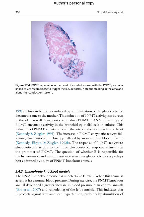
Figure 17.4 PNMT expression in the heart of an adult mouse with the PNMT promoterlinked to Cre recombinase to trigger the lacZ reporter. Note the staining in the atria andalong the conduction system.
368 Richard Kvetnansky et al.
Author's personal copy
1991). This can be further induced by administration of the glucocorticoid
dexamethasone to the mother. This induction of PNMT activity can be seen
in the adult as well. Glucocorticoids induce PNMTmRNA in the lung and
PNMT enzymatic activity in the bronchial epithelial cells in culture. This
induction of PNMT activity is seen in the arteries, skeletal muscle, and heart
(Kennedy & Ziegler, 1991). The increase in PNMT enzymatic activity fol-
lowing glucocorticoid is closely paralleled by an increase in blood pressure
(Kennedy, Elayan, & Ziegler, 1993b). The response of PNMT activity to
glucocorticoids is due to the three glucocorticoid response elements in
the promoter of PNMT. The question of whether E is responsible for
the hypertension and insulin resistance seen after glucocorticoids is perhaps
best addressed by study of PNMT knockout animals.
2.4.3 Epinephrine knockout modelsThe PNMT knockout mouse has undetectable E levels. When this animal is
at rest, it has a normal blood pressure. During exercise, the PNMTknockout
animal developed a greater increase in blood pressure than control animals
(Bao et al., 2007) and remodeling of the left ventricle. This indicates that
E protects against stress-induced hypertension, probably by stimulation of

369Stress and Peripheral Catecholamines
Author's personal copy
vasodilating b2-receptors. Not only did E protect against blood pressure
increase during exercise, but also endogenous E protected against diabetes
(Ziegler, Milic, Sun, et al., 2011). These experiments demonstrate that
E may actually protect against the metabolic syndrome (Ziegler, Elayan,
Milic, Sun, & Gharaibeh, 2012), in part by protecting against obesity-
induced insulin resistance. Although the short-term effect of
pharmacological doses of E is to increase blood glucose and diminish insulin
sensitivity, long-term knockout of E production exacerbated diet-induced
hyperglycemia and insulin resistance. There is reason to believe this is due to
E stimulation of b2-receptors. E stimulates b2-receptors better than NE but
has similar potency at a-, b1-, and b3- receptors. A chronic E infusion
enhanced rat muscle insulin sensitivity. b2-agonists in pharmacological doses
lead to muscle hypertrophy in rats, cattle, pigs, poultry, and sheep by
decreasing breakdown of muscle protein. E facilitates insulin binding to
exercising muscle and increases muscle blood flow. The short-term effect
of E is to raise blood pressure and blood sugar. The longer-term effect of
E is the opposite. These long-term effects can be mimicked by the admin-
istration of b2-agonists. Stimulation of b2-receptors induces the glucose
transporter GLUT4 and increases muscle mitochondria, permitting muscle
to take up and metabolize glucose. In addition, b2-receptors in the vascu-
lature cause vasodilation, lowering blood pressure and increasing cardiac
output. Thus, although it was once suspected that E might increase blood
pressure and cause diabetes, over the long term, E appears to lower both
blood pressure and blood sugar. This might account for some of the bene-
ficial effect of exercise to prevent both hypertension and diabetes. Human
athletes have used and abused the muscle growth-stimulating effects of
b2-agonist drugs, leading to regulations prohibiting their use in many pro-
fessional sports and in the Olympics.
2.4.4 The effect of stress on nonneuronal catecholamine systemsThe cardiac nonneuronal system producing E is also affected by stress. Stress
exposure induced PNMT gene expression not only in the adrenal medulla
but also in the cardiac atria and ventricles (Elayan, Kennedy, & Ziegler,
1990; Krizanova et al., 2001; Kvetnansky et al., 2006) and in the spleen
(Jelokova et al., 2002). PNMT mRNA levels in heart tissues were several
fold increased by immobilization stress (Krizanova et al., 2001;
Kvetnansky et al., 2006). In the atria, PNMT mRNA levels were increased
by hypoxia but decreased by cold stress. In the ventricles, no significant
changes were observed. In the atria, gene expression of PNMT is clearly

370 Richard Kvetnansky et al.
Author's personal copy
modulated by glucocorticoids, because adrenalectomy or hypophysectomy
prevents the increase in PNMTmRNA levels in response to immobilization
stress. Levels of PNMT mRNA remain elevated in the cardiac atria of
repeatedly immobilized rats and mice (Kvetnansky et al., 2006). This finding
suggests that increased E synthesis in the heart of mammals is a part of the
adaptation process of the organism to chronic or repeated stress exposure.
It is estimated that about one-third of cardiac E is synthesized by the heart
itself. However, there are two different enzymes in the heart that can syn-
thesize E. Atrial E-forming activity resembled adrenal PNMT. The ventric-
ular tissue nonspecifically methylated both NE and DA. These results help
explain why PNMT mRNA is found in the atria much more than in the
ventricles, even though both synthesize E. A nonspecific methylating
enzyme in the cardiac ventricles can synthesize E, but the activity of that
enzyme for E formation is much lower than PNMT activity in the atria
(Elayan et al., 1990). PNMTmRNA levels in humanmyocardiumwere sig-
nificantly higher during the first three years following cardiac transplantation
as compared to further periods after the transplantation (Goncalvesova et al.,
2004). A decrease in the PNMT gene expression with years after transplan-
tation could be a consequence of the reinnervation process. The PNMT
gene is also expressed in the lymphoid tissues such as the spleen and thymus
and in sympathetic ganglia. In these organs, PNMTmRNA levels were also
found to be elevated by stress (Kubovcakova et al., 2006; Kvetnansky
et al., 2006).
Epinephrine production by extra-adrenomedullary tissues increased fol-
lowing stress. The primary mechanism appears to be corticosteroid induc-
tion of PNMT (Kvetnansky et al., 2006), which can act on NE released in
response to stress. It is not clear how large a role this extra-adrenal E plays in
relation to circulating E derived from the adrenal medulla. There is fair evi-
dence for participation of cardiac E in pacemaking during embryogenesis,
but PNMT activity in the heart is very high at day 12 of rat embryogenesis
and declines thereafter.
2.4.5 Catecholamine systems in the adipose tissueCatecholamines regulate lipolysis in the adipose tissue and are produced
mainly by the sympathoadrenal system. Endogenous catecholamine produc-
tion has also been found in nonneuronal cells of adipose tissues—in adipo-
cytes (Vargovic et al., 2011). Rat adipocytes from mesenteric adipose tissue
express genes of catecholamine biosynthetic enzymes and produce catechol-
amines de novo (Kvetnansky et al., 2012). Acute or chronic cold exposure

371Stress and Peripheral Catecholamines
Author's personal copy
increased intracellular NE and E levels in isolated rat mesenteric adipocytes.
A single immobilization stress caused increased NE, E, catecholamine bio-
synthetic enzymes, and vesicular monoamine transporter 1 (VMAT1), but
repeated exposure to immobilization stress showed a highly exaggerated
effect (Vargovic et al., 2013). In the adipose tissues, both the sympathoadrenal
system and adipocyte catecholamine systems can produce catecholamines, and
both systems are activated by stress.
2.5. Adaptation to specific stressorsIn 1936, Selye described a pathological triad—adrenal enlargement, gas-
trointestinal ulceration, and thymicolymphatic involution—which
should be elicited by any stressor. In 1989, Vigas proposed the idea that
stress responses are specific, which modified Selye’s theory of stress. More
than a half century elapsed before Selye’s doctrine of nonspecificity
underwent testing by an NIH group, which failed to confirm it (Pacak
et al., 1998). Stressor-specific responses of catecholamine systems have
been described (Kvetnansky, Pacak, Sabban, Kopin & Goldstein, 1998;
Pacak & Palkovits, 2001; Pacak et al., 1998). Goldstein recently intro-
duced a new definition of stress (Goldstein, 2003; Goldstein & Kopin,
2007). Central to his stress theory is that the body possesses numerous
homeostatic comparators, which have been called “homeostats.” Differ-
ent homeostats can regulate the activity of the same effector system. The
definition of stress formulated by Goldstein is: “Stress is a condition in
which expectations, whether genetically programmed, established by
prior learning, or deduced from circumstances, do not match the current
or anticipated perceptions of the internal or external environment, and
this discrepancy between what is observed or sensed and what is expected
or programmed elicits patterned, compensatory responses.” Allostasis is
the process of adaptation of the body upon the exposure to various
stressors (Goldstein, 2003; Goldstein & Kopin, 2007; McEwen 1998;
2004).
In general, stressors can be divided into four main categories (Pacak &
Palkovits, 2001):
1) Physical stressors, for example, cold, heat, radiation, noise, vibration,
chemical stressors, pain, and immobilization.
2) Psychological stressors that affect emotional processes and may result in
behavioral changes such as anxiety, fear, or frustration. This can be trig-
gered in animals by handling or restraint.

372 Richard Kvetnansky et al.
Author's personal copy
3) Social stressors, reflecting disturbed interactions among individuals, for
example, unemployment, marital separation, death of partner, and
dominancy in animals.
4) Stressors that challenge cardiovascular and metabolic homeostasis, for example,
exercise, orthostasis, upright tilt, hypoglycemia, and hemorrhage.
Different stressors elicit different patterns of activation of the sympathetic
nervous, adrenomedullary, hypothalamic–pituitary–adrenocortical (HPA),
and other effectors.
In terms of duration, stressors may be either
(a) acute stressors (single, intermittent, and time-limited exposure) or
(b) chronic or repeated stressors (continuous long-term prolonged exposure
and intermittent long-term exposure).
There are different specific responses of sympathoadrenomedullary, HPA, and
sympathoneural systems to various stressors from different categories. This
specificity in responses of the organism to various stressors exists not only
at the level of plasma E, NE, and corticosteroids (Kvetnansky, 2004;
Kvetnansky et al., 1998; Pacak & Palkovits, 2001; Pacak et al., 1998) but also
at the level of gene expression and transcription factors of enzymes involved in
catecholamine biosynthesis (Kvetnansky, 2004; Kvetnansky, Jelokova, et al.,
2002; Kvetnansky, Nankova, et al., 2002; Liu, Kvetnansky, Serova, Sollas, &
Sabban, 2005; Sabban, Hebert, Liu, Nankova, & Serova, 2004; Sabban,
Nankova, Serova, Kvetnansky, and Liu, 2004; Wong & Tank, 2007).
Goldstein and Kopin (2008) reported results of a meta-analysis of the
literature examining interrelationships among responses to stressors, as
measured by plasma E, adrenocorticotropic hormone (ACTH), and NE
levels. Mean E responses were strongly positively correlated with mean
ACTH responses (r¼0.93) and less strongly with NE responses (r¼0.40).
Plasma E responses were disproportionately larger than NE responses during
hypoglycemia and smaller than NE responses during cold exposure without
hypothermia, orthostasis, and active escape/avoidance. Plasma NE
responses were disproportionately larger than ACTH responses during cold
exposure without hypothermia and severe/exhausting exercise and smaller
than ACTH responses during hypoglycemia. The results of this meta-
analysis indicate a close association between adrenomedullary and HPA
responses across a variety of stressors. This association seems to be stronger
than that between adrenomedullary and sympathetic noradrenergic
responses. The findings therefore favor the concept of a unitary adrenal sys-
tem over that of a unitary sympathoadrenal system (Goldstein &
Kopin, 2008).

373Stress and Peripheral Catecholamines
Author's personal copy
3. THE EFFECT OF STRESS ON ORGAN SYSTEMS
3.1. Release of catecholamines in stress
Individual stressors such as heat, cold, hypoglycemia, exercise, hemorrhage,and psychological challenges require specific responses. Appropriately,
different stressors elicit different patterns of activation, closing negative feed-
back loops. Regulation of E and NE release is highly stressor specific. Hypo-
glycemia and immobilization elicited mainly E release. However, stressors
such as cold and pain induced NE release (Goldstein & Kopin, 2008). Med-
ullary cellular hypertrophy, but not hyperplasia, is a general consequence of
chronic stress.
The mechanism of release of catecholamines is similar in the adrenal
medulla and the sympathetic nerve endings. Acetylcholine released from
the sympathetic preganglionic nerve terminals binds to nicotinic cholinergic
receptors and leads to a depolarization of the cell membrane, resulting in an
increase inmembrane permeability to sodium. This initiates a series of events
that lead to an increase in the influx of calcium. Then, catecholamine storage
vesicles fuse with the chromaffin or sympathetic neuronal cell membranes
and, via exocytosis, release their contents of catecholamines, together with
chromogranins, other neuropeptides, ATP, and a fraction of the soluble
DBH. NE and ATP are stored in both small synaptic and large dense-core
vesicles, but neuropeptides are stored only in the large ones. Release of
NPY, therefore, does not parallel that of NE and ATP, as exocytosis from
small and large vesicles is regulated differently (Zukowska-Grojec, 1995).
After exocytosis, the vesicle membrane is retrieved from the plasma
membrane and recycled into newly formed vesicles.
Following exocytosis, catecholamines that escape reuptake and local
metabolism diffuse into the circulation and constitute the circulating pool
of catecholamines. Plasma catecholamines turn over very rapidly. The half-
time of disappearance of human NE is about 2.5 min (Esler et al., 1979).
Under resting conditions, low levels of catecholamines are released into the
blood from the adrenal medulla and sympathetic nerve terminals. During
stressful stimulation, however, a huge amount of E (about 95%) and a signif-
icant amount of NE (which may comprise up to 30% of the total circulating
NE) may be released from the adrenal medulla. The remaining 70% of NE is
released from the sympathetic nerve terminals and enters blood capillaries
from the site of release at the neuroeffector junction. Human plasma normally
contains six catecholamines: E, NE, and DA; the catecholamine precursor

374 Richard Kvetnansky et al.
Author's personal copy
L-DOPA; the metabolite of NE—dihydroxyphenylglycol (DHPG); and a
metabolite of DA—dihydroxyphenylacetic acid (DOPAC) (Fig. 17.2).
Plasma NE is released mainly from the sympathetic nerves and the major-
ity of NE is metabolized before entry into plasma. Plasma NE levels depend
both on the rate of NE release into plasma and the rate of its removal from
the plasma. In some pathological situations, NE may be released from the
sympathetic terminals by a nonexocytotic mechanism.
Plasma E levels parallel neural outflow to the adrenal medulla. Under the
effect of various stressors, E responses are more closely related to ACTH
levels than to NE. The fate of E that enters the bloodstream differs quanti-
tatively from that of NE. E is a poorer substrate than NE for uptake-1 and a
better substrate than NE for extraneuronal uptake-2. E is also a better sub-
strate than NE for catechol-O-methyltransferase (COMT). Therefore,
more of circulating E than NE is metabolized by extraneuronal uptake
and O-methylation (Goldstein, 2010).
Plasma DA levels are low and similar to those of E, but circulating DA
does not act as a hormone (Goldstein, 2010). Stressors that elicit release of
NE from sympathetic neurons produce much larger increases in plasma NE
levels than in plasma DA levels. Free plasma DA is mostly released from the
sympathetic nerves (Goldstein & Holmes, 2008). In humans, at least 95% of
circulating DA is present in sulfoconjugated form (Goldstein, 2010). Levels
of DA sulfate respond relatively little to acute exposure to various stressors,
for example, exercise. However, meal ingestion markedly increases plasma
DA sulfate levels (Goldstein, 2010).
Plasma DOPA levels exceed those of NE by about tenfold, due to more
rapid clearance of NE than of L-DOPA from plasma. L-DOPA is the pre-
cursor of catecholamines and the product of the rate-limiting step in biosyn-
thesis. Immobilization stress in rats increases L-DOPA levels in plasma
within a few minutes, and blockade of catecholamine biosynthesis prevents
these increases (Kvetnansky, Armando, et al., 1992).
Plasma DHPG is formed from NE in sympathoneural cytosol by deam-
ination and reduction. DHPG diffuses rapidly across cell membranes into the
extracellular fluid and then to the bloodstream. Plasma DHPG levels offer a
biochemical index of NE turnover (Goldstein, 2010).
Plasma DOPAC is a DA metabolite. DOPAC levels are about 50 times
higher than DA, due to much slower clearance of DOPAC than of DA from
the circulation. Immobilization stress in rats rapidly increases plasma
DOPAC levels (Kvetnansky, Goldstein, et al., 1992), and blockade of cat-
echolamine biosynthesis prevents the stress-induced increases in plasma

375Stress and Peripheral Catecholamines
Author's personal copy
DOPAC (Kvetnansky, Armando, et al., 1992). Plasma DOPAC might also
be formed from metabolism of DA in nonneuronal cells of the
gastrointestinal tract.
Decapitation of animals produced an 80-fold increase in plasma E and
an eightfold increase in plasma NE levels compared to values obtained
from blood collected via a permanently inserted arterial catheter
(Kvetnansky et al., 1978). Even minor disturbances like handling or trans-
fer of animals produce highly significant increases in E and NE levels
(Kvetnansky et al., 1978). Many stressors increase not only plasma cate-
cholamine levels but also the catecholamine precursor DOPA and cate-
cholamine metabolites. This indicates that stress increased
catecholamine synthesis, release, and metabolism. Adrenal medullectomy
completely prevents the stress-induced increases in plasma E but reduces
plasma NE levels only by about 30%. Combined adrenal medullectomy
and sympathectomy almost completely abolishes plasma NE. Therefore,
during stress, the increment in plasma E is derived almost completely from
the adrenal medulla, whereas most plasma NE (about 70%) is derived from
the sympathetic nerves (Kvetnansky, Weise, Thoa, & Kopin, 1979). Reg-
ulation of E and NE release is highly stressor-specific (Goldstein & Kopin,
2008). Adrenal medullary E release is mainly induced by hypoglycemia,
immobilization, and emotional stressors (Kvetnansky et al., 1998; Pacak
et al., 1998). Conversely, cold or pain exposure does not activate
E release but highly stimulates NE release (Kvetnansky et al., 1998;
Pacak et al., 1998).
When immobilization stress is applied daily for several weeks, baseline
levels of plasma catecholamines are elevated but the stress-induced incre-
ment is reduced (Kvetnansky, Nemeth, Vigas, Oprsalova, & Jurcovicova,
1984; Stone & McCarty, 1983). Repeated handling diminished E and
ACTH responses but not NE responses compared to the first handling pro-
cedure (Dobrakovova, Kvetnansky, Oprsalova, & Jezova, 1993). When
handling-adapted animals are handled by a different person, E responses
are enhanced. This dissociation of plasma E and NE responses illustrates dif-
fering control of sympathoadrenomedullary and sympathoneural systems.
Rats exposed to repeated immobilization have significantly increased plasma
DBH activities (Kvetnansky et al., 1984; Weinshilboum, Kvetnansky,
Axelrod, & Kopin, 1971). An exaggerated response of plasma catechol-
amines occurred in rats adapted to a homotypic stressor after a heterotypic
novel stressor (Dronjak, Ondriska, Svetlovska, Jezova, & Kvetnansky,
2002). Thus, a novel stress can exaggerate the response to an ongoing stress.

376 Richard Kvetnansky et al.
Author's personal copy
3.2. Inactivation and uptake of catecholamines in stressThe physiological effects of catecholamines released into the synaptic cleft are
terminated very rapidly by uptake back into the sympathetic nerve endings
and effector cells with some conversion to inactive metabolites. Sympathetic
nerve endings take up catecholamines from the extracellular fluid by a process
distinct from the intraneuronal uptake of catecholamine by the storage gran-
ules. Neuronal uptake is known as “uptake-1” (Eisenhofer, 2001) and the
uptake by nonneuronal tissues as “uptake-2” (Iversen, 1965).
Uptake-1 serves to recapture NE (Fig. 17.2). The carrier can transport NE
against large concentration gradients. This uptake plays a less important role in
the inactivation of circulating E. Uptake-1 increases in parallel with increased
NE release during exposure of the organism to stressors (Eisenhofer, Cox and
Esler, 1990; Eisenhofer, Esler, et al., 1991; Eisenhofer, Kopin, Goldstein,
2004b). About 90% of released NE is taken up by neurons (Eisenhofer et al.,
1990; Eisenhofer, Esler, et al., 1991; Eisenhofer, Goldstein, Kopin, 1989;
Eisenhofer, Smolich, Cox, Esler, 1991) mediated by NE transporter (NET)
andDA transporter (DAT) proteins.NE is translocated byNET about twofold
more effectively than E. DA is a much better substrate for DAT thanNE or E.
Extraneuronal uptake-2 (U-2) is an active process of transport into non-
neuronal cells. The extraneuronal monoamine transporter of uptake-2
(Iversen, 1965) has little if any stereospecificity and has low affinity (higher
Km). Uptake-2 favors E over NE and is not a Naþ- or Cl�-dependent pro-cess. Uptake-2 is sensitive to inhibition by O-methylated metabolites
normetanephrine and metanephrine and by corticosteroids. Uptake-2 is
responsible for formation of catecholamine metabolites in the liver, kidney,
and lung and is highly sensitive to inhibition by glucocorticoids. It now
appears that there are at least three nonneuronal catecholamine transporters
functioning at extraneuronal locations. The classic transporter is corticoste-
rone sensitive—the uptake-2 transporter, also referred as OCT3 “organic
cation transporter.” Other two extraneuronal organic cation transporters,
which also transport catecholamines, were identified as OCT1 and OCT2.
Vesicular monoamine transporters (VMAT).Varicosities in theperipheral sym-
pathetic neurons contain cytoplasmic vesicles.These vesicles actively store syn-
thesized or recaptured cytoplasmic catecholamines and have specific carrier
proteins called vesicular monoamine transporters (Johnson, 1988). Cloning
studies revealed the existence of two isoforms of this transporter—VMAT1
(the “neuroendocrine” isoform) and VMAT2 (the “neuronal” isoform)
(Eidenet al., 2002; Schuldiner, 1994).These twoVMAT isoforms are encoded

377Stress and Peripheral Catecholamines
Author's personal copy
by separate genes with different cellular distribution. Neurons express only
VMAT2. In contrast, the adrenal medullary chromaffin cells express both
isoformswithVMAT1predominant in rodents andVMAT2 in humans. Para-
crine SIF cells of the sympathetic ganglia express predominantly the VMAT1
isoform(Eidenet al., 2002).Catecholamines are, ingeneral, better substrates for
VMAT2 than for VMAT1 (Goldstein, 2001). The energy for vesicular uptake
by VMAT is provided by the proton gradient, and the acidic vesicles attract
basic amines. This is in contrast to transport of neurotransmitters across the
plasmamembrane (NET),which is sodiumdependent.Underbasal conditions,
VMAT1 is widely expressed in all adrenal chromaffin cells while VMAT2 is
colocalized with TH but not PNMT, indicating its expression in NE, but
not E-synthesizing chromaffin cells. After exposure to immobilization stress,
VMAT2 mRNA together with TH mRNA was elevated, reflected by
increased VMAT2. The stress-induced expression of the VMAT2 gene could
be mediated by a rise in glucocorticoids (Sabban, Tillinger, Nostramo, &
Serova, 2012; Tillinger, Sollas, Serova, Kvetnansky, & Sabban, 2010). Cate-
cholamines takenupbymonoamine cellmembrane transporters are then trans-
ported into storage vesicles or metabolized by monoamine oxidase (MAO) in
the cytoplasm of neurons or by COMT in nonneuronal cells (Fig. 17.2).
Eisenhofer, Kopin, and Goldstein (2004a) and Eisenhofer et al. (2004b) found
increasedNErelease andreuptakebutunchangedNEleakage fromstorageves-
icles during exercise, indicating increased use ofNET andVMATduring stress
(Eisenhofer et al., 2004a).
Under basal conditions, VMAT1 is widely expressed in all adrenal chro-
maffin cells while VMAT2 is colocalized with TH but not PNMT, indicat-
ing its expression in NE, but not E-synthesizing chromaffin cells. After
exposure to stress, there was no change in levels of VMAT1 mRNA. How-
ever, VMAT2 mRNA together with TH mRNA was elevated after expo-
sure of rats to a single or repeated immobilization for 6 days. The changes in
VMAT2 mRNA were reflected by increased VMAT2 protein (Sabban
et al., 2012; Tillinger et al., 2010). Sabban et al. (2012) have shown that
the stress-induced expression of the VMAT2 gene could be mediated by
a rise in glucocorticoids.
3.3. Degradation and vesicular leakage of catecholaminesin stress
Catecholamines are subjected to chemical degradation by 3-O-methylation
(COMT, EC 2.1.1.6) and oxidative deamination (MAO, EC 1.4.3.4) and

378 Richard Kvetnansky et al.
Author's personal copy
by conjugation as sulfate and glucuronide. After neuronal reuptake, cyto-
plasmic NE can undergo metabolism catalyzed by MAO to form DHPG
or translocation back into the storage vesicles via VMAT. The latter consti-
tutes the predominant pathway (see Fig. 17.2) where MAO catalyzes deam-
ination of amines, with production of aldehydes that are metabolized to
carboxylic acids or alcohols. The MAO-A subtype has a higher affinity
for NE and E.MAO-B is responsible for degradation of DA; both are mainly
localized in the liver. MAO participates in regulation of NE storage in the
nerve terminals. COMT is primarily an extraneuronal enzyme, which
metabolizes circulating catechols, mainly in the liver and kidney. Phenolic
hydroxyl groups of catecholamines can also be conjugated to sulfates or glu-
curonides. Eisenhofer and coworkers (2004a, 2004b) demonstrated that
most metabolism of catecholamines takes place within the same cells in
which the amines are synthesized. In sympathetic nerves, the aldehyde pro-
duced from NE by MAO is converted to 3,4-DHPG and not to
3,4-dihydroxymandelic acid. Subsequent extraneuronal O-methylation
leads to production of 3-methoxy-4-hydroxyphenylglycol and not to
vanillylmandelic acid (see Fig. 17.2). This acid is instead formed in the liver
by oxidation catalyzed by alcohol and aldehyde dehydrogenases. Compared
to intraneuronal deamination, extraneuronal O-methylation of NE and
E represents a minor pathway of catecholamine metabolism.
Most of the MAO metabolite DHPG, produced under resting conditions,
comes from NE leakage from vesicles. In the resting human heart, about
73% of NE turnover is due to intraneuronal metabolism of NE leaking from
storage vesicles (Eisenhofer et al., 1998, 2004a; Eisenhofer & Lenders, 1998).
Under conditions of exercise (at 50% of maximal work capacity), rates of NE
release and reuptake exceed the rate of NE leakage from storage vesicles,
which as a passive process operates independently of exocytotic release
and remains relatively constant. Vesicular leakage may therefore be viewed
as a mechanism that metabolizes catecholamines that are produced in prep-
aration for stress responses. Because the ability to increase TH activity is lim-
ited, this leakage mechanism provides sympathetic nerves with a capacity for
a more extended range of sustainable release rates than would otherwise be
possible (Eisenhofer et al., 2004a; 2004b).
Immobilization stress decreased both MAO-A and MAO-B activities
in rats (Obata & Yamanaka, 1994). Exposure to cold decreased liver
MAO-A activity and also the ratio MAO-A/MAO-B. Decreased
MAO-A activity and unchanged MAO-B activity are also observed in
the hearts and brains of rats exposed to foot shock (Lemoine, Armando,
379Stress and Peripheral Catecholamines
Author's personal copy
Brun, Segura, & Barontini, 1990). MAO activity was unchanged in the
adrenal medulla of stressed animals (Kvetnansky, Torda, Jahnova, &
Saleh, 1975). Animals immobilized repeatedly have shown lower COMT
activity in both parts of the adrenal. MAO and COMT activities were also
studied in the adrenals of rats that spent 18–20 days in space onboard three
COSMOS biosatellites. The data suggest that a prolonged stay in weight-
lessness does not appear to be a stressful stimulus for catecholaminergic sys-
tems (Kvetnansky et al., 1981). MAO activity declined in the sympathetic
ganglia of stress-predisposed animals after a stressor, while in stress-resistant
animals, it increased (Gorbunova & Kashtanov, 1983). Thus, stress-
induced changes in catecholamine-degrading enzyme activity are not uni-
form and depend on the strain of the animals, model of stress, timing of
stressor, and many other factors. COMT activity (Kvetnansky et al.,
1976) and also COMT mRNA levels were significantly decreased in
the liver of adult mice exposed to stress (Mikhailova, Gulyaeva, &
Filipenko, 2005). In aged mice, however, the effect of stress on COMT
mRNA levels in the liver is absent (Mikhailova et al., 2005). Genetic dif-
ferences in COMTmay underlie individual differences in response to psy-
chological and physical stressful challenges (Smolka et al., 2005; Zubieta
et al., 2003). An association between COMT genotype and history of vio-
lent behavior has been seen in some human studies (Jabbi et al., 2007;
Lachman, Nolan, Mohr, Saito, & Volavka, 1998), and COMT is a possible
candidate gene involved in the pathogenesis of major depressive disorders
and schizophrenia.
After exposure to stress, the activity of MAO and COMT is reduced in
some organs. The reduced degradation process, together with stress-induced
increases in catecholamine production, release, and secretion, might be
involved in the enhanced availability of catecholamines for adrenergic
receptors and for increased activity of metabolic and physiological processes
under stress. Although the general trend is for catecholamine degradative
enzymes to decrease with stress, the opposite is true for VMAT. This can
lead to greater availability of NE and E and greater vesicular stores after stress.
However, the cardiovascular consequences of inhibition of VMAT and
MAO are not so straightforward. Inhibition of both can cause postural
hypotension, even though their inhibition should increase intrasynaptic
NE, leading to a pressor, not a depressor, effect. The postural hypotension
could be due to intrasynaptic NE stimulation of presynaptic a2 receptors,leading to inhibition of NE release, or due to the prominent central nervous
effects of these drugs.

380 Richard Kvetnansky et al.
Author's personal copy
3.4. Renin activation and accumulated cell proliferation in theadrenal gland
All components of the renin–angiotensin system, including prorenin, renin,
angiotensinogen, angiotensin-converting enzyme, and angiotensins Ι and II,were expressed in both the adrenal cortex and the adrenal medulla
(Armando, Jezova, Bregonzio, Baiardi, & Saavedra, 2004). Local angioten-
sin II production could regulate the production of aldosterone and gluco-
corticoids and stimulate catecholamine secretion in the adrenal medulla
(Armando et al., 2004). Angiotensin II served as a paracrine amplifier of
the morphogenic signal and had a critical role in adrenocortical cellular
proliferation and development.
3.5. Corticosteroid responses to stressorsAdrenocortical cells of adult mammals have low proliferative activity. Stress
induces corticotropin-releasing hormone (CRH) release from the para-
ventricular nucleus of the hypothalamus, ACTH release from the anterior
lobe of the pituitary gland, and glucocorticoid release from the adrenal cor-
tex. Acute ACTH administration induced hyperplasia in the zona fasciculate
of the adrenal cortex. Chronic stress affected not only ACTH but also the
adrenal cortex. When animals were exposed to a continuous or intense
stressor, ACTH gradually returned to normal levels. However, glucocorti-
coid levels remained high. Moreover, many patients with depression had
increased basal plasma cortisol and enlarged adrenals.
3.6. The effect of maternal stress on offspringThe adrenal medulla develops internal to the adrenal cortex because of adre-
nocortical release of glucocorticoids. Transgenic animals that fail to express
adrenomedullary glucocorticoids have dispersed chromaffin cells that fail to
form a discrete adrenal medulla. Furthermore, the expression of PNMT and
E synthesis depends on stimulation of glucocorticoid response elements in
the PNMT promoter. Maternal stress increases glucocorticoid release
through the HPA axis. The effect of maternal glucocorticoid excess is easily
modeled by the administration of exogenous glucocorticoids. Glucocorti-
coids are used in obstetric practice, primarily to enhance lung maturation
in cases of threatened preterm labor to reduce mortality in preterm infants.
Although these treatments improve survival, they are not without
adverse effects.

381Stress and Peripheral Catecholamines
Author's personal copy
Prenatal exposure to glucocorticoids during their final week of preg-
nancy is sufficient to produce permanent adult hypertension in the rat
(Levitt, Lindsay, Holmes, & Seckl, 1996). Even physiological levels of
administered glucocorticoid for just 2 days were sufficient to develop hyper-
tension in the rat offspring (Singh et al., 2007). The 11b-hydroxysteroiddehydrogenase type 2 (11b-HSD2) enzyme, which serves as the placental
barrier for glucocorticoid transfer, is present in syncytiotrophoblasts in the
placenta of humans andmany other mammalian species. It catalyzes the con-
version of active glucocorticoids into inactive 11-keto metabolites, thereby
protecting the fetus from elevated concentrations of maternal glucocorti-
coids. Nevertheless, increased levels of endogenous glucocorticoids can
result in hypertension in the rat offspring (Singh et al., 2007).
Glucocorticoids regulate PNMT in two ways, posttranslationally by
indirectly preventing the degradation of the PNMT enzyme and transcrip-
tionally by regulating the expression of PNMT mRNA (Wong, Lesage,
Siddall, & Funder, 1992). Dexamethasone induces PNMTmRNA as much
as 20-fold. Though animal studies suggest that fetal exposure to glucocor-
ticoids contributes to adult hypertension, long-term effects of fetal exposure
to glucocorticoids in human are difficult to study but suggest a link to higher
blood pressure after age 30.
3.7. Human models of chronic sympathetic nervous systemstress responses
Patients with Alzheimer’s can be disruptive and need careful supervision day
and night. Spousal caregivers to Alzheimer’s patients are placed in a stressful
situation and frequently complain of work overload without respite. These
caregivers have an accelerated risk of developing hypertension over a period
of 5 years (Shaw et al., 1999). This may be a response to an increase in their
allostatic load or in response to their sleep disturbance. Multivariate analysis
shows a close relation betweenNE levels and sleep disturbance in caregivers.
The severity of the increase in sympathetic nerve activity among caregivers is
attested to by a decrease in sensitivity of their b2-adrenergic receptor. Theincreased sympathetic nerve activity is accompanied not only by increased
blood pressure but also by impaired function of their vascular endothelium
and atherosclerosis (von Kanel et al., 2011).
Sleep is disturbed in Alzheimer caregivers (von Kanel et al., 2010) but is
much more disturbed in obstructive sleep apnea. Patients with sleep apnea
fail to lower their blood pressure at night as much as normal subjects and
have increased blood pressure day and night (Ziegler, Milic, Sun, et al.,

382 Richard Kvetnansky et al.
Author's personal copy
2011). They have increased plasma NE throughout the day and night and
desensitized b2-adrenergic receptors. These sympathetic nervous system dis-
orders are largely ameliorated by treatment of the sleep apnea with contin-
uous positive airway pressure, which also lowers blood pressure. It is
noteworthy that pharmacological treatment of hypertension that accom-
panies sleep apnea with drugs that block excess sympathetic nerve activity
tends to be more effective than with other types of drugs (Ziegler,
Milic, & Elayan, 2011). Slow-wave (deep) sleep is the period of least sym-
pathetic nerve electrical activity, lowest heart rate, and lowest blood pres-
sure. A study of elderly normotensive men found that those with the least
deep sleep were the most likely to develop hypertension (Fung et al., 2011).
Psychological stress has long been considered a potential cause of human
hypertension. Human studies suggest that severe stresses such as caregiving
for Alzheimer’s patients or sleep apnea might lead to hypertension through
chronic activation of the sympathetic nervous system. Sleep disruption may
play a role in the sympathetic nervous activation, since deep sleep is the
period of least sympathetic nerve activity.
3.8. Hypoglycemia-associated autonomic failureMedication-induced hypoglycemia affects most people with type 1 diabetes
and many people with type 2 diabetes. Recurrent hypoglycemic stress can
cause hypoglycemia-associated autonomic failure (HAAF), leading to both
defective glucose counterregulation by reducing adrenomedullary E response
and unawareness by reducing the symptomatic response to hypoglycemia.
Three independent studies demonstrated that scrupulous avoidance of
hypoglycemia reverses hypoglycemia unawareness and improves the
reduced E component of defective glucose counterregulation in most
affected patients. Recently, studies (Ramanathan & Cryer, 2011) demon-
strated that adrenergic blockade prevented the effect of hypoglycemia to
reduce the plasma catecholamine responses to subsequent hypoglycemia.
Recurrent hypoglycemia and the attendant defective counterregulation
are the main limiting factors for long-term health benefits of intensive insulin
therapy in diabetes. Recent studies from the Nankova lab by Edmund La
Gamma compared the effects of single daily versus twice-daily episodes of
insulin-induced recurrent hypoglycemia for 3 days in normal rats on adrenal
E release and its production as reflected by altered TH mRNA levels,
PNMT, and preproenkephalin (ppEnk, a costored and coreleased neuro-
peptide). Single daily episodes of recurrent hypoglycemia resulted in an

383Stress and Peripheral Catecholamines
Author's personal copy
early-onset increase in plasma E, corticosterone, and glucagon with a
corresponding later-onset upregulation of TH, PNMT, and ppEnk mRNA
during a hypoglycemic clamp study on day 4. Exposure to twice-daily recur-
rent hypoglycemia reduced the immediate counterregulatory response of
E and glucagon but not corticosterone. Attenuation was associated with a
limited elevation of TH mRNA without affecting PNMT or ppEnk
mRNA. The frequency of the antecedent hypoglycemic episodes can either
increase THmRNA (once daily), to sustain biosynthesis and thus the releas-
able pool of E or, paradoxically, attenuate its elevation (twice daily). These
mechanisms appear to require circulating factors in vivo not present in in vitro
models. Nuclear run-on assays show butyrate-dependent decreases of both
TH mRNA and TH protein in PC12 cells, the cultured adrenal cells from
rat origin. This signaling may represent a significant contributing factor to
the clinical syndrome of HAAF.
4. MOLECULAR GENETIC MECHANISMS OF PERIPHERALCATECHOLAMINERGIC RESPONSES TO STRESS
4.1. Stress and induction of catecholaminebiosynthetic enzymes
Increased expression of catecholamine biosynthetic enzymes is a prevalent
response to many types of stressors. The adrenal medulla rapidly responds
to single stressors by targeting individual genes in response to cholinergic
stimulation. In contrast, the sympathetic ganglia are especially responsive
to activation of the HPA axis (Sabban & Kvetnansky, 2001). Several reviews
deal with various aspects of the regulation of CA biosynthetic enzyme activ-
ity, protein levels, gene expression, and molecular genetics during exposure
to stressors (Kobayashi & Nagatsu, 2005; Kvetnansky & McCarty, 2007;
Kvetnansky & Sabban, 1993, 1998; Kvetnansky et al., 2009; Sabban,
2007; Sabban et al., 1995; Sabban & Kvetnansky, 2001; Wong & Tank,
2007). The paraventricular, periventricular, and dorsomedial hypothalamic
nuclei are important centers regulating neuroendocrine and autonomic sys-
tems during stress. Immobilization stress increased THmRNA and TH pro-
tein levels in these brain nuclei and in the peripheral sympathetic nervous
system (Kiss, Mravec, Palkovits, & Kvetnansky, 2008).
Administration of the transcriptional inhibitor actinomycin D to rats
blocks the stress-evoked increases in adrenal DBH and TH activity and
TH and PNMTmRNA levels. Repeated exposure to immobilization stress
leads to increased enzymatic activity and TH and DBH protein levels. No

384 Richard Kvetnansky et al.
Author's personal copy
significant changes were observed with a single exposure to stress (Sabban &
Kvetnansky, 2001). However, both single and repeated immobilization stress
led to increased initiation of TH and DBH gene transcription to levels about
threefold higher than in control animals. A single 5 min stressor caused a
three- to fourfold increase in TH andDBH transcription rate. Thus, transcrip-
tion of TH and DBH genes is almost as fast as the stress-triggered rise in levels
of adrenal glucocorticoids. However, this rise is transient if the stress is not
prolonged. Following repeated stress (2 h daily for several consecutive days),
the activation of TH and DBH transcription is stabilized and significantly ele-
vated even 24 h after the last stress exposure (Nankova & Sabban, 1999;
Nankova, Tank, & Sabban, 1999). Repeated immobilization kept levels of
TH and DBHmRNA elevated in both the sympathetic ganglia and the adre-
nal medulla, but did not produce further increase compared to already ele-
vated levels in adapted control groups. Thus, elevated transcription of
catecholamine biosynthetic enzymes plays an important role in response of
the peripheral catecholaminergic systems to stressors.
Quantitative evaluation of gene expression of catecholamine biosyn-
thetic enzymes (Kvetnansky et al., 2004) showed that in the adrenal medulla,
the basal concentration of TH mRNA was about 0.5 amol/ng of total
RNA. DBH mRNA was about 12 times higher (6.0 amol/ng RNA) and
PNMT mRNA about 56 times higher (28.0 amol/ng RNA) than TH
mRNA. In the stellate ganglia, the basal concentration of TH mRNA
(0.02 amol/ng RNA) was about 25 times lower than in the adrenal medulla,
but DBHmRNA in ganglia (2.6 amol/ng RNA) was present at similar con-
centration as in the adrenal medulla (Kvetnansky et al., 2004; Micutkova,
Rychkova, Sabban, Krizanova, & Kvetnansky, 2003). Repeated immobili-
zation (2 h daily for 7 days) kept levels of TH and DBH mRNA elevated
both in the sympathetic ganglia and the adrenal medulla. PNMT gene
expression was also increased after repeated stress in both the stellate ganglia
and the adrenal medulla of rats and mice (Kubovcakova et al., 2006;
Kvetnansky et al., 2006, 2004). Thus, gene expression of catecholamine bio-
synthetic enzymes in both the sympathetic ganglia and the adrenal medulla is
markedly elevated as part of the adaptation mechanism of the organism to
long-term exposure to a homotypic stressor.
4.2. Transcription factors4.2.1 Adrenal medullaTranscription factors are part of a dynamic interplay that converts short-term
transient activation of transcription to prolonged, potentially maladaptive

385Stress and Peripheral Catecholamines
Author's personal copy
changes in gene expression (reviewed by Kvetnansky et al., 2009; Sabban &
Kvetnansky, 2001; Sabban, Liu, Serova, Gueorguiev, & Kvetnansky, 2006;
Sabban et al., 2012). The rapid activation of gene transcription for TH and
DBH after 5 min of immobilization is too soon to reflect de novo synthesis of
induced transcription factors. Increased phosphorylation of cAMP response
element binding protein (CREB) occurs in the adrenal medulla of rats
immobilized for only 5 min (Sabban, Nankova, et al., 2004). The activation
of CREB at the CRE motif of the TH promoter is a well-characterized
mechanism that is important for activation of TH gene transcription. Phos-
phorylation of a CREB serine residue needed for transcriptional activation
begins after 5 min of immobilization and is more pronounced after 30 min.
However, by 2 h of immobilization, levels of phosphorylated CREB returns
to basal levels, reflecting the transient nature of this response. The phosphor-
ylation of CREB on Ser-133 can be mediated by a number of kinases,
including protein kinase A, mitogen activated protein kinases, and calmod-
ulin dependent kinase.
After a single exposure to stress, the rise in mRNA levels is transient
(Nankova et al., 1996; 1999). However, with a second exposure to the same
stressor, there is already “memory” of the first experience such that the
increase in TH mRNA is more prolonged. With prolonged repeated stress
over 5–7 days, TH transcription remains high for as long as 2 days following
cessation of stress. A single stress induces c-Fos in the adrenal medulla, which
is correlated with its increased binding to the AP-1-like sites on the TH and
DBH promoters (Nankova, Rivkin, Kelz, Nestler, & Sabban, 2000). The
c-Fos is probably not the only AP-1 factor manifesting the second wave
of gene expression in response to stressors. Fra-2 is not significantly changed
during the first 30 min of stress. After 2 h, however, Fra-2 increases and is
potentiated by repeated stress. This might be one of the mechanisms
involved in permanently elevated TH mRNA levels in the adrenal medulla
of repeatedly immobilized rats.
The immediate early gene Egr1 is also induced in rat adrenal medulla by
a single stress exposure. Egr1 stimulates PNMT transcription in combina-
tion with glucocorticoids (Wong, Tai, Wong-Faull, Claycomb, &
Kvetnansky, 2004). Egr1 also participates in cholinergic stimulation of
the PNMT promoter. Reduced levels of Egr1 in the adrenal medulla of
CRH knockout mice exposed to immobilization stress correlate well with
reduced PNMT mRNA levels in those animals (Kvetnansky et al., 2006).
Egr1 can also regulate transcription of the TH gene. Thus, a single tran-
scription factor can stimulate catecholamine genes by more than one path-
way and can stimulate more than one gene.

386 Richard Kvetnansky et al.
Author's personal copy
Following a single stress for 2 h, several hundred genes are significantly
up- and downregulated (651 up- and 487 downregulated). With repeated
stress, 370 genes were up- and 195 downregulated (Liu, Serova,
Kvetnansky, & Sabban, 2008). A substantial number of gene changes were
from the group of transcriptional factors and growth factor-related tran-
scripts. Interestingly, an analysis of the direct interactions among the genes
affected by stress indicates that Fos, Egr1, and nuclear receptor family mem-
ber NR4A1 (also known as Nurr 77), which can interact with NBREmotif
on the TH promoter, are likely to play a central role in interacting with
many of the adrenomedullary genes regulated by a single immobilization
stress. Repeated stress triggers an enormous induction of Fra-2 (Sabban
et al., 2006), which is also phosphorylated with the repeated stress (Liu
et al., 2005). The adrenal medulla does not appear to desensitize after
repeated episodes of immobilization stress (even for 42 days); the final
stressor still triggers induction of TH mRNA and Fos-related antigens such
as Fra-2 (Kvetnansky, Jelokova, et al., 2002; Kvetnansky, Nankova,
et al., 2002).
Stress also alters the expression of metabolism-, lipid-, protease-, and
kinase/phosphatase-related genes. With repeated stress, a higher percentage
of transcripts are devoted to neuropeptides. Most of the genes (>80%)
altered by a single stress were transiently elevated. In contrast, approximately
half of the genes elevated by repeated stress were also responsive to a single
stress indicating that they mediate a prolonged response. There was also
sustained phosphorylation of CREB throughout the entire course of
repeated stress. Adrenal medullae of rats exposed to chronically repeated
immobilization stress increased phosphorylated CREB 60-fold over
unstressed controls.
Cold stress, like immobilization stress, triggered elevation of TH tran-
scription in the adrenal medulla; however, the modified transcription factors
were very different. Cold triggered increased binding to the AP-1 motif of
the adrenal TH promoter, and a transient elevation of c-Fos was observed
within the first few hours of cold exposure (Liu et al., 2005). However, there
were no significant changes in phosphorylation of CREB or induction of
Egr1. Fra-2 was also induced by cold stress, but the time course of changes
differed from that observed with c-Fos. Levels of Fra-2 were not signifi-
cantly changed with 1 or 7 days of cold stress but were about double basal
levels after 28 days of continual cold stress (Liu et al., 2005). This is still a
much smaller change than the changes in Fra-2 with repeated immobiliza-
tion stress.

387Stress and Peripheral Catecholamines
Author's personal copy
Microarray analysis reveals that immobilization and cold activate a very
different repertoire of transcription factors in the adrenal medulla. Cold trig-
gers significant changes in expression of fewer genes than immobilization
with overlap in only about 5% of the genes up- or downregulated. Thus,
there is specificity of response of the organism exposed to various stressors
even at the molecular genetic level of expression of different genes. Animals
preexposed to immobilization had a greater cold response in P-CREB,
Egr1, and Fra-2. Rats preexposed to 28 days of cold displayed a significantly
higher response to immobilization. Thus, different stressors can accentuate
the molecular responses to each other.
4.2.2 Sympathetic gangliaThemechanisms of stress-triggered activation of TH and DBH gene expres-
sion differ between sympathetic nerves and the adrenal medulla (reviewed
by Sabban, 2007; Sabban, Nankova, et al., 2004). The most intriguing dif-
ferences are in the response of TH and DBH gene expression to ACTH.
ACTH can have a direct effect on expression of genes in sympathetic ganglia
but not in the adrenal medulla (Kvetnansky et al., 2004). The marked induc-
tion of AP-1 factors, c-Fos, and Fra-2 observed in the adrenal medulla does
not appear to be important in sympathetic ganglia (Kvetnansky et al., 2009;
Sabban, Nankova, et al., 2004). Immobilization stress increased CREB
binding at the CRE element on the TH gene promoter in the superior cer-
vical ganglia (Sabban, Nankova, et al., 2004), suggesting increased expres-
sion of CREB and showing that related transcriptional mechanisms are
likely involved. In contrast, in the adrenal medulla, the same stressor did
not alter CREB levels, but rather triggered increased phosphorylation of
CREB. Thus, induction of CREB might play a critical role in the stress
response in the SCG, and this might mediate a sustained elevation of tran-
scription of TH, DBH, and other CREB-responsive genes. The c-Jun
N-terminal kinase is also selectively induced in the adrenal medulla, but
not in the superior cervical ganglion.
The time course of stress-induced changes in mRNA levels is also dif-
ferent in sympathetic ganglia compared to the adrenal medulla. In stellate
ganglia, the time course of the elevation of TH and DBH mRNA levels
was more gradual and the extent of elevation more modest than in the adre-
nal medulla. In contrast, in the adrenal medulla, there was a rapid change in
TH gene expression with only a single exposure to immobilization.
Although stress-induced PNMT gene expression is regulated by the
HPA axis in both the adrenal medulla and sympathetic ganglia of rats and

388 Richard Kvetnansky et al.
Author's personal copy
mice (Kubovcakova et al., 2006; Kvetnansky et al., 2006), the involvement
of the HPA axis in the stress-triggered increase in TH and DBH gene
expression differs between the sympathetic ganglia and adrenal medulla.
In summary, the sympathoneural system, at least as it relates to TH and
DBH, is regulated distinctly from the adrenal medulla.
4.2.3 Regulation by the HPA axisTrans-synaptic stimulation regulates catecholamine biosynthetic enzymes in
sympathetic nerves. Nerve stimulation increased TH and DBH activities and
elevated TH protein andmRNA levels in rat sympathetic ganglia. Denervation
prevented the elevation of TH in response to reserpine. However, trans-
synaptic inputs are not the only regulatory inputs. Decentralization greatly
reduced basal TH activity, but exposure to cold stress triggered a large induction
in TH activity in sympathetic ganglia even following decentralization.
Regulation of TH and DBH (in contrast to PNMT) gene expression in
the adrenal medulla with stress is not very much affected by the HPA axis,
and increased TH and DBH expression is still observed in hypophysecto-
mized animals. However, the activation of the HPA axis by stress appears
to be involved in the regulation of catecholamine biosynthetic enzymes
in the sympathoneural system. A single dose of glucocorticoid increased
TH activity and TH mRNA and enhanced the response to cold in sympa-
thetic ganglia. Furthermore, injection of ACTH triggered a large rise in TH
mRNA in superior cervical ganglia and enhanced expression of ACTH
(MC2) receptor mRNA in the superior cervical and stellate ganglia
(Nankova, Kvetnansky, & Sabban, 2003; Sabban, Nankova, et al., 2004).
The effect of ACTH appears to occur, at least partially, by an adrenal-
independent mechanism. Adrenalectomy, which, as expected, eliminated
circulating E and increased plasma ACTH, also markedly raised plasma
NE levels (Kvetnansky et al., 1995). Moreover, TH and DBH mRNAs
in rat superior cervical ganglia are also elevated in adrenalectomized rats
(Sabban et al., 2006; Serova, Gueorguiev, Cheng, & Sabban, 2008). These
findings suggest that ACTH may have a direct effect on gene expression of
catecholamine biosynthetic enzymes in sympathetic ganglia. Catechol-
amines also are involved in central nervous stimulation of ACTH release.
The locus coeruleus has widespread NE projections, notably to the medial
prefrontal cortex, which inhibits HPA responses to emotional stress. Injec-
tion of axonally transported immunotoxin to selectively ablate NE inputs to
the prefrontal cortex diminished the stress-induced output of the HPA
(Radley et al. 2008). Thus, central NE pathways can enhance the release

389Stress and Peripheral Catecholamines
Author's personal copy
of ACTH in response to stress. This feed forward mechanism in which stress
activates CNS catecholamine release of ACTH and ACTH activates periph-
eral catecholamine production has implications for stress-induced hyperten-
sion. In addition, these pathways might help explain the occurrence of
human hypertension following administration of glucocorticoids such as
dexamethasone that do not cause sodium retention.
Substantial evidence supports the importance of the activation of the
HPA axis for regulation of PNMT gene expression. Hypophysectomy pre-
vents the immobilization-triggered induction of PNMT gene expression.
Glucocorticoids regulate PNMT in at least two ways: posttranscriptionally
and transcriptionally (Wong & Tank, 2007). The PNMT promoter contains
three glucocorticoid response elements (Tai, Claycomb, Her, Bloom, &
Wong, 2002).The glucocorticoid receptor interacts with the PNMT
promoter alone and also synergistically with Egr1 and AP-2 transcription
factors. The HPA axis is also important in regulation of PNMT in the sym-
pathoneural system. PNMT was detected in stellate ganglia and is elevated
by immobilization stress in wild-type but not in CRH-deficient mice
(Kubovcakova et al., 2006; Kvetnansky et al., 2006). A comparison of the
regulation of gene expression for catecholamine biosynthetic enzymes in
sympathetic ganglia with the adrenal medulla shows that in contrast to
the original designation of the “sympathoadrenal” system, these are clearly
two distinct systems—sympathoadrenomedullary and sympathoneural.
5. CONCLUSION
Stressors such as hypoglycemia, cold, and restraint selectively activate
catecholamine release from sympathetic nerves and the adrenal medulla.
Trans-synaptic stimulation induces catecholamine biosynthetic enzymes
in nerves and the adrenal medulla by different pathways of CRE phosphor-
ylation and expression factor synthesis. On the other hand, glucocorticoids
are the primary stimulus to enzyme synthesis in nonneuronal catecholamine-
producing cells. Because of these differences, stressors selectively activate
different parts of peripheral catecholamine systems. However, chronic stress
leads to longer-lasting elevations of catecholamine-synthesizing enzymes,
and the adaptation to one stressor may potentiate the response to a single
exposure of a different stressor.
Human studies indicate that chronic stress can lead to prolonged catechol-
amine increases and deleterious cardiovascular responses. Molecular studies in
animals show that prolonged catecholamine activation following stress not

390 Richard Kvetnansky et al.
Author's personal copy
only is a consequence of central nervous memories but also reflects intracel-
lular changes in the peripheral sympathetic nervous system. In addition, the
additive effects of glucocorticoids, nerve traffic, and angiotensin II on cate-
cholamine synthetic enzymes suggest potential therapeutic agents that can
block these effects to reverse excess catecholamine release in patients with
chronic stress. Rodent studies indicate that maternal stress sufficient to elevate
glucocorticoid levels may induce hypertension in the offspring. We conclude
that stress-induced changes in the peripheral sympathetic system are individ-
ualized and complex but comprehensible. These changes appear to have
important cardiovascular consequences and probably play a role in stress-
induced cardiac arrhythmias, hypertension, and the metabolic syndrome.
CONFLICT OF INTERESTThe authors have no conflicts of interest to declare.
ACKNOWLEDGMENTSSupported in part by NIH grants 1UL1RR039180 and HL58120 (to M.Z.) and by Slovak
Research and Development Agency (No. APVV-0148-06 and APVV-0088-10) and VEGA
grant (2/0036/11) (to R.K.).
REFERENCESArmando, I., Jezova,M., Bregonzio, C., Baiardi, G., & Saavedra, J. M. (2004). Angiotensin II
AT1 and AT2 receptor types regulate basal and stress-induced adrenomedullary cate-cholamine production through transcriptional regulation of tyrosine hydroxylase. Annalsof the New York Academy of Sciences, 1018, 302–309.
Bao, X., Lu, C. M., Liu, F., Gu, Y., Dalton, N. D., Zhu, B. Q., et al. (2007). Epinephrine isrequired for normal cardiovascular responses to stress in the phenylethanolamineN-methyltransferase knockout mouse. Circulation, 116, 1024–1031.
Culman, J., Torda, T., Petrikova, M., & Murgas, K. (1988). Effect of corticosterone treat-ment and adrenalectomy on phenylethanolamine N-methyltransferase and catechol-amines in brain stem and hypothalamic nuclei and superior cervical ganglion of rats.Endocrinologia Experimentalis, 22, 117–128.
Dimsdale, J. E., & Moss, J. (1980). Plasma catecholamines in stress and exercise. Journal of theAmerican Medical Association, 243(4), 340–342.
Dobrakovova, M., Kvetnansky, R., Oprsalova, Z., & Jezova, D. (1993). Specificity of theeffect of repeated handling on sympathetic-adrenomedullary and pituitary-adrenocortical activity in rats. Psychoneuroendocrinology, 18, 163–174.
Dronjak, S., Ondriska, M., Svetlovska, D., Jezova, D., & Kvetnansky, R. (2002). Effects ofnovel stressors on plasma catecholamine levels in rats exposed to long-term cold. InR. McCarty, G. Aguilera, E. L. Sabban, & R. Kvetnansky (Eds.), Stress neural, endocrineand molecular studies (pp. 83–89). London: Taylor and Francis.
Eiden, L. E., Schutz, B., Anlauf, M., Depboylu, C., Schafer, M. K. H., & Weihe, E. (2002).The vesicular monoamine transporters (VMATs): Role in the chemical coding of neu-ronal transmission and monoamine storage in amine handling immune and inflammatorycells. In T. Nagatsu, T. Nabeshima, R. McCarty, & D. S. Goldstein (Eds.),Catecholamine

391Stress and Peripheral Catecholamines
Author's personal copy
research: From molecular insights to clinical medicine (pp. 23–33). New York: KluwerAcademic/Plenum.
Eisenhofer, G. (2001). The role of neuronal and extraneuronal plasmamembrane transportersin the inactivation of peripheral catecholamines. Pharmacology & Therapeutics, 91, 35–62.
Eisenhofer, G., Cox, H. S., & Esler, M. D. (1990). Parallel increases in noradrenaline reup-take and release into plasma during activation of the sympathetic nervous system in rab-bits. Naunyn-Schmiedeberg’s Archives of Pharmacology, 342, 328–335.
Eisenhofer, G., Esler, M. D., Meredith, I. T., Ferrier, C., Lanbert, G., & Jennings, G. (1991).Neuronal re-uptake of noradrenaline by sympathetic nerves in humans. Clinical Science,80, 257–263.
Eisenhofer, G., Goldstein, D. S., & Kopin, I. J. (1989). Plasma dihydroxyphenylglycol forestimation of noradrenaline neuronal re-uptake in the sympathetic nervous systemin vivo. Clinical Science, 76, 171–182.
Eisenhofer, G., Keiser, H., Friberg, P., Mezey, E., Huynh, T. T., Hiremagalur, B., et al.(1998). Plasma metanephrines are markers of pheochromocytoma produced bycatechol-O-methyltransferase within tumors. Journal of Clinical Endocrinology andMetabolism, 83, 2175–2185.
Eisenhofer, G., Kopin, I. J., & Goldstein, D. S. (2004a). Catecholamine metabolism:A contemporary view with implications for physiology and medicine. PharmacologicalReviews, 56, 331–349.
Eisenhofer, G., Kopin, I. J., & Goldstein, D. S. (2004b). Leaky catecholamine stores: Unduewaste or a stress response coping mechanism? Annals of the New York Academy of Sciences,1018, 224–230.
Eisenhofer, G., & Lenders, J. W. (1998). Clues to the diagnosis of pheochromocytoma fromthe differential tissue metabolism of catecholamines. Advances in Pharmacology, 42,374–377.
Eisenhofer, G., Smolich, J. J., Cox, H. S., & Esler, M. D. (1991). Neuronal reuptake of nor-epinephrine and production of dihydroxyphenylglycol by cardiac sympathetic nerves inthe anesthetized dog. Circulation, 84, 1354–1363.
Elayan, H. H., Kennedy, B. P., & Ziegler, M. G. (1990). Cardiac atria and ventricles containdifferent inducible adrenaline synthesising enzymes. Cardiovascular Research, 24, 53–56.
Esler, M., Jackman, G., Bobik, A., Kelleher, D., Jennings, G., Leonard, P., et al. (1979).Determination of norepinephrine apparent release rate and clearance in humans. LifeSciences, 25, 1461–1470.
Fung, M. M., Peters, K., Redline, S., Ziegler, M. G., Ancoli-Israel, S., Barrett-Connor, E.,et al. (2011). Decreased slow wave sleep increases risk of developing hypertension inelderly men. Hypertension, 58, 596–603.
Goldstein, D. S. (1995). Stress, catecholamines, and cardiovascular disease. New York: OxfordUniversity Press p. 539.
Goldstein, D. S. (2001). The autonomic nervous system in health and disease. New York: MarcelBekker p. 618.
Goldstein, D. S. (2003). Catecholamines and stress. Endocrine Regulations, 37, 69–80.Goldstein, D. S. (2010). Catecholamines 101. Clinical Autonomic Research, 20, 331–352.Goldstein, D. S., & Holmes, C. (2008). Neuronal source of plasma dopamine. Clinical
Chemistry, 54, 1864–1871.Goldstein, D. S., & Kopin, I. J. (2007). Evolution of concepts of stress. Stress, 10, 109–120.Goldstein, D. S., & Kopin, I. J. (2008). Adrenomedullary, adrenocortical, and sym-
pathoneural responses to stressors: A meta-analysis. Endocrine Regulations, 42, 111–119.Goncalvesova, E., Micutkova, L., Mravec, B., Ksinantova, L., Krizanova, O., Fabian, J.,
et al. (2004). Changes in gene expression of phenylethanolamine N-methyltransferasein the transplanted human heart. Annals of the New York Academy of Sciences, 1018,430–436.
392 Richard Kvetnansky et al.
Author's personal copy
Gorbunova, A. V., & Kashtanov, S. I. (1983). Monoamine oxidase activity in autonomicganglia of rabbits with differing resistance of cardiovascular functions to emotional stress.Biulleten’ Eksperimental’noı Biologii i Meditsiny, 96, 111–113.
Huang, M. H., Friend, D. S., Sunday, M. E., Singh, K., Haley, K., Austen, K. F., et al.(1996). An intrinsic adrenergic system in mammalian heart. The Journal of Clinical Inves-tigation, 98, 1298–1303.
Iversen, L. (1965). The uptake of catecholamines at high perfusion concentrations in the ratisolated heart: A novel catecholamine uptake process. British Journal of Pharmacology, 25,18–33.
Jabbi, M., Kema, I. P., van der Pompe, G., Meerman, G. J., Ormel, J., & den Boer, J. A.(2007). Catechol-o-methyltransferase polymorphism and susceptibility to majordepressive disorder modulates psychological stress response. Psychiatric Genetics, 17,183–193.
Jelokova, J., Rusnak, M., Kubovcakova, L., Buckendahl, P., Krizanova, O., Sabban, E. L.,et al. (2002). Stress increases gene expression of phenylethanolamine N-methyltransferasein spleen of rats via pituitary-adrenocortical mechanism. Psychoneuroendocrinology, 27,619–633.
Johnson, G. R. J. (1988). Accumulation of biological amines into chromaffin granules:Amodel for hormone and neurotransmitter transport. Physiological Reviews, 68, 232–307.
Kennedy, B., Bigby, T. D., & Ziegler, M. G. (1995). Nonadrenal epinephrine-formingenzymes in humans. Characteristics, distribution, regulation, and relationship to epi-nephrine levels. The Journal of Clinical Investigation, 95(6), 2896–2902.
Kennedy, B., Elayan, H., & Ziegler, M. G. (1993a). Glucocorticoid elevation of mRNAencoding epinephrine-forming enzyme in lung. American Journal of Physiology. LungCellular and Molecular Physiology, 265, L117–L120.
Kennedy, B., Elayan, H., & Ziegler, M. G. (1993b). Glucocorticoid hypertension and non-adrenal phenylethanolamine N-methyltransferase. Hypertension, 21, 415–419.
Kennedy, B., & Ziegler, M. G. (1991). Cardiac epinephrine synthesis. Regulation by a glu-cocorticoid. Circulation, 84(2), 891–895.
Kiran, B. K., & Ulus, I. H. (1992). Selective response of rat peripheral sympathetic nervoussystem to various stress situations. In R. Kvetnansky, R. McCarty, & J. Axelrod (Eds.),Stress: Neuroendocrine and molecular approaches (pp. 561–568). New York: Gordon andBreach.
Kiss, A., Mravec, B., Palkovits, M., & Kvetnansky, R. (2008). Stress-induced changes intyrosine hydroxylase gene expression in rat hypothalamic paraventricular, per-iventricular, and dorsomedial nuclei. Annals of the New York Academy of Sciences, 1148,74–85.
Kobayashi, K., & Nagatsu, T. (2005). Molecular genetics of tyrosine 3-monooxygenase andinherited diseases. Biochemical and Biophysical Research Communications, 338, 267–270.
Krizanova, O., Micutkova, L., Jelokova, J., Filipenko, M., Sabban, E., & Kvetnansky, R.(2001). Existence of cardiac PNMT mRNA in adult rats: Elevation by stress in aglucocorticoid-dependent manner. American Journal of Physiology. Heart and CirculatoryPhysiology, 281, H1372–H1379.
Kubovcakova, L., Micutkova, L., Bartosova, Z., Sabban, E. L., Krizanova, O., &Kvetnansky, R. (2006). Identification of phenylethanolamine N-methyltransferase geneexpression in stellate ganglia and its modulation by stress. Journal of Neurochemistry, 97,1419–1430.
Kvetnansky, R. (2004). Stressor specificity and effect of prior experience on catecholaminebiosynthetic enzyme phenylethanolamine N-methyltransferase. Annals of the New YorkAcademy of Sciences, 1032, 117–129.
Kvetnansky, R., Albrecht, I., Torda, T., Saleh, N., Jahnova, E., & Mikulaj, L. (1976). Effectof stress on catecholamine synthesizing and degrading enzymes in control and

393Stress and Peripheral Catecholamines
Author's personal copy
spontaneously hypertensive rats. In E. Usdin, R. Kvetnansky, & I. J. Kopin (Eds.), Cat-echolamines and stress (pp. 237–249). Oxford: Pergamon.
Kvetnansky, R., Armando, I., Weise, V. K., Holmes, C., Fukuhara, K., Deka-Starosta, A.,et al. (1992). Plasma dopa responses during stress: Dependence on sympathoneural activ-ity and tyrosine hydroxylation. Journal of Pharmacology and Experimental Therapeutics, 261,899–909.
Kvetnansky, R., Goldstein, D. S., Weise, V. K., Holmes, C., Szemeredi, K., Bagdy, G., et al.(1992). Effects of handling or immobilization on plasma levels of3,4-dihydroxyphenylalanine, catecholamines, and metabolites in rats. Journal of Neuro-chemistry, 58, 2296–2302.
Kvetnansky, R., Jelokova, J., Rusnak,M., Dronjak, S., Serova, L., Nankova, B., et al. (2002).Novel stressors exaggerate tyrosine hydroxylase gene expression in the adrenal medulla ofrats exposed to long-term cold stress. In R. McCarty, G. Aguilera, E. L. Sabban, &R. Kvetnansky (Eds.), Stress neural, endocrine and molecular studies (pp. 121–128).New York: Taylor and Francis.
Kvetnansky, R., Kubovcakova, L., Tillinger, A., Micutkova, L., Krizanova, O., &Sabban, E. L. (2006). Gene expression of phenylethanolamine N-methyltransferase incorticotropin-releasing hormone knockout mice during stress exposure. Cellular andMolecular Neurobiology, 26, 733–752.
Kvetnansky, R., & McCarty, R. (2007). Adrenal medulla. In G. Fink (Ed.), Encyclopedia ofstress (pp. 52–59). New York: Academic.
Kvetnansky, R., Micutkova, L., Rychkova, N., Kubovcakova, L., Mravec, B.,Filipenko, M., et al. (2004). Quantitative evaluation of catecholamine enzymes geneexpression in adrenal medulla and sympathetic ganglia of stressed rats. Annals of theNew York Academy of Sciences, 1018, 356–369.
Kvetnansky, R., & Mikulaj, L. (1970). Adrenal and urinary catecholamines in rats duringadaptation to repeated immobilization stress. Endocrinology, 87, 738–743.
Kvetnansky, R., Nankova, B., Rusnak, M., Micutkova, L., Kubovcakova, L., Dronjak, S.,et al. (2002). Differential gene expression of tyrosine hydroxylase in rats exposed long-term to various stressors. In T. Nagatsu, T. Nabeshima, R. McCarty, & D. S. Goldstein(Eds.), Catecholamine research: From molecular insights to clinical medicine (pp. 317–320).New York: Plenum.
Kvetnansky, R., Nemeth, S., Vigas, M., Oprsalova, Z., & Jurcovicova, J. (1984). Plasma cat-echolamines in rats during adaptation to intermittent exposure to different stressors. InE. Usdin, R. Kvetnansky, & J. Axelrod (Eds.), Stress: The role of catecholamines and otherneurotransmitters (pp. 537–562). New York: Gordon and Breach Science.
Kvetnansky, R., Pacak, K., Fukuhara, K., Viskupic, E., Hiremagalur, B., Nankova, B., et al.(1995). Sympathoadrenal system in stress. Interaction with the hypothalamic-pituitary-adrenocortical system. Annals of the New York Academy of Sciences, 771, 131–158.
Kvetnansky, R., Pacak, K., Sabban, E. L., Kopin, I. J., & Goldstein, D. S. (1998). Stressorspecificity of peripheral catecholaminergic activation. Advances in Pharmacology, 42,556–560.
Kvetnansky, R., & Sabban, E. L. (1993). Stress-induced changes in tyrosine hydroxylase andother catecholamine biosynthetic enzymes. In M. Naoi & S. H. Parvez (Eds.), Tyrosinehydroxylase: From discovery to cloning. Utrecht: VSP.
Kvetnansky, R., & Sabban, E. L. (1998). Stress and molecular biology of neurotransmitter-related enzymes. Annals of the New York Academy of Sciences, 851, 342–356.
Kvetnansky, R., Sabban, E. L., & Palkovits, M. (2009). Catecholaminergic systems in stress:Structural and molecular genetic approaches. Physiological Reviews, 89, 535–606.
Kvetnansky, R., Sun, C. L., Lake, C. R., Thoa, N., Torda, T., &Kopin, I. J. (1978). Effect ofhandling and forced immobilization on rat plasma levels of epinephrine, norepinephrine,and dopamine-beta-hydroxylase. Endocrinology, 103, 1868–1874.

394 Richard Kvetnansky et al.
Author's personal copy
Kvetnansky, R., Torda, T., Jahnova, E., & Saleh, N. (1975). Activity of catecholaminedegrading enzymes in rat adrenal medulla and cortex after acute and repeated stress.Endocrinologia Experimentalis, 9, 79–86.
Kvetnansky, R., Torda, T., Macho, L., Tigranian, R. A., Serova, L., & Genin, A. M. (1981).Effect of weightlessness on sympathetic-adrenomedullary activity of rats. ActaAstronautica, 8, 469–481.
Kvetnansky, R., Ukropec, J., Laukova, M., Manz, B., Pacak, K., & Vargovic, P. (2012).Stress stimulates production of catecholamines in rat mesenteric adipocytes. Cellularand Molecular Neurobiology, 32, 801–813.
Kvetnansky, R., Weise, V. K., & Kopin, I. J. (1970). Elevation of adrenal tyrosine hydrox-ylase and phenylethanolamine-N-methyl transferase by repeated immobilization of rats.Endocrinology, 87, 744–749.
Kvetnansky, R., Weise, V. K., Thoa, N. B., & Kopin, I. J. (1979). Effects of chronic gua-nethidine treatment and adrenal medullectomy on plasma levels of catecholamines andcorticosterone in forcibly immobilized rats. Journal of Pharmacology and ExperimentalTherapeutics, 209, 287–291.
Lachman, H. M., Nolan, K. A., Mohr, P., Saito, T., & Volavka, J. (1998). Associationbetween catechol O-methyltransferase genotype and violence in schizophrenia andschizoaffective disorder. The American Journal of Psychiatry, 155, 835–837.
Lemoine, A. P., Armando, I., Brun, J. C., Segura, E. T., & Barontini, M. (1990). Footshockaffects heart and brainMAO andMAO inhibitory activity and open field behavior in rats.Pharmacology, Biochemistry, and Behavior, 36, 85–88.
Levitt, N. S., Lindsay, R. S., Holmes, M. C., & Seckl, J. R. (1996). Dexamethasone in the lastweek of pregnancy attenuates hippocampal glucocorticoid receptor gene expression andelevates blood pressure in the adult offspring in the rat. Neuroendocrinology, 64, 412–418.
Liu, X., Kvetnansky, R., Serova, L., Sollas, A., & Sabban, E. L. (2005). Increased suscepti-bility to transcriptional changes with novel stressor in adrenal medulla of rats exposed toprolonged cold stress. Molecular Brain Research, 141, 19–29.
Liu, X., Serova, L., Kvetnansky, R., & Sabban, E. L. (2008). Identifying the stress trans-criptome in the adrenal medulla following acute and repeated immobilization. Annalsof the New York Academy of Sciences, 1148, 1–28.
McEwen, B. S. (1998). Stress, adaptation, and disease. Allostasis and allostatic load. Annals ofthe New York Academy of Sciences, 840, 33–44.
McEwen, B. S. (2004). Protection and damage from acute and chronic stress: Allostasis andallostatic overload and relevance to the pathophysiology of psychiatric disorders. Annalsof the New York Academy of Sciences, 1032, 1–7.
Mezey, E., Eisenhofer, G., Hansson, S., Harta, G., Hoffman, B. J., Gallatz, K., et al. (1999).Non-neuronal dopamine in the gastrointestinal system.Clinical and Experimental Pharma-cology & Physiology. Supplement, 26, S14–S22.
Mezey, E., Eisenhofer, G., Harta, G., Hansson, S., Gould, L., Hunyady, B., et al. (1996).A novel nonneuronal catecholaminergic system: Exocrine pancreas synthesizes andreleases dopamine. Proceedings of the National Academy of Sciences of the United States ofAmerica, 93, 10377–10382.
Micutkova, L., Rychkova, N., Sabban, E. L., Krizanova, O., & Kvetnansky, R. (2003).Quantitation of changes in gene expression of norepinephrine biosynthetic enzymesin rat stellate ganglia induced by stress. Neurochemistry International, 43, 235–242.
Mikhailova, O. N., Gulyaeva, L. F., & Filipenko, M. L. (2005). Gene expression of drugmetabolizing enzymes in adult and aged mouse liver: A modulation by immobilizationstress. Toxicology, 210, 189–196.
Nankova, B., Kvetnansky, R., Hiremagalur, B., Sabban, B., Rusnak, M., & Sabban, E. L.(1996). Immobilization stress elevates gene expression for catecholamine biosynthetic

395Stress and Peripheral Catecholamines
Author's personal copy
enzymes and some neuropeptides in rat sympathetic ganglia: Effects of adrenocorticotro-pin and glucocorticoids. Endocrinology, 137, 5597–5604.
Nankova, B. B., Kvetnansky, R., & Sabban, E. L. (2003). Adrenocorticotropic hormone(MC-2) receptor mRNA is expressed in rat sympathetic ganglia and up-regulated bystress. Neuroscience Letters, 344, 149–152.
Nankova, B. B., Rivkin, M., Kelz, M., Nestler, E. J., & Sabban, E. L. (2000). Fos relatedantigen 2: Potential mediator of the transcriptional activation in rat adrenal medullaevoked by repeated immobilization stress. Journal of Neuroscience, 20, 5647–5653.
Nankova, B. B., & Sabban, E. L. (1999). Multiple signalling pathways exist in the stress-triggered regulation of gene expression for catecholamine biosynthetic enzymes and sev-eral neuropeptides in the rat adrenal medulla. Acta Physiologica Scandinavica, 167, 1–9.
Nankova, B. B., Tank, A. W., & Sabban, E. L. (1999). Transient or sustained transcriptionalactivation of the genes encoding rat adrenomedullary catecholamine biosyntheticenzymes by different durations of immobilization stress. Neuroscience, 94, 803–808.
Obata, T., & Yamanaka, Y. (1994). Changes in monoamine oxidase activity in rat liver dur-ing stress. Japanese Journal of Pharmacology, 66, 149–150.
Pacak, K., & Palkovits, M. (2001). Stressor specificity of central neuroendocrine responses:Implications for stress-related disorders. Endocrine Reviews, 22, 502–548.
Pacak, K., Palkovits, M., Yadid, G., Kvetnansky, R., Kopin, I. J., & Goldstein, D. S. (1998).Heterogeneous neurochemical responses to different stressors: A test of Selye’s doctrineof nonspecificity. American Journal of Physiology. Regulatory, Integrative and ComparativePhysiology, 275, R1247–R1255.
Paivarinta, H., Pickel, V. M., Eranko, L., & Joh, T. H. (1989). Glucocorticoid inducedPNMT-immunoreactive sympathetic cells in the superior cervical ganglion of the rat.Journal of Electron Microscopy Technique, 12, 389–396.
Radley, J. J., Williams, B., & Sawchenko, P. E. (2008). Noradrenergic innervation of thedorsal medial prefrontal cortex modulates hypothalamo-pituitary-adrenal responses toacute emotional stress. Journal of Neuroscience, 28(22), 5806–5816.
Ramanathan, R., & Cryer, P. E. (2011). Adrenergic mediation of hypoglycemia-associatedautonomic failure. Diabetes, 60(2), 602–606.
Sabban, E. L. (2007). Catecholamines in stress: Molecular mechanisms of gene expression.Endocrine Regulations, 41, 61–73.
Sabban, E. L., Hebert, M. A., Liu, X., Nankova, B., & Serova, L. (2004). Differential effectsof stress on gene transcription factors in catecholaminergic systems.Annals of the New YorkAcademy of Sciences, 1032, 130–140.
Sabban, E. L., Hiremagalur, B., Nankova, B., & Kvetnansky, R. (1995). Molecular biologyof stress-elicited induction of catecholamine biosynthetic enzymes. Annals of the NewYork Academy of Sciences, 771, 327–338.
Sabban, E. L., & Kvetnansky, R. (2001). Stress-triggered activation of gene expression incatecholaminergic systems: Dynamics of transcriptional events. Trends in Neurosciences,24, 91–98.
Sabban, E. L., Liu, X., Serova, L., Gueorguiev, V., & Kvetnansky, R. (2006). Stress triggeredchanges in gene expression in adrenal medulla: Transcriptional responses to acute andchronic stress. Cellular and Molecular Neurobiology, 26, 843–854.
Sabban, E. L., Nankova, B. B., Serova, L. I., Kvetnansky, R., & Liu, X. (2004). Molecularregulation of gene expression of catecholamine biosynthetic enzymes by stress: Sympa-thetic ganglia versus adrenal medulla. Annals of the New York Academy of Sciences, 1018,370–377.
Sabban, E. L., Tillinger, A., Nostramo, R., & Serova, L. (2012). Stress triggered changes inexpression of gene for neurosecretory granules in adrenal medulla. Cellular and MolecularNeurobiology, 32, 795–800.

396 Richard Kvetnansky et al.
Author's personal copy
Saygili, E., Gunzel, C., Saygili, E., Noor-Ebad, F., Schwinger, R. H., Mischke, K., et al.(2011). Irregular electrical activation of intrinsic cardiac adrenergic cells increasescatecholamine-synthesizing enzymes. Biochemical and Biophysical Research Communica-tions, 413, 432–435.
Schalling, M., Franco-Cereceda, A., Hemsen, A., Dagerlind, A., Seroogy, K., Persson, H.,et al. (1991). Neuropeptide Y and catecholamine synthesizing enzymes and theirmRNAs in rat sympathetic neurons and adrenal glands: Studies on expression, synthesisand axonal transport after pharmacological and experimental manipulations usinghybridization techniques and radioimmunoassay. Neuroscience, 41, 753–766.
Schuldiner, S. (1994). A molecular glimpse of vesicular monoamine transporters. Journal ofNeurochemistry, 62, 2067–2078.
Serova, L. I., Gueorguiev, V., Cheng, S. Y., & Sabban, E. L. (2008). Adrenocorticotropichormone elevates gene expression for catecholamine biosynthesis in rat superior cervicalganglia and locus coeruleus by an adrenal independent mechanism. Neuroscience, 153,1380–1389.
Shaw, W. S., Patterson, T. L., Ziegler, M. G., Dimsdale, J. E., Semple, S. J., & Grant, I.(1999). Accelerated risk of hypertensive blood pressure recordings among Alzheimercaregivers. Journal of Psychosomatic Research, 46, 215–227.
Singh, R. R., Cullen-McEwen, L. A., Kett, M. M., Boon, W. M., Dowling, J.,Bertram, J. F., et al. (2007). Prenatal corticosterone exposure results in altered AT1/AT2, nephron deficit, and hypertension in the rat offspring. The Journal of Physiology,579(Pt 2), 503–513.
Slavikova, J., Kuncova, J., Reischig, J., & Dvorakova, M. (2003). Catecholaminergic neu-rons in the rat intrinsic cardiac nervous system. Neurochemical Research, 28, 593–598.
Smolka, M. N., Schumann, G., Wrase, J., Grusser, S. M., Flor, H., Mann, K., et al. (2005).Catechol-O-methyltransferase val158met genotype affects processing of emotional stim-uli in the amygdala and prefrontal cortex. Journal of Neuroscience, 25, 836–842.
Stone, E. A., & McCarty, R. (1983). Adaptation to stress: Tyrosine hydroxylase activity andcatecholamine release. Neuroscience and Biobehavioral Reviews, 7, 29–34.
Tai, T. C., Claycomb, R., Her, S., Bloom, A. K., & Wong, D. L. (2002). Glucocorticoidresponsiveness of the rat phenylethanolamine N-methyltransferase gene. Molecular Phar-macology, 61, 1385–1392.
Tillinger, A., Novakova, M., Pavlovicova, M., Lacinova, L., Zatovicova, M.,Pastorekova, S., et al. (2006). Modulation by 6-hydroxydopamine of expression ofthe phenylethanolamine N-methyltransferase (PNMT) gene in the rat heart duringimmobilization stress. Stress, 9, 207–213.
Tillinger, A., Sollas, A., Serova, L. I., Kvetnansky, R., & Sabban, E. L. (2010). Vesicularmonoamine transporters (VMAT) in adrenal chromaffin cells: Stress-triggered inductionof VMAT2 and expression in epinephrine synthesizing cells. Cellular and Molecular Neu-robiology, 30, 1459–1464.
Vargovic, P., Ukropec, J., Laukova, M., Cleary, S., Manz, B., Pacak, K., et al. (2011). Adi-pocytes as a new source of catecholamine production. FEBS Letters, 585, 2279–2284.
Vargovic, P., Ukropec, J., Laukova, M., Kurdiova, T., Balaz, M., Manz, B., et al. (2013).Repeated immobilization stress induces catecholamine production in rat mesenteric adi-pocytes. Stress, 16(3), 340–352.
von Kanel, R., Ancoli-Israel, S., Dimsdale, J. E.,Mills, P. J., Mausbach, B. T., Ziegler,M. G.,et al. (2010). Sleep and biomarkers of atherosclerosis in elderly Alzheimer caregivers andcontrols. Gerontology, 56, 41–50.
von Kanel, R., Mausbach, B. T., Dimsdale, J. E., Mills, P. J., Patterson, T. L., Ancoli-Israel, S.,et al. (2011). Cardiometabolic effects in caregivers of nursing home placement and death oftheir spouse with Alzheimer’s disease. Journal of American Geriatrics Society, 9, 2037–2044.

397Stress and Peripheral Catecholamines
Author's personal copy
Weinshilboum, R. M., Kvetnansky, R., Axelrod, J., & Kopin, I. J. (1971). Elevation ofserum dopamine-beta-hydroxylase activity with forced immobilization. Nature: NewBiology, 230, 287–288.
Wittstein, I. S. (2012). Stress cardiomyopathy: A syndrome of catecholamine-mediated myo-cardial stunning? Cellular and Molecular Neurobiology, 32, 847–857.
Wolfovitz, E., Grossman, E., Folio, C. J., Keiser, H. R., Kopin, I. J., & Goldstein, D. S.(1993). Derivation of urinary dopamine from plasma dihydroxyphenylalanine inhumans. Clinical Science, 84, 549–557.
Wong, D. L., Lesage, A., Siddall, B., & Funder, J. W. (1992). Glucocorticoid regulation ofphenylethanolamine N-methyltransferase in vivo. The FASEB Journal, 6, 3310–3315.
Wong, D. L., Tai, T. C., Wong-Faull, D. C., Claycomb, R., & Kvetnansky, R. (2004).Genetic mechanisms for adrenergic control during stress. Annals of the New York Academyof Sciences, 1018, 387–397.
Wong, D. L., & Tank, A. W. (2007). Stress-induced catecholaminergic function: Transcrip-tional and post-transcriptional control. Stress, 10, 121–130.
Ziegler, M. G., Bao, X., Kennedy, B. P., Joyner, A., & Enns, R. (2002). Location, devel-opment, control, and function of extra-adrenal phenylethanolamine N-methyltransferase. Annals of the New York Academy of Sciences, 971, 76–82.
Ziegler, M. G., Elayan, H., Milic, M., Sun, P., & Gharaibeh, M. (2012). Epinephrine and themetabolic syndrome. Current Hypertension Reports, 14, 1–7.
Ziegler, M. G., Kennedy, B. P., &Houts, F.W. (1998). Extra-adrenal nonneuronal epineph-rine and phenylethanolamine-N-methyltransferase. Advances in Pharmacology, 42,843–846.
Ziegler, M. G., Milic, M., & Elayan, H. (2011). Cardiovascular regulation in obstructivesleep apnea. Drug Discovery Today: Disease Models, 8, 155–160.
Ziegler, M. G., Milic, M., Sun, P., Tang, Ch.-M., Elayan, H., Bao, X., et al. (2011). Endog-enous epinephrine protects against obesity induced insulin resistance. Autonomic Neuro-science, 162, 32–34.
Zubieta, J. K., Heitzeg, M. M., Smith, Y. R., Bueller, J. A., Xu, K., Xu, Y., et al. (2003).COMT val158met genotype affects mu-opioid neurotransmitter responses to a painstressor. Science, 299, 1240–1243.
Zukowska-Grojec, Z. (1995). Neuropeptide Y. A novel sympathetic stress hormone andmore. Annals of the New York Academy of Sciences, 771, 219–233.
Copyright © 2022 FDOKUMEN