Bioinspired Polymeric Nanocomposites for Regenerative Medicine
Upload
khangminh22Category
view
0download
0
HAL Id: tel-03217727https://tel.archives-ouvertes.fr/tel-03217727
Submitted on 5 May 2021
HAL is a multi-disciplinary open accessarchive for the deposit and dissemination of sci-entific research documents, whether they are pub-lished or not. The documents may come fromteaching and research institutions in France orabroad, or from public or private research centers.
L’archive ouverte pluridisciplinaire HAL, estdestinée au dépôt et à la diffusion de documentsscientifiques de niveau recherche, publiés ou non,émanant des établissements d’enseignement et derecherche français ou étrangers, des laboratoirespublics ou privés.
Enzymatically triggered polymeric drug deliverysystems for colon targeting
Youcef Benzine
To cite this version:Youcef Benzine. Enzymatically triggered polymeric drug delivery systems for colon targeting. Humanhealth and pathology. Université de Lille, 2019. English. �NNT : 2019LILUS036�. �tel-03217727�

i
Université de Lille
Faculté des Sciences Pharmaceutiques et Biologiques
Ecole Doctorale Biologie Santé
ENZYMATICALLY TRIGGERED POLYMERIC DRUG DELIVERY
SYSTEMS FOR COLON TARGETING
FORMES GALÉNIQUES INNOVANTES SENSIBLES AUX BACTERIES
POUR LE CIBLAGE DU COLON
Thesis submitted to obtain the degree of Doctor in Pharmaceutical Sciences
Defended on 18th December 2019
by
Youcef BENZINE
Supervised
by
Dr. Youness KARROUT
Laboratoire de Pharmacotechnie Industrielle (INSERM U1008)
3, rue du Professeur Laguesse
59006 Lille, France
Jury members:
Dr. Mohamed SKIBA (referee)
Prof. Dr. Amélie BOCHOT (referee)
Prof. Dr. Juergen SIEPMANN (president of the jury, examinant)
Dr. Youness KARROUT (supervisor)


ACKNOWLEDGEMENTS
First of all, I would like to thank Prof. Juergen Siepmann, for his expertise, guidance and his
funding of this thesis. This project has received funding from the Interreg 2 Seas programme 2014-
2020, co-funded by the European Regional Development Fund under subsidy contract 2S01-
059_IMODE. The authors are very grateful for this support.
I would like to extend my sincere gratitude to my supervisor Dr. Youness Karrout for his
continuous support, his generous and helpful suggestions throughout this project and during the
writing of this thesis; without your help this paper would not have been possible. Allow me to
express my highest consideration and respect.
Also, I would like to thank Prof. Florence Siepmann, for her welcoming, assistance and help
during this thesis.
Thanks are also extended to Prof. Amélie Bochot and Dr. Mohamed Skiba for taking time to
read this manuscript and serving as members of my thesis jury.
I am extremely grateful to Dr. Christel Neut and all the members at the laboratory INSERM U
995 for their help and advices during my microbiological experiments. Also, to Dr. Jean-François
Willart at the laboratory CNRS UMR 8207 for his help, interesting discussions and also for his
insightful suggestions and comments during the redaction of my first article.
Furthermore, I deeply thank Dr. Mounira Hamoudi, Dr. Susanne Muschert and all my friends
and colleagues at the laboratory INSERM U1008 for their help, good humour and support; they
have made my time during this 3 years more positive and enjoyable.
Last of all, I would like to give special thanks to my parents, for their continuous support,
encouragement and endless love despite of the distance.


TABLE OF CONTENTS
I. INTRODUCTION ............................................................................................... 1
1. General ...................................................................................................................................... 3
2. Oral controlled drug delivery ................................................................................................. 5
3. Hot melt extrusion and injection molding technology .......................................................... 8
4. Colon targeting ....................................................................................................................... 19
5. Purposes of this work ............................................................................................................. 27
II. MATERIALS AND METHODS ..................................................................... 29
1. Materials ................................................................................................................................. 31
2. Preparation of dosage forms ................................................................................................. 33
3. Physicochemical characterizations ....................................................................................... 36
III. RESULTS AND DISCUSSIONS ..................................................................... 41
CHAPTER 1: POLYVINYL ACETATE:POLYSACCHARIDE BLENDS
FOR COLON TARGETING ................................................................................. 43
1. Effect of polysaccharide nature and amount in thin polymeric films ............................... 46
2. Effect of polysaccharide nature on pH level ........................................................................ 49
3. Impact of hydrophobic polymer nature in thin polymeric films ....................................... 51
4. Effect of the amount of polysaccharide in hot melt extrudates ......................................... 54
5. Impact of drug loading in hot melt extrudates .................................................................... 66
CHAPTER 2: POLYURETHANE:POLYSACCHARIDE BLENDS FOR
COLON TARGETING ........................................................................................... 73
1. Influence of polyurethane nature in hot melt extrudates ................................................... 76
2. Effect of polysaccharide and plasticizer nature in hot melt extrudates ............................ 84
3. Impact of polymer:polymer blend ratio in hot melt extrudates ........................................ 87


4. Influence of polymer:polymer blend ratio in injection-molded capsules ......................... 92
CHAPTER 3: HOT MELT EXTRUDED POLYSACCHARIDE BLENDS
FOR CONTROLLED DRUG DELIVERY .......................................................... 97
1. Impact of the type of polymer blend & plasticizer addition ............................................ 101
2. Ethylcellulose:guar gum blends .......................................................................................... 111
IV. CONCLUSIONS AND PERSPECTIVES .................................................... 129
V. RESUME IN DETAIL (FRENCH) ............................................................... 133
VI. REFERENCES ................................................................................................ 151
VII. PUBLICATIONS AND PRESENTATIONS .............................................. 179


1
I. INTRODUCTION

2

INTRODUCTION
3
1. General
Inflammatory bowel diseases (IBD) are either intermittent or continuous inflammatory diseases
of different parts of the intestinal mucosa that mostly affect young adults. It is a term that refers in
particular to two diseases of the gastro intestinal tract (GIT): Crohn's disease and Ulcerative Colitis.
They are characterized by the inflammation of the mucosa of a part of the digestive tract linked to
a hyper-activation of the digestive immune system. Although, the etiology of inflammatory bowel
diseases remains unknown. Several risk factors are suspected, including microbiota dysbiosis,
genetic and environmental such as pollution, smoking [1,2]. Both are chronic diseases that involve
inflammation of the gut mucosa [3]. The main difference between Crohn’s disease and Ulcerative
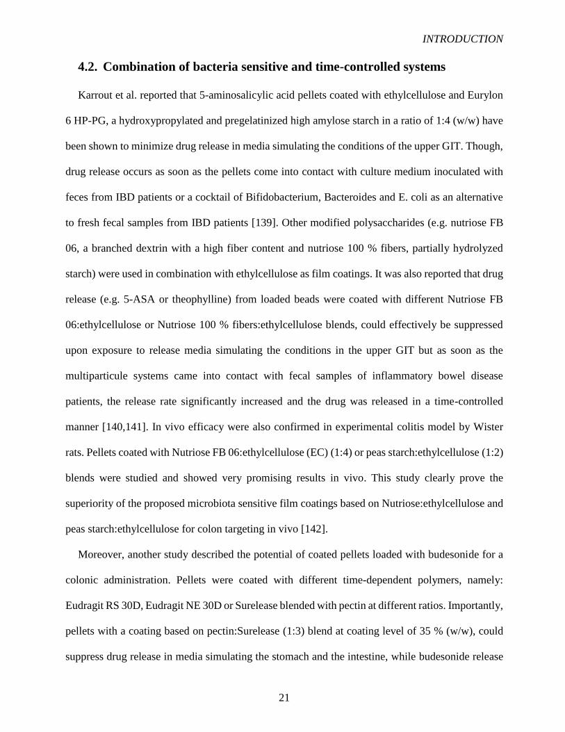
Colitis is the location and nature of the inflammation (figure 1). Crohn’s disease can affect any part
of GIT from the mouth to the anus but in most cases attacks the ileum. In contrast, Ulcerative Colitis
is restricted to the colon and the rectum [4].
Figure 1: Location of inflammation of gastro intestinal tract (dark areas) in the case of a) Crohn’s
disease and b) Ulcerative Colitis (Printed from [5]).
a) b)
INTRODUCTION
4
Symptoms consist of pain, diarrhea, and diminished appetite. Complications can occur such as
toxic dilation of the colon that sometimes cause colon cancer, hence, the need for regular monitoring
[6].
The different therapeutic strategies used to treat chronic inflammatory bowel diseases can be related
to drug treatments, biological drugs, immunosuppressants, symptomatic treatments and surgery in
case of the failure of the treatment. Drug treatments include anti-inflammatory drugs that require
frequently and high dose administration in order to control the state of the disease (more prolonged
remission) [7].
Colonic site specific delivery can provide major advantages such as:
Reducing drug dosing ensured by local drug delivery at the site of action [8].
Oral administration of peptide and protein drugs, which are fragile in the upper gastro
intestinal tract [9].
The colon is a site, where both local and systemic drug delivery could be achieved (topical
treatment of inflammatory bowel disease, e.g. Ulcerative Colitis or Crohn’s disease). Such
inflammatory conditions are usually treated with glucocorticoids and 5-aminosalycilic acid
(5-ASA) [9].
A number of others serious diseases of the colon, e.g. colorectal cancer, might also be
capable of being treated more effectively if drugs were targeted to the colon [8].
Reducing systemic side effects [8].
Conventional dosage forms cannot be used due to its premature drug release in the stomach and
small intestine. Thus, drug will be absorbed into the blood stream, provoking considerable side
effects. Nevertheless, drug concentration will be very low at the site of the action (distal part of
GIT), which can lead to less efficacy of the treatment. In order to overcome this problem, an ideal
dosage form should effectively protect the drug in the upper GIT and subsequently release the drug

INTRODUCTION
5
in the distal part of GIT in a time controlled manner, depending on the disease state. Reservoir
systems (coated pellets, capsules…) or matrices systems (tablets, extrudates…) can be used to
protect the drug in the upper GIT. Polysaccharides that are only degraded by bacterial enzymes
localized in the colon can offer a very useful tool for colonic dosage forms in the treatment of IBD.
2. Oral controlled drug delivery
2.1. Definitions
There are many routes of administration of active pharmaceutical ingredients (API): local, oral,
rectal, and parenteral. Therefore, the oral route is the most used route due to its several advantages,
such as ease of administration, the ambulant treatment, the less psychological anxiety during the
administration and the reduction of the infectious risks. In addition, it allows relatively easy
industrial production and therefore low production costs (pharmaceutical dosage forms are not
sterilized for instance) [10]. However, many disadvantages can be occurred. Indeed, the API can be
toxic for certain tissues and aggressive if drugs are absorbed in inappropriate location. It can also
be degraded before reaching the site of action and then loss of the efficacy of the treatment. This
requires the administration of several doses and thus increases in side effects. Furthermore, drug
absorption and the bioavailability of a drug can be different from a patient to another due to several
physiopathological factors (e.g. gastric transit, pH variations in the gastro intestinal tract, food
administration and also the state of the mucosa) and therefore, in-vitro studies should take in
consideration all these factors [11].
Controlled release systems have been developed for years, allowing the administration of high
amounts of drugs and releasing them according to a desired profile. An active substance is mixed
with specific excipients in order to improve drug efficacy and minimize undesirable side effects.
These systems can (i) protect a drug against gastric degradation and therefore, avoiding gastric
INTRODUCTION
6
irritation and loss of drug efficacy, (ii) release a drug progressively in time, (iii) delay drug release
in order to reach a specific absorption window or (iv) deliver site specifically drugs (e.g. colon
targeting) [12].
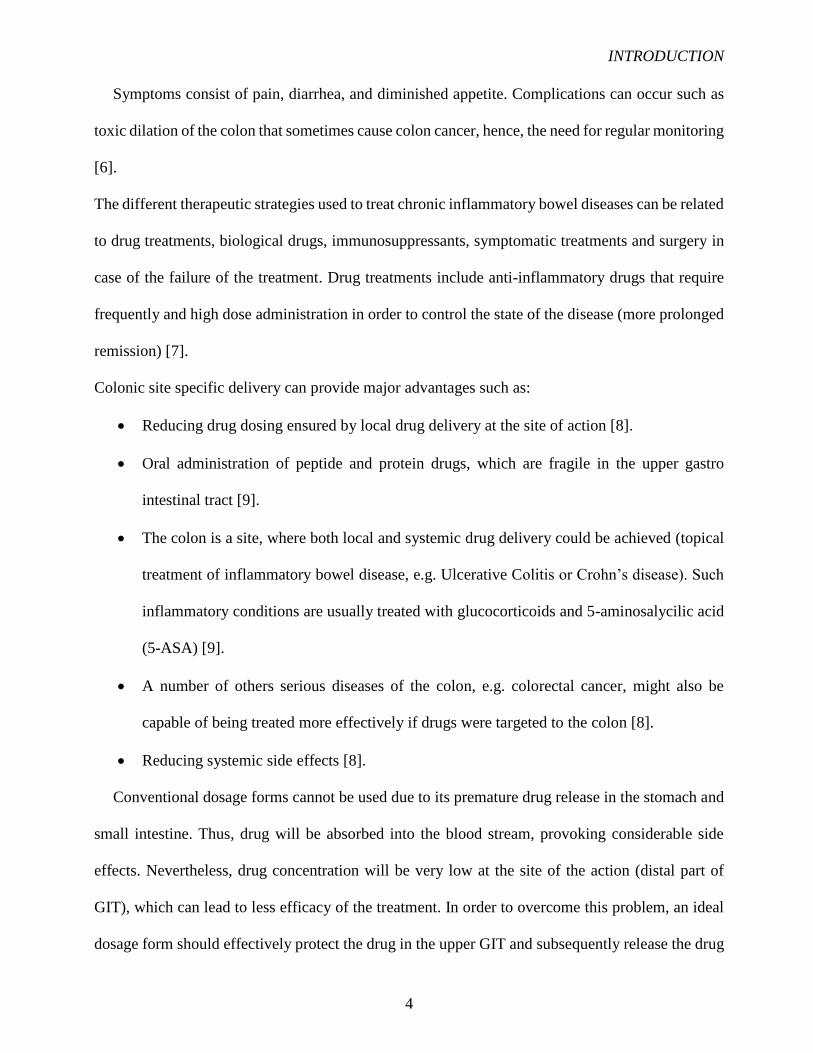
Furthermore, controlled drug delivery systems are very useful tools in order to achieve and maintain
an optimal therapeutic drug plasma concentration during a desired period of time (figure 2).
By controlling the delivery rate of the drug, the duration of the therapeutic action can be sustained.
Figure 2: Plasma concentrations of an immediate versus controlled release dosage form.
In general, the development of controlled release formulations offers many benefits over
conventional dosage forms: controlled administration of a therapeutic dose at the desired delivery
rate, which leads to less fluctuations of plasma concentrations [12]. This results in reduced toxicity
Minimum
efficacy level
Toxic level
Controlled release
Dru
g p
las
ma
co
nc
en
tra
tio
n
Time
Therapeutic window
Minimal toxic concentration
Minimal effective
concentration
Dru
g p
las
ma
co
nc
en
tra
tio
n
Time
Immediate release
Therapeutic window
Minimum
efficacy level
Toxic levelMinimal toxic concentration
Minimal effective
concentration

INTRODUCTION
7
as well as serious side effects and increased efficacy of the therapy. That can decrease the frequency
of the drug dose and improve patient compliance. Disadvantages include the longer time required
to achieve therapeutic blood concentrations, which can increase the variation in bioavailability after
oral administration, enhanced first-pass effect, risk on dose dumping (especially for reservoir-based
systems), lack of dose flexibility and usually higher costs [13].
Figure 3: Schematic representation of a) a matrix and b) a reservoir system (Printed from [14]).
Generally, controlled release delivery systems can be divided in two groups (figure 3):
Matrix systems where a drug is homogenously dissolved/dispersed in a polymeric
network to obtain a system allowing for controlled drug release. These systems have
several advantages including the easy-manufacture, the capacity for incorporating a high
amount of drug and avoiding toxic concentrations if the system gets accidentally
damaged. Various types of matrix systems can be achieved and thus, the drug release
mechanisms will be different [15-17].
Reservoir systems in this case, the drug is layered around an inert core or incorporated
into a water-soluble excipient matrix. The API core is surrounded by a coating layer.
Drug dispersed into
a polymeric matrix
Polymeric coating Drug dispersed into
a soluble core
b) a)

INTRODUCTION
8
Different drug release profiles can be achieved by varying the polymer nature and
properties [18,19].
2.2. Drug release mechanisms
The presence of the appropriate macromolecular networks can avoid immediate drug release and
then, different types of mass transport processes can be involved in the control of drug release.
This includes for instance, water diffusion into the system, drug dissolution, polymer swelling,
polymer dissolution and potentially matrix erosion [20-22]. The resulting drug release kinetics can
strongly depend on the drug properties (e.g. solubility in water) and drug loading. Sometimes, it can
be very challenging to provide desired drug release kinetics for a given drug and drug dose [23].
It is therefore possible to mathematically predict the drug release rate. However, numerous
parameters should be taken into account including type of the drug, the dosage form and the
environmental conditions during drug release. Importantly, drug release is impacted by several
factors such as the solubility and permeability; the preparation process of different types of dosage
forms (e.g. matrix or reservoir); the initial drug concentration in the system compered to drug
solubility, drug-polymer/excipients interactions and the device geometry (e.g. cubic, sphere,
cylinder, film) [24,25].
3. Hot melt extrusion and injection molding technology
3.1. Definitions
Hot melt extrusion (HME) has been used in many diverse industrial fields, mostly with the
processing of foods and the manufacturing of plastics. The first industrial use of single-screw
extruders was in the mid-nineteenth century with the extrusion of thermoplastic materials. Since
then, it became a very interesting technique used in the pharmaceutical industry with proven

INTRODUCTION
9
robustness for numerous drug delivery systems (DDS) [26,27]. Hot melt extrusion related patents,
which have been issued for pharmaceutical systems have steadily increased since the early 1980’s.
So far, the U.S.A and Germany are the leading of issued patents for hot melt extrusion in the market
[28].
Hot melt extrusion is a continuous process in which material melts or softens under elevated
temperatures and is further forced through a die, usually with the help of one or two conveyer screws
in a barrel. During the process, the material is exposed to heating and intense shear allowing a
homogeneous distribution of drug particles in a molten carrier [29].
The marked hot melt extruders have either one screw or two screws. The latter leads to better mixing
of the drug and polymer. Two screws are disposed side-by-side, allowing for different
configurations in all zones from the start of the product mixing to the output of the dosage form.
Moreover, the screws are either rotated in the same (co-rotating) or in the opposite direction (counter
rotating). The latter option is able to blend drug and polymer even in case of high viscosity, which
required high shear forces. Furthermore, the friction between the barrel, blending, and rotating
screws provides the driving force for the material in order to reach the die. It is to emphasize that
depending on the marked hot melt extruder, the material can be fed at different locations, which can
be very practically beneficial in case of the addition of additives during the process. Importantly,
liquids can also be introduced using a liquid pump and liquid injection system [29]. Due to increased
barrel temperature, the materials melts or fuses after it enters into the transition zone. The drug is
embedded homogenously within the carrier (polymer) with help of screws and the mixture moves
along the barrel towards the die. When the material reaches the output, it is delivered through the
die cavity and sized to obtain its final shape (figure 4) [30].

INTRODUCTION
10
Figure 4: Schematic representation of the hot melt extruder (Printed from [30]).
The adjustment of extrusion parameters is of utmost importance in the manufacturing of such
polymeric drug delivery systems. Adjustable parameters include screw speed, processing
temperature and feeding rate, which impact the shear stress (torque) and mean residence time and
in the long term also dissolution rate and stability of the final product [29]. In this purpose, extruder
sensors are able to measure the barrel and die temperatures, the torque generated, the melt pressure,
the feed rate, the screw speed and the melt temperature during the process. Moreover, other thermos
analytical techniques such as hot-stage microscopy, differential scanning calorimetry, micro
calorimetry, X-ray diffraction (XRD), dynamic mechanical thermal analysis (DMTA)
and thermogravimetric analysis (TGA) are often applied to investigate the chemical stability,
thermal behavior and crystalline properties of actives: and/or excipients in the final dosage form
[31].
Die Extrudate
Heating zones
Screw (s)
Feeder
API-polymer
mixture
Mixing elements
Barrel

INTRODUCTION
11
3.2. Pharmaceutical applications of hot melt extrusion
Hot melt extrusion is a continuous and free solvent process allowing for the preparation of solid
dispersions or solutions, depending on drug solubility in the matrix former (often a polymer) as well
as drug concentration in the system. Such solid dosage forms have the potential to improve drug
bioavailability, efficacy and safety of the drug treatment. Hot melt extrusion is used to produce
different polymeric drug delivery systems administrated via oral route such as polymeric films
[32,33] and tablets [34-36] but also transdermal [37], transmucosal [38,39] and intra ocular
(implants) route [40,41]. The production of three types of solid dispersions are described: (i) an
amorphous solid dispersion where both the drug and the carrier are amorphous but the drug is
dispersed in a particular form within the polymer (DSC thermogramm shows two Tg corresponding
to those of drug and polymer) (ii) a crystalline solid dispersion, where all/a fraction of the drug
remains crystalline in an amorphous polymer (DSC thermogramm shows a Tg corresponding to the
polymer and a fusion/dissolution pic corresponding to the drug) and (iii) an amorphous solid
solution where both the drug and the carrier are amorphous and completely miscible, which can be
indicated by one Tg in the DSC thermogramm. Note that the drug is dispersed at the molecular level
in case of an amorphous solid solution [42]. An amorphous solution have the advantage to increase
drug availability by improving its solubility. In a crystalline suspension, the drug crystals are
dispersed in the amorphous matrix which is suitable for a controlled drug delivery. Amorphous
solid suspension are the less stable with a high tendency to recrystallization. Indeed, the drug is in
an amorphous state and is dispersed in the formulation. The type of the solid dispersion obtained
depends on the drug solubility in the polymer, the stability of the final form and the drug - polymer
interactions [43].
INTRODUCTION
12
The choice of the polymer nature is critical in the formulation process and its application. Some
of the most well-known applications of hot melt extrusion are:
Taste masking of drugs [44-47].
Solubility enhancement of poorly water-soluble compounds for an immediate drug release
[48-51].
Sustained and time controlled release [52-55], and extended drug delivery systems [56].
Site specific drug delivery systems [57-59].
In addition to these current applications, some recent innovations have to be mentioned, such as
co-extrusion [60-62], co-crystallization [63-66], 3D printing [67-69] and injection molding [70-73].
3.3. Materials used for hot melt extrusion
3.3.1. Drugs
The properties of the active drug substance often limit the formulation and processing choices
available to the pharmaceutical scientist in the development of dosage forms. Hot melt extrusion is
a free solvent process, which avoids potential hydrolytic degradation pathways. In addition, poorly
compactable materials can be prepared as tablets without a compression process by cutting an
extruded rod to the desired dimensions. Furthermore, depending on the materials used, hot melt
extrusion can be useful to enhance the bioavailability of poorly soluble drugs. It can also protect a
drug from enzymatic degradation as well as avoid the irritation of the mucosa in gastro intestinal
tract. Moreover, if the drug has a specific absorption window or need to be targeted at a specific
area of the gastro intestinal tract for systemic or localized treatment of diseases (e.g. Crohn’s disease
and Ulcerative Colitis), hot melt extrusion can be used.
Prior to the hot melt extrusion process, deep knowledge of the physicochemical properties of the
drug as well as the polymer used is essential for the feasibility of the extrusion process. It is to
INTRODUCTION
13
emphasize that physicochemical characterization of the drug is extremely important for the
development of drug delivery systems (e.g. solubility, physical state, particle size, flowability,
melting/glass transition temperature and thermal degradation) [74].
3.3.2. Carriers
In hot melt extruded drug delivery systems, the active compound is embedded in a carrier
formulation often comprised of one or more “meltable” substances and other functional excipients.
The meltable substance is generally a polymer (natural or synthetic) or a lipid. The selection of the
carrier material for pharmaceutical drug delivery systems strongly depends on the application (taste
masking, immediate or sustained release). The choice of the pharmaceutically approved polymer is
critical in the formulation process as its properties not only dictate the processing conditions but
also govern the dissolution characteristics of the dosage form (drug release kinetics and release
mechanism).
As for drugs, some relevant characteristics of the carrier should be well investigated proceed to hot
melt extrusion [31]. Here there are mostly important factors: the chemical structure, the solubility,
the glass transition temperature/melting temperature, the melt viscosity that can be improved by the
addition of plasticizer, the flowability, the lipophilicity/hydrophylicity, the thermal stability, the
drug-carrier interactions and compatibility. Moreover, the characterization of rheological properties
can help in selecting the appropriate carriers and setting the process conditions, which can be cost
and time consuming [74].
Some of the carriers used for hot melt extrusion and their appropriate studies are listed below:
Ethylene vinyl acetate (EVA): a water-insoluble copolymer of ethylene and vinyl acetate
(VA). By simple varying the ethylene/VA ratio, large spectrum of drug release profiles can
be provided [75-77].

INTRODUCTION
14
Poly-vinyl acetate (PVAc): a water insoluble polymer, which is suitable for hot melt
extrusion [78,79].
Cellulose derivatives: Hydroxy Propyl Methyl Cellulose (HPMC) is a non-ionic water
soluble polymer, which is widely used for the preparation of controlled release tablets,
microparticles and films by HME [80-83]. Hydroxypropyl cellulose (HPC) is also a non-
ionic water-soluble and pH insensitive cellulose ether, which has been successfully used as
matrix former and solubility-enhancing agent [84,85]. Ethyl Cellulose (EC) is a non-ionic
water insoluble polymer [86,87].
Copolymers derived from esters of acrylic and methacrylic acid have various
physicochemical properties due to the nature of their functional groups. Commercially
available “Eudragit ®” for immediate release (E100, E PO), delayed release (S100, L100)
or sustained release (NE, NM, RL, RS) [88-97].
Poly Ethylene Oxides (PEO) have the same composition as Poly Ethylene Glycol (PEG) but
can be obtained at much higher molecular weights (100 KDa to 7000 KDa), they can be
used in controlled drug delivery systems [42,98-100] as well as plasticizers [101-103]
depending on their molecular weight.
Polyurethane are produced by a reaction between polyols (polyethers or polyesters) and
diisocyanates. They are copolymers containing soft and hard segments through which they
have thermoplastic and elastic properties. Polyurethanes have been used as carriers for hot
melt extrusion for the preparation of vaginal rings [104,105] and tablets for oral controlled
drug delivery [106,107].
Biopolymers (e.g. polysaccharides) such as chitosan (animal origin) and xanthan gum have
been used as matrix in hot melt extrusion for oral sustained release formulation [108,109].

INTRODUCTION
15
3.3.3. Plasticizers and additives
The addition of plasticizers can lower the processing extrusion temperatures that can avoid heat
degradation of drugs and carriers [110-112]. A plasticizer is a low molecular weight compound that
softens the polymer to make it more flexible, decreases the glass transition temperature (Tg) and
lowers the melt viscosity [113-115]. The plasticizer can increase the free volume between polymer
chains, providing more mobility for the polymer chains, which results softer and more flexible
mixture. Being more flexible, the processing temperature can be decreased, which leads to lower
torque generated during the process [116,117]. Furthermore, the physical and mechanical properties
of the final product can be improved. Most common plasticizers used are low molecular weight
polyethylene glycols, triacetin, triethyl citrate, glycerol, dibutyl sebacate...etc. Moreover, several
drugs (e.g. ibuprofen, guaifenesin, metoprolol tartrate) have been shown to be effective plasticizers
for certain polymers [118-120]. Since the drug and polymer are exposed to elevated temperatures,
high pressure and extensive mixing during hot-melt extrusion, it is important to evaluate the stability
of the API and polymer after the manufacturing process in order to avoid degradation. In order to
be efficient, several prerequisites have to be fulfilled by plasticizers, namely good efficiency,
polymer-plasticizer compatibility/affinity and thermostability. The efficiency of a plasticizer refers
to the concentration of plasticizer necessary to lower the glass transition temperature of the polymer.
Compatibility refers to the similarity in chemical structure between plasticizer and polymer, which
results in a better compatibility. The nature of the plasticizer can have an impact on drug release
kinetics: a hydrophobic plasticizer could be remained into the polymeric network, retarding the
release of a drug into the bulk media but in contrast the leaching out of the hydrophilic plasticizer
from the polymeric network can be rapid, which can creat more pores into the system and thus,
accelerating drug release [121,122].

INTRODUCTION
16
Other additives can also be used in these systems (e.g. antioxidants, lubricants). The stability of
polymers that are susceptible to degradation can be improved with the addition of antioxidants. The
latter can enhance the stability of polymers susceptible to degradation during hot melt extrusion.
3.4. Advantages and disadvantages of hot melt extrusion
As described above, hot melt extrusion is a continuous and free-solvent process. It offers several
advantages compared to conventionally available pharmaceutical processing techniques such as
[28]:
Increases solubility and bioavailability of poorly water soluble drugs.
The final product is free of residual solvent as no solvents are required during the process.
Improves the stability and avoids the degradation of the final product.
Reduced the production time (economical process).
Requires few processing steps.
A continuous operation with efficient scale-up from laboratory to pilot scale to production.
However, hot melt extrusion has some disadvantages as well. The main inconvenient of hot melt
extrusion is the thermal process, which is risks of drug/polymer stability and the physical
transformation of the extrusion process. Nevertheless, the use of a plasticizer sometimes solve this
problem. Furthermore, continuous industrial production can cause a high energy input and most
important costs related to the purchase of the equipment. Moreover, after the extrusion of
hydrophobic polymers, the cleaning of the machine can be problematic and time consuming.
Specific hydrophilic excipients are available in the market (HME cleaner Plus®) and are required
for the cleaning of different parts of the machine (eg. the screw, the barrel) after the extrusion
process [28,31].

INTRODUCTION
17
3.5. Commercially marketed hot melt extrusion formulations
As described above hot melt extrusion is a continuous free solvent process, which show various
advantages. It became a very interesting pharmaceutical technology in the industry used in many
research studies with an increase of number of publications per year. Despite the large and extensive
application in academic and industrial fields, only a few commercialized hot melt extrusion
pharmaceutical products (table 1) are currently marketed.

INTRODUCTION
18
Table 1: Some of hot melt extrusion marketed products. Reported from [123,124]
Name/company Drug
Carrier
(polymer)
Administration
route
Indication
Ozurdex®/Allergan Dexamethasone PLGA
Intraocular
(implants)
Macular edema
Implanon®/Merck Etonogestrel EVA
Subcutaneous
(amplants)
Contraceptive
Gris-PEG®/Pedinol Griseofulvin PEG
Oral
(tablets)
Onychomycosis
Kaletra®/Abbott Lopinavir/ritonavir PVP/PVA
Oral
(tablets)
Viral infection
(HIV)
Covera- HS®/Pfizer Verapamil, HCl HPC
Oral
(tablets)
Hypertension
Noxafil®/Merck Posaconazole
HPMCAS and
PVA
Oral
(tablets)
Antifungal
Nurofen®/Reckitt
Benckiser
Healthcare
Ibuprofen HPMC
Oral
(tablets)
Analgesic
Zithromax®/Pfizer Azithromycin
Pregelatinized
starch
Oral
(tablets)
Bacterial
infection

INTRODUCTION
19
4. Colon targeting
4.1. Strategies for colonic drug delivery systems
The site-specific drug delivery to the colon can improve local treatments of inflammatory bowel
diseases (IBD), such as Crohn’s disease and Ulcerative Colitis. The suppression of premature drug
release in the upper part of the gastrointestinal tract (GIT) avoids drug absorption into the blood
stream and thus minimizes undesired side-effects. Once the colon is reached, the drug is released,
ideally in a time-controlled manner, in order to ensure optimized drug concentration time profiles
at the site of action, leading to improved therapeutic effects [125]. Different strategies have been
reported in the literature aiming at site-specific drug delivery to the colon [126].
Immediate drug release upon contact with aqueous body fluids is avoided by the presence of the
matrix former/film coating. The latter should be insoluble and impermeable for the drug in the
stomach and small intestine but should dissolve or become permeable as soon as the target site is
reached. To provide the required site-specific system properties, the following approaches have
been described in the literature: (i) pH-dependent systems using pH-dependent polymers, which are
soluble at different pH levels in the different part of the GIT. The polymers described as pH-
dependent in colonic specific drug delivery are insoluble at low pH levels but become increasingly
soluble as pH rises [127]. Even if a pH-dependent polymer can protect a drug delivery system in
the stomach, the duodenum and the jejunum, it may start to dissolve in the ileum, and the site-
specificity of formulations can be poor [128], (ii) time-dependent systems are also very promising
drug delivery systems. Nevertheless, due to potentially large variations of gastric transit time for
humans, nature and amount of food intake, colonic arrival time is unfortunately unpredictable. This
results in poorly colonic drug availability [129]. It had to be pointed out that the transit time, the
gastrointestinal movement, especially peristalsis or contraction in the stomach result in change in
gastrointestinal transit of the drug [130] but also to the accelerated transit time observed in IBD-

INTRODUCTION
20
patients [131-133], (iii) bacteria sensitive systems have been found to be the most promising and
adapted to the disease state because of the significantly increased colonic microbiota and enzymatic
activities in the distal part of the GIT [134]. The use of natural polysaccharides is attracting a lot of
attention for colon targeting since these polymers are found in abundance and have wide
availability. These polymers are inexpensive and are available in a verity of a structures with varied
physicochemical properties. They are highly stable, safe, nontoxic, and hydrophilic and in addition,
are biodegradable. These include polysaccharides obtained from plant (guar gum, inulin), animal
(chitosan), algal (alginates) or microbial (dextran) origin [135]. In combination with a time or pH
dependent polymer, the system should be able to minimize or completely suppress drug release in
the upper part of the GIT. But in contact with colonic fluid, drug release is triggered by the
enzymatic fermentation of these polysaccharides.
The use of multiparticulate dosage forms (e.g. pellets and mini-matrices) can be very interesting
and provides major advantages compared to single unit dosage forms (e.g. tablets or capsules) such
as the all-or-nothing effect that can be avoided: if a tablet gets accidentally damaged within the
upper GIT, the entire drug dose is lost and the gastric emptying time is less variable, because the
pylorus can be passed even in the contracted state. Furthermore, significant drug amounts can be
incorporated in the core of coated dosage forms. This is particularly important for highly dosed
drugs, such as 5-aminosalicylic acid (5-ASA), which is the standard treatment for inflammatory
bowel diseases-patients [136-138]. From the literature point of view, care should be taken when
using just one approach for targeting the 5-ASA to the colon even for multiparticulate dosage forms
because of inter and intra variability of physiopathological conditions of patients. However, the
combination of different approaches in one delivery system could make drug delivery to the colon
more assured.
INTRODUCTION
21
4.2. Combination of bacteria sensitive and time-controlled systems
Karrout et al. reported that 5-aminosalicylic acid pellets coated with ethylcellulose and Eurylon
6 HP-PG, a hydroxypropylated and pregelatinized high amylose starch in a ratio of 1:4 (w/w) have
been shown to minimize drug release in media simulating the conditions of the upper GIT. Though,
drug release occurs as soon as the pellets come into contact with culture medium inoculated with
feces from IBD patients or a cocktail of Bifidobacterium, Bacteroides and E. coli as an alternative
to fresh fecal samples from IBD patients [139]. Other modified polysaccharides (e.g. nutriose FB
06, a branched dextrin with a high fiber content and nutriose 100 % fibers, partially hydrolyzed
starch) were used in combination with ethylcellulose as film coatings. It was also reported that drug
release (e.g. 5-ASA or theophylline) from loaded beads were coated with different Nutriose FB
06:ethylcellulose or Nutriose 100 % fibers:ethylcellulose blends, could effectively be suppressed
upon exposure to release media simulating the conditions in the upper GIT but as soon as the
multiparticule systems came into contact with fecal samples of inflammatory bowel disease
patients, the release rate significantly increased and the drug was released in a time-controlled
manner [140,141]. In vivo efficacy were also confirmed in experimental colitis model by Wister
rats. Pellets coated with Nutriose FB 06:ethylcellulose (EC) (1:4) or peas starch:ethylcellulose (1:2)
blends were studied and showed very promising results in vivo. This study clearly prove the
superiority of the proposed microbiota sensitive film coatings based on Nutriose:ethylcellulose and
peas starch:ethylcellulose for colon targeting in vivo [142].
Moreover, another study described the potential of coated pellets loaded with budesonide for a
colonic administration. Pellets were coated with different time-dependent polymers, namely:
Eudragit RS 30D, Eudragit NE 30D or Surelease blended with pectin at different ratios. Importantly,
pellets with a coating based on pectin:Surelease (1:3) blend at coating level of 35 % (w/w), could
suppress drug release in media simulating the stomach and the intestine, while budesonide release

INTRODUCTION
22
can be triggered in the presence of rat cecal content. In vivo results confirmed therapeutic efficacy
of pectin/Surelease coated pellets loaded with budesonide, indicating the improvement of
macroscopic and microscopic parameters of experimental induced colitis [143].
Furthermore, in another study, chitosan was mixed with Aquacoat® ECD 30 (a suspension of
ethylcellulose) and used as coating for pellets loaded with 5-ASA. In-vitro dissolution studies upon
exposure to simulated intestinal fluid, chitosan level as well as the thickness of the coating layer
have been shown to impact/influence drug release profile. Pellets coated with Aquacoat:chitosan
blend ratio (23:2) at 60 % coating level released around 5 % of 5-ASA over 12 h in simulating
intestinal fluid (SIF). This formulation was selected for further studies. Drug release was more
pronounced in presence of fecal rat contents (simulated colonic fluid = SCF) and pores were visible
on the surface of the pellets using environmental scanning electron microscopy (ESEM). These
multiparticulate coated systems might offer a promising tool for colon targeting. However, further
in-vivo test should be investigated in order to confirm the potential of these systems for colonic
delivery [144].
Li-Fang et al. reported that tablets contained theophylline and coated with a blend of Kollidon
SR:chitosan (3.5:1) is able to protect the drug in fluids simulating the upper part of the GIT. Drug
release in simulating colonic fluid have been shown high and more effective digestion of chitosan
with rat fecal bacterial enzymes secreted during the test than β-glucosidase enzyme incorporated
[145].
In another study, Wilson et al., showed that tablets loaded with mesalazine and coated with
amylose:ethylcellulose blend was suitable for the treatment of IBD by targeting the release of
mesalazine in the colon. In-vitro tests showed that the ratio of the two polymers as well as the
thickness of the film are key parameters to control drug release. Importantly, tablets coated with
amylose:ethylcellulose blend ratios of 1:3 and 1:2 at different coating levels (2, 4 or 6 %) are able
INTRODUCTION
23
to completely suppress mesalazine release in media simulating the stomach and small intestine (less
than 10 % of mesalazine released after 6 h upon exposure to dissolution media). Furthermore, drug
release significantly increased in contact with human fecal samples, which confirmed these results
in vivo [146].
Liu et al., developed a capsule with an enteric coating for pulsatile drug release. The capsule was
filled with a rapid-disintegrating tablet of 5-ASA and a pectin-based plug in the opening of capsule
body. Pectin-based plug was consisted of either high methoxy pectin (HM-pectin) and lactose or
low methoxy pectin (LM-pectin) and HPMC. HM-pectin alone led to a long lag time because of its
high viscosity, while the lactose addition can shorten the lag time. LM-pectin due to its weak gel
ability was used along with HPMC in order to extend the lag time. The lag time prior to drug release
was shortened in the presence of pectinase or rat cecal content in the release medium, which
confirmed the responsiveness of pectin plug to the enzymatic action. Determination of 5-ASA in
the plasma samples of beagle dogs after administration of a single dose of the orally pulsatile
capsule, revealed site-specific delivery to the colon [147].
4.3. Combination of bacteria sensitive and pH-dependent systems
Patole et al., prepared 5-ASA loaded alginate microspheres and filled them in capsules based on
HPMC. Capsules were coated with Eudragit FS 30D, an enteric coating. These systems were able
to suppress 5-ASA release in gastric fluid due to the enteric coating. However, drug release was
triggered in the presence of rat fecal samples by bacterial degradability of alginate. Also, in-vivo
studies showed a significant reduction of ulcer index in rats treated with these alginate based
microspheres [148].
In two another studies, Jain et al. and Mohanty et al. used Eudragit S 100 coating for
microspheres based on pectin and loaded with 5-ASA, or alginate [149] for colonic delivery

INTRODUCTION
24
multiparticulate systems. 5-ASA microspheres based on pectin were prepared by water-in-oil (w/o)
emulsification and then coated with Eudragit S 100. These multiparticulate systems were in vitro
evaluated for their drug release. In both studies, it was shown that Eudragit S100 was able to
suppress 5-ASA release from pectin microspheres in media simulating the upper GIT (stomach and
small intestine) and releasing drug in the colon (around 98 % drug release after 16 h). This can be
attributed to the dissolution of the enteric coating (Eudragit S100) at pH above 7 and the
biodegradation of pectin by colonic bacteria [150,151].
Eudragit S 100 was also used as an enteric coating for chitosan based microparticules to deliver
mesalamine into the colon. In this study, in vitro drug release from chitosan microparticles coated
with Eudragit S 100 was compared to mesalamine suspensions. Importantly, 100 % of mesalamine
was released from the suspension, contrary to drug release from chitosan coated microparticules,
which was almost suppressed in media simulating the stomach and small intestine, while in the
presence of rat fecal samples, 71 % of mesalamine was released within 20 h. Furthermore, in vivo
tests performed by Sprague Dawley rats showed that microparticles containing chitosan and coated
with Eudragit S 100 have the potential to maintain the concentration of mesalamine in the targeted
region for a long time (Cmax of mesalamine in rats’ plasma after 8 h of administration). These
systems can offer a very interesting tool to treat inflammations localized in the distal part of GIT
[152].
Combination of pH-dependent and bacterial degradation approaches for the design of colonic
delivery of 5-ASA as a single unit dosage form were also studied. In one study, chitosan capsules
filled with 5-ASA were enteric coated to achieve colonic drug delivery. Drug release from the
capsules increased significantly when dissolution was performed in media containing rat cecal
contents. For the treatment of colitis in rats, 5-ASA chitosan capsules orally administered and better

INTRODUCTION
25
therapeutic effects were observed compared to administration of 5-ASA in carboxymethyl cellulose
suspension [153].
Furthermore, 5-ASA tablets were coated with chitosan and then overcoated with Eudragit L100.
Eudragit L100 prevented dissolution of chitosan layer in gastric region and the chitosan layer
protected drug release in the small intestine due to its low solubility in basic pH. The developed
formulation indicates a degradation of chitosan by bacterial enzymes located in the colon, resulting
in colonic delivery of 5-ASA [154].
4.4. Combination of three different approaches
Other studies associated three different approaches: pH-time-enzyme dependent systems for the
delivery of 5-ASA to the colon. Multiparticulate systems (coated pellets) were investigated by Fude
et al. Three different coating layers were applied on 5-ASA loaded pellets prepared by extrusion-
spheronisation. Calcium pectinate as an enzyme dependent polymer, which could be digested by
enzymes secreted by colonic bacteria allowing for colonic administration of 5-ASA, ethylcellulose,
a time-dependent polymer, which could protect drug release in the small intestine and an enteric
coating (Eudragit L 30D-55), which prevented drug release in the stomach. No significant drug
release was observed in 0.1 M HCl while the drug release after 3-4 h lag time at pH 6.8 was observed
during in-vitro dissolution test. An increase in drug release in contact with colonic enzymes might
support the suitability of using this system to achieve colon targeted drug delivery [155].
Design of 5-ASA microparticulates with matrix structure using combination of different
approaches was studied by Mladenovska et al. Microparticles consisting of 5-ASA dispersed in the
chitosan-Ca-alginate matrix were prepared by spray drying. Both pH dependency and bacterial
degradability were used as main mechanisms for colonic delivery of the drug in these particles. This
system was evaluated in vitro as well as in vivo by Wistar rats. It was shown that chitosan-alginate
INTRODUCTION
26
complex was able to erode slowly upon medium with pH values higher than 6.5. This property
prevented drug release in the upper parts of GIT and controlled drug release in the colon. In addition,
degradation of chitosan by the colonic microflora resulted in specific drug release in the colon.
Colonic delivery of the microparticles was confirmed in biodistribution studies in rats, resulting in
suggested usefulness of the system for clinical treatment in human IBD [156,157].
The combination of three approaches for colonic delivery of 5-ASA tablets was studied by
Nunthanid et al., who proposed a delivery system which was sensitive to time, pH and enzyme. 5-
ASA tablets were coated by combinations of spray-dried chitosan acetate (as a pH-dependent and
bacterial sensitive polymer) and HPMC (as a time-dependent polymer) at different ratios. The pH-
dependent swellability and solubility of spray-dried chitosan acetate, its degradation by the β-
glucosidase enzyme and gradual dissolution of HPMC contributed to drug release underlying
mechanisms. The results clearly demonstrated the suitability of the combination of HPMC and
spray-dried chitosan acetate at an appropriate ratio as a coating material for 5-ASA colonic delivery
[158].

INTRODUCTION
27
5. Purposes of this work
The major objective of this work was to develop novel dosage forms prepared by hot melt
extrusion/injection molding allowing for site-specific drug delivery to the colon in the treatment of
inflammatory bowel diseases such as Crohn’s disease and Ulcerative Colitis.
The strategy used is a combination of a polysaccharide (pectin, guar gum, inulin, maltodextrin…),
which permits for colon targeting by enzymatic degradation and a hydrophobic thermoplastic
polymer (ethylcellulose, polyurethane, polyvinyl acetate…), which can reduce the hydrophilicity of
the biodegraded polysaccharide. However, the mixture of the two polymers should not hidden
bacterial enzymatic degradation of the polysaccharide. Furthermore, hot melt extruded
polysaccharide blends for controlled drug delivery were studied. Particular aims included:
The Preparation and physicochemical characterization of thin polymeric films of different
polysaccharide:hydrophobic polymer blends suitable for colonic delivery.
The preparation of hot melt extrudates and injection-molded capsules for colonic delivery
and the set of the process parameters.
The physicochemical characterization of extrudates and capsules and the identification of
promising systems for colonic drug delivery.
The preparation of polysaccharides-based hot melt extudates for controlled drug delivery
and the optimization of the process parameters.

28

29
II. MATERIALS AND METHODS

30
MATERIALS AND METHODES
31
1. Materials
1.1. Polymer aqueous dispersions
Polyvinyl acetate (Kollicoat SR 30D; BASF, Ludwigshafen, Germany); ethyl acrylate methyl
methacrylate copolymer (Eudragit NE 30D; Evonik Degussa GmbH, Darmstadt, Germany);
ammonioalkyl methacrylate copolymers (Eudragit RL 30D and Eudragit RS 30D; Evonik Degussa
GmbH, Darmstadt, Germany).
1.2. Polymer powders
Polyvinyl acetate (Kollidon SR 30D; BASF, Ludwigshafen, Germany); hydrophobic grades of
polyurethane: Carbothane PC-3575A (Carbothane) and Techothane TT-1074A (Tecothane);
hydrophilic grades of polyurethane: Tecophillic SP 93A-100 (Tecophilic SP93) and Tecophilic SP
60D-60 (Tecophilic SP60) (Lubrizol corporation, Wilmington, MA, USA); ethylcellulose (EC,
Ethocel Standard 10 premium; Colorcon, Massy, France); hydroxypropyl methylcellulose (HPMC,
Methocel E5 Premium LV; Colorcon, Kent, England).
1.3. Polysaccharide powders
Pectin and maltodextrin (UniPectin and C*Actistar 11700 Tapioca maltodextrin , Cargill,
Krefeld, Germany); maize starch (C*PharmGel 03406; Cargill, Gent, Belgium); guar gum
(viscosity of a 1 % solution in water at 25 °C: ~5000 cP; Cooper, Melun, France); inulin (Inulin
HPX and Inulin Synergy 1; Beneo, Oreye, Belgium); chitosan (Crab Shell chitosan, Mw = 800 kDa,
degree of deacetylation = 80-90 %; Bio 21, Chonburi, Thailand); sodium alginate (Keltone LVCR;
FMC Bio-polymer, Girvan, UK); carrageenan (Gelcarine GP-91-NF; Philadelphia, USA); Arabic
gum (Cooper, Melun, France); xanthan gum (Keltrol*TF; Kelco, London, UK).

MATERIALS AND METHODES
32
1.4. Plasticizers
Dibutyl sebacate (DBS; Stearinerie Dubois, Boulogne-Billancourt, France); triethyl citrate
(TEC; Alfa Aesar, Karlsruhe, Germany); poly ethylene glycol (PEG 1500; Pluracare E 1500 Flasks;
BASF, Ludwigshafen, Germany).
1.5. Drug powders
Anhydrous theophylline and diprophylline (BASF, Ludwigshafen, Germany)
Table 2: Physicochemical properties of model drugs [159-161].
Drug Anhydrous theophylline Diprophylline
Chemical structure
Chemical formula
C7H8N4O2 C10H14N4O4
Molecular weight 180.17 g/mol 254.24 g/mol
Solubility (in water at 25 °C) 7.36 g/L 333 g/L
Melting point 270 - 274 °C 155 - 157 °C
pKa 8.81 13.91
Log P - 0.02 - 1.9

MATERIALS AND METHODES
33
1.6. Ingredients for culture medium preparation
Extracts from beef and tryptone (Pancreatic digest of casein; Becton Dickinson, Sparks, USA);
yeast extract (Oxoid Ltd, Hants, UK); sodium chloride (J. T. Baker, Deventer, Netherlands);
L-cysteine hydrochloride hydrate (Acros Organics, Geel, Belgium); Ringer solution (Merck,
Darmstadt, Germany).
1.7. Ingredients for mobile phase preparation
Acetonitrile (CWR, Fontenay sous bois, France); sodium acetate (Sodium acetate, anhydrous,
99 %, ThermoFisher GmbH, Kandel, Germany).
2. Preparation of dosage forms
2.1. Thin polymeric films
Thin polymeric films were prepared by casting blends of different aqueous hydrophobic polymer
dispersions and a polysaccharide into Teflon molds and subsequent drying for 1 day at 60 °C. The
polysaccharide was dispersed in purified water. Aqueous polymer dispersion (30 % w/w solids
content) was plasticized for 24 h with 30 % DBS or TEC (w/w, referred to the solid content of the
dispersion) under stirring. The polysaccharide and plasticized polymer dispersions were blended at
room temperature at different polymer:polymer (w/w) blend ratios. The mixtures were stirred for 6
h prior to casting.
2.2. Hot melt extrudates
Drug and polymer powders were blended for 10 min at 98 rpm in a Turbula T2A (Willy A.
Bachofen Maschinenfabrik, Muttenz, Switzerland), followed by manual mixing in a mortar with a

MATERIALS AND METHODES
34
plasticizer (TEC, DBS or PEG 1500). The compositions were varied as indicated. Polymer:polymer
blend ratios are expressed in weight:weight, plasticizer percentages are referring to the mass of
1st polymer (mass of 1st polymer = 100 %), drug percentages refer to the total mass of the hot melt
extrudates (mass of extrudate = 100 %). The plasticized blends were kept at room temperature for
24 h, followed by extrusion with a Nano 16 twin screw extruder (Leistritz, Nuremberg, Germany),
equipped with a 4 mm diameter die (screw diameter = 16 mm, length/diameter ratio = 26.25).
Figure 5 shows the setting of the screw elements. The process temperatures were kept constant at
170 – 170 – 170 – 170 °C, 130 – 130 – 130 – 130 °C or 100 – 100 – 100 – 100 °C
[zone 4 (die) – zone 3 – zone 2 – zone 1], as indicated. The feed rate was set at 3 mL/min.
After cooling, the hot melt extrudates were manually cut into cylinders.
Figure 5: Setting of the screw elements used for hot melt extrusion.
2.3. Injection-molded capsules
Injection molding process was performed with a bench-top micromolding machine (BabyPlast
6/10P; Cronoplast S.L., Barcelona, Spain; Rambaldi S.r.l., Molteno, Lecco, IT) equipped with a
capsular mold furnished with a hot-runner and with two interchangeable inserts for the
manufacturing of matching cap and body items of 600 μm thickness. Hot melt extrudates prepared
as mentioned before were manually cut into cylinders of 2 mm length prior to manually load in the

MATERIALS AND METHODES
35
plasticizing unit of the injection molding machine and successively pushed in the injection chamber
by means of the loading plunger. Figure 6 shows the different zones of the micromolding machine.
Figure 6: Zones and temperatures of the micromolding machine used for the preparation of
capsules (printed from babyplast® 6/10P instruction manual).
Afterwards, two different and consecutives injection pressures (P1 - P2), maintained for a
selected time (t1 - t2) in order to inject the polymeric melt into the mold cavity. The injection
molding temperatures were set as during the hot melt extrusion process and then adjusted. For the
injection pressures and times, minimum values were set up and progressively increased until the
obtaining of complete capsule shell. Final process parameters for the manufacturing of 600 μm
capsules are reported in table 3.
Table 3: Injection molding conditions for the manufacturing of capsules
Plasticizing
chamber
temperature
Injecting
temperature
Nozzle
temperature
Mold
temperature
Pressure
P1 – P2
Injection
time t1 – t2
Cooling
time
100 °C 100 °C 100 °C 107 °C
100-80
bar
6-5 s 10 s

MATERIALS AND METHODES
36
3. Physicochemical characterizations
3.1. Optical microscopy
Macroscopic pictures of hot melt extrudates and capsules were taken with an optical image
analysis system (Nikon SMZ-U; Nikon, Tokyo, Japan), equipped with a Zeiss camera (Axiocam
ICc1; Zeiss, Jena, Germany). Cross-sections of hot melt extrudates were obtained by manual
breaking.
3.2. Fermentation studies
Polysaccharide powders (approximately 500 mg) were incubated 24 h in (i) culture medium
inoculated with feces from inflammatory bowel disease patients (ii) culture medium inoculated with
bacterial strains: Baterioides, Bifidobacterium and E.coli, (iii) culture medium free of feces and
bacteria for reasons of comparison.
Culture medium was prepared by dissolving 1.5 g beef extract, 3 g yeast extract, 5 g tryptone,
2.5 g NaCl and 0.3 g L-cysteine hydrochloride hydrate in 1 L distilled water (pH 7.0 ± 0.2) and
subsequent sterilization in an autoclave. Feces of patients (approximately 1 g) with Crohn’s disease
or ulcerative colitis were diluted 1:200 with cysteinated Ringer solution; 2.5 ml of this suspension
was diluted with culture medium to 100 ml. The samples were agitated (50 rpm; Stuart, Cole-
Parmer; Villepinte, France) at 37 °C under anaerobic conditions (AnaeroGen 2.5 L; Thermo
Scientific; Illkirch, France). The pH was measured using color-fixed pH indicator sticks (Fisher
Scientific; Leicestershire, UK).

MATERIALS AND METHODES
37
3.3. Water content and dry mass
The water content and the dry mass loss kinetics of hot melt extrudates and thin polymeric films
were measured gravimetrically upon exposure to (i) simulated gastric fluid (0.1 M HCl pH 1.2) (ii)
simulated intestinal fluid (phosphate buffer pH 6.8; USP 41) (iii) simulated colonic fluid (culture
medium inoculated with feces from inflammatory bowel disease patients) (iv) culture medium
inoculated with bacterial strains: Baterioides, Bifidobacterium and E.coli as substitutes of fecal
samples and (v) culture medium free of feces for reasons of comparison.
Samples were placed into flasks (1 sample per flask), filled with 200 mL 0.1 M HCl or phosphate
buffer pH 6.8 and agitated at 80 rpm (in a horizontal shaker, 37 °C; GFL 3033, Gesellschaft fuer
Labortechnik, Burgwedel, Germany). The incubation with fecal samples was performed under
anaerobic conditions (AnaeroGen 2.5 L; Thermo Scientific; Illkirch, France). Culture medium was
prepared as described above. Samples were placed into 120 ml containers filled with 100 ml pre-
heated medium, followed by horizontal shaking at 37 °C (50 rpm; Stuart, Cole-Parmer; Villepinte,
France). At predetermined time points, samples were withdrawn and rinsed with water.
Excess water was removed by careful blotting with Kimtech precision wipes (Kimberly Clark,
Roswell, GA). Samples were accurately weighed (wet mass) and dried to constant weight at 60 °C
(dry mass). The water uptake and the dry mass (%) at time t was calculated as follows:
Water content (%) (t) = wet mass (t) − dry mass (t)
wet mass (t) . 100 % (1)
Dry mass (%) (t) = dry mass (t)
dry mass (t = 0) . 100 % (2)
MATERIALS AND METHODES
38
3.4. In vitro drug release
Under conditions simulating the upper gastro intestinal tract: Hot melt extrudates or injection-
molded capsules (filled with 10 mg of drug) were placed into flasks (1 sample per flask), filled with
200 mL 0.1 M HCl and agitated at 80 rpm (in a horizontal shaker, 37 °C; GFL 3033, Gesellschaft
fuer Labortechnik, Burgwedel, Germany). After 2 h, the release medium was completely exchanged
with phosphate buffer pH 6.8 (USP 41). At pre-determined time points, 3 mL samples were
withdrawn and analyzed UV-spectrophotometrically (UV-1800; Shimadzu, Kyoto, Japan)
at λ = 275 nm (theophylline) or λ = 274 nm (diprophylline) for their drug content.
Under conditions simulating the entire gastro intestinal tract: Hot melt extrudates or injection-
molded capsules (filled with 10 mg of drug) were exposed to 0.1 M HCl for 2 h and subsequently
to phosphate buffer pH 6.8 (USP 41) for 6 h, in a USP Apparatus 3 (20 dpm, 37 °C, Bio-Dis; Varian,
Paris, France). Afterwards, the samples were transferred into 120 mL flasks filled with: (i) 100 mL
culture medium inoculated with human fecal samples, or (ii) culture medium free of feces for
reasons of comparison.
Culture medium was prepared as described above. The samples were agitated (50 rpm; Stuart,
Cole-Parmer; Villepinte, France) at 37 °C under anaerobic conditions (AnaeroGen 2.5 L; Thermo
Scientific; Illkirch, France). At pre-determined time points, 2 mL samples were withdrawn,
centrifuged at 10000 rpm for 10 min (Centrifuge Universal 320; Hettich, Tuttlingen, Germany),
filtered (0.45 μm, Millex-HU; Merck Millipore, Tullagreen, Ireland) and analyzed by HPLC for
their drug content using a Thermo Fisher Scientific Ultimate 3000, equipped with a LPG 3400
SD/RS pump, an auto sampler (WPS-3000 SL) and a UV-Vis detector (VWD-3400RS) (Thermo
Fisher Scientific, Waltham, USA). In the case of theophylline, the mobile phase consisted of 10 %
acetonitrile and 90 % water (v/v). Samples were injected into a C18 column (Kinetex 5 µm EVO
C18 100 Å, 250 mm x 4.6 mm; Phenomenex, Le Pecq, France) and the flow rate was set

MATERIALS AND METHODES
39
at 1.0 mL/min. The drug was detected UV-spectrophotometrically at λ = 275 nm. In the case of
diprophylline, the mobile phase consisted of 35 % acetonitrile and 65 % 0.01 M aqueous sodium
acetate solution (v/v). Samples were injected into a Polar C18 column (Luna Omega 3 µm Polar
C18 100 Å, 150 mm x 4.6 mm; Phenomenex, Le Pecq, France) and the flow rate was set at 1.0
mL/min. The drug was detected UV-spectrophotometrically at λ = 274 nm.
3.5. Differential scanning calorimetry (DSC)
DSC thermograms were recorded with a Q200 calorimeter (TA Instruments, Guyancourt,
France). During all measurements the calorimeter head was flushed with highly pure nitrogen gas.
Temperature and enthalpy readings were calibrated using pure indium at the same scan rates as used
in the experiments. The samples were placed in hermetic high volume pans, resistant to pressure.
Approximately 6 mg samples were heated from - 40 to 300 °C at 10 °C/min.
3.6. Powder X-ray diffraction (PXRD)
PXRD experiments were conducted with a PanAlytical X’PERT PRO MPD diffractometer
(λCuKα = 1.5418 Å for combined K1 and K2), equipped with an X’celerator detector (Almlo,
The Netherlands). Samples were placed into Lindemann glass capillaries (diameter = 0.7 mm) and
installed on a rotating sample holder to avoid artifacts due to preferential orientations of crystallites.
Heating experiments have been performed in situ, at 10 °C/min, using the furnace HTC 9634 from
Huber (Rimsting, Germany).
3.7. Thermogravimetric analysis (TGA)
TGA experiments were conducted with a Q500 TGA from TA Instruments (Guyancourt,
France). Samples were placed in open aluminum pans, and the furnace was flushed with highly pure

MATERIALS AND METHODES
40
nitrogen gas (50 mL/min). The temperature reading was calibrated using the Curie points of alumel
and nickel, while the mass reading was calibrated using balance tare weights provided by TA
Instruments. All scans were performed at 10 °C/min.

41
III. RESULTS AND DISCUSSIONS

42
43
CHAPTER 1:
POLYVINYL ACETATE:POLYSACCHARIDE
BLENDS FOR COLON TARGETING

RESULTS AND DISCUSSION: CHAPTER 1
44

RESULTS AND DISCUSSION: CHAPTER 1
45
POLYVINYL ACETATE:POLYSACCHARIDE BLENDS FOR COLON
TARGETING
Insoluble polymers such as polyvinyl acetate (PVAc) are widely used as oral controlled release
matrix prepared by hot melt extrusion [78,79]. Commercially available Kollidon® SR is a matrix
retarding agent based on 80 % PVAc, 19 % povidone, 0.8 % sodium lauryl sulfate and 0.2 % Silica
[162]. It was reported in the literature that polyvinyl acetate and different polysaccharides blends
are suitable for a colonic drug delivery [145]. Li-Fang et al. showed that tablets contained
theophylline and coated with a blend of Kollidon SR:chitosan (3.5:1) is able to protect the drug in
fluids simulating the upper part of the gastro intestinal tract (GIT). Drug release in simulating
colonic fluid have been shown high and more effective digestion of chitosan in contact with rat
fecal samples than β-glucosidase enzyme incorporated [145].
The aim of this study was to evaluate the ability of blends of Kollidon SR with a second
polysaccharide as hot melt extrudates to provide site-specific drug delivery to the colon. Kollidon
SR was used to minimize drug release in the upper GIT due to its hydrophobicity. Thus, the second
polysaccharide (inulin and maltodextrin) allowing for colon targeting is more permeable and
fermentable by colonic bacteria. Dibutyl sebacate was added as a plasticizer in order to reduce the
process temperature of hot melt extrudates containing theophylline as model drug. The water
content and dry mass loss kinetics of hot melt extrudates free of drug as well as in vitro drug release
from hot melt extrudates containing different amounts of theophylline were measured under
conditions simulating the contents of the entire GIT (0.1 M HCl, phosphate buffer pH 6.8 and
culture medium inoculated with fecal samples from IBD-patients). For reasons of comparison,
in vitro drug release was also performed upon exposure to 0.1 M HCl, followed by phosphate buffer
pH 6.8, followed by culture medium free of fecal samples.
RESULTS AND DISCUSSION: CHAPTER 1
46
1. Effect of polysaccharide nature and amount in thin polymeric films
Figures 7 and 8 show the impact of different types of polysaccharides and their amount (as
indicated in the figures) in thin polymeric films on the water content and dry mass loss kinetics upon
exposure to culture medium inoculated with a cocktail of Bifidobacterium, Bacterioides and E.coli
(solid curves). For reasons of comparison, the films were also exposed to culture medium free of
bacteria (dashed curves). Thin polymeric films contained blends of kollicoat SR (polyvinyl acetate
suspension) and different types of polysaccharides. The polymer:polymer blend ratio was varied as
indicated and the films were plasticized with 30 % TEC (based on the solid mass of Kollicoat SR).
Importantly, water content and dry mass loss rate and extent increased by increasing the amount
of the water soluble polysaccharide in the film. This can be explained by the increase of the
hydrophilicity of the systems, which leads to the formation of more pores in the polymeric network,
allowing for high amount of water penetration into the system (figures 7 and 8).
Furthermore, all blends did not show any significant difference of water content and dry mass loss
either in the presence or in the absence of bacteria. This can be attributed to the fact that Kollicoat
SR in this mixture (Kollicoat SR and the 2nd polysacharide) protect the polysaccharides form
enzymatic degradation.

RESULTS AND DISCUSSION: CHAPTER 1
47
Figure 7: Water uptake of thin films based on: a) kollicoat SR:alginate, b) kollicoat SR:arabic gum,
c) kollicoat SR:inulin HPX, d) kollicoat SR:maltodextrin and e) kolicoat SR:pectin blends upon
exposure to culture medium inoculated with a cocktail of Bifidobacterium, Bacterioides and E.coli
and culture medium free of bacteria for reasons of comparison. The polymer:polymer blend ratio
was varied as indicated, the films were plasticized with 30 % TEC (referring to kollicoat SR).
0
25
50
75
100
0 6 12 18 24
Wate
r co
nte
nt,
%
Time, h
70:30 kollicoat SR:alginate
80:20
100:0
0
25
50
75
100
0 6 12 18 24
Wa
ter
co
nte
nt,
%
Time, h
70:30 kollicoat SR:arabic gum
80:20
100:0
0
25
50
75
100
0 6 12 18 24
Wate
r co
nte
nt,
%
Time, h
70:30 kollicoat SR:maltodextrin
80:20
100:0
a) b)
d)
0
25
50
75
100
0 6 12 18 24
Wate
r co
nte
nt,
%
Time, h
50:50 kollicoat SR:inulin HPX
70:30
80:20
100:0
c)
0
25
50
75
100
0 6 12 18 24
Wate
r co
nte
nt,
%
Time, h
50:50 kollicoat SR:pectin
70:30
100:0
e)
Cocktail of bacteria
Culture medium

RESULTS AND DISCUSSION: CHAPTER 1
48
Figure 8: Dry mass of thin films based on: a) kollicoat SR:alginate, b) kollicoat SR:arabic gum, c)
kollicoat SR:inulin HPX, d) kollicoat SR:maltodextrin and e) kolicoat SR:pectin blends upon
exposure to culture medium inoculated with a cocktail of Bifidobacterium, Bacterioides and E.coli
and culture medium free of bacteria for reasons of comparison. The polymer:polymer blend ratio
was varied as indicated, the films were plasticized with 30 % TEC (referring to kollicoat SR).
0
25
50
75
100
0 6 12 18 24
Dry
fil
m m
ass, %
Time, h
100:0 kollicoat SR:alginate
80:20
70:30
0
25
50
75
100
0 6 12 18 24
Dry
fil
m m
as
s, %
Time, h
100:0 kollicoat SR:arabic gum
80:20
70:30
0
25
50
75
100
0 6 12 18 24
Dry
fil
m m
ass, %
Time, h
100:0 kollicoat SR:inulin HPX
80:20
70:30
50:50
0
25
50
75
100
0 6 12 18 24
Dry
fil
m m
as
s, %
Time, h
100:0 kollicoat SR:maltodextrin
80:20
70:30
0
25
50
75
100
0 6 12 18 24
Dry
fil
m m
as
s, %
Time, h
100:0 kollicoat SR:pectin
70:30
50:50
c)
a) b)
d)
e)
Cocktail of bacteria
Culture medium

RESULTS AND DISCUSSION: CHAPTER 1
49
2. Effect of polysaccharide nature on pH level
Figure 9: Impact of polysaccharide nature on pH level of culture medium (i) inoculated with a
cocktail of Bifidobacterium, Bacterioides and E.coli, (ii) inoculated with feces of patients suffering
from crohn’s disease and (iii) free of fecal samples for reasons of comparison.
0
1
2
3
4
5
6
7
8
9
10
De
cre
as
e in
pH
Culture Medium
Bifidobacterium, Bacterioides and E.Coli
Feces of IBD-patients
RESULTS AND DISCUSSION: CHAPTER 1
50
Polysaccharides fermentation results in a final stage to short-chain fatty acid production [163]
such as acetate and butyrate [164]. These final products could have an impact on the pH of the
release media and could be used as indicator for polysaccharides fermentation by bacterial enzymes
in the colon. Figure 9 shows the impact of different polysaccharides on the pH of culture medium
under anaerobic conditions in the presence of fecal samples from patients suffering of inflammatory
bowel diseases or a cocktail of different bacterial strains (E.Coli, Bacterioides and Bifidobacterium)
as a substitutes of fecal samples [140,141]. For reasons of comparison, polysaccharides were also
incubated in culture medium free of fecal samples.
The initial pH of culture medium was around 7 (± 0.2) and remained constant in the absence of
polymer after 24 h incubation with fecal samples or with the cocktail of bacteria. As it can be seen,
for samples containing inulin and maltodextrin, pH values of the medium decreased in the presence
of fecal samples (approximately from 7 to 5). This can be attributed to the fact that inulin and
maltodextrin served as substrate for bacteria in the fecal samples. The decrease of pH of culture
medium inoculated with the cocktail of bacteria was not pronounced as with fecal samples
(approximately from 7 to 6.5) even in the case of inulin and maltodextrin. This can be explained by
the fact that probably the quantitative and qualitative composition of the cocktail of bacterial strains
(E.Coli, Bacterioides and Bifidobacterium) is not sufficient to degrade the polysaccharides into
short chain fatty acids. Thus, this cocktail of bacteria can be used only with precaution as an
alternative drug release media simulating colonic conditions, and also depending on the type of
polysaccharide and the film polymeric mixture.
In case of sodium alginate, carrageenan and xanthan gum, no changes in pH values was observed
neither in the presence of fecal samples nor upon exposure to bacterial strains. It is to emphasize
that inulin as well as maltodextrin might offer an interesting tool as excipients for colon targeting
and therefore they will be selected for further studies in this work.

RESULTS AND DISCUSSION: CHAPTER 1
51
3. Impact of hydrophobic polymer nature in thin polymeric films
Figure 10: Macroscopic pictures of thin polymeric films consisting of different types of polymers
blended with inulin. The 1st polymer:inulin blend ratio was 80:20, the films were plasticized with
30 % DBS (referring to the 1st polymer).
Eudragit RL:inulin
Kollicoat SR:inulin Eudragit NE:inulin
Eudragit RS:inulin

RESULTS AND DISCUSSION: CHAPTER 1
52
Figure 10 shows the macroscopic pictures of casted thin polymeric films (pieces obtained by
manual cutting) based on different types of hydrophobic polymers and inulin blends. Figure 11
shows the impact of different hydrophobic polymers nature (as indicated) in thin films based on
inulin blends on the water content and dry mass loss kinetics upon exposure to culture medium
inoculated with fecal samples from IBD-patients (white bars). For reasons of comparison, thin
polymeric films based on inulin blends upon exposure to culture medium free of feces are shown
(black bars). The polymer:polymer blend ratio was 80:20. Films were plasticized with 30 % DBS
(referring to the 1st hydrophobic polymer).
Importantly, dry mass loss of kollicoat SR:inulin blend upon contact to fecal samples was much
more pronounced than upon exposure to culture medium free of feces (around 25 % vs 5 %). This
can be attributed to the fact that inulin is degraded by the enzymes secreted by the bacteria present
in the human feces. Furthermore, the water content of the films was even higher in the presence of
fecal samples. This can be explained by the fermentation of inulin, which allows high amount of
water penetration into the polymeric network. Other polymer blends did not show this difference
neither in the presence nor in the absence of feces. This can be attributed to the hydrophobic nature
of the 1st polymer, which probably covered entirely the polysaccharide that should be digested by
bacterial enzymes.
Clearly, kollidcoat SR:inulin blends can offer a promising tool for colon targeting by releasing
an incorporated drug as soon as the target site is reached.

RESULTS AND DISCUSSION: CHAPTER 1
53
Figure 11: Water content and dry mass of thin polymeric films consisting of different types of
polymers blended with inulin upon exposure to culture medium inoculated with feces of patients
suffering from inflammatory bowel diseases (white bars) and culture medium free of feces (black
bars) for reasons of comparison. The 1st polymer:inulin blend ratio was 80:20, the films were
plasticized with 30 % DBS (referring to the 1st polymer).
50
60
70
80
90
100
Eudragit RL Eudragit RS Eudragit NE Kollicoat SR
Dry
m
as
s, %
Culture Medium
Feces of IBD-patients
0
25
50
75
100
Eudragit RL Eudragit RS Eudragit NE Kollicoat SR
Wa
ter
co
nte
nt,
%
Culture Medium
Feces of IBD-patients

RESULTS AND DISCUSSION: CHAPTER 1
54
4. Effect of the amount of polysaccharide in hot melt extrudates
4.1. Processability of hot melt extrudates
Figure 12 shows microscopic pictures of hot melt extrudates surfaces’ (obtained by manual
breaking) based on different types of polymer:polymer blends: Kollidon SR (polyl vinyl acetate)
was blended with inulin or maltodextrin. The Kollidon SR:2nd polysaccharide blend ratio was varied
as indicated in the figure. Hot melt extrudates were plasticized with 30 % DBS (referring to Kollidon
SR) prior to hot melt extrusion at 100 °C. For reasons of comparison, hot melt extrudates based
“only” on Kollidon SR are shown (plasticized with 30 % DBS). Clearly, the structure of all systems
appeared to be rather homogeneous and the surface relatively smooth, except for Kollidon
SR:maltodextrin blend at the ratio of 20:80, which lead to breakable extrudates with a granular
surface.
Moreover, no visible sign of polymer degradation was observed. The cross-section of hot melt
extrudates based “only” on Kollidon SR (plasticized with 30 % DBS) increases by die output
(this phenomenon is called die swelling). The extent of swelling, which was not observed in case
of Kollidon SR:2nd polysaccharide blends depends on the viscoelastic properties of the material, a
lower viscosity leads to less extensive polymer swelling on the viscosity of the blend [165].
This can be at least confirmed by the torque measurement during the extrusion, which remained
stable (around 30 %) in case of Kollidon SR:inulin blends. Please note that low and stable torque
will lead to simple extrusion processing (figure 13a). However, the manufacturing of 20:80
Kollidon SR:maltodextrin blend showed variations of torque measurement during the process (high
standard deviation, around 13), which can be explained by the fact that maltodextrin remained in a
solid state at 100 °C and the amount of Kollidon SR was not enough to embed all the polysaccharide
powder (figure 13b).

RESULTS AND DISCUSSION: CHAPTER 1
55
Figure 12: Macroscopic pictures of hot melt extrudates based on Kollidon SR and different types
of a polysaccharide (inulin or maltodextrin, as indicated in the figure). The polymer:polymer blend
ratio is indicated in the figure, the extrudates were extruded at 100 °C and plasticized with 30 %
DBS (referring to Kollidon SR). For reasons of comparison, also hot melt extrudates based “only”
on Kollidon SR (plasticized with 30 % DBS) are illustrated.

RESULTS AND DISCUSSION: CHAPTER 1
56
a)
b)
Figure 13: Impact of the poymer:polymer blend ratio on the torque generated during the
preparation of hot melt extrudates based on a) Kollidon SR:inulin and b) Kollidon SR:maltodextrin.
The extrudates were extruded at 100 °C and plasticized with 30 % DBS (referring to Kollidon SR).
0
25
50
75
100
100:0 70:30 50:50 30:70 20:80
To
rqu
e,
%
Kollidon SR:inulin
Stable
0
25
50
75
100
100:0 70:30 50:50 30:70 20:80
To
rqu
e,
%
Kollidon SR:maltodextrin

RESULTS AND DISCUSSION: CHAPTER 1
57
4.2. Hot melt extrudates properties in the upper gastro intestinal tract
The water content and dry mass loss kinetics of a polymeric system can be used as indicators for
its permeability to drug, upon exposure to the release medium. With increasing water content and
increasing dry mass loss, the density of the macromolecular network decreases, resulting in
increased polymer chain mobility and, thus, increased drug permeability.
Figures 14 to 17 shows the impact of polymer:polymer blend ratio on the water content and dry
mass loss kinetics of hot melt extrudates upon exposure to 0.1 M HCl (figures a) and phosphate
buffer pH 6.8 (figures b) simulating the gastric and intestinal fluid respectively. Hot melt extrudates
based on kollidon SR:inulin (figures 14 and 15) or Kollidon SR:maltodextrin (figures 16 and 17)
blends are shown. Extrudates were plasticized with 30 % DBS (referring to Kollidon SR) and
extruded at 100 °C. For reasons of comparison, water content and dry mass loss of hot melt
extrudates based “only” on plasticized Kollidon SR are shown (Kollidon SR:2nd polysaccharide
100:0). As it can be seen, water content and dry mass loss kinetics upon exposure to 0.1 M HCl and
phosphate buffer pH 6.8 increased by increasing the 2nd polysaccharide amount for all cases with a
higher rate for Kollidon SR:inulin blends compared to Kollidon:maltodextrin blends (for instance,
25 % water content vs 15 % and 30 % dry mass loss vs 2 % for the blend ratio 30:70, respectively
upon exposure to 0.1 M HCl for 2 h). This can be probably due to the physicochemical properties
of polysaccharides and particularly their solubility.
Furthermore, the investigated 2nd polysaccharides (inulin and maltodextrin) exhibit a higher
affinity to water, more than Kollidon SR, which is insoluble in water. It was also noticed that pure
(plasticized) Kollidon SR hot melt extrudates loose minor amounts of dry mass. The low dry mass
loss can be attributed to the leaching out of povidone (polyvinylpyrrolidone: a water soluble
component) into the bulk media.

RESULTS AND DISCUSSION: CHAPTER 1
58
Figure 14: Water content kinetics of hot melt extrudates consisting of different Kollidon SR:inulin
blend ratios (indicated in the figures) upon exposure to: a) 0.1 M HCl and b) phosphate buffer pH
6.8. The extrudates were extruded at 100 °C and plasticized with 30 % DBS (referring to Kollidon
SR). Extrudates consisting “only” on Kollidon SR (plasticized with 30 % DBS) are shown for
reasons of comparison.
Kollidon SR:inulin
0
25
50
75
100
0 0.5 1 1.5 2
Wa
ter
co
nte
nt,
%
Time, h
20:80
30:70
50:50
70:30
100:0
0
25
50
75
100
0 2 4 6 8
Wate
r co
nte
nt,
%
Time, h
20:80
30:70
50:50
70:30
100:0
Kollidon SR:inulin
b)
a)

RESULTS AND DISCUSSION: CHAPTER 1
59
Figure 15: Dry mass loss kinetics of hot melt extrudates consisting of different Kollidon SR:inulin
blend ratios (indicated in the figures) upon exposure to: a) 0.1 M HCl and b) phosphate buffer pH
6.8. The extrudates were extruded at 100 °C and plasticized with 30 % DBS (referring to Kollidon
SR). Extrudates consisting “only” on Kollidon SR (plasticized with 30 % DBS) are shown for
reasons of comparison.
Kollidon SR:inulin
Kollidon SR:inulin
0
25
50
75
100
0 0.5 1 1.5 2
Dry
ma
ss
, %
Time, h
100:0
70:30
50:50
30:70
20:80
0
25
50
75
100
0 2 4 6 8
Dry
mass, %
Time, h
100:0
70:30
50:50
30:70
20:80
a)
b)

RESULTS AND DISCUSSION: CHAPTER 1
60
Figure 16: Water content kinetics of hot melt extrudates consisting of different Kollidon
SR:maltodextrin blend ratios (indicated in the figures) upon exposure to: a) 0.1 M HCl and b)
phosphate buffer pH 6.8. The extrudates were extruded at 100 °C and plasticized with 30 % DBS
(referring to Kollidon SR). Extrudates consisting “only” on Kollidon SR (plasticized with 30 %
DBS) are shown for reasons of comparison.
0
25
50
75
100
0 2 4 6 8
Wate
r co
nte
nt,
%
Time, h
30:70
50:50
70:30
100:0
0
25
50
75
100
0 0.5 1 1.5 2
Wa
ter
co
nte
nt,
%
Time, h
30:70
50:50
70:30
100:0
Kollidon SR:maltodextrin
Kollidon SR:maltodextrin
b)
a)

RESULTS AND DISCUSSION: CHAPTER 1
61
a)
b)
Figure 17: Dry mass loss kinetics of hot melt extrudates consisting of different Kollidon
SR:maltodextrin blend ratios (indicated in the figures) upon exposure to: a) 0.1 M HCl and b)
phosphate buffer pH 6.8. The extrudates were extruded at 100 °C and plasticized with 30 % DBS
(referring to Kollidon SR). Extrudates consisting “only” on Kollidon SR (plasticized with 30 %
DBS) are shown for reasons of comparison.
0
25
50
75
100
0 2 4 6 8
Dry
ma
ss
, %
Time, h
100:0
70:30
50:50
30:70
0
25
50
75
100
0 0.5 1 1.5 2
Dry
ma
ss
, %
Time, h
100:0
70:30
50:50
30:70
Kollidon SR:maltodextrin
Kollidon SR:maltodextrin

RESULTS AND DISCUSSION: CHAPTER 1
62
In order to avoid premature drug release in the stomach and small intestine, a polymeric system
allowing for colon targeting should be poorly permeable for the incorporated drug upon exposure
to media simulating the conditions in the stomach and the small intestine. Thus, the most promising
systems for a colonic administration should show a very low water content and dry mass loss extent.
Comparing the water content and dry mass loss kinetics of hot melt extrudats upon exposure to
0.1 M HCl and phosphate buffer pH 6.8 in figures 14 to 17, it can be seen that the type of 2nd
polysaccharide as well as the polymer:polymer blend ratio have a significant impact on the
polymeric network and drug mobility. Importantly, hot melt extrudates based on Kollidon
SR:maltodextrin blends showed the lowest water content and dry mass loss and can effectively
decrease the drug release in media simulating the upper GIT. Furthermore, Kollidon SR:Inulin hot
melt extrudates at the ratio 70:30 and 50:50 showed a low rate and amount of water content and dry
mass loss (less than 10 % of dry mass loss upon exposure to phosphate buffer pH 6.8 for 8 h) and
might be a very promising tool for a colonic administration. However, hot melt extrudates based on
Kollidon SR:inulin blends (ratio of 30:70 and 20:80) showed a high water content and dry mass
loss (75 % of dry mass loss for kolidon SR:inulin 20:80 blend upon exposure to phosphate buffer
pH 6.8 for 8 h), which can probably lead to a premature drug release from these hot melt extrudates
in the upper part of GIT. Further studies are required in order to determine if these promising
systems are able to release the incorporated drug at the site of action in a time controlled manner
(by enzymatic fermentation of the polysaccharide).

RESULTS AND DISCUSSION: CHAPTER 1
63
4.3. Hot melt extrudates properties in the colon
Figure 18: Water content of hot melt extrudates consisting of different a) Kollidon SR:inulin and
b) Kollidon SR:maltodextrin blend ratios (as indicated) upon exposure to culture medium and
culture medium inoculated with feces of patients suffering from inflammatory bowel diseases (as
indicated in the figures). The extrudates were extruded at 100 °C and plasticized with 30 % DBS
(referring to Kollidon SR). Extrudates consisting “only” on Kollidon SR (plasticized with 30 %
DBS) are shown for reasons of comparison.
0
25
50
75
100
100:0 70:30 50:50 30:70
Wa
ter
co
nte
nt,
%
Culture Medium
Feces of IBD-patients
0
25
50
75
100
100:0 70:30 50:50 30:70 20:80
Wa
ter
co
nte
nt,
%
Culture Medium
Feces of IBD-patients
Kollidon SR:inulin
Kollidon SR:maltodextrin
a)
b)

RESULTS AND DISCUSSION: CHAPTER 1
64
Figure 19: Dry mass of hot melt extrudates consisting of different a) Kollidon SR:inulin and b)
Kollidon SR:maltodextrin blend ratios (as indicated) upon exposure to culture medium and culture
medium inoculated with feces of patients suffering from inflammatory bowel diseases (as indicated
in the figures). The extrudates were extruded at 100 °C and plasticized with 30 % DBS (referring
to Kollidon SR). Extrudates consisting “only” on Kollidon SR (plasticized with 30 % DBS) are
shown for reasons of comparison.
Kollidon SR:inulin
Kollidon SR:maltodextrin
a)
b)
25
40
55
70
85
100
100:0 70:30 50:50 30:70 20:80
Dry
m
as
s, %
Culture Medium
Feces of IBD-patients
25
40
55
70
85
100
100:0 70:30 50:50 30:70
Dry
m
ass, %
CultureMedium
Feces ofIBD-patients

RESULTS AND DISCUSSION: CHAPTER 1
65
Experimental water content and dry mass loss of hot melt extrudates were studied (figures 18
and 19) upon exposure to culture medium inoculated with fecal samples from IBD-patients.
For reasons of comparison, also water content and dry mass loss kinetics of hot melt extrudates
upon exposure to culture medium free of feces are studied. Hot melt extrudates based on Kollidon
SR:inulin or Kollidon SR:maltodextrin blends have been shown in figures 18 and 19. Thirty percent
of DBS was added to powders blends prior to hot melt extrusion at 100 °C.
Clearly, in case of hot melt extrudates based on Kollidon SR:inulin blends (ratios 50:50, 30:70
and 20:80), dry mass loss was more pronounced upon exposure to culture medium inoculated with
feces than culture medium free of fecal samples (around 20 % vs 5 % for the ratio 50:50).
This indicates that inulin serves as a substrate for the enzymes secreted by the colonic bacteria
(figure 19a). Furthermore, the water content of extrudates, which were exposed to patient feces was
higher than those exposed to sterile culture medium free of feces (figure 18a). However, these
differences in water content and dry mass loss were not observed in case of extrudates containing
Kollidon SR:maltodextrin blends (figures 18b and 19b). This can be probably due to the fact that
Kollidon SR protects the maltodextrin against enzymatic degradation, thus, these systems were not
studied any further.
Appropriate hot melt extrudates for colon targeting should take up considerable amounts of water
and show significant dry mass loss upon exposure to fecal samples from IBD-patients in order to
trigger the embedded drug release at the site of inflammation in the distal part of GIT. As it can be
seen in figures 18a and 19a, hot melt extrudates based on Kollidon SR:inulin 50:50 showed
significant water uptake and dry mass loss upon exposure to fecal samples of patients suffering
from IBD. Interestingly, these extrudates showed a low water content and dry mass loss extent in
media simulating the upper GIT. Moreover, also blends ratio of 30:70 and 20:80 (Kollidon
SR:inulin) look very promising for a colonic drug delivery (significant water content and dry mass

RESULTS AND DISCUSSION: CHAPTER 1
66
loss behavior upon exposure to fecal samples). However, these systems already take up considerable
amounts of water and remarkably loose in dry mass upon contact with media simulating the contents
of the upper GIT (figures 14 and 15).
5. Impact of drug loading in hot melt extrudates
5.1. Processability of hot melt extrudates
In order to investigate the potential of hot melt extrudates used as matrices systems for a colonic
administration, extrudates based on Kollidon SR:inulin 50:50 blend were prepared and loaded with
different amounts of theophylline as model drug.
Figure 20: Impact of the theophylline loading on the torque generated during the preparation of
hot melt extrudates based on Kollidon SR:inulin (50:50). The extrudates were extruded at 100 °C,
loaded with 10 to 60 % theophylline and plasticized with 30 % DBS (referring to Kollidon SR). For
reasons of comparison, also hot melt extrudates free of drug are shown.
0
25
50
75
100
0% 10% 20% 30% 40% 50% 60%
To
rqu
e,
%
Kollidon SR:inulin (50:50)
Theophylline

RESULTS AND DISCUSSION: CHAPTER 1
67
Hot melt extrudates based on Kollidon SR:inulin containing various concentrations of
theophylline, which is a slightly water-soluble drug were prepared by varying the drug loading (10,
20, 30, 40, 50 and 60 %, referring to the total extrudate mass). In all formulations, the Kollidon
SR:inulin blend ratio was kept constant: 50:50 (w/w). Hot melt extrudates were plasticized with 30
% DBS (referring to Kollidon SR) and extruded at 100 °C. In all cases a homogeneous system
structures and relatively smooth surfaces were obtained. Please note that in no cases, any visible
sign of polymer degradation was observed.
Figure 20 shows the respective torque values observed during extrusion. As it can be seen, the
torque values were stable and constant (around 35 %) during the extrusion for the formulation
loaded from 0 to 40 % theophylline. However, torque values increased with increasing the drug
loading from 40 to 60 % (around 35 % to 55 %), which is in good agreement with data reported in
the literature [45]. This can probably be attributed to the fact that theophylline do not melt at
100 °C (figure 55; chapter 3) and the relative amounts of the plasticized polymer blends in the
formulations decrease (increase viscosity). Besides, this increase in torque values were still in the
limits of the extruder and did not cause any difficulty during processing.
5.2. Physical characterization of hot melt extrudates
Since blends powder were exposed to heating during hot melt extrusion process, it was important
to evaluate the physical state modifications of drug during the manufacturing process.
The X-ray diffraction patterns of the raw materials and of the investigated hot melt extrudates are
shown in figure 21. Hot melt extrudates were based on Kollidon SR:inulin (50:50) blend containing
various amounts of theophylline (0, 20, 40 and 60 % referring to the total extrudate mass).
Extrudates were plasticized with 30 % DBS (referring to Kollidon SR) and extruded at 100 °C.
As it can be seen, the pure drug exhibited sharp X-ray diffraction, indicates that drug is in crystalline

RESULTS AND DISCUSSION: CHAPTER 1
68
nature. In contrast, Kollidon SR and inulin powders as well as hot melt extrudates free of drug were
X-ray amorphous and showed any Bragg pics. Importantly, X-ray diffraction Bragg peaks of
theophylline were clearly visible in the investigated hot melt extrudates of Kollidon SR:inulin blend
(50:50) even at 20 % drug loading but mostly in the range between 20 and 30 degrees. Importantly,
theophylline is at least partially distributed in the form of small crystals throughout the hot melt
extrudates at all the investigated drug loadings (figure 21).
Figure 21: X-ray diffractograms of hot melt extrudates based on 50:50 Kollidon SR:inulin blend.
The amount of theophylline was varied as indicated. The extrusion temperature was 100 °C, the
extrudates were plasticized with 30 % DBS (referring to Kollidon SR). For reasons of comparison,
also the X-ray diffractograms of hot melt extrudates free of drug as well as the pure theophylline,
Kollidon SR and inulin powders (all as received) are illustrated.
0
25000
50000
75000
100000
125000
150000
175000
200000
0 10 20 30 40 50 60
Inte
nsit
y, co
un
ts
2θ, degree
60 % theophylline HME
40 % theophylline HME
20 % theophylline HME
0 % theophylline HME
Pure kollidon SR
Pure inulin
Pure theophylline

RESULTS AND DISCUSSION: CHAPTER 1
69
5.3. In vitro drug release from hot melt extrudates
Figure 22: Impact of the theophylline loading on drug release from hot melt extrudates in 0.1 M
HCl for 2 h, followed by phosphate buffer pH 6.8. The systems were based on 50:50 Kollidon
SR:inulin blends, the extrusion temperature was 100 °C, the extrudates were plasticized with 30 %
DBS (referring to Kollidon SR).
The impact of drug loading on the experimental in vitro drug release was studied. The
percentages of slightly water-soluble theophylline were varied from 10 to 60 %. Figure 22 shows
0
25
50
75
100
0 2 4 6 8
Th
eo
ph
yll
ine r
ele
ased
, %
Time, h
60% theophylline
50%
40%
30%
20%
10%
pH 1.2 pH 6.8
Kollidon SR:inulin (50:50)

RESULTS AND DISCUSSION: CHAPTER 1
70
the resulting drug release kinetics from hot melt extrudates of Kollidon SR:inulin blend (50:50)
containing different drug loading in 0.1 M HCl for 2 h, followed by phosphate buffer pH 6.8 for
6 h. Hot melt extrudates were plasticized with 30 % DBS (referring to Kollidon SR) and extruded
at 100 °C. As it can be seen, the resulting drug release rates clearly increased with increasing drug
loading. This can be explained by the fact that less drug release retarding polymer is present in the
systems. On the other hand, the systems upon contact to the release media, which facilitate the
ingress of water into the systems and subsequently increasing the solubility and diffusion of drug.
Most of the drug (almost 75 %) is released during 8 h at an initial drug loading of 60 %.
An ideal dosage form allowing for the site-specific drug delivery to the colon should completely
suppress drug release in the upper GIT. However, once the colon is reached, drug release should be
triggered in a time controlled manner. Importantly, at a drug loading of 10 %, premature drug release
from these hot melt extrudates based of Kollidon SR:inulin blend (50:50) could be minimized (less
than 25 % of theophylline was released after 8 h upon exposure to the release media). Thus, these
type of systems shows a promising potential for colon targeting. Based on results shown in figure
22, hot melt extrudates of Kollidon SR:inulin (50:50) containing 10 % theophylline were selected
for drug release studies in media simulating colonic fluid.
This system was exposed to 0.1 M HCl for 2 h, followed by 6 h in phosphate buffer pH 6.8
followed by 10 h in culture medium inoculated with feces from inflammatory bowel disease patients
(solid curve in figure 23). For reasons of comparison, the extrudates were also exposed to 0.1 M
HCl, followed by phosphate buffer pH 6.8 and culture medium free of feces (dotted curve in figure
23). As it can be seen, the drug release rate did not increase upon contact with fecal samples
(approximately the same percentage of drug released in presence or absence of fecal samples:
around 30 %). This can be at least explained by the fact that despite of the considerable amount of
water uptake (figure 18a) by these hot melt extrudates and the significant dry mass loss (figure 19a)

RESULTS AND DISCUSSION: CHAPTER 1
71
upon exposure to fecal samples from IBD-patients, this system was not susceptible to bacterial
enzymes located in the colon of IBD-patients (figure 23).
Figure 23: Theophylline release from hot melt extrudates based on 50:50 Kollidon SR:inulin blends
under conditions simulating the transit through the entire gastro intestinal tract: 2 h in 0.1 M HCl,
followed by 6 h in phosphate buffer pH 6.8, followed by culture medium inoculated with feces of
patients suffering from inflammatory bowel diseases (solid curves). For reasons of comparison also
drug release in 0.1 M HCl, phosphate buffer pH 6.8 and culture medium without fecal samples is
shown (dotted curves). The drug loading was 10 %, the extrudates were extruded at 100 °C and
plasticized with 30 % DBS (referring to Kollidon SR).
0
25
50
75
100
0 4 7 11 14 18
Th
eo
ph
yll
ine
re
lea
se
d, %
Time, h
pH 1.2 pH 6.8 Feces or culture medium
Culture medium
Feces
Kollidon SR:inulin (50:50)

72
73
CHAPTER 2:
POLYURETHANE:POLYSACCHARIDE
BLENDS FOR COLON TARGETING
RESULTS AND DISCUSSION: CHAPTER 2
74

RESULTS AND DISCUSSION: CHAPTER 2
75
POLYURETHANE:POLYSACCHARIDE BLENDS FOR COLON
TARGETING
Recently, thermoplastic polyurethanes (TPU) were found to be promising matrix formers for the
production of high drug loaded oral sustained release formulations prepared by hot melt extrusion
and injection molding (HME/IM). They are used in order to reduce the amount of excipients needed
and creating a major advantage for patient’s compliance [166]. These polymers are produced by a
reaction between polyols (polyethers or polyesters) and diisocyanates. They are copolymers
containing soft and hard segments through which they have thermoplastic and elastic properties.
Polyurethanes have been used as carriers for hot melt extrusion for the preparation of vaginal rings
[104,105] as well as tablets for oral controlled drug delivery [106,107].
The aim of this study was to evaluate the suitability of different thermoplastic polyurethane
blends with a second polysaccharide for hot melt extrudates and injection-molded capsules,
providing colonic site-specific drug delivery. Carbothane, tecothane, tecophilic SP 60 and tecophilic
SP93 were used to minimize drug release in conditions simulating the upper gastro intestinal tract
(GIT). Thus, these polysaccharides (inulin, maltodextrin, guar gum and maize starch) allowing for
colon targeting are more permeable and fermentable by colonic bacteria. Dibutyl sebacate was
added as a plasticizer in order to reduce the process temperature of hot melt extrudates and injection-
molded capsules containing theophylline as model drug. The water content and dry mass loss
kinetics of hot melt extrudates free of drug were measured under conditions simulating the contents
of the entire GIT (0.1 M HCl, phosphate buffer pH 6.8 and culture medium inoculated with fecal
samples from IBD-patients). Capsules were prepared via injection molding and in vitro drug release
was measured under conditions simulating the contents of the upper GIT (0.1 M HCl and phosphate
buffer pH 6.8), as well as the colon (upon exposure to human fecal samples). For reasons of

RESULTS AND DISCUSSION: CHAPTER 2
76
comparison, in vitro drug release, water uptake and dry mass loss kinetics were also performed upon
exposure to 0.1 M HCl, phosphate buffer pH 6.8 and culture medium free of fecal samples.
1. Influence of polyurethane nature in hot melt extrudates
1.1. Processability of hot melt extrudates
Figure 24: Macroscopic pictures of (i) different grades of polyurethane pellets as received (ii) hot
melt extrudates based on different polyurethane grades, extrudates were plasticized with 30 % DBS,
and (iii) hot melt extrudates based on different polyurethane grades blended with inulin, the
polymer:polymer blend ratio was 40:60, extrudates were plasticized with 30 % DBS (referring to
polyurethane). The extrusion temperature and screw speed were varied as indicated.

RESULTS AND DISCUSSION: CHAPTER 2
77
Figure 24 shows macroscopic appearance of hot melt extrudates (obtained by manual breaking):
different types of polyurethane (hydrophobic and hydrophilic) were blended with inulin. The
polyurethane:inulin blend ratio was kept constant at 40:60. Hot melt extrudates were plasticized
with 30 % DBS (referring to polyurethane weight) prior to hot melt extrusion at 100 °C.
Please note that it was not possible to extrude blends based on tecothane:inulin at 100 °C (the torque
values were too high). This is why these extrudates were obtained at 130 °C processing temperature.
For reasons of comparison, hot melt melt extrudates based “only” on different polyurethane grades
(plasticized with 30 % DBS) as well as polyurethanes pellets as received are shown. Clearly, the
structure of all systems appears to be rather homogeneous and the surface, which is relatively
smooth. Moreover, any visible sign of polymer degradation was observed. Hot melt extrudates
based on carbothane: inulin and tecophilic SP93:inulin blends showed an swelling behavior which
was not observed in case of other formulations.
Moreover, the torque measured during extrusion of hot melt extrudates based on “only”
polyurethane grades (plasticized with 30 % DBS) was similar and stable (55 % on average), which
leads to easy extrusion process. However, torque values decreased during the preparation of hot
melt extrudates of polyurethane:inulin blends (figure 25). This can be explained by the fact that
inulin is in a rubbery state at the extrusion temperature. During the extrusion process, the viscosity
of blends decreased and facilitate more the preparation of hot melt extrudates based on
carbothane:inulin blends.

RESULTS AND DISCUSSION: CHAPTER 2
78
Figure 25: Impact of the polyurethane nature on the torque generated during the preparation of
hot melt extrudates based on different types of polyurethane blended with inulin (black histograms),
the polymer:polymer blend ratio was 40:60, extrudates were plasticized with 30 % DBS (referring
to polyurethane). For reasons of comparison, also plasticized extrudates based “only” on different
types of polyurethane (white histograms) are illustrated.
1.2. Hot melt extrudates properties in the upper gastro intestinal tract
In many cases polymeric drug delivery systems upon exposure to dissolution media can reach a
critical water content, which leads to a significant increase in polymer chains mobility and, thus, an
increase permeability of the polymeric network for a drug. Moreover, the water content and dry
mass loss kinetics of polymeric reservoir/matrices systems can give an important insight to its
permeability for a drug. Water content and dry mass loss kinetics of hot melt extrudates of different
polyurethane:inulin blends were investigated upon exposure to media simulating conditions in the
upper GIT (figures 26 and 27).
100 C
130 C100 C
100 C
0
25
50
75
100
Carbothane Tecothane TecophilicSP60
Tecophilic SP93
To
rqu
e,
%
Polyurethane
Polyurethane:inulin

RESULTS AND DISCUSSION: CHAPTER 2
79
Figure 26: Water content kinetics of hot melt extrudates based on different polyurethane:inulin
blends (right hand side) plasticized with 30 % DBS (referring to polyurethane) upon exposure to:
a) 0.1 M HCl and b) phosphate buffer pH 6.8. The polyurethane:inulin blend ratio was 40:60. For
reasons of comparison, also water content of plasticized hot melt extrudates based “only” on
different types of polyurethane (left hand side) are illustrated.
b)
a)
0
25
50
75
100
0 0.5 1 1.5 2
Wate
r co
nte
nt,
%
Time, h
Techophilic SP93
Techophilic SP60
Techothane
Carbothane
0
25
50
75
100
0 1.5 3 4.5 6
Wate
r co
nte
nt,
%
Time, h
Tecophilic SP93
Tecophilic SP60
Tecothane
Carbothane
0
25
50
75
100
0 0.5 1 1.5 2
Wa
ter
co
nte
nt,
%
Time, h
Tecophilic SP93:inulin
Tecothane:inulin
Tecophilic SP60:inulin
Carbothane:inulin
0
25
50
75
100
0 1.5 3 4.5 6
Wate
r co
nte
nt,
%
Time, h
Tecophilic SP93:inulin
Tecothane:inulin
Tecophilic SP60:inulin
Carbothane:inulin

RESULTS AND DISCUSSION: CHAPTER 2
80
Figure 27: Dry mass loss kinetics of hot melt extrudates based on different polyurethane:inulin
blends (right hand side) plasticized with 30 % DBS (referring to polyurethane) upon exposure to:
a) 0.1 M HCl and b) phosphate buffer pH 6.8. The polyurethane:inulin blend ratio was 40:60. For
reasons of comparison, also dry mass of plasticized hot melt extrudates based “only” on different
types of polyurethane (left hand side) are illustrated.
b)
a)
0
25
50
75
100
0 0.5 1 1.5 2
Dry
mass, %
Time, h
Carbothane
Tecothane
Tecophilic SP60
Tecophilic SP93
0
25
50
75
100
0 1.5 3 4.5 6
Dry
mass, %
Time, h
Carbothane
Tecothane
Tecophilic SP60
Tecophilic SP93
0
25
50
75
100
0 0.5 1 1.5 2
Dry
mass, %
Time, h
Carbothane:inulin
Tecophilic SP60:inulin
Tecophilic SP93:inulin
Tecothane:inulin
0
25
50
75
100
0 1.5 3 4.5 6
Dry
mass, %
Time, h
Carbothane:inulin
Tecophilic SP60:inulin
Tecophilic SP93:inulin
Tecothane:inulin

RESULTS AND DISCUSSION: CHAPTER 2
81
The polyurethane:inulin blend ratio was kept constant in all cases at 40:60. Extrudates were
plasticized with 30 % DBS (referring to polyurethane) and extruded at 100 °C or 130 °C in case of
tecothane:inulin blend. For reasons of comparison, water content and dry mass loss of hot melt
extrudates on pure polyurethane grades are shown (plasticized with 30 % DBS). As it can be seen,
water content rates upon exposure to 0.1 M HCl and phosphate buffer pH 6.8 (figure 26) were much
more higher for tecophilic SP 60 and tecophilic SP 93 hot melt extrudates compared to hot melt
extrudates of tecothane and carbothane (e.g. around 25 % of water uptake for tecophilic SP 93 vs
less than 5 % for carbothane hot melt extrudates upon exposure to 0.1 M HCl for 2 h). This can be
probably due to the fact that hydrophilic polyurethanes have more affinity to water compared to
polyurethane of hydrophobic nature (tecophilic SP 93 and SP 60 are water soluble polyurethane
grades while carbothane and tecothane are insoluble in water). Furthermore, the investigated
polyurethane:inulin hot melt extruded blends exhibit similar water content profiles as for hot melt
extrudates based “only” on polyurethane grades, more than Kollidon SR which is insoluble in water.
It was also noticed that pure (plasticized) Kollidon SR hot melt extrudates loose minor amounts of
dry mass. This can be attributed to leaching out of povidone, which is soluble in water
(polyvinylpyrrolidone: a water soluble component) into the bulk media.
The most promising systems for a colonic administration should show a very low water content
and dry mass loss extent. Comparing the water content and dry mass loss kinetics of hot melt
extrudats upon exposure to 0.1 M HCl and phosphate buffer pH 6.8 (figures 26 and 27), it can be
observed that the type of polyurethane have a significant impact on the polymeric network mobility
and thus, drug diffusion. Importantly, hot melt extrudates based on 40:60 carbothane;inulin blend
showed the lowest water content and dry mass loss in both media simulating the stomach and the
intestine (less than 20 % of water content and dry mass loss upon exposure to phosphate buffer pH
6.8 for 6 h) and, thus, can effectively decrease the drug release in media simulating the upper GIT.

RESULTS AND DISCUSSION: CHAPTER 2
82
1.3. Hot melt extrudates properties in the colon
Figure 28: Water content and dry mass of hot melt extrudates consisting of different types of
polyurethane blended with inulin upon exposure to culture medium and culture medium inoculated
with feces of patients suffering from inflammatory bowel diseases (as indicated in the figures). The
polymer:polymer blend ratio was 40:60, the extrudates were plasticized with 30 % of DBS
(referring to polyurethane).
0
20
40
60
80
100
Wate
r u
pta
ke, %
Culture Medium
Feces of IBD patients
25
40
55
70
85
100
Dry
ma
ss
, %
Culture Medium
Feces of IBD patients

RESULTS AND DISCUSSION: CHAPTER 2
83
Water content and dry mass loss kinetics of hot melt extrudates were based on different types of
polyurethane blended with inulin as a 2nd polysaccharide upon exposure to culture medium
inoculated with fecal samples from IBD-patients are illustrated in figure 28. For reasons of
comparison, also water content and dry mass loss kinetics of hot melt extrudates upon exposure to
culture medium free of feces are shown. The polyurethane:inulin blend ratio was kept constant at
40:60. Extrudates were plasticized with 30 % DBS (referring to polyurethane) and extruded at
100 °C or at 130 °C in case of tecothane:inulin blend. Importantly, dry mass loss of hot melt
extrudates of carbothane or tecophilic SP 93:inulin blends (ratio 40:60) are more pronounced upon
exposure to culture medium inoculated with feces than culture medium free of fecal samples. This
can be at least attributed to the fact that inulin serves as a substrate for the enzymes secreted by the
colonic bacteria. Furthermore, the water content of extrudates, which were exposed to patient feces
was similar to hot melt extrudates which, were exposed to sterile culture medium free of feces.
Appropriate hot melt extrudates that could be used for a site-specific delivery in the colon should
take up considerable amounts of water and show significant dry mass loss upon exposure to fecal
samples from IBD-patients. As it can be seen, hot melt extrudates based on carbothane:inulin blend
are the most promising systems for colonic drug delivery systems. A slightly significant dry mass
loss upon exposure to fecal samples of patients suffering from IBD compared to culture medium
free of feces and showed lower water content and dry mass loss in media simulating the upper GIT
(figures 26, 27). Moreover, hot melt extrudates based on tecophilic SP93:inulin blend showed the
same properties in media simulating colonic fluid (a slightly significant dry mass loss upon exposure
to fecal samples). Nevertheless, these systems showed a high water content and dry mass loss extent
and rate upon exposure to media simulating the stomach and the intestine, which could lead to
premature drug release in the upper GIT. Systems based on 40:60 carbothane:inulin might be
promising for a colonic drug delivery and should be further investigated.

RESULTS AND DISCUSSION: CHAPTER 2
84
2. Effect of polysaccharide and plasticizer nature in hot melt extrudates
Figure 29: Water content and dry mass of hot melt extrudates consisting of carbothane blended
with different types of polysaccharides (as indicated) upon exposure to culture medium and culture
medium inoculated with feces of patients suffering from inflammatory bowel diseases (as indicated
in the figures). The polymer:polymer blend ratio was 40:60. The extrudates were extruded at
100 °C and plasticized with 30 % of DBS (referring to carbothane).
0
20
40
60
80
100
Wate
r u
pta
ke, %
Culture Medium
Feces of IBD patients
25
40
55
70
85
100
Dry
mass, %
Culture Medium
Feces of IBD patients

RESULTS AND DISCUSSION: CHAPTER 2
85
Carbothane, a hydrophobic grade of polyurethane was selected for the rest of the study. Hot melt
extrudates based on 40 % carbothane blended with 60 % inulin (weight/weight) showed the most
promising behavior of water content and dry mass loss upon exposure to media simulating the distal
part of GIT (figure 29). For reasons of comparison hot melt extrudates based on carbothane blended
with different types of 2nd polysaccharides namely guar gum, maltodextrin and maize starch, were
investigated for their water content and dry mass loss upon exposure to feces from IBD-patients or
culture medium free of feces. The hot melt extrudates of carbothane:2nd polysaccharide blends
(40:60) were plasticized with 30 % DBS and extruded at 100 °C. Clearly, no significant dry mass
loss difference from the investigated extrudates was observed in the presence or absence of fecal
bacteria compared to those of carbothane:inulin blend. This can probably attributed to the fact that
guar gum, maltodextrin and maize starch are not susceptible to bacterial enzymes in the colon,
contrary to inulin, which serve as substrate and is more accessible for bacterial degradation. These
systems were not studied any further.
Furthermore, the impact of the plasticizer nature on physicochemical properties of hot melt
extrudates in the colon was studied. Water content and dry mass loss were measured upon exposure
to fecal samples from IBD-patients from hot melt extrudates of carbothane:inulin blend 40:60
containing 30 % DBS the hydrophobic plasticizer or TEC the hydrophilic plasticizer referring to
the solid mass of carbothane (extruded at 100 °C). For reasons of comparison, water content and
dry mass loss were measured upon exposure to culture medium free of feces (figure 30).
Importantly, dry mass loss from hot melt extrudates plasticized with TEC did not show any
significant difference after exposure to fecal samples or culture medium free of feces, this can
partially indicate that enzymes secreted by fecal bacteria did not degrade inulin. Moreover, hot melt
extrudates plasticized with TEC showed a pronounced water content and dry mass loss compared
to hot melt extrudates plasticized with DBS. This can be due to leaching out of the water soluble
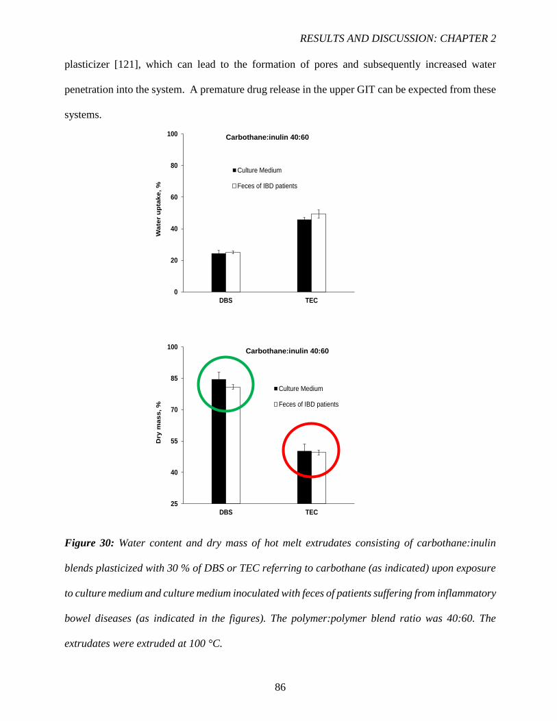
RESULTS AND DISCUSSION: CHAPTER 2
86
plasticizer [121], which can lead to the formation of pores and subsequently increased water
penetration into the system. A premature drug release in the upper GIT can be expected from these
systems.
Figure 30: Water content and dry mass of hot melt extrudates consisting of carbothane:inulin
blends plasticized with 30 % of DBS or TEC referring to carbothane (as indicated) upon exposure
to culture medium and culture medium inoculated with feces of patients suffering from inflammatory
bowel diseases (as indicated in the figures). The polymer:polymer blend ratio was 40:60. The
extrudates were extruded at 100 °C.
0
20
40
60
80
100
DBS TEC
Wa
ter
up
tak
e, %
Culture Medium
Feces of IBD patients
25
40
55
70
85
100
DBS TEC
Dry
ma
ss
, %
Culture Medium
Feces of IBD patients
Carbothane:inulin 40:60
Carbothane:inulin 40:60

RESULTS AND DISCUSSION: CHAPTER 2
87
3. Impact of polymer:polymer blend ratio in hot melt extrudates
3.1. Processability of hot melt extrudates
Figure 31: Macroscopic pictures of hot melt extrudates based on carbothane:inulin blends. The
polymer:polymer blend ratio was varied as indicated, the extrudates were extruded at 100 °C and
plasticized with 30 % DBS (referring to carbothane). For reasons of comparison, also hot melt
extrudates based “only” on carbothane (plasticized with 30 % DBS) are illustrated.
Figure 32: Impact of the polymer:polymer blend ratio on the torque generated during the
preparation of hot melt extrudates based on carbothane:inulin blends. The polymer:polymer blend
ratio was varied as indicated, the extrudates were extruded at 100 °C and plasticized with 30 %
DBS (referring to carbothane).
0
25
50
75
100
100:0 40:60 30:70 20:80 10:90
To
rqu
e,
%
Carbothane:inulin
Stable

RESULTS AND DISCUSSION: CHAPTER 2
88
Macroscopic appearance of hot melt extrudates based on carbothane used as a 1st hydrophobic
polymer, blended with inulin as a 2nd polysaccharide are illustrated in figure 31.
The carbothane:inulin blend ratio was varied as indicated in the figure. Hot melt extrudates were
plasticized with 30 % (w/w) DBS (referring to carbothane) and extruded at 100 °C. For reasons of
comparison, hot melt melt extrudates based “only” on carbothane plasticized with 30 % DBS are
shown. As it can be seen, the morphology of all systems appeares to be rather homogeneous and
the surface relatively smooth. Hot melt extrudates with a ratio of 40:60 and 30:70 were flexible,
contrary to those with a ratio of 20:80 and 10:90, which were hard and brittle. This is due to less
amount of DBS into hot melt extrudates. Moreover, any visible sign of polymer degradation was
observed. Hot melt extrudates of carbothane:inulin blend 40:60 showed an increase of diameter at
the output of die. This swelling behavior was not observed by increasing the amount of inulin into
hot melt estrudates (polymer:polymer ratios: 30:70, 20:80 and 10:90).
In all cases, the torque measured during the extrusion process remained stable (around 60 %),
which have been identified to be beneficial for the extrusion process (Figure 32).
The torque value was higher during the preparation of hot melt extrudates of pure carbothane
plasticized with 30 % DBS. This can be explained by the fact that inulin is in a rubbery state at the
extrusion temperature (100 °C). During the extrusion process, the viscosity of blends is decreased
with increasing inulin amount, which facilitate more the preparation of hot melt extrudates under
these conditions.
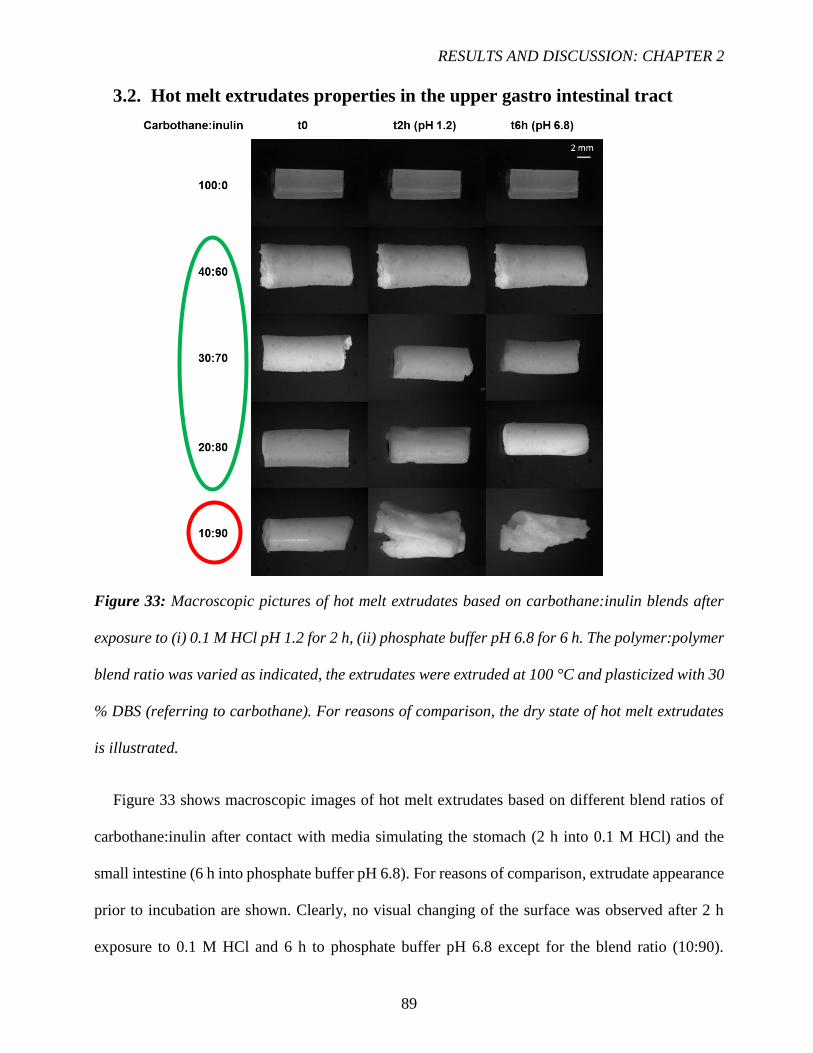
RESULTS AND DISCUSSION: CHAPTER 2
89
3.2. Hot melt extrudates properties in the upper gastro intestinal tract
Figure 33: Macroscopic pictures of hot melt extrudates based on carbothane:inulin blends after
exposure to (i) 0.1 M HCl pH 1.2 for 2 h, (ii) phosphate buffer pH 6.8 for 6 h. The polymer:polymer
blend ratio was varied as indicated, the extrudates were extruded at 100 °C and plasticized with 30
% DBS (referring to carbothane). For reasons of comparison, the dry state of hot melt extrudates
is illustrated.
Figure 33 shows macroscopic images of hot melt extrudates based on different blend ratios of
carbothane:inulin after contact with media simulating the stomach (2 h into 0.1 M HCl) and the
small intestine (6 h into phosphate buffer pH 6.8). For reasons of comparison, extrudate appearance
prior to incubation are shown. Clearly, no visual changing of the surface was observed after 2 h
exposure to 0.1 M HCl and 6 h to phosphate buffer pH 6.8 except for the blend ratio (10:90).

RESULTS AND DISCUSSION: CHAPTER 2
90
This can be explained by the increase of the hydrophilicity of the system when increasing the inulin
amount into the extrudates. Systems based on 40:60, 30:70 or 20:80 carbothane:inulin blends can
be very promising for colonic drug delivery because of the very low drug release in the upper GIT.
3.3. Hot melt extrudates properties in the colon
Physicochemical properties of hot melt extrudates in the colon upon exposure to culture medium
inoculated with fecal samples from IBD-patients are shown in figure 34. For reasons of comparison,
also water content and dry mass loss of hot melt extrudates upon exposure to culture medium free
of feces are shown. Hot melt extrudates were based on carbothane:inulin blends and the ratio of
blend was varied as indicated. Extrudates were plasticized with 30 % DBS (referring to carbothane
mass) and prepared at 100 °C. Clearly, in case of dry mass loss was more pronounced upon exposure
to culture medium inoculated with feces compared to culture medium free of fecal samples (around
55 % vs 45 % for the ratio 30:70). This can be attributed to the fact that inulin serves as a substrate
for colonic bacterial enzymes. Furthermore, the water content of extrudates, which were exposed to
patient feces was higher than the one to sterile culture medium free of feces. This indicates that the
new developed systems contain more pores created by the fermentation of inulin. In order to trigger
the drug release at the site of inflammation in the distal part of GIT, appropriate hot melt extrudates
should take up considerable amounts of water and show significant dry mass loss upon exposure to
fecal samples. As it can be seen in figure 33, hot melt extrudates of carbothane:inulin blends (40:60,
30:70 and 20:80) show a significant water uptake and dry mass loss upon exposure to fecal samples
of patients suffering from IBD. Furthermore, these extrudates show no visible changing of the initial
structure upon exposure to media simulating the upper GIT. Moreover, also blend ratio of 10:90
(carbothane:inulin) look promising for a colonic drug delivery (significant water uptake and weight
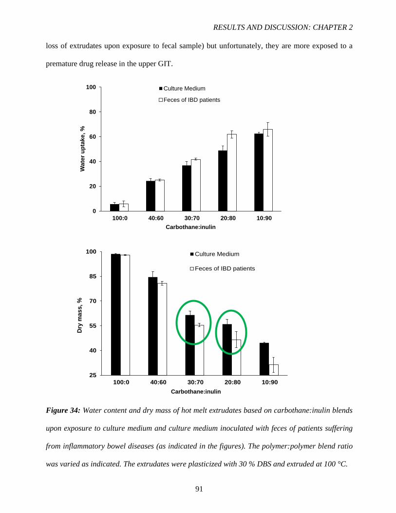
RESULTS AND DISCUSSION: CHAPTER 2
91
loss of extrudates upon exposure to fecal sample) but unfortunately, they are more exposed to a
premature drug release in the upper GIT.
Figure 34: Water content and dry mass of hot melt extrudates based on carbothane:inulin blends
upon exposure to culture medium and culture medium inoculated with feces of patients suffering
from inflammatory bowel diseases (as indicated in the figures). The polymer:polymer blend ratio
was varied as indicated. The extrudates were plasticized with 30 % DBS and extruded at 100 °C.
0
20
40
60
80
100
100:0 40:60 30:70 20:80 10:90
Wa
ter
up
tak
e, %
Culture Medium
Feces of IBD patients
25
40
55
70
85
100
100:0 40:60 30:70 20:80 10:90
Dry
ma
ss
, %
Culture Medium
Feces of IBD patients
Carbothane:inulin
Carbothane:inulin

RESULTS AND DISCUSSION: CHAPTER 2
92
4. Influence of polymer:polymer blend ratio in injection-molded capsules
4.1. Processability of injection-molded capsules
Figure 35: Macroscopic pictures of injecting-molded capsules based on carbothane:inulin blends.
The polymer:polymer blend ratio was varied as indicated, the capsules were prepared at 100 °C
and injected into a mold at 107 °C. For reasons of comparison, also hot melt extrudates based on
carbothane:inulin blends (plasticized with 30 % DBS referring to carbothane) are illustrated.
Figure 35 shows macroscopic appearance of closed and opened capsules prepared by injection
molding process. Hot melt extrudates based on carbothane:inulin blend ratios (30:70 and 20:80)
extruded at 100 °C (plasticized with 30 % DBS referred to carbothane weight) were manually cut
prior to injection molding. The plasticizing chamber, injecting chamber and nozzle temperatures
were set at 100 °C. The blends were injected into a mold heated at 107 °C.
Importantly, the injection was complete and capsules obtained showed a regular and smooth surface
for all polymer:polymer ratios (carbothane:inulin 30:70, 20:80 and 10:90).

RESULTS AND DISCUSSION: CHAPTER 2
93
4.2. In vitro drug release from capsules and hot melt extrudates
Figure 36: Theophylline release from injection-molded capsules based on carbothane:inulin blends
under conditions simulating the transit through the entire gastro intestinal tract: 2 h in 0.1 M HCl,
followed by 6 h in phosphate buffer pH 6.8, followed by culture medium inoculated with feces of
patients suffering from inflammatory bowel diseases (solid curves). For reasons of comparison also
drug release in 0.1 M HCl, phosphate buffer pH 6.8 and culture medium without fecal samples is
shown (dotted curves).
0
25
50
75
100
0 6 12 18 24 30
Th
eo
ph
yll
ine r
ele
as
ed
, %
Time, h
10:90 carbothane:inulin
20:80
30:70
Feces
Culture medium
pH 1.2 pH 6.8 Feces or culture medium

RESULTS AND DISCUSSION: CHAPTER 2
94
The best candidates of hot melt extrudates have been selected for further analysis as well as
capsules manufacturing. The selected blend ratios (30:70, 20:80 and 10:90) of injection-molded
capsules bases on carbothane:inulin where characterized for their drug release kinetics in media
simulating the entire GIT. Capsules were filled with 10 mg of theophylline and then exposed to 0.1
M HCl for 2 h, followed by 6 h in phosphate buffer pH 6.8 and then 22 h in culture medium
inoculated with fecal samples from inflammatory bowel disease patients (solid curve in figure 36).
For reasons of comparison, the extrudates were also exposed to 0.1 M HCl, followed by phosphate
buffer pH 6.8 and culture medium free of feces (dotted curve in figure 36). As it can be seen,
theophylline release rate did not increase upon contact with fecal samples. Approximately the same
percentage of drug released in presence or absence of fecal samples. For instance, around 25 % for
the blend ratios 20:80 and 5 % for the ratio 30:70. This can be at least explained by the fact that
despite of the considerable amount of water uptake by hot melt extrudates and the significant dry
mass loss of hot melt extrudates (figure 34) upon exposure to fecal samples from IBD-patients,
capsules prepared from these extrudates did not show the same behavior due probably to non-
fermentation of the polysaccharide under these conditions. For reasons of comparison, hot melt
extrudates of carbothane:inulin blends (30:70, 20:80 and 10:90) containing 10 % theophylline were
prepared. Hot melt extrudates were plasticized with 30 % DBS referring to the dry mass of
carbothane and extruded at 100 °C. The in-vitro drug release from these hot melt extrudates was
performed upon exposure to 0.1 M HCl for 2 h, followed by phosphate buffer pH 6.8 (figure 37).
Clearly, almost 100 % of theophylline was released from these hot melt extrudates into the
dissolution media for all ratios. However, less than 30 % was released from injection-molded
capsules. This can be probably explained by the thermoplastic polymer “carbothane”, which is able
to coat the polysaccharide and then hinders the bacterial fermentation.

RESULTS AND DISCUSSION: CHAPTER 2
95
Figure 37: Theophylline released from hot melt extrudates based on carbothane:inulin in 0.1 M
HCl for 2 h, followed by phosphate buffer pH 6.8. Hot melt extrudates were loaded with 10 %
theophylline. The polymer:polymer blend ratio was varied as indicated. The extrudates were
plasticized with 30 % DBS and extruded at 100 °C.
0
25
50
75
100
0 6 12 18 24
Th
eo
ph
yll
ine r
ele
ased
, %
Time, h
10:90 carbothane:inulin
20:80
30:70
pH 1.2 pH 6.8

96
97
CHAPTER 3:
HOT MELT EXTRUDED POLYSACCHARIDE
BLENDS FOR CONTROLLED DRUG DELIVERY
RESULTS AND DISCUSSION: CHAPTER 3
98

RESULTS AND DISCUSSION: CHAPTER 3
99
HOT MELT EXTRUDED POLYSACCHARIDE BLENDS FOR
CONTROLLED DRUG DELIVERY
Polymer blends offer an interesting potential for controlled drug delivery systems [167-172],
both as matrix formers [173-176] and as coating materials [177-181]. By simply varying the
polymer:polymer blend ratio, the resulting key properties of the systems can be effectively varied,
allowing to provide large spectra of possible drug release kinetics [182-184]. For example, a variety
of blends of enteric and non-enteric polymer blends has been used to control the resulting drug
release kinetics from coated pellets [185,186].
Importantly, the presence of the non-enteric polymer can effectively hinder the leaching of the
enteric polymer out of the film coating at neutral pH [187]. Thus, one polymer can efficiently
“mask” key properties of the other polymer, if the two compounds are intimately mixed [188]. The
polymer:polymer blend ratio as well as the manufacturing technique (determining the inner system
structure) can strongly affect the efficiency of such “masking” phenomena [189]. The type of
polymers used, the polymer blend ratio as well as the manufacturing conditions determine the
resulting system properties and, thus, the control of drug release. The basic principle is that the drug
is “trapped” within the polymeric system and different types of mass transport phenomena can be
involved in the control of drug release, such as water diffusion into the system, drug dissolution and
diffusion, polymer swelling and dissolution, osmotic effects, polymer degradation and pore
formation upon leaching of water soluble compounds into the surrounding bulk fluid (to mention
just a few). In the case of polymer blends, the properties of both compounds might be decisive, or
one of them might dominate. For example, when blending a polymer that is permeable for many
drugs with a polymer that is poorly permeable, broad spectra of drug release patterns might be
obtained by simply varying the polymer:polymer blend ratio [190]. Also differences in drug
solubility or drug loading might be compensated by adjusting the polymer:polymer blend ratio. For

RESULTS AND DISCUSSION: CHAPTER 3
100
example, high loadings of a freely water-soluble drug in a matrix system generally lead to fast drug
release. This might be compensated by increasing the portion of the poorly permeable polymer in
the dosage form. Also, one polymer might assure the mechanical stability of the drug delivery
system within the gastrointestinal tract, whereas the other polymer might trigger drug release in
specific segments (e.g. small intestine or colon) [191-196].
Furthermore, the solubilities of the two polymers might be complementary: For example,
ethylcellulose:guar gum blends have been proposed as film coating materials to provide controlled
drug release that is not susceptible to the co-consumption of alcoholic beverages [197-199]. The
basic idea is that ethylcellulose is not soluble in water, but in ethanol. Viceversa, guar gum is soluble
in water, but not in ethanol. Appropriate ethylcellulose:guar gum blends were shown to be able to
release theophylline from coated pellets with release rates that were very similar in release media
containing 0, 20 or 40 % ethanol.
The aim of this study was to explore the potential of blends of ethylcellulose with a second
polysaccharide as matrix former in controlled release hot melt extrudates. Ethylcellulose was
chosen to provide good mechanical stability of the systems. But it is known to be poorly permeable
for many drugs [200]. Thus, it was combined with different types and amounts of a more permeable
second polysaccharide, namely hydroxypropyl methylcellulose (HPMC), pectin, maize starch,
inulin, maltodextrin, guar gum, and chitosan. Optionally, different types and amounts of plasticizers
(dibutyl sebacate, triethyl citrate and polyethylene glycol) were added. Theophylline was selected
as model drug exhibiting slight water solubility, diprophylline as freely water-soluble model drug.
The drug loading was varied from 10 to 60 %. The key manufacturing parameters (temperature and
screw speed) as well as the polymer:polymer blend ratio were varied. Drug release was measured
under conditions simulating the contents of the upper GIT (0.1 M HCl and phosphate buffer pH
6.8), as well as the colon (upon exposure to human fecal samples).
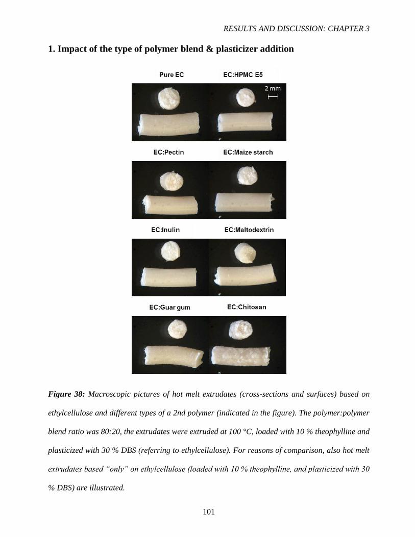
RESULTS AND DISCUSSION: CHAPTER 3
101
1. Impact of the type of polymer blend & plasticizer addition
Figure 38: Macroscopic pictures of hot melt extrudates (cross-sections and surfaces) based on
ethylcellulose and different types of a 2nd polymer (indicated in the figure). The polymer:polymer
blend ratio was 80:20, the extrudates were extruded at 100 °C, loaded with 10 % theophylline and
plasticized with 30 % DBS (referring to ethylcellulose). For reasons of comparison, also hot melt
extrudates based “only” on ethylcellulose (loaded with 10 % theophylline, and plasticized with 30
% DBS) are illustrated.

RESULTS AND DISCUSSION: CHAPTER 3
102
Figure 38 shows optical macroscopy pictures of hot melt extrudates based on different types of
polymer:polymer blends: Ethylcellulose was blended with a selection of other polysaccharides, as
indicated. The ethylcellulose:2nd polysaccharide blend ratio was 80:20 (weight:weight) in all cases.
Thirty percent DBS (referring to ethylcellulose) was added as a plasticizer, the systems were loaded
with 10 % theophylline, and the extrusion temperature was 100 °C in all cases. Cross-sections of
the hot melt extrudates (obtained by manual breaking) are shown at the top, pictures of surfaces
right below. As it can be seen, the inner structure of all systems appeared to be rather homogeneous
and the surface relatively smooth, except for ethylcellulose:chitosan blends, which lead to
extrudates with a slightly rough surface and somehow “granular” inner structure.
Importantly, in all cases the torque measured during extrusion was similar (around 30 %), not
causing any difficulty during processing (figure 39a). The resulting drug release kinetics upon
exposure to 0.1 M HCl for 2 h, followed by phosphate buffer pH 6.8 for 22 h are illustrated in figure
39b. Clearly, the type of polymer blend had a pronounced effect on theophylline release. This can
be attributed to the different chemical structures of the polysaccharides and the resulting difference
in permeability of the polymeric matrices for the drug. In many cases, the observed release rates
were too low for oral administration (e.g. less than about 18 % after 24 h). For this reason, only the
3 most rapidly releasing polymer blends were selected for further studies: ethylcellulose:chitosan,
ethylcellulose:guar gum and ethylcellulose:pectin.

RESULTS AND DISCUSSION: CHAPTER 3
103
Figure 39: Impact of the type of polymer blend (indicated in the diagrams) used as matrix former
on: a) the torque generated during hot melt extrusion, and b) theophylline release from the
extrudates in 0.1 M HCl for 2 h, followed by phosphate buffer pH 6.8. The extrudates were extruded
at 100 °C, loaded with 10 % drug and plasticized with 30 % DBS (referring to ethylcellulose).
0
25
50
75
100
0 6 12 18 24
Th
eo
ph
yll
ine
re
lea
se
d, %
Time, h
(80:20) (Ethylcellulose:Chitosan)
(80:20) (Ethylcellulose:Guar gum)
(80:20) (Ethylcellulose:Pectin)
(80:20) (Ethylcellulose:HPMC E5)
(80:20) (Ethylcellulose:Maltodextrin)
(80:20) (Ethylcellulose:Inulin)
(80:20) (Ethylcellulose:Maize starch)
Pure Ethylcellulose
Stable
a)
b)
0
25
50
75
100
To
rqu
e, %
(80:20) (Ethylcellulose:2nd polymer)

RESULTS AND DISCUSSION: CHAPTER 3
104
In an attempt to simplify the formulation, the plasticizer DBS was omitted. Figure 40 shows
macroscopic pictures of cross-sections and surfaces of hot melt extrudates based on
ethylcellulose:pectin/guar gum/chitosan blends free of plasticizer. The blend ratio was varied as
indicated, hot melt extrudates based “only” on ethylcellulose (loaded with 10 % theophylline) are
shown for reasons of comparison. To allow for extrusion without a plasticizer, the processing
temperature had to be increased, here to 170 °C. As it can be seen in the top row, this temperature
degraded pectin under the given conditions. Also, in the case of hot melt extrudates loaded with
high amounts of chitosan some color changes at the systems’ surface was observed (Figure 40,
bottom row). Due to the important pectin degradation, these hot melt extrudates were not studied
any further.
The resulting theophylline release kinetics of systems based on different ethylcellulose:guar gum
and ethylcellulose:chitosan blends are illustrated in figure 41. The release medium was 0.1 M HCl
for the first 2 h, followed by phosphate buffer pH 6.8 for the subsequent 6 h. The ethylcellulose:2nd
polysaccharide blend ratio was varied as follows: 100:0, 90:10, 80:20, 70:30 and 60:40. As it can
be seen, the resulting release rate was very low at all blend ratios in the case of
ethylcellulose:chitosan (4 - 15 % after 8 h). Also in the case of ethylcellulose:guar gum blends,
theophylline release from the plasticizer-free implants was relatively slow, much slower compared
to the respective DBS-plasticized systems extruded at 100 °C: For example, after 8 h only 6 versus
15 % drug was released in the case of ethylcellulose:guar gum 80:20 blends (see also figure 41
versus figure 39b). This reduced drug release rate in plasticizer-free systems can at least partially
be attributed to the fact that the presence of the plasticizer increases the mobility of the polymer
chains and, thus, also of the drug molecules.

RESULTS AND DISCUSSION: CHAPTER 3
105
Figure 40: Macroscopic pictures of hot melt extrudates (cross-sections and surfaces) based on
ethylcellulose and different types of a 2nd polymer (indicated in the figure). The polymer:polymer
blend ratio is indicated in the figure, the extrudates were extruded at 170 °C, loaded with 10 %
theophylline and free of plasticizer.

RESULTS AND DISCUSSION: CHAPTER 3
106
Figure 41: Impact of the polymer:polymer blend ratio (indicated in the diagrams) on theophylline
release from hot melt extrudates based on ethylcellulose:guar gum or ethylcellulose:chitosan
blends (as indicated) in 0.1 M HCl for 2 h, followed by phosphate buffer pH 6.8. The extrudates
were extruded at 170 °C, loaded with 10 % drug and free of plasticizer.
0
25
50
75
100
0 2 4 6 8
Th
eo
ph
yll
ine
re
lea
se
d, %
Time, h
(60:40) (Ethylcellulose:Guar gum)
(70:30)
(80:20)
(90:10)
(100:0)
0
25
50
75
100
0 2 4 6 8
Th
eo
ph
yll
ine
re
lea
se
d, %
Time, h
(60:40) (Ethylcellulose:Chitosan)
(70:30)
(80:20)
(90:10)
(100:0)
pH 1.2 pH 6.8

RESULTS AND DISCUSSION: CHAPTER 3
107
Figure 42: DSC thermogram of ethylcellulose plasticized with 30 % DBS.
For example, figure 42 shows the DSC thermogram of ethylcellulose plasticized with 30 % DBS.
The glass transition temperature decreased from about 130 to 53 °C in the dry state compared to
pure ethylcellulose (figure 55 vs. figure 42). In addition to DBS, water can also be expected to be
acting as a plasticizer for this polymer during drug release. Since the observed release rates were
too low in plasticizer-free hot melt extrudates, these systems were not studied any further.
Instead, the suitability of ethylcellulose:guar gum, ethylcellulose:chitosan and
ethylcellulose:HPMC E5 blends, plasticized with 30 % DBS (referring to ethylcellulose) were
studied in more detail (note that “pectin was replaced by HPMC E5”, since it showed important
thermal degradation at 170 °C, and similar drug release rates: figure 40 and figure 39b). In the
presence of the plasticizer, the extrusion temperature was again reduced to 100 °C.

RESULTS AND DISCUSSION: CHAPTER 3
108
Figure 43: Macroscopic pictures of hot melt extrudates (cross-sections and surfaces) based on
ethylcellulose and different types of a 2nd polymer (indicated in the figure). The polymer:polymer
blend ratio was varied as indicated in the figure, the extrudates were extruded at 100 °C, loaded
with 10 % theophylline and plasticized with 30 % DBS (referring to ethylcellulose).
Figure 44: Impact of the type of ethylcellulose and different types of a 2nd polymer blend (indicated
in the diagrams) on the torque generated during hot melt extrusion. The polymer:polymer blend
ratio was varied as indicated. The extrudates were extruded at 100 °C, loaded with 10 % drug and
plasticized with 30 % DBS (referring to ethylcellulose).
0
25
50
75
100
80:20 70:30 60:40 50:50 40:60 30:70 20:80
To
rqu
e,
%
Ethylcellulose:Guar gum
Ethylcellulose:Chitosan
Ethylcellulose:HPMC

RESULTS AND DISCUSSION: CHAPTER 3
109
Figure 43 shows macroscopic pictures of cross-sections and surfaces of hot melt extrudates based
on these polymer:polymer blends, varying the blend ratio from 100:0 to 20:80, in 10 % increments.
As it can be seen, the inner structure of all systems appeared to be rather homogeneous and the
surface relatively smooth, except for ethylcellulose:chitosan blends, which lead to extrudates with
a slightly rough surface and somehow “granular” inner structure for all the blend ratios. Importantly,
in all cases the torque measured during extrusion was similar (around 30 %) with a slightly increase
for the blends ratio 30:70 and 20:80 (40 - 45 %), without causing any difficulty during processing
(figure 44).
Figure 45 shows the resulting theophylline release kinetics from. The release medium was
0.1 M HCl for 2 h, followed by phosphate buffer pH 6.8 for 22 h. Clearly, in all cases a broad
spectrum of drug release patterns could be obtained, by simply varying the polymer:polymer blend
ratio. In the investigated cases, ethylcellulose was less permeable for theophylline than the other
polymers. This is why in all cases, the release rate increased with decreasing ethylcellulose content.
Since ethylcellulose:guar gum blends showed the largest spectrum of possible drug release patterns
(e.g. the highest theophylline release rate at the 20:80 ethylcellulose:2nd polysaccharide blend
ratio), this blend was selected for further studies.

RESULTS AND DISCUSSION: CHAPTER 3
110
Figure 45: Impact of the polymer:polymer blend ratio (indicated in the figure) on theophylline
release from hot melt extrudates based on ethylcellulose and different types of a 2nd polysaccharide
in 0.1 M HCl for 2 h, followed by phosphate buffer pH 6.8. The extrudates were extruded at 100 °C,
loaded with 10 % drug and plasticized with 30 % DBS (referring to ethylcellulose).
0
25
50
75
100
0 6 12 18 24
Th
eo
ph
yll
ine
re
lea
se
d, %
Time, h
(20:80)
(30:70)
(40:60)
(50:50)
(60:40)
(70:30)
(80:20)
(100:0)
Ethylcellulose:Guar gum
0
25
50
75
100
0 6 12 18 24
Th
eo
ph
yll
ine
re
lea
se
d, %
Time, h
(20:80)
(30:70)
(40:60)
(50:50)
(60:40)
(70:30)
(80:20)
(100:0)
Ethylcellulose:Chitosan
0
25
50
75
100
0 6 12 18 24
Th
eo
ph
yll
ine
re
lea
se
d, %
Time, h
(20:80)
(30:70)
(40:60)
(50:50)
(60:40)
(70:30)
(80:20)
(100:0)
Ethylcellulose:HPMC E5
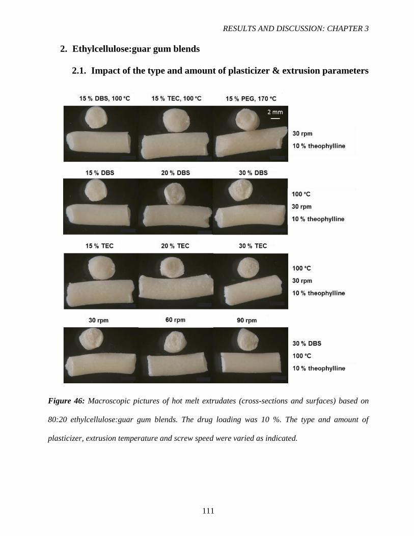
RESULTS AND DISCUSSION: CHAPTER 3
111
2. Ethylcellulose:guar gum blends
2.1. Impact of the type and amount of plasticizer & extrusion parameters
Figure 46: Macroscopic pictures of hot melt extrudates (cross-sections and surfaces) based on
80:20 ethylcellulose:guar gum blends. The drug loading was 10 %. The type and amount of
plasticizer, extrusion temperature and screw speed were varied as indicated.

RESULTS AND DISCUSSION: CHAPTER 3
112
Figure 46 shows macroscopic pictures of cross-sections and surfaces of different types of
ethylcellulose:guar gum based hot melt extrudates: The following parameters were varied: (i) the
type of plasticizer (DBS, TEC and PEG), (ii) the percentage of plasticizer (15, 20 and 30 %,
referring to ethylcellulose), (iii) the extrusion temperature (100 and 170 °C) and (iv) the screw
speed (30, 60 and 90 rpm). In all cases, the ethylcellulose:guar gum blend ratio was kept constant:
80:20 (weight:weight). As it can be seen, in all cases rather homogeneous inner system structures
and relatively smooth surfaces were obtained. In no case, any visible sign of polymer degradation
was observed. Extrudates containing 0, 15 or 20 % plasticizer were hard and brittle. All other
systems were flexible.
Figure 47a shows the torque values measured during the extrusion of ethylcellulose:guar gum
80:20 blends plasticized with 15 % PEG (PEG 1500), DBS or TEC. The systems were loaded with
10 % theophylline. For reasons of comparison, also the torque values observed with “pure”
ethylcellulose hot melt extrudates (loaded with 10 % theophylline) are shown. Please note that it
was not possible to extrude plasticizer-free and 15 % PEG containing formulations at 100 °C (the
torque values were too high). This is why these extrudates were obtained at 170 °C processing
temperature. In contrast, blends plasticized with 15 % DBS or TEC could be obtained at 100 °C
processing temperature, although the corresponding torque values were high (figure 47a).
The respective theophylline release kinetics from these hot melt extrudates are illustrated in figure
47b. As it can be seen, the following rank order with respect to the resulting drug release rate was
observed: 15 % PEG > 15 % DBS > 15 % TEC > no plasticizer. Thus, the plasticizer facilitates
drug release, probably due to increased polymer chain mobility and/or plasticizer leaching into the
surrounding bulk fluid. Please note that a direct comparison of PEG with DBS & TEC should be
viewed with caution, since the extrusion temperature was different. In all cases, the resulting drug
release rates were rather low (e.g., less than 36 % after 6 h).

RESULTS AND DISCUSSION: CHAPTER 3
113
Figure 47: Impact of the type of plasticizer and extrusion temperature on: a) the generated torque,
and b) theophylline release from extrudates in 0.1 M HCl for 2 h, followed by phosphate buffer pH
6.8. The systems were based on 80:20 ethylcellulose:guar gum blends, the drug loading was 10 %.
0
25
50
75
100
0 1 2 3 4 5 6
Th
eo
ph
yll
ine
re
lea
se
d, %
Time, h
15 % PEG, 170 °C
15 % DBS, 100 °C
15 % TEC, 100 °C
No plasticizer, 170 °C
pH 1.2 pH 6.8
170 C 170 C
100 C
100 C
0
25
50
75
100
Noplasticizer
15 % PEG 15 % DBS 15 % TEC
To
rqu
e, %
(80:20) Ethylcellulose:Guar gum
(80:20) Ethylcellulose:Guar gum
a)
b)

RESULTS AND DISCUSSION: CHAPTER 3
114
Figure 48a shows the impact of varying the plasticizer content (here DBS and TEC) on the torque
measured during extrusion of ethylcellulose:guar gum 80:20 blends, loaded with 10 % theophylline.
The extrusion temperature was 100 °C. Clearly, the torque values substantially decreased with
increasing plasticizer level, irrespective of the type of plasticizer. The theophylline release kinetics
from the obtained hot melt extrudates are shown in figure 48b. Interestingly, the freely water-soluble
plasticizer TEC leads to slower drug release rates than the lipophilic plasticizer DBS. Thus, in these
cases, the increase in polymer chain mobility seems to play a more important role than potential
plasticizer leaching into the surrounding bulk fluid (eventually creating water-filled pores).
TEC seems to be a more efficient plasticizer for the polymeric matrix than DBS, resulting in a
denser (and less permeable) system (overcompensating potential increased drug mobility effects).
But again, in all cases the resulting theophylline release rates were rather low (e.g., less than 27 %
drug was released after 6 h).
The effects of varying the screw speed when manufacturing ethylcellulose:guar gum 80:20 based
hot melt extrudates loaded with 10 % theophylline at 100 °C (plasticized with 30 % DBS) on: a)
the torque measured during extrusion, and b) drug release in 0.1 M HCl for 2 h, followed by
phosphate buffer pH 6.8 for subsequent 6 h, are illustrated in figure 49. Clearly, the variation of the
screw speed in the investigated range (30 – 60 – 90 rpm) did neither substantially impact the torque,
nor theophylline release.
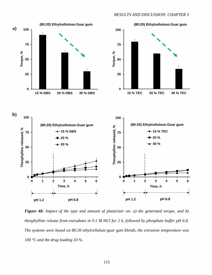
RESULTS AND DISCUSSION: CHAPTER 3
115
Figure 48: Impact of the type and amount of plasticizer on: a) the generated torque, and b)
theophylline release from extrudates in 0.1 M HCl for 2 h, followed by phosphate buffer pH 6.8.
The systems were based on 80:20 ethylcellulose:guar gum blends, the extrusion temperature was
100 °C and the drug loading 10 %.
0
25
50
75
100
0 1 2 3 4 5 6
Th
eo
ph
yll
ine
re
lea
se
d, %
Time, h
15 % DBS
20 %
30 %
0
25
50
75
100
15 % DBS 20 % DBS 30 % DBS
To
rqu
e, %
0
25
50
75
100
15 % TEC 20 % TEC 30 % TEC
To
rqu
e, %
0
25
50
75
100
0 1 2 3 4 5 6
Th
eo
ph
yll
ine
re
lea
se
d, %
Time, h
15 % TEC
20 %
30 %
(80:20) Ethylcellulose:Guar gum (80:20) Ethylcellulose:Guar gum
(80:20) Ethylcellulose:Guar gum (80:20) Ethylcellulose:Guar gum
pH 1.2 pH 6.8 pH 1.2 pH 6.8
a)
b)
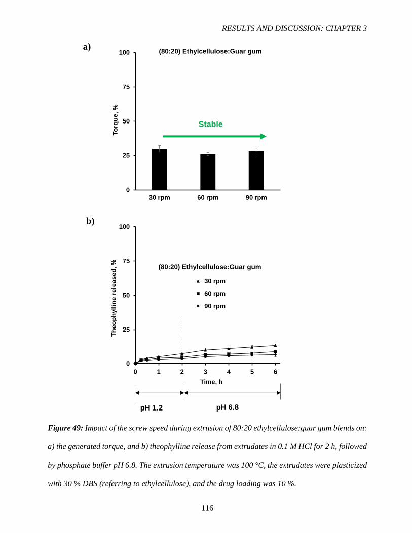
RESULTS AND DISCUSSION: CHAPTER 3
116
Figure 49: Impact of the screw speed during extrusion of 80:20 ethylcellulose:guar gum blends on:
a) the generated torque, and b) theophylline release from extrudates in 0.1 M HCl for 2 h, followed
by phosphate buffer pH 6.8. The extrusion temperature was 100 °C, the extrudates were plasticized
with 30 % DBS (referring to ethylcellulose), and the drug loading was 10 %.
0
25
50
75
100
30 rpm 60 rpm 90 rpm
To
rqu
e, %
(80:20) Ethylcellulose:Guar gum
0
25
50
75
100
0 1 2 3 4 5 6
Th
eo
ph
yll
ine
re
lea
se
d, %
Time, h
30 rpm
60 rpm
90 rpm
pH 1.2 pH 6.8
(80:20) Ethylcellulose:Guar gum
a)
b)
Stable

RESULTS AND DISCUSSION: CHAPTER 3
117
2.2. Impact of the type and amount of drug
Figure 50 shows macroscopic pictures of cross-sections and surfaces of different types of
ethylcellulose:guar gum based hot melt extrudates: The following parameters were varied: (i) the
type of drug (theophylline and diprophylline, being slightly and freely water-soluble), and (ii) the
drug loading (10, 30 and 60 %, referring to the total extrudate mass). In all cases, the
ethylcellulose:guar gum blend ratio was kept constant: 80:20 (weight:weight). As it can be seen, in
all cases rather homogeneous inner system structures and relatively smooth surfaces were obtained.
In no case, any visible sign of polymer degradation was observed. Extrudates loaded with 60 %
drug were hard and brittle. All other systems were flexible.
Figure 50: Macroscopic pictures of hot melt extrudates (cross-sections and surfaces) based on
80:20 ethylcellulose:guar gum blends. The extrudates were plasticized with 30 % DBS (referring
to ethylcellulose), the extrusion temperature was 100 °C and the screw speed 30 rpm. The type and
amount of drug were varied as indicated.

RESULTS AND DISCUSSION: CHAPTER 3
118
Figure 51: Impact of the theophylline or diprophylline loading on the generated torque during hot
melt extrusion. The systems were based on 80:20 ethylcellulose:guar gum blends, the extrusion
temperature was 100 °C, the extrudates were plasticized with 30 % DBS.
0
25
50
75
100
10% 30% 60%
To
rqu
e, %
(80:20) Ethylcellulose:Guar gum
Theophylline
0
25
50
75
100
10% 30% 60%
To
rqu
e, %
(80:20) Ethylcellulose:Guar gum
Diprophylline
RESULTS AND DISCUSSION: CHAPTER 3
119
Furthermore, the impact of the type of drug and drug loading was studied (figures 51 and 52):
The percentages of slightly water-soluble theophylline and freely water-soluble diprophylline were
varied from 10 to 60 %. Figure 51 shows the respective torque values observed during extrusion
(at 100 °C, with 30 % DBS). Figure 52 shows the resulting drug release kinetics in 0.1 M HCl for
2 h, followed by phosphate buffer pH 6.8 for 22 h. As it can be seen, the torque values increased
with increasing drug loading (especially in the case of theophylline). This can probably be attributed
to the fact that both drugs do not melt at 100 °C (figure 55) and that the relative amounts of more
easily extrudable, plasticized polymer blends in the formulations decrease. Figure 52 shows that
also the resulting drug release rates clearly increased with increasing drug loading. This can at least
partially be explained by the fact that less drug release retarding polymer is present in the systems.
Or, in other words: Upon drug release, the systems become more and more porous and remaining
drug can more easily diffuse out.
This is very important from a practical point of view: Most of the drug is released after 24 h at
an initial drug loading of 60 %. Also, as it can be seen, about zero order release kinetics can be
provided during major parts of the release periods: Theophylline and diprophylline were released
at a rate of approximately 3 %/h during 24 h. Please note that perfect sink conditions were provided
throughout the experiments. Thus, the observed relatively constant drug release rates can probably
be attributed to drug saturation effects within the hot melt extrudates: The amounts of water
penetrating into the systems are limited and most likely not sufficient to dissolve the entire drug
loadings. Thus, non-dissolved and dissolved drug co-exist within the systems. Importantly, only
dissolved drug is available for diffusion. This results in about constant drug concentration
differences: saturated solutions inside the hot melt extrudates and perfect sink conditions in the
surrounding bulk fluids.

RESULTS AND DISCUSSION: CHAPTER 3
120
Figure 52: Impact of the theophylline or diprophylline loading on drug release from extrudates in
0.1 M HCl for 2 h, followed by phosphate buffer pH 6.8. The systems were based on 80:20
ethylcellulose:guar gum blends, the extrusion temperature was 100 °C, the extrudates were
plasticized with 30 % DBS.
0
25
50
75
100
0 6 12 18 24
Dip
rop
hyllin
e r
ele
as
ed
, %
Time, h
60% diprophylline
30%
10%
0
25
50
75
100
0 6 12 18 24
Th
eo
ph
yll
ine
re
lea
se
d, %
Time, h
60% theophylline
30%
10%
(80:20) Ethylcellulose:Guar gum
pH 1.2 pH 6.8
(80:20) Ethylcellulose:Guar gum

RESULTS AND DISCUSSION: CHAPTER 3
121
Since guar gum has been reported to be preferentially degraded by enzymes secreted by bacteria
present in the colon [201-203] and since the provided drug release periods were rather long, it was
important to evaluate whether or not the exposure to fecal samples potentially altered the resulting
drug release kinetics from the investigated hot melt extrudates. Due to the intimate mixture of guar
gum with ethylcellulose, it might be that the latter effectively protects the guar gum against
enzymatic degradation. For this reason, hot melt extrudates loaded with 60 % theophylline or 60 %
diprophylline, based on ethylcellulose:guar gum 80:20 blends (plasticized with 30 % DBS referred
to ethylcellulose) were prepared at 100 °C and exposed to 0.1 M HCl for 2 h, followed by phosphate
buffer pH 6.8 for 6 h, and fecal samples for 10 h (the latter under anaerobic conditions). For reasons
of comparison, the extrudates were also exposed to 0.1 M HCl and phosphate buffer pH 6.8,
followed by culture medium free of fecal bacteria. The 0.1 M HCl was intended to simulate the
conditions in the stomach, the phosphate buffer pH 6.8 in the small intestine, and the fecal samples
the conditions in the colon. The solid curves in figure 53 show the experimentally measured drug
release rates with this set up using fecal samples, whereas the dashed curves show the respective
release rates observed with culture medium free of feces. As it can be seen, the presence of fecal
bacteria did not have a noteworthy impact on drug release, irrespective of the type of drug
(figure 53).
Furthermore, the long term stability of the investigated hot melt extrudates was studied. Figure
54 shows the release rates of diprophylline and theophylline from ethylcellulose:guar gum 80:20-
based systems in 0.1 M HCl for 2 h, followed by 22 h in phosphate buffer pH 6.8. The drug loading
were varied from 10 to 60 %, the extrudates were prepared at 100 °C and contained 30 % DBS
(referring to ethylcellulose). The solid curves illustrate drug release prior to storage, the dashed
curves drug release after 1 year storage at ambient conditions. In all cases, drug release did not
change to a noteworthy extent.

RESULTS AND DISCUSSION: CHAPTER 3
122
Figure 53: Drug release from hot melt extrudates based on 80:20 ethylcellulose:guar gum blends
under conditions simulating the transit through the entire gastro intestinal tract: 2 h in 0.1 M HCl,
followed by 6 h in phosphate buffer pH 6.8, followed by culture medium inoculated with human
fecal samples (solid curves). For reasons of comparison also drug release in 0.1 M HCl, phosphate
buffer pH 6.8 and culture medium without fecal samples is shown (dotted curves). The drug loading
was 60 %, the extrusion temperature 100 °C. The extrudates were plasticized with 30 % DBS
(referring to ethylcellulose).
0
25
50
75
100
0 5 9 14 18
Dru
g r
ele
as
ed
, %
Time, h
60 % diprophylline
60 % theophylline
pH 6.8 Feces or culture medium pH 1.2
Feces
Culture medium
(80:20) (Ethylcellulose:Guar gum)

RESULTS AND DISCUSSION: CHAPTER 3
123
Figure 54: Storage stability of hot melt extrudates based on 80:20 ethylcellulose:guar gum blends
loaded with theophylline or diprophylline (as indicated). The extrusion temperature was 100 °C,
the extrudates were plasticized with 30 % DBS. Drug release was measured in 0.1 M HCl (for 2 h),
followed by phosphate buffer pH 6.8 before (solid curves) and after 1- year open storage at ambient
conditions (dotted curves).
0
25
50
75
100
0 6 12 18 24
Th
eo
ph
yll
ine
re
lea
se
d, %
Time, h
60% theophylline
30%
10%
(80:20) Ethylcellulose:Guar gum
0
25
50
75
100
0 6 12 18 24
Dip
rop
hyll
ine
re
lea
se
d, %
Time, h
60% diprophylline
30%
10%
(80:20) Ethylcellulose:Guar gum
pH 1.2 pH 6.8
Before storage
After 1 year storage

RESULTS AND DISCUSSION: CHAPTER 3
124
2.3. Physical characterization
Figure 55: DSC thermograms of hot melt extrudates based on 80:20 ethylcellulose:guar gum
blends. The type and amount of drug were varied as indicated. The extrusion temperature was 100
°C, the extrudates were plasticized with 30 % DBS (referring to ethylcellulose). For reasons of
comparison, also the DSC thermograms of the pure drugs, polymer powders and DBS (all as
received) are illustrated.
-40 10 60 110 160 210 260
He
at
flo
w
Temperature, C
Pure ethylcellulose
Pure DBS
Pure guar gum
Pure diprophylline
10 % diprophylline HME
30 % diprophylline HME
60 % diprophylline HME
Pure theophylline
10 % theophylline HME
30 % theophylline HME
60 % theophylline HME
exo
5 W/g
X10
X10

RESULTS AND DISCUSSION: CHAPTER 3
125
Figure 55 shows DSC thermograms of hot melt extrudates loaded with 10 to 60 % diprophylline
or theophylline, based on ethylcellulose:guar gum 80:20 blends plasticized with 30 % DBS
(referring to ethylcellulose) prepared at 100 °C. For reasons of comparison, also the DSC
thermograms of the following raw materials (as received) are illustrated: ethylcellulose, guar gum,
DBS, diprophylline and theophylline. Pure ethylcellulose showed 2 thermal events: (i) a glass
transition (Tg) at about 130 °C, which is in good agreement with data reported in the literature
[204], and (ii) an exothermic event at approximately 185 °C. Lai et al., [205] have shown that for
“Ethocel Standard 100 Premium” (an ethylcellulose with a higher molecular weight than the
investigated polymer) (Dow, Midland, MI, USA) a similar exotherm corresponded to an oxidative
degradation (the pans were hermetically closed, the nitrogen flushing was outside the pans), which
masks the melting of a small crystalline fraction
Figure 56: X-ray diffractograms of pure ethylcellulose and pure guar gum powders (as received).
500
1900
3300
4700
6100
0 10 20 30 40 50 60
Inte
nsit
y, co
un
ts
2θ, degree
Pure ethylcellulose
Pure guar gum
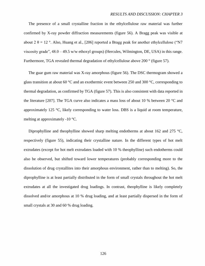
RESULTS AND DISCUSSION: CHAPTER 3
126
The presence of a small crystalline fraction in the ethylcellulose raw material was further
confirmed by X-ray powder diffraction measurements (figure 56). A Bragg peak was visible at
about 2 θ = 12 °. Also, Huang et al., [206] reported a Bragg peak for another ethylcellulose (“N7
viscosity grade”, 48.0 – 49.5 w/w ethoxyl groups) (Hercules, Wilmington, DE, USA) in this range.
Furthermore, TGA revealed thermal degradation of ethylcellulose above 200 ° (figure 57).
The guar gum raw material was X-ray amorphous (figure 56). The DSC thermogram showed a
glass transition at about 60 °C and an exothermic event between 250 and 300 °C, corresponding to
thermal degradation, as confirmed by TGA (figure 57). This is also consistent with data reported in
the literature [207]. The TGA curve also indicates a mass loss of about 10 % between 20 °C and
approximately 125 °C, likely corresponding to water loss. DBS is a liquid at room temperature,
melting at approximately -10 °C.
Diprophylline and theophylline showed sharp melting endotherms at about 162 and 275 °C,
respectively (figure 55), indicating their crystalline nature. In the different types of hot melt
extrudates (except for hot melt extrudates loaded with 10 % theophylline) such endotherms could
also be observed, but shifted toward lower temperatures (probably corresponding more to the
dissolution of drug crystallites into their amorphous environment, rather than to melting). So, the
diprophylline is at least partially distributed in the form of small crystals throughout the hot melt
extrudates at all the investigated drug loadings. In contrast, theophylline is likely completely
dissolved and/or amorphous at 10 % drug loading, and at least partially dispersed in the form of
small crystals at 30 and 60 % drug loading.
RESULTS AND DISCUSSION: CHAPTER 3
127
Figure 57: TGA thermograms of pure ethylcellulose and pure guar gum powders (as received).
Hot melt extrudates based on ethylcellulose:guar gum blends offer an interesting potential as
controlled drug delivery systems: They can be prepared at temperatures of about 100 °C, provide
broad spectra of drug release patterns (in particular about constant drug release rates) and are long
term stable. The ethylcellulose can effectively protect the guar gum against potential enzymatic
degradation in the colon.
60
70
80
90
100
20 55 90 125 160 195 230 265 300
Weig
ht,
%
Temperature, C
Pure ethylcellulose
Pure guar gum

128

129
IV. CONCLUSIONS AND PERSPECTIVES

CONCLUSIONS AND PERSPECTIVES
130

CONCLUSIONS AND PERSPECTIVES
131
CONCLUSIONS AND PERSPECTIVES
In this work, different dosage forms (thin polymeric films, hot melt extrudates and capsules)
have been developed and characterized in vitro. These new developed systems are based on
polysaccharides; allowing for colon targeting and thermoplastic polymers, which play an important
role in controlled dug delivery and especially in the site-specific drug delivery to the colon.
We have identified novel dosage forms for oral colonic drug delivery, which are adapted to the
physiopathological conditions of patients. These systems showed low water uptake and mass loss
upon exposure to simulated gastric and intestinal fluid. However, in contact with simulated colonic
fluid, these systems showed a considerable water content and mass loss due to the fermentation of
the polysaccharide by colonic bacteria. Furthermore, novel dosage forms for controlled drug
delivery have been developed. These systems offered large spectrum of drug delivery patterns.
These systems should be deeply investigated and studied for optimization. It has been pointed
out that care should be taken when changing the dosage form (e.g. manufacturing process) as well
as the composition of the formulation. This can result different polymer-polymer distribution into
the dosage form.

132

133
V. RESUME IN DETAIL
(FRENCH)

134
RESUME IN DETAIL (FRENCH)
135
1. Introduction Générale
Les maladies inflammatoires chroniques de l’intestin (MICI) sont des maladies inflammatoires
continues ou intermittentes de différentes parties de la muqueuse intestinale qui touchent
principalement les jeunes adultes. Ce terme désigne notamment deux maladies du tractus gastro-
intestinal (TGI) : la maladie de Crohn et la rectocolite hémorragique. Elles se caractérisent par une
inflammation de la muqueuse d'une partie du tube digestif liée à une hyper-activation du système
immunitaire digestif. Bien que l'étiologie des MICI reste inconnue, plusieurs facteurs à risque sont
suspectés, notamment génétiques et environnementaux tels que la pollution, le tabagisme, ou bien
liés à une dysbiose du microbiote [1,2]. Les deux sont des maladies chroniques impliquant une
inflammation de la muqueuse intestinale [3]. La principale différence entre la maladie de Crohn et
la rectocolite hémorragique réside dans le lieu et la nature de l’inflammation. La maladie de Crohn
peut toucher n’importe quelle partie du tractus gastro-intestinal, de la bouche à l’anus mais dans la
plupart des cas, elle atteint l’iléon. En revanche, la rectocolite hémorragique est limitée au côlon et
au rectum [4].
Parmi les symptômes on compte des douleurs abdominales, des diarrhées et la perte de poids.
Des complications peuvent survenir telles que la dilatation toxique du côlon pouvant parfois causer
le cancer du côlon, d'où la nécessité d'une surveillance régulière [6].
Les différentes stratégies thérapeutiques utilisées pour traiter les maladies inflammatoires
chroniques de l'intestin peuvent être liées aux traitements médicamenteux, aux médicaments
biologiques, aux immunosuppresseurs, aux traitements symptomatiques et enfin à la chirurgie.
Les traitements médicamenteux comprennent les anti-inflammatoires qui nécessitent une
administration fréquente et élevée de médicaments afin de contrôler la croissance et de maintenir
les patients en rémission plus prolongée, ce qui peut entraîner des effets indésirables graves [7].
RESUME IN DETAIL (FRENCH)
136
Une administration locale de médicaments au niveau colique peut fournir des avantages majeurs
tels que:
Réduire les doses d’administration élevées du médicament grâce à l'administration locale au
site d'action [8].
Le système d'administration de médicaments au niveau du colon pourrait permettre
l'administration orale de médicaments à base de peptides et de protéines [9].
Le côlon est un site où l’administration locale ou systémique du médicament pourrait être
réalisée (traitement topique des maladies inflammatoires de l’intestin). Ces affections
inflammatoires sont généralement traitées avec des glucocorticoïdes et de l'acide
5-aminosalycilique.
Un certain nombre de maladies graves du côlon, par exemple le cancer colorectal, pourraient
également être traitées plus efficacement si les médicaments étaient ciblés au niveau du
côlon [8].
Réduire les effets secondaires systémiques [9].
Les formes classiques ne peuvent pas être utilisées en raison de leur libération prématurée de la
substance active dans l'estomac et l'intestin grêle. De ce fait, la substance active sera absorbée dans
la circulation sanguine, provoquant des effets secondaires considérables. De plus, la concentration
de l’actif sera très faible au site d'action (partie distale du TGI), ce qui peut réduire l'efficacité du
traitement. Afin de surmonter ce problème, une forme galénique idéale devrait protéger
efficacement le médicament dans le haut TGI et ensuite libérer la substance active dans la partie
distale du TGI de manière contrôlée dans le temps, en fonction de l'état pathologique. Des systèmes
réservoirs (granules enrobés, capsules…) ou des systèmes matriciels (comprimés, extrudats…)
peuvent être utilisés pour protéger le médicament dans la partie supérieure du tube digestif.
Les polysaccharides qui ne sont dégradés que par des enzymes bactériennes localisées dans le côlon
RESUME IN DETAIL (FRENCH)
137
ou partiellement dans l’intestin grêle [140] peuvent constituer un outil très utile pour le traitement
des maladies inflammatoires chroniques de l’intestin.
2. Libération contrôlée par voie orale
Il existe de nombreuses voies d'administration de substances actives (SA) : locale, orale, rectale
et parentérale. La voie orale est la voie la plus utilisée en raison de ses nombreux avantages, tels
que la facilité d'administration, le traitement ambulatoire, moins d'anxiété psychologique lors des
piqures intraveineuses et la réduction des risques infectieux. De plus, cela permet une production
industrielle relativement facile et donc des coûts de production faibles (les formes pharmaceutiques
ne sont pas stérilisées, par exemple) [10]. Cependant, de nombreux inconvénients peuvent être
rencontrés. En effet, la substance active peut être toxique pour certains tissus et agressive si elle est
absorbée à un endroit inapproprié. Elle peut également être dégradée avant d'atteindre le site
d'action, puis perdre l'efficacité du traitement. Cela nécessite l'administration de plusieurs doses e
qui entraine des effets secondaires très importants. En outre, l'absorption et la biodisponibilité d'un
médicament peuvent être différentes d'un patient à l'autre en raison de plusieurs facteurs
physiopathologiques (ex. le transit gastrique, les variations de pH dans le tractus gastro-intestinal,
l'administration de nourriture ainsi que l'état de la muqueuse). De ce fait, les études in vitro doivent
prendre en compte tous ces facteurs [11].
Des systèmes à libération contrôlée ont été développés pendant des années, permettant
l’administration de grandes quantités de substance active et leur libération selon le profil souhaité.
Une substance active est mélangée avec des excipients spécifiques afin d’améliorer l’efficacité du
médicament et de réduire les effets secondaires indésirables. Ces systèmes peuvent (i) protéger un
médicament contre la dégradation gastrique, évitant ainsi les lésions gastriques et la perte

RESUME IN DETAIL (FRENCH)
138
d’efficacité du médicament, (ii) libérer un médicament progressivement, (iii) délivrer
spécifiquement des médicaments au niveau du site d’action (ex. le ciblage du colon) [12].
Les systèmes à libération contrôlée sont très utiles pour atteindre et maintenir une concentration
plasmatique optimale de substance active pendant une période de temps souhaitée. En contrôlant le
taux d'administration de la substance active, la durée d'action thérapeutique peut être prolongée.
En général, le développement de formulations à libération contrôlée offre de nombreux avantages
par rapport aux formes galéniques conventionnelles : administration contrôlée d'une dose
thérapeutique au débit souhaité, ce qui entraîne moins de fluctuations des concentrations
plasmatiques [12]. Il en résulte une toxicité réduite des effets secondaires et une efficacité accrue
du traitement. Cela peut diminuer la fréquence de la dose de médicament, améliorant ainsi
l'observance du patient. Les inconvénients comprennent le temps plus long nécessaire pour atteindre
les concentrations sanguines thérapeutiques ce qui peut augmenter la variation de la biodisponibilité
après administration orale, un effet de premier passage accru, le risque de décharge de dose toxique
(en particulier pour les systèmes à réservoir) et des coûts généralement plus élevés [13].
En règle générale, les systèmes d'administration à libération contrôlée peuvent être divisés en
deux groupes :
Les systèmes matriciels dans lesquels un médicament est dissous/dispersé de manière
homogène dans un réseau polymérique. Ces dispositifs offrent plusieurs avantages tels que
la facilité de fabrication, le faible coût et la capacité d’incorporer une grande quantité de
substance active. Différents types de systèmes matriciels peuvent être formulés et ainsi les
mécanismes de libération de la substance active seront différents [15-17].
Les systèmes réservoir dans ce cas, la substance active est superposée à un noyau inerte ou
incorporé à une matrice formée par un excipient soluble dans l’eau, le tout entouré d’une ou
de plusieurs couches polymériques. Selon les propriétés du polymère, les mécanismes de

RESUME IN DETAIL (FRENCH)
139
libération du médicament varient et par conséquent, les profils de libération seront différents
[18,20].
Figure 1 : Représentation schématique d’un système a) matriciel et b) réservoir [14].
3. Extrusion par fusion à chaud et moulage par injection
3.1. Définition
L'extrusion par fusion à chaud a été utilisée dans de nombreux domaines industriels,
principalement dans le traitement des aliments et la fabrication de plastiques. La première utilisation
industrielle des extrudeuses à une vis remonte au milieu du XIXe siècle avec l'extrusion de
matériaux thermoplastiques. Depuis lors, cette technique est devenue très intéressante dans
l’industrie pharmaceutique et sa robustesse a été démontrée pour de nombreux systèmes de
distribution de médicaments [26,27]. Les brevets relatifs aux systèmes pharmaceutiques fondés sur
l'extrusion à chaud ont augmenté régulièrement depuis le début des années 80. Jusqu'à présent, les
États-Unis et l’Allemagne sont à la pointe des brevets sur le marché de l'extrusion par fusion [28].
L'extrusion par fusion à chaud est un processus continu dans lequel le matériau fond ou se
ramollit à des températures élevées. Il est ensuite forcé à travers une filière, généralement à l'aide
d'une ou deux vis de transport dans un cylindre. Au cours du processus, le matériau est soumis à un
Substance active
dispersée dans une
matrice
polymériques
Film polymérique Substance active
dispersée dans un
noyau soluble
b) a)
RESUME IN DETAIL (FRENCH)
140
chauffage et à un mélange intense permettant une dispersion homogène des particules de substance
active dans un mélange de polymères fondus [29].
L'extrudeuse peut avoir une ou deux vis. L’extrudeuse à deux vis est devenue le dispositif préféré
en raison de sa meilleure capacité de mélange. Elle utilise deux vis, disposées côte à côte et
permettant d’obtenir un certain nombre de configurations différentes dans toutes les zones de
chauffe jusqu’à la filière. Les vis peuvent tourner dans le même sens (co-rotation) ou dans le sens
opposé (contre-rotation, utilisée lorsque des forces de cisaillement élevées sont requises).
Le frottement entre le cylindre, le mélange et les vis en rotation fournit la force motrice nécessaire
pour que le matériau atteigne l’orifice. Selon la conception de l'extrudeuse, le matériau peut être
alimenté à différents endroits. De plus, des additifs peuvent être ajoutés à la masse fondue à
plusieurs endroits en aval, en utilisant un distributeur latéral. Les liquides peuvent également être
introduits à l'aide d'une pompe à liquide et d'un système d'injection de liquide [29]. Une fois que le
matériau a pénétré dans la zone de transition, il fond en raison de la température accrue dans le
cylindre. Le matériau se mélange ensuite à la substance active à l’aide des vis et se déplace le long
du cylindre en direction de l’orifice. Lorsque le matériau atteint la fin du cylindre sous forme de
masse fondue ou de dispersion homogène, il est acheminé à travers l’orifice et est dimensionné pour
obtenir sa forme finale [30].
L'ajustement des paramètres d'extrusion a une importance capitale dans la fabrication de tels
systèmes d'administration de substances actives. Les paramètres ajustables comprennent la vitesse
des vis, la température de traitement et le taux d'alimentation qui ont une incidence sur la contrainte
de cisaillement, le temps de séjour moyen et à long terme le taux de dissolution et la stabilité du
produit final [29]. À cet effet, les extrudeuses sont équipées de capteurs capables de mesurer les
températures du cylindre et de l’orifice, le débit d'alimentation, la vitesse des vis, la contrainte de
cisaillement, la pression et la température de fusion au cours du processus. De plus, d'autres

RESUME IN DETAIL (FRENCH)
141
techniques thermo-analytiques telles que la microscopie, la calorimétrie différentiel à balayage,
l'analyse dynamique thermomécanique (DMTA) et l'analyse thermogravimétrique (TGA) sont
souvent utilisées pour étudier la stabilité chimique, le comportement thermique et les propriétés
cristallines des actifs et/ou excipients dans la forme galénqiue finale [31].
3.2. Les applications pharmaceutiques de l’extrusion par fusion à chaud
L’extrusion par fusion à chaud est un procédé continu permettant la préparation de dispersions
ou de solutions solides, en fonction de la solubilité d’une substance active dans une matrice (souvent
un polymère) ainsi que de sa concentration dans le système. De telles formes galéniques solides ont
le potentiel d’améliorer la biodisponibilité, l’efficacité et la sécurité du traitement médicamenteux.
L’extrusion par fusion à chaud est utilisée pour produire différents systèmes d’administration de
substance active par voie orale, tels que des films polymériques [32,33] et des comprimés [34-36]
mais également par voie transdermique [37], trans-mucosale [38,39] et intraoculaire (implants).
[40,41]. Trois types de dispersions solides peuvent être produites: (i) une dispersion amorphe où la
substance active et le polymère sont amorphes mais où elle est dispersée sous une forme particulaire
dans le polymère, le thermogramme DSC montre deux Tg correspondant à celles de l’actif et celle
du polymère , (ii) une dispersion cristalline, dans laquelle toute/une fraction de la substance active
reste cristalline dans le polymère amorphe, le thermogramme DSC montre une Tg correspondant à
celle du polymère et un pic de fusion/dissolution correspondant à celui de l’actif et enfin (iii) une
solution amorphe dans laquelle la substance active et le polymère sont amorphes et complètement
miscibles, ce qui peut être indiqué par une seule Tg dans le thermogramme DSC. Notez que dans le
cas d'une solution amorphe, la substance active est dispersée au niveau moléculaire [42].
Les solutions amorphes ont l’avantage d’augmenter la disponibilité d’une substance active peu
soluble dans l’eau. Dans une dispersion cristalline, les cristaux de substance active sont dispersés

RESUME IN DETAIL (FRENCH)
142
dans la matrice amorphe qui la libère de manière prolongée. Les dispersions amorphes sont les
moins stables avec une forte tendance à une recristallisation de la substance active qui est dispersée
sous forme amorphe dans la matrice. La formation de l'une de ces trois dispersions solides dépend
de la solubilité du médicament dans le polymère, des interactions médicament-polymère et de la
stabilité du mélange [43].
Le choix de la nature du polymère est essentiel dans le processus de formulation et de son
application. Certaines des applications les plus connues de l'extrusion par fusion à chaud sont:
Le masquage du goût de substances actives [44-47].
L’augmentation de la solubilité de substances actives faiblement solubles dans l'eau pour
une libération immédiate [48-51].
La mise au point de systèmes à libération prolongée d’une substance active [52-55].
La fabrication de systèmes à libération ciblée d’une substance active [57–59].
Outre ces applications actuelles, il convient de mentionner certaines innovations récentes, telles que
la co-extrusion [60-62], la co-cristallisation [63-66], l'impression 3D [67-69] et le moulage par
injection [70-73].
4. Le ciblage du colon
L’administration de médicaments dans le côlon peut améliorer les traitements locaux des
maladies inflammatoires chroniques de l’intestin, telles que la maladie de Crohn et la rectocolite
hémorragique. La suppression de la libération prématurée du médicament dans la partie supérieure
du tractus gastro-intestinal (TGI) évite l’absorption de la substance active dans le sang et minimise
ainsi les effets secondaires indésirables. Une fois le côlon atteint, la substance active est libérée
idéalement de manière contrôlée dans le temps, afin d’optimiser les concentrations d’actif au site

RESUME IN DETAIL (FRENCH)
143
d’action, conduisant à de meilleurs effets thérapeutiques [125]. Différentes stratégies ont été
rapportées dans la littérature visant à administrer un médicament au niveau du côlon [126].
Des systèmes enrobés peuvent être utilisés pour une administration locale et ciblée au niveau
du colon. Cet enrobage doit être insoluble et imperméable à la diffusion de l’actif dans l'estomac et
dans l'intestin grêle, mais devrait se dissoudre ou devenir perméable dès que le site cible est atteint.
Afin de fournir les propriétés des systèmes spécifiques au site d’action, les approches suivantes ont
été décrites dans la littérature: (i) Systèmes dépendant du pH utilisant des polymères sensible à des
valeurs de pH, basé sur les différences de niveaux de pH dans les différentes parties du TGI. Les
polymères décrits comme dépendant du pH dans l'administration de médicaments spécifiques du
côlon sont insolubles à des niveaux de pH bas, mais le deviennent de plus en plus à mesure que le
pH augmente [127]. Bien qu'un polymère dépendant du pH puisse protéger une formulation dans
l'estomac et l'intestin grêle, il peut commencer à se dissoudre dans l'intestin grêle distale et donc la
spécificité de la formulation peut être médiocre [128], (ii) systèmes dépendant du temps sont
également des systèmes de libération de substances actives très prometteurs. Cependant, en raison
des variations potentiellement importantes du temps de vidange gastrique chez l'homme et du type
et de la quantité de nourriture ingérée, l'heure d'arrivée du médicament au côlon ne peut pas être
prédit avec précision, ce qui entraîne une faible biodisponibilité au niveau du côlon [129].
La variation du temps de transit peut être provoquée par des mouvements gastro-intestinaux, en
particulier le péristaltisme. La contraction de l'estomac entraînerait également une modification du
transit gastro-intestinal [130]. Cela peut être également expliqué par le transit accéléré dans
différentes régions du côlon chez les patients atteint de MICI [131-133], (iii) systèmes sensibles
aux bactéries se sont avérés les plus prometteurs et les mieux adaptés à l'état physiopathologique
des patients en raison de la concentration élevée du microbiote colique et des activités enzymatiques
dans la partie distale du TGI [134]. L'utilisation de polysaccharides attire beaucoup d'attention pour

RESUME IN DETAIL (FRENCH)
144
le développement de médicaments ciblant le côlon, puisque ces polymères se trouvent en
abondance, ont une grande disponibilité, sont peu coûteux et sont disponibles dans des structures
aux propriétés variées. Ils sont hautement stables, sûrs, non toxiques, hydrophiles et biodégradables.
Ceux-ci comprennent des polysaccharides obtenus à partir de plantes (gomme de guar, inuline),
d'origine animale (chitosan), d'algues (alginates) ou microbiennes (dextrane) [135]. En combinaison
avec un polymère dépendant du temps ou du pH, le système devrait être capable de minimiser ou
de supprimer complètement la libération de la substance active dans la partie supérieure du TGI.
Mais au contact du liquide colique, la libération est déclenchée par la fermentation du
polysaccharide par l'action d'enzymes sécrétées par les souches bactériennes du côlon.
L'utilisation de formes galéniques multiparticulaires de diamètre entre 1 et 3 mm (ex. les granules
ou les mini-matrices) peut être très intéressante et offre un avantage majeur par rapport aux formes
galéniques unitaires (ex. les comprimés ou les gélules), comme l'effet tout ou rien qui peut être
évité: si un comprimé est accidentellement endommagé dans la partie supérieure du TGI, la dose de
substance active entière est perdue et le temps de vidange gastrique est moins variable. De plus, des
quantités significatives de substance active peuvent être incorporées dans le noyau des formes
galéniques enrobées. Ceci est particulièrement important pour les médicaments fortement dosés en
substance active, telle que l'acide 5-aminosalicylique (5-ASA), qui est l’anti-inflammatoire standard
utilisé pour le traitement local des maladies inflammatoires chroniques de l'intestin [136-138].
En raison de variations des conditions physiologiques des patients, l'utilisation d'une seule approche
pour cibler le côlon ne serait pas fiable, même pour les formes galéniques multiparticulaires.
Cependant, la combinaison d'approches différentes dans un même système d'administration pourrait
rendre la délivrance de médicaments au côlon plus fiable.

RESUME IN DETAIL (FRENCH)
145
5. Objectifs de la thèse
L’objectif principal de ce travail était de mettre au point de nouvelles formes galéniques
préparées par extrusion par fusion à chaud/injection et moulage permettant la libération de
substances actives au niveau du colon, destinées au traitement des maladies inflammatoires
chroniques de l’intestin telles que la maladie de Crohn et la rectocolite hémorragique.
La stratégie utilisée consiste à associer un polysaccharide (pectine, gomme de guar, inuline,
maltodextrine…) permettant le ciblage du colon par dégradation enzymatique et un polymère
thermoplastique hydrophobe (éthylcellulose, polyuréthane, polyvinyle acétate…) permettant de
réduire le caractère hydrophile du polysaccharide biodégradé. Cependant, le mélange des deux
polymères ne doit pas masquer la dégradation enzymatique bactérienne du polysaccharide. De plus,
des mélanges de polysaccharides extrudés à chaud pour une libération contrôlée ont été étudiés. Les
objectifs particuliers inclus:
La préparation et la caractérisation physicochimique de films polymériques de différents
mélanges de polysaccharides et de polymères hydrophobes adaptés au ciblage du côlon.
La préparation d’extrudats et des capsules moulées par injection ainsi que l’ajustement de
l'ensemble des paramètres des procédés de fabrication.
La caractérisation physicochimique des extrudats et des gélules et l'identification de
systèmes prometteurs pour l'administration de médicaments dans le côlon.
La préparation d’extrudats à base de polysaccharides pour une libération contrôlée de
substances actives et l'optimisation des paramètres du procédé de fabrication.

RESUME IN DETAIL (FRENCH)
146
6. Résultats et discussions
Le premier chapitre avait pour objectif le développement de formes galéniques à base de
mélanges de polyvinyle acétate (Kollidon SR) et de différents types de polysaccharides pour une
libération colique. Dans un premier temps, un screening de différents polysaccharides a permis
d’identifier l’inuline et la maltodextrine comme deux candidats prometteurs pour le développement
de formes galéniques permettant la libération de la substance active au niveau du colon. En effet,
ces deux polysaccharides ont démontré une sensibilité aux bactéries présentes au niveau du colon.
Des extrudats à base de mélanges de Kollidon SR et d’inuline ou de maltotextrine ont été préparés
à des températures d’extrusion de 100 °C en ajoutant 30 % de dibutyle sebacate (DBS) comme
plastifiant. Le ratio du mélange variait de 80:20, 70:30, 50:50, 30:70 à 20:80. Il a été démontré que
des extrudats à base de mélange de Kollidon SR et d’inuline au ratio 50:50 absorbent peu d’eau et
perdent peu de masse après exposition aux milieux simulant le haut tractus gastro-intestinal (une
solution d’acide HCl 0,1 M pour simuler les conditions de l’estomac et un tampon phosphaté pH
6,8 pour simuler conditions de l’intestin grêle) grâce aux propriétés hydrophobes du Kollidon SR.
Cependant, après exposition au milieu simulant le colon (un milieu de culture ensemencé de
microbiote dans des selles de patients atteints de MICI), ces systèmes montrent une perte en masse
considérable provoquée par la fermentation de l’inuline par des enzymes sécrétées par les bactéries
coliques. Ces systèmes prometteurs ont été chargés avec 10 % de théophylline utilisée comme
substance active modèle et la libération de la théophylline a été mesurée dansune solution d’acide
HCl 0,1 M pendant 2 h, ensuite dans un tampon phosphaté pH 6,8 et enfin du microbiote dans des
selles de patients pour simuler le côlon (dans des conditions anaérobies). Les mesures DSC ont été
utilisées pour caractériser l'état physique des systèmes. Il est à noter, que ces extrudats a base de
mélange de Kollidon SR et d’inuline au ratio 50:50 et contenant 10 % de théophylline libèrent un
faible taux de substance active dans les milieux simulant le haut TGI. Cependant, le contact avec le

RESUME IN DETAIL (FRENCH)
147
microbiote dans des selles de patients ne permet pas de déclencher la libération de la substance
active et donc de pouvoir administrer localement la substance active au niveau du colon.
Cette étude a été poursuivie dans le deuxième chapitre où le Kollidon SR a été remplacé par
différents types de polyuréthanes utilisés comme polymères hydrophobes. Des extrudats à base de
mélanges d’inuline et de différent types de polyuréthanes (grades hydrophobes ou hydrophiles) ont
été préparés à des températures d’extrusion de 100 °C en ajoutant 30 % de dibutyle sebacate (DBS)
comme plastifiant. Le ratio du mélange polyuréthane:inuline était fixé à 40:60. Il a été démontré
que des extrudats à base de mélange de carbothane (polyuréthane hydrophobe) et d’inuline
absorbent peu d’eau et perdent peu de masse après exposition aux milieux simulant le haut tractus
gastro-intestinal (une solution d’acide HCl 0,1 M pour simuler l’estomac et un tampon phosphaté
pH 6,8 pour simuler l’intestin grêle) grâce aux propriétés hydrophobes du carbothane. Cependant,
après exposition au milieu simulant le colon (un milieu de culture ensemencé de microbiote dans
des selles de patients atteints de MICI), ces systèmes montrent une perte en masse plus prononcée
que celle obtenue en absence de microbiote dans des selles de patients. Cela peut être expliqué par
le fait que l’inuline sert de substrat aux enzymes sécrétées par les bactéries coliques. Des extrudats
à base de mélanges de carbothane et d’inuline (à des ratios allant de 40:60, 30:70, 20:80 à 10:90)
ont été préparés. Ces extrudats montrent une sensibilité aux bactéries coliques plus importante en
augmentant le taux d’inuline dans l’extrudats. Cela est expliqué par le fait que les enzymes sont
exposées à une quantité plus importante en substrat (inuline). A partir de ces extrudats, des gélules
ont été préparées par moulage par injection à des températures similaires à celles de l’extrusion par
fusion à chaud (100 °C). Les gélules ont été remplies avec 10 mg de théophylline et le test de
libération de la substance active a été réalisé dans une solution d’acide HCl 0,1 M pendant 2 h,
ensuite dans un tampon phosphaté pH 6,8 et enfin des échantillons de microbiote dans des selles de
patients pour simuler le côlon (dans des conditions anaérobies). Pour des raisons de comparaisons,

RESUME IN DETAIL (FRENCH)
148
le test de libération a aussi été réalisé sur des extrudats de la même composition que les gélules et
contenants 10 % de théophylline. Il est à noter, que ces gélules à base de mélange de carbothane et
d’inuline libèrent une faible quantité de substance active dans les milieux simulant le haut TGI mais
également dans le milieu simulant le colon. Comparés aux extrudats, qui eux libèrent une quantité
beaucoup plus importante et dans certains cas (carbothane:inuline aux ratios 20:80 et 10:90) libèrent
100 % de théophylline après 24 h d’exposition aux milieux de libérations.
Enfin, dans le dernier chapitre, différents types d'extrudats ont été préparés à base de plusieurs
mélanges d'éthylcellulose et d'un second polysaccharide, à savoir l'hydroxypropylméthylcellulose
(HPMC), la pectine, l'amidon de maïs, l'inuline, la maltodextrine, la gomme de guar et le chitosan.
Le ratio du mélange polymère:polymère variait de 80:20, 70:30, 60:40, 50:50, 40:60, 30:70 à 20:80.
L'ajout de quantités appropriées de plastifiants (30 % de dibutyl sebacate ou de triethyl citrate) a
permis de réduire la température d'extrusion à environ 100 ° C. Les impacts de la vitesse des vis, de
la température d'extrusion, de la quantité et du type de plastifiant ainsi que de la quantité et du type
de la substance active (10 à 60 % de théophylline ou de diprophylline) ont été étudiés. La libération
de la substance active a été mesurée dans une solution d’acide HCl 0,1 M pendant 2 h, ensuite dans
un tampon phosphaté pH 6,8 et (éventuellement) des échantillons de microbiote dans des selles de
patients pour simuler le côlon (dans des conditions anaérobies). Les mesures DSC et la microscopie
optique ont été utilisées pour caractériser l'état physique et la morphologie des systèmes. Il est
intéressant de noter que des extrudats à base de mélanges de gomme de guar et d’éthylcellulose
peuvent être facilement préparés et offrent un large spectre de profils de libération de la théophylline
ainsi que la diprophylline. Des taux de libération de médicament à peu près constants pourraient
être obtenus pendant des périodes prolongées. Il est important de noter que les taux de libération de
substance active obtenus à partir d'extrudats à base d'éthylcellulose:gomme de guar au ratio 80:20
étaient similaires en présence et en absence de bactéries du côlon, indiquant que l'éthylcellulose

RESUME IN DETAIL (FRENCH)
149
semble protéger la gomme de guar contre la dégradation lors de l'exposition à des échantillons de
microbiote dans des selles de patients. De plus, ces systèmes étaient stables à long terme pendant
au moins 1 an dans des conditions ambiantes. De ce fait, ces systèmes peuvent offrir un potentiel
intéressant en tant que systèmes à libération contrôlée administrés par voie orale.
7. Conclusions et perspectives
Dans ce travail, différentes formes galéniques (films polymériques, extrudats et capsules) ont
été développées et caractérisées in vitro. Ces nouveaux systèmes développés sont à base de
polysaccharides permettant le ciblage du côlon et de polymères thermoplastiques, qui jouent un rôle
important dans la libération contrôlée de la substance active, en particulier pour une administration
colique.
Nous avons identifié de nouvelles formes galéniques pour l'administration de médicaments par
voie orale dans le côlon, adaptées aux conditions physiopathologiques des patients. Ces systèmes
ont montré une faible absorption en eau et une faible perte en masse après exposition aux milieux
simulant l’estomac et l’intestin grêle. En outre, au contact de fluides coliques, ces systèmes ont
montré une absorption en eau et une perte en masse considérables en raison de la fermentation du
polysaccharide par les bactéries coliques. De plus, de nouvelles formes galéniques pour une
libération contrôlée de la substance active ont été développées. Ces systèmes offraient un large
spectre de profils de libération de substances actives.
Ces systèmes doivent être étudiés en profondeur pour être optimisés. Il a été souligné qu’il fallait
prendre des précautions lors du changement de la forme galénique (ex. du procédé de fabrication)
ainsi que de la composition de la formulation. Cela peut entraîner une distribution différente du
polymère dans la forme finale.

150

151
VI. REFERENCES

REFERENCES
152

REFERENCES
153
REFERENCES
[1] O. Rahmouni, L. Dubuquoy, P. Desreumaux, C. Neut, Microbiote intestinal et développement
des maladies inflammatoires chroniques de l’intestin, médecine/sciences 32 (2016) 968-973.
[2] O. Nitzan, M. Elias, A. Peretz, and W. Saliba, Role of antibiotics for treatment of inflammatory
bowel disease, World J. Gastroenterol. 22 (2016) 1078-1087.
[3] A. Iannone, M. Ruospo, G. Wong, M. Principi, M. Barone, G.F.M. Strippoli, A. Di Leo,
Chromoendoscopy for Surveillance in Ulcerative Colitis and Crohn’s Disease: A Systematic
Review of Randomized Trials, Clin. Gastroenterol. H. 15 (2017) 1684-1697.
[4] L. Beaugerie, E. Langholz, N. Nybo-Andersen, B. Pigneur, H. Sokol, Differences in
epidemiological features between ulcerative colitis and Crohn’s disease: The early life-
programmed versus late dysbiosis hypothesis, Med. Hypotheses 115 (2018) 19-21.
[5] https://www.crohnscolitisfoundation.org/diet-and-nutrition/malnutrition-and-ibd
[6] http://www.digestscience.com/fr/pathologies-digestives.
[7] N. Singh and R.C. Khanna, Colon targeted drug delivery systems- A Potential Approach, The
pharma journal 1 (2012) 40-47.
[8] S. K. Bajpai, M. Bajpai, R. Dengree, Chemically treated hard gelatin capsules for colon‐targeted
drug delivery: A novel approach. J. Appl. Polym. Sci. 89 (2003) 2277-2282.
[9] M. K. Chourasia, S. K. Jain, Pharmaceutical approaches to colon targeted drug delivery systems,
J. Pharmaceutical. Sci. 6 (2003) 33-66.
[10] H. Lennernäs and B. Abrahamsson, The use of biopharmaceutic classification of drugs in drug
discovery and development: current status and future extension, J. Pharm. Pharmacol. 57 (2005)
273-285.
[11] P. Fyhr, K. Downie, Extended release drug delivery technology, Innov. Pharm. Technol. (2013)
80–86.

REFERENCES
154
[12] M. N. Kumar, N. Kumar, Polymeric controlled drug-delivery systems: Perspective Issues and
opportunities. Drug Dev. Ind. Pharm. 27 (2001) 1-30.
[13] P. Colombo, F. Sonvico, G. Colombo, R. Bettini, Novel platforms for oral drug delivery. Pharm.
Res. 26 (2009) 601-611.
[14] J. Siepmann, F. Siepmann, Mathematical modeling of drug delivery, Int. J. Pharma. 364 (2008)
328-343.
[15] P. Veres, A.M. Lopez-Periago, I. Lazar, J. Saurina, C. Domingo, Hybrid aerogel preparations as
drug delivery matrices for low water-solubility drugs. Int. J. Pharm. 496 (2015) 360-370.
[16] A. Balogh, B. Farkas, A. Domokos, A. Farkas, B. Demuth. E. Borbas, B. Nagy, G. Marosi, Z.
K. Nagy, Controlled-release solid dispersions of Eudragit FS 100 and poorly soluble
spironolactone prepared by electrospinning and melt extrusion. Eur. Polym. J. 95 (2017) 406-
417.
[17] C. Maderuelo, A. Zarzuelo, J. M. Lanao, Critical factors in the release of drugs from sustained
release hydrophilic matrices, J. Control. Release 154 (2011) 2-19.
[18] J. Fahier, S. Muschert, B. Fayard, C. Velghe, G. Byrne, J. Doucet, F. Siepmann, J. Siepmann,
Importance of air bubbles in the core of coated pellets: Synchrotron X-ray microtomography
allows for new insights, J. Control. Release 237 (2016) 125-137.
[19] R. Soravoot, B. Roland, Modified release from hydroxypropyl methylcellulose compression-
coated tablets, Int. J. Pharma. 402 (2010) 72-77.
[20] N.S. Berchane, K.H. Carson, A.C. Rice-Ficht, M.J. Andrews, Effect of mean diameter and
polydispersity of PLG microspheres on drug release: experiment and theory, Int. J. Pharm. 337
(2007) 118-126.
[21] S. Chirico, A. Dalmoro, G. Lamberti, G. Russo, G. Titomanlio, Analysis and modeling of
swelling and erosion behavior for pure HPMC tablet, J. Control. Release 122 (2007) 181-188.

REFERENCES
155
[22] M. J. Abdekhodaie, X. Y. Wu, Drug release from ion-exchange microspheres: mathematical
modeling and experimental verification, Biomaterials 29 (2008) 1654-1663.
[23] J. Siepmann, H. Kranz, R. Bodmeier, N. A. Peppas, HPMC-Matrices for Controlled Drug
Delivery: A New Model Combining Diffusion, Swelling, and Dissolution Mechanisms and
Predicting the Release Kinetics, Pharm. Res. 16 (1999) 1748-1756.
[24] J. B. Clarke, J. W. Hastie, L. H. E. Kihlborg, R. Metselaar, M. M. Thackeray, Definitions of
terms relating to phase transitions of the solid state (IUPAC Recommendations 1994), Pure
Appl. Chem. 66 (1994) 577-594.
[25] J. Siepmann, F. Siepmann, Mathematical modeling of drug dissolution, Int. J. Pharm. 453 (2013)
12–24.
[26] M. Wilson, M. A. Williams, D. S. Jones, G. P. Andrews, Hot-melt extrusion technology and
pharmaceutical application, Ther. Deliv. 3 (2012) 787-797.
[27] M. Maniruzzaman, J. S. Boateng, M J. Snowden, and D. Douroumis, A review of hot-melt
extrusion: process technology to pharmaceutical products, I. S. R. N. Pharm. (2012) Article ID
436763.
[28] S. Bruin, D. J. Van Zuilichem and W. Stolp, Review of Fundamental and Engineering Aspects
of Extrusion of Biopolymers in a Single-Screw Extruder, J. Food. Process. Eng. 2 (1978) 1-37.
[29] J. Breitenbach, Melt extrusion: from process to drug delivery technology, Eur. J. Pharm.
Biopharm. 54 (2002) 107-117.
[30] D. Douroumis, Hot-melt Extrusion: Pharmaceutical applications, John Wiley & Sons, 2012,
ISBN: 978-0-470-71118-7.
[31] D. Markl, P. R. Wahl, J. C. Menezes, D. M. Koller, B. Kavsek, K. Francois, E. Roblegg, J. G.
Khinast, Supervisory Control System for Monitoring a Pharmaceutical Hot Melt Extrusion
Process, AAPS Pharm. Sci. Tech. 14 (2013) 1034-1044.

REFERENCES
156
[32] M. B. Pimparade, A. Vo, A. S. Maurya, J. Bae, J. T. Morott, X. X. Feng, D. W. Kim, V. I.
Kulkarni, R. Tiwari, K. Vanaja, R. Murthy, H. N. Shivakumar, D. Neupane, S. R. Mishra, S. N.
Murthy, M. A. Repka, Development and evaluation of an oral fast disintegrating anti-allergic
film using hot-melt extrusion technology, Eur. J. Pharm. Biopharm. 119 (2017) 81-90.
[33] I. Speer, M. Preis, J. Breitkreutz, Prolonged drug release properties for orodispersible films by
combining hot-melt extrusion and solvent casting methods, Eur. J. Pharm. Biopharm. 129
(2018) 66-73.
[34] F. Zhang and J. W. McGinity, Properties of hot-melt extruded theophylline tablets containing
poly(vinyl acetate), Drug Dev. Ind. Pharm. 26 (2000) 931-942.
[35] M. M. Crowley, F. Zhang, J. J. Koleng, J. W. McGinity, Stability of polyethylene oxide in matrix
tablets prepared by hot-melt extrusion, Biomaterials 23 (2002) 4241-4248.
[36] A. Melocchi, G. Loreti, M. D. Del Curto, A. Maroni, A. Gazzaniga, L. Zema, Evaluation of hot-
melt extrusion and injection molding for continuous manufacturing of immediate-release
tablets, J. Pharm. Sci. 104 (2015) 1971-1980.
[37] E. Albarahmieh, S. Qi, D. Q. M. Craig, Hot melt extruded transdermal films based on amorphous
solid dispersions in Eudragit RS PO: the inclusion of hydrophilic additives to develop moisture-
activated release systems, Int. J. Pharm. 514 (2016) 270-281.
[38] N. S. Mendonsa, P. Thipsay, D. W. Kim, S. T. Martin, M. A. Repka, Bioadhesive drug delivery
system for enhancing the permeability of a BCS class III drug via hot-melt extrusion technology,
AAPS Pharm. Sci. Tech. 18 (2017) 2639-2647.
[39] M. Alhijjaj, J. Bouman, N. Wellner, P. Belton, S. Qi, Creating drug solubilization compartments
via phase separation in multicomponent buccal patches prepared by direct hot melt extrusion-
injection molding, Mol. Pharm. 12 (2015) 4349-4362.
REFERENCES
157
[40] C. Bode, H. Kranz, A. Fivez, F. Siepmann, J. Siepmann, Often neglected: PLGA/PLA swelling
orchestrates drug release: HME implants, J. Control. Release 306 (2019) 97-107.
[41] C. Bode, H. Kranz, F. Siepmann, J. Siepmann, Coloring of PLGA implants to better understand
the underlying drug release mechanisms, Int. J. Pharma. 569 (2019) 118563.
[42] O. Cantin, F. Siepmann, F. Danede, J. F. Willart, Y. Karrout, J. Siepmann, PEO hot melt
extrudates for controlled drug delivery: Importance of the molecular weight, J. Drug. Deliv. Sci.
Technol. 36 (2016) 130-140.
[43] D. Ridhurkar, A. Vajdai, and Z. Zsigmond, Hot-melt extrusion (HME) and its application for
pharmacokinetic improvement of poorly water soluble drugs, PTB Reports 2 (2016) 47-51.
[44] L. F. B. Malaquias, L. C. L. Sá-Barreto, D. O. Freire, I. C. R. Silva, K. Karan, T. Durig, E. M.
Lima, R. N. Marreto, G. M. Gelfuso, T. Gratieri, M. Cunha-Filho, Taste masking and rheology
improvement of drug complexed with beta-cyclodextrin and hydroxypropyl-beta-cyclodextrin
by hot melt extrusion. Carbohydr. Polym. 185 (2018) 19-26.
[45] A. V. Keating, J. Soto, C. Tuleu, C. Forbes, M. Zhao, D. Q. M. Craig, Solid state characterisation
and taste masking efficiency evaluation of polymer based extrudates of isoniazid for paediatric
administration, Int. J. Pharm. 536 (2018) 536-546.
[46] R. V. Tiwari, A.N. Polk, H. Patil, X. Ye, M.B. Pimparade, M.A. Repka, Rat Palatability Study
for Taste Assessment of Caffeine Citrate Formulation Prepared via Hot-Melt Extrusion
Technology, AAPS Pharm. Sci. Tech. 18 (2015) 341-348.
[47] M. B. Pimparade, J. T. Morott, J. B. Park, V. I. Kulkarni, S. Majumdar, S. N. Murthy, Z. Lian,
E. Pinto, V. Bi, T. Durig, R. Murthy, H. N. Shivakumar, K. Vanaja, P. C. Kumar, M. A. Repka,
Development of taste masked caffeine citrate formulations utilizing hot melt extrusion
technology and in vitro–in vivo evaluations, Int. J. Pharm. 487 (2015) 167-176.

REFERENCES
158
[48] H. McFall, S. Sarabu, V. Shankar, S. Bandari, S. N. Murthy, K. Kolter, N. Langley, D. WW.
Kim, M. A. Repka, (2018) Formulation of aripiprazole-loaded pH-modulated solid dispersions
via hot-melt extrusion technology: in vitro and in vivo studies, Int. J. Pharm. 554 (2019) 302-
311.
[49] L. Xi, H. Song, Y. Wang, H. Gao, Q. Fu, Lacidipine amorphous solid dispersion based on hot
melt extrusion: good miscibility, enhanced dissolution, and favorable stability. AAPS Pharm.
Sci. Tech. 19 (2018) 3076-3084.
[50] L. Martinez-Marcos, D. A. Lamprou, R. T. McBurney, G. W. Halbert, A novel hot-melt extrusion
formulation of albendazole for increasing dissolution properties, Int. J. Pharm. 499 (2016) 175–
185.
[51] S. S. Penumetcha, L. N. Gutta, H. Dhanala, S. Yamili, S. Challa, S. Rudraraju, V. Rudraraju, Hot
melt extruded Aprepitant-Soluplus solid dispersion: preformulation considerations, stability and
in vitro study, Drug Dev. Ind. Pharm. 42 (2016) 1609-1620.
[52] U. Quintavalle, D. Voinovich, B. Perissutti, F. Serdoz, G. Grassi, A. Dal Col, M. Grassi,
Preparation of sustained release co-extrudates by hot melt extrusion and mathematical
modelling of in vitro/in vivo drug release profiles, Eur. J. Pharm. Sci. 33 (2003) 282-293.
[53] A. Cossé, C. König, A. Lamprecht, K.G. Wagner, Hot Melt Extrusion for Sustained Protein
Release: Matrix Erosion and In Vitro Release of PLGA-Based Implants, AAPS Pharm. Sci.
Tech. 18 (2016) 5-26.
[54] R. Yang, Y. Wang, X. Zheng, J. Meng, X. Tang, X. Zhang, Preparation and evaluation of
ketoprofen hot-melt extruded enteric and sustained-release tablets, Drug Dev. Ind. Pharm. 34
(2008) 83-89.

REFERENCES
159
[55] M. Maniruzzaman, M. T. Islam, S. Halsey, D. Amin, D. Douroumis, Novel Controlled Release
Polymer-Lipid Formulations Processed by Hot Melt Extrusion, AAPS Pharm. Sci. Tech. 17
(2016) 191-199.
[56] K. P. Sawant, R. Fule, M. Maniruzzaman, P. D. Amin, Extended release delivery system of
metoprolol succinate using hot-melt extrusion: effect of release modifier on methacrylic acid
copolymer, Drug Deliv. Transl. Res. 8 (2018) 1679-1693.
[57] E. Mehuys, J. P. Remon, C. Vervaet, Production of enteric capsules by means of hot-melt
extrusion. Eur. J. Pharm. Sci. 24 (2005) 207-212.
[58] D. A. Miller, J. C. Di Nunzio, W. Yang, J. W. McGinity, R. O. Williams, Targeted intestinal
delivery of supersaturated itraconazole for improved oral absorption, Pharm. Res. 25 (2008)
1450-1459
[59] L. D. Bruce, N. H. Shah, A. W. Malick, M. H. Infeld, J. W. McGinity, Properties of hot-melt
extruded tablet formulations for the colonic delivery of 5-aminosalicylic acid, Eur. J. Pharm.
Biopharm. 59 (2005) 85-97.
[60] A. K. Vynckier, L. Dierickx, J. Voorspoels, Y. Gonnissen, J. P. Remon, C. Vervaet, Hot-melt
co-extrusion: requirements, challenges and opportunities for pharmaceutical applications, J.
Pharm. Pharmacol. 66 (2014) 167-79.
[61] L. Dierickx, B. Van Snick, T. Monteyne, T. De Beer, J. P. Remon, C. Vervaet, Co-extruded solid
solutions as immediate release fixed-dose combinations, Eur. J. Pharm. Biopharm. 88 (2014)
502-509.
[62] E. J. Laukamp, A. K. Vynckier, J. Voorspoels, M. Thommes, J. Breitkreutz, Development of
sustained and dual drug release co-extrusion formulations for individual dosing, Eur. J. Pharm.
Biopharm. 89 (2015) 357-364.

REFERENCES
160
[63] S. Li, T. Yu, Y. Tian, C. P. McCoy, D. S. Jones, G. P. Andrews, Mechanochemical Synthesis of
Pharmaceutical Cocrystal Suspensions via Hot Melt Extrusion: Feasibility Studies and
Physicochemical Characterization, Mol. Pharm. 13 (2016) 3054-3068.
[64] K. Boksa, A. Otte, R. Pinal, Matrix-Assisted Cocrystallization (MAC) Simultaneous Production
and Formulation of Pharmaceutical Cocrystals by Hot-Melt Extrusion, J. Pharm. Sci. 103 (2014)
2904-2910.
[65] X. Liu, M. Lu, Z. Guo, L. Huang, X. Feng, C. Wu, Improving the Chemical Stability of
Amorphous Solid Dispersion with Cocrystal Technique by Hot Melt Extrusion, Pharm. Res. 29
(2012) 806-817.
[66] H. G. Moradiya, M. T. Islam, S. Halsey, M. Maniruzzaman, B. Z. Chowdhry, M. J. Snowden
and D. Douroumis, Continuous cocrystallisation of carbamazepine and trans-cinnamic acid via
melt extrusion processing, Cryst. Eng. Comm. 16 (2014) 3573-3583.
[67] C. I. Gioumouxouzis, O. L. Katsamenis, N. Bouropoulos, D. G. Fatouros, 3D printed oral solid
dosage forms containing hydrochlorothiazide for controlled drug delivery, J. Drug Deliv. Sci.
Technol. 40 (2017) 164-171.
[68] Q. Li, H. Wen, D. Jia, X. Guan, H. Pan, Y. Yang, S. Yu, Z. Zhu, R. Xiang, W. Pan, Preparation
and investigation of controlled-release glipizide novel oral device with three-dimensional
printing, Int. J. Pharma. 525 (2017) 5-11.
[69] G. Verstraete, A. Samaro, W. Grymonpré, V. Vanhoorne, B. Van Snick, M. N. Boone, T.
Hellemansc, L. Van Hoorebekeb, J. P. Remona, C. Vervaeta, 3D printing of high drug loaded
dosage forms using thermoplastic polyurethanes, Int. J. Pharma. 536 (2018) 318-325.
[70] L. Zema, G. Loreti, A. Melocchi, A. Maroni, L. Palugan, A. Gazzaniga, Gastroresistant capsular
device prepared by injection molding, Int. J. Pharma. 440 (2013) 264-272.
REFERENCES
161
[71] A. Gazzaniga, M. Cerea, A. Cozzi, A. Foppoli, A. Maroni, and L. Zema, A Novel Injection-
Molded Capsular Device for Oral Pulsatile Delivery Based on Swellable/Erodible Polymers,
AAPS Pharm. Sci. Tech. 12 (2011) 295-303.
[72] F. Briatico-Vangosa, A. Melocchi, M. Uboldi, A. Gazzaniga, L. Zema, A. Maroni, Effect of
Polyethylene Glycol Content and Molar Mass on Injection Molding of Hydroxypropyl
Methylcellulose Acetate Succinate-Based Gastroresistant Capsular Devices for Oral Drug
Delivery, Polymers 11 (3) (2019).
[73] F. Casati, F. Briatico-Vangosa, F. Baldi, A. Melocchi, A. Maroni, A. Gazzaniga, L. Zema,
Assessment of hot-processability and performance of ethylcellulose-based materials for
injection-molded prolonged-release systems: An investigational approach, Int. J. Pharma. 548
(2018) 400-407.
[74] R. J. Chokshi, H. K. Sandhu, R. M. Iyer, N. H. Shah, A. W. Malick, H. Zia, Characterization of
physic-mechanical properties of indomethacin and polymers to assess their suitability for hot-
melt extrusion process as a means to manufacture solid dispersion/solution, J. Pharm. Sci. 94
(2005) 2463-2474.
[75] A. Almeida, S. Possemiers, M. N. Boone, T. De Beer, T. Quinten, L. Van Hoorebeke, J. P.
Remon, C. Vervaet, Ethylene vinyl acetate as matrix for oral sustained release dosage forms
produced via hot-melt extrusion, Eur. J. Pharm. Biopharm. 77 (2011) 297-305.
[76] N. Genina, J. Holländer, H. Jukarainen, E. Mäkilä, J. Salonen, N. Sandler, Ethylene vinyl acetate
(EVA) as a new drug carrier for 3D printed medical drug delivery devices, Eur. J. Pharm. Sci.
90 (2016) 53-63.
[77] A. Almeida, L. Brabant, F. Siepmann, T. De Beer, W. Bouquet, L. Van Hoorebeke, J. Siepmann,
J. P. Remon, C. Vervaet, Sustained release from hot-melt extruded matrices based on ethylene
vinyl acetate and polyethylene oxide, Eur. J. Pharm. Biopharm. 82 (2012) 526-533.

REFERENCES
162
[78] I. Özgüney, D. Shuwisitkul, R. Bodmeier, Development and characterization of extended release
Kollidon SR mini-matrices prepared by hot-melt extrusion, Eur. J. Pharm. Biopharm. 73 (2009)
140-145.
[79] L. Saerens, L. Dierickx, T. Quinten, P. Adriaensens, R. Carler, C. Vervaet, J. P. Remon, T. De
Beer, In-line NIR spectroscopy for the understanding of polymer–drug interaction during
pharmaceutical hot-melt extrusion, Eur. J. Pharm. Biopharm. 81 (2012) 230-237.
[80] B. Rambali, G. Verreck, L. Baert, D. L. Massart, Itraconazole formulation studies of the melt-
extrusion process with mixture design, Drug Dev. Ind. Pharm. 29 (2003) 641-652.
[81] D. A. Miller, J. T. McConville, W. Yang, R. O. Williams III, J. W. McGinity, Hot-melt extrusion
for enhanced delivery of drug particles, J. Pharm. Sci. 96 (2007) 361-376.
[82] M. A. Repka, K. Gutta, S. Prodduturi, M. Munjal, S. P. Stodghill, Characterization of cellulosic
hot-melt extruded films containing lidocaine, Eur. J. Pharm. Biopharm. 59 (2005) 189-196.
[83] C. De Brabander, C. Vervaet, J. P. Remon, Development and evaluation of sustained release
mini-matrices prepared via hot melt extrusion, J. Control. Release 89 (2003) 235-247.
[84] N. N. Mohammed, S. Majumdar, A. Singh, W. Deng, N. S. Murthy, E. Pinto, D. Tewari, T. Durig
and M. A. Repka, Klucel EF and ELF polymers for immediate-release oral dosage forms
prepared by melt extrusion technology, AAPS Pharm. Sci. Tech. 13 (2012) 1158-1169.
[85] G. Loreti, A. Maroni, M. D. Del Curto, A. Melocchi, A. Gazzaniga, L. Zema, Evaluation of hot-
melt extrusion technique in the preparation of HPC matrices for prolonged release, Eur. J.
Pharm. Sci. 52 (2014) 77-85.
[86] E. Verhoeven, T. R. M. De Beer, G. Van den Mooter, J. P. Remon, C. Vervaet, Influence of
formulation and process parameters on the release characteristics of ethylcellulose sustained-
release mini-matrices produced by hot-melt extrusion, Eur. J. Pharm. Biopharm. 69 (2008) 312-
319.

REFERENCES
163
[87] Y. Yang, H. Wang, H. Li, Z. Ou, G. Yang, 3D printed tablets with internal scaffold structure
using ethyl cellulose to achieve sustained ibuprofen release, Eur. J. Pharm. Sci. 115 (2018) 11-
18.
[88] S. Qi, A. Gryczke, P. Belton, D. Q. M. Craig, Characterisation of solid dispersions of paracetamol
and Eudragit E prepared by hot-melt extrusion using thermal, microthermal and spectroscopic
analysis, Int. J. Pharm. 354 (2008) 158-167.
[89] S. U. Schilling, C. D. Bruce, N. H. Shah, A. W. Malick, J. W. McGinity, Citric acid monohydrate
as a release-modifying agent in melt extruded matrix tablets, Int. J. Pharm. 361 (2008) 158-168.
[90] A. Q. Vo, X. Feng, J. T. Morott, M. B. Pimparade, R. V Tiwari, F. Zhang, M. A. Repka, A novel
floating controlled release drug delivery system prepared by hot-melt extrusion, Eur. J. Pharm.
Biopharm. 98 (2015) 108-121.
[91] B. Claeys, R. Vandeputte, B. G. De Geest, J. P. Remon, C. Vervaet, pH-independent immediate
release polymethacrylate formulations – an observational study, Drug Dev. Ind. Pharm. 42
(2016) 578-583.
[92] J. Albers, R. Alles, K. Matthée, K. Knop, J. S. Nahrup, P. Kleinebudde, Mechanism of drug
release from polymethacrylate-based extrudates and milled strands prepared by hot-melt
extrusion, Eur. J. Pharm. Biopharm. 71 (2009) 387-394.
[93] H. Liu, P. Wang, X. Zhang, F. Shen, C. G. Gogos, Effects of extrusion process parameters on
the dissolution behavior of indomethacin in Eudragit E PO solid dispersions, Int. J. Pharm. 383
(2010) 161-169.
[94] C. R. Young, J. J. Koleng, J. W. McGinity, Production of spherical pellets by a hot-melt extrusion
and spheronization process, Int. J. Pharm. 242 (2002) 87-92.
[95] M. Fukuda, N. A. Peppas, J. W. McGinity, Floating hot-melt extruded tablets for gastroretentive
controlled drug release system, J. Control. Release 115 (2006) 121-129.

REFERENCES
164
[96] Y. Zhu, N. H. Shah, A. W. Malick, M. H. Infeld, J. W. McGinity, Influence of thermal processing
on the properties of chlorpheniramine maleate tablets containing an acrylic polymer, Pharm.
Dev. Technol. 7 (2002) 481-489.
[97] C. R. Young, C. Dietzsch, M. Cerea, T. Farrell, K. A. Fegely, A. Rajabi-Siahboomi, J. W.
McGinity, Physicochemical characterization and mechanisms of release of theophylline from
melt-extruded dosage forms based on a methacrylic acid copolymer, Int. J. Pharm. 301 (2005)
112-120.
[98] M. Windbergs, C. J. Strachan, P. Kleinebudde, Influence of structural variations on drug release
from lipid/polyethylene glycol matrices, Eur. J. Pharm. Sci. 37 (2009) 555-562.
[99] U. Quintavalle, D. Voinovich, B. Perissutti, F. Serdoz, G. Grassi, A. Dal Col, M. Grassi,
Preparation of sustained release co-extrudates by hot-melt extrusion and mathematical
modelling of in vitro/in vivo drug release profiles, Eur. J. Pharm. Sci. 33 (2008) 282-293.
[100] J. B. Park, C. Y. Kang, W. S. Kang, H. G. Choi, H. K. Han, B. J. Lee, New investigation of
distribution imaging and content uniformity of very low dose drugs using hot-melt extrusion
method, Int. J. Pharm. 458 (2013) 245-253.
[101] S. U. Schilling, J. W. McGinity, Novel application of hot-melt extrusion for the preparation of
monolithic matrices containing enteric-coated particles, Int. J. Pharm. 400 (2010) 24-31.
[102] S. U. Schilling, N. H. Shah, A. W. Malick, J. W. McGinity, Properties of melt extruded enteric
matrix pellets, Eur. J. Pharm. Biopharm. 74 (2010) 352-361.
[103] M. A. Repka, T. G. Gerding, S. L. Repka, J. W. McGinity, Influence of plasticizers and drugs
on the physical-mechanical properties of hydroxypropylcellulose films prepared by hot melt
extrusion, Drug Dev. Ind. Pharm. 25 (1999) 625-633.

REFERENCES
165
[104] M. R. Clark, T. J. Johnson, R. T. Mccabe, J. T. Clark, A. Tuitupou, H. Elgendy, D. R. Friend,
P. F. Kiser, A Hot-Melt Extruded Intravaginal Ring for the Sustained Delivery of the
Antiretroviral Microbicide UC781, J. Pharm. Sci. 101 (2012) 576-587.
[105] G. Verstraete, L. Vandenbussche, S. Kasmi, L. Nuhn, D. Brouckaert, J. Van Renterghem, W.
Grymonpré, V. Vanhoorne, T. Coenye, B. G. De Geest, T. De Beer, J. P. Remon, C. Vervaet,
Thermoplastic polyurethane-based intravaginal rings for prophylaxis and treatment of
(recurrent) bacterial vaginosis, Int. J. Pharma. 529 (2017) 218-226.
[106] G. Verstraete, P. Mertens, W. Grymonpré, P. J. Van Bockstal, T. De Beer, M. N. Boone, L.
Van Hoorebeke, J. P. Remon, C. Vervaet, A comparative study between melt
granulation/compression and hot melt extrusion/injection molding for the manufacturing of oral
sustained release thermoplastic polyurethane matrices, Int. J. Pharma. 513 (2016) 602-611.
[107] G. Verstraete, J. Van Renterghem, P. J. Van Bockstal, S. Kasmi, B. G. De Geest, T. De Beer,
J. P. Remon, C. Vervaet, Hydrophilic thermoplastic polyurethanes for the manufacturing of
highly dosed oral sustained release matrices via hot melt extrusion and injection molding, Int.
J. Pharma. 506 (2016) 214-221.
[108] M. Fukuda, N. A. Peppas, J. W. McGinity, Properties of sustained release hot-melt extruded
tablets containing chitosan and xanthan gum, Int. J. Pharm. 310 (2006) 90-100.
[109] T. Phaechamud, G. C. Ritthidej, Sustained-release from Layered Matrix System Comprising
Chitosan and Xanthan Gum, Drug Dev. Ind. Pharm. 33 (2008) 595-605.
[110] H. Karandikar, R. Ambardekar, A. Kelly, T. Gough, A. Paradkar, Systematic identification of
thermal degradation products of HPMCP during hot melt extrusion process, Int. J. Pharm. 486
(2015) 252-258.
[111] M. M. Crowley, F. Zhang, J. J. Koleng, J. W. McGinity, Stability of polyethylene oxide in
matrix tablets prepared by hot-melt extrusion, Biomaterials 23 (2002) 4241-4248.

REFERENCES
166
[112] S. Thumma, M. A. ElSohly, S. Q. Zhang, W. Gul, M. A. Repka, Influence of plasticizers on the
stability and release of a prodrug of Delta(9)-tetrahydrocannabinol incorporated in poly
(ethylene oxide) matrices., Eur. J. Pharm. Biopharm. 70 (2008) 605-614.
[113] J. S. La Fountaine, J. W. McGinity, R. O. Williams, Challenges and Strategies in Thermal
Processing of Amorphous Solid Dispersions: A Review, AAPS Pharm. Sci. Tech. 17 (2016) 43-
55.
[114] S. U. Schilling, H. L. Lirola, N. H. Shah, A. W. Malick, J.W. McGinity, Influence of plasticizer
type and level on the properties of Eudragit S100 matrix pellets prepared by hot-melt extrusion,
J. Microencapsul. 27 (2010) 521-532.
[115] M. A. Repka, J. W. McGinity, Influence of vitamin E TPGS on the properties of hydrophilic
films produced by hot-melt extrusion, Int. J. Pharm. 202 (2000) 63-70.
[116] S. M. Aharoni, Increased glass transition temperature in motionally constrained semicrystalline
polymers. Polym. Adv. Technol. 9 (1998) 169-201.
[117] L. Liu, T. Z. Jin, D. R. Coffin, K. B. Hicks, Preparation of antimicrobial membranes:
coextrusion of poly(lactic acid) and Nisaplin in the presence of Plasticizers, J. Agric. Food
Chem. 57 (2009) 8392-8398.
[118] F. Siepmann, V. Le Brun, J. Siepmann, Drugs acting as plasticizers in polymeric systems: A
quantitative treatment, J. Control. Release 115 (2006) 298-306.
[119] M. M. Crowley, A. Fredersdorf, B. Schroeder, S. Kucera, S. Prodduturi, M. A. Repka, J. W.
McGinity. The influence of guaifenesin and ketoprofen on the properties of hot-melt extruded
polyethylene oxide films, Eur. J. Pharm. Sci. 22 (2004) 409-418.
[120] Y. Karrout, F. Siepmann, Y. Benzine, L. Paccou, Y. Guinet, A. Hedoux, J. Siepmann, When
drugs plasticize film coatings: Unusual formulation effects observed with metoprolol and
Eudragit RS, Int. J. Pharma. 539 (2018) 39-49.

REFERENCES
167
[121] Y. Cuppok, S. Muschert, M. Marucci, J. Hjaertstam, F. Siepmann, A. Axelsson, J. Siepmann,
Drug release mechanisms from Kollicoat SR:Eudragit NE coated pellets. 409 (2011) 30-37.
[122] J. Kazlauske, M. M. Cafaro, D. Caccavo, M. Marucci, G. Lamberti, A. A. Barba, A. Larsson.
Determination of the release mechanism of Theophylline from pellets coated with Surelease –
a water dispersion of ethyl cellulose. Int. J. Pharma. 528 (2017) 345-353.
[123] M. Stanković, H. W. Frijlink, W. L. J. Hinrichs, Polymeric formulations for drug release
prepared by hot melt extrusion: application and characterization, Drug Discov. Today. 20 (2015)
812-823.
[124] R. V. Tiwari, H. Patil, M. A. Repka, Contribution of hot-melt extrusion technology to advance
drug delivery in the 21st century, Expert. Opin. Drug Deliv. 13 (2016) 451-464.
[125] H. K. Han, G. L. Amidon, Targeted prodrug design to optimize drug delivery. AAPS Pharm.
Sci. 2 (2000) 48-58.
[126] L. Yang, J.S. Chu, J. A. Fix, Colon-specific drug delivery: new approaches and in vitro/in vivo
evaluation. Int. J. Pharm 235 (2002) 1–15.
[127] S. Milojevic, J. M. Newton, J. H. Cummings, G. R. Gibson, R. L. Botham, S. G. Ring, M.
Stockham, M. C. Allwood, Amylose as a coating for drug delivery to the colon: preparation and
in vitro evaluation using glucose pellets, J. Control. Release 38 (1996) 85-94.
[128] E. Fukui, N. Miyamura, M. Kobayashi, An in vitro investigation of the suitability of press
coated tablets with hydroxypropylmethylcellulose acetate succinate (HPMCAS) and
hydrophobic additives in the outer shell for colon targeting. J. Control. Release 70 (2000) 97-
107.
[129] A. Gazzaniga, P. Iamartino, G. Maffino, M. E. Sangalli, Oral delayed release system for colonic
specific drug delivery, Int. J. Pharmceut. 108 (1994) 77-83.

REFERENCES
168
[130] E. Fukui, N. Miyamura, K. Uemura, M. Kobayashi, Preparation of enteric coated timed-release
press-coated tablets and evaluation of their function by in vitro and in vivo tests for colon
targeting, Int. J. Pharmceut. 1 (2000) 7-15.
[131] S. M. Reddy, V. R. Sinha, D. S. Reddy, Novel oral colon-specific drug delivery systems for
pharmacotherapy of peptide and non-peptide drugs, Drug Today 35 (1999) 537-580.
[132] M. Vassallo, M. Camilleri, S. F. Phillips, M. L. Brown, N. J. Chapman, G. M. Thomforde,
Transit through the proximal colon influences stool weight in the irritable bowel syndrome,
Gastroenterology 102 (1992) 102-108.
[133] M. R. Von der Ohe, M. Camilleri, L. K. Kvols, G. M. Thomforde, Motor dysfunction of the
small bowel and colon in patients with the carcinoid syndrome and diarrhea, N. Engl. J. Med.
329 (1993) 1073-1078.
[134] Y. Karrout, C. Neut, D. Wils, F. Siepmann, L. Deremaux, L. Dubreuil, P. Desreumaux, J.
Siepmann, Colon targeting with bacteria-sensitive films adapted to the disease state, Eur. J.
Pharm. Biopharm. 73 (2009) 74-81.
[135] M. Ashord, J.T. Fell, D. Attwood, H. Sharma, P. Woodhead, An evaluation of pectin as a carrier
for drug targeting to the colon, J. Control. Release 29 (1993) 213-220.
[136] P. Desreumaux, L. Dubuquoy, S. Nutten, M. Peuchmaur, W. Englaro, K. Schoonjans, B.
Derijard, B. Desvergne, W. Wahli, P. Chambon, M. D. Leibowitz, J. F. Colombel, J. Auwerx,
Attenuation of colon inflammation through activators of the retinoid X receptor
(RXR)/peroxisome proliferator-activated receptor Ƴ (PPAR Ƴ) heterodimer: a basis for new
therapeutic strategies, J. Exp. Med. 193 (2001) 827-838.
[137] C. Rousseaux, B. Lefebvre, L. Dubuquoy, P. Lefebvre, O. Romano, J. Auwerx, D. Metzger, W.
Wahli, B. Desvergne, G. C. Naccari, P. Chavatte, A. Farce, P. Bulois, A. Cortot, J. F. Colombel,
REFERENCES
169
P. J. Desreumaux, Intestinal anti-inflammatory effect of 5-aminosalicylic acid is dependent on
peroxisome proliferator-activated receptorgamma, Exp. Med. 201 (2005) 1205-1215.
[138] L. Dubuquoy, C. Rousseaux, X. Thuru, L. Peyrin-Biroulet, O. Romano, P. Chavatte, M.
Chamaillard, P. Desreumaux, PPAR as a new therapeutic target in inflammatory bowel diseases,
Gut 55 (2006) 1341-1349.
[139] Y. Karrout, C. Neut, F. Siepmann, D. Wils, P. Ravaux, L. Deremaux, M-P. Flament, L.
Dubreuil, M. Lemdani, P. Desreumaux and J. Siepmann, Enzymatically degraded Eurylon 6
HP-PG: ethylcellulose film coatings for colon targeting in inflammatory bowel disease, J.
Pharm. Pharmacol. 62 (2010) 1676-1684.
[140] Y. Karrout, C. Neut, D. Wils, F. Siepmann, L. Deremaux, M-P. Flament, L. Dubreuil, P.
Desreumaux, J. Siepmann, Novel polymeric film coatings for colon targeting: Drug release from
coated pellets, Eur. J. Pharm. Sci. 37 (2009) 427-433.
[141] Y. Karrout, C. Neut, D. Wils, F. Siepmann, L. Deremaux, M-P. Flament, L. Dubreuil, P.
Desreumaux, J. Siepmann, Enzymatically activated coated multiparticulates containing
theophylline for colon targeting, J. Drug Del. Sci. Technol. 20 (2010) 193-199.
[142] Y. Karrout, L. Dubuquoy, C.Piveteau, F. Siepmann, E.Moussa, D.Wils, T.Beghyna, C.Neut,
M-P. Flament, L. Guerin-Deremaux, L. Dubreuil, B. Deprez, P. Desreumaux, J. Siepmann, In
vivo efficacy of microbiota-sensitive coatings for colon targeting: A promising tool for IBD
therapy, J. Control. Release 197 (2015) 121-130.
[143] J. Varshosaz, J. Emami, N. Tavakoli, M. Minaiyan, N. Rahmani, F. Dorkoosh and P. Mahzouni,
Pectin Film Coated Pellets for Colon-targeted Delivery of Budesonide: In-vitro/In-vivo
Evaluation in Induced Ulcerative Colitis in Rat, Iran. J. Pharm. Res. 11 (2012) 733-745.
[144] W. S. Omwancha, R. Mallipeddi, B. L. Valle, S. H. Neau, Chitosan as a pore former in coated
beads for colon specific drug delivery of 5-ASA, Int. J. Pharmceut. 441 (2013) 343-351.
REFERENCES
170
[145] F. Li-Fang, H. Wei, C. Yong-Zhen, X. Bai, D. Qing, W. Feng, Q. Min, C. De-Ying, Studies of
chitosan/Kollicoat SR 30D film-coated tablets for colonic drug delivery, Int. J. Pharma. 375
(2009) 8-15.
[146] P.J. Wilson, A. W. Basit, Exploiting gastrointestinal bacteria to target drugs to the colon: an in
vitro study using amylose coated tablets, Int. J. Pharma. 300 (2005) 89-94.
[147] J. Liu, L. Zhang, Y. Jia, W. Hu, J. Zhang, H. Jiang, Preparation and evaluation of pectin-based
colon-specific pulsatile capsule in vitro and in vivo, Arch. Pharm. Res. 35 (2012) 1927-1934.
[148] V. C. Patole, A. P. Pandit, Mesalamine-loaded alginate microspheres filled in enteric coated
HPMC capsules for local treatment of ulcerative colitis: in vitro and in vivo characterization, J.
Pharm. Investig. 48 (2018) 257-267.
[149] H. Duan, S. Lü, C. Gao, X. Bai, H. Qin, Y. Wei, X. Wu, M. Liu, Mucoadhesive
microparticulates based on polysaccharide for target dual drug delivery of 5-aminosalicylic acid
and curcumin to inflamed colon, Colloids. Surf. B. Biointerfaces. 1 (2016) 510-519.
[150] V. Jain, D. Prasad, D. Jain, S. K. Mishra, R. Singh, Factorial design-based development of
measlamine microspheres for colonic delivery, Biomatter 1 (2011) 182-188.
[151] S. Mohanty, A. K. Panigrahi, Multiparticulate drug delivery system for colon targeting, Int. J.
Pharm. Pharm. Sci. 7 (2015) 433-436.
[152] L. Jin, Y. Ding, Y. Zhang, X. Xu, Q. Cao, A novel pH – enzyme-dependent mesalamine colon-
specific delivery system, Drug Des. Dev. Ther. 10 (2016) 2021-2028.
[153] H. Tozaki, T. Odoriba, N. Okada, T. Fujita, A. Terabe, T. Suzuki, S. Okabe, S. Muranishi, A.
Yamamoto, Chitosan capsules for colon-specific drug delivery: enhanced localization of 5-
aminosalicylic acid in the large intestine accelerates healing of TNBS-induced colitis in rats, J.
Control. Release 82 (2002) 51-61.

REFERENCES
171
[154] D. A. Quinteros, R. H. Manzo, D. A. Allemandi, Design of a colonic delivery system based on
cationic polymethacrylate (Eudragit E100)-mesalamine complexes, Drug Deliv. 17 (2010) 208-
213.
[155] C. Fude, Y. Lei, J. Jie, P. Hongze, L. Wenhui, C. Dongmei, C, Preparation and in vitro
evaluation of pH, time-based and enzyme-degradable pellets for colonic drug delivery, Drug
Dev. Ind. Pharm. 33 (2007) 999-1007.
[156] K. Mladenovska, O. Cruaud, P. Richomme, E. Belamie, R. S. Raicki, M. C. Venier-Julienne,
E. Popovski, J. P. Benoit, K. Goracinova, 5-ASA loaded chitosan-Caalginate microparticles:
preparation and physicochemical characterization, Int. J. Pharma. 345 (2007) 59-69.
[157] K. Mladenovska, R. S. Raicki, E. I. Janevik, T. Ristoski, M. J. Pavlova, Z. Kavrakovski, M. G.
Dodov, K. Goracinova, Colon-specific delivery of 5-aminosalicylic acid from chitosan-Ca-
alginate microparticles, Int. J. Pharma. 342 (2007) 124-136
[158] J. Nunthanid, K. Huanbutta, M. Luangtana-anan, P. Sriamornsak, S. Limmatvapirat, S.
Puttipipatkhachorn, Development of time-, pH-, and enzyme-controlled colonic drug delivery
using spray-dried chitosan acetate and hydroxypropyl methylcellulose, Eur. J. Pharm.
Biopharm. 68 (2008) 253-259.
[159] http://www.pharmacopeia.cn/
[160] http://www.drugbank.ca/
[161] https://pubchem.ncbi.nlm.nih.gov/
[162] https://pharmaceutical.basf.com/en/Drug-Formulation/Kollidon-SR.html.
[163] F. Turroni, C. Milani, S. Duranti, L. Mancabelli, M. Mangifesta, A. Viappiani, G. A. Lugli, C.
Ferrario, L. Gioiosa, A. Ferrarini, Deciphering bifidobacterial-mediated metabolic interactions
and their impact on gut microbiota by a multi-omics approach, ISME J. 10 (2016) 1656-1668.
REFERENCES
172
[164] A. Rivière, M. Gagnon, S. Weckx, D. Roy, V. L. De, Mutual cross-feeding interactions between
Bifidobacterium longum subsp. Longum NCC2705 and Eubacterium rectale ATCC 33656
explain the bifidogenic and butyrogenic effects of arabinoxylan oligosaccharides, Appl.
Environ. Microbiol. 81 (2015) 7767-7781.
[165] A. Patel, D. Sahu, A. Dashora, R. Garg, P. Agraval, P. Patel, P. Patel, G. Patel, A Review of
Hot Melt Extrusion Technique, Int, J. Innov. Res. Sci. Eng. Technol. 2 (2013) 2194-2198.
[166] B. Claeys, Non-conventional polymers as matrix excipients for hot melt extruded oral-release
formulations. PhD Thesis. Ghent University, Faculty of Pharmaceutical Sciences, Ghent,
Belgium (2015) 171–172.
[167] L. Palugan, M. Cerea, L. Zema, A. Gazzaniga, A. Maroni, Coated pellets for oral colon delivery,
J. Drug Deliv. Sci. Technol. 25 (2015) 1-15.
[168] H. Häbel, H. Andersson, A. Olsson, E. Olsson, A. Särkkä, Characterization of pore structure of
polymer blended films used for controlled drug release, J. Control. Release 222 (2016) 151-158.
[169] H. Andersson Moore, M. Marucci, L. Härdelin, J. Hjärtstam, A. Larsson, New insights on the
influence of manufacturing conditions and molecular weight on phase-separated films intended
for controlled release, Int. J. Pharma. 536 (2018) 261-271.
[170] K. Madhumathi, L. Jeevana Rekha, T. S. Sampath Kumar, Tailoring antibiotic release for the
treatment of periodontal infrabony defects using bioactive gelatin-alginate/apatite
nanocomposite films, J. Drug Deliv. Sci. Technol. 43 (2018) 57-64.
[171] H. Li, Q. Sang, J. Wu, G. R. Williams, L-M. Zhu, Dual-responsive drug delivery systems
prepared by blend electrospinning, Int. J. Pharma. 543 (2018) 1-7.
[172] N.U.Okur, M. Filippousi, M. E. Okur, S. Ayla, P. I. Siafaka, A novel approach for skin
infections: Controlled release topical mats of poly(lactic acid)/poly(ethylene succinate) blends
containing Voriconazole, J. Drug Deliv. Sci. Technol. 46 (2018) 74-86.
REFERENCES
173
[173] R. Ali, A. Dashevsky, R. Bodmeier, Poly vinyl acetate and ammonio methacrylate copolymer
as unconventional polymer blends increase the mechanical robustness of HPMC matrix tablets,
Int. J. Pharma. 516 (2017) 3-8.
[174] J. D. N. Ogbonna, A. A. Attama, K. C. Ofokansi, S. B. Patil, G. D. Basarkar, Optimization of
formulation processes using Design Expert® software for preparation of polymeric blends-
artesunate-amodiaquine HCl microparticles, J. Drug Deliv. Sci. Technol. 39 (2017) 36-49.
[175] Z. Mirzaie, A. Reisi-Vanani, M. Barati, Polyvinyl alcohol-sodium alginate blend, composited
with 3D-graphene oxide as a controlled release system for curcumin, J. Drug Deliv. Sci.
Technol. 50 (2019) 380-387.
[176] A. Amarjargal, M. Brunelli, G. Fortunato, F. Spano, R. M. Rossi, On-demand drug release from
tailored blended electrospun nanofibers, J. Drug Deliv. Sci. Technol. 52 (2019) 8-14.
[177] S. Ensslin, K. P. Moll, H. Metz, M. Otz, K. Mäder, Polymer blends for controlled release
coatings, J. Control. Release 125 (2008) 1-15.
[178] Y. Cuppok, S. Muschert, M. Marucci, J. Hjaertstam, J. Siepmann, pH-independent release from
coated pellets: Effect of coating composition on solubilization processes and drug release, Eur.
J. Pharm. Biopharm. 72 (2009) 111-118.
[179] L. Ho, Y. Cuppok, S. Muschert, K. C. Gordon, M. Pepper, Y. C. Shen, YC, F. Siepmann, J.
Siepmann, P. F. Taday, T. Rades, Effects of film coating thickness and drug layer uniformity
on in vitro drug release from sustained-release coated pellets: A case study using terahertz
pulsed imaging, Int. J. Pharma. 382 (2009) 151-159.
[180] L. Ho, Y. Cuppok, S. Muschert, K. C. Gordon, T. Rades, Drug release mechanisms from
Kollicoat SR:Eudragit NE coated pellets, Int. J. Pharma. 409 (2011) 30-37.
REFERENCES
174
[181] E. Villar López, A. Luzardo Álvarez, J. Blanco Méndez, F. J. Otero Espinar, Cellulose-
polysaccharide film-coating of cyclodextrin based pellets for controlled drug release, J. Drug
Deliv. Sci. Technol. 42 (2017) 273-283
[182] R. T. Liggins, H. M. Burt, Paclitaxel-loaded poly(l-lactic acid) microspheres 3: blending low
and high molecular weight polymers to control morphology and drug release, Int. J. Pharma.
282 (2004) 61-71.
[183] Z-W. Ye, P. Rombout, J. P. Remon, C. Vervaet, G. Van den Mooter, Correlation between the
permeability of metoprolol tartrate through plasticized isolated ethylcellulose/hydroxypropyl
methylcellulose films and drug release from reservoir pellets, Eur. J. Pharm. Biopharm. 67
(2007) 485-490.
[184] Y. El-Malah, S. Nazzal, Novel use of Eudragit® NE 30D/Eudragit® L 30D-55 blends as
functional coating materials in time-delayed drug release applications, Int. J. Pharma. 357
(2008) 219-227.
[185] H. Kranz, S. Gutsche, Evaluation of the drug release patterns and long term stability of aqueous
and organic coated pellets by using blends of enteric and gastrointestinal insoluble polymers,
Int. J. Pharma. 380 (2009) 112-119.
[186] R. Wulff, C. S. Leopold, Coatings from blends of Eudragit® RL and L55: A novel approach in
pH-controlled drug release, Int. J. Pharma. 476 (2014) 78-87.
[187] F. Lecomte, J. Siepmann, M. Walther, R. J. MacRae, R. Bodmeier, Blends of enteric and GIT-
insoluble polymers used for film coating: physicochemical characterization and drug release
patterns, J. Control. Release 89 (2003) 457-471.
[188] F. Lecomte, J. Siepmann, M. W. Ross, J. Mac Rae, R. Bodmeier, Polymer blends used for the
Coating of multiparticulates: Comparison of aqueous and organic coating techniques, Pharm.
Res. 21 (2004) 882-890.

REFERENCES
175
[189] F. Siepmann, J. Siepmann, M. Walther, R. J. MacRae, R. Bodmeier, Blends of aqueous polymer
dispersions used for pellet coating: Importance of the particle size, J. Control. Release 105
(2005) 226-239.
[190] S. P. Lyu, R. Sparer, C. Hobot, K. Dang, Adjusting drug diffusivity using miscible polymer
blends, J. Control. Release 102 (2005) 679-687.
[191] R. Semdé, K. Amighi, D. Pierre, M. J. Devleeschouwer, A. J. Moës, Leaching of pectin from
mixed pectin/insoluble polymer films intended for colonic drug delivery, Int. J. Pharma. 174
(1998) 233-241.
[192] L. F. Siew, A. W. Basit, J. M. Newton, The properties of amylose–ethylcellulose films cast
from organic-based solvents as potential coatings for colonic drug delivery, Eur. J. Pharma. Sci.
11 (2000) 133-139.
[193] R. Semdé, K. Amighi, M. J. Devleeschouwer, A. J. Moës, Studies of pectin HM/Eudragit®
RL/Eudragit® NE film-coating formulations intended for colonic drug delivery, Int. J. Pharma.
197 (2000) 181-192.
[194] L. F. Siew, S. M. Man, J. M. Newton, A. W. Basit, Amylose formulations for drug delivery to
the colon: a comparison of two fermentation models to assess colonic targeting performance in
vitro, Int. J. Pharma. 273 (2004) 129-134.
[195] P. J. Wilson, A. W. Basit, Exploiting gastrointestinal bacteria to target drugs to the colon: An
in vitro study using amylose coated tablets, Int. J. Pharma. 300 (2005) 89-94.
[196] S. Rujivipat, R. Bodmeier, Improved drug delivery to the lower intestinal tract with tablets
compression-coated with enteric/nonenteric polymer powder blends, Eur. J. Pharma. Biopharm.
76 (2010) 486-492.
[197] Y. Rosiaux, S. Muschert, R. Chokshi, B. Leclercq, J. Siepmann, Ethanol-resistant polymeric
film coatings for controlled drug delivery, J. Control. Release 169 (2013) 1-9.

REFERENCES
176
[198] Y. Rosiaux, C. Velghe, S. Muschert, R. Chokshi, J. Siepmann, Ethanol-resistant
ethylcellulose/guar gum coatings – Importance of formulation parameters, Eur. J. Pharma.
Biopharm. 85 (2013) 1250-1258.
[199] Y. Rosiaux, C. Velghe, S. Muschert, R. Chokshi, B. Leclercq, F. Siepmann, J. Siepmann,
Mechanisms controlling theophylline release from ethanol-resistant coated pellets, Pharm. Res.
31 (2014) 731-741.
[200] J. Siepmann, F. Lecomte, R. Bodmeier, Diffusion-controlled drug delivery systems: calculation
of the required composition to achieve desired release profiles, J. Control. Release 60 (1999)
379-389.
[201] Y. S. R. Krishnaiah, V. Satyanarayana, B. D. Kumar, R. S. Karthikeyan, In vitro drug release
studies on guar gum-based colon targeted oral drug delivery systems of 5-fluorouracil. Eur. J.
Pharm. Sci. 16 (2002) 185-192.
[202] S. M. Al-Saidan, Y. S. Krishnaiah, V. Satyanarayana, G. S. Rao, In vitro and in vivo evaluation
of guar gum-based matrix tablets of Rofecoxib for colonic drug delivery, Curr. Drug. Deliv. 2
(2005) 155-63.
[203] R. Kaur, M. Gulati, S. K. Singh, Role of synbiotics in polysaccharide assisted colon targeted
microspheres of mesalamine for the treatment of ulcerative colitis, Int. J. Biol. Macromol. 95
(2017) 438-450.
[204] M. M. Crowley, B. Schroeder, A. Fredersdorf, S. Obara, M. Talarico, S. Kucera, J. W.
McGinity, Physicochemical properties and mechanism of drug release from ethyl cellulose
matrix tablets prepared by direct compression and hot-melt extrusion. Int. J. Pharma. 269 (2004)
509-522.

REFERENCES
177
[205] H. L. Lai, K. Pitt, D. Q. M. Craig, Characterisation of the thermal properties of ethylcellulose
using differential scanning and quasi-isothermal calorimetric approaches. Int. J. Pharma. 286
(2010) 178-184.
[206] J. Huang, R. J. Wigent, J. B. Schwartz. Drug–Polymer interaction and its significance on the
physical stability of nifedipine amorphous dispersion in microparticles of an ammonio
methacrylate copolymer and ethylcellulose binary blend. J. Pharma. Sci. 97 (2008) 251–262.
[207] D. Mudgil, S. Barak, B.S. Khatkar. X-ray diffraction, IR spectroscopy and thermal
characterization of partially hydrolyzed guar gum. Int. J. Biol. Macromol. 50 (2012) 1035-1039.

178

179
VII. PUBLICATIONS AND PRESENTATIONS

PUBLICATIONS AND PRESENTATIONS
180

PUBLICATIONS AND PRESENTATIONS
181
Original Research Articles
1. Benzine Y, Siepmann F, Neut C, Danede F, Willart J-F, Siepmann J, Karrout Y. Hot melt
extruded polysaccharide blends for controlled drug delivery. J. Drug. Deliv. Sci. Technol. 54
(2019) 101317.
2. Benzine Y, Siepmann F, Neut C, Danede F, Willart J-F, Siepmann J, Karrout Y. Oral controlled
drug delivery dosage forms based on water insoluble polymers (PVAc or carbothane) blended
with inulin – In preparation.
Oral Presentations
1. Benzine Y, Siepmann F, Siepmann J, Karrout Y. Hot melt extrudates for colon targeting.
Pharmaceutical Solid State Research Cluster (PSSRC), 12th Annual Symposium, Leuven,
Belgium, 2018.
2. Benzine Y, Siepmann F, Siepmann J, Karrout Y. Formes galéniques pour le ciblage du colon.
2ème Journée de la Recherche, Lille, France, 2018.
PUBLICATIONS AND PRESENTATIONS
182
Poster Presentations
1. Benzine Y, Siepmann F, Siepmann J, Karrout Y. Hot melt extrudates for colon targeting. 11th
World Meeting on Pharmaceutics, Biopharmaceutics and Pharmaceutical technology,
(ADRITELF/APGI/APV), Granada, Proceedings # 30, 2018.
2. Benzine Y, Siepmann F, Siepmann J, Karrout Y. Ethylcellulose:guar gum hot melt extrudates
for controlled drug release: Impact of plasticizers & processing parameters. 3rd European
Conference on Pharmaceutics, (ADRITELF/APGI/APV), Bologna, Italy, Proceedings # 61,
2019.
3. Benzine Y, Siepmann F, Siepmann J, Karrout Y. Hot melt extruded polysaccharide blends for
controlled drug delivery. 3rd European Conference on Pharmaceutics, (ADRITELF/APGI/APV),
Bologna, Italy, Proceedings # 62, 2019.
Copyright © 2022 FDOKUMEN