Polymeric Nanoparticle Engineering: From Temperature-Responsive Polymer Mesoglobules to Gene...
19
Polymeric Nanoparticle Engineering: From Temperature-Responsive Polymer Mesoglobules to Gene Delivery Systems Emi Haladjova, † Natalia Toncheva-Moncheva, † Margarita D. Apostolova, ‡ Barbara Trzebicka, § Andrzej Dworak, § Petar Petrov, † Ivaylo Dimitrov, † Stanislav Rangelov, † and Christo B. Tsvetanov* ,† † Institute of Polymers, Bulgarian Academy of Sciences, “Akad. G. Bonchev” St. 103A, 1113 Sofia, Bulgaria ‡ Institute of Molecular Biology “Roumen Tsanev”, Bulgarian Academy of Sciences, “Akad. G. Bonchev” St. 21, 1113 Sofia, Bulgaria § Centre of Polymer and Carbon Materials, Polish Academy of Sciences, M. Curie-Sklodowskiej 34, 41-819 Zabrze, Poland ABSTRACT: A novel approach for the preparation of nano- and microcapsules in aqueous solutions by using thermoresponsive polymer (TRP) templates (mesoglobules) is described. The method comprised three steps: formation of mesoglobules, coating the templates by seeded radical copolymerization, followed by core dissolution and core removal upon cooling. When mesoglobule entraps biomacromolecules during the process of their formation, it makes it possible to load a controlled amount of bioactive compounds without covalent attachment. Special attention is paid to the mesoglobule dissolution upon cooling, as well as their loading efficiency. Details on the outer shell formation and the possibilities for targeting ligands incorporation and control of the shell porosity are discussed. Finally, the seeded radical copolymerization was used for covering DNA complexes with cationic copolymers bearing TRP blocks. This Review is an attempt to convince researchers of the promising perspectives for using mesoglobules as potential reservoirs, carriers, and transferring agents for biologically active substances. ■ INTRODUCTION Polymeric capsules are composed of a hollow core and a polymeric shell. Due to the large compartment for hydrophilic load and a membrane with easy to modify properties, they could find applications as drug carriers and microreactors. 1−3 In contrast to polymeric micelles, polymeric capsules can accommodate large quantities of cargo molecules and, importantly, large-size molecules such as therapeutic poly- peptides, proteins, as well as RNA and DNA. 4 Two of the most widely used approaches for the synthesis of polymeric capsules are the template-free and template-assisted techniques. The template-free technique is based on self-assembly of amphi- philic block copolymers with appropriate composition. In this way, vesicular structures with an aqueous core enclosed by a bi- or multilayer membrane analogous to liposomes are formed, which are called polymersomes. They have advantages over liposomes in terms of superior stability and toughness, reduced permeability, restricted chain mobility within the membrane, and better resistance to dissolution. 5 The biggest drawback of polymersomes is their polydispersity in size, which hinders their application as drug carriers. In contrast to template-free method, the use of a matrix allows production of capsules having much narrower particle size distribution. For the past 20 years the template-assisted layer-by-layer (LbL) deposition technique has been widely used. 6−8 The clear advantage of the LbL approach is the precise control over the outer shell properties, especially the shell thickness and the shell morphology. A serious deficiency of this method is that the templates have to be sacrificed in a final step to prepare capsules. The core removal process strongly depends on the nature of the template such as Au-nanoparticles, SiO 2 , CaCO 3 , polystyrene, or melamine formaldehyde particles. 9 The elimination of the core materials can require tedious multistep procedures: hydration, enzymatic degradation, or wet etching with toxic or corrosive chemicals, such as HF, KCN, or EDTA complexes. Moreover, in most cases, the LbL technique requires multiple polymer adsorption steps, which is time and material consuming. Another drawback of most LbL capsules is their micrometer scale size, which sets limits to their application as intravenous drug delivery systems. Furthermore, in most cases, the cargo can be loaded in and released from the capsules only by diffusion, which makes loading/release processes difficult to control. Most chemotherapeutic treatments require the drug nano- carriers to be administered intravascularly. The injection site is usually quite far from the target (e.g., tumor location), which requires the polymeric nanocarrier to travel through the circulatory system, perhaps for a long time, before coming in contact with the target. Obviously, the two approaches for preparation of hollow particles described here do not fully meet the essential requirements for biomedical applications, namely, narrow-size distribution, ability to carry and release biologically Received: August 14, 2014 Revised: October 10, 2014 Published: October 16, 2014 Review pubs.acs.org/Biomac © 2014 American Chemical Society 4377 dx.doi.org/10.1021/bm501194g | Biomacromolecules 2014, 15, 4377−4395
-
Upload
independent -
Category
Documents
-
view
4 -
download
0
Transcript of Polymeric Nanoparticle Engineering: From Temperature-Responsive Polymer Mesoglobules to Gene...

Polymeric Nanoparticle Engineering: From Temperature-ResponsivePolymer Mesoglobules to Gene Delivery SystemsEmi Haladjova,† Natalia Toncheva-Moncheva,† Margarita D. Apostolova,‡ Barbara Trzebicka,§
Andrzej Dworak,§ Petar Petrov,† Ivaylo Dimitrov,† Stanislav Rangelov,† and Christo B. Tsvetanov*,†
†Institute of Polymers, Bulgarian Academy of Sciences, “Akad. G. Bonchev” St. 103A, 1113 Sofia, Bulgaria‡Institute of Molecular Biology “Roumen Tsanev”, Bulgarian Academy of Sciences, “Akad. G. Bonchev” St. 21, 1113 Sofia, Bulgaria§Centre of Polymer and Carbon Materials, Polish Academy of Sciences, M. Curie-Sklodowskiej 34, 41-819 Zabrze, Poland
ABSTRACT: A novel approach for the preparation of nano- andmicrocapsules in aqueous solutions by using thermoresponsivepolymer (TRP) templates (mesoglobules) is described. Themethod comprised three steps: formation of mesoglobules,coating the templates by seeded radical copolymerization,followed by core dissolution and core removal upon cooling.When mesoglobule entraps biomacromolecules during the processof their formation, it makes it possible to load a controlled amountof bioactive compounds without covalent attachment. Specialattention is paid to the mesoglobule dissolution upon cooling, aswell as their loading efficiency. Details on the outer shellformation and the possibilities for targeting ligands incorporation and control of the shell porosity are discussed. Finally, theseeded radical copolymerization was used for covering DNA complexes with cationic copolymers bearing TRP blocks. ThisReview is an attempt to convince researchers of the promising perspectives for using mesoglobules as potential reservoirs,carriers, and transferring agents for biologically active substances.
■ INTRODUCTION
Polymeric capsules are composed of a hollow core and apolymeric shell. Due to the large compartment for hydrophilicload and a membrane with easy to modify properties, theycould find applications as drug carriers and microreactors.1−3 Incontrast to polymeric micelles, polymeric capsules canaccommodate large quantities of cargo molecules and,importantly, large-size molecules such as therapeutic poly-peptides, proteins, as well as RNA and DNA.4 Two of the mostwidely used approaches for the synthesis of polymeric capsulesare the template-free and template-assisted techniques. Thetemplate-free technique is based on self-assembly of amphi-philic block copolymers with appropriate composition. In thisway, vesicular structures with an aqueous core enclosed by a bi-or multilayer membrane analogous to liposomes are formed,which are called polymersomes. They have advantages overliposomes in terms of superior stability and toughness, reducedpermeability, restricted chain mobility within the membrane,and better resistance to dissolution.5 The biggest drawback ofpolymersomes is their polydispersity in size, which hinders theirapplication as drug carriers. In contrast to template-freemethod, the use of a matrix allows production of capsuleshaving much narrower particle size distribution. For the past 20years the template-assisted layer-by-layer (LbL) depositiontechnique has been widely used.6−8 The clear advantage of theLbL approach is the precise control over the outer shellproperties, especially the shell thickness and the shellmorphology. A serious deficiency of this method is that the
templates have to be sacrificed in a final step to preparecapsules. The core removal process strongly depends on thenature of the template such as Au-nanoparticles, SiO2, CaCO3,polystyrene, or melamine formaldehyde particles.9 Theelimination of the core materials can require tedious multistepprocedures: hydration, enzymatic degradation, or wet etchingwith toxic or corrosive chemicals, such as HF, KCN, or EDTAcomplexes. Moreover, in most cases, the LbL techniquerequires multiple polymer adsorption steps, which is time andmaterial consuming. Another drawback of most LbL capsules istheir micrometer scale size, which sets limits to their applicationas intravenous drug delivery systems. Furthermore, in mostcases, the cargo can be loaded in and released from the capsulesonly by diffusion, which makes loading/release processesdifficult to control.Most chemotherapeutic treatments require the drug nano-
carriers to be administered intravascularly. The injection site isusually quite far from the target (e.g., tumor location), whichrequires the polymeric nanocarrier to travel through thecirculatory system, perhaps for a long time, before coming incontact with the target. Obviously, the two approaches forpreparation of hollow particles described here do not fully meetthe essential requirements for biomedical applications, namely,narrow-size distribution, ability to carry and release biologically
Received: August 14, 2014Revised: October 10, 2014Published: October 16, 2014
Review
pubs.acs.org/Biomac
© 2014 American Chemical Society 4377 dx.doi.org/10.1021/bm501194g | Biomacromolecules 2014, 15, 4377−4395

active substances, an outer membrane (shell) with complexfunctionality and tuned porosity, appropriate mechanicalstrength, as well as biocompatibility, biodegradability, andlack of toxicity.So far, biodrugs, particularly peptides and proteins, have been
most commonly encapsulated by using organic solvents.10 It isdesirable, however, that the processes of preparation ofnanocarriers and encapsulation of biomacromolecules payloadare carried out in an environmentally benign medium such aswater without application of significant shear stress, whichmight contribute to disruption of the higher structural hierarchyorder of proteins. The high requirements for the quality of thenanocarriers as a promising tool to treat cancer and otherdiseases have led scientists to work on improving the alreadyexisting methods for their preparation as well as to search fornew synthetic approaches. Scheme 1 presents the desired
structure of a polymeric capsule to be used as a drugnanocarrier. Particularly important features are the inclusionof a predetermined amount of biomacromolecules as payload,carrying out the reactions in an aqueous medium, andpreparation of strongly elongated, rod-like nanocarriers.Based on the above requirements, we looked into a novel
approach for preparation of nano- and microcapsules usingthermoresponsive polymer (TRP) templates. TRPs are solublein cold water and phase separate upon heating above the phasetransition temperature (PTT) or lower critical solutiontemperature (LCST). It is well-known that, in dilute aqueoussolutions (in most cases below 0.5 g/L), the TRPs form stable,monomodal spherical nanoaggregates, called mesoglobules,which disaggregate and dissolve at temperatures lower than theLCST.11,12 The mesoglobules can be prepared in a very narrowsize distribution with dimensions ranging from ca. 50 to 400nm, making them potential templates for fabrication ofnanocapsules. The size of mesoglobules strongly depends onthe initial concentration, the heating protocol, and frequently
on the molar mass of the individual macromolecules. Theability of the mesoglobules to disintegrate and dissolve uponcooling is the basis of our new approach. If the mesoglobulesare coated with a water-swollen cross-linked polymericmembrane (i.e., as a result of the formation of core−shellparticles), it can be expected that the core will start to dissolveupon cooling down below the LCST of the TRP, and thedissolved macromolecules will diffuse through the polymericmembrane, thus creating empty spaces in the interior of theparticles. Complete removal of the TRP can be achieved bycentrifugation or extensive dialysis, thus producing polymericnanocapsules. The outer polymeric membrane can beconstructed by heterophase copolymerization on the surfaceof the mesoglobules (e.g., seeded radical copolymerization).Scheme 2 represents the different synthetic stages of this newmethod. The most valuable advantage of the process is that alloperations are carried out sequentially in one pot and only inan aqueous medium by simply changing the temperature, whichis of particular importance as far as biomacromolecules areconcerned. The possibility of adjusting the size of the templateas well as the ease of formation of membranes of desirablethickness, porosity, and functional characteristics are otherbenefits of the proposed method.This contribution highlights a thorough examination of both
the successes and the problems associated with each of thestages of preparation of hollow nanospheres by using themethod depicted in Scheme 2. In the first two parts, thepreparation and characteristics of mesoglobules of differentTRP and the control of their dimensions and loading efficiencyare discussed. Next, details for the formation of an outermembrane and the possibilities for integrating vectors orimportant functional groups are presented followed bydissolution and extraction of the template core by dialysis atlow temperatures. Results on encapsulation of gene transfectionagents in the last section reveal the feasibility of the newapproach.
■ MESOGLOBULES AS TEMPLATES FORPREPARATION OF POLYMERIC NANOCAPSULES
In this part we will discuss in more detail the mechanism ofaggregation and the reverse process of disintegration anddissolution in aqueous solution of the three most commonlystudied TRPs: poly(N- isopropylacrylamide) (PNIPAM),poly(2-isopropyl-2-oxazoline) (PIPOX), and polymers ofoligoethylene glycol (meth)acrylates (Figure 1). Bearingamide functionalities, PNIPAM and PIPOX are representativesof the largest group of TRPs. Both polymers are structuralisomers and can be considered as simple models for proteindenaturation in aqueous solution. PNIPAM has a nonpolarcarbon backbone and hydrophilic amide groups in its side
Scheme 1. Desired Structure of Polymeric Nanocapsules forIncorporation of Biomacromolecules
Scheme 2. Schematic Presentation of the New Method for Preparation of Nanocapsules
Biomacromolecules Review
dx.doi.org/10.1021/bm501194g | Biomacromolecules 2014, 15, 4377−43954378

chains, whereas PIPOX has polar backbone and amide group asa part of the main chain (Figure 1). The formation of >CO···H−N intrachain hydrogen bonds plays an important role in thetemperature responsiveness of PNIPAM in aqueous solution,whereas such bonds do not exist in PIPOX aqueous solution.As it will be discussed below, the presence or absence ofhydrogen bonding during the aggregation process leads tosignificant differences in the mechanism of formation ofmesoglobules and their morphology and properties.PNIPAM Mesoglobules. Aggregation (Demixing) of
PNIPAM. Water is a good solvent for TRPs at temperatureslower than the LCST. Even at lower temperatures (i.e., at least20 °C below PPT) and in dilute solutions, the TRP dissolves asindividual chains. It has been found for PNIPAM that anaverage number of 11 water molecules per monomer unit areattached to the polymer chain.13 As a result, at least threehydration layers are formed around the chain: direct >CO···H−O−H bonding to amide groups, bridges between the boundwater molecules, and outer hydration layer. When aqueoussolutions of PNIPAM are heated above their LCST, twoprocesses simultaneously occur: intrachain contraction, leadingto collapse of individual chains from water-swollen coils tocompact globules, and interchain association, resulting inaggregation. Both interactions compete with each other fordehydration in the heating process. It is very important to notethat the dehydration starts already at least 15−20 °C below thephase transition. The driving force for this transition isassociated with the temperature-dependent molecular inter-actions, such as hydrogen bonding and hydrophobic associa-
tion. The mode of heating is of particular importance for themesoglobule formation and reverse dissolution, because interms of kinetics, the chain folding and association cannotfollow the temperature change. A proper adjustment of therates of interchain association and intrachain contraction leadsto formation of mesoglobules with rather different sizes andstructures. A nonequilibrium, fast heating leads to formation ofsmall aggregates due to more pronounced intrachaincontraction and less interchain association. Furthermore, itfavors the vitrifying of the polymer-rich phase.14 At slow ratesof heating, the chains start to associate with each other beforetheir collapse, thus forming larger size mesoglobules.Once formed, the PNIPAM mesoglobules neither precipitate
nor disintegrate upon dilution. According to Wu et al.15 theinterchain aggregation occurs before the individual chains reachtheir fully collapsed globule state. Upon increasing temperatureabove the LCST, micelle-like structures are initially formed, dueto the partial selective dehydration of the macromolecules(Scheme 3). This assumption is in accordance with the fact thatPNIPAM meso-dyads are more hydrophobic than the racemo-dyads.16 It has been shown that the phase separation behaviorstrongly depends on the PNIPAM tacticity.17 From trans-mittance measurements, it was revealed that the LCST tends todecrease with increasing meso-diad content of PNIPAM.Similarly to the amphiphilic copolymers, for a very narrowtemperature range, one can consider meso- and racemic dyadsas hydrophobic and hydrophilic monomer units, respectivelyrandomly distributed along the polymer chain (see Scheme 3).The first step is their aggregation into loose or molten
Figure 1. Structural formulas of poly(N- isopropylacrylamide) (PNIPAM) (a), poly(2-isopropyl-2-oxazoline) (PIPOX) (b), and polymers ofpoly(oligoethylene glycol (meth)acrylates) (POEG(M)A) (c).
Scheme 3. Schematic Presentation of Chain Aggregation and Dissolution of PNIPAM in Water during a Heating and CoolingCyclea
aModified with permission from ref 18. Copyright 2006 American Chemical Society.
Biomacromolecules Review
dx.doi.org/10.1021/bm501194g | Biomacromolecules 2014, 15, 4377−43954379

mesoglobules, meaning that initially the latter are not dense andfully packed and contain a significant amount of water (up to70%) in the form of “bound water” and “peripheral water”. Thepolymer chains are not uniformly distributed inside the particle,and they keep certain mobility. Moreover, to form stableparticles, the outer shell should be hydrophilic.11 Presumably,the mesoglobule periphery is composed of small loops ofpartially hydrated PNIPAM sequences, implying the formationof rough surface. Scheme 3 represents chain association(demixing) and dissolution (remixing) of PNIPAM macro-molecules during a heating and cooling cycle.Obviously, the heating rate and the initial polymer
concentration are essential for the PNIPAM mesoglobulesize. The most suitable for the preparation of mesoglobules aredilute solutions of the TRP below the overlapping concen-tration (typically in the 0.1−1.0 g/L range). The addition of asurfactant,19 ionic liquids,20 or osmolyte21 results in a significantsize reduction (Figure 2).
Evidently, at a specified condition, one may generatereproducibly spherical PNIPAM nanoparticles with desiredsize. It is noteworthy that the prolonged incubation at elevatedtemperatures produces templates that dissolve much moreslowly and incompletely in cold water due to chainentanglement and partial vitrifying.Dissolution (Remixing) of PNIPAM Mesoglobules. The
dissolution is largely influenced by the thermal history of thepreceding phase separation events. The rate of remixing ofPNIPAM mesoglobules is always slower than the rate ofdemixing.22 A retardation of the effects of disintegration anddissolution of mesoglobules (hysteresis) is typically observed,implying that additional intrachain hydrogen bonds are formedupon chain collapse. Actually, the dissolution of PNIPAMmesoglobules involves two processes: disruption of hydrogenbonding, formed in the collapsed state, and dissolution ofcollapsed and entangled chains due to rehydration.23 Uponcooling, the interchain hydrogen bonds are progressivelyreplaced by hydrogen bonds between water molecules andmonomer units. The interchain hydrogen bonds act as physicalcross-linkers and may impart gel-like behavior of the polymer.It is noteworthy that a portion of them remains active evenupon cooling below the LCST.18 Therefore, initially swellingassociated with a large increase in hydrodynamic dimensions(Figure 3) instead of direct dissolution is observed.19 The sharp
increase in Rh (swelling) upon cooling could be a problemwhen PNIPAM mesoglobules are used as templates forpreparation of nanocapsules (Scheme 2). It imparts enormousstrain on the outer shell, which may cause deformation or evenrupture of the latter.The polymer chains in mesoglobules are strongly entangled
since the TRP is in a condensed phase state. The higher themolar mass, the more entangled the chains. Hence, one wouldexpect that the polymer matrix in the mesoglobules woulddisintegrate rapidly if it is composed of a TRP of lower molarmass. Another reason for the hysteresis in the cooling−heatingcycles is that PNIPAM mesoglobules are partially vitrified,consequently, longer time for disintegration is required forcomplete remixing.14 Importantly, the inter- and intrachaininteractions are so strong that they can be completely overcomeonly by deep cooling to 4 °C, when water becomes anextremely good solvent for PNIPAM.
PIPOX Mesoglobules. PIPOX is often considered as astructural isomer of PNIPAM. The two polymers share manysimilarities from the macroscopic viewpoint. Both polymers aresoluble in cold water, and their aqueous solutions undergo aphase transition upon heating. The LCST of PIPOX rangesfrom 36 to 63 °C depending on polymer molar mass andconcentration. The crucial difference between PNIPAM andPIPOX is in the mechanism of heat-driven phase transition.This difference can be explained with respect to the strength ofhydrogen bonds between the monomer units and watermolecules that those polymers form. Weaker hydrogen bondsare formed in the aqueous solutions of PIPOX, as the hydrogenbond-forming amide groups are absent in PIPOX (Figure 1).The clouding of aqueous PIPOX solutions resembles theformation of two liquid phases. This liquid/liquid phaseseparation is characteristic for PIPOX solutions.24 Within thepolymer-rich phase, the polymer chains adopt exclusively trans-conformation.25 This conformation facilitates interchain dipolarinteractions between amide groups, which promote partialorganization of the chains, leading to crystallization of PIPOX.Crystallization occurs by nucleation, which requires introduc-tion of thermal energy. Therefore, prolonged heating at (50−70°C) temperatures of PIPOX aqueous solutions has been foundto induce crystallization.26,27 Scheme 4 clearly shows themechanism of change in the conformation of the individualphases of the heating process.
Figure 2. Hydrodynamic radius of PNIPAM mesoglobules as afunction of surfactant (SDS) concentration, reprinted from ref 19.Copyright 2008, with permission from Elsevier.
Figure 3. Hydrodynamic radius Rh90 distribution of PNIPAM
mesoglobules at 60 and 25 °C; [c]PNIPAM = 1 g/L, SDS/PNIPAM =0.2. Reprinted from ref 19. Copyright 2008 with permission fromElsevier.
Biomacromolecules Review
dx.doi.org/10.1021/bm501194g | Biomacromolecules 2014, 15, 4377−43954380

In general, the thermal response of aqueous PIPOX solutionsis faster than that of PNIPAM solutions, both for aggregationand disintegration, which is most likely due to the relativelyweaker hydrogen bonding capacity of PIPOX and the absenceof partial vitrifying during mesoglobule formation. The largerdimensions of PIPOX mesoglobules (see Table 1) are probablydue to different thermal transition behavior. Importantly,
prolonged heating at higher temperatures should be avoidedin order to prevent crystallization of PIPOX and to preparemesoglobule templates that easily dissolve upon cooling downto room temperature.
Mesoglobules of Oligoethylene Glycol Acrylate andMethacrylate. Recently, polymers of oligo(ethylene glycol)acrylate and methacrylate (POEGA or POEGMA) have been
Scheme 4. Changes in the Conformation of PIPOX in Water as a Function of Temperaturea
aReprinted with permission from ref 25. Copyright 2012 American Chemical Society.
Table 1. Experimental conditions, molecular characteristics and properties of mesoglobules prepared from different TRPs
polymer Mn (Da) C (g/L) T (°C) additive (SDS) s/pb mean size (Rh, nm) heating mode references
PNIPAM 15500 0.08 50 - 108 gradual 11, 12, 19160000a 0.25 50 - 8684000 1.0 60 - 50
1.0 60 0.2 25PVCL 330000a 0.2 50 - 129 gradual 11, 12PVME 9400 0.02 50 - 150 gradual 11, 12
14000 0.02 50 - 200PIPOX 3660 0.5 80 - 490 abrupt 37
0.5 4005540 0.1 70 - 350
0.5 1508940 0.5 123
PDEGMA 6400 0.5 70 - 81 abrupt 33, 340.3 39
11400 - 920.3 41
14000 0.5 - 165112000 0.1 0.3 50
0.5 16- 960 gradual- 520
PETEGA 7000 1.0 70 - 50 gradual 3240 90
20000 70 3540000 70 25
40 50aMw.
bs/p - surfactant to polymer weight ratio.
Biomacromolecules Review
dx.doi.org/10.1021/bm501194g | Biomacromolecules 2014, 15, 4377−43954381

extensively studied as thermoresponsive polymers.28 Thesepolymers are composed of a hydrophobic polymethacrylate orpolyacrylate backbone, grafted with short hydrophilic poly-(ethylene glycol) chains. The thermoresponsive properties aregoverned by the balance between hydrophobic and hydrophilicmoieties. The most important advantage, as compared toPNIPAM, is the fully reversible phase transition withoutmarked hysteresis. The range of application of these polymers isextremely broad as shown by the excellent reviews, which haverecently appeared.29,30 Since our goal is to identify the mostsuitable templates for preparing polymeric capsules, we willfocus primarily on the mode of formation and properties ofcolloidal mesoglobules from POEGA and POEGMA.In spite of the considerable interest in POEGA and
POEGMA, only a few studies have reported on the formationof mesoglobules.31−34 When dilute (0.1 g/L − 1g/L) aqueoussolutions of POEGMA or POEGA are heated above theirLCST, stable colloidal spherical mesoglobules of narrow sizedistribution are formed. Their dimensions depend on thepolymer molar mass, rate of heating, and solution concen-tration. Importantly, the hydrophilic PEG moieties are locatedpreferably at the periphery of the mesoglobules, thus improvingtheir stability in aqueous media.Usually, the slow, gradual heating produces large meso-
globules from hundreds of nanometers to more than 1 μm inradius. In all cases, their dimensions, formed at higherconcentrations, are larger than those formed at lowerconcentrations of TRP. Well-defined mesoglobules withconsiderably smaller dimensions are obtained when a shock,abrupt heating, is used (Figure 4). Besides the heating protocol,
the polymer molar mass and surfactant addition can alsoinfluence the size of the mesoglobules as shown by Toncheva etal.32 for templates prepared from poly(ethoxy triethylene glycolacrylate) (PETEGA; Figure 5). Recent light scattering studieshave clearly shown that the mesoglobules remain highlyhydrated34 implying that hydrophilic biomolecules can beeffectively loaded using an appropriate protocol. Thissuggestion was supported by recent studies35 showing thatthe pentapeptide−PDEGMA conjugate readily forms stablemesoglobules.Importantly, Hu and co-workers36 demonstrated that
POEGMA mesoglobules self-assemble into a colloidal crystal-line phase at higher polymer concentrations between 3.8 and
6.3 wt %. While this finding is promising in microelectronics, itis not desirable in the use of mesoglobules as templates forpolymeric capsules, since the formation of crystal phase makesit difficult to dissolve the template under cooling.
Summary and Setting Preferences for the Templates.The size of the template is crucial for the capsule engineering.Table 1 summarizes data for dimensions of mesoglobules,prepared from different TRPs, experimental conditions for theirpreparation, as well as other data such as molar mass, and thepresence and quantity of additives. It is quite obvious that viaselection of a proper preparation protocol one can control thesize of the templates. In all cases, the temperature, the heatingrate, and the initial polymer concentration are essential for theparticle size. The most suitable conditions are dilute solutionsof the TRP, that is, below the overlapping concentration. Withthe exception of PNIPAM, the higher the molar mass, the morecompact (with smaller sizes) the mesoglobules. The mostpronounced molar mass dependence of mesoglobules dimen-sions is observed for PDEGMA polymers (Table 1). In allcases, the slow (equilibrium) heating results in aggregates oflarger size. More compact (smaller dimensions) and betterdefined (narrower size distribution) particles are obtained byabrupt, shock heating. Size reduction is observed upon additionof a surfactant,19 ionic liquids,20 and osmolyte.21
The ideal system for preparation of nanocapsules viamesoglobule templates should possess the following character-istics:
• The aqueous colloidal dispersions of mesoglobulesshould be stable in time even in the absence ofsurfactants. The mesoglobules should be uniform inshape with a maximum size of 100 nm and narrow sizedistribution.
• The mesoglobules should be able to quickly dissolveupon cooling, without any significant increase in volumedue to swelling.
• Low molar mass polymers (preferably below 10 000)with narrow dispersity and flexible chains withoutfunctional groups able to interact with the membranewould facilitate diffusion of the polymer chains throughthe membrane and, hence, their more effective removalfrom the interior of the particles.
• The process of mesoglobule formation should not behampered by interactions with molecules that are to beloaded in the nanocapsules.
■ CAN THE MESOGLOBULES BE LOADED WITHBIOMACROMOLECULES?
The main objective of the present contribution is to show thefeasibility of creating scaffolds that would keep biomolecules,such as peptides, proteins, or nucleotides, in a fully functionalstate during their storage and delivery to a specific target.Chemically conjugated biological species cannot be applied inthis case, because conjugation leads to a substantial decrease ofthe biological activity.38 Therefore, it is essential that theincorporation of biomolecules is based on physical entrapmentinto the mesoglobules in aqueous solution, that is, in theabsence of harsh organic solvents, acids, bases, or other toxiccompounds. Detailed studies of incorporation of biomoleculesduring the process of formation of the template wouldcontribute to achieve controlled loading. As already mentioned,an important advantage over other methods is that the loading
Figure 4. Size distribution of PDEGMA mesoglobules, (PDEGMA Mn= 112 000) prepared via abrupt and gradual heating of aqueoussolution with a concentration 0.1 g/L. Reprinted from ref 34.Copyright 2012 Wiley Periodicals, Inc. With permission from JohnWiley and Sons.
Biomacromolecules Review
dx.doi.org/10.1021/bm501194g | Biomacromolecules 2014, 15, 4377−43954382

operations can be performed entirely in aqueous medium in amild and nondestructive fashion.Loading of Biomolecules by Physical Entrapment. For
the last two decades, peptides and proteins have been availablein sufficient quantities and purity to use them successfully astherapeutic agents. Their nature and short-time activity make itdifficult to be delivered to the desired site in the human body.Very often they can be easily eliminated from the systemiccirculation due to enzymatic degradation and excretion.Therefore, efficient delivery systems are required, and variousapproaches are tested. Many researchers use well-known drugcarriers such as polymeric micelles, nanogels, or polymer-somes.4
Undoubtedly, our approach can be applied only ifmesoglobule templates are able to incorporate sufficientamounts of biomolecules during the phase transition process.However, studies on the incorporation of biodrugs intomesoglobules via heating of mixed aqueous solution attemperatures above the LCST of the TRP are very few (seeTable 2).Let us consider the mode of interaction between a protein
and a TRP in aqueous solution below the PTT by referring torecent studies.39,40 When a protein such as bovine serumalbumin (BSA) interacts with an uncharged water-solublepolymer such as PNIPAM at temperatures below the LCST ofthe latter, the main driving forces are hydrogen bonding andhydrophobic interactions.40 Because of the weak protein−polymer interactions, the binding is in a reversible dynamicequilibrium state as shown in Scheme 5. With increasing
PNIPAM/BSA molar ratio, rmixing, the interchain interactions ofPNIPAM are enhanced, which hinders the binding affinitybetween BSA and PNIPAM. It is noteworthy that the authorsperformed their experiments at 25 °C, a temperature very closeto the LCST of PNIPAM at which water is no longer a goodsolvent for PNIPAM. As a result, two interactions competitiveto BSA-PNIPAM, namely, polymer intrachain contraction and
Figure 5. Influence of preparation protocol on the mesoglobule size: (a) heating temperature; (b) SDS as additive. PETEGA Mn = 8000, polymerconcentration 1 g/L. Reprinted from ref 32. Copyright 2011 Springer-Verlag. With kind permission from Springer Science and Business Media.
Table 2. Loading Efficiency of Mesoglobules and Their Higher Aggregates Towards Different Proteins and Dyes
loading efficiency
polymerloadedmolecule 25 °C >37 °C protocol mean size (Dh) and shape references
PNIPAM cross-linked lysozyme 132 mg/g 4.91 mg/g water, penetration andadsorption
sphere, 80 μm 43ovalbumin 120 mg/g 6.92 mg/g
PNIPAM Nile Red fluorescent analysis water, two dye populations na 44PNIPAM hydrogel α-lactalbumin 2.5 mg/g 7.0 mg/g water, adsorption desorption sphere, 1500 nm (25 °C), 400 nm
(40 °C)45
lysozyme 3.5 mg/g 3.0 mg/gmyoglobin 3.0 mg/g 19.5 mg/g
PEO-b-PtNEA FITC-Lys - 46 mg/g phosphate buffer, heating sphere, 450 nm 46poly(NIPAM-co-AAm) blue dextran 10% - water, penetration sphere, 160−200 μm 47poly(NIPAM-co-BMA-co-AA)
FITC-insulin - 91% water, phosphate buffer sphere, 2−4 mm 48cytochrome C - 44%angiotensin II - 33%
Scheme 5. Schematic Illustration of the Complex Formationof the BSA with PNIPAM at Temperatures Lower than theLCST of PNIPAMa
armixing is the molar ratio of PNIPAM to BSA. Reproduced fromreference 40. Copyright 2013, with permission of The Royal Society ofChemistry. http://dx.doi.org/10.1039/c3ra43146k.
Biomacromolecules Review
dx.doi.org/10.1021/bm501194g | Biomacromolecules 2014, 15, 4377−43954383

polymer−polymer interchain association, took place. In thiscase, PNIPAM showed well-pronounced temperature-depend-ent protein absorption and adsorption.For better understanding of the process of protein loading, it
must be considered that the properties of mesoglobules verymuch resemble those of the smart polymeric nanogels (PNGs),because they represent a gel phase, which is finely dispersed.41
The essential difference is that the PNGs are chemically cross-linked materials and, hence, not able to disintegrate. Highprotein-loading capacities have been found for hydrophilicPNGs. The main reason for this is that the swollen PNGs, inwhich the water content prevails, provide a large cargo space forincorporation of biomolecules. The microphase separation,which takes place inside the mesoglobules, results in theformation of hydrophilic and hydrophobic domains, similarly tothe structure of PNG. This assumption has been confirmed byJunk et al.41 who found that the mesoglobules fromdendronized TRPs, formed by slow heating at high polymerconcentrations, were composed of a dense polymeric layer(skin barrier) at the periphery of the particle thus entrappingconsiderable amount of water (Figure 6). The release of
molecules, accommodated in the aqueous core is obviouslygreatly hindered. Detailed insights into the impact of theheating rate on the mesoglobules morphology provideimportant information about how to tailor their structure. Asa general rule, hydrophobic guest molecules can be loaded intocompletely collapsed aggregate, whereas uptake of hydrophilicmolecules is facilitated by a slow heating rate leading toformation of an aqueous core (Figure 6) or aqueous voids asshown in Scheme 3.In most cases hydrophilic protein or hydrophilic peptide
sorption into PNIPAM mesoglobules does not occur, althougheven the compact mesoglobules are highly hydrated. Instead,adsorption on the surface is predominant, resulting in coveringof mesoglobules by protein (or peptide) molecules. This is inagreement with the aggregation behavior of a peptide-conjugated TRP, which formed nanoparticles composed of a
compact core of TRP moieties, surrounded by a thin peptideshell.35,42 On the contrary, hydrophobic peptides occupy themesoglobules’ interior, where they can be protected fromenzymatic hydrolysis.49 PNIPAM showed a temperaturedependence of protein adsorption. Importantly, most of theproteins adsorbed at temperatures higher than the PTT can bedesorbed by lowering temperature. Adsorption of flexibleproteins is preferable, since the process is reversible, due to thefact that flexible proteins can easily acquire the structuralrearrangement into the native state.It is logical to assume that mesoglobules can be used for
entrapment of nucleotides. Encapsulation of DNA oroligonucleotides into mesoglobules has not been observedyet. Chen et al.50 have recently shown that the DNAconformation in an aqueous PNIPAM solution can becontrolled by changing the solution temperature as presentedin Figure 7. Experimental results show that not all compactedDNA condensates dissolve because some are observed alsoafter the heating (not shown in Figure 7). Importantly, theparticles formed at low and higher temperatures are similar insize (200 nm) and shape (spherical). In their study Chen et al.have not mentioned whether some PNIPAM macromoleculesare incorporated into the DNA condensates. Partial PNIPAMincorporation onto the template can be suggested from theheterogeneous surface composition. The mechanism of DNAcompaction by PNIPAM should be much different from thatwith polycationic polymers as ionic interactions betweenPNIPAM and DNA are unlikely, since there are no positivelycharged groups in the PNIPAM macromolecules. Twomechanisms of binding have been considered:50 (i) weakbinding analogous to DNA−polyamide systems51 and (ii)compaction of DNA in a crowding environment of PNIPAM,similar to the effect of PEGs.52 Importantly, analogous topeptide−PNIPAM systems, DNA and PNIPAM are compatibleonly in chilled aqueous medium. This was confirmed by recentstudies of Moura et al.53 and Martinho et al.54 who were able toimmobilize oligonucleotide (OND) and DNA on thermores-ponsive shell of PNIPAM, copolymerized with a small amountof positively charged comonomer, aminoethyl methacrylatehydrochloride, in order to increase the absorption of negativelycharged DNA and OND. At temperatures lower than PTT, thenucleotides are distributed into the interior of the shell. Byincreasing the temperature above the phase transition, DNAand OND are anchored in the dense surface layer, not locatedinside the shell but oriented toward the water phase. Otherresults, published elsewhere,53,54 suggest that the incorporationinto the TRP of small amounts of hydrophobic, weakly acidic,or basic comonomers is an important factor for successfulbiomedical and biotechnological applications. In that case, theelectrostatic interactions were the dominant force. Thus, theloading efficiency of proteins, peptides, or nucleotides would besignificantly enhanced.
Figure 6. Simplified sketch on the effect of heating rate onmorphology of mesoglobules. Reprinted with permission from ref41. Copyright 2011 American Chemical Society.
Figure 7. Schematic presentation of DNA compaction and temperature-induced decompaction in PNIPAM aqueous solutions. Reprinted withpermission from ref 50. Copyright 2010 American Chemical Society.
Biomacromolecules Review
dx.doi.org/10.1021/bm501194g | Biomacromolecules 2014, 15, 4377−43954384
Loading by Electrostatic Interactions. Temperature-Responsive, Mesoglobule-like Polyplexes. Polyion com-plexes (polyplexes) with plasmid DNA have a great potential toact as nonviral gene delivery vectors. However, their poorsolubility combined with nonspecific interactions withcomponents of the reticulo-endothelial system (RES), may
result in rapid bloodstream clearance. Another serious problem,when using polyplexes, is their relatively low cell transfectionefficiency.55 Polyplexes are formed by mixing DNA (or OND)and a cationic polymer (or a copolymer bearing cationicmoieties) in aqueous solutions at different molar ratios betweenthe cationic groups of the (co)polymer and the phosphate
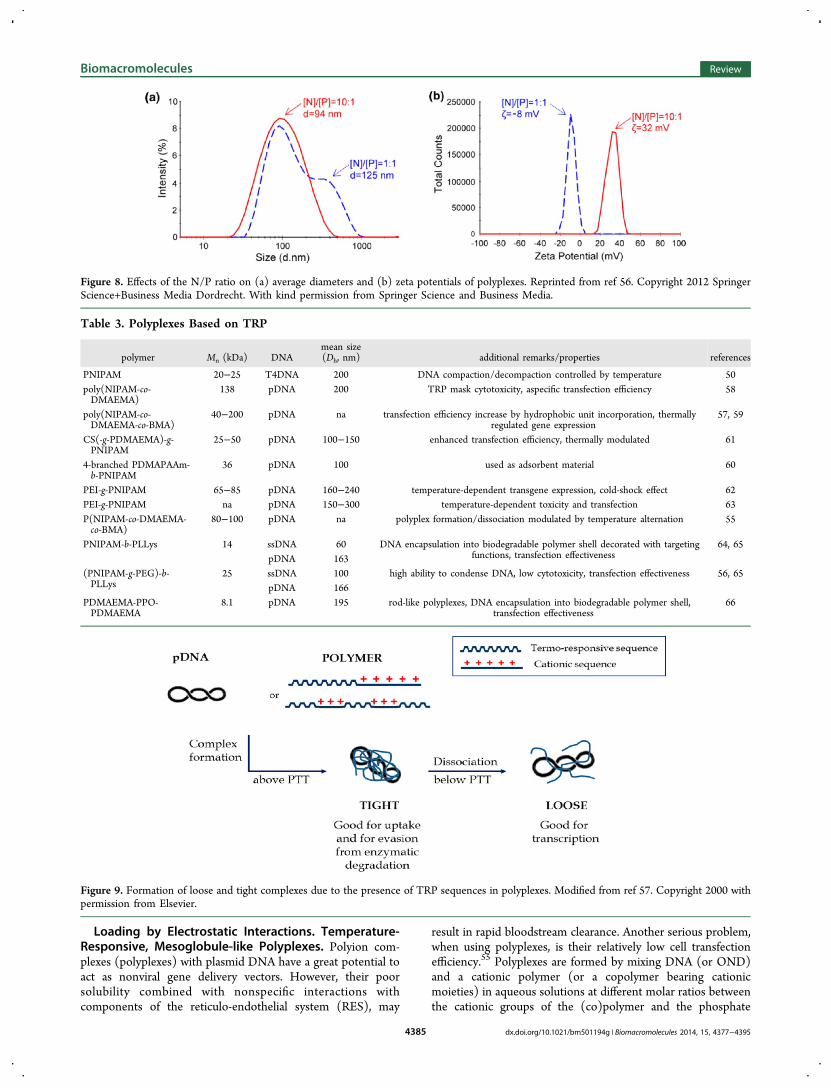
Figure 8. Effects of the N/P ratio on (a) average diameters and (b) zeta potentials of polyplexes. Reprinted from ref 56. Copyright 2012 SpringerScience+Business Media Dordrecht. With kind permission from Springer Science and Business Media.
Table 3. Polyplexes Based on TRP
polymer Mn (kDa) DNAmean size(Dh, nm) additional remarks/properties references
PNIPAM 20−25 T4DNA 200 DNA compaction/decompaction controlled by temperature 50poly(NIPAM-co-DMAEMA)
138 pDNA 200 TRP mask cytotoxicity, aspecific transfection efficiency 58
poly(NIPAM-co-DMAEMA-co-BMA)
40−200 pDNA na transfection efficiency increase by hydrophobic unit incorporation, thermallyregulated gene expression
57, 59
CS(-g-PDMAEMA)-g-PNIPAM
25−50 pDNA 100−150 enhanced transfection efficiency, thermally modulated 61
4-branched PDMAPAAm-b-PNIPAM
36 pDNA 100 used as adsorbent material 60
PEI-g-PNIPAM 65−85 pDNA 160−240 temperature-dependent transgene expression, cold-shock effect 62PEI-g-PNIPAM na pDNA 150−300 temperature-dependent toxicity and transfection 63P(NIPAM-co-DMAEMA-co-BMA)
80−100 pDNA na polyplex formation/dissociation modulated by temperature alternation 55
PNIPAM-b-PLLys 14 ssDNA 60 DNA encapsulation into biodegradable polymer shell decorated with targetingfunctions, transfection effectiveness
64, 65pDNA 163
(PNIPAM-g-PEG)-b-PLLys
25 ssDNA 100 high ability to condense DNA, low cytotoxicity, transfection effectiveness 56, 65pDNA 166
PDMAEMA-PPO-PDMAEMA
8.1 pDNA 195 rod-like polyplexes, DNA encapsulation into biodegradable polymer shell,transfection effectiveness
66
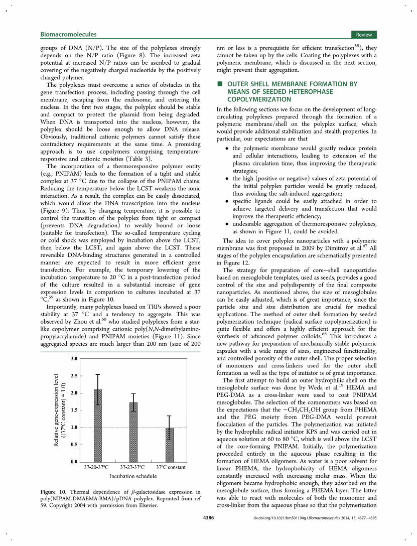
Figure 9. Formation of loose and tight complexes due to the presence of TRP sequences in polyplexes. Modified from ref 57. Copyright 2000 withpermission from Elsevier.
Biomacromolecules Review
dx.doi.org/10.1021/bm501194g | Biomacromolecules 2014, 15, 4377−43954385
groups of DNA (N/P). The size of the polyplexes stronglydepends on the N/P ratio (Figure 8). The increased zetapotential at increased N/P ratios can be ascribed to gradualcovering of the negatively charged nucleotide by the positivelycharged polymer.The polyplexes must overcome a series of obstacles in the
gene transfection process, including passing through the cellmembrane, escaping from the endosome, and entering thenucleus. In the first two stages, the polyplex should be stableand compact to protect the plasmid from being degraded.When DNA is transported into the nucleus, however, thepolyplex should be loose enough to allow DNA release.Obviously, traditional cationic polymers cannot satisfy thesecontradictory requirements at the same time. A promisingapproach is to use copolymers comprising temperature-responsive and cationic moieties (Table 3).The incorporation of a thermoresponsive polymer entity
(e.g., PNIPAM) leads to the formation of a tight and stablecomplex at 37 °C due to the collapse of the PNIPAM chains.Reducing the temperature below the LCST weakens the ionicinteraction. As a result, the complex can be easily dissociated,which would allow the DNA transcription into the nucleus(Figure 9). Thus, by changing temperature, it is possible tocontrol the transition of the polyplex from tight or compact(prevents DNA degradation) to weakly bound or loose(suitable for transfection). The so-called temperature cyclingor cold shock was employed by incubation above the LCST,then below the LCST, and again above the LCST. Thesereversible DNA-binding structures generated in a controlledmanner are expected to result in more efficient genetransfection. For example, the temporary lowering of theincubation temperature to 20 °C in a post-transfection periodof the culture resulted in a substantial increase of geneexpression levels in comparison to cultures incubated at 37°C,59 as shown in Figure 10.Importantly, many polyplexes based on TRPs showed a poor
stability at 37 °C and a tendency to aggregate. This wasobserved by Zhou et al.60 who studied polyplexes from a star-like copolymer comprising cationic poly(N,N-dimethylamino-propylacrylamide) and PNIPAM moieties (Figure 11). Sinceaggregated species are much larger than 200 nm (size of 200
nm or less is a prerequisite for efficient transfection58), theycannot be taken up by the cells. Coating the polyplexes with apolymeric membrane, which is discussed in the next section,might prevent their aggregation.
■ OUTER SHELL MEMBRANE FORMATION BYMEANS OF SEEDED HETEROPHASECOPOLYMERIZATION
In the following sections we focus on the development of long-circulating polyplexes prepared through the formation of apolymeric membrane/shell on the polyplex surface, whichwould provide additional stabilization and stealth properties. Inparticular, our expectations are that
• the polymeric membrane would greatly reduce proteinand cellular interactions, leading to extension of theplasma circulation time, thus improving the therapeuticstrategies;
• the high (positive or negative) values of zeta potential ofthe initial polyplex particles would be greatly reduced,thus avoiding the salt-induced aggregation;
• specific ligands could be easily attached in order toachieve targeted delivery and transfection that wouldimprove the therapeutic efficiency;
• undesirable aggregation of thermoresponsive polyplexes,as shown in Figure 11, could be avoided.
The idea to cover polyplex nanoparticles with a polymericmembrane was first proposed in 2009 by Dimitrov et al.67 Allstages of the polyplex encapsulation are schematically presentedin Figure 12.The strategy for preparation of core−shell nanoparticles
based on mesoglobule templates, used as seeds, provides a goodcontrol of the size and polydispersity of the final compositenanoparticles. As mentioned above, the size of mesoglobulescan be easily adjusted, which is of great importance, since theparticle size and size distribution are crucial for medicalapplications. The method of outer shell formation by seededpolymerization technique (radical surface copolymerization) isquite flexible and offers a highly efficient approach for thesynthesis of advanced polymer colloids.68 This introduces anew pathway for preparation of mechanically stable polymericcapsules with a wide range of sizes, engineered functionality,and controlled porosity of the outer shell. The proper selectionof monomers and cross-linkers used for the outer shellformation as well as the type of initiator is of great importance.The first attempt to build an outer hydrophilic shell on the
mesoglobule surface was done by Weda et al.19 HEMA andPEG-DMA as a cross-linker were used to coat PNIPAMmesoglobules. The selection of the comonomers was based onthe expectations that the −CH2CH2OH group from PHEMAand the PEG moiety from PEG-DMA would preventflocculation of the particles. The polymerization was initiatedby the hydrophilic radical initiator KPS and was carried out inaqueous solution at 60 to 80 °C, which is well above the LCSTof the core-forming PNIPAM. Initially, the polymerizationproceeded entirely in the aqueous phase resulting in theformation of HEMA oligomers. As water is a poor solvent forlinear PHEMA, the hydrophobicity of HEMA oligomersconstantly increased with increasing molar mass. When theoligomers became hydrophobic enough, they adsorbed on themesoglobule surface, thus forming a PHEMA layer. The latterwas able to react with molecules of both the monomer andcross-linker from the aqueous phase so that the polymerization
Figure 10. Thermal dependence of β-galactosidase expression inpoly(NIPAM-DMAEMA-BMA)/pDNA polyplex. Reprinted from ref59. Copyright 2004 with permission from Elsevier.
Biomacromolecules Review
dx.doi.org/10.1021/bm501194g | Biomacromolecules 2014, 15, 4377−43954386

continued within the layer and the shell thickness grew. Acorrelation between the initiator to monomer ratio and shellthickness was found, thus providing an effective control overthe thickness of the polymeric membrane. Importantly, thespherical shape of the resulting core−shell particles waspreserved.An essential advantage of a membrane, which is built from a
thermoresponsive polymer, is the fact that its hydrophilicity andpermeability can be easily tuned by changing the temperature.The latter is a desired function for both loading and release ofbiologically active substances.In the case of core-forming PIPOX and the shell-forming
PNIPAM, rather unexpectedly, raspberry-like morphology wasobserved as shown in Figure 13.37 The anisotropy arises fromthe immiscibility of the monomers and polymers when formingthe cross-linked membrane.69
When an oil-soluble initiator AIBN is added to thesuspension of hydrophobic seed particles in water, radicalsare generated on the mesoglobule surface. In this case thepartition between the monomer and the seed particle dependsstrongly on the interfacial tensions of the particle and theaqueous phase γP,A, the particle and the monomer, γP,M, and themonomer and the aqueous phase γM,A as shown by Mock etal.69 The shape of the monomer droplets is determined by themonomer/polymer wetting properties, kinetics of the polymernetwork contraction, and kinetics of monomer polymerization(Scheme 6).When a hydrophilic initiator such as KPS was used, NIPAM
oligomers were initially formed in the aqueous phase. At acertain moment, they collapsed and began to adsorb on thePIPOX mesoglobules surface forming a nodule and, by trappingmonomer and cross-linker from the aqueous phase, furtherevolved as polymeric particles attached to the surface. The
Figure 11. Aggregation of thermoresponsive polyplexes after prolonged heating. Reprinted from ref 60. Copyright 2007 with permission fromElsevier.
Figure 12. Scheme of polyplex encapsulation. Reprinted from ref 67. Copyright 2009 WILEY-VCH Verlag GmbH & Co. KGaA, Weinheim. Withpermission from John Wiley & Sons.
Figure 13. TEM image showing raspberry-like morphology of thePIPOX(core)/PNIPAM(shell) particles. Reprinted from ref 37.Copyright 2013, with permission from Elsevier.
Scheme 6. Possible Swelling Geometries at Different Statesof the Monomer−Seed Particle Systems: (a) Very Favorable;(b) Moderately Favorable; (c) Not Favorable; (d) ExtremelyUnfavorablea
aReprinted with permission from ref 69. Copyright 2006 AmericanChemical Society.
Biomacromolecules Review
dx.doi.org/10.1021/bm501194g | Biomacromolecules 2014, 15, 4377−43954387

immiscibility between PIPOX and PNIPAM revealed by DSCled to the formation of an inhomogeneous layer with raspberry-like morphology, as shown in Scheme 7.
A strong advantage of the approach of using heterophaseseeded radical polymerization is the possibility to incorporatemonomer units bearing specific functions so that the resultingparticles meet a set of preferable properties such as thefollowing:
• Stimuli-responsiveness of the shell. Monomers such asNIPAM, acrylic acid, oligoethylene glycol methacrylate,other acrylate monomers, etc. have been successfullyused. Due to the collapse of the shell chains understimuli, channels can be formed. Glucose and temper-ature dual-responsive nanospheres were fabricated byincorporation of glucose-responsive monomer (3-acryl-amidophenylboronic acid) (APBA) as reported by Du etal.70
• Biodegradability of the shell. By using biodegradablecross-linking agents such as N,N′-bis(acryloyl) cystamine
(BAC), disulfide bonds were incorporated in the networkof the shell.64−66 They can be degraded under reducingconditions or via thiol−disulfide exchange occurring inthe cells.
• Attachment of cell recognizable targeting ligands. Thiswas demonstrated by Dimitrov64 by incorporating afolate-terminated PEO-macromonomer.
Seeded radical copolymerization has been successively usedfor encapsulation of temperature-responsive polyplexes with abiodegradable cross-linked shell.64−66 For this purpose, firstplasmid or linear DNA was compacted via complex formationwith cationic diblock copolymer (PNIPAM-b-PLLys or(PNIPAM-g-PEG)-b-PLLys64,65 (Scheme 8) or via interactionof plasmid DNA with cationic worm-like micelles comprising apoly(propylene oxide) core and a PDMAEMA corona66
(Scheme 9). The polyplex particles exhibit good colloidalstability at high temperature (up to 70 °C), allowing formationof a cross-linked shell via copolymerization of NIPAM andN,N′-bis(acryloyl) cystamine. The formation of the outer shellwas confirmed by the increase of apparent hydrodynamicdiameter and change in zeta potential from highly positive toless positive or from highly negative to less negative (Table 4).
■ CORE RELEASE: FORMATION OF HOLLOWNANOPARTICLES
According to our approach, nanocapsules are fabricated bydissolution and removal of the core template. When core−shellparticles are cooled down to a temperature below LCSTcore, thepolymer chains of the core dissolve and go into the water phaseby penetrating through the cross-linked shell into the waterphase. The outer polymeric shell (membrane) acts as a barrierto the diffusion of macromolecules from the core to theparticle/water interface. Therefore, to obtain hollow structures(capsules), construction of a permeable outer membrane ismandatory. However, systematic information on factorsaffecting the membrane permeability is not available. Factors,
Scheme 7. Schematic Presentation of the Cross-Section andFront View of Core-Shell Nanoparticles with Raspberry-LikeMorphology, Formed by Seeded Copolymerization ofNIPAM and BIS on the Surface of PIPOX Mesoglobulesa
aReprinted from ref 37. Copyright 2013, with permission fromElsevier.
Scheme 8. Polyplex Formation from Thermoresponsive PNIPAM−PLLys Block Copolymers Followed by Coating withBiodegradable Polymer Layera
aReprinted from ref 65. Copyright 2013, with permission from Elsevier.
Biomacromolecules Review
dx.doi.org/10.1021/bm501194g | Biomacromolecules 2014, 15, 4377−43954388

generally considered to govern the shell permeability are givenbelow.
• chemical composition and the hydrophilic/hydrophobicbalance of the shell
• cross-linking density• thickness• formation of channels within the membrane• charged/uncharged state of the shell as well as the
encapsulated macromolecules (core polymer)• molecular weight and dispersity of the core-polymer• flexibility of the core-polymer• the disintegration rate of the core template
Some of the above factors, e.g. chemical composition, cross-linking density, membrane thickness, are quite obvious. Othersare discussed below.The seeded copolymerization allows preparation of shell
membranes with controllable thickness and permeability. Thethickness increases with increasing monomer to initiator ratioas well as the reaction time. Obviously, a thicker shell wouldhinder polymer chain diffusion, which would result in a reducedrelease rate of the core polymer during the core-removalprocess. The flexibility of the core-polymer is of significantimportance as well: a unique feature of the flexible macro-molecules is their ability to permeate the pores despite the factthat the coil size can be larger than the pore size.75
It should be noted that the hindered transport of core-forming polymers is greatly influenced by the molecular weightand dispersity. Low MW TRPs with narrow MW distribution
are easily permeating through the shell membrane, while largemacromolecules are retained.70,73 Thus, the fact that perme-ability is limited to a certain molecular weight allows efficientencapsulation of large biomacromolecules, which are not ableto leave the core.The formation of channels within the membrane is beneficial
since it may accelerate the release of the template. It should benoted, however, that the macromolecule diffusion in thechannel is restricted as shown by a model study for a polymerin a tube.76 The size and the pore geometry can be adjusted byseveral approaches. The first approach consists of anappropriate selection of polymers forming the core and theshell. The core is invariably composed of mesoglobules of aTRP or copolymers comprising thermoresponsive sequences.The cross-linked shell, however, can be constructed bypolymers with properties that are quite different from thoseof the core-forming polymer. In recent articles, thermores-ponsive PNIPAM37,70,7,73 or the hydrogel-forming PHEMA19,73
have been used for construction of the membrane. As discussedabove, upon cooling, the mesoglobules swell and then dissolve(Figure 3). The presence of a cross-linked polymeric shell ontheir surface, however, may prevent the mesoglobules fromswelling to equilibrium (e.g., effect of core compression).PNIPAM and PHEMA form membranes with a differentmorphology and mechanical strength, which inevitably leads toa different mechanism and kinetics of core-TRP extractionupon cooling. The membrane composed of cross-linkedPNIPAM is raspberry-like and uneven in contrast to that ofcross-linked PHEMA, which is smooth, uniform in thickness,
Scheme 9. Schematic Illustration of the Formation and Encapsulation of the Polyplex between pDNA and Cationic Micellesa
aReproduced from ref 66. Copyright 2013, with permission of The Royal Society of Chemistry. http://dx.doi.org/10.1039/c3ra21890b.
Table 4. Preparation of Polymeric Capsules and Encapsulated Polyplexes via Seeded Interfacial Copolymerization
hollow particles (Dh, nm)
polymer coremean size ofcore (Dh, nm) polymer shell
shellthickness(nm) 25 °C >40 °C additional remarks references
PNIPAM 50−100 PHEMA, PEG-DMA
3−25 300 125 capsules like deflated balloon 19
P(St-co-NIPAM) 40−230 PNIPAM, BIS 30−90 460 175 hollow microgels with temperature sensitivity 71
PNIPAM - PNIPAM, APBA,DVB
80−100 300−400 200−300 temperature and glucose responsive capsules 70
PIPOX 300−2000 PNIPAM,BIS 40−70 345 705 raspberry-like shell 37
PNIPAM 108−285 PNIPAM,BIS 80−135 1400 300 core made via semicontinuous heterophasepolymerization, fully soluble mesoglobules
72
PDEGMA 100−350 PNIPAM, BIS 10−35 100−250 - raspberry-like shell 73
PHEMA, BIS 20 131 - smooth shell
P(G-co-EGC) 225−350 PNIPAM,BIS 25−30 - no dissolution of the core because of too high MW 74
P(PNIPAM-b-PLLys)/ssDNA
80−150 PNIPAM,BIS,PEG-DA or
BAC
15−90 - for the first time encapsulated polyplex, folic acidused as targeting vector
64
P(PNIPAM-b-PLLys)/pDNA
163 PNIPAM, PEG-A,BAC
15 - encapsulated polyplex, reduced transfection efficiencycompared to the corresponding polyplexes
65
P(PNIPAM-g-PEG)-b-PLLys/pDNA
116 PNIPAM, PEG-A,BAC
25 - encapsulated polyplex, reduced transfection efficiencycompared to the corresponding polyplexes
65
PDMAEMA-PPO-PDMAEMA/pDNA
195 PNIPAM, BAC 10 - encpsulated polyplex protecting shell decreased thecytotoxicity
66
Biomacromolecules Review
dx.doi.org/10.1021/bm501194g | Biomacromolecules 2014, 15, 4377−43954389

and homogeneous. The raspberry-like morphology of thehollow particles is shown in Figure 14.
Let us consider the cases where the core and the shell arecomposed of different TRP characterized by LCSTcore andLCSTshell. In the case of LCSTcore > LCSTshell, when thetemperature is below LCSTcore and close to or higher thanLCSTshell (Scheme 10b), the collapse of the shell results in theformation of channels, which greatly facilitates the diffusion ofthe core polymer and the trans-membrane transport.37 In thecase when dialysis is carried out to remove the core polymer attemperatures lower than the LCST for both polymer andLCSTcore ≤ LCSTshell (Scheme 10c), water is a good solvent forall components of the system. At such conditions, the initialcore−shell particles consist of swollen core and shell that arecross-linked physically and chemically, respectively. The coredimensions increase until an equilibrium between the forceexerted by the shell and the resisting elastic force developed inthe core is reached. In the next step, the macromolecules fromthe mesoglobules are expected to pass through the cross-linkedshell hydrogel. However, due to the increase in the macro-molecule dimensions, the process of core release is slow andfrequently incomplete.An alternative method of preparing porous membranes
involves seeded copolymerization in the presence of a
particulate porogen matrix such as sucrose or glucose, orglucose comonomers. Dergunov and Pinkhassik77 successfullyused pore forming molecules (glucose pentaacetate and glucosepentabenzoate) to synthesize nanocapsules with pores ofcontrolled size (0.8 ± 0.2 nm). The porous nanocapsulesretain molecules larger than the pore size but provide ultrafastaccess to their interior for molecules and ions smaller than thepore size (Figure 15). Membranes can also become porous in
response to the presence of glucose in the medium, whenglucose-responsive phenylboronic acid derivative is introducedvia NIPAM and 3-acrylamidophenilboronic acid seededcopolymerization as shown elsewhere.70 Channels can beconstructed also when the seeded copolymerization isperformed with comonomers creating incompatible (immis-cible) domains. For example, channels can be further modifiedby the formation of a mixed PEG/PNIPAM shell.78
Figure 14. TEM images of hollow nanoparticles composed ofPNIPAM membrane. The particles were obtained using templatesfrom (a) PIPOX mesoglobules37 and (b) PDEGMA mesoglobules.73
Reprinted from refs 37 (Copyright 2013) and 73 (Copyright 2014),with permission from Elsevier.
Scheme 10. Release of Core Macromolecules from Core/Shell Particles: (a) Synthesized by Weda et al.19 and Haladjova et al.;73
(b) Synthesized by Toncheva et al.;37 (c) synthesized by Du,70 Chen,72 and Haladjova et al.73
Figure 15. Nanometer thin film with functionalized nanopores.Reprinted from ref 77. Copyright 2008 WILEY-VCH Verlag GmbH &Co. KGaA, Weinheim. With permission from John Wiley and Sons.
Biomacromolecules Review
dx.doi.org/10.1021/bm501194g | Biomacromolecules 2014, 15, 4377−43954390

■ NONVIRAL GENE DELIVERY BYPOLYMER-COATED POLYPLEXES
The method for preparation of core/shell nanoparticles (NP)by surface copolymerization (or seeded radical copolymeriza-tion) was used to cover DNA complexes with a desirablepolymeric coating as shown in Schemes 8 and 9. Plasmid DNAis unstable in biological media. Therefore, association with acarrier is required to protect it from degradation. Plasmid DNAis generally incorporated into nanoscale formulations bycomplex formation with cationic lipids (lipoplexes) or cationicpolymers (polyplexes), which provide greater stability andfunctionality.79 Unfortunately, the therapeutic use of polyplexesis limited mainly due to their instability in vivo and tointeractions with cells of the immune system (RES) and bloodplasma proteins during their transportation in the bloodstream.Moreover, most of the polyplexes exhibit a high positive surfacecharge, which causes undesirable responses in the body such asaggregation and tissue damage. Construction of coatings on thepolyplex surface via seeded radical copolymerization (the outershell referred to as the envelope is of the same structure as themembranes of the polymeric nanocapsules) should provide aphysical barrier and stealth properties, thus significantlyimproving stability and increasing circulation time in thesystem circulation.It is noteworthy that in the case of a polymer-coated
polyplex, it is the membrane that interacts with the cell surfaceand other cellular compartments and proteins. Hence, under-standing the interactions of the polyplex envelope with the cellsis crucial for improving their functionality both in vitro and invivo. A major advantage of the approach is that the seededsurface polymerization process allows preparation of envelopeswith properties that can be modulated. For instance, theproperties of the membrane can be altered using monomersbearing specific side groups, thus introducing defined ligands.Furthermore, when two monomers are copolymerized, themembranes can be tuned (e.g., hydrophilic/hydrophobicbalance) simply by changing the relative ratio between thecomonomers.The polymer coating would first significantly reduce the
positive surface potential of the polyplexes resulting in nearlyneutral nanoparticles. As the positively charged polyplexes arehighly toxic and not approved for clinical applications, thecoating would resolve the toxicity problems. Figure 16 clearlyshows that the polyplex coated with a polymeric shell isinvariably less toxic than the parent (noncoated) polyplex.The applicability of polyplexes, coated by a biodegradable
shell, for nonviral gene delivery was proven in a series ofexperiments showing successful transfection in HEK 293 cellswith a plasmid carrying the enhanced green fluorescenceprotein (EGFP) gene (Table 5). It should be noted that theproper selection of the initiating system as well as the mildconditions for formation of the coating are very important,since the free radicals generated by initiators such as KPS candamage DNA. Therefore, the surface radical copolymerizationwas initiated by 2,2′-azobis(2-methylpropionamidine) dihydro-chloride (AAPH)a water-soluble, nondestructive for pDNAinitiator, and in the presence of N,N′-bis(acryloyl)cystamine, across-linking agent. The use of disulfide-containing BACimparted biodegradability to the polymer shell in theintracellular environment, which was important for pDNAtransfection.
The transfection efficiency strongly depends on the structureof the cationic copolymers used for complex formation withpDNA. Thus, for example, both the parent polyplexes and theircoated counterparts, comprising PNIPAm-b-PLLys exhibitedhigher transfection efficiency compared to those formed by theblock copolymer containing PEG-grafts (PNIPAm-g-PEG)-b-PLLys.65 The results were attributed to the PEG shieldingeffect and were consistent with other studies, showing that thetransfection efficiency of PEG-conjugated polyplexes wasgenerally 2-fold lower compared to non-PEGylated particlesdue to a decrease in cell binding and uptake.80,81 On the otherhand, for the two systems, the initial noncoated polyplexesdisplayed higher transfection efficiency as compared to thecorresponding coated derivatives (Figure 17).Obviously, the polymer coating hindered the cellular uptake
and decreased the effect. Similar tendency was observed forsystems based on PDMAEMA13-b-PPO69-b-PDMAEMA13 rod-like micelles.66 The detected transfection efficiency of the initialpolyplex and the coated one at incubation for 24 h are 13% and8%, respectively (Table 5). After incubation for 48 h, themaximum transfection efficiency of the coated PDMAEMA13-b-PPO69-b-PDMAEMA13/pDNA polyplex increased to 27%.Although the transfection efficiency of coated polyplexes wasnot as high as that of Lipofectamine, these results areencouraging and suggesting that these systems could beconsidered as potential nonviral gene delivery vectors. Itmight be expected that the coating reaction would result inaggregation of the polyplex nanoparticles, which is a highlyundesirable process, as it prohibits their use as transfectionagents. However, as seen from Figure 18, the width of the Dhdistribution of the coated polyplexes was slightly smaller thanthat of the original polyplexes, implying that aggregation didnot take place during the coating reaction.
■ CONCLUSIONS AND FUTURE PERSPECTIVESA novel synthetic strategy for the preparation of hollownanoparticles was developed and reported by us in 2008.19 Itinvolves several steps: temperature-induced PT of TRP, shellformation by seeded radical copolymerization, and subsequentdissolution of TRP by dialysis at lower temperatures, as shownin Scheme 2.Almost at the same time Zhang and Wang71 suggested
nanocapsule preparation from core−shell particles with
Figure 16. Comparison of HEK 293 cell viability followingtransfection with naked or coated polyplex, obtained by MTT assayon 24 h. In all experiments P/N = 2. Data are means of threeindependent experiments, and the error bars represent the SD, p <0.05 against transfection with naked polyplex. Adapted from ref 66(Supporting Information). Copyright 2013, with permission of TheRoyal Society of Chemistry. http://dx.doi.org/10.1039/c3ra21890b.
Biomacromolecules Review
dx.doi.org/10.1021/bm501194g | Biomacromolecules 2014, 15, 4377−43954391

P(styrene-co-NIPAM) core and cross-linked PNIPAM shell.The capsules were obtained by dissolving and removing theP(styrene-co-NIPAM) core in tetrahydrofuran. A key advantageof our method over the one reported in ref 71 is that all stepsare performed entirely in aqueous medium in a very mild andnondestructive fashion, as shown in Scheme 11.The aqueous conditions used in our approach are beneficial,
since water is the preferred environment for the biomoleculesas they are metastable. Water molecules form a solvating layeraround the hydrophilic surface residues of proteins and nucleicacids. This water layer has a damping effect on the attractive
forces between biomolecules. Moreover, it is well-known thatbiomacromolecules such as enzymes lose their activity inorganic solvents.Polymer nanocapsules reported so far are able to load and
release guest molecules from their interior only by diffusion.Therefore, it is rather difficult to control the loading process.Another drawback of the current methods is that loading can bedone only after the formation of the nanocontainer. The loadincludes mostly low molecular weight compounds, such asdoxorubicin, paclitaxel, etc. Our approach overcomes thisdrawback, as it provides the possibility for incorporation of
Table 5. Characterization Data and Transfection Efficiency of Polyplexes and the Corresponding Polyplexes Covered by aPolymeric Shell
polyplexmean size(Dh, nm)
ζ-potential(mV)
transfection(%)
coated polyplex monomer,comonomer, initiator
mean size(Dh, nm)
ζ-potential(mV)
transfection(%) references
PNIPAM-b-PLLys/pDNA(N/P = 20)
163 19.5 22 NIPAM, PEG-DA, BAC,AAPH
195 8.4 12 65
P(NIPAM-g-PEG)-b-PLLys/pDNA(N/P = 20)
116 27.8 15 NIPAM, PEG-DA, BAC,AAPH
161 11.7 7 65
PDMAEMA-PPO-PDMAEMA/pDNA (N/P = 0.5; rod-like)
195 −32.3 13 NIPAM, BAC, AAPH 210 −28.3 8 (24 h) 27(48 h)
66
Figure 17. Transfection efficiency of the polyplex formed at N/P = 20 from pDNA and PNIPAm-b-PLLys. HEK 293 cells were transfected withpEGFP-C2 containing polyplex for 24 h. 48 h after plasmid transfection the percentage of GFP-positive cells was measured by flow cytometry andassessed by fluorescent microscopy. The GFP positive population was gated in area R3. Data are means of three independent experiments. (A) Non-transfected cells (negative control); (B) cells transfected with Lipofectamine 2000 containing 4 μg of pEGFP-C2 (positive control); (C) transfectionof initial polyplex; and (D) after polyplex stealth coating with a biodegradable PNIPAm membrane. Reprinted from ref 65. Copyright 2013, withpermission from Elsevier.
Figure 18. Average diameters and PDI before and after coating of polyplexes, formed at N/P = 20 from pDNA and (A) PNIPAM-b-PLLys and (B)(PNIPAM-g-PEG)-b-PLLys. Reprinted from ref 65. Copyright 2013 with permission from Elsevier.
Biomacromolecules Review
dx.doi.org/10.1021/bm501194g | Biomacromolecules 2014, 15, 4377−43954392

biopolymers during the formation of mesoglobule coretemplate. Thus, it enables correct determination of the amountof the entrapped biologically active substance, or, in otherwords, to achieve a controlled load.The final goal cannot be achieved unless we know how to
include a sufficient amount of biological substance during theprocess of mesoglobule formation. In all cases a reversible andsite-specific interactions are desired. The results, presented inTable 2, show an insufficient loading efficiency. Moresystematic investigation of the loading process into meso-globules is needed. We believe that the optimization ofmesoglobules properties to achieve a reasonable loadingwithout the so-called nonspecific interactions is feasible. It isquite obvious that better results can be obtained usingfunctionalized TRP, bearing positive or negative charges,hydrophobic, ligating, or polyoxyethylene functions. Moreover,charges play a major role in protein binding since all proteinscarry positive or negative charges on their surface. Anappropriate example represents the use of poly(NIPAM-co-vinylbenzyl iminodiacetic acid) copolymer by Tsai et al.82
Divalent nickel ions interact with the diacetic acid ligandsallowing selective metal affinity attachment to a His6-Cyspeptide. Moreover, the peptide can be successfully releasedupon the addition of the competitive ligand imidazole.The importance of the mesoglobule morphology for the
effective loading of biomolecules should also be noted.Obviously, the “loose” mesoglobule structure is preferable.Unfortunately, the internal voids (see Scheme 3) are notcontinuous, which will greatly hinder the biomoleculeabsorption. In the case of a dense mesoglobule, the ligandsand charges bound to the TRP are less accessible to thebiomolecule.According to our approach, mesoglobule templates are used
as seeds (nuclei) for the formation of the shell. The radicalcopolymerization on the seed surface is a convenient methodfor the outer layer modification. There are numerous vinylmonomers that can be used to create desired properties, such asstimulus-sensitivity, stealth properties, biodegradability, con-trolled porosity, tuned hydrophobic/hydrophilic balance, ligandattachment, etc. The copolymerization proceeds at thenanoparticle’s surface. The initial mesoglobule grows by theaddition of fresh comonomers from the aqueous medium. Aserious problem that should be mentioned is the difference inreactivity ratios of the comonomers and the cross-linker. Whenthe reactivity of the first monomer is higher, it is preferablyconsumed. This leads to inhomogeneity of the outer
membrane. When the cross-linker (e.g., BIS) reacts fasterthan the monomer (e.g., NIPAM), the cross-link density ismuch higher in the interior of the membrane, giving the outersurface a less dense network structure. In that case, the additionof NaNO2 as a water-soluble inhibitor that is able to deactivatewater-soluble radicals is of great importance.83 The outer shellthickness in most of the experiments is between 10 and 70 nm.The thickness and the overall cross-linked density can be easilycontrolled. Thus, mesoglobules are protected by polymericstealth layer that can minimize adsorption of proteins wheninjected into the bloodstream.The permeability control of the membrane is still a challenge.
Unfortunately, it has not been studied thoroughly. Thethickness of the membrane and the cross-linked density arethe most obvious parameters that can influence thepermeability. While the small molecules can easily cross themembrane, the release of TRP macromolecules is problematic.Obviously, the membrane permeability has to be tuned. Thus,for example the permeability can be temperature-enhanced ifthe membrane contains thermoresponsive-sequences. A prom-ising approach is the inclusion of pore-forming glucosepentaacetate and glucose pentabenzoate into the membranestructure, as proposed by Dergunow and Pinkhassik.77 An idealouter shell would contain a membrane protein as it was alreadydemonstrated with some polymersomes.84 Thus, the perme-ability can be selectively tuned depending on the nature of thechannel protein incorporated into the membrane.Finally, mesoglobules comprise a template, able to
disintegrate and dissolve at low temperature. Obviously, themembrane can be prepared through any well-known methodfor nanoparticle coating with a stealth polymer layer. Thus, theradical copolymerization can be successfully replaced by anioniccopolymerization of cyanoacrylates or even by the very popularrecently layer-by-layer deposition technique. So far this has notbeen done, but it is a real challenge.Overall, our approach allows the formation of structures that
resemble artificial cell structure or multicompartment archi-tecture as presented in Scheme 12.
This review is an attempt to convince researchers of theprospects for further, much deeper study of mesoglobules aspotential reservoirs, carriers, and transferring agents forbiologically active substances.
Scheme 11. Schematic Illustration of the Differences in thePreparation of Hollow Polymeric Particles: (a) CoreDissolution by Organic Solvent;71 (b) Core Dissolution inCold Water Due to Phase Transition19,37,70,72,73
Scheme 12. Sketch of Formation of Sphere-in-SphereStructure with Two Compartments A and B Loaded withTwo Different Biomolecules
Biomacromolecules Review
dx.doi.org/10.1021/bm501194g | Biomacromolecules 2014, 15, 4377−43954393
■ AUTHOR INFORMATIONCorresponding Author*Mailing address: Institute of Polymers, Bulgarian Academy ofSciences, “Akad. G. Bonchev” str. 103A, 1113 Sofia, Bulgaria. E-mail:[email protected] ContributionsThe manuscript was written through contributions of allauthors. All authors have given approval to the final version ofthe manuscript.NotesThe authors declare no competing financial interest.
■ ACKNOWLEDGMENTSMost of the studies were supported by the National ScienceFund of Bulgaria under Grant DO 02-247/2008 as well as fromthe Ministry of Science and Higher Education of Poland, GrantNN 209 1441 36. The paper benefited from discussions atevents of P2M Research Networking Programme of ESF andfrom bilateral cooperation between the Bulgarian and PolishAcademies of Sciences. We are also grateful to the followingcolleagues, who participated in the experiments: P. Weda, Dr.Ph. Dimitrov, E. Ivanova, R. G. Kozarova, N. I. Ivanova, B.Gajewska, and E. B. Petrova.
■ ABBREVIATIONSA acrylic acidAAm acrylamideAAPH 2,2′-azobis(2-methylpropionamidine) dihydro-
chlorideAIBN α,α′-azoisobutyronitrileAPBA 3-acrylamidophenylboronic acidBAC N,N′-bis(acryloyl)cystamineBIS N,N′-methylenebis(acrylamide)BSA bovine serum albuminBMA butyl methacrylateCS chitosanDVB divinylbenzeneFITC Lys−fluorescein isothiocyanate-lysozomeHEMA hydroxyethyl methacrylateKPS potassium persulfateLCST lower critical solution temperatureLbL layer-by-layerNIPAM N-isopropylacrylamideNP nanoparticleOND oligonucleotidePTT phase transition temperaturePDI polydispersity indexPNG polymeric nanogelPNIPAM poly(N- isopropylacrylamide)PHEMA poly(hydroxyethyl methacrylate)PDMAEMA poly(2-dimethylaminoethyl methacrylate)PEG poly(ethylene glycol)PEG-A poly(ethylene glycol) acrylatePEG-DA poly(ethylene glycol) diacrylatePEG-DMA poly(ethylene glycol)-dimethacrylatePETEGA poly(ethoxy triethylene glycol acrylate)PIPOX poly(2-isopropyl-2-oxazoline)PLLys poly(L-lysine)POEGA poly(oligo(ethylene glycol) acrylate)POEGMA poly(oligo(ethylene glycol) methacrylate)PDEGMA poly(di(ethylene glycol) methacrylate)PVCL poly(N-vinyl caprolactam)
PVME poly(vinyl methyl ether)
PtNEApoly(trans-N-(2-ethoxy-1,3-dioxan-5-yl) acryla-mide)
PEI polyethylene iminePPO poly(propylene oxide)RES reticulo-endothelial systemSDS sodium dodecyl sulfateSt styreneTRP thermoresponsive polymerTg glass transition temperature
■ REFERENCES(1) Discher, D. E.; Eisenberg, A. Science 2002, 297, 267−273.(2) LoPresti, C.; Lomas, H.; Massignani, M.; Smart, T.; Battaglia, G.J. Mater. Chem. 2009, 19, 3576−3590.(3) Chang, Y. W.; Silas, J. A.; Ugaz, K. M. Langmuir 2010, 26 (4),12132−12139.(4) Kowalczuk, A.; Trzcinska, R.; Trzebicka, B.; Muller, A. H. E.;Dworak, A.; Tsvetanov, Ch.B. Prog. Polym. Sci. 2014, 39, 43−86.(5) Rangelov, S.; Pispas, S. Polymer and Polymer-Hybrid Nanoparticles:From Synthesis to Biomedical Applications; CRC Press: Taylor andFrancis Group, 2014.(6) Donath, E.; Sukhorukov, G. B.; Caruso, F.; Davis, S. A.;Mohwald, H. Angew. Chem., Int. Ed. 1998, 37, 2201−2205.(7) Gittins, D. T.; Caruso, F. Adv. Mater. 2000, 12 (24), 1947−1949.(8) Becker, A. L.; Johnston, A. P. R.; Caruso, F. Small 2010, 6 (17),1836−1852.(9) Cui, J.; van Koeverden, M. P.; Mullner, M.; Kempe, K.; Caruso, F.Adv. Colloid Interface Sci. 2014, 207, 14−31.(10) Ando, S.; Putnam, D.; Pack, D. W.; Langer, R. J. Pharm. Sci.1999, 88, 126−130.(11) Aseyev, V.; Hietala, S.; Laukkanen, A.; Nuopponen, M.;Confortini, O.; Du Prez, F. E.; Tenhu, H. Polymer 2005, 46, 7118−7131.(12) Kujawa, P.; Aseyev, V.; Tenhu, H.; Winnik, F. M. Macro-molecules 2006, 39, 7686−7693.(13) Tanaka, F.; Koga, T.; Kaneda, I.; Winnik, F. M. J. Phys.: Condens.Matter 2011, 23 (28), 284105.(14) Van Durme, K.; Van Assche, G.; Van Mele, B. Macromolecules2004, 37, 9596−9605.(15) Wu, C.; Li, W.; Zhu, X. X. Macromolecules 2004, 37, 4989−4992.(16) Nishi, K.; Hiroi, T.; Hashimoto, K.; Fujii, K.; Han, Y. S.; Kim, T.H.; Katsumoto, Y.; Shibayama, M. Macromolecules 2013, 46, 6225−6232.(17) Katsumoto, Y.; Kubosaki, N. Macromolecules 2008, 41, 5955−5956.(18) Cheng, H.; Shen, L.; Wu, C. Macromolecules 2006, 39, 2325−2339.(19) Weda, P.; Trzebicka, B.; Dworak, A.; Tsvetanov, Ch.B. Polymer2008, 49, 1467−1474.(20) Debeljuch, N. J.; Sutti, A.; Barrow, C. J.; Byrne, N. J. Phys. Chem.B 2013, 117, 8430−8435.(21) Reddy, P. M.; Taha, M.; Venkatesu, P.; Kumar, A.; Lee, M. J. J.Chem. Phys. 2012, 136, 234904.(22) Aseyev, V.; Tenhu, H.; Winnik, F. M. Adv. Polym. Sci. 2006, 196,1−85.(23) Ding, Y.; Ye, X.; Zhang, G. Macromolecules 2005, 38, 904−908.(24) Takahashi, R.; Sato, T.; Terao, K.; Qiu, X. P.; Winnik, F. M.Macromolecules 2012, 45, 6111−6119.(25) Katsumoto, Y.; Sato, T.; Terao, K.; Qiu, X. P.; Winnik, F. M.Macromolecules 2012, 45, 3531−3541.(26) Demirel, A.; Meyer, M.; Schlaad, H. Angew. Chem., Int. Ed. 2007,46, 8622−8624.(27) Diehl, C.; Cernoch, P.; Zenke, I.; Runge, H.; Pitschke, R.;Hartmann, J.; Tiersch, B.; Schlaad, H. Soft Matter 2010, 6, 3784−3788.(28) Lutz, J. F.; Akdemir, O.; Hoth, A. J. Am. Chem. Soc. 2006, 128(40), 13046−13047.
Biomacromolecules Review
dx.doi.org/10.1021/bm501194g | Biomacromolecules 2014, 15, 4377−43954394
(29) Hu, Z.; Cai, T.; Chi, C. Soft Matter 2010, 6 (10), 2115−2123.(30) Lutz, J. F. Adv. Mater. 2011, 23 (19), 2237−2243.(31) Lutz, J. F.; Weichenhan, K.; Akdemir, O.; Hoth, A.Macromolecules 2007, 40 (7), 2503−2508.(32) Toncheva-Moncheva, N.; Dimitrov, Ph.; Tsvetanov, Ch.B.;Robak, B.; Trzebicka, B.; Dworak, A.; Rangelov, S. Polym. Bull. 2011,67, 1335−1346.(33) Rangelov, S.; Simon, P.; Toncheva-Moncheva, N.; Dimitrov,Ph.; Gajewska, B.; Tsvetanov, Ch. B. Polym. Bull. 2012, 68, 2175−2185.(34) Trzebicka, B.; Szweda, D.; Rangelov, S.; Kowalczuk, A.;Mendrek, B.; Utrata-Wesolek, A.; Dworak, A. J. Polym. Sci., Part A:Polym. Chem. 2013, 51, 614−623.(35) Trzcinska, R.; Szweda, D.; Rangelov, S.; Suder, P.; Silberring, J.;Dworak, A.; Trzebicka, B. J. Polym. Sci., Part A: Polym. Chem. 2012, 50(15), 3104−3115.(36) Hu, Z.; Cai, T.; Chi, C. Soft Matter 2010, 6 (10), 2115−2123.(37) Toncheva-Moncheva, N.; Tsvetanov, Ch.; Rangelov, S.;Trzebicka, B.; Dworak, A. Polymer 2013, 54 (19), 5166−5173.(38) Gauthier, M.; Klok, H. A. Chem. Commun. 2008, 23, 2591−2611.(39) Su, Z.; Zhang, L.; Zhao, J.; Chen, X. Polymer 2011, 52, 1084−1091.(40) Li, J.; Zhao, J.; Zhao, X.; Jandt, K. D.; Su, Z. RSC Adv. 2013, 3(43), 20254−20260.(41) Junk, M.; Li, W.; Schluter, A. D.; Wegner, G.; Spiess, H.; Zhang,A.; Hinderberger, D. J. Am. Chem. Soc. 2011, 133, 10832−10838.(42) Trzebicka, B.; Robak, B.; Trzcinska, R.; Szweda, D.; Suder, P.;Silberring, J.; Dworak, A. Eur. Polym. J. 2013, 49, 499−509.(43) Grabstein, V.; Bianco-Peled, H. Biotechnol. Prog. 2003, 19,1728−1733.(44) Locatelli-Champagne, C.; Cloitre, M. Colloid Polym. Sci. 2013,291 (12), 2911−2916.(45) Fujimoto, K.; Mizuhara, Y.; Tamura, N.; Kawaguchi, H. J. Intell.Mater. Syst. Struct. 1993, 4, 184−189.(46) Qiao, Z. Y.; Ji, R.; Huang, X. N.; Du, F. S.; Zhang, R.; Liang, D.H.; Li, Z. C. Biomacromolecules 2013, 14, 1555−1563.(47) Fundueanu, G.; Constantin, M.; Ascensi, P. Int. J. Pharm. 2009,379, 9−17.(48) Ramkissoon-Ganorkar, C.; Liu, F.; Baudys, M.; Kim, S. W. J.Controlled Release 1999, 59, 287−298.(49) Molawi, K.; Studer, A. Chem. Commun. 2007, 48, 5173−5175.(50) Chen, N.; Zinchenko, A. A.; Kidoaki, S.; Murata, S.; Yoshikawa,K. Langmuir 2010, 26 (5), 1995−1998.(51) Dervan, P. B.; Burli, R. W. Curr. Opin. Chem. Biol. 1999, 3, 688−693.(52) Zinchenko, A. A.; Yoshikawa, K. Biophys. J. 2005, 88 (6), 4118−4123.(53) Moura, L. M.; Martinho, J. M. G.; Farinha, J. P. S.ChemPhysChem 2010, 11, 1749−1756.(54) Martinho, J. M. G.; Prazeres, T. J. V.; Moura, L. M.; Farinha, J.P. S. Pure. Appl. Chem. 2009, 81 (9), 1615−1634.(55) Tran, N. T. D.; Truong, N. P.; Gu, W.; Jia, Z.; Cooper, M. A.Biomacromolecules 2013, 14, 495−502.(56) Ivanova, E.; Dimitrov, I.; Kozarova, R.; Turmanova, S.;Apostolova, M. D. J. Nanopart. Res. 2013, 15, 1358.(57) Kurisawa, M.; Yokoyama, M.; Okano, T. J. Controlled Release2000, 69, 127−137.(58) Hinrichs, W. L. J.; Schuurmans-Nieuwenbroek, N. M. E.; van deWetering, P.; Hennink, W. E. J. Controlled Release 1999, 60, 249−259.(59) Takeda, N.; Nakamura, E.; Yokoyama, M.; Okano, T. J.Controlled Release 2004, 95, 343−355.(60) Zhou, Y. M.; Ishikawa, A.; Okahashi, R.; Uchida, K.; Nemoto,Y.; Nakayama, M.; Nakayama, Y. J. Controlled Release 2007, 123, 239−246.(61) Bao, H.; Ping, Y.; Pan, Y.; Li, L.; Li, J.; Gan, L. H. Soft Matter2012, 8, 2518−2526.(62) Lavigne, M. D.; Pennadam, S. S.; Ellis, J.; Yates, L. L.; Alexander,C.; Gorecki, D. G. J. Gene Med. 2007, 9, 44−54.
(63) Bisht, H. S.; Manickam, D. S.; You, Y.; Oupicky, D.Biomacromolecules 2006, 7, 1169−1178.(64) Dimitrov, I. V.; Petrova, E. B.; Kozarova, R. G.; Apostolova, M.D.; Tsvetanov, Ch.B. Soft Matter 2011, 7, 8002−8004.(65) Ivanova, E. D.; Ivanova, N. I.; Apostolova, M. D.; Turmanova, S.C.; Dimitrov, I. V. Bioorg. Med. Chem. Lett. 2013, 23, 4080−4084.(66) Petrov, P. D.; Ivanova, N. I.; Apostolova, M. D.; Tsvetanov,Ch.B. RSC Adv. 2013, 3, 3508−3511.(67) Dimitrov, Ph.; Toncheva, N.; Weda, P.; Rangelov, S.; Trzebicka,B.; Dworak, A.; Tsvetanov, Ch.B. Macromol. Symp. 2009, 278, 89−95.(68) Kim, J. W.; Suh, K. D. J. Ind. Eng. Chem. . 2008, 14, 1−9.(69) Mock, E. B.; De Bruyn, H.; Hawkett, B. S.; Gilbert, R. G.;Zukoski, C. F. Langmuir 2006, 22, 4037−4043.(70) Du, P.; Mu, B.; Wang, Y.; Liu, P. Mater. Lett. 2012, 75, 77−79.(71) Zhang, F.; Wang, C. C. Colloid Polym. Sci. 2008, 286, 889−895.(72) Chen, Y.; Sajjadi, S. Polymer 2013, 54 (21), 5467−5472.(73) Haladjova, E.; Rangelov, S.; Tsvetanov, Ch.; Simon, P. Polymer2014, 55, 1621−1627.(74) Trzebicka, B.; Weda, P.; Utrata-Wesolek, A.; Dworak, A.;Tsvetanov, Ch. J. Polym. Sci., Part A: Polym. Chem. 2010, 48, 4074−4083.(75) Bayley, H.; Martin, C. R. Chem. Rev. 2000, 100, 2575−2594.(76) Bezrukov, S. M.; Vodyanoy, I.; Parsegian, V. A. Nature 1994,370, 279−281.(77) Dergunov, S. A.; Pinkhassik, E. Angew. Chem. 2008, 47, 8264−8267.(78) Wu, C.; Ma, R.; He, H.; Zhao, L.; Gao, H.; An, Y.; Shi, L.Macromol. Biosci. 2009, 9, 1185−1193.(79) Mastrobattista, E.; Hennink, W. E.; Schiffelers, R. M. Pharm. Res.2007, 24 (8), 1561−1563.(80) van den Berg, J. H.; Oosterhuis, K.; Hennink, W. E.; Storm, G.;van der Aa, L. J.; Engbersen, J. F.; Haanen, J. B.; Beijnen, J. H.;Schumacher, T. N.; Nuijen, B. J. Controlled Release 2010, 141, 234−240.(81) Mishra, S.; Webster, P.; Davis, M. E. Eur. J. Cell Biol. 2004, 83,97−111.(82) Tsai, H. Y.; Lee, A.; Peng, W.; Yates, M. Z. Colloids Surf., B2014, 114, 104−110.(83) Yuyama, H.; Hashimoto, T.; Ma, G. H.; Nagai, M.; Omi, S. J.Appl. Polym. Sci. 2000, 78, 1025−1043.(84) Nardin, C.; Thoeni, S.; Widmer, J.; Mathias, W.; Meier, W.Chem. Commun. 2000, 15, 1433−1434.
Biomacromolecules Review
dx.doi.org/10.1021/bm501194g | Biomacromolecules 2014, 15, 4377−43954395