Perilipin Expression Reveals Adipogenic Potential of hADSCs inside Superporous Polymeric Cellular...
10
Research Article Perilipin Expression Reveals Adipogenic Potential of hADSCs inside Superporous Polymeric Cellular Delivery Systems Sorina Dinescu, 1 Bianca Galateanu, 1 Adriana Lungu, 2 Eugen Radu, 3 Sorin Nae, 4 Horia Iovu, 2 and Marieta Costache 1 1 Department of Biochemistry and Molecular Biology, University of Bucharest, 91-95 Splaiul Independentei, 050095 Bucharest, Romania 2 Advanced Polymer Materials Group, Department of Bioresources and Polymer Science, University Politehnica of Bucharest, 149 Calea Victoriei, 010072 Bucharest, Romania 3 Molecular Biology and Pathology Research Lab “Molimagex”, University Hospital Bucharest, 169 Splaiul Independentei, 050098 Bucharest, Romania 4 Faculty of Medicine, Ovidius University of Constanta, 1 Aleea Universitatii, 900527 Constanta, Romania Correspondence should be addressed to Marieta Costache; [email protected] Received 6 February 2014; Accepted 6 April 2014; Published 4 May 2014 Academic Editor: Kibret Mequanint Copyright © 2014 Sorina Dinescu et al. is is an open access article distributed under the Creative Commons Attribution License, which permits unrestricted use, distribution, and reproduction in any medium, provided the original work is properly cited. Recent progress in tissue engineering and regenerative medicine envisages the use of cell-scaffold bioconstructs to best mimic the natural in vivo microenvironment. Our aim was not only to develop novel 3D porous scaffolds for regenerative applications by the association of gelatin (G), alginate (A), and polyacrylamide (PAA) major assets but also to evaluate their in vitro potential to support human adipose-derived stem cells (hADSCs) adipogenesis. G-A-PAA biomatrix investigated in this work is an interesting substrate combining the advantages of the three individual constituents, namely, biodegradability of G, hydrophilicity of A and PAA, superior elasticity at compression with respect to the G-A and PAA controls, and the capacity to generate porous scaffolds. hADSCs inside these novel interpenetrating polymer networks (IPNs) were able to populate the entire scaffold structure and to display their characteristic spindle-like shape as a consequence of a good interaction with G component of the matrices. Additionally, hADSCs proved to display the capacity to differentiate towards mature adipocytes, to accumulate lipids inside their cytoplasm, and to express perilipin late adipogenic marker inside novel IPNs described in this study. On long term, this newly designed biomatrix aims to represent a stem cell delivery system product dedicated for modern regenerative strategies. 1. Introduction Modern tissue engineering (TE) applications require the correlation between the composition, structure, and charac- teristics of the material and the biological component. e interaction of the scaffold with cells, fluids, and tissues is strongly dependent on the chemistry of the material, since the physicochemical features of the material can decisively influence cell adherence. Polymers are versatile natural and synthetic compounds displaying a large panel of properties that make them suitable for a wide range of TE applications. Despite their specific biodegradability and biocompatibility, some mate- rials do not possess appropriate mechanical properties or biodegradation rate. In this context, we recently developed and investigated various multicomponent scaffolds based on semi- and interpenetrating polymer networks (IPNs) by combining natural, synthetic, biodegradable, and/or non- biodegradable macromolecular components such as gelatin- alginate [1, 2], gelatin-alginate-polyacrylamide (PAA) [3], fibroin-PAA [4], gelatin-poly(2−hydroxyethyl methacrylate) (PHEMA) [5], and collagen-sericin [6, 7]. e underlying principle was that the natural polymers (i.e., collagen, gelatin, and alginate) would impair biodegradability to the resulting bi- or tricomponent scaffolds, while displaying improved overall properties. Furthermore, the presence of collagen or gelatin in a scaffold’s formulation confers cell adhesion properties, while also ensuring enzymatic biodegradation. In addition, macromolecular elements with high water affinity, such as PAA and alginate, enhance the degradation rate Hindawi Publishing Corporation BioMed Research International Volume 2014, Article ID 830791, 9 pages http://dx.doi.org/10.1155/2014/830791
-
Upload
independent -
Category
Documents
-
view
0 -
download
0
Transcript of Perilipin Expression Reveals Adipogenic Potential of hADSCs inside Superporous Polymeric Cellular...
Research ArticlePerilipin Expression Reveals Adipogenic Potential of hADSCsinside Superporous Polymeric Cellular Delivery Systems
Sorina Dinescu1 Bianca Galateanu1 Adriana Lungu2 Eugen Radu3 Sorin Nae4
Horia Iovu2 and Marieta Costache1
1 Department of Biochemistry andMolecular Biology University of Bucharest 91-95 Splaiul Independentei 050095 Bucharest Romania2 Advanced Polymer Materials Group Department of Bioresources and Polymer Science University Politehnica of Bucharest149 Calea Victoriei 010072 Bucharest Romania
3Molecular Biology and Pathology Research Lab ldquoMolimagexrdquo University Hospital Bucharest 169 Splaiul Independentei050098 Bucharest Romania
4 Faculty of Medicine Ovidius University of Constanta 1 Aleea Universitatii 900527 Constanta Romania
Correspondence should be addressed to Marieta Costache marietacostacheyahoocom
Received 6 February 2014 Accepted 6 April 2014 Published 4 May 2014
Academic Editor Kibret Mequanint
Copyright copy 2014 Sorina Dinescu et alThis is an open access article distributed under the Creative Commons Attribution Licensewhich permits unrestricted use distribution and reproduction in any medium provided the original work is properly cited
Recent progress in tissue engineering and regenerative medicine envisages the use of cell-scaffold bioconstructs to best mimic thenatural in vivo microenvironment Our aim was not only to develop novel 3D porous scaffolds for regenerative applications bythe association of gelatin (G) alginate (A) and polyacrylamide (PAA) major assets but also to evaluate their in vitro potential tosupport human adipose-derived stem cells (hADSCs) adipogenesis G-A-PAA biomatrix investigated in this work is an interestingsubstrate combining the advantages of the three individual constituents namely biodegradability ofG hydrophilicity ofA andPAAsuperior elasticity at compression with respect to the G-A and PAA controls and the capacity to generate porous scaffolds hADSCsinside these novel interpenetrating polymer networks (IPNs) were able to populate the entire scaffold structure and to display theircharacteristic spindle-like shape as a consequence of a good interaction with G component of the matrices Additionally hADSCsproved to display the capacity to differentiate towardsmature adipocytes to accumulate lipids inside their cytoplasm and to expressperilipin late adipogenic marker inside novel IPNs described in this study On long term this newly designed biomatrix aims torepresent a stem cell delivery system product dedicated for modern regenerative strategies
1 Introduction
Modern tissue engineering (TE) applications require thecorrelation between the composition structure and charac-teristics of the material and the biological component Theinteraction of the scaffold with cells fluids and tissues isstrongly dependent on the chemistry of the material sincethe physicochemical features of the material can decisivelyinfluence cell adherence
Polymers are versatile natural and synthetic compoundsdisplaying a large panel of properties that make themsuitable for a wide range of TE applications Despite theirspecific biodegradability and biocompatibility some mate-rials do not possess appropriate mechanical properties orbiodegradation rate In this context we recently developed
and investigated various multicomponent scaffolds basedon semi- and interpenetrating polymer networks (IPNs) bycombining natural synthetic biodegradable andor non-biodegradable macromolecular components such as gelatin-alginate [1 2] gelatin-alginate-polyacrylamide (PAA) [3]fibroin-PAA [4] gelatin-poly(2minushydroxyethyl methacrylate)(PHEMA) [5] and collagen-sericin [6 7] The underlyingprinciple was that the natural polymers (ie collagen gelatinand alginate) would impair biodegradability to the resultingbi- or tricomponent scaffolds while displaying improvedoverall properties Furthermore the presence of collagenor gelatin in a scaffoldrsquos formulation confers cell adhesionproperties while also ensuring enzymatic biodegradation Inaddition macromolecular elements with high water affinitysuch as PAA and alginate enhance the degradation rate
Hindawi Publishing CorporationBioMed Research InternationalVolume 2014 Article ID 830791 9 pageshttpdxdoiorg1011552014830791
2 BioMed Research International
of multicomponent scaffolds due to improved accessibilityof the substrate to hydrolytic attack [3 5] thus improvingthe overall water affinity of such multicomponent scaffoldsTaking together all these features we recently synthesized andcharacterized a tricomponent gelatin-alginate-PAA system asappealing substrates for soft tissue regeneration [3]
In a dynamic view adipose tissue (AT) through itscellular component the adipocytes generates a wide rangeof signal molecules such as growth factors proteins relatedto the immune system and adipokines [8] Particularlysubcutaneous adipose depots are accessible and abundantin contrast with the bone marrow (BM) the traditionalmesenchymal stem cells (MSCs) harvesting source In thisperspective AT has become an attractive option for adipose-derived stem cells isolation (ADSCs) ADSCs found in thestromal-vascular fraction (SVF) of the AT have the ability todifferentiate into cells of several lineages such as adipocytesosteoblasts chondrocytes myocytes endothelial cellshematopoietic cells hepatocytes and neuronal cells [9ndash18]
Themain promoters of adipogenic differentiation PPAR120574and CEBP120572 act synergistically to activate transcriptionof genes that produce the adipocyte phenotype althoughhormones are required for terminal differentiation [19 20]Mature adipocytes synthesize AT-specific products suchas adipocyte fatty acid-binding protein (aP2) and perilipin[21] Lipid droplet-associated protein perilipin coats lipiddroplets in mature adipocytes and acts as a protective layeragainst the physiological lipases
In this context our aim was not only to develop acombinatory approach of the gelatin alginate andPAAmajorassets to design novel 3D porous scaffolds for soft tissueregenerative applications but also to evaluate their in vitropotential to support hADSCs differentiation towards matureand functional adipocytes On long term this newly designedbiomatrix aims to represent a stem cell delivery system prod-uct dedicated for modern regenerative strategies Thereforeessential functional properties such as the water affinity themechanical properties and the enzymatic degradation ofthe porous tricomponent gelatin-alginate-PAA scaffolds wereevaluated In addition cell behavior and distribution as wellas the potential to accumulate lipid droplets and to expresslate adipogenic markers such as perilipin during in vitroadipogenesis were also assessed
2 Materials and Methods
21 Materials Gelatin B (further named Gel) from bovineskin (Sigma) was used as 20 (wv) aqueous solutionSodium alginate (SA) was used as 4 (wv) aqueous solutionAcrylamide (AAm) for electrophoresis gt99 (HPLC) NN1015840-methylenebis(acrylamide) (MBA) 99 triethanolamine(TEA) ammonium persulfate (APS) glutaric aldehyde (GA)as aqueous solution 25 and calcium chloride anhydrous(CaCl
2) were purchased from Sigma and used without
further purification Ethylene diamine tetra-acetic acid(tetrasodium salt tetrahydrate) (EDTA) from Sigma-Aldrichwas used as received Sodium azide (99) was purchasedfrom Avocado Research Chemicals Ltd Collagenase type I of
Clostridium histolyticum with a collagen activity ge125 unitsper mg (collagen digestion units) was from Sigma All thesalts necessary to prepare phosphate buffer saline (PBS) weresupplied by Sigma-Aldrich
Human subcutaneous adipose tissue which served asstem cells source for this study was harvested from adultpatients undergoing elective abdominoplasty All the sub-jects offered their written informed consent to participatein this study and none of them had diabetes or severesystemic illness or was taking medication known to affectadipose tissue metabolism All the medical procedures wereperformed in compliance with the Helsinki Declarationwith the approval of the Emergency Hospital for PlasticSurgery and Burns Ethical Committee (Reference number307610062010) hADSCs were manipulated using sterileThermo Scientific Nunc labware disposables MesenPRO RSculture medium and StemPro Adipogenesis DifferentiationKit (Gibco Life Technologies Foster City CA) were used topropagate and differentiate hADSCs Glutaraldehyde bovineserum albumin (BSA) Triton X-100 and Oil Red O dye werepurchased from Sigma-Aldrich Co RNA extraction was per-formed using TRIzol Reagent provided by Invitrogen FosterCity CA USA and the qPCR LightCycler FastStart DNAMaster SYBRGreen I Kit was provided by RocheMannheimGermany All the primary and secondary antibodies used inthis study were purchased from Santa Cruz BiotechnologyInc (Heidelberg Germany)
22 Methods
221 Scaffold Synthesis IPNs based on G SA and PAA wereprepared using a previously described three-step procedure[3] which was adapted for this particular study Briefly semi-IPNs were initially generated by the free-radical copolymer-ization of AA and MBA in water in the presence of G andSA A weight ratio of 14 1 20 between G SA and AA wasused with a total solid content (119879)of 21A redox initiatingsystem based on APS (1 molar with respect to AA andMBA) and TEA (12 molar with respect to APS) was usedto perform the polymerization reaction at room temperature(RT) The molar ratio between MBA and AA was 18 100 Acopolymerization stock solution was prepared through thedissolution of AA MBA and the corresponding amount ofAPS in distilled water under stirring at RT 1mL of thissolution was further mixed with 8mL of G solution andwith 1mL of SA solution at 40∘C The resulting mixture wasdegassed using an ultrasound bath (Elma S 30H Elmasonic)for 15 minutes at 40∘C and finally TEA was added undervigorous stirring The copolymerization reaction of AA andMBA was allowed for 24 hours at RT and consequentlysemi-IPNs consisting in crosslinked PAA and uncrosslinkedG and SA were obtained Furthermore the materials werecooled for 2 hours at 4∘C to allow physical gelation of G Ina second step the crosslinking of G was performed throughtheir immersion in GA 05 for 24 hours at RT The thirdphase of the synthesis consisted in the crosslinking of SA byimmersing the samples for 24 hours in a 1 CaCl
2aqueous
solution As a result calcium alginate (A) was formed TheG-A-PAA hydrogel was further extracted in distilled water
BioMed Research International 3
at 40∘C for four days Gravimetric measurement was usedto confirm the success of the IPNs formation The synthesisof porous scaffolds was performed through a freeze-dryingtreatment as previously reported [6]
In order to investigate the potential advantages of thetricomponent IPN control G-A and PAA hydrogels withthe same 119879 (21) were synthesized following similar pro-cedures and submitted to lyophilization to generate porousmaterials While PAA was obtained through the redoxinitiated free-radical polymerization of the correspondingmonomer and crosslinker (molar ratio MBAAA of 18100119879 = 21) G-A hydrogels were obtained using a three-stepcrosslinking Briefly the preparation of the bicomponent G-SA solution (GelSA of 141 and119879 = 21)was followed by thephysical gelation of G (2 hours at 4∘C)Then the crosslinkingof G was performed through the immersion of the specimensin GA 05 for 24 hours at RT The third phase consistedin the crosslinking of SA by immersion of the samples for24 hours in a 1 CaCl
2aqueous solution to generate A All
the control samples were purified as described for G-A-PAAfollowed by freeze-drying
222 Determination of Water Affinity The swelling behaviorof the G-A-PAA and control freeze-dried hydrogels wasinvestigated in ddw at 37∘C using the conventional gravi-metric method The swelling ratio (SR) was calculated atpredefined time intervals using the following equation
SR =119908
119905minus 119908
0
119908
0
lowast 100 (1)
where 119908119905is the weight of swollen hydrogel at time 119905 and 119908
0
is the initial weight of the dry hydrogel before incubationin ddw The samples were weighed after the excess of waterwas removed with filter paperThemaximum swelling degree(MSD) represents the maximum value obtained after reach-ing equilibrium After the swelling experiment the sampleswere dried and the dry mass was measured in order to allowcomparison with the initial dry mass
223 Mechanical Properties The mechanical behaviour ofthe investigated hydrogels swollen at equilibrium (in ddwat 40∘C) was studied using Brookfield CT3 texture analyzerat room temperature Cylinder samples with the diameterof 10mm and a thickness of 5mm were fixed on a baseplate and uniaxially compressed by the upper plate connectedto a 4500-gram cell The test speed was set at 05mmsA stress versus strain graph was plotted The compressionmodulus was calculated from the slope of the linear part ofthe compression curve at 10 strain
224 In Vitro Degradation by Collagenase In vitro degrada-tion of the hydrogel G-A-PAA and of the control G-A hydro-gel was investigated by incubation of cylindrical freeze-driedsamples (Φ = 8 times 5mm) in collagenase solution followinga procedure reported elsewhere [3 5] Briefly the sampleswere initially immersed in 05mL Tris-HCl buffer (01MpH 74) in the presence of NaN
3(00005 wv) and CaCl
2
(5mM) at 37∘C After one hour 05mL collagenase solution
(200UmL) dissolved in Tris-HCl buffer was added Thedegradation of G was stopped at predefined time intervalsby adding 01mL EDTA solution (025M) and subsequentcooling on iceThen the samples were washed three times for10 minutes with ice-cooled Tris-HCl buffer and three timeswith double-distilled water The remaining materials weredried for the determination of the gel fraction (the insolublepolymer fraction remaining after degradation)
Degradation sdot extentddw =119908
0minus 119908
119891
119908
0
times 100 (2)
where 1199080is the initial mass of the sample while 119908
119891is the
weight of the sample after degradation treatment
225 In Vitro Cell Culture Model and Cell-Scaffold BiohybridAchievement hADSCs were isolated as previously described[22] and seeded on top of G-A and G-A-PAA biomatricesat an initial density of 6 times 105 cellscm2 after propagation inMesenPRO RS Medium In our experiments the porous 3Dconstructs resulted after hADSCs populated G-A and G-A-PAA biomatrices were defined as biohybrids Consequentlythey are further addressed as hADSCsG-A and hADSCsG-A-PAA respectively
Regarding the adipogenic differentiation protocol thebioconstructs were exposed to proadipogenic conditionsfor 28 days using StemPro Adipogenesis Differentiation Kit(Gibco Life Technologies Foster City CA) Bioconstructsinduction towards the adipogenic lineage was started onlyafter 48 hours after cell seeding into scaffolds hADSCspotential of differentiation towards the adipogenic lineagewas assessed at 3 7 14 21 and 28 days after inductionThe time point when the systems were first exposed to thechondrogenic cocktail was considered T0
226 Scanning Electron Microscopy Evaluation of Biohybridsduring Adipogenesis Theresulting biohybrids were subjectedto SEM analysis at 2 days after hADSCs seeding and at 3 7 1421 and 28 days after adipogenic induction All the sampleswere fixed with 25 glutaraldehyde (Sigma-Aldrich Co) for24 hours at 4∘C and then subjected to freeze-drying Thecross-sections were gold-coated and then analyzed using aQuanta Inspect F SEM device equipped with a field emissiongun (FEG) with 12 nm resolution and with an X-ray energydispersive spectrometer (EDS)
227 Intracellular Lipid Accumulation inside Biohybrids Sub-jected to Adipogenesis The presence of neutral lipid dropletsin the cytoplasm of the differentiating cells inside G-A-PAAandG-A scaffolds was investigated by Oil Red O staining at 37 14 21 and 28 days of induction For this purpose the bio-hybrids were fixed overnight in 4 paraformaldehyde Afterpermeabilization in 2 bovine serum albumin (BSA)01Triton X-100 solution for 2 hours both hADSCsG-A-PAAand hADSCsG-A bioconstructs were exposed to Oil RedO staining solution for 24 hours at 4∘C The resultingbioconstructs were analyzed by bright-fieldmicroscopy usingan Olympus IX71 inverted microscope and images were
4 BioMed Research International
Table 1 Primer sequences used for adipogenic marker identifica-tion by qPCR
Target Nucleotide sequencePerilipin F 51015840-ATGCTTCCAGAAGACCTACA-31015840
Perilipin R 51015840-CAGCTCAGAAGCAATCTTTT-31015840
TBP F 51015840-AGGCATCTGTCTTTGCACAC-31015840
TBP R 51015840-GGGTCAGTCCAGTGCCATAA-31015840
RPL13A F 51015840-CCTGGAGGAGAAGAGGAAAGAGA-31015840
RPL13A R 51015840-TTGAGGACCTCTGTGTATTTGTCAA-31015840
TBP TATAA-box binding protein RPL13A ribosomal protein L13A
captured with Cell F Imaging Software (Olympus HamburgGermany 2008)
228 qPCR Evaluation of Perilipin Late Adipogenic MarkerExpression After hADSCsG-A and hADSCsG-A-PAA bio-constructs fragments were exposed to TRIzol Reagent inaccordance with the manufacturerrsquos instructions total RNAwas isolated and assessed for concentration purity (Nan-oDrop spectrophotometer Shimadzu Duisburg Germany)and integrity on the BioAnalyzer 2100 (Agilent TechnologiesWaldbronn Germany) Late adipogenic marker perilipinexpression was evaluated by qPCR performed on a LightCy-cler 20 carrousel-based system using LightCycler FastStartDNA Master SYBR Green I Kit The genes coding forribosomal protein L13A (RPL13A) and TATAA-box bindingprotein (TBP) were used as reference genes in order tonormalize the levels of perilipin adipogenic marker duringdata processing Primer sequences used for gene expressionassessment are presented in Table 1
229 Confocal Fluorescence Microscopy Assessment of Per-ilipin Protein Expression in 3D Culture Conditions Perilipinprotein expressionwas assessed at 3 7 14 21 and 28 days afteradipogenic induction using a Carl Zeiss LSM710 confocalmicroscope Briefly both hADSCsG-A and hADSCsG-A-PAA biohybrids were fixed with 4 PFA permeabilized with2 BSA01 Triton X-100 solution incubated overnight at4∘C with rabbit polyclonal anti-perilipin antibody solutionand finally exposed toTRITC conjugated goat anti-rabbit sec-ondary antibody Cell nuclei were stained using DAPI dye for30min and the resulting labeled constructs were visualized inconfocal fluorescence microscopy using a confocal aperturethat corresponded to a back-projected size of 1 airy unitThe 405 and 543 nm laser lines were used for excitation andfluorescence emission was detected at 490ndash515 nm for DAPIand 600ndash680 nm for TRITC Carl Zeiss Zen 2010 softwareversion 60 was used for image acquisition and analysis
2210 Statistical Analysis The spectrophotometric and geneexpression data were statistically analyzed using GraphPadPrism 303 Software one-way ANOVA and Bonferroni testThe experiments were performed with 119899 = 3 biologicalreplicates and each data set is presented as the average of threereplicates (mean plusmn standard deviation)
100200300400500600700800900
0
Swel
ling
degr
ee (
)
G-APAA
G-A-PAA
0 200 400 600 800 1000 1200 1400 1600 1800
Swelling time (min)
(a)
Com
pres
sive m
odul
us (P
a)
80000
70000
60000
50000
40000
30000
20000
10000
0G-A PAA G-A-PAA
(b)
Figure 1 (a) Evolution of the swelling degree of the studied hydrogeland of the control samples in distilled water as obtained after 1800minutes (b) Effect of the composition on the compressive modulusobtained at room temperature and compressive strain of 10
3 Results and Discussions
31 Scaffold Synthesis and Physicochemical Characterization
311 Swelling Behavior Theaffinity againstwater is extremelyimportant since it directly impacts various key propertiessuch as (1) the mechanical behavior (2) the capacity of thehydrogels to generate porous scaffolds through freeze-dryingand (3) the ability to be loaded with or to deliver water-soluble speciesThe results of the swelling test are graphicallydepicted in Figure 1(a) The tricomponent hydrogel G-A-PAA had the highest MSD value namely 790 plusmn 23 whencompared to the control hydrogels The lowest swellingwith a MSD value of 464 plusmn 6 was noticed for G-Awhile the control PAA had a MSD value of 632 plusmn 4 Theswelling is directly determined by the crosslinking densityof each network Interestingly the combination of the threepolymers in a complex IPN with the same 119879 as the controlhydrogels increased the affinity for waterThis is normal sinceeach additional component intercalated at molecular levelplays the role of a diluent for the resulting tricomponentIPN Accordingly the maximum swelling degree increasesFurthermore with respect to the time needed to reach theswelling equilibrium the tricomponent IPN is again the
BioMed Research International 5
1009080706050403020100D
egra
datio
n ex
tent
()
0 100 200 300 400 500
Degradation time (min)
G-AG-A-PAA
lowast lowast lowast lowast
Figure 2 In vitro degradation with collagenase I at 37∘C degra-dation extents versus degradation time lowastThese degradation extentvalues do not indicate a total enzymatic degradation but thedisintegration of the scaffolds which become too fragile due to thedegradation of G
leader swelling faster than the control materials (PAA needs300minutes to reach equilibrium andG-Aneeds 180minuteswhile G-A-PAA swells in only 90 minutes maximum) Inorder to estimate if uncrosslinked products are releasedfrom the hydrogels during swelling the final dry mass afterthe maximum swelling was compared with the initial drymass of each sample No significant differences were noticedconfirming the insolubility of the analyzed materials
312 Mechanical Behavior The compression moduli werecalculated using the linear part of the stress-strain curvesobtained at 10 strain Figure 1(b) is representative in thisrespect and it shows that the compressive moduli decreasein the order G-A gt PAA gt G-A-PAA The maximum value(65 plusmn 5 kPa) was obtained for the IPN G-A The followingvalue 52 plusmn 4 kPa was obtained for the synthetic hydrogelPAA The lowest value 15 plusmn 2 kPa was obtained for G-A-PAA First these values indicate that the tricomponentIPN is the most elastic hydrogel when compared to the twocontrol materials prepared using the same 119879 Furthermorethe elasticity perfectly correlates the swelling behavior ofthe hydrogels increasing with the amount of water from thehydrogel swollen at the maximum G-A lt PAA lt G-A-PAA
313 Enzymatic Degradation Enzymatic degradation wasestimated through incubation of the samples in a solutionof collagenase The results are graphically presented inFigure 2 The enzyme degrades the G macromolecules fromthe IPN The maximum degradation extent is reached inapproximately 6 hours and it corresponds to the degradationof the whole amount of gelatin to water-soluble fractionsSince gelatin is the main component of the studied G-A-PAA IPN (67 G) it appears that the degradation extentshould have a value of approximately 67 However thetricomponent hydrogel is strongly degraded following theenzymatic attack and it completely loses its integrity after360 minutes On the other hand it can be expected thatthe presence of the synthetic polymer in the complex IPNincreases the stability of the G-A-PAA hydrogel to enzymatic
degradation The obtained results do not contradict thetheoretical expectation but indicate that after the degradationof the protein the remaining network is very fragile and hasan increased tendency to disintegrate when immersed in aliquid medium Furthermore it can be observed that G-A-PAA is faster degraded when compared to the control G-A(needing 10 hours to reach the maximum degradation levelof approximately 94) Such behavior has a dual explanation(1) easier access of the enzyme in G-A-PAA due to the largerpores and thinner separation walls and (2) ldquodiluentrdquo effectplayed by PAA network with respect to the density of Gnetwork in the tricomponent IPN In this context it can bespeculated that if implanted such a material would degradein time and only a fragile network of A and PAA will remainembedded in the newly formed tissue
32 Cell-Scaffold Interaction and the Adipogenic Potential ofhADSCs inside G-A-PAA and G-A IPNs BioconstructsBehavior during Adipogenesis
321 hADSCs Morphology and Distribution inside G-A-PAAand G-A Scaffolds during Induced Adipogenic DifferentiationSEM analysis of the cross-sections performed in hADSCsG-A and hADSCsG-A-PAA biohybrids at 2 days after seedingrevealed that both biomatrices were entirely populated withcells proving the pores interconnectivity pattern of G-Aand G-A-PAA scaffolds (Figures 3(a) and 3(b)) In additionthe embedded cells displayed a characteristic spindle-likeshape inside the tested scaffolds probably due to their goodinteraction with the biomaterial (Figures 3(a) and 3(b))
During 4 weeks of adipogenic induction hADSCs dis-played dramatic changes with respect to their volume andmorphology features most likely as a consequence of thedifferentiation process that they were undergoing In Figures3(c) and 3(d) we presented micrographs of hADSCsG-Aand hADSCsG-A-PAA biohybrids captured at 28 days afteradipogenic induction revealing the adipocyte-like sphericalshape of the committed hADSCs inside the scaffolds
322 Intracellular Lipid Droplet Accumulation insidehADSCsG-A-PAA and hADSCsG-A Bioconstructs duringAdipogenesis Oil Red O staining of hADSCsG-A andhADSCsG-A-PAA biohybrids revealed that after 7 days ofadipogenic induction hADSCs started to accumulate lipids(Figures 4(a) and 4(b)) The small initial lipid containingvesicles started to grow during the exposure of bothbiohybrids to proadipogenic condition and resulted in largedroplets stained in red with Oil Red O dye (Figures 4(c) and4(d)) at 4 weeks of experiment
323 Perilipin Late Adipogenic Marker ExpressionAssessment inside hADSCsG-A-PAA and hADSCsG-ABioconstructs during Adipogenesis
Perilipin Gene Expression Once the adipogenic signaling isactivated via PPAR inducer the transcription of downstreamtargets involved in inducing and maintaining the adipocyte
6 BioMed Research International
hADSCsG-A
2da
ys aft
er se
edin
g
(a)
hADSCsG-A-PAA
(b)
4w
eeks
after
adip
ogen
ic in
duct
ion
(c) (d)
Figure 3 SEM micrographs showing hADSCs morphology and distribution inside hADSCsG-A-PAA (b d) and hADSCsG-A (a c)bioconstructs at 2 days after cell seeding (a b) and after 4 weeks of induced in vitro adipogenesis (c d)
phenotype is initiated Late adipogenic marker perilipinresponsible for lipid storage was evaluated in terms of geneexpression pattern in the conditions of hADSCsG-A andhADSCsG-A-PAA 3D cultures during 28 days of inducedadipogenic differentiation (Figure 5)
In our conditions perilipin was first statistically detected(119875 lt 0001) at 7 days after adipogenic induction inboth hADSCsG-A and hADSCsG-A-PAA biohybrids ascompared to 3 days of adipogenesis Perilipin levels of geneexpression highly increased (119875 lt 0001) between 7 and 14days of induced adipogenesis in both constructs suggestinga continuous differentiation process supported by the G-Aand G-A-PAA materials and the gradual accumulation oflipids inside differentiating cells Interestingly a statisticallysignificant difference (119875 lt 005) was found between perilipintranscript levels in hADSCsG-A and hADSCsG-A-PAAbioconstructs whereas this difference was not detected at 7days Fourweeks after adipogenic induction perilipinmRNAlevels significantly increased (119875 lt 0001) in both systemsas compared to 14 days with no statistic difference betweenhADSCsG-A-PAA and control biohybrids According to ourresults the increasing profile of perilipin gene expression
suggests that an adipogenic differentiation process was activeinside the 3D bioconstructs while similar patterns of geneexpression between the hADSCsG-A and hADSCsG-A-PAA suggest the nontoxic effect of PAA in the compositionof the tested scaffolds
Perilipin Protein Expression Late adipogenicmarker perilipinprotein expression was evaluated by confocal fluorescencemicroscopy at 2 days after seeding and at 3 7 14 21 and28 days after adipogenic induction of hADSCsG-A andhADSCsG-A-PAA biohybrids Images displaying the earliestpositive expression of perilipin at 7 days after induction andimages of 4-week differentiating hADSCs were capturedat sim90120583m in the scaffoldsrsquo depth and are presented inFigure 6
After one week of adipogenic commitment TRITClabeled perilipin molecules were observed attached to smallvesicles surrounding the blue DAPI stained nucleus in fewcells (Figures 6(a) and 6(b)) confirming the Oil Red Ostaining observations In addition 28 days after adipogenicinduction hADSCs inside G-A-PAArsquos pores highly expressedperilipin as all the nuclei were proved to be surrounded
BioMed Research International 7
hADSCsG-A
1w
eek
100120583m
(a)
hADSCsG-A-PAA
100120583m
(b)
4w
eeks
100120583m
(c)
100120583m
(d)
Figure 4 Lipid droplet accumulation inside hADSCsG-A-PAA (b d) and hADSCsG-A (a c) bioconstructs after 1 week (a b) and 4 weeksof adipogenic differentiation (c d) as revealed by Oil Red O staining
hADSCsG-AhADSCsG-A-PAA
003
002
001
000
Fold
incr
ease
T0 3 d 7 d 14 d 28 d
lowast
Perilipin
∙ ∙ ∙
Figure 5 Perilipin gene expression pattern in hADSCsG-A and hADSCsG-A-PAA bioconstructs during 28 days of induced adipogenicdifferentiation as revealed by qPCR Statistical meaning ( lowast
119875
lt 005 (hADSCsG-A-PAA versus hADSCsG-A bioconstruct 14 days) 119875
lt
0001 (hADSCsG-A-PAA 7 days versus 3 days) ∙∙∙119875
lt 0001 (hADSCsG-A 7 days versus 3 days))
by bright red vesicles (Figures 6(c) and 6(d)) Perilipinexpression inside hADSCsG-A biohybrid was found to besimilar
Our experiments show that as a result of the adi-pogenic differentiation progress adipocytes inside G-A and
G-A-PAA biomatrices start to gradually accumulate lipidsConsequently this increase of intracellular lipids determinesthe overexpression of perilipin since perilipin functions as alipid droplet gatekeeper that controls lipases access to lipidsin a phosphorylation-dependent manner
8 BioMed Research International
hADSCsG-A
1w
eek
(a)
hADSCsG-A-PAA
(b)
4w
eeks
(c) (d)
Figure 6 Confocal microscopy micrographs displaying perilipin protein expression in hADSCsG-A and hADSCsG-A-PAA biohybridsduring adipogenesis
4 Conclusions
The tricomponent material G-A-PAA investigated in thiswork is an interesting substrate combining the advantagesof the three individual constituents namely biodegradabilityof G hydrophilicity of A and PAA superior elasticity atcompression with respect to the G-A and PAA controls andnevertheless the capacity to generate porous scaffolds
hADSCs inside these novel IPNs were able to populatethe entire scaffold structure and to display their characteristicspindle-like shape as a consequence of a good interactionwith G component of the matrices Additionally hADSCsproved to display the capacity to differentiate towards matureadipocytes to accumulate lipids inside their cytoplasm andto express perilipin late adipogenic marker inside novelIPNs described in this study Consequently the presenceof the synthetic PAA component in the formulation ofthe scaffold did not influence either hADSCs behavior incontact with the material or the evolution of the adipogenicdifferentiation process but it ensured a more appropriate 3D
microenvironment for cells resembling in vivo conditionsFurthermore as a perspective for tissue engineering advancehADSCsG-A-PAA could be an eligible candidate as cellulardelivery system at the injury sites for regenerative purposes
Conflict of Interests
The authors declare that there is no conflict of interestsregarding the publication of this paper
Authorsrsquo Contribution
Sorina Dinescu and Bianca Galateanu have contributedequally to this work
Acknowledgments
This research was supported by the European Social Fundingthrough the Sectorial Operational Programme for Human
BioMed Research International 9
Resources Development POSDRU15915S133391 and bythe Romanian CNCS-UEFISCDI Complex ExploratoryResearch Project (Grant no PCCE2482010) The authorsthank Dr Eugeniu Vasile for the help he provided with theScanning Electron Microscopy assays
References
[1] I-C Stancu D M Dragusin E Vasile R Trusca I Antoniacand D S Vasilescu ldquoPorous calcium alginate-gelatin interpen-etrated matrix and its biomineralization potentialrdquo Journal ofMaterials Science Materials in Medicine vol 22 no 3 pp 451ndash460 2011
[2] DM Dragusin D E Giol E Vasile et al ldquoInfluence of physicalinteractions on the porosity of gelatin-alginate scaffoldsrdquo Opto-electronics and Advanced MaterialsmdashRapid Communicationsvol 5 no 4 pp 459ndash464 2011
[3] I C Stancu A Lungu D M Dragusin E Vasile C Petreaand H Iovu ldquoPorous gelatine-alginate-polyacrilamide scaffoldswith interpenetrating network structure synthesis and charac-terizationrdquo Soft Materials vol 11 no 4 pp 384ndash393 2013
[4] C Zaharia M-R Tudora I-C Stancu B Galateanu A Lunguand C Cincu ldquoCharacterization and deposition behavior of silkhydrogels soaked in simulated body fluidrdquoMaterials Science andEngineering C vol 32 no 4 pp 945ndash952 2012
[5] D M Dragusin S V Vierberghe P Dubruel et al ldquoNovelgelatin-PHEMA porous scaffolds for tissue engineering appli-cationsrdquo Soft Materials vol 8 no 37 pp 9589ndash9602 2012
[6] A Lungu M G Albu N M Florea I C Stancu E Vasileand H Iovu ldquoThe influence of glycosaminoglycan type on thecollagen-glycosaminoglycan porous scaffoldsrdquoDigest Journal ofNanomaterials and Biostructures vol 6 no 4 pp 1867ndash18752011
[7] S Dinescu B Galateanu M Albu A Cimpean A Dinis-chiotu and M Costache ldquoSericin enhances the bioperfor-mance of collagen-based matrices preseeded with human-adipose derived stem cells (hADSCs)rdquo International Journal ofMolecular Sciences vol 14 pp 1870ndash1889 2013
[8] M H Fonseca-Alaniz J Takada M I C Alonso-Vale and FB Lima ldquoAdipose tissue as an endocrine organ from theory topracticerdquo Jornal de Pediatria vol 83 no 5 pp S192ndash203 2007
[9] J M Gimble and F Guilak ldquoAdipose-derived adult stemcells isolation characterization and differentiation potentialrdquoCytotherapy vol 5 no 5 pp 362ndash369 2003
[10] J M Gimble A J Katz and B A Bunnell ldquoAdipose-derivedstem cells for regenerative medicinerdquo Circulation Research vol100 no 9 pp 1249ndash1260 2007
[11] J I Huang P A Zuk N F Jones et al ldquoChondrogenic potentialof multipotential cells from human adipose tissuerdquo Plastic andReconstructive Surgery vol 113 no 2 pp 585ndash594 2004
[12] V Planat-Benard J-S Silvestre B Cousin et al ldquoPlasticity ofhuman adipose lineage cells toward endothelial cells physio-logical and therapeutic perspectivesrdquo Circulation vol 109 no5 pp 656ndash663 2004
[13] K M Safford K C Hicok S D Safford et al ldquoNeurogenicdifferentiation of murine and human adipose-derived stromalcellsrdquo Biochemical and Biophysical Research Communicationsvol 294 no 2 pp 371ndash379 2002
[14] M J Seo S Y Suh Y C Bae and J S Jung ldquoDifferentiationof human adipose stromal cells into hepatic lineage in vitro and
in vivordquo Biochemical and Biophysical Research Communicationsvol 328 no 1 pp 258ndash264 2005
[15] K Timper D Seboek M Eberhardt et al ldquoHuman adi-pose tissue-derived mesenchymal stem cells differentiate intoinsulin somatostatin and glucagon expressing cellsrdquo Biochem-ical and Biophysical Research Communications vol 341 no 4pp 1135ndash1140 2006
[16] A Winter S Breit D Parsch et al ldquoCartilage-like gene expres-sion in differentiated human stem cell spheroids a comparisonof bone marrow-derived and adipose tissue-derived stromalcellsrdquo Arthritis and Rheumatism vol 48 no 2 pp 418ndash4292003
[17] P A Zuk M Zhu P Ashjian et al ldquoHuman adipose tissue is asource of multipotent stem cellsrdquoMolecular Biology of the Cellvol 13 no 12 pp 4279ndash4295 2002
[18] P A Zuk M Zhu H Mizuno et al ldquoMultilineage cells fromhuman adipose tissue implications for cell-based therapiesrdquoTissue Engineering vol 7 no 2 pp 211ndash228 2001
[19] T C Otto and M D Lane ldquoAdipose development fromstem cell to adipocyterdquo Critical Reviews in Biochemistry andMolecular Biology vol 40 no 4 pp 229ndash242 2005
[20] A Schaffler and C Buchler ldquoConcise review adipose tissue-derived stromal cellsmdashbasic and clinical implications for novelcell-based therapiesrdquo StemCells vol 25 no 4 pp 818ndash827 2007
[21] A S Greenberg J J Egan S AWekM CMoos Jr C Londosand A R Kimmel ldquoIsolation of cDNAs for perilipins A and Bsequence and expression of lipid droplet-associated proteins ofadipocytesrdquo Proceedings of the National Academy of Sciences ofthe United States of America vol 90 no 24 pp 12035ndash120391993
[22] B Galateanu S Dinescu A Cimpean D Dinischiotu and MCostache ldquoModulation of adipogenic conditions for prospec-tive use of hADSCs in adipose tissue engineeringrdquo InternationalJournal of Molecular Sciences vol 13 pp 15881ndash15900 2012
Submit your manuscripts athttpwwwhindawicom
Stem CellsInternational
Hindawi Publishing Corporationhttpwwwhindawicom Volume 2014
Hindawi Publishing Corporationhttpwwwhindawicom Volume 2014
MEDIATORSINFLAMMATION
of
Hindawi Publishing Corporationhttpwwwhindawicom Volume 2014
Behavioural Neurology
EndocrinologyInternational Journal of
Hindawi Publishing Corporationhttpwwwhindawicom Volume 2014
Hindawi Publishing Corporationhttpwwwhindawicom Volume 2014
Disease Markers
Hindawi Publishing Corporationhttpwwwhindawicom Volume 2014
BioMed Research International
OncologyJournal of
Hindawi Publishing Corporationhttpwwwhindawicom Volume 2014
Hindawi Publishing Corporationhttpwwwhindawicom Volume 2014
Oxidative Medicine and Cellular Longevity
Hindawi Publishing Corporationhttpwwwhindawicom Volume 2014
PPAR Research
The Scientific World JournalHindawi Publishing Corporation httpwwwhindawicom Volume 2014
Immunology ResearchHindawi Publishing Corporationhttpwwwhindawicom Volume 2014
Journal of
ObesityJournal of
Hindawi Publishing Corporationhttpwwwhindawicom Volume 2014
Hindawi Publishing Corporationhttpwwwhindawicom Volume 2014
Computational and Mathematical Methods in Medicine
OphthalmologyJournal of
Hindawi Publishing Corporationhttpwwwhindawicom Volume 2014
Diabetes ResearchJournal of
Hindawi Publishing Corporationhttpwwwhindawicom Volume 2014
Hindawi Publishing Corporationhttpwwwhindawicom Volume 2014
Research and TreatmentAIDS
Hindawi Publishing Corporationhttpwwwhindawicom Volume 2014
Gastroenterology Research and Practice
Hindawi Publishing Corporationhttpwwwhindawicom Volume 2014
Parkinsonrsquos Disease
Evidence-Based Complementary and Alternative Medicine
Volume 2014Hindawi Publishing Corporationhttpwwwhindawicom
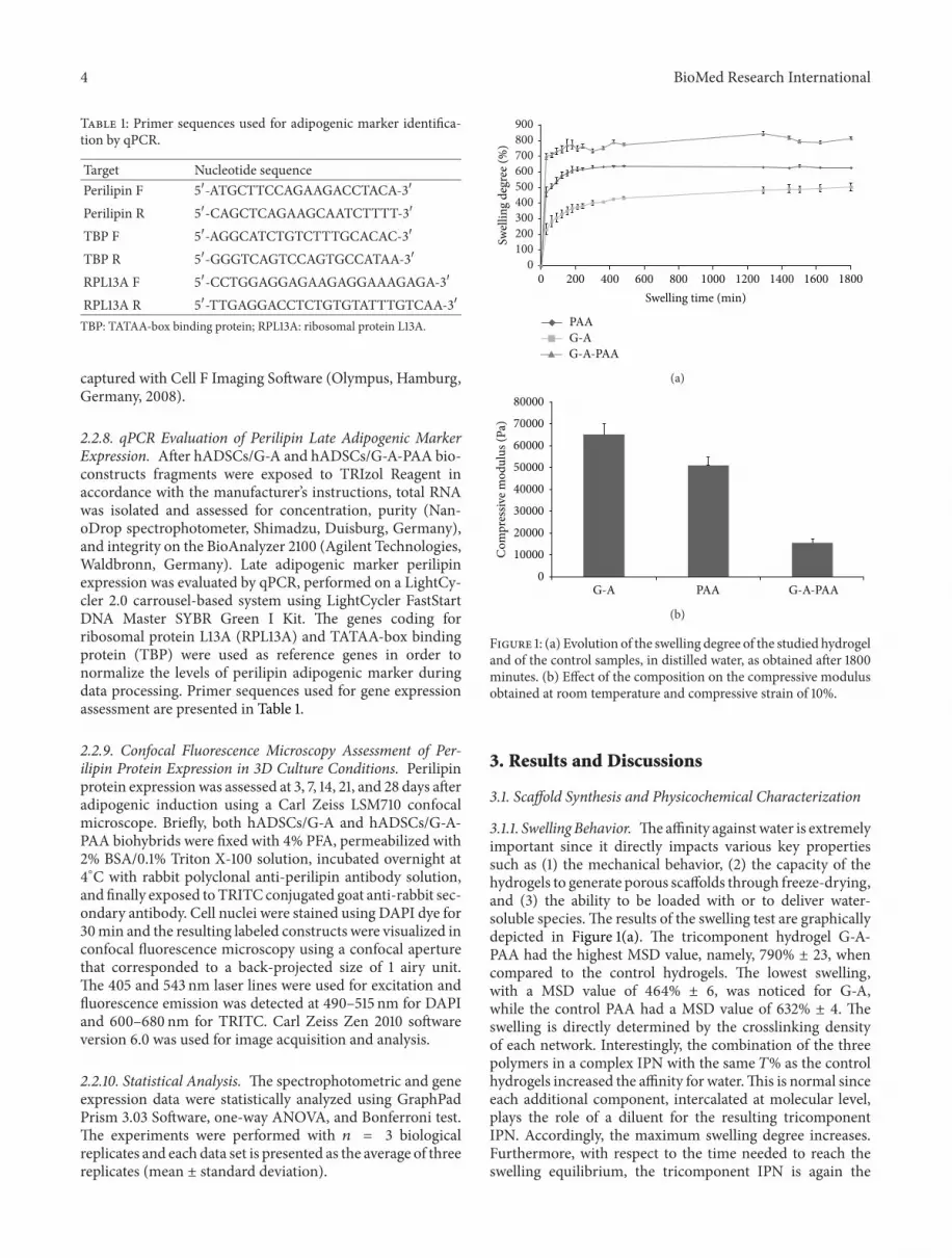
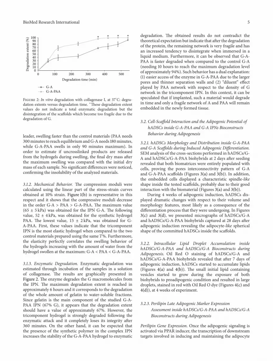
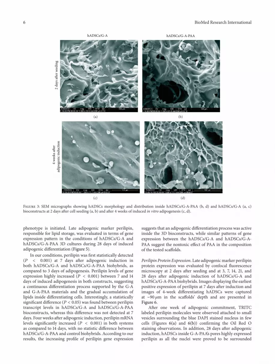
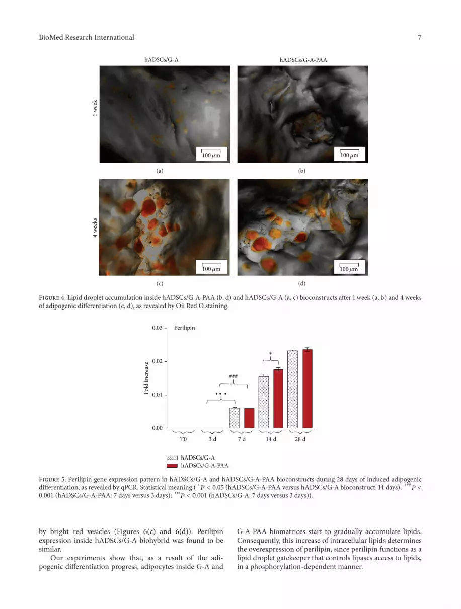
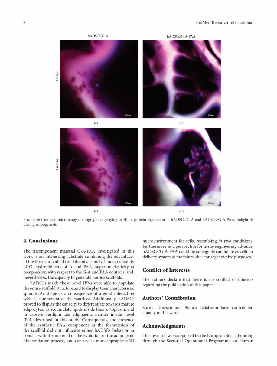