CXCR4-independent rescue of the myeloproliferative defect of the gata1low myelofibrosis mouse model...
10
CXCR4-Independent Rescue of the Myeloproliferative Defect of the Gata1 low Myelofibrosis Mouse Model by Aplidin 1 MARIA VERRUCCI, 1 ALESSANDRO PANCRAZZI, 2 MIGUEL ARACIL, 3 FABRIZIO MARTELLI, 1 PAOLA GUGLIELMELLI, 2 MARIA ZINGARIELLO, 4 BARBARA GHINASSI, 4,5 EMANUELA D’AMORE, 1 JOSE ´ JIMENO, 3 ALESSANDRO M. VANNUCCHI, 2 AND ANNA RITA MIGLIACCIO 1,5 * 1 Department of Hematology, Oncology and Molecular Medicine, Istituto Superiore di Sanita`, Rome, Italy 2 Department of Hematology, University of Florence, Florence, Italy 3 Pharma Mar S.A., Colmenar Viejo, Madrid, Spain 4 Department of Biomorphology, University of Chieti G. D’Annunzio, Chieti, Italy 5 Tisch Cancer Institute, Mount Sinai School of Medicine, New York, New York The discovery of JAK2 mutations in Philadelphia-negative myeloproliferative neoplasms has prompted investigators to evaluate mutation- targeted treatments to restore hematopoietic cell functions in these diseases. However, the results of the first clinical trials with JAK2 inhibitors are not as promising as expected, prompting a search for additional drugable targets to treat these disorders. In this paper, we used the hypomorphic Gata1 low mouse model of primary myelofibrosis (PMF), the most severe of these neoplasms, to test the hypothesis that defective marrow hemopoiesis and development of extramedullary hematopoiesis in myelofibrosis is due to insufficient p27 Kip1 activity and is treatable by Aplidin 1 , a cyclic depsipeptide that activates p27 Kip1 in several cancer cells. Aplidin 1 restored expression of Gata1 and p27 Kip1 in Gata1 low hematopoietic cells, proliferation of marrow progenitor cells in vitro and maturation of megakaryocytes in vivo (reducing TGF-b/VEGF levels released in the microenvironment by immature Gata1 low megakaryocytes). Microvessel density, fibrosis, bone growth, and marrow cellularity were normal in Aplidin 1 -treated mice and extramedullary hematopoiesis did not develop in liver although CXCR4 expression in Gata1 low progenitor cells remained low. These results indicate that Aplidin 1 effectively alters the natural history of myelofibrosis in Gata1 low mice and suggest this drug as candidate for clinical evaluation in PMF. J. Cell. Physiol. 225: 490–499, 2010. ß 2010 Wiley-Liss, Inc. Primary myelofibrosis (PMF) is a myeloproliferative neoplasm characterized by abnormalities in the interaction between the hematopoietic stem/progenitor cells and their bone marrow niches which lead to increased stem/progenitor cell trafficking and development of extramedullary hematopoiesis (Tefferi, 2000). Although the dominant positive JAK2 V617F mutation recently described in patients with myeloproliferative neoplasms is present only in approximately 50% of PMF patients (Zhan and Spivak, 2009), new data are emerging on additional molecular abnormalities associated with the disease. The molecular signature of PMF includes epigenetic absent/reduced expression of CXCR4 in stem/progenitor cells (Shi et al., 2007; Bogani et al., 2008) [CXCR4 is the receptor for the chemokine SDF1, also known as CXCL12, required for interaction of these cells with the marrow vascular niche (Broxmeyer, 2008)] and reduced levels of GATA1 in megakaryocytes (MK) (Vannucchi et al., 2005b) [GATA1 is a transcription factor essential for MK maturation (Orkin and Zon, 2008)]. Therefore in PMF, MK proliferate and accumulate in great numbers in the marrow releasing numerous cytokines (TGF-b, VEGF, osteoprotegerin/ BMP4, etc.) which activate stromal cells leading to fibrosis (fibroblasts), angiogenesis (endothelial cells), and osteosclerosis (osteoblasts) (Lataillade et al., 2008). These observations suggest that treatment of PMF may require drugs that target both stem cell and microenvironmental functions (Lataillade et al., 2008; Lane et al., 2009). In an attempt to understand the pathogenesis of PMF, several mouse mutants have been generated as models for the disease (reviewed in Varricchio et al., 2009). Transgenic mice carrying the JAK2 V617F mutation readily develop, based on genetic background and dosage of JAK2 V617F expression, the myeloproliferative neoplasms polycythemia vera and essential thrombocythemia (Varricchio et al., 2009). These mice may develop secondary myelofibrosis but do not represent good Conflict of interest disclosure: AP, FM, PG, MZ, BG, E D’A, AMV, and ARM declare no competing financial interest. MA and JJ are/ were employed by Pharma Mar and MV received a fellowship from Pharma Mar. Additional Supporting Information may be found in the online version of this article. Contract grant sponsor: Ministero per la Ricerca Scientifica, Alleanza sul Cancro. Contract grant sponsor: Toscana Life Foundation, Italy. Contract grant sponsor: National Cancer Institute; Contract grant number: P01-CA108671. Contract grant sponsor: NY STAR, NY, USA. Contract grant sponsor: Pharma Mar S.A.. Jose ´ Jimeno’s present address is Pangaea Biotech SA, Barcelona, Spain. *Correspondence to: Anna Rita Migliaccio, Department of Medicine, Mount Sinai School of Medicine, One Gustave L. Levy Place, Box #1079, New York, NY 10029. E-mail: [email protected] Received 5 March 2010; Accepted 21 April 2010 Published online in Wiley Online Library (wileyonlinelibrary.com). 10 May 2010. DOI: 10.1002/jcp.22228 ORIGINAL ARTICLE 490 Journal of Journal of Cellular Physiology Cellular Physiology ß 2010 WILEY-LISS, INC.
-
Upload
independent -
Category
Documents
-
view
3 -
download
0
Transcript of CXCR4-independent rescue of the myeloproliferative defect of the gata1low myelofibrosis mouse model...
ORIGINAL ARTICLE 490J o u r n a l o fJ o u r n a l o f
CellularPhysiologyCellularPhysiology
CXCR4-Independent Rescue ofthe Myeloproliferative Defect ofthe Gata1low MyelofibrosisMouse Model by Aplidin1
MARIA VERRUCCI,1 ALESSANDRO PANCRAZZI,2 MIGUEL ARACIL,3 FABRIZIO MARTELLI,1
PAOLA GUGLIELMELLI,2 MARIA ZINGARIELLO,4 BARBARA GHINASSI,4,5
EMANUELA D’AMORE,1 JOSE JIMENO,3 ALESSANDRO M. VANNUCCHI,2
AND ANNA RITA MIGLIACCIO1,5*1Department of Hematology, Oncology and Molecular Medicine, Istituto Superiore di Sanita, Rome, Italy2Department of Hematology, University of Florence, Florence, Italy3Pharma Mar S.A., Colmenar Viejo, Madrid, Spain4Department of Biomorphology, University of Chieti G. D’Annunzio, Chieti, Italy5Tisch Cancer Institute, Mount Sinai School of Medicine, New York, New York
The discovery of JAK2 mutations in Philadelphia-negative myeloproliferative neoplasms has prompted investigators to evaluate mutation-targeted treatments to restore hematopoietic cell functions in these diseases. However, the results of the first clinical trials with JAK2inhibitors are not as promising as expected, prompting a search for additional drugable targets to treat these disorders. In this paper, weused the hypomorphic Gata1low mouse model of primary myelofibrosis (PMF), the most severe of these neoplasms, to test the hypothesisthat defective marrow hemopoiesis and development of extramedullary hematopoiesis in myelofibrosis is due to insufficient p27Kip1
activity and is treatable by Aplidin1, a cyclic depsipeptide that activates p27Kip1 in several cancer cells. Aplidin1 restored expression ofGata1 and p27Kip1 in Gata1low hematopoietic cells, proliferation of marrow progenitor cells in vitro and maturation of megakaryocytesin vivo (reducing TGF-b/VEGF levels released in the microenvironment by immature Gata1low megakaryocytes). Microvessel density,fibrosis, bone growth, and marrow cellularity were normal in Aplidin1-treated mice and extramedullary hematopoiesis did not develop inliver although CXCR4 expression in Gata1low progenitor cells remained low. These results indicate that Aplidin1 effectively alters thenatural history of myelofibrosis in Gata1low mice and suggest this drug as candidate for clinical evaluation in PMF.
J. Cell. Physiol. 225: 490–499, 2010. � 2010 Wiley-Liss, Inc.
Conflict of interest disclosure: AP, FM, PG, MZ, BG, E D’A, AMV,and ARM declare no competing financial interest. MA and JJ are/were employed by Pharma Mar and MV received a fellowship fromPharma Mar.
Additional Supporting Information may be found in the onlineversion of this article.
Contract grant sponsor: Ministero per la Ricerca Scientifica,Alleanza sul Cancro.Contract grant sponsor: Toscana Life Foundation, Italy.Contract grant sponsor: National Cancer Institute;Contract grant number: P01-CA108671.Contract grant sponsor: NY STAR, NY, USA.Contract grant sponsor: Pharma Mar S.A..
Jose Jimeno’s present address is Pangaea Biotech SA, Barcelona,Spain.
*Correspondence to: Anna Rita Migliaccio, Department ofMedicine, Mount Sinai School of Medicine, One Gustave L. LevyPlace, Box #1079, New York, NY 10029.E-mail: [email protected]
Received 5 March 2010; Accepted 21 April 2010
Published online in Wiley Online Library(wileyonlinelibrary.com). 10 May 2010.DOI: 10.1002/jcp.22228
Primary myelofibrosis (PMF) is a myeloproliferative neoplasmcharacterized by abnormalities in the interaction between thehematopoietic stem/progenitor cells and their bone marrowniches which lead to increased stem/progenitor cell traffickingand development of extramedullary hematopoiesis (Tefferi,2000). Although the dominant positive JAK2 V617F mutationrecently described in patients with myeloproliferativeneoplasms is present only in approximately 50% of PMF patients(Zhan and Spivak, 2009), new data are emerging on additionalmolecular abnormalities associated with the disease. Themolecular signature of PMF includes epigenetic absent/reducedexpression of CXCR4 in stem/progenitor cells (Shi et al., 2007;Bogani et al., 2008) [CXCR4 is the receptor for the chemokineSDF1, also known as CXCL12, required for interaction of thesecells with the marrow vascular niche (Broxmeyer, 2008)] andreduced levels of GATA1 in megakaryocytes (MK) (Vannucchiet al., 2005b) [GATA1 is a transcription factor essential for MKmaturation (Orkin and Zon, 2008)]. Therefore in PMF, MKproliferate and accumulate in great numbers in the marrowreleasing numerous cytokines (TGF-b, VEGF, osteoprotegerin/BMP4, etc.) which activate stromal cells leading tofibrosis (fibroblasts), angiogenesis (endothelial cells), andosteosclerosis (osteoblasts) (Lataillade et al., 2008). Theseobservations suggest that treatment of PMF may require drugsthat target both stem cell and microenvironmental functions(Lataillade et al., 2008; Lane et al., 2009).
In an attempt to understand the pathogenesis of PMF, severalmouse mutants have been generated as models for the disease(reviewed in Varricchio et al., 2009). Transgenic mice carryingthe JAK2 V617F mutation readily develop, based on genetic
� 2 0 1 0 W I L E Y - L I S S , I N C .
background and dosage of JAK2 V617F expression, themyeloproliferative neoplasms polycythemia vera and essentialthrombocythemia (Varricchio et al., 2009). These mice maydevelop secondary myelofibrosis but do not represent good
R E S C U E O F G a t a 1 l o w M Y E L O F I B R O S I S B Y A P L I D I N 1 491
models for primary PMF. Models more clearly resembling PMFhave been developed based on mutations that interfere witheither the extrinsic (thrombopoietin, TPO, its receptor,MPL and LNK, a protein that sequesters MPL preventing itstrafficking to the cell membrane) or intrinsic (the transcriptionfactor GATA1) control of megakaryopoiesis. The TPOhigh
model dies of aggressive myelofibrosis within a few months(Varricchio et al., 2009) and is therefore unsuitable as a tool toidentify treatment strategies. The observation that the TPOhigh
mutation reduces Gata1 expression in MK (Vannucchi et al.,2005a) led to the discovery that deletion of the regulatorysequences that control Gata1 expression in these cells(hypomorphic Gata1low mutation) also induces myelofibrosiswith age (Martelli et al., 2005). Gata1low mice have a life spanmore than 2 years and develop the disease in precise sequentialstages (Martelli et al., 2005) (Fig. 1). Mice are born anemic andthrombocytopenic and recover from anemia at 1 month bydeveloping extramedullary hematopoiesis in the spleen. Themice remain thrombocytopenic and develop increased boneformation at 1 month, fibrosis and increased angiogenesis at6–8 months, and increased hematopoietic stem/progenitorcell trafficking and extramedullary hematopoiesis in liver at10 months (Martelli et al., 2005). The normal life span ofGata1low mice in spite of hemopoietic failure in their marrow isprobably due to the efficiency of the spleen as anextramedullary hematopoietic site in response to stress(Migliaccio et al., 2009). This precisely timed diseaseprogression makes these mice suitable for identification ofdrugs that can alter the natural history of the disease. In fact,although the initiating lesion of PMF may not involve the Gata1gene (Vannucchi et al., 2005b), the phenotype of these miceclosely recapitulates the human disease because Gata1low
mice express the same MK, stem/progenitor cell, andmicroenvironmental abnormalities expressed by PMF patients(Migliaccio et al., 2008). Gata1low stem/progenitor cells, inparticular, lack CXCR4 expression (Migliaccio et al., 2008)while their marrow microenvironment expresses high levels ofVEGF, TGF-b, CXCL12, and other growth factors known toactivate stromal cells (Varricchio et al., 2009). Therefore,in these mice, as in PMF patients, defective hematopoiesis inthe marrow, abnormal stem/progenitor cell trafficking, anddevelopment of hematopoiesis in extramedullary sites isprobably the result of alterations in both stem cells (that lackCXCR4 expression) (Migliaccio et al., 2008) and theirmicroenvironment (increased osteoblast and endothelial cellproliferation in the bone marrow) (Varricchio et al., 2009). Thecyclic depsipeptide Aplidin1 originally isolated from aMediterranean marine tunicate, Aplidium albicans and presentlyproducted by synthesis, is undergoing late phase II clinical trialsfor solid tumors, myeloma and T-cell lymphoma, with noreported marrow toxicity (Ferme et al., 2008; Mateos et al.,2008). Aplidin1 does not inhibit colony formation from normalhuman progenitor cells (Depenbrock et al., 1998; Albella et al.,2002; Gomez et al., 2003) but induces cell cycle arrest andapoptosis of many tumor cell types (Munoz-Alonso et al., 2009).The ability of tumor cells to respond to Aplidin1 is predicted byreduced expression of p27kip1 (Moneo et al., 2007), an inhibitorof cyclin D (Ray et al., 2009) necessary to maintain stem cells inquiescence (Cheng et al., 2000). Low concentrations ofAplidin1 blocked G1-to-S transition by decreasing the level ofthe cyclin D1/cyclin-dependent kinase (Cdk)4/p21 complex,leading to an increase in the levels of hypophosphorylatedretinoblastoma protein (Rb-1) in undifferentiated, anaplasticthyroid carcinoma cells (Bravo et al., 2005), induced G1 and G2/M arrest and decreased the expression of cyclins A and B in SK-MEL-28 and UACC-257 melanoma cell lines (Munoz-Alonsoet al., 2008), and decreased cyclin D1 and Cdk4 in murinemyeloma 5T33MM cells (Caers et al., 2008). The direct effectsof Aplidin1 include rapid (within 1 min) activation of Rac1
JOURNAL OF CELLULAR PHYSIOLOGY
(Gonzalez-Santiago et al., 2006; Munoz-Alonso et al., 2008),a member of the guanine triphosphatase family activated bythe canonical Wnt signaling (Wu et al., 2008) that exertsmultiple functions on hematopoietic stem/progenitor cells.Rac1 facilitates stem cell engraftment in the marrow andincreases p27Kip1 expression (Gu et al., 2003; Walmsley et al.,2003; Cancelas et al., 2005). Indirect effects of Aplidin1 oncancer growth are related to anti-angiogenic properties(Broggini et al., 2003; Biscardi et al., 2005; Straight et al., 2006).In vivo, Aplidin1 inhibits spontaneous as well as VEGF- andFGF-2-induced angiogenesis in the chick allantoid assay(Taraboletti et al., 2004; Straight et al., 2006; Mitsiades et al.,2008). In vitro, Aplidin1 interferes with endothelial cell growthby inhibiting both expression and signaling of VEGF (Brogginiet al., 2003; Mitsiades et al., 2008). Since Gata1low stem/progenitor cells were found to express low levels of p27kip1
(this article), we hypothesized that Aplidin1, by activating Rac1and p27kip1 in stem/progenitor cells and by reducing the numberof vascular niches, might restore both stem cell andmicroenvironmental function in this mouse model of PMF.
To test this hypothesis, Gata1low mice were treated withconsecutive Aplidin1 cycles and evaluated for diseaseprogression. Aplidin1 treatment increased expression ofp27kip1 and Gata1 in stem/progenitor cells, reduced TGF-b andVEGF production, and inhibited angiogenesis. In addition,Gata1low stem/progenitor cells from treated mice generatedGata1pos MK in the marrow, increasing peripheral bloodplatelets counts by threefold. Normal cellularity and reducedfibrosis and bone formation were also observed in Aplidin1-treated animals. These results suggest that Aplidin1 altered thenatural history of myelofibrosis in Gata1low mice by restoringboth the microenvironmental and stem/progenitor cell defects,providing a rationale for clinical evaluation of Aplidin1 in PMFpatients.
Materials and MethodsMice
Male wild-type and Gata1low mice were generated in the animalfacility of Istituto Superiore di Sanita as described (Martelli et al.,2005). All the experiments were performed with sex- andage-matched mice under protocols approved by the institutionalanimal care committee.
Drug formulation
Vials containing Aplidin1 (0.5 mg/vial) and mannitol (as additive,25 mg/vial) were reconstituted with 1 ml of reconstitution solution(cremophor EL/ethanol/water, 15:15:70 v/v/v) that was furtherdiluted with 9 mg/ml (0.9% w/v) of sodium chloride.
Drug treatment
Gata1low mice were divided into three age groups: disease-free(1–6 months old), early myelofibrosis (8–12 months old, whenpresence of the disease is detectable only in bone marrow), andmyelofibrosis (15 months old to natural death, when the completeclinical picture of the human disease is manifested) (Martelli et al.,2005). Three experiments with slightly different doses andschedules were performed (see Fig. 1 and Supplemental Fig. 1 fordetails). Briefly, Gata1low mice were individually weighed andinjected intraperitoneally (i.p.) with Aplidin1 or with equivalentvolumes of vehicle. The dose of Aplidin1 administered wasadjusted on the basis of weight with each animal receiving 60mg ofAplidin1/kg/day for 9 days for either one cycle (experiment 1) ortwo cycles 28 days apart (experiment 2), or 100mg of Aplidin1/kg/day for five consecutive days for two or four cycles 21 days apart(experiment 3). Since the three separate experiments gave similarresults, only the third experiment is described in detail for clarity.

Fig. 1. Aplidin1 treatment is well tolerated in mice and modestly affects body weight. A: Natural history of myelofibrosis in Gata1low mice(modified from Varricchio et al., 2009) and schedule of Aplidin1 treatment used in the experiments presented. Treatment of Gata1low mice wasinitiated at 10 months of age. The arrows and asterisk indicate day of blood sampling and mice sacrifice, respectively. B: Kaplan–Meier analyses ofsurvival of control Gata1low mice (untreated or injected with vehicle, black) and of Gata1low mice treated with Aplidin1 for four cycles (blue).The number of mice included in each group is indicated in parenthesis. C: Body weight of Gata1low mice treated either with vehicle (veh, blue) orwith Aplidin1 (Apl, shaded blue) during the course of the experiment. The day (d) of Aplidin1 treatment is indicated on the x-axis. Results arepresented as mean (WSD) determinations of at least three mice per experimental group and are compared to historic determinations of bodyweight of untreated wild-type (blue shaded area) and Gata1low (yellow shaded area) mice of comparable age. Values statistically different from thecorresponding vehicle group are indicated by M (P < 0.05).
JOURNAL OF CELLULAR PHYSIOLOGY
492 V E R R U C C I E T A L .

R E S C U E O F G a t a 1 l o w M Y E L O F I B R O S I S B Y A P L I D I N 1 493
Aplidin1 reduced animal mobility and normal drinking behavior.Therefore, in the second and third experiments mice were injectedwith 200ml of sodium chloride (0.9% w/v) subcutaneously toprevent dehydration.
Hematological parameters
Blood was collected from the retro-orbital plexus intoethylenediaminetetraacetic acid (EDTA)-coated microcapillarytubes (20–40ml/sampling). Hematocrit (Hct), platelet (plt), andwhite blood cell (WBC) counts were determined manually.
Flow cytometry and cell sorting
Cells obtained from bone marrow were counted manually byTrypan Blue (0.4% w/v, Sigma, St. Louis, MO) staining and incubatedfor 30 min on ice first with a Fcg blocker (CD16/CD32) and thenwith phycoerythrin (PE)-conjugated anti-CD34, fluoresceinisothiocyanate (FITC)-conjugated anti-CXCR4, andallophycocyanin (APC)-conjugated anti-CD117 (cKit) (all fromPharMingen, San Diego, CA). Dead cells and non-specific signalswere excluded by propidium iodide staining (5mg/ml, Sigma) andappropriate isotype controls (PharMingen). Cells in theprospective stem/progenitor cell gates deprived of lineage positivecells (cKitpos/CD34pos or CD34neg) were isolated by sorting withan ARIA cell sorter (Becton Dickinson, Franklin Lakes, NJ) (>90%pure based upon reanalysis) (Migliaccio et al., 2008).
Progenitor cell determination
Prospectively isolated progenitor cells (1,000 cells/ml) andunfractionated liver cells (5� 104 cells/ml) were cultured inmethylcellulose cultures (0.9% w/v) containing fetal bovine serum(30% v/v, Sigma) and ratSCF (100 ng/ml), mouseIL3 (10 ng/ml),G-CSF and GM-CSF (50 ng/ml in both cases) (all from Sigma), andhuman erythropoietin (2 U/ml) (Roche Welwyn Garden City,Hertfordshire, UK). Cultures were incubated at 378C in ahumidified incubator containing 5% CO2 in air and scored for thepresence of hematopoietic colonies on day 8 (Ghinassi et al., 2007;Migliaccio et al., 2009).
Histology
Femurs, tibias, and liver sections were stained with hematoxylin–eosin, Gomory-silver (MicroStain MicroKit, Diapath, Bologna,Italy) or Mallory-trichromic staining (Migliaccio et al., 2009). Forimmunohistochemistry, sections were incubated with an anti-Gata1 antibody (Santa Cruz Biotechnology, Santa Cruz, CA)(Migliaccio et al., 2009) and the immunoreaction detected with theavidin–biotin immunoperoxidase system (Vectastain Elite ABC Kit;Vector Laboratories, Burlingame, CA) on slides counterstainedwith hematoxylin–eosin. Histological observations were carriedout using a ZEISS AXIOSKOPE light microscope (Jena, Germany)equipped with a Coolsnap Videocamera and the acquired imageswere analyzed with the MetaMorph 6.1 Software (UniversalImaging Corp, Downingtown, PA). Levels of Gata1 immunostainingper MK, numbers of reticulinic fibers and Mallory positive areaswere determined by analyzing with the MethaMorph 6.1 programfive randomly chosen non-overlapping areas per each section. Formicrovessel density determination, sections from the femur wereincubated with a rabbit anti-mouse CD34 antibody (cloneMEC14.7, code CL8927AP, Cedarlane Laboratories, Burlington,NC) or a rabbit anti-mouse CD31 (Becton Dickinson) (dilution1:200 for both). Antibody dilution and blocking of non-specificbinding sites was performed with Ultra V-Block (code TA-060-UB,Thermo Scientific, Walthman, MA). Secondary labeled antibodyincubation and stain development was performed usingSuperPicTure Kit HRP Broad Spectrum (code 87–8963, Histo-LineLaboratories, Milano, Italy, dilution 1:1). Microvessel density was
JOURNAL OF CELLULAR PHYSIOLOGY
calculated as the mean number of stained vessels per 400� highpower field, calculating the mean of five randomly chosen non-overlapping areas. Frequency of MK and percentage of Gata1pos
MK were similarly obtained. Counting was performed by twoseparate investigators in a blinded fashion.
Quantitative RT-PCR analysis
Total RNA was prepared by lysing single erythroid colonies orprospectively isolated cell populations into Trizol (Gibco BRL,Paisley, UK). RNA was reverse transcribed with 2.5mM randomhexamers using the superscript kit (Invitrogen, Milan, Italy) and geneexpression levels quantified by Real Time RT-PCR, as described(Ghinassi et al., 2007; Migliaccio et al., 2009). Beta-2 microglobulincDNA was amplified as an internal standard. Reactions wereperformed in an ABI PRISM 7700 Sequence Detection System(Applied Biosystems, Carlsband, CA). Cycle threshold (Ct) werecalculated with the SDS software and mRNA levels expressed as2�DCt (DCt ¼ target gene Ct � Beta � 2 microglobulin CtD).
Statistical analysis
Statistical analysis was performed by analysis of variance (ANOVAtest) using Origin 3.5 software for Windows (Microcal Software,Inc., Northampton, MA) or by Kruskal–Wallis and Bonferroni–Dunn’s post hoc and Mann–Whiteny’s tests using GraphPad Prism4.0, as appropriate.
ResultsAplidin1 treatment is well tolerated by Gata1low miceand causes only modest reduction in body weight
Gata1low mice were treated for two or four cycles of Aplidin1
and sacrificed for analysis 14 days after the second or fourthcycle (Fig. 1A and Supplemental Fig. 1). The slightly lowersurvival rate (1 death out of 10 treated mice, 85%) of theAplidin1-treated mice with respect to controls (5 out of 70mice, 93%) is not statistically significant. In addition, death in thetreated group occurred during the first cycle of therapy while inthe control mice mortality was observed throughout the periodof observation and was consistent with the natural course of thedisease (Martelli et al., 2005).
Body weight was monitored as a measure of morbidity/toxicity (Teicher and Andrews, 2004). Gata1low mice areslightly smaller than wild-type littermates but the reduction isnot statistically significant (P> 0.05) (Fig. 1C). Small (10–15%)but significant reductions in body weight were observed inthe Aplidin1-treated Gata1low animals which persistedthroughout treatment (P< 0.05). These reductions arebelow the threshold that define toxicity (20%) and are likelydue to the improvement in mobility observed in the treatedanimals. No sign of organ toxicity was observed upon postmortem examination of the sacrificed mice by the end of thetreatment.
Progenitor cells prospectively isolated from the marrowof Gata1low mice express reduced levels of p27Kip1 whichpredicts Aplidin1 responsiveness
Since functional GATA binding sites are present in theregulatory regions of p27kip1 (Fukuchi et al., 2006), it isconceivable that progenitor cells carrying the hypomorphicGata1low mutation express reduced levels of p27Kip1 and,therefore, meet the criteria for Aplidin1 responsiveness.To test this hypothesis, progenitor cells were prospectivelyisolated from the marrow of Gata1low and wild-type littermatesand the levels of p27Kip1 expressed by the various populationscompared (Fig. 2A). Progenitor cells were identified on the
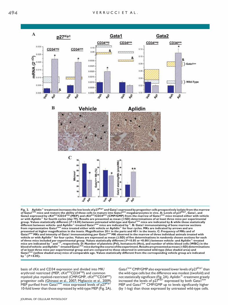
Fig. 2. Aplidin1 treatment increases the low levels of p27Kip1 and Gata1 expressed by progenitor cells prospectively isolate from the marrowof Gata1low mice and restore the ability of these cells to mature into Gata1pos megakaryocytes in vivo. A: Levels of p27Kip1, Gata1, andGata2 expressed by cKitpos/CD34neg (MEP) and cKitpos/CD34pos (CMP/GMP) from the marrow of Gata1low mice treated either with vehicleor with Aplidin1 for fourth cycles (day 79). Results are presented as mean (WSD) determinations of at least three mice per experimentalgroup. Values statistically different (P < 0.05) between untreated wild-type and Gata1low mice are indicated by & while those statisticallydifferent between vehicle- and Aplidin1-treated Gata1low mice are indicated by M. B: Gata1 immunostaining of bone marrow sectionsfrom representative Gata1low mice treated either with vehicle or Aplidin1 for four cycles. MKs are indicated by arrows and arepresented at higher magnification in the insets. Magnification 20T in the parts and 40T in the insets. C: Frequency of MKs and ofGata1pos MKs and intensity of Gata1 immunostaining per Gata1pos MK observed in the marrow of three individual animals treated withvehicle or with Aplidin1 for four cycles. Values are expressed as mean (WSD) of five determinations in randomly chosen sections for eachof three mice included per experimental group. Values statistically different (P < 0.05 or <0.001) between vehicle- and Aplidin1-treatedmice are indicated by M and MM, respectively. D: Number of platelets (Ptl), hematocrit (Hct), and number of white blood cells (WBC) in thebloodofvehicle-andAplidin1-treatedGata1lowmiceduringthecourseoftheexperiment.Resultsarepresentedasmean(WSD)determinationsof at least three mice per experimental group and are compared to those observed in untreated wild-type (blue shaded area) andGata1low (yellow shaded area) mice of comparable age. Values statistically different from the corresponding vehicle group are indicatedby M (P < 0.05).
494 V E R R U C C I E T A L .
basis of cKit and CD34 expression and divided into MK/erythroid restricted (MEP, cKitposCD34neg) and commonmyeloid plus myeloid-restricted (CMP/GMP, cKitposCD34pos)progenitor cells (Ghinassi et al., 2007; Migliaccio et al., 2009).MEP purified from Gata1low mice expressed levels of p27Kip1
10-fold lower than those expressed by wild-type MEP (Fig. 2A).
JOURNAL OF CELLULAR PHYSIOLOGY
Gata1low CMP/GMP also expressed lower levels of p27Kip1 thanthe wild-type cells but the difference was modest (twofold) andnot statistically significant (Fig. 2A). Aplidin1-treatment greatlyincreased the levels of p27Kip1 expressed by both Gata1low
MEP and Gata1low CMP/GMP up to levels significantly higher(by 1-log) than those expressed by untreated wild-type cells.
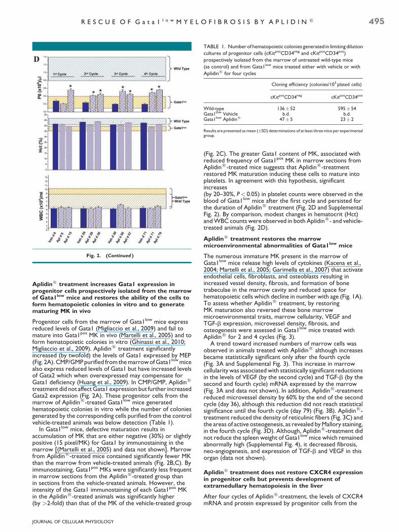
TABLE 1. Number of hematopoietic colonies generated in limiting dilution
cultures of progenitor cells (cKitposCD34neg and cKitposCD34pos)
prospectively isolated from the marrow of untreated wild-type mice
(as control) and from Gata1low mice treated either with vehicle or with
Aplidin1 for four cycles
Cloning efficiency (colonies/103 plated cells)
cKitposCD34neg cKitposCD34pos
Wild-type 136� 52 595� 54Gata1low Vehicle b.d. b.d.Gata1low Aplidin1 47� 5 23� 2
Results are presented as mean (�SD) determinations of at least three mice per experimentalgroup.
Fig. 2. (Continued )
R E S C U E O F G a t a 1 l o w M Y E L O F I B R O S I S B Y A P L I D I N 1 495
Aplidin1 treatment increases Gata1 expression inprogenitor cells prospectively isolated from the marrowof Gata1low mice and restores the ability of the cells toform hematopoietic colonies in vitro and to generatematuring MK in vivo
Progenitor cells from the marrow of Gata1low mice expressreduced levels of Gata1 (Migliaccio et al., 2009) and fail tomature into Gata1pos MK in vivo (Martelli et al., 2005) and toform hematopoietic colonies in vitro (Ghinassi et al., 2010;Migliaccio et al., 2009). Aplidin1 treatment significantlyincreased (by twofold) the levels of Gata1 expressed by MEP(Fig. 2A). CMP/GMP purified from the marrow of Gata1low micealso express reduced levels of Gata1 but have increased levelsof Gata2 which when overexpressed may compensate forGata1 deficiency (Huang et al., 2009). In CMP/GMP, Aplidin1
treatment did not affect Gata1 expression but further increasedGata2 expression (Fig. 2A). These progenitor cells from themarrow of Aplidin1-treated Gata1low mice generatedhematopoietic colonies in vitro while the number of coloniesgenerated by the corresponding cells purified from the controlvehicle-treated animals was below detection (Table 1).
In Gata1low mice, defective maturation results inaccumulation of MK that are either negative (30%) or slightlypositive (15 pixel/MK) for Gata1 by immunostaining in themarrow [(Martelli et al., 2005) and data not shown]. Marrowfrom Aplidin1-treated mice contained significantly fewer MKthan the marrow from vehicle-treated animals (Fig. 2B,C). Byimmunostaining, Gata1pos MKs were significantly less frequentin marrow sections from the Aplidin1-treated group thanin sections from the vehicle-treated animals. However, theintensity of the Gata1 immunostaining of each Gata1pos MKin the Aplidin1-treated animals was significantly higher(by >2-fold) than that of the MK of the vehicle-treated group
JOURNAL OF CELLULAR PHYSIOLOGY
(Fig. 2C). The greater Gata1 content of MK, associated withreduced frequency of Gata1pos MK in marrow sections fromAplidin1-treated mice suggests that Aplidin1-treatmentrestored MK maturation inducing these cells to mature intoplatelets. In agreement with this hypothesis, significantincreases(by 20–30%, P< 0.05) in platelet counts were observed in theblood of Gata1low mice after the first cycle and persisted forthe duration of Aplidin1 treatment (Fig. 2D and SupplementalFig. 2). By comparison, modest changes in hematocrit (Hct)and WBC counts were observed in both Aplidin1- and vehicle-treated animals (Fig. 2D).
Aplidin1 treatment restores the marrowmicroenvironmental abnormalities of Gata1low mice
The numerous immature MK present in the marrow ofGata1low mice release high levels of cytokines (Kacena et al.,2004; Martelli et al., 2005; Garimella et al., 2007) that activateendothelial cells, fibroblasts, and osteoblasts resulting inincreased vessel density, fibrosis, and formation of bonetrabeculae in the marrow cavity and reduced space forhematopoietic cells which decline in number with age (Fig. 1A).To assess whether Aplidin1 treatment, by restoringMK maturation also reversed these bone marrowmicroenvironmental traits, marrow cellularity, VEGF andTGF-b expression, microvessel density, fibrosis, andosteogenesis were assessed in Gata1low mice treated withAplidin1 for 2 and 4 cycles (Fig. 3).
A trend toward increased numbers of marrow cells wasobserved in animals treated with Aplidin1 although increasesbecame statistically significant only after the fourth cycle(Fig. 3A and Supplemental Fig. 3). This increase in marrowcellularity was associated with statistically significant reductionsin the levels of VEGF (by the second cycle) and TGF-b (by thesecond and fourth cycle) mRNA expressed by the marrow(Fig. 3A and data not shown). In addition, Aplidin1-treatmentreduced microvessel density by 60% by the end of the secondcycle (day 36), although this reduction did not reach statisticalsignificance until the fourth cycle (day 79) (Fig. 3B). Aplidin1-treatment reduced the density of reticulinic fibers (Fig. 3C) andthe areas of active osteogenesis, as revealed by Mallory staining,in the fourth cycle (Fig. 3D). Although, Aplidin1-treatment didnot reduce the spleen weight of Gata1low mice which remainedabnormally high (Supplemental Fig. 4), it decreased fibrosis,neo-angiogenesis, and expression of TGF-b and VEGF in thisorgan (data not shown).
Aplidin1 treatment does not restore CXCR4 expressionin progenitor cells but prevents development ofextramedullary hematopoiesis in the liver
After four cycles of Aplidin1-treatment, the levels of CXCR4mRNA and protein expressed by progenitor cells from the

Fig. 3. Aplidin1 treatment increases cellularity and decreases VEGF and TGF-b expression, vessel density, fiber and bone formation in themarrowofGata1lowmice.A:NumberofcellsandVEGFandTGF-bexpressioninthefemurofvehicle-andAplidin1-treatedGata1lowmiceafterthesecond and fourth cycle. mRNA levels were determined by quantitative RT-PCR and expressed as ratio with the levels of expression in the vehiclecontrol group. Results are presented as mean (WSD) determinations of at least three mice per experimental group. Values statistically differentfrom the corresponding vehicle group are indicated by M (P < 0.05). B: Microvessel density (by CD34-staining), (C) fibrosis (by Gomory-silverstaining), and(D)bone formation(byMallorystaining)of the femur fromGata1low micetreatedeither withvehicleorwithAplidin1 for fourcycles.ResultswerequantifiedwiththeMetaMorph6.1programandpresentedasmean(WSD)determinationswiththreemiceperexperimentalgroupinthe parts on the right. Magnification 20T in (B) and (C) and 10T in (E). Values statistically different (P < 0.05 and 0.0001) from those observed invehicle mice are indicated by M and MM, respectively.
JOURNAL OF CELLULAR PHYSIOLOGY
496 V E R R U C C I E T A L .

Fig. 4. Aplidin1 treatment does not restore CXCR4 expression in the stem/progenitor cells and does not halt progenitor cell trafficking butprevents development of extramedullary hematopoiesis in the liver of Gata1low mice. A: Flow cytometrical determinations of CXCR4 expressionin progenitor cells (CD117pos) of the marrow of wild-type and Aplidin1-treated (4 cycles) Gata1low mice. CXCR4 expression is presented both ascontour plotandashistogramtohighlight thepopulation profilesandthemeanfluorescence intensity, indicatedbyanarrow, respectively. Isotypecontrols for CXCR4 analyses are presented in gray. See Table 2 for further details. B: Levels of CXCR4 mRNA expressed by progenitor cells(cKitposCD34neg and cKitposCD34pos) isolated from the marrow of vehicle- or Aplidin1-treated mice, respectively. The levels of CXCR4 expressedby cells purified from wild-type and Gata1low mice are indicated by the blue and yellow shaded areas, respectively. Values statistically significant(P < 0.05) from those observed in untreated Gata1low mice are indicated by M. All the results observed in Gata1low mice are statistically lower(P < 0.001) thanthoseobserved inwild-typemice.C:Hematoxylin–eosinstainingof liver sections fromavehicle-andanAplidin1-treatedGata1low
mice, respectively.ThepresenceofMKinthe liverparenchymais indicatedbyarrows.Magnification20T.D:Numberofhematopoieticprogenitorcells (CFC) present in the liver from vehicle- and Aplidin1-treated Gata1low mice. Results are presented as mean (WSD) of those observed in threemice per experimental group. The number of progenitor cells present in the liver of untreated wild-type and Gata1low mice of comparableage are indicated by the shaded blue and yellow areas, respectively. Values statistically greater (P < 0.01) than those observed in vehicle-treatedmice are indicated by M.
R E S C U E O F G a t a 1 l o w M Y E L O F I B R O S I S B Y A P L I D I N 1 497
marrow of Gata1low mice remained barely detectable (Fig. 4A,Band Supplemental Table 1).
Hematopoietic cells were not detected by histologicalanalyses in the liver parenchyma of Gata1low mice treated withAplidin1 while numerous MK were observed in theparenchyma of the vehicle-treated control group (Fig. 4C).Because of the underlying extramedullary hematopoiesis, inuntreated and vehicle-treated animals the liver increased in sizeand were spongy in consistency, while size and consistency ofthe liver in Aplidin1-treated mice were normal (result notshown). These observations indicate that Aplidin1-treatment
JOURNAL OF CELLULAR PHYSIOLOGY
prevented development of hematopoiesis in the liver of thesemice. However, greater numbers of colony forming cells weredetectable in the liver of Gata1low mice treated with Aplidin1
(Fig. 4D). Given the extensive vascularization of the liver, thepresence of such great numbers of progenitor cells in this organindicates that Aplidin1 treatment did not prevent, and mayeven have favored, stem/progenitor cell trafficking in theseanimals. These data suggest that increased stem/progenitorcell trafficking induced by lack of CXCR4 expression may benecessary but is not sufficient for extramedullaryhematopoiesis to occur.
498 V E R R U C C I E T A L .
Discussion
Primary myelofibrosis is the most severe of the Philadelphianeg
myeloproliferative neoplasms (Tefferi, 2000; Tefferi et al.,2007). The only effective treatment currently available for thisdisease is bone marrow transplantation (Cervantes et al., 2007),a therapy that can only be provided to eligible patients forwhom suitable donors exist. Palliative treatment may includeremoval of the enlarged spleen. However, splenectomy inpatients with PMF has been associated with a higher incidence ofdevelopment of extramedullary hematopoiesis in liver and ahigher rate of blast transformation (Cervantes et al., 2007).Clinical trials with JAK2 inhibitors in patients with PMF led to adramatic rapid reduction of spleen size but did not improveother hematological parameters and even induced severethrombocytopenia (reviewed in Pardanani, 2008). Alternativestrategies to cure this disease are therefore warranted(Lataillade et al., 2008; Lane et al., 2009).
We have extensively characterized the myelofibroticphenotype expressed by Gata1low mice (Varricchio et al., 2009).Previous studies have determined that the hematopoieticfunctions of Gata1low stem/progenitor cells are exquisitelyrescued by the spleen microenvironment (Migliaccio et al.,2009). Therefore, removal of the spleen from heterozygousGata1low/þ females [since Gata1 is on the X chromosome (Zonet al., 1990), due to the lionization process these females haveboth Gata1low and Gata1þ stem/progenitor cells] restoredwild-type hematopoiesis in the marrow normalizing many of themyelofibrotic traits expressed by these animals (increasedplatelets counts in the blood, restored marrow cellularity, anddecreased angiogenesis and bone formation) (Migliaccio et al.,2009). Therefore, removal of the spleen rescued the stemcell abnormalities of the Gata1low/þ females. However,splenectomy did not restore the cytokine expression profile ofthe marrow which remained abnormally high and, similar to theeffect induced in PMF patients, promoted development ofextramedullary hematopoiesis in liver (Ghinassi et al., 2010).Therefore restoration of wild-type hematopoiesis in themarrow of Gata1low/þ females did not prevent all of the diseasemanifestations indicating that, as suggested for PMF patients(Lataillade et al., 2008; Lane et al., 2009), also in this animalmodel treatment of myelofibrosis may require normalization ofboth stem/progenitor cell and microenvironmental functions.
The dual action of Aplidin1 on cancer growth (induction ofapoptosis of cancer cells and inhibition of angiogenesis) suggeststhat this drug may be effective in altering the natural history ofmyelofibrosis in Gata1low mice. Indeed, Aplidin1-treatmentrescued both the microenvironmental and progenitor cellfunction in the marrow of Gata1low mice preventing these micefrom developing myelofibrosis: it increased blood plateletcounts, induced normal marrow cellularity, decreased fibrosis,angiogenesis, and bone formation and prevented, or at leastdelayed, development of extramedullary hematopoiesis in liver.Decreased angiogenesis, fibrosis, and bone formation were theresult of reduced VEGF, TGF-b and, probably, bone-morphogenic proteins that are released by Gata1low MK(Kacena et al., 2004; Garimella et al., 2007) which are greatlydecreased in number in the marrow of Aplidin1-treatedanimals. Restoration of MK maturation was due to increasedGata1 expression while increases in the expression of bothp27Kip1 (at the mRNA level) and Gata1 (both as mRNA andprotein) probably contributed to rescue the function ofhematopoietic cells from the marrow. Although technicallimitations prevented direct measurements of p27kip1 proteinlevels in this experimental model, Aplidin1 has been reportedto increase both p27kip1 mRNA and protein in sarcoma cell lines(Moneo et al., 2007). The protein p27Kip1 regulates stem/progenitor cell fate decisions with low levels of p27Kip1 favoringproliferation and high levels promoting differentiation (Cheng
JOURNAL OF CELLULAR PHYSIOLOGY
et al., 2000; Messina et al., 2005) and, by affecting microtubulemodeling, inhibits migration, and tissue invasion of sarcoma cells(Baldassarre et al., 2005), suggesting that increased levels ofp27Kip1 expression contributed to rescue the hematopoieticfunctions of Gata1low progenitor cells from the marrow andprevented establishment of hematopoiesis in extramedullarysites. Increased levels of Gata1 expression may havecooperated with p27Kip1 to restore in vitro and in vivohematopoietic functions of the progenitor cells from themarrow. Kinetic studies indicate that the first detectable(within 60 sec) change induced by Aplidin1 in several cancercells is represented by Rac1 activation, a protein that regulatesp27kip1 expression (Gonzalez-Santiago et al., 2006; Munoz-Alonso et al., 2008). The observations that Gata1low stem/progenitor cells express low levels of p27Kip1 and that Aplidin1
restored p27Kip1 expression in these cells, suggest that thephenotype of Gata1low stem/progenitor cells includesinsufficient levels of Rac1 activation. Unfortunately, due to itslow level of basal activity (Gu et al., 2003), levels of Rac1activation in the bone marrow of Gata1low and wild-type micecould not be directly compared. However, in agreement withthe hypothesis that Rac1 is down regulated in Gata1low mice,stem/progenitor cells from Rac1 deficient mice display, as thosefrom Gata1low mice, CXCR4-independent enhancedmobilization and engraft poorly when transplanted into NOD/SCID mice (Gu et al., 2003). It is, therefore, conceivable that,similar to findings observed in other cell systems, Aplidin1
induced p27Kip1 expression in Gata1low cells through Rac1activation.
In conclusion, Aplidin1-treatment was well tolerated byGata1low mice with acceptable toxicity (death) and morbidity(changes in body weight) and prevented development ofmyelofibrosis by partially restoring progenitor cell andmicroenvironmental functions in the marrow. On the basis ofthese encouraging results, the design for a clinical trial withAplidin1 in PMF patients is under development (Dr. A. Tefferi,personal communication, April 2, 2010).
Acknowledgments
This study was supported by Ministero per la RicercaScientifica, Alleanza sul Cancro (ARM and AMV), Toscana LifeFoundation, Italy (AMV), National Cancer Institute (grant no.P01-CA108671), and NY STAR, NY, USA (ARM). MV wassupported by a fellowship from Pharma Mar S.A.
Literature Cited
Albella B, Faircloth G, Lopez-Lazaro L, Guzman C, Jimeno J, Bueren JA. 2002. In vitro toxicityof ET-743 and aplidine, two marine-derived antineoplastics, on human bone marrowhaematopoietic progenitors. Comparison with the clinical results. Eur J Cancer 38:1395–1404.
Baldassarre G, Belletti B, Nicoloso MS, Schiappacassi M, Vecchione A, Spessotto P, MorrioneA, Canzonieri V, Colombatti A. 2005. p27(Kip1)-stathmin interaction influences sarcomacell migration and invasion. Cancer Cell 7:51–63.
Biscardi M, Caporale R, Balestri F, Gavazzi S, Jimeno J, Grossi A. 2005. VEGF inhibition andcytotoxic effect of aplidin in leukemia cell lines and cells from acute myeloid leukemia. AnnOncol 16:1667–1674.
Bogani C, Ponziani V, Guglielmelli P, Desterke C, Rosti V, Bosi A, Le Bousse-Kerdiles MC,Barosi G, Vannucchi AM. 2008. Hypermethylation of CXCR4 promoter in CD34þ cellsfrom patients with primary myelofibrosis. Stem Cells 26:1920–1930.
Bravo SB, Garcia-Rendueles ME, Seoane R, Dosil V, Cameselle-Teijeiro J, Lopez-Lazaro L,Zalvide J, Barreiro F, Pombo CM, Alvarez CV. 2005. Plitidepsin has a cytostatic effect inhuman undifferentiated (anaplastic) thyroid carcinoma. Clin Cancer Res 11:7664–7673.
Broggini M, Marchini SV, Galliera E, Borsotti P, Taraboletti G, Erba E, Sironi M, Jimeno J,Faircloth GT, Giavazzi R, D’Incalci M. 2003. Aplidine, a new anticancer agent of marineorigin, inhibits vascular endothelial growth factor (VEGF) secretion and blocks VEGF-VEGFR-1 (flt-1) autocrine loop in human leukemia cells MOLT-4. Leukemia 17:52–59.
Broxmeyer HE. 2008. Chemokines in hematopoiesis. Curr Opin Hematol 15:49–58.Caers J, Menu E, De Raeve H, Lepage D, Van Valckenborgh E, Van Camp B, Alvarez E,
Vanderkerken K. 2008. Antitumour and antiangiogenic effects of Aplidin in the 5TMMsyngeneic models of multiple myeloma. Br J Cancer 98:1966–1974.
Cancelas JA, Lee AW, Prabhakar R, Stringer KF, Zheng Y, Williams DA. 2005. Rac GTPasesdifferentially integrate signals regulating hematopoietic stem cell localization. Nat Med11:886–891.
Cervantes F, Mesa R, Barosi G. 2007. New and old treatment modalities in primarymyelofibrosis. Cancer J 13:377–383.

R E S C U E O F G a t a 1 l o w M Y E L O F I B R O S I S B Y A P L I D I N 1 499
Cheng T, Rodrigues N, Dombkowski D, Stier S, Scadden DT. 2000. Stem cell repopulationefficiency but not pool size is governed by p27(kip1). Nat Med 6:1235–1240.
Depenbrock H, Peter R, Faircloth GT, Manzanares I, Jimeno J, Hanauske AR. 1998. In vitroactivity of aplidine, a new marine-derived anti-cancer compound, on freshly explantedclonogenic human tumour cells and haematopoietic precursor cells. Br J Cancer 78:739–744.
Ferme C, Mateos MV, Szyldergemajn S, Zucca E, Espinoza J, Briones J, Morschhauser F,Gisselbrecht C, Ribrag V. 2008. Plitidepsin is active in peripheral T-cell lynphoma (PTCL): Asubset analysis from an ongoing multicenter phase II trial. Blood 112:1566.
Fukuchi Y, Yamato K, Kawamura C, Ikeda Y, Kizaki M. 2006. p27KIP1 and GATA-1 arepotential downstream molecules in activin A-induced differentiation and apoptosispathways in CML cells. Oncol Rep 16:1099–1103.
Garimella R, Kacena MA, Tague SE, Wang J, Horowitz MC, Anderson HC. 2007. Expressionof bone morphogenetic proteins and their receptors in the bone marrow megakaryocytesof GATA-1(low) mice: A possible role in osteosclerosis. J Histochem Cytochem 55:745–752.
Ghinassi B, Sanchez M, Martelli F, Amabile G, Vannucchi AM, Migliaccio G, Orkin SH,Migliaccio AR. 2007. The hypomorphic Gata1low mutation alters the proliferation/differentiation potential of the common megakaryocytic-erythroid progenitor. Blood109:1460–1471.
Ghinassi B, Martelli F, Verrucci M, D’Amore E, Migliaccio G, Vannucchi AM, Hoffman R,Migliaccio AR. 2010. Evidence for organ-specific stem cell microenvironments. J CellPhysiol 223:460–470.
Gomez SG, Bueren JA, Faircloth GT, Jimeno J, Albella B. 2003. In vitro toxicity of three newantitumoral drugs (trabectedin, aplidin, and kahalalide F) on hematopoietic progenitors andstem cells. Exp Hematol 31:1104–1111.
Gonzalez-Santiago L, Suarez Y, Zarich N, Munoz-Alonso MJ, Cuadrado A, Martinez T, GoyaL, Iradi A, Saez-Tormo G, Maier JV, Moorthy A, Cato AC, Rojas JM, Munoz A. 2006. Aplidininduces JNK-dependent apoptosis in human breast cancer cells via alteration of glutathionehomeostasis, Rac1 GTPase activation, and MKP-1 phosphatase downregulation. CellDeath Differ 13:1968–1981.
Gu Y, Filippi MD, Cancelas JA, Siefring JE, Williams EP, Jasti AC, Harris CE, Lee AW,Prabhakar R, Atkinson SJ, Kwiatkowski DJ, Williams DA. 2003. Hematopoietic cellregulation by Rac1 and Rac2 guanosine triphosphatases. Science 302:445–449.
Huang Z, Dore LC, Li Z, Orkin SH, Feng G, Lin S, Crispino JD. 2009. GATA-2 reinforcesmegakaryocyte development in the absence of GATA-1. Mol Cell Biol. 29:5168–5180.
Kacena MA, Shivdasani RA, Wilson K, Xi Y, Troiano N, Nazarian A, Gundberg CM, BouxseinML, Lorenzo JA, Horowitz MC. 2004. Megakaryocyte-osteoblast interaction revealed inmice deficient in transcription factors GATA-1 and NF-E2. J Bone Miner Res 19:652–660.
Lane SW, Scadden DT, Gilliland DG. 2009. The leukemic stem cell niche: Current conceptsand therapeutic opportunities. Blood 114:1150–1157.
Lataillade JJ, Pierre-Louis O, Hasselbalch HC, Uzan G, Jasmin C, Martyre MC, Le Bousse-Kerdiles MC. 2008. Does primary myelofibrosis involve a defective stem cell niche? Fromconcept to evidence. Blood 112:3026–3035.
Martelli F, Ghinassi B, Panetta B, Alfani E, Gatta V, Pancrazzi A, Bogani C, Vannucchi AM,Paoletti F, Migliaccio G, Migliaccio AR. 2005. Variegation of the phenotype induced by theGata1low mutation in mice of different genetic backgrounds. Blood 106:4102–4113.
Mateos MV, Cibeira MT, Richardson P, Blade J, Prosper F, Oriol A, Rubia J, Alegre A, LahuertaJJ, Garcia-Sanz R, Mitsiades CS. 2008. Final results of a phase II trial with plitidepsin (Aplidin)alone and in combination with dexamethasone in patients with relapsed/refractorymultiple myeloma. Blood 112:3700.
Messina G, Blasi C, La Rocca SA, Pompili M, Calconi A, Grossi M. 2005. p27Kip1 actsdownstream of N-cadherin-mediated cell adhesion to promote myogenesis beyond cellcycle regulation. Mol Biol Cell 16:1469–1480.
Migliaccio AR, Martelli F, Verrucci M, Migliaccio G, Vannucchi AM, Ni H, Xu M, Jiang Y,Nakamoto B, Papayannopoulou T, Hoffman R. 2008. Altered SDF-1/CXCR4 axis inpatients with primary myelofibrosis and in the Gata1 low mouse model of the disease. ExpHematol 36:158–171.
Migliaccio AR, Martelli F, Verrucci M, Sanchez M, Valeri M, Migliaccio G, Vannucchi AM,Zingariello M, Di Baldassarre A, Ghinassi B, Rana RA, van Hensbergen Y, Fibbe WE. 2009.GATA1 expression driven by the alternative HS2 enhancer in the spleen rescues the
JOURNAL OF CELLULAR PHYSIOLOGY
hematopoietic failure induced by the hypomorphic GATA1low mutation. Blood 114:2107–2120.
Mitsiades CS, Ocio EM, Pandiella A, Maiso P, Gajate C, Garayoa M, Vilanova D, Montero JC,Mitsiades N, McMullan CJ, Munshi NC, Hideshima T, Chauhan D, Aviles P, Otero G,Faircloth G, Mateos MV, Richardson PG, Mollinedo F, San-Miguel JF, Anderson KC. 2008.Aplidin, a marine organism-derived compound with potent antimyeloma activity in vitroand in vivo. Cancer Res 68:5216–5225.
Moneo V, Serelde BG, Leal JF, Blanco-Aparicio C, Diaz-Uriarte R, Aracil M, Tercero JC,Jimeno J, Carnero A. 2007. Levels of p27(kip1) determine Aplidin sensitivity. Mol CancerTher 6:1310–1316.
Munoz-Alonso MJ, Gonzalez-Santiago L, Zarich N, Martinez T, Alvarez E, Rojas JM, Munoz A.2008. Plitidepsin has a dual effect inhibiting cell cycle and inducing apoptosis via Rac1/c-JunNH2-terminal kinase activation in human melanoma cells. J Pharmacol Exp Ther 324:1093–1101.
Munoz-Alonso MJ, Gonzalez-Santiago L, Martinez T, Losada A, Galmarini CM, Munoz A.2009. The mechanism of action of plitidepsin. Curr Opin Investig Drugs 10:536–542.
Orkin SH, Zon LI. 2008. Hematopoiesis: An evolving paradigm for stem cell biology. Cell132:631–644.
Pardanani A. 2008. JAK2 inhibitor therapy in myeloproliferative disorders: Rationale,preclinical studies and ongoing clinical trials. Leukemia 22:23–30.
Ray A, James MK, Larochelle S, Fisher RP, Blain SW. 2009. p27Kip1 inhibits cyclin D-cyclin-dependent kinase 4 by two independent modes. Mol Cell Biol 29:986–999.
Shi J, Zhao Y, Ishii T, Hu W, Sozer S, Zhang W, Bruno E, Lindgren V, Xu M, Hoffman R. 2007.Effects of chromatin-modifying agents on CD34þ cells from patients with idiopathicmyelofibrosis. Cancer Res 67:6417–6424.
Straight AM, Oakley K, Moores R, Bauer AJ, Patel A, Tuttle RM, Jimeno J, Francis GL. 2006.Aplidin reduces growth of anaplastic thyroid cancer xenografts and the expression ofseveral angiogenic genes. Cancer Chemother Pharmacol 57:7–14.
Taraboletti G, Poli M, Dossi R, Manenti L, Borsotti P, Faircloth GT, Broggini M, D’Incalci M,Ribatti D, Giavazzi R. 2004. Antiangiogenic activity of aplidine, a new agent of marine origin.Br J Cancer 90:2418–2424.
Tefferi A. 2000. Myelofibrosis with myeloid metaplasia. N Engl J Med 342:1255–1265.Tefferi A, Thiele J, Orazi A, Kvasnicka HM, Barbui T, Hanson CA, Barosi G, Verstovsek S,
Birgegard G, Mesa R, Reilly JT, Gisslinger H, Vannucchi AM, Cervantes F, Finazzi G,Hoffman R, Gilliland DG, Bloomfield CD, Vardiman JW. 2007. Proposals and rationale forrevision of the World Health Organization diagnostic criteria for polycythemia vera,essential thrombocythemia, and primary myelofibrosis: Recommendations from an ad hocinternational expert panel. Blood 110:1092–1097.
Teicher B, Andrews P. 2004. In vivo methods. In: Teicher BA, editor. Anticancer drugdevelopment guide, 2nd edition. Totama, NJ: Humana Press. p 110.
Vannucchi AM, Bianchi L, Paoletti F, Pancrazzi A, Torre E, Nishikawa M, Zingariello M,Di Baldassarre A, Rana RA, Lorenzini R, Alfani E, Migliaccio G, Migliaccio AR. 2005a.A pathobiologic pathway linking thrombopoietin, GATA-1, and TGF-beta1 in thedevelopment of myelofibrosis. Blood 105:3493–3501.
VannucchiAM, Pancrazzi A, Guglielmelli P, Di Lollo S, Bogani C, Baroni G, Bianchi L, MigliaccioAR, Bosi A, Paoletti F. 2005b. Abnormalities of GATA-1 in megakaryocytes from patientswith idiopathic myelofibrosis. Am J Pathol 167:849–858.
Varricchio L, Mancini A, Migliaccio AR. 2009. Pathological interactions betweenhematopoietic stem cells and their niche revealed by mouse models of primarymyelofibrosis. Expert Rev Hematol 2:315–334.
Walmsley MJ, Ooi SK, Reynolds LF, Smith SH, Ruf S, Mathiot A, Vanes L, Williams DA, CancroMP, Tybulewicz VL. 2003. Critical roles for Rac1 and Rac2 GTPases in B cell developmentand signaling. Science 302:459–462.
Wu X, Tu X, Joeng KS, Hilton MJ, Williams DA, Long F. 2008. Rac1 activation controls nuclearlocalization of beta-catenin during canonical Wnt signaling. Cell 133:340–353.
Zhan H, Spivak JL. 2009. The diagnosis and management of polycythemia vera, essentialthrombocythemia, and primary myelofibrosis in the JAK2 V617F era. Clin Adv HematolOncol 7:334–342.
Zon LI, Tsai SF, Burgess S, Matsudaira P, Bruns GA, Orkin SH. 1990. The major humanerythroid DNA-binding protein (GF-1): Primary sequence and localization of the gene tothe X chromosome. Proc Natl Acad Sci USA 87:668–672.