Novel Inhibitors of Alkaline Phosphatase Suppress Vascular Smooth Muscle Cell Calcification
Upload
independentCategory
view
0download
0
2008;68:7819-7827. Cancer Res Rebecca Marlow, Phyllis Strickland, Ji Shin Lee, et al.
within Breast EpitheliumSdf1/Cxcr4 by Silencing In vivoSLITs Suppress Tumor Growth
Updated version
http://cancerres.aacrjournals.org/content/68/19/7819
Access the most recent version of this article at:
Material
Supplementary
http://cancerres.aacrjournals.org/content/suppl/2008/09/29/68.19.7819.DC1.html
Access the most recent supplemental material at:
Cited Articles
http://cancerres.aacrjournals.org/content/68/19/7819.full.html#ref-list-1
This article cites by 45 articles, 17 of which you can access for free at:
Citing articles
http://cancerres.aacrjournals.org/content/68/19/7819.full.html#related-urls
This article has been cited by 8 HighWire-hosted articles. Access the articles at:
E-mail alerts related to this article or journal.Sign up to receive free email-alerts
Subscriptions
Reprints and
To order reprints of this article or to subscribe to the journal, contact the AACR Publications
Permissions
To request permission to re-use all or part of this article, contact the AACR Publications
Research. on November 27, 2013. © 2008 American Association for Cancercancerres.aacrjournals.org Downloaded from
Research. on November 27, 2013. © 2008 American Association for Cancercancerres.aacrjournals.org Downloaded from
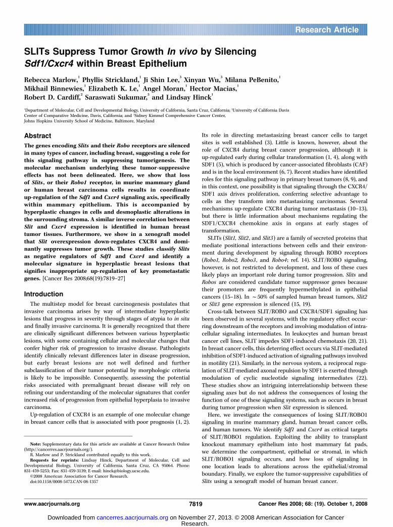
SLITs Suppress Tumor Growth In vivo by Silencing
Sdf1/Cxcr4 within Breast Epithelium
Rebecca Marlow,1Phyllis Strickland,
1Ji Shin Lee,
3Xinyan Wu,
3Milana PeBenito,
1
Mikhail Binnewies,1Elizabeth K. Le,
1Angel Moran,
1Hector Macias,
1
Robert D. Cardiff,2Saraswati Sukumar,
3and Lindsay Hinck
1
1Department of Molecular, Cell and Developmental Biology, University of California, Santa Cruz, California; 2University of California DavisCenter of Comparative Medicine, Davis, California; and 3Sidney Kimmel Comprehensive Cancer Center,Johns Hopkins University School of Medicine, Baltimore, Maryland
Abstract
The genes encoding Slits and their Robo receptors are silencedin many types of cancer, including breast, suggesting a role forthis signaling pathway in suppressing tumorigenesis. Themolecular mechanism underlying these tumor-suppressiveeffects has not been delineated. Here, we show that lossof Slits , or their Robo1 receptor, in murine mammary glandor human breast carcinoma cells results in coordinateup-regulation of the Sdf1 and Cxcr4 signaling axis, specificallywithin mammary epithelium. This is accompanied byhyperplastic changes in cells and desmoplastic alterations inthe surrounding stroma. A similar inverse correlation betweenSlit and Cxcr4 expression is identified in human breasttumor tissues. Furthermore, we show in a xenograft modelthat Slit overexpression down-regulates CXCR4 and domi-nantly suppresses tumor growth. These studies classify Slitsas negative regulators of Sdf1 and Cxcr4 and identify amolecular signature in hyperplastic breast lesions thatsignifies inappropriate up-regulation of key prometastaticgenes. [Cancer Res 2008;68(19):7819–27]
Introduction
The multistep model for breast carcinogenesis postulates thatinvasive carcinoma arises by way of intermediate hyperplasticlesions that progress in severity through stages of atypia to in situand finally invasive carcinoma. It is generally recognized that thereare clinically significant differences between various hyperplasticlesions, with some containing cellular and molecular changes thatconfer higher risk of progression to invasive disease. Pathologistsidentify clinically relevant differences later in disease progression,but early breast lesions are not well defined and furthersubclassification of their tumor potential by morphologic criteriais likely to be impossible. Consequently, assessing the potentialrisks associated with premalignant breast disease will rely onrefining our understanding of the molecular signatures that conferincreased risk of progression from epithelial hyperplasia to invasivecarcinoma.Up-regulation of CXCR4 is an example of one molecular change
in breast cancer cells that is associated with poor prognosis (1, 2).
Its role in directing metastasizing breast cancer cells to targetsites is well established (3). Little is known, however, about therole of CXCR4 during breast cancer progression, although it isup-regulated early during cellular transformation (1, 4), along withSDF1 (5), which is produced by cancer-associated fibroblasts (CAF)and is in the local environment (6, 7). Recent studies have identifiedroles for this signaling pathway in primary breast tumors (8, 9), andin this context, one possibility is that signaling through the CXCR4/SDF1 axis drives proliferation, conferring selective advantage tocells as they transform into metastasizing carcinomas. Severalmechanisms up-regulate CXCR4 during tumor metastasis (10–13),but there is little information about mechanisms regulating theSDF1/CXCR4 chemokine axis in organs at early stages oftransformation.SLITs (Slit1, Slit2 , and Slit3) are a family of secreted proteins that
mediate positional interactions between cells and their environ-ment during development by signaling through ROBO receptors(Robo1, Robo2, Robo3 , and Robo4 ; ref. 14). SLIT/ROBO signaling,however, is not restricted to development, and loss of these cueslikely plays an important role during tumor progression. Slits andRobos are considered candidate tumor suppressor genes becausetheir promoters are frequently hypermethylated in epithelialcancers (15–18). In f50% of sampled human breast tumors, Slit2or Slit3 gene expression is silenced (15, 19).Cross-talk between SLIT/ROBO and CXCR4/SDF1 signaling has
been observed in several systems, with the regulatory effect occur-ring downstream of the receptors and involving modulation of intra-cellular signaling intermediates. In leukocytes and human breastcancer cell lines, SLIT impedes SDF1-induced chemotaxis (20, 21).In breast cancer cells, this deterring effect occurs via SLIT-mediatedinhibition of SDF1-induced activation of signaling pathways involvedin motility (21). Similarly, in the nervous system, a reciprocal regu-lation of SLIT-mediated axonal repulsion by SDF1 is exerted throughmodulation of cyclic nucleotide signaling intermediates (22).These studies show an intriguing interrelationship between thesesignaling axes but do not address the consequences of losing thefunction of one of these signaling systems, such as occurs in breastduring tumor progression when Slit expression is silenced.Here, we investigate the consequences of losing SLIT/ROBO1
signaling in murine mammary gland, human breast cancer cells,and human tumors. We identify Sdf1 and Cxcr4 as critical targetsof SLIT/ROBO1 regulation. Exploiting the ability to transplantknockout mammary epithelium into host mammary fat pads,we determine the compartment, epithelial or stromal, in whichSLIT/ROBO1 signaling occurs, and how loss of signaling inone location leads to alterations across the epithelial/stromalboundary. Finally, we explore the tumor-suppressive capabilities ofSlits using a xenograft model of human breast cancer.
Note: Supplementary data for this article are available at Cancer Research Online(http://cancerres.aacrjournals.org/).
R. Marlow and P. Strickland contributed equally to this work.Requests for reprints: Lindsay Hinck, Department of Molecular, Cell and
Developmental Biology, University of California, Santa Cruz, CA 95064. Phone:831-459-5253; Fax: 831-459-3139; E-mail: [email protected].
I2008 American Association for Cancer Research.doi:10.1158/0008-5472.CAN-08-1357
www.aacrjournals.org 7819 Cancer Res 2008; 68: (19). October 1, 2008
Research Article
Research. on November 27, 2013. © 2008 American Association for Cancercancerres.aacrjournals.org Downloaded from
Materials and Methods
Clinical samples. Frozen or formalin-fixed paraffin-embedded tissue
specimens were collected at Johns Hopkins University (Baltimore, MD). All
human tissue was collected using protocols approved by the Institutional
Review Board. Informed consent was obtained from each individual who
provided tissue linked with clinical data.
Animals. The study conformed to guidelines set by University of
California at Santa Cruz animal care committee (Chancellor’s Animal
Research Committee). Mouse Slit2, Slit3, and Robo1 nulls were generated
and genotyped as described (23).
Transplant techniques. Mammary anlage was rescued from E16-20
embryos and transplanted into precleared fat pads of athymic nude mice
(24). Tissue fragments from the resulting outgrowths were contralaterally
transplanted to generate knockout and wild-type tissue controls (25).
Implantation of Elvax beads. Elvax, an ethylene vinyl copolymer
capable of sustained slow release of bioactive molecules, was prepared as
described (26), with pellets containing 225 ng SDF1 and 0.45 mg bovine
serum albumin (BSA) for control. Pellets were contralaterally implanted
into the fat pad of wild-type CD1 mice (n = 3), and tissue was harvested
after 6 d.
Cell lines, DNA constructs, and antibodies. MCF7 and MDA-MB-231
cells were maintained in DMEM supplemented with 10% FCS. pGL-
CXCR4(�375) contains CXCR4 between �357 and +51 relative to the
transcription site followed by the luciferase gene (12). pCRII-SDF1
( for riboprobes) contains 538-nucleotide fragment of the mouse Sdf1
cDNA (27). Mouse image clone 3385804 (American Type Culture Collection).
Small interfering RNA (siRNA) directed against Robo1 was from Santa Cruz
Biotechnology. pSecTagB-hSlit3 -C-myc was from Dr. Roy Bicknell
(University of Birmingham, Birmingham, United Kingdom). The following
antibodies were used: anti-CK14 (AF64, Covance), anti-SMA (1A4, Sigma),
anti-Ki67 (Santa Cruz Biotechnology), anti-CXCR4 (Abcam), anti-SDF1(Santa Cruz Biotechnology), anti-SLIT3 (Chemicon), anti-SLIT2 (Chemicon),
anti-HA (Dr. Doug Kellog, University of California, Santa Cruz, CA), anti-
Myc (9E10), anti-ROBO1 (Abcam), and anti-extracellular signal-regulated
kinase (Santa Cruz Biotechnology).Generation of stable cell lines. MDA-MB-231 cells were transfected
with pSecTagB-Slit2-HA and pSecTagB-Slit3-Myc and selected in zeocin
(Invitrogen). n = 3 lines were generated expressing SLIT2-HA and n = 2 linesexpressing SLIT3-Myc.
Tumor generation. Stable cell lines (106 cells) were injected into
precleared fat pads of nude mice. Tumor volume was calculated using the
formula (length � width)2/2.Immunohistochemistry. Tissue was fixed in 4% paraformaldehyde.
Paraffin-embedded tissue was sectioned at 6 Am and serially mounted.
Standard protocols were used and avidin-biotin complex method (Vector
Labs) was used for amplification.Scoring of immunohistochemistry. Immunostaining was scored
according to percentage positive cells (P) and staining intensity (I). Score
equals P + I. P scores 0 (none), 1 (<1%), 2 (1–10%), 3 (10–30%), 4 (30–60%),
and 5 (>60%). I scores 0 (none), 1 (weak), 2 (intermediate), and 3 (strong).siRNA transfection. MCF7 cells were transiently transfected using
Robo1 siRNA (Santa Cruz Biotechnology) and Lipofectamine 2000(Invitrogen) according to the manufacturers’ instructions. For three-dimensional culture, the ‘‘on-top’’ method was used (28). For luciferaseassay, 48 h before harvest, cells were cotransfected with pGL-CXCR4(�375) (F-luciferase) and pRL-TK (R-luciferase). Cells were lysedusing passive lysis buffer and assay was carried out in triplicate usingthe Dual-Luciferase Assay System (Promega) and Wallac Victor Lumin-ometer (Perkin-Elmer Life Sciences) according to the manufacturers’instructions. F-luciferase activity was normalized to R-luciferase activity(transfection efficiency).
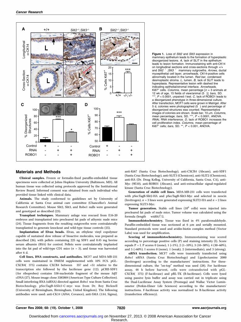
Figure 1. Loss of Slit2 and Slit3 expression inmammary epithelium leads to the formation of hyperplasticdisorganized lesions. A, lack of SLIT in the epitheliumleads to lesion formation. Immunostaining with anti-CK14on longitudinal sections and cross-sections through +/+and Slit2�/�;Slit3�/� mammary outgrowths. Arrows, ductalmyoepithelial cell layer; arrowheads, CK14-positive cellsabnormally located in the lumen. Red bar, condenseddesmoplastic stroma. L , lumen. B, lack of SLIT leads tohyperplasia. Representative lesion with dashed lineindicating epithelial/stromal interface. Arrowheads,Ki67+ cells. Columns, mean percentage [n = 3 animals at12 wk of age, 15 fields of view/animal (5�)]; bars, SD.***, P < 0.0001, unpaired t test. C, lack of ROBO1 leads toa disorganized phenotype in three-dimensional culture.After transfection, MCF7 cells were grown in Matrigel. After5 d, colonies were photographed (5�) and percentage ofdisorganized structures was counted. Representativeimages of colonies are shown. Scale bar, 10 Am. Columns,mean percentage; bars, SD. ***, P < 0.0001, ANOVA.RNAi, RNA interference. D, lack of ROBO1 increases thecell proliferation index. Columns, mean percentage ofKi67+ cells; bars, SD. **, P < 0.001, ANOVA.
Cancer Research
Cancer Res 2008; 68: (19). October 1, 2008 7820 www.aacrjournals.org
Research. on November 27, 2013. © 2008 American Association for Cancercancerres.aacrjournals.org Downloaded from
Western blotting. Western blotting was performed using standardprocedures (29). Band intensity was scanned using Typhoon 9410 imager
and quantified using ImageQuant 5 software.
Quantitative real-time reverse transcription-PCR analysis. Real-time
reverse transcription-PCR (RT-PCR) analysis was done as previouslydescribed (30). Data were first analyzed using the Sequence Detector
Software SDS 2.0 (Applied Biosystems). Results were calculated and
normalized relative to glyceraldehyde-3-phosphate dehydrogenase
(GAPDH) control. All of the PCR assays were done in triplicate, and meanvalues are shown in figures.
In situ hybridization. In situ hybridization was carried out as described
previously (23, 25).
Primary cell isolation. Primary mammary epithelial cells were preparedfrom mild collagenase and dispase digestion, as described (23). Cells were
plated overnight and then trypsinized and placed onto Matrigel-coated
coverslips.Chemotaxis assay. Chemotaxis was examined as described before (29).
Phase-contrast images were acquired at 0 and 60 min. The change in cell
area in the directed quadrant was calculated using ImageJ.
Statistical analysis. We used factorial design ANOVA, unpaired t tests,or Mann-Whitney tests to analyze data as appropriate. Significant ANOVA
values were subsequently subjected to post-test using the Tukey-Kramer
comparison. We report P values for each statistical test; all P values were
<0.05.
Results
Loss of Slit or Robo1 in mammary epithelium leads to theformation of hyperplastic, disorganized lesions. Given the
expanding role of SLITs in epithelial biology, we hypothesized atumor-suppressive function for Slits in breast. We previouslyshowed that two Slit family members, Slit2 and Slit3 , are expressedin murine mammary gland (23). The homozygous Slit2�/�
mutation causes perinatal lethality. Therefore, to investigate theconsequence of its loss in mature mammary gland, we generatedSlit2�/�;Slit3�/� outgrowths by contralateral transplantation ofknockout and wild-type anlage into cleared fat pads of immuno-compromised mice (24).We examined mature Slit2�/�;Slit3�/� mammary outgrowths for
morphology. Compared with the open lumens and organizedbilayers of ducts in control outgrowths, Slit2�/�;Slit3�/� ductsdisplayed striking abnormalities (Fig. 1A). The phenotype was 100%penetrant, with f30% of ducts having lesions extending between0.3 and 5.0 mm. We categorized the lesions as mild and severe.Mild lesions contained cells in the luminal space (10.1% F SE 1.9;n = 621 ducts; 5 outgrowths), and many of these cells were peeledaway from the myoepithelial layer, similar to an adhesive defectpreviously described in Ntn1�/�;Slit2�/� glands (23). In severelesions (17.8% F SE 8.1; n = 621 ducts; 5 outgrowths), ductallumens were occluded with a disorganized mass of cells (Fig. 1A).These excess cells suggested disrupted growth control due to eitherincreased proliferation and/or decreased apoptosis. We labeledproliferating cells and observed a significant increase in thepercentage of Ki67+ cells in Slit2�/�;Slit3�/� , compared with +/+,ducts (Fig. 1B). This increase is responsible for the excess cells
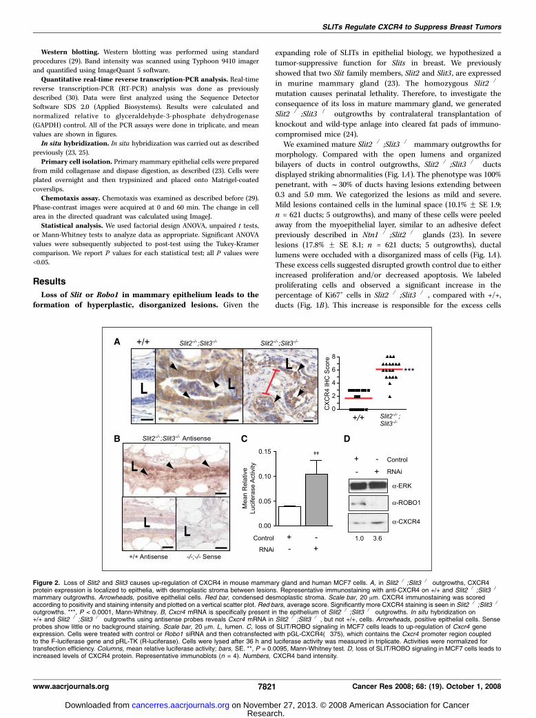
Figure 2. Loss of Slit2 and Slit3 causes up-regulation of CXCR4 in mouse mammary gland and human MCF7 cells. A, in Slit2�/�;Slit3�/� outgrowths, CXCR4protein expression is localized to epithelia, with desmoplastic stroma between lesions. Representative immunostaining with anti-CXCR4 on +/+ and Slit2�/�;Slit3�/�
mammary outgrowths. Arrowheads, positive epithelial cells. Red bar, condensed desmoplastic stroma. Scale bar, 20 Am. CXCR4 immunostaining was scoredaccording to positivity and staining intensity and plotted on a vertical scatter plot. Red bars, average score. Significantly more CXCR4 staining is seen in Slit2�/�;Slit3�/�
outgrowths. ***, P < 0.0001, Mann-Whitney. B, Cxcr4 mRNA is specifically present in the epithelium of Slit2�/�;Slit3�/� outgrowths. In situ hybridization on+/+ and Slit2�/�;Slit3�/� outgrowths using antisense probes reveals Cxcr4 mRNA in Slit2�/�;Slit3�/� , but not +/+, cells. Arrowheads, positive epithelial cells. Senseprobes show little or no background staining. Scale bar, 20 Am. L, lumen. C, loss of SLIT/ROBO signaling in MCF7 cells leads to up-regulation of Cxcr4 geneexpression. Cells were treated with control or Robo1 siRNA and then cotransfected with pGL-CXCR4(�375), which contains the Cxcr4 promoter region coupledto the F-luciferase gene and pRL-TK (R-luciferase). Cells were lysed after 36 h and luciferase activity was measured in triplicate. Activities were normalized fortransfection efficiency. Columns, mean relative luciferase activity; bars, SE. **, P = 0.0095, Mann-Whitney test. D, loss of SLIT/ROBO signaling in MCF7 cells leads toincreased levels of CXCR4 protein. Representative immunoblots (n = 4). Numbers, CXCR4 band intensity.
SLITs Regulate CXCR4 to Suppress Breast Tumors
www.aacrjournals.org 7821 Cancer Res 2008; 68: (19). October 1, 2008
Research. on November 27, 2013. © 2008 American Association for Cancercancerres.aacrjournals.org Downloaded from
because we evaluated apoptosis using activated caspase-3 stainingand observed no difference (data not shown). Histopathologicanalyses concurred with our observations that Slit2�/�;Slit3�/�
tissue contains hyperplasias. Condensed and desmoplastic stromasurrounding the lesions were also noted in the diagnosis (Fig. 1A),as was a large influx of immune infiltrates in the knockout,compared with wild-type, tissue.ROBO1 is a SLIT receptor that could mediate the observed effects
in the gland (23). Robo1�/� animals are viable so we evaluated theloss-of-function phenotype using intact glands. Ducts in Robo1�/�
glands were hyperplastic and disorganized, displaying a phenotypethat was indistinguishable from Slit2�/�;Slit3�/� ductal lesions(Supplementary Fig. S1). As was the case for Slit2�/�;Slit3�/� tissue,the penetrance of the phenotype was 100%, with f30% of ductsdisplaying lesions that extended between 0.3 and 5.0 mm.To investigate whether a similar phenotype occurred when
SLIT/ROBO1 signaling was disrupted in human breast cells,we used the MCF7 line that retains several characteristics
of differentiated mammary epithelium, including expression ofSlit2, Slit3 , and Robo1 (data not shown; ref. 31). Cells were treatedwith Robo1 siRNA to down-regulate SLIT/ROBO1 signaling(Supplementary Fig. S2) and then cultured in Matrigel. MCF7cells formed smooth, nonpolarized colonies without centrallumens. In contrast, the siRNA-treated colonies were largeand disorganized, a phenotype that was rescued by reexpressionof Robo1 (Fig. 1C). Immunostaining with Ki67 revealed asignificantly higher fraction of proliferating cells in coloniestreated with Robo1 siRNA compared with control (Fig. 1D). Thiswas similar to the elevated proliferation observed in Slit2�/�;Slit3�/� outgrowths and Robo1�/� glands (Fig. 1B ; SupplementaryFig. S2). Together, these data show that a consequence ofSlit/Robo1 loss is elevated proliferation leading to hyperplasticlesions.Loss of Slit up-regulates Cxcr4 expression. We sought
candidates whose misexpression in the absence of SLIT/ROBO1signaling is responsible for the observed hyperplastic phenotype.
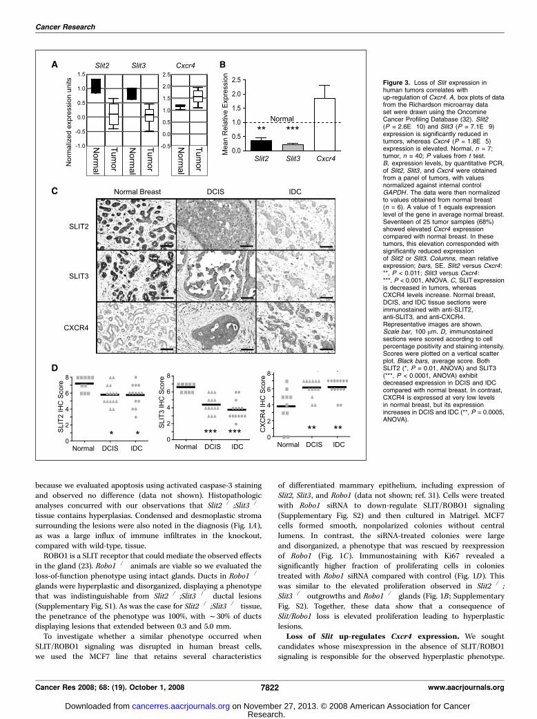
Figure 3. Loss of Slit expression inhuman tumors correlates withup-regulation of Cxcr4. A, box plots of datafrom the Richardson microarray dataset were drawn using the OncomineCancer Profiling Database (32). Slit2(P = 2.6E�10) and Slit3 (P = 7.1E�9)expression is significantly reduced intumors, whereas Cxcr4 (P = 1.8E�5)expression is elevated. Normal, n = 7;tumor, n = 40; P values from t test.B, expression levels, by quantitative PCR,of Slit2, Slit3 , and Cxcr4 were obtainedfrom a panel of tumors, with valuesnormalized against internal controlGAPDH . The data were then normalizedto values obtained from normal breast(n = 6). A value of 1 equals expressionlevel of the gene in average normal breast.Seventeen of 25 tumor samples (68%)showed elevated Cxcr4 expressioncompared with normal breast. In thesetumors, this elevation corresponded withsignificantly reduced expressionof Slit2 or Slit3. Columns, mean relativeexpression; bars, SE. Slit2 versus Cxcr4 :**, P < 0.011; Slit3 versus Cxcr4 :***, P < 0.001, ANOVA. C, SLITexpressionis decreased in tumors, whereasCXCR4 levels increase. Normal breast,DCIS, and IDC tissue sections wereimmunostained with anti-SLIT2,anti-SLIT3, and anti-CXCR4.Representative images are shown.Scale bar, 100 Am. D, immunostainedsections were scored according to cellpercentage positivity and staining intensity.Scores were plotted on a vertical scatterplot. Black bars, average score. BothSLIT2 (*, P = 0.01, ANOVA) and SLIT3(***, P < 0.0001, ANOVA) exhibitdecreased expression in DCIS and IDCcompared with normal breast. In contrast,CXCR4 is expressed at very low levelsin normal breast, but its expressionincreases in DCIS and IDC (**, P = 0.0005,ANOVA).
Cancer Research
Cancer Res 2008; 68: (19). October 1, 2008 7822 www.aacrjournals.org
Research. on November 27, 2013. © 2008 American Association for Cancercancerres.aacrjournals.org Downloaded from
One candidate is CXCR4 because it is expressed early during breasttumorigenesis (1, 4), and blocking its expression or functioninhibits breast tumor growth (8, 9). Western blots of whole glandlysates showed elevated CXCR4 expression in Slit2�/�;Slit3�/�,compared with +/+, tissue (Supplementary Fig. S3). Immunohisto-chemistry revealed CXCR4 expression in a large fraction of cells inSlit2�/�;Slit3�/� epithelium, with little or no expression in +/+
epithelium (Fig. 2A). We also observed condensed and desmo-plastic stroma surrounding these CXCR4-positive lesions (Fig. 2A).Because CXCR4 is regulated by transcriptional and posttranscrip-tional mechanisms, we performed in situ hybridization studies andobserved Cxcr4 in Slit knockout, but not wild-type, epithelium(Fig. 2B). A transcriptional mechanism also occurred in Robo1siRNA-treated MCF7 cells because we observed increased Cxcr4
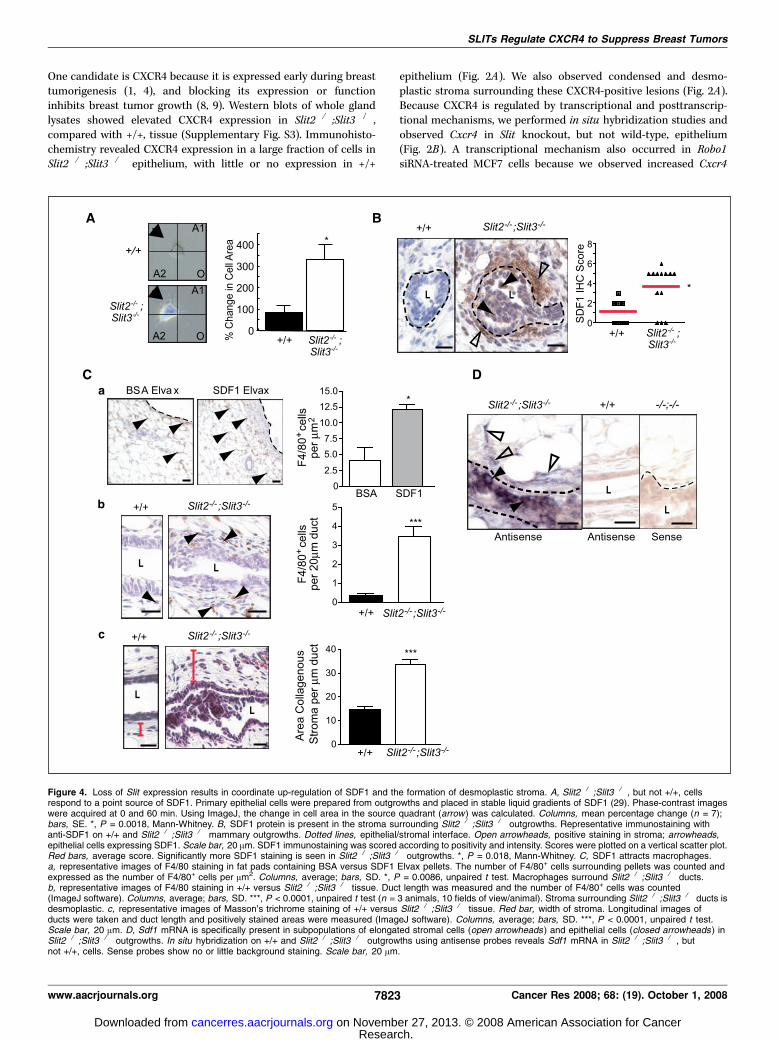
Figure 4. Loss of Slit expression results in coordinate up-regulation of SDF1 and the formation of desmoplastic stroma. A, Slit2�/�;Slit3�/� , but not +/+, cellsrespond to a point source of SDF1. Primary epithelial cells were prepared from outgrowths and placed in stable liquid gradients of SDF1 (29). Phase-contrast imageswere acquired at 0 and 60 min. Using ImageJ, the change in cell area in the source quadrant (arrow) was calculated. Columns, mean percentage change (n = 7);bars, SE. *, P = 0.0018, Mann-Whitney. B, SDF1 protein is present in the stroma surrounding Slit2�/�;Slit3�/� outgrowths. Representative immunostaining withanti-SDF1 on +/+ and Slit2�/�;Slit3�/� mammary outgrowths. Dotted lines, epithelial/stromal interface. Open arrowheads, positive staining in stroma; arrowheads,epithelial cells expressing SDF1. Scale bar, 20 Am. SDF1 immunostaining was scored according to positivity and intensity. Scores were plotted on a vertical scatter plot.Red bars, average score. Significantly more SDF1 staining is seen in Slit2�/�;Slit3�/� outgrowths. *, P = 0.018, Mann-Whitney. C, SDF1 attracts macrophages.a, representative images of F4/80 staining in fat pads containing BSA versus SDF1 Elvax pellets. The number of F4/80+ cells surrounding pellets was counted andexpressed as the number of F4/80+ cells per Am2. Columns, average; bars, SD. *, P = 0.0086, unpaired t test. Macrophages surround Slit2�/�;Slit3�/� ducts.b, representative images of F4/80 staining in +/+ versus Slit2�/�;Slit3�/� tissue. Duct length was measured and the number of F4/80+ cells was counted(ImageJ software). Columns, average; bars, SD. ***, P < 0.0001, unpaired t test (n = 3 animals, 10 fields of view/animal). Stroma surrounding Slit2�/�;Slit3�/� ducts isdesmoplastic. c, representative images of Masson’s trichrome staining of +/+ versus Slit2�/�;Slit3�/� tissue. Red bar, width of stroma. Longitudinal images ofducts were taken and duct length and positively stained areas were measured (ImageJ software). Columns, average; bars, SD. ***, P < 0.0001, unpaired t test.Scale bar, 20 Am. D, Sdf1 mRNA is specifically present in subpopulations of elongated stromal cells (open arrowheads ) and epithelial cells (closed arrowheads ) inSlit2�/�;Slit3�/� outgrowths. In situ hybridization on +/+ and Slit2�/�;Slit3�/� outgrowths using antisense probes reveals Sdf1 mRNA in Slit2�/�;Slit3�/� , butnot +/+, cells. Sense probes show no or little background staining. Scale bar, 20 Am.
SLITs Regulate CXCR4 to Suppress Breast Tumors
www.aacrjournals.org 7823 Cancer Res 2008; 68: (19). October 1, 2008
Research. on November 27, 2013. © 2008 American Association for Cancercancerres.aacrjournals.org Downloaded from
reporter gene activity and increased levels of CXCR4 in treated,compared with control, cells (Fig. 2C and D). Together, our resultsshow that SLIT/ROBO1 signaling negatively regulates Cxcr4expression, with loss of this regulation leading to elevated levelsof CXCR4 in murine tissue and human breast cancer cells.If Slits silence Cxcr4 in normal breast, we hypothesize that loss
of Slits in tumors will correspond with elevated Cxcr4. Toinvestigate, we analyzed microarray data sets from human breasttumor samples available at Oncomine.org (32) and found aninverse correlation between Slit and Cxcr4 expression (Fig. 3A). Weconfirmed this by performing quantitative RT-PCR on a panel ofhuman tumors; in 68% of tumors with elevated Cxcr4 expression,
Slit2 or Slit3 levels were significantly reduced compared with theirexpression in normal tissue (Fig. 3B). We further verified theseobservations at the protein level using immunohistochemistry onsamples of normal breast, ductal carcinoma in situ (DCIS), andinfiltrating ductal carcinoma (IDC; Fig. 3C and D). Again, therewere robust levels of SLIT2 and SLIT3 in normal tissue thatsignificantly decreased with increasing tumor grade. In contrast,and as previously shown (1, 4), little or no CXCR4 was detectable innormal breast, but its expression significantly increased withhigher tumor grade.Loss of Slit expression results in coordinate up-regulation of
SDF1. Although CXCR4 is up-regulated in the vast majority ofsampled premalignant lesions, studies on human breast cancer celllines have suggested that it is only active in metastatic cells (33).To evaluate CXCR4 activity in Slit2�/�;Slit3�/� cells, we performedchemotaxis assays. Slit2�/�;Slit3�/� cells did not exhibit robustmigration as expected of primary cells harvested from premalig-nant tissue but instead responded to SDF1 by reorganizing theircell membrane and sending membranous projections toward apoint source (Fig. 4A). Wild-type cells were unresponsive to SDF1.This result suggested that CXCR4 expressed on Slit2�/�;Slit3�/�
cells is active and reacts to its ligand.This raised the question of whether SDF1 surrounded Slit2�/�;
Slit3�/� lesions because recent studies have placed it in the tumormicroenvironment (6, 7). We found abundant SDF1 expression inthe epithelium and the surrounding stroma of knockout, butnot wild-type, tissue (Fig. 4B). The presence of SDF1 is consistentwith the histopathologic diagnosis that noted the infiltrationof immune cells within desmoplastic stroma surroundingSlit2�/�;Slit3�/� lesions. Macrophages, which express CXCR4,represent a major component of immune infiltrates surroundingtumors and play a key role in promoting the angiogenicswitch during malignant transition (34). To determine whethermacrophages are attracted to SDF1, we implanted point sources ofSDF1 or vehicle (BSA) into wild-type mammary glands (Fig. 4C, a).Significantly more macrophages (F4/80+) infiltrated into the tissuesurrounding SDF1, compared with control, showing that SDF1 is achemoattractant for macrophages and suggesting a role inrecruiting these immune cells to tumors (Fig. 4C, a). Next, we
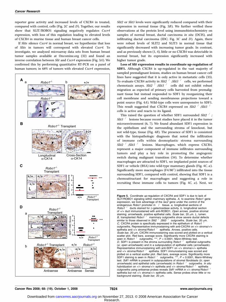
Figure 5. Coordinate up-regulation of CXCR4 and SDF1 is due to lack ofSLIT/ROBO1 signaling within mammary epithelia. A, to examine Robo1 geneexpression, we took advantage of the lacZ gene under the control of theendogenous Robo1 promoter in �/� tissue. a, longitudinal sections ofRobo1�/� ducts stained for h-galactosidase activity. b, longitudinal sectionof +/+ duct immunostained with anti-ROBO1. Open arrows, positive stromalstaining; arrowheads, positive epithelial cells. Scale bar, 20 Am. L , lumen.B, transplanted Robo1�/� mammary outgrowths show severe ductal defectssimilar to those observed in Slit2�/�;Slit3�/� outgrowths. Scale bar, 20 Am.C, CXCR4 protein is specifically expressed in the epithelium of Robo1�/�
outgrowths. Representative immunostaining with anti-CXCR4 on +/+ stroma/+/+epithelia and +/+ stroma/Robo1�/� epithelia. Arrows, positive cells.Scale bar, 20 Am. CXCR4 immunostaining was scored and plotted on a verticalscatter plot. Red bars, average score. Significantly more CXCR4 staining isseen in Robo1�/� outgrowths. ***, P < 0.0001, Mann-Whitney test.D, SDF1 is present in the stroma surrounding Robo1�/� epithelial outgrowths(a ; open arrowheads ) and in a subpopulation of epithelial cells (arrowheads ).Representative immunostaining with anti-SDF1 on +/+ stroma/+/+ epitheliaand +/+ stroma/Robo1�/� epithelia. SDF1 immunostaining was scored andplotted on a vertical scatter plot. Red bars, average score. Significantly moreSDF1 staining is seen in Robo1�/� outgrowths. ***, P < 0.0001, Mann-Whitneytest. Sdf1 mRNA is present in subpopulations of stromal fibroblasts (b ; openarrowheads ) and epithelial cells (arrowheads ) in Robo1�/� outgrowths. In situhybridization on +/+ stroma/+/+ epithelia and +/+ stroma/Robo1�/� epitheliaoutgrowths using antisense probes reveals Sdf1 mRNA in +/+ stroma/Robo1�/�
epithelia but not +/+ stroma/+/+ epithelia cells. Sense probes show little or nobackground staining. Scale bar, 20 Am.
Cancer Research
Cancer Res 2008; 68: (19). October 1, 2008 7824 www.aacrjournals.org
Research. on November 27, 2013. © 2008 American Association for Cancercancerres.aacrjournals.org Downloaded from

performed F4/80 immunohistochemistry on Slit2�/�;Slit3�/� andcontrol tissue and found a significant increase in macrophagessurrounding knockout tissue (Fig. 4C, b). We also evaluated thestromal expression of collagen, a major constituent of desmoplasticstroma (Fig. 4C, c). Stroma surrounding Slit2�/�;Slit3�/� epithe-lium contained significantly more condensed, collagenous stroma,compared with +/+, consistent with the histopathologic analysis.To define the cellular source of SDF1, we performed in situ hybri-dization analyses and discovered Sdf1 in a fraction of epithelialcells and in a subset of elongated stromal cells that are likely to befibroblasts based on their morphology (Fig. 4D). Thus, both CXCR4and SDF1 are initially up-regulated in the epithelium, as has beenrecently observed in a xenograft model of DCIS (5). A local sourceof SDF1 may function to transform myoepithelial cells into CAFsor to recruit CAFs from circulating cells (35).Epithelial regulation of CXCR4/SDF1 chemokine signaling
axis. Together, the data show that loss of Slit expression leads tothe coordinate up-regulation of Cxcr4 in epithelia and Sdf1 in bothepithelia and stroma. This suggests that SLIT/ROBO1 signalingkeeps SDF1/CXCR4 expression in check, but the regulatorynetworks may be complicated. Slit genes are expressed in theepithelia, but they encode a secreted cue that may act on any celltype expressing ROBO1 receptors. During mammary development,ROBO1 is expressed on myoepithelial cells (23), but as the glandmatures, we observed a switch in its expression to include asubpopulation of luminal cells (Fig. 5A). ROBO1 was also expressedon stromal fibroblasts (Fig. 5A). Consequently, loss of Slitexpression could regulate Sdf1 and Cxcr4 independently bydisrupting ROBO1 signaling in both the stromal and epithelialcompartments. Alternatively, loss of SLIT/ROBO1 signaling injust one compartment could up-regulate Sdf1 and Cxcr4 in bothcompartments.To investigate, we eliminated SLIT/ROBO1 signaling selectively
in the epithelial compartment by transplanting Robo1�/� epithe-lium into wild-type stroma. In these chimeric glands, we observeddisorganized, hyperplastic epithelial lesions (Fig. 5B), which
were similar in phenotype, penetrance (100%), and expressivity(19.64% F SE 9.77; n = 669 ducts; 6 outgrowths) to those seen inSlit2�/�;Slit3�/� transplants (Fig. 1A). We evaluated the chemokineaxis and again found up-regulation of CXCR4 in Robo1�/�
epithelium (Fig. 5C), and coordinate up-regulation of SDF1 in thesurrounding +/+ stroma (Fig. 5D, a), which was desmoplastic andcontained immune infiltrates similar to stroma surroundingSlit2�/�Slit3�/� tissue (data not shown). These data show thatloss of SLIT/ROBO1 signaling in the epithelial compartment, alone,up-regulates SDF1 and CXCR4. This leads to phenotypic changessimilar to those occurring in Slit2�/�;Slit3�/� transplants in whichSLIT/ROBO1 signaling is disrupted in both compartments. Todefine the source of SDF1 in the transplanted tissue, we performedin situ hybridization studies and found Sdf1 mRNA in cellsubpopulations in the epithelia and stroma (Fig. 5D, b), suggestingthat loss of SLIT/ROBO1 signaling in breast epithelia at early stagesof transformation both generates a local source of Sdf1 and up-regulates Cxcr4 . We therefore conclude that loss of SLIT/ROBO1signaling in the epithelia, alone, is sufficient to drive the observedmorphologic and molecular changes, resulting in hyperplasticlesions, surrounded by desmoplastic stroma.SLITs suppress CXCR4 expression and inhibit tumor growth.
Given that SLITs exert this regulatory function by inhibiting theexpression of Sdf1 and Cxcr4 within the mammary epithelium, wewondered whether overexpression of Slits in human breastcarcinoma cells would suppress Cxcr4 expression and inhibittumor growth. Previous studies have shown that the metastatichuman cell line MDA-MB-231 expresses CXCR4, but not SDF1 (36),and that inhibiting CXCR4 expression or function in these cellsblocks primary tumor growth (8, 9). Because MDA-MB-231 cellsexpress ROBO1 and ROBO2 (21),4 signaling through these receptorscould down-regulate CXCR4 expression and suppress tumorformation. To investigate, we transiently expressed Myc-Slit2
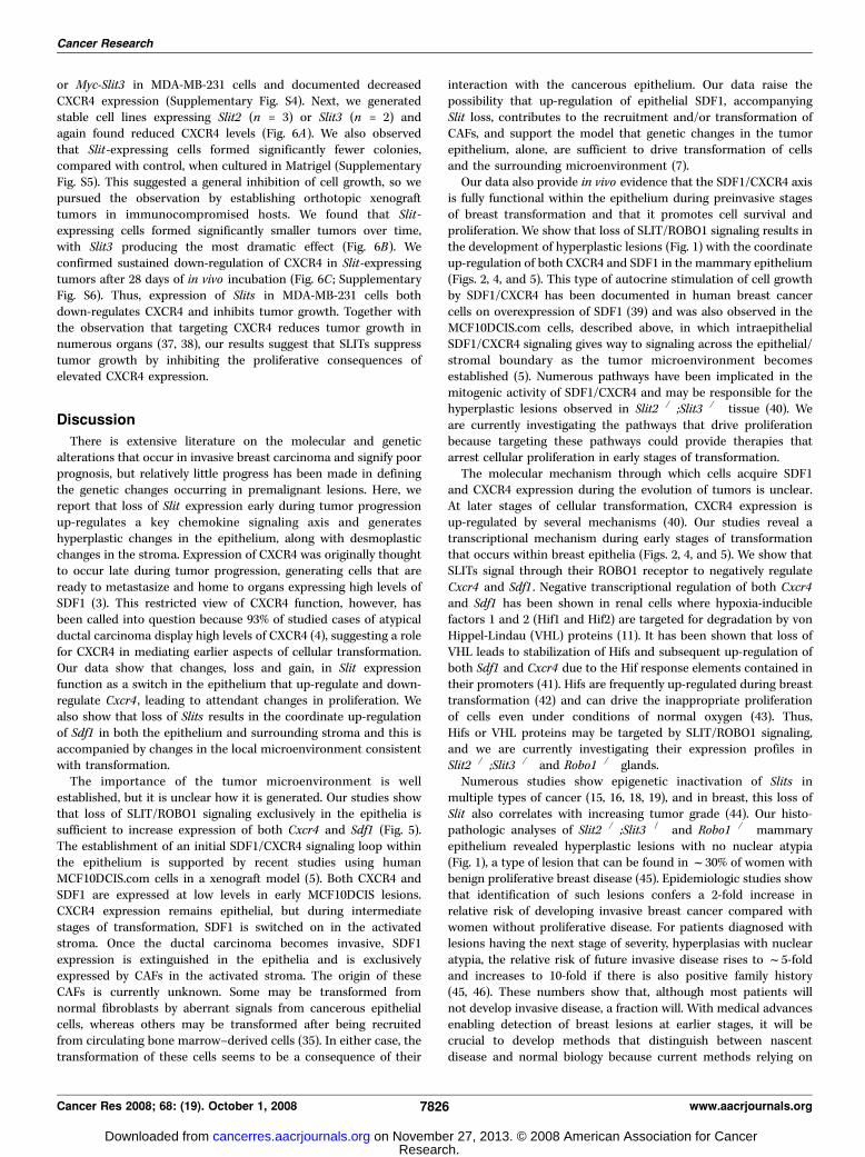
Figure 6. Slit expression in MDA-MB-231 cellsblocks tumor growth by reducing CXCR4expression. A, Slit2 -HA and Slit3 -Myc stable celllines express low levels of CXCR4 compared withvector alone control lines. Stable Slit2 -HA (n = 3)and Slit3 -myc (n = 2) cell lines were generated byclonal selection. Stable cell line extracts wereprobed with anti-CXCR4. Columns, mean CXCR4band intensity (n = 2 for each line); bars, SE.**, P < 0.001, ANOVA. B, expression of Slit2 orSlit3 resulted in smaller tumor size. Tumors weregenerated using Slit and control stable cell lines.n = 12 mice for each line. Points, mean tumorvolume at each day; bars, SE. ***, P < 0.0001;**, P < 0.001; *, P < 0.05, ANOVA. Representativeimages of orthotopic tumors are shown. Scale bar,0.25 mm. C, tumors expressing Slit2 or Slit3contain significantly less CXCR4 protein comparedwith control tumors. Columns, mean CXCR4immunoblot band intensity from n = 3 tumors; bars,SE. **, P = 0.01, ANOVA.
4 R. Marlow, unpublished data.
SLITs Regulate CXCR4 to Suppress Breast Tumors
www.aacrjournals.org 7825 Cancer Res 2008; 68: (19). October 1, 2008
Research. on November 27, 2013. © 2008 American Association for Cancercancerres.aacrjournals.org Downloaded from
or Myc-Slit3 in MDA-MB-231 cells and documented decreasedCXCR4 expression (Supplementary Fig. S4). Next, we generatedstable cell lines expressing Slit2 (n = 3) or Slit3 (n = 2) andagain found reduced CXCR4 levels (Fig. 6A). We also observedthat Slit-expressing cells formed significantly fewer colonies,compared with control, when cultured in Matrigel (SupplementaryFig. S5). This suggested a general inhibition of cell growth, so wepursued the observation by establishing orthotopic xenografttumors in immunocompromised hosts. We found that Slit-expressing cells formed significantly smaller tumors over time,with Slit3 producing the most dramatic effect (Fig. 6B). Weconfirmed sustained down-regulation of CXCR4 in Slit-expressingtumors after 28 days of in vivo incubation (Fig. 6C ; SupplementaryFig. S6). Thus, expression of Slits in MDA-MB-231 cells bothdown-regulates CXCR4 and inhibits tumor growth. Together withthe observation that targeting CXCR4 reduces tumor growth innumerous organs (37, 38), our results suggest that SLITs suppresstumor growth by inhibiting the proliferative consequences ofelevated CXCR4 expression.
Discussion
There is extensive literature on the molecular and geneticalterations that occur in invasive breast carcinoma and signify poorprognosis, but relatively little progress has been made in definingthe genetic changes occurring in premalignant lesions. Here, wereport that loss of Slit expression early during tumor progressionup-regulates a key chemokine signaling axis and generateshyperplastic changes in the epithelium, along with desmoplasticchanges in the stroma. Expression of CXCR4 was originally thoughtto occur late during tumor progression, generating cells that areready to metastasize and home to organs expressing high levels ofSDF1 (3). This restricted view of CXCR4 function, however, hasbeen called into question because 93% of studied cases of atypicalductal carcinoma display high levels of CXCR4 (4), suggesting a rolefor CXCR4 in mediating earlier aspects of cellular transformation.Our data show that changes, loss and gain, in Slit expressionfunction as a switch in the epithelium that up-regulate and down-regulate Cxcr4 , leading to attendant changes in proliferation. Wealso show that loss of Slits results in the coordinate up-regulationof Sdf1 in both the epithelium and surrounding stroma and this isaccompanied by changes in the local microenvironment consistentwith transformation.The importance of the tumor microenvironment is well
established, but it is unclear how it is generated. Our studies showthat loss of SLIT/ROBO1 signaling exclusively in the epithelia issufficient to increase expression of both Cxcr4 and Sdf1 (Fig. 5).The establishment of an initial SDF1/CXCR4 signaling loop withinthe epithelium is supported by recent studies using humanMCF10DCIS.com cells in a xenograft model (5). Both CXCR4 andSDF1 are expressed at low levels in early MCF10DCIS lesions.CXCR4 expression remains epithelial, but during intermediatestages of transformation, SDF1 is switched on in the activatedstroma. Once the ductal carcinoma becomes invasive, SDF1expression is extinguished in the epithelia and is exclusivelyexpressed by CAFs in the activated stroma. The origin of theseCAFs is currently unknown. Some may be transformed fromnormal fibroblasts by aberrant signals from cancerous epithelialcells, whereas others may be transformed after being recruitedfrom circulating bone marrow–derived cells (35). In either case, thetransformation of these cells seems to be a consequence of their
interaction with the cancerous epithelium. Our data raise thepossibility that up-regulation of epithelial SDF1, accompanyingSlit loss, contributes to the recruitment and/or transformation ofCAFs, and support the model that genetic changes in the tumorepithelium, alone, are sufficient to drive transformation of cellsand the surrounding microenvironment (7).Our data also provide in vivo evidence that the SDF1/CXCR4 axis
is fully functional within the epithelium during preinvasive stagesof breast transformation and that it promotes cell survival andproliferation. We show that loss of SLIT/ROBO1 signaling results inthe development of hyperplastic lesions (Fig. 1) with the coordinateup-regulation of both CXCR4 and SDF1 in the mammary epithelium(Figs. 2, 4, and 5). This type of autocrine stimulation of cell growthby SDF1/CXCR4 has been documented in human breast cancercells on overexpression of SDF1 (39) and was also observed in theMCF10DCIS.com cells, described above, in which intraepithelialSDF1/CXCR4 signaling gives way to signaling across the epithelial/stromal boundary as the tumor microenvironment becomesestablished (5). Numerous pathways have been implicated in themitogenic activity of SDF1/CXCR4 and may be responsible for thehyperplastic lesions observed in Slit2�/�;Slit3�/� tissue (40). Weare currently investigating the pathways that drive proliferationbecause targeting these pathways could provide therapies thatarrest cellular proliferation in early stages of transformation.The molecular mechanism through which cells acquire SDF1
and CXCR4 expression during the evolution of tumors is unclear.At later stages of cellular transformation, CXCR4 expression isup-regulated by several mechanisms (40). Our studies reveal atranscriptional mechanism during early stages of transformationthat occurs within breast epithelia (Figs. 2, 4, and 5). We show thatSLITs signal through their ROBO1 receptor to negatively regulateCxcr4 and Sdf1 . Negative transcriptional regulation of both Cxcr4and Sdf1 has been shown in renal cells where hypoxia-induciblefactors 1 and 2 (Hif1 and Hif2) are targeted for degradation by vonHippel-Lindau (VHL) proteins (11). It has been shown that loss ofVHL leads to stabilization of Hifs and subsequent up-regulation ofboth Sdf1 and Cxcr4 due to the Hif response elements contained intheir promoters (41). Hifs are frequently up-regulated during breasttransformation (42) and can drive the inappropriate proliferationof cells even under conditions of normal oxygen (43). Thus,Hifs or VHL proteins may be targeted by SLIT/ROBO1 signaling,and we are currently investigating their expression profiles inSlit2�/�;Slit3�/� and Robo1�/� glands.Numerous studies show epigenetic inactivation of Slits in
multiple types of cancer (15, 16, 18, 19), and in breast, this loss ofSlit also correlates with increasing tumor grade (44). Our histo-pathologic analyses of Slit2�/�;Slit3�/� and Robo1�/� mammaryepithelium revealed hyperplastic lesions with no nuclear atypia(Fig. 1), a type of lesion that can be found in f30% of women withbenign proliferative breast disease (45). Epidemiologic studies showthat identification of such lesions confers a 2-fold increase inrelative risk of developing invasive breast cancer compared withwomen without proliferative disease. For patients diagnosed withlesions having the next stage of severity, hyperplasias with nuclearatypia, the relative risk of future invasive disease rises to f5-foldand increases to 10-fold if there is also positive family history(45, 46). These numbers show that, although most patients willnot develop invasive disease, a fraction will. With medical advancesenabling detection of breast lesions at earlier stages, it will becrucial to develop methods that distinguish between nascentdisease and normal biology because current methods relying on
Cancer Research
Cancer Res 2008; 68: (19). October 1, 2008 7826 www.aacrjournals.org
Research. on November 27, 2013. © 2008 American Association for Cancercancerres.aacrjournals.org Downloaded from
morphologic criteria are insufficient. Improved understanding ofmolecular signatures within breast lesions holds the promise ofidentifying those at high risk so they receive appropriate treatmentwhile also identifying the majority who are not at risk so theirmedical concerns are dispelled (47). The findings presented in thisreport identify the loss of Slit expression as a marker of early lesionsthat have the potential to progress to invasive disease due to up-regulation of metastasis markers SDF1/CXCR4. We propose thatthese molecular alterations define a specific subclass of breastlesions whose early detection could lead to treatment strategies thatprevent development of invasive disease.
Disclosure of Potential Conflicts of Interest
No potential conflicts of interest were disclosed.
Acknowledgments
Received 4/16/2008; revised 6/30/2008; accepted 7/25/2008.Grant support: American Cancer Society grant RSG0218001MGO, California
Breast Cancer Research Program grant 10PB-0188, and National Cancer Institute grantR01CA128902 (L. Hinck) and Congressionally Directed Medical Research Programgrant BC043200 and National Cancer Institute grant U01 CA105490 (R.D. Cardiff).
The costs of publication of this article were defrayed in part by the payment of pagecharges. This article must therefore be hereby marked advertisement in accordancewith 18 U.S.C. Section 1734 solely to indicate this fact.
Santa Cruz Biotechnology generously provided antibodies and siRNA reagents.Other generous gifts were the following: anti-Dutt1 (Dr. Rabbitts, University College,London, United Kingdom), anti-HA (Dr. Doug Kellogg), MCF7 and MDA-MB-231 celllines (Dr. Bissell, Lawrence Labs, Berkeley, CA), pGL-CXCR4(�375) (Dr. Avraham,Harvard Medical School, Boston, MA), pCRII-SDF1 (Dr. Goffinet, University ofLouvain Medical School, Brussels, Belgium), pRL-TK (Dr. Haussler, University ofCalifornia, Santa Cruz, CA), pSecTagB-hSlit3 -C-myc (Dr. Roy Bicknell), Slit3�/� mice(Dr. Ornitz, Washington University, St. Louis, MO), and Slit2�/� mice and Robo1�/�
(Dr. Tessier-Lavigne, Genentech, Inc., South San Francisco, CA).
SLITs Regulate CXCR4 to Suppress Breast Tumors
www.aacrjournals.org 7827 Cancer Res 2008; 68: (19). October 1, 2008
References
1. Salvucci O, Bouchard A, Baccarelli A, et al. The role ofCXCR4 receptor expression in breast cancer: a large tissuemicroarray study. Breast Cancer Res Treat 2006;97:275–83.2. Holm NT, Byrnes K, Li BD, et al. Elevated levels ofchemokine receptor CXCR4 in HER-2 negative breastcancer specimens predict recurrence. J Surg Res 2007;141:53–9.3. Muller A, Homey B, Soto H, et al. Involvement ofchemokine receptors in breast cancer metastasis.Nature 2001;410:50–6.4. Schmid BC, Rudas M, Rezniczek GA, Leodolter S,Zeillinger R. CXCR4 is expressed in ductal carcinomain situ of the breast and in atypical ductal hyperplasia.Breast Cancer Res Treat 2004;84:247–50.5. Tait LR, Pauley RJ, Santner SJ, et al. Dynamic stromal-epithelial interactions during progression ofMCF10DCIS.com xenografts. Int J Cancer 2007;120:2127–34.6. Orimo A, Gupta PB, Sgroi DC, et al. Stromal fibroblastspresent in invasive human breast carcinomas promotetumor growth and angiogenesis through elevated SDF-1/CXCL12 secretion. Cell 2005;121:335–48.7. Allinen M, Beroukhim R, Cai L, et al. Molecularcharacterization of the tumor microenvironment inbreast cancer. Cancer Cell 2004;6:17–32.8. Lapteva N, Yang AG, Sanders DE, Strube RW, ChenSY. CXCR4 knockdown by small interfering RNAabrogates breast tumor growth in vivo . Cancer GeneTher 2005;12:84–9.9. Smith MC, Luker KE, Garbow JR, et al. CXCR4regulates growth of both primary and metastatic breastcancer. Cancer Res 2004;64:8604–12.10. Helbig G, Christopherson KW, Bhat-Nakshatri P, et al.NF-nB promotes breast cancer cell migration andmetastasis by inducing the expression of the chemokinereceptor CXCR4. J Biol Chem 2003;278:21631–8.11. Staller P, Sulitkova J, Lisztwan J, Moch H, Oakeley EJ,Krek W. Chemokine receptor CXCR4 downregulated byvon Hippel-Lindau tumour suppressor pVHL. Nature2003;425:307–11.12. Lee BC, Lee TH, Zagozdzon R, Avraham S, Usheva A,Avraham HK. Carboxyl-terminal Src kinase homologouskinase negatively regulates the chemokine receptorCXCR4 through YY1 and impairs CXCR4/CXCL12(SDF-1a)-mediated breast cancer cell migration. CancerRes 2005;65:2840–5.13. Li YM, Pan Y, Wei Y, et al. Upregulation of CXCR4 isessential for HER2-mediated tumor metastasis. CancerCell 2004;6:459–69.14. Hinck L. The versatile roles of ‘‘axon guidance’’ cuesin tissue morphogenesis. Dev Cell 2004;7:783–93.15. Dallol A, Dickinson RE, Latif F. Epigenetic disruptionof the SLIT-ROBO interactions in human cancer. In:Ablin RJ, Jiang WG, Esteller M, editors. DNA methyla-tion, epigenetic and metastasis: New York: SpringerNetherlands; 2005. p. 191–214.16. Narayan G, Goparaju C, Arias-Pulido H, et al.Promoter hypermethylation-mediated inactivation of
multiple Slit-Robo pathway genes in cervical cancerprogression. Mol Cancer 2006;5:16.17. Schmid BC, Rezniczek GA, Fabjani G, Yoneda T,Leodolter S, Zeillinger R. The neuronal guidance cueSlit2 induces targeted migration and may play a role inbrain metastasis of breast cancer cells. Breast CancerRes Treat 2007;106:333–42.18. Latil A, Chene L, Cochant-Priollet B, et al. Quantifi-cation of expression of netrins, slits and their receptorsin human prostate tumors. Int J Cancer 2003;103:306–15.19. Sharma G, Mirza S, Prasad CP, Srivastava A, GuptaSD, Ralhan R. Promoter hypermethylation of p16INK4A,p14ARF, CyclinD2 and Slit2 in serum and tumor DNAfrom breast cancer patients. Life Sci 2007;80:1873–81.20. Wu JY, Feng L, Park HT, et al. The neuronal repellentSlit inhibits leukocyte chemotaxis induced by chemo-tactic factors. Nature 2001;410:948–52.21. Prasad A, Fernandis AZ, Rao Y, Ganju RK. Slitprotein-mediated inhibition of CXCR4-induced chemo-tactic and chemoinvasive signaling pathways in breastcancer cells. J Biol Chem 2004;279:9115–24.22. Chalasani SH, Sabelko KA, Sunshine MJ, Littman DR,Raper JA. A chemokine, SDF-1, reduces the effectivenessof multiple axonal repellents and is required for normalaxon pathfinding. J Neurosci 2003;23:1360–71.23. Strickland P, Shin GC, Plump A, Tessier-Lavigne M,Hinck L. Slit2 and netrin 1 act synergistically as adhesivecues to generate tubular bi-layers during ductalmorphogenesis. Development 2006;133:823–32.24. Robinson GW, Accili D, Hennighausen L. Rescue ofmammary epithelium of early lethal phenotypes byembryonic mammary gland transplantation as exem-plified with insulin receptor null mice. New York: KluwerAcademic/Plenum Press; 2000. p. 307–16.25. Srinivasan K, Strickland P, Valdes A, Shin GC, HinckL. Netrin-1/neogenin interaction stabilizes multipotentprogenitor cap cells during mammary gland morpho-genesis. Dev Cell 2003;4:371–82.26. Silberstein GB, Daniel CW. Investigation of mousemammary ductal growth regulation using slow-releaseplastic implants. J Dairy Sci 1987;70:1981–90.27. Tissir F, Wang CE, Goffinet AM. Expression of thechemokine receptor Cxcr4 mRNA during mouse braindevelopment. Brain Res Dev Brain Res 2004;149:63–71.28. Lee GY, Kenny PA, Lee EH, Bissell MJ. Three-dimensional culture models of normal and malignantbreast epithelial cells. Nat Methods 2007;4:359–65.29. Bartoe JL, McKenna WL, Quan TK, et al. Proteininteracting with C-kinase 1/protein kinase Ca-mediatedendocytosis converts netrin-1-mediated repulsion toattraction. J Neurosci 2006;26:3192–205.30. Wu X, Chen H, Parker B, et al. HOXB7, ahomeodomain protein, is overexpressed in breast cancerand confers epithelial-mesenchymal transition. CancerRes 2006;66:9527–34.31. Dallol A, Da Silva NF, Viacava P, et al. SLIT2, a humanhomologue of the Drosophila Slit2 gene, has tumorsuppressor activity and is frequently inactivated in lungand breast cancers. Cancer Res 2002;62:5874–80.32. Richardson AL, Wang ZC, De Nicolo A, et al. X
chromosomal abnormalities in basal-like human breastcancer. Cancer Cell 2006;9:121–32.33. Holland JD, Kochetkova M, Akekawatchai C, DottoreM, Lopez A, McColl SR. Differential functional activationof chemokine receptor CXCR4 is mediated by G proteinsin breast cancer cells. Cancer Res 2006;66:4117–24.34. Lin EY, Li JF, Gnatovskiy L, et al. Macrophagesregulate the angiogenic switch in a mouse model ofbreast cancer. Cancer Res 2006;66:11238–46.35. Orimo A, Weinberg RA. Stromal fibroblasts in cancer:a novel tumor-promoting cell type. Cell Cycle 2006;5:1597–601.36. Kang H, Watkins G, Parr C, Douglas-Jones A, ManselRE, Jiang WG. Stromal cell derived factor-1: its influenceon invasiveness and migration of breast cancer cellsin vitro , and its association with prognosis and survival inhuman breast cancer. Breast Cancer Res 2005;7:R402–10.37. Rubin JB, Kung AL, Klein RS, et al. A small-moleculeantagonist of CXCR4 inhibits intracranial growth ofprimary brain tumors. Proc Natl Acad Sci U S A 2003;100:13513–8.38. De Falco V, Guarino V, Avilla E, et al. Biological roleand potential therapeutic targeting of the chemokinereceptor CXCR4 in undifferentiated thyroid cancer.Cancer Res 2007;67:11821–9.39. Kang H, Mansel RE, Jiang WG. Genetic manipulationof stromal cell-derived factor-1 attests the pivotal role ofthe autocrine SDF-1-CXCR4 pathway in the aggressive-ness of breast cancer cells. Int J Oncol 2005;26:1429–34.40. Luker KE, Luker GD. Functions of CXCL12 andCXCR4 in breast cancer. Cancer Lett 2006;238:30–41.41. Zagzag D, Krishnamachary B, Yee H, et al. Stromalcell-derived factor-1a and CXCR4 expression in heman-gioblastoma and clear cell-renal cell carcinoma: vonHippel-Lindau loss-of-function induces expression of aligand and its receptor. Cancer Res 2005;65:6178–88.42. Zhong H, De Marzo AM, Laughner E, et al. Over-expression of hypoxia-inducible factor 1a in commonhuman cancers and their metastases. Cancer Res 1999;59:5830–5.43. Dang DT, Chen F, Gardner LB, et al. Hypoxia-inducible factor-1a promotes nonhypoxia-mediatedproliferation in colon cancer cells and xenografts.Cancer Res 2006;66:1684–936.44. Miller LD, Smeds J, George J, et al. An expressionsignature for p53 status in human breast cancer predictsmutation status, transcriptional effects, and patientsurvival. Proc Natl Acad Sci U S A 2005;102:13550–5.45. Hartmann LC, Sellers TA, Frost MH, et al. Benignbreast disease and the risk of breast cancer. N Engl JMed 2005;353:229–37.46. Fitzgibbons PL, Henson DE, Hutter RV. Benign breastchanges and the risk for subsequent breast cancer: anupdate of the 1985 consensus statement. CancerCommittee of the College of American Pathologists.Arch Pathol Lab Med 1998;122:1053–5.47. Jeffrey SS, Pollack JR. The diagnosis and managementof pre-invasive breast disease: promise of new technol-ogies in understanding pre-invasive breast lesions.Breast Cancer Res 2003;5:320–8.
Research. on November 27, 2013. © 2008 American Association for Cancercancerres.aacrjournals.org Downloaded from
Copyright © 2022 FDOKUMEN