Mutations that suppress the thermosensitivity of green fluorescent protein
11
Mutations that suppress the thermosensitivity of green fluorescent protein Kirby R. Siemering*, Ralph Golbik † , Richard Sever* and Jim Haseloff* Background: The green fluorescent protein (GFP) of the jellyfish Aequorea victoria has recently attracted great interest as the first example of a cloned reporter protein that is intrinsically fluorescent. Although successful in some organisms, heterologous expression of GFP has not always been straight forward. In particular, expression of GFP in cells that require incubation temperatures around 37 °C has been problematic. Results: We have carried out a screen for mutant forms of GFP that fluoresce more intensely than the wild-type protein when expressed in E. coli at 37 °C. We have characterized a bright mutant (GFPA) with reduced sensitivity to temperature in both bacteria and yeast, and have shown that the amino-acids substituted in GFPA act by preventing temperature-dependent misfolding of the GFP apoprotein. We have shown that the excitation and emission spectra of GFPA can be manipulated by site-directed mutagenesis without disturbing its improved folding characteristics, and have produced a thermostable folding mutant (GFP5) that can be efficiently excited using either long-wavelength ultraviolet or blue light. Expression of GFP5 results in greatly improved levels of fluorescence in both microbial and mammalian cells cultured at 37 °C. Conclusions: The thermotolerant mutants of GFP greatly improve the sensitivity of the protein as a visible reporter molecule in bacterial, yeast, and mammalian cells. The fluorescence spectra of these mutants can be manipulated by further mutagenesis without deleteriously affecting their improved folding characteristics, so it may be possible to engineer a range of spectral variants with improved tolerance to temperature. Such a range of sensitive reporter proteins will greatly improve the prospects for GFP-based applications in cells that require relatively high incubation temperatures. Background The green fluorescent protein (GFP) from the bio- luminescent jellyfish Aequorea victoria emits green light (l max = 509 nm) upon excitation with long-wavelength ultraviolet (UV) or blue light (maximally at 400 nm, with a secondary peak at 475 nm) [1]. GFP is unusual amongst photoproteins and fluorescent proteins in that its chro- mophore is intrinsic rather than extrinsic: the tripeptide Ser65–Tyr66–Gly67 of the GFP primary sequence under- goes cyclization and oxidation to form a p-hydroxy- benzylideneimidazolidinone chromophore [2–4]. This process is believed either to be autocatalytic or to require only ubiquitous cellular factors, because the gfp cDNA has been cloned [5] and fluorescence observed upon expression in a diverse range of organisms (see [4] for review). This characteristic makes GFP extremely attractive as an alter- native to current reporter molecules, because it allows the direct visualization of gene expression and subcellular local- ization of fusion proteins in living cells, without the need for invasive techniques or the addition of cofactors [6,7]. The fluorescence spectra of GFP are generally well suited for its application as a fluorescent reporter molecule. The 400 nm excitation peak is useful for direct visual screening of whole organisms or colonies of cells with a long-wave- length UV lamp. The 475 nm excitation peak is important for applications such as fluorescence microscopy, confocal laser scanning microscopy and fluorescence-activated cell sorting, where filter sets optimized for the detection of fluorescein derivatives or blue laser excitation are com- monly used. The list of potential applications of GFP has been further extended by the isolation of mutants with altered excitation and emission spectra [3,8–10] which open the way for simultaneous monitoring of multiple cel- lular events [11] or analysis of protein–protein interactions by fluorescence resonance energy transfer [12]. Although GFP has many obvious advantages as a fluores- cent reporter molecule, expression of the protein in het- erologous systems is not necessarily straight forward [4]. For example, safe expression of high levels of GFP in Addresses: *MRC Laboratory of Molecular Biology, Hills Road, Cambridge CB2 2QH, UK. † MRC Unit for Protein Engineering and Design, Hills Road, Cambridge CB2 2QH, UK, and Martin-Luther- University Halle-Wittenberg, Department of Biochemistry, Institute of Biochemistry/Biotechnology, Weinbergweg 18a, 06124 Halle (Saale), Germany. Correspondence to: Jim Haseloff E-mail: [email protected] Received: 4 September 1996 Accepted: 25 October 1996 Current Biology 1996, Vol 6 No 2:000–000 © Current Biology Ltd ISSN 0960-9822 Research Paper 1653 Wednesday, 13 November 1996 17:14 BB6C35.QXD
Transcript of Mutations that suppress the thermosensitivity of green fluorescent protein
Mutations that suppress the thermosensitivity of greenfluorescent proteinKirby R. Siemering*, Ralph Golbik†, Richard Sever* and Jim Haseloff*
Background: The green fluorescent protein (GFP) of the jellyfish Aequoreavictoria has recently attracted great interest as the first example of a clonedreporter protein that is intrinsically fluorescent. Although successful in someorganisms, heterologous expression of GFP has not always been straightforward. In particular, expression of GFP in cells that require incubationtemperatures around 37 °C has been problematic.
Results: We have carried out a screen for mutant forms of GFP that fluorescemore intensely than the wild-type protein when expressed in E. coli at 37 °C. Wehave characterized a bright mutant (GFPA) with reduced sensitivity totemperature in both bacteria and yeast, and have shown that the amino-acidssubstituted in GFPA act by preventing temperature-dependent misfolding of theGFP apoprotein. We have shown that the excitation and emission spectra ofGFPA can be manipulated by site-directed mutagenesis without disturbing itsimproved folding characteristics, and have produced a thermostable foldingmutant (GFP5) that can be efficiently excited using either long-wavelengthultraviolet or blue light. Expression of GFP5 results in greatly improved levels offluorescence in both microbial and mammalian cells cultured at 37 °C.
Conclusions: The thermotolerant mutants of GFP greatly improve the sensitivityof the protein as a visible reporter molecule in bacterial, yeast, and mammaliancells. The fluorescence spectra of these mutants can be manipulated by furthermutagenesis without deleteriously affecting their improved foldingcharacteristics, so it may be possible to engineer a range of spectral variantswith improved tolerance to temperature. Such a range of sensitive reporterproteins will greatly improve the prospects for GFP-based applications in cellsthat require relatively high incubation temperatures.
BackgroundThe green fluorescent protein (GFP) from the bio-luminescent jellyfish Aequorea victoria emits green light(lmax = 509 nm) upon excitation with long-wavelengthultraviolet (UV) or blue light (maximally at 400 nm, with asecondary peak at 475 nm) [1]. GFP is unusual amongstphotoproteins and fluorescent proteins in that its chro-mophore is intrinsic rather than extrinsic: the tripeptideSer65–Tyr66–Gly67 of the GFP primary sequence under-goes cyclization and oxidation to form a p-hydroxy-benzylideneimidazolidinone chromophore [2–4]. Thisprocess is believed either to be autocatalytic or to requireonly ubiquitous cellular factors, because the gfp cDNA hasbeen cloned [5] and fluorescence observed upon expressionin a diverse range of organisms (see [4] for review). Thischaracteristic makes GFP extremely attractive as an alter-native to current reporter molecules, because it allows thedirect visualization of gene expression and subcellular local-ization of fusion proteins in living cells, without the needfor invasive techniques or the addition of cofactors [6,7].
The fluorescence spectra of GFP are generally well suitedfor its application as a fluorescent reporter molecule. The400 nm excitation peak is useful for direct visual screeningof whole organisms or colonies of cells with a long-wave-length UV lamp. The 475 nm excitation peak is importantfor applications such as fluorescence microscopy, confocallaser scanning microscopy and fluorescence-activated cellsorting, where filter sets optimized for the detection offluorescein derivatives or blue laser excitation are com-monly used. The list of potential applications of GFP hasbeen further extended by the isolation of mutants withaltered excitation and emission spectra [3,8–10] whichopen the way for simultaneous monitoring of multiple cel-lular events [11] or analysis of protein–protein interactionsby fluorescence resonance energy transfer [12].
Although GFP has many obvious advantages as a fluores-cent reporter molecule, expression of the protein in het-erologous systems is not necessarily straight forward [4].For example, safe expression of high levels of GFP in
Addresses: *MRC Laboratory of Molecular Biology,Hills Road, Cambridge CB2 2QH, UK. †MRC Unitfor Protein Engineering and Design, Hills Road,Cambridge CB2 2QH, UK, and Martin-Luther-University Halle-Wittenberg, Department ofBiochemistry, Institute ofBiochemistry/Biotechnology, Weinbergweg 18a,06124 Halle (Saale), Germany.
Correspondence to: Jim HaseloffE-mail: [email protected]
Received: 4 September 1996Accepted: 25 October 1996
Current Biology 1996, Vol 6 No 2:000–000
© Current Biology Ltd ISSN 0960-9822
Research Paper 1653
Wednesday, 13 November 1996 17:14 BB6C35.QXD
transgenic Arabidopsis thaliana has required the eliminationof a cryptic intron from the coding sequence and compart-mentalization of the protein to the endoplasmic reticulum([13]; J.H., K.R.S., D. Prasher and S. Hodge, unpublishedobservations). Also, expression of GFP in mammalian cellshas been described as highly variable [14], often requiringa strong promoter and decreased incubation temperaturefor good results [15–17]. Other researchers have found thatdevelopment of fluorescence is similarly favoured by alower incubation temperature during expression of GFP inbacteria [18] and yeast [19]. These observations suggestthat expression of GFP in cells that require higher incuba-tion temperatures may be far from optimal.
In this study, we have isolated a mutant of GFP (GFPA)that fluoresces 35-fold more intensely than wild-type GFPwhen expressed in Escherichia coli at 37 °C. We demonstratethat this increased fluorescence results primarily from sup-pression of a temperature-dependent defect in the foldingof the GFP apoprotein. We also describe manipulation ofthe fluorescence spectra of GFPA by site-directed mutage-nesis to produce a thermostable folding mutant, GFP5,which can be efficiently excited using either long-wave UVor blue light, and expression of which results in signifi-cantly improved levels of fluorescence in mammalian cellscultured at 37 °C. These variants will be of particularbenefit for the application of GFP in experimental systemsthat require incubation temperatures around 37°C.
ResultsIsolation and characterization of a GFP mutant withenhanced fluorescence at 37 °CWe have altered the codon usage of the jellyfish cDNAcoding for GFP in order to eliminate a cryptic intron thatis efficiently recognized during expression in A. thaliana([13]; J.H., K.R.S., D. Prasher and S. Hodge, unpublishedobservations). The modified gene, mgfp4, was subjected torandom mutagenesis using the polymerase chain reaction(PCR), and a library of mutant genes was constructed inthe plasmid Bluescript II KS (+). The mutant library wasintroduced into E. coli and expressed overnight at 37 °C.Approximately 10 000 colonies on agar plates were illumi-nated with long-wavelength UV light and visuallyscreened for increased fluorescence. Two of the brightestclones (designated GFPA and GFPB) obtained from thisscreen were chosen for further analysis.
In order to localize the positions of the mutations responsi-ble for the bright phenotypes of GFPA and GFPB, variousrestriction fragments of the mutant genes (mgfpA andmgfpB, respectively) were recombined with the wild-typemgfp4 gene (see Materials and methods for details). In eachcase, the mutation(s) responsible for the bright phenotypewere found to lie within a 336 base-pair ClaI–SacI fragmentat the 3′ end of the gene. Sequencing of the ClaI–SacI frag-ment of mgfpB revealed the presence of a single coding
alteration, Val163→Ala (V163A). This same change wasfound in combination with a second coding alteration,Ser175→Gly (S175G), in the ClaI–SacI fragment of mgfpA.The S175G change contributed to the phenotype ofGFPA: cells expressing the GFPA protein were clearlymore fluorescent than those expressing GFPB, so theGFPA mutant was selected for further characterization.
In order to assess quantitatively the difference in fluores-cence between strains expressing GFP and GFPA, wecloned mgfp4 and mgfpA downstream of the inducible trcpromoter of the expression vector pSE380. The fluores-
1654 Current Biology 1996, Vol 6 No 12 or Vol 7 No 1
Figure 1
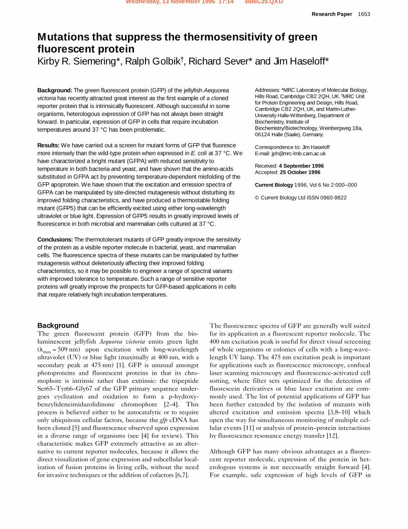
Induction of GFP and GFPA expression in E. coli. (a) Fluorescence ofcells expressing GFP or GFPA. E. coli XL1Blue cells containing pSE-GFP or pSE-GFPA were grown at 37 °C to an optical density of 0.2 at600 nm and then induced with 0.5 mM IPTG. At the times shown,samples were withdrawn, diluted to an optical density of 0.2 at 600 nm,and the fluorescence (lex = 397 nm, lem = 508 nm) measured at 25 °C.(b) Accumulation of recombinant protein in cells expressing GFP orGFPA. Total protein prepared from cells collected 270 min after inductionwith IPTG was western blotted (with anti-GFP antibodies) as described inMaterials and methods. Protein was from cells carrying the pSE380vector (lane marked Vector), pSE-GFP (GFP) and pSE-GFPA (GFPA).
0
200
400
600
800
1000
0 200 400 600 800 1000 1200
GFP
GFPA
Fluo
resc
ence
(arb
itrar
y un
its)
Time after induction (min)
(a)
(b)
17.2 kDa
30.0 kDa
42.7 kDaVec
tor
GFP GFPA
cence of similar numbers (equal optical densities) of E. colicells containing the resulting plasmids was then measuredat various times following induction of protein synthesis at37 °C with isopropyl b-D-thiogalactoside (IPTG; see Fig.1a). At 4.5 hours after induction, cells expressing GFPAwere observed to fluoresce 20-fold more intensely thanthose expressing GFP, a figure which increased to 35-foldby the time the cells had entered stationary phase (9 hoursafter induction). To determine whether the enhanced fluo-rescence of cells expressing GFPA was due to increasedlevels of protein expression, total protein was prepared fromcells sampled at the 4.5 hour time point and the amount ofintracellular GFP estimated by western immunoblot. Ascan be seen in Figure 1b, GFPA accumulates inside cells toa significantly higher level than does GFP. However, thedifference in protein levels as estimated by quantificationof band intensities is 2.4-fold, not nearly enough to accountfor the 20-fold difference in fluorescence levels observed atthis time point. This result suggests either that a large pro-portion of GFP that is expressed in cells at 37 °C is non-flu-orescent and that the substitutions present in GFPAenhance the formation or stability of the mature fluorescentform, or that the substitutions in GFPA substantiallyincrease the intrinsic fluorescence of the mature protein.Comparing the growth curves of strains expressing GFP orGFPA with the growth curve of a non-expressing strain(data not shown) indicated that expression of these proteinshas little adverse effect on the growth of bacterial cells.
The amino-acid substitutions present in GFPA enhancematuration at elevated temperatures in bacteria and yeastExperiments with a GFP–nucleoplasmin fusion proteinhave indicated that maturation of GFP to the fluorescentform may be sensitive to temperature during expression inthe yeast Saccharomyces cerevisiae [19]. To test whether the
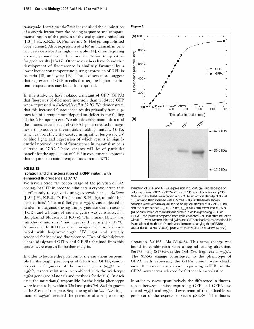
same could be true of expression in E. coli, and whetherthe substitutions present in GFPA enhance maturation bysuppressing any such sensitivity, we examined expressionof GFP and GFPA over a range of temperatures. Strainsexpressing GFP or GFPA were grown overnight at tem-peratures between 25 °C and 42 °C. For each culture, thefluorescence of an equal optical density of cells was mea-sured at 25 °C and the amount of intracellular recombi-nant protein determined by western blot (Table 1).Fluorescence values were then normalized against theamount of recombinant protein present in the cells to givea relative measure of the proportion of intracellular GFPthat is fluorescent for each culture. The results (Fig. 2)clearly show that the proportion of GFP in the cells that isfluorescent steadily decreases with increasing incubationtemperature, indicating that either mature GFP or thepathway leading to its formation is temperature-sensitive.
In order to test whether mature GFP itself is thermosensi-tive, we examined the effect of temperature on its fluores-cence in bacterial cells. E. coli cells expressing GFP weregrown to mid-log phase at 25 °C and treated with 50 mgml–1 chloramphenicol to inhibit further protein synthesis.The culture was then divided in two and one half incubatedat 25 °C and the other at 42 °C for an additional 4.5 hours.Comparison of the fluorescence intensities of the twosamples revealed no significant difference (data not shown),indicating that the fluorescence of mature GFP synthesizedprior to the chloramphenicol block is unaffected by temper-ature. This finding is consistent with the fact that mature
Research Paper Thermotolerant green fluorescent protein Siemering et al. 1655
Table 1
Fluorescence and accumulation of recombinant protein inbacteria expressing GFP or GFPA at different temperatures.
Temperature Fluorescence Relative amount of intracellular(°C) (arbitrary units)* recombinant protein†
GFP GFPA GFP GFPA
25 328.4 722.2 0.29 0.58
30 100.5 541.1 0.21 0.82
37 67.9 2273.0 0.23 1.00
42 9.2 369.4 0.17 0.44
*XL1-Blue cells containing pSE-GFP or pSE-GFPA were grown to anoptical density of 0.2 at 600 nm and then induced overnight with 0.2 mMIPTG. The fluorescence of cell samples was measured as in Fig. 1a. †Total protein prepared from the cell samples used for fluorescencemeasurements was western blotted as in Fig. 1b and the amounts ofintracellular recombinant protein determined by quantification of bandintensities. Data have been normalized to the highest value that wasobtained (7.8 3 105 arbitrary units for GFPA expressed at 37 °C).
Figure 2
Relative proportions of GFP and GFPA that are fluorescent in bacterialcells grown at different temperatures. Values were calculated bynormalization of fluorescence values against the amount of intracellularrecombinant protein for cultures grown at 25, 30, 37 or 42°C (Table 1).
0
500
1000
1500
2000
2500
25 30 37 42
GFPA
Fluo
resc
ence
(nor
mal
ized
)
Temperature (°C)
GFP
GFP is a highly stable molecule whose fluorescence in vitrois unaffected by temperatures up to 65 °C [20], and withthe results of a similar experiment that was carried out inyeast cells using a GFP–nucleoplasmin fusion protein [19].We therefore conclude that higher incubation temperaturesinterfere with the post-translational maturation of GFPrather than causing inactivation of the mature protein.Moreover, the primary effect of the substitutions present inGFPA is to enhance the proportion of GFP that is fluores-cent at higher temperatures, rather than simply enhancingthe intrinsic fluorescence properties of mature GFP.
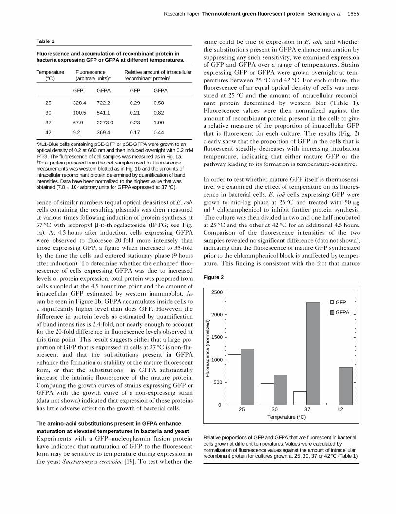
To determine whether the substitutions present in GFPAwere also capable of suppressing thermosensitivity of GFPmaturation in yeast, strains of S. cerevisiae expressingeither GFP or GFPA were grown on agar plates at either25 °C or 37 °C. The results, shown in Figure 3, confirmthat expression of GFP is temperature-sensitive in yeastand demonstrate that this phenomenon is also suppressedby the substitutions present in GFPA. These results indi-cate that the thermosensitivity of GFP maturation may bea common phenomenon that can be suppressed by theamino-acid substitutions present in GFPA.
Chromophore oxidation during maturation of GFP and GFPAThe post-translational maturation of GFP to the fluorescentform involves a number of steps [2–4]. First, the GFPapoprotein must presumably fold into a catalytically activeconformation that facilitates cyclization and oxidation of thetripeptide Ser65–Tyr66–Gly67. The mature protein mustthen be correctly folded to maintain its fluorescent proper-ties, presumably to protect the chromophore from solventeffects [21–23]. In principle, any of these steps could besensitive to temperature and could thus be responsible forthe observed thermosensitivity of GFP during maturation.
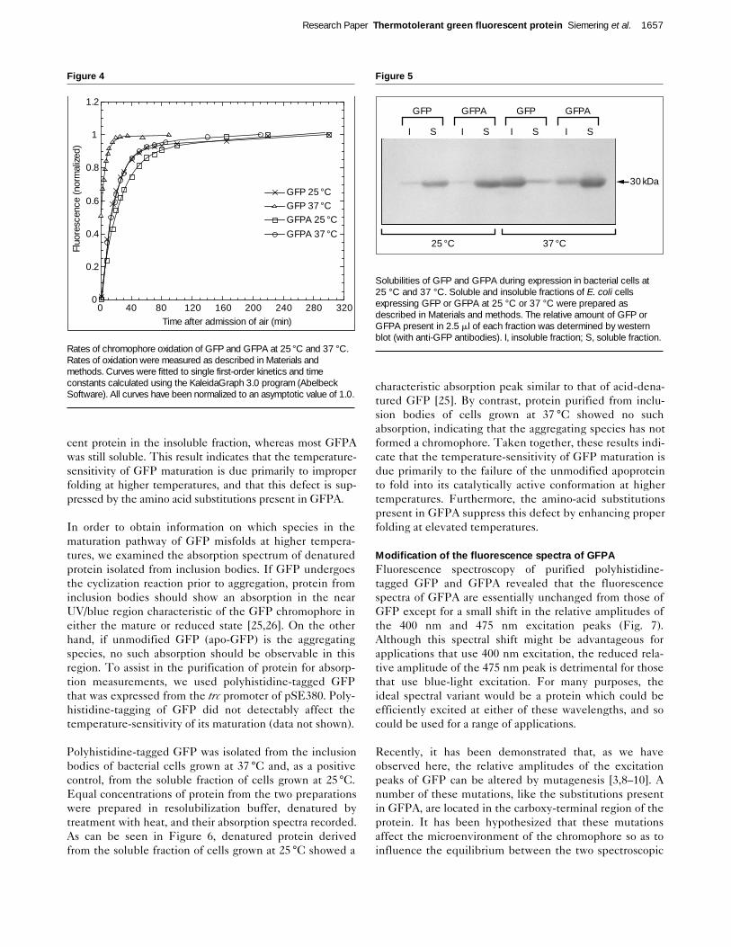
Because the oxidation reaction involved in chromophoreformation appears to require molecular oxygen, Heim et al.[10] were able to measure the reaction rate by expressingGFP in E. coli under anaerobic conditions and then moni-toring the development of fluorescence after admission ofair. To determine whether this reaction is temperature-sensitive and whether the substitutions present in GFPAenhance its rate at higher temperatures, we measured therates of oxidation of GFP and GFPA at both 25 °C and37 °C. For our experiments we chose to use a yeast expres-sion system which provided for better growth and expres-sion levels under anaerobic conditions than bacteria. S.cerevisiae cells expressing either GFP or GFPA were grownunder anaerobic conditions overnight at 30 °C. As was thecase in bacteria, S. cerevisiae cells exhibited no fluorescencefollowing anaerobic growth. Air was then admitted to thecultures and the development of fluorescence followed ateither 25 °C or 37 °C. As reported previously [10], each oxi-dation proceeded as a pseudo-first-order reaction (Fig. 4).The time constant measured for the oxidation of GFP at 37
°C (5.9 ± 0.1 min) was found to be approximately 3-foldfaster than that measured at 25 °C (16.2 ± 0.3 min), indicat-ing that the post-translational oxidation of the GFP chro-mophore is not the step responsible for the temperaturesensitivity of maturation. In confirmation of this conclu-sion, the time constants derived for GFPA at both 25 °Cand 37 °C (22.5 ± 1.4 min and 18.1 ± 0.4 min, respectively)were actually slower than those measured for GFP.
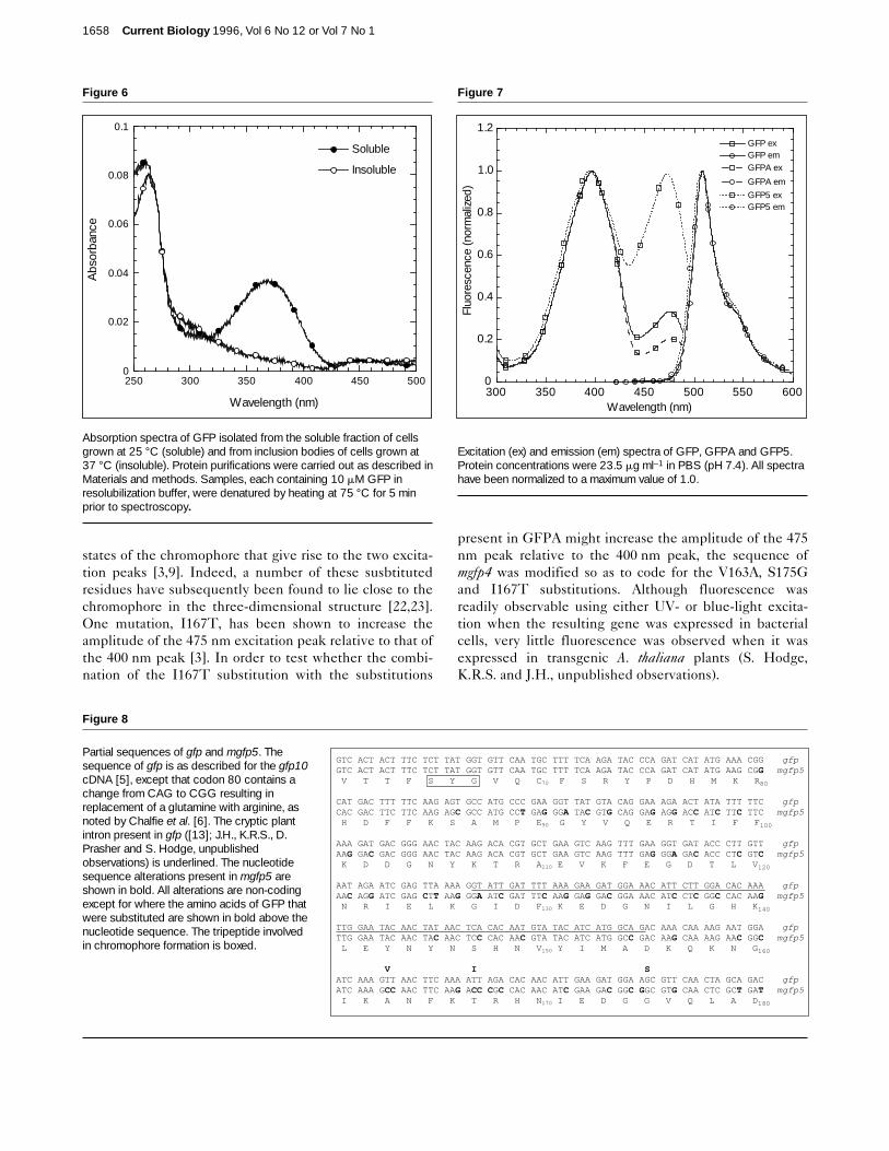
Apo-GFP folds improperly at elevated temperaturesThe improper folding of proteins often results in their aggre-gation into insoluble inclusion bodies during expression inE. coli [24]. To determine whether the proper folding ofGFP might be temperature-sensitive and whether the sub-stitutions present in GFPA act by enhancing proper foldingat increased temperatures, we examined the solubilities ofthe two proteins during expression in E. coli at 25 °C and 37°C. Bacterial cells expressing GFP or GFPA were grownovernight at either temperature, lysed, and the soluble andinsoluble fractions separated by centrifugation (see Materi-als and methods for details). In each case, fluorescence wasfound almost exclusively in the soluble fraction. Theamount of GFP or GFPA present in each fraction was thenestimated by western blot (Fig. 5). At 25 °C, both GFP andGFPA were found predominantly in the soluble fraction,indicating relatively efficient folding of both proteins. At 37°C, however, the majority of GFP was found as non-fluores-
1656 Current Biology 1996, Vol 6 No 12 or Vol 7 No 1
Figure 3
The substitutions present in GFPA cure the thermosensitivity of GFPexpression in yeast. Strains of S. cerevisiae MGLD-4a expressingeither GFP or GFPA were incubated at 25 °C and 37 °C on syntheticdrop-out agar lacking uracil [33]. Colonies (each approximately 1.5 mmin diameter) were visualized as described in Materials and methodsusing the Leitz-D filter set suitable for UV excitation of GFP.
25ºC
GFP
GFPA
37ºC
cent protein in the insoluble fraction, whereas most GFPAwas still soluble. This result indicates that the temperature-sensitivity of GFP maturation is due primarily to improperfolding at higher temperatures, and that this defect is sup-pressed by the amino acid substitutions present in GFPA.
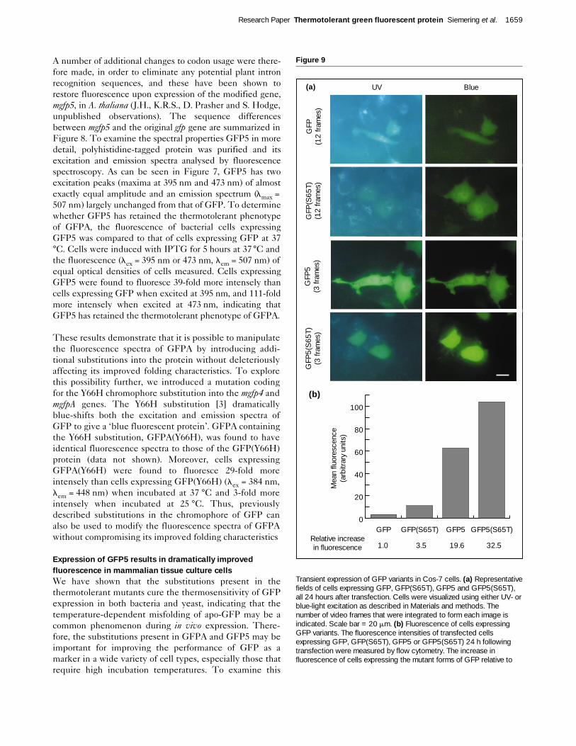
In order to obtain information on which species in thematuration pathway of GFP misfolds at higher tempera-tures, we examined the absorption spectrum of denaturedprotein isolated from inclusion bodies. If GFP undergoesthe cyclization reaction prior to aggregation, protein frominclusion bodies should show an absorption in the nearUV/blue region characteristic of the GFP chromophore ineither the mature or reduced state [25,26]. On the otherhand, if unmodified GFP (apo-GFP) is the aggregatingspecies, no such absorption should be observable in thisregion. To assist in the purification of protein for absorp-tion measurements, we used polyhistidine-tagged GFPthat was expressed from the trc promoter of pSE380. Poly-histidine-tagging of GFP did not detectably affect thetemperature-sensitivity of its maturation (data not shown).
Polyhistidine-tagged GFP was isolated from the inclusionbodies of bacterial cells grown at 37 °C and, as a positivecontrol, from the soluble fraction of cells grown at 25°C.Equal concentrations of protein from the two preparationswere prepared in resolubilization buffer, denatured bytreatment with heat, and their absorption spectra recorded.As can be seen in Figure 6, denatured protein derivedfrom the soluble fraction of cells grown at 25 °C showed a
characteristic absorption peak similar to that of acid-dena-tured GFP [25]. By contrast, protein purified from inclu-sion bodies of cells grown at 37 °C showed no suchabsorption, indicating that the aggregating species has notformed a chromophore. Taken together, these results indi-cate that the temperature-sensitivity of GFP maturation isdue primarily to the failure of the unmodified apoproteinto fold into its catalytically active conformation at highertemperatures. Furthermore, the amino-acid substitutionspresent in GFPA suppress this defect by enhancing properfolding at elevated temperatures.
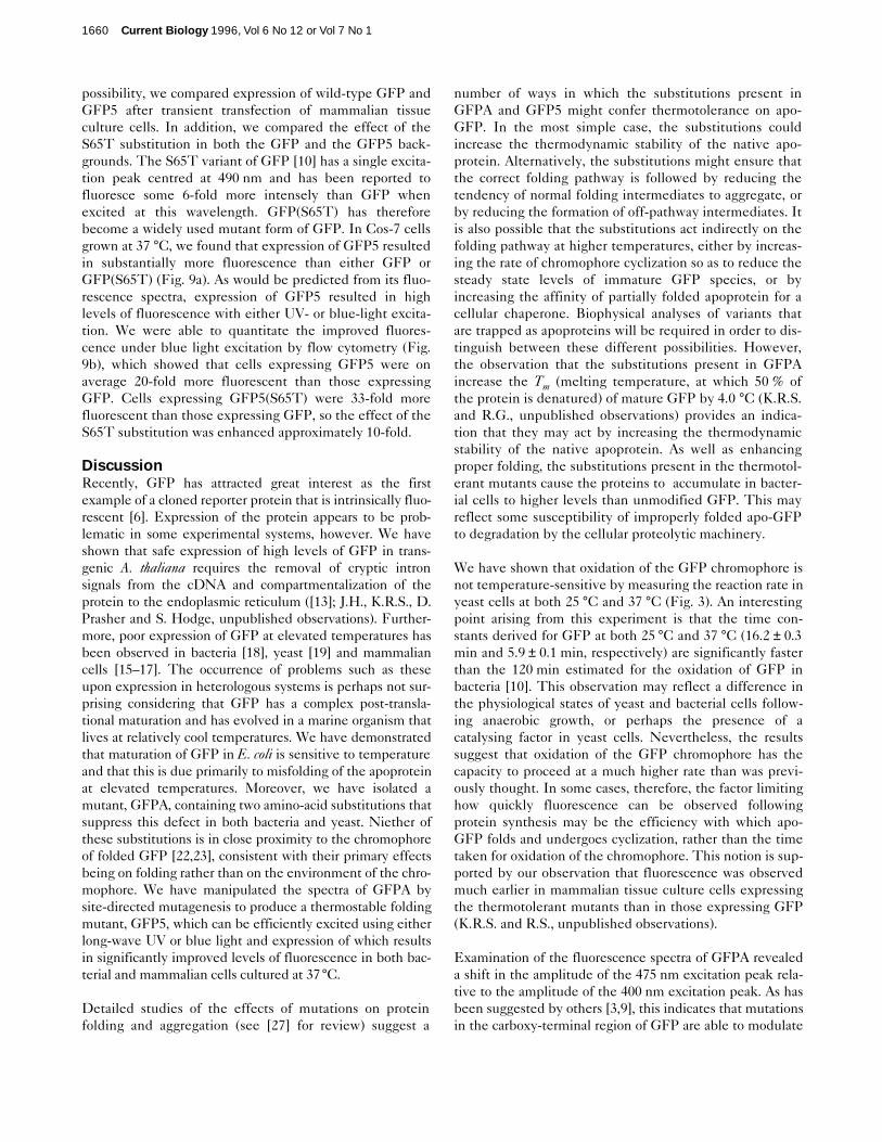
Modification of the fluorescence spectra of GFPAFluorescence spectroscopy of purified polyhistidine-tagged GFP and GFPA revealed that the fluorescencespectra of GFPA are essentially unchanged from those ofGFP except for a small shift in the relative amplitudes ofthe 400 nm and 475 nm excitation peaks (Fig. 7).Although this spectral shift might be advantageous forapplications that use 400 nm excitation, the reduced rela-tive amplitude of the 475 nm peak is detrimental for thosethat use blue-light excitation. For many purposes, theideal spectral variant would be a protein which could beefficiently excited at either of these wavelengths, and socould be used for a range of applications.
Recently, it has been demonstrated that, as we haveobserved here, the relative amplitudes of the excitationpeaks of GFP can be altered by mutagenesis [3,8–10]. Anumber of these mutations, like the substitutions presentin GFPA, are located in the carboxy-terminal region of theprotein. It has been hypothesized that these mutationsaffect the microenvironment of the chromophore so as toinfluence the equilibrium between the two spectroscopic
Research Paper Thermotolerant green fluorescent protein Siemering et al. 1657
Figure 4
Rates of chromophore oxidation of GFP and GFPA at 25°C and 37 °C.Rates of oxidation were measured as described in Materials andmethods. Curves were fitted to single first-order kinetics and timeconstants calculated using the KaleidaGraph 3.0 program (AbelbeckSoftware). All curves have been normalized to an asymptotic value of 1.0.
0
0.2
0.4
0.6
0.8
1
1.2
0 40 80 120 160 200 240 280 320
GFPA 37 °CGFPA 25 °CGFP 37 °CGFP 25 °C
Fluo
resc
ence
(nor
mal
ized
)
Time after admission of air (min)
Figure 5
Solubilities of GFP and GFPA during expression in bacterial cells at25 °C and 37 °C. Soluble and insoluble fractions of E. coli cellsexpressing GFP or GFPA at 25 °C or 37 °C were prepared asdescribed in Materials and methods. The relative amount of GFP orGFPA present in 2.5 ml of each fraction was determined by westernblot (with anti-GFP antibodies). I, insoluble fraction; S, soluble fraction.
30 kDa
25 °C 37 °C
I S I S I S I S
GFP GFPA GFP GFPA
states of the chromophore that give rise to the two excita-tion peaks [3,9]. Indeed, a number of these susbtitutedresidues have subsequently been found to lie close to thechromophore in the three-dimensional structure [22,23].One mutation, I167T, has been shown to increase theamplitude of the 475 nm excitation peak relative to that ofthe 400 nm peak [3]. In order to test whether the combi-nation of the I167T substitution with the substitutions
present in GFPA might increase the amplitude of the 475nm peak relative to the 400 nm peak, the sequence ofmgfp4 was modified so as to code for the V163A, S175Gand I167T substitutions. Although fluorescence wasreadily observable using either UV- or blue-light excita-tion when the resulting gene was expressed in bacterialcells, very little fluorescence was observed when it wasexpressed in transgenic A. thaliana plants (S. Hodge,K.R.S. and J.H., unpublished observations).
1658 Current Biology 1996, Vol 6 No 12 or Vol 7 No 1
Figure 6
Absorption spectra of GFP isolated from the soluble fraction of cellsgrown at 25 °C (soluble) and from inclusion bodies of cells grown at37 °C (insoluble). Protein purifications were carried out as described inMaterials and methods. Samples, each containing 10 mM GFP inresolubilization buffer, were denatured by heating at 75 °C for 5 minprior to spectroscopy.
0
0.02
0.04
0.06
0.08
0.1
250 300 350 400 450 500
Soluble
Insoluble
Abs
orba
nce
Wavelength (nm)
Figure 7
Excitation (ex) and emission (em) spectra of GFP, GFPA and GFP5.Protein concentrations were 23.5 mg ml–1 in PBS (pH 7.4). All spectrahave been normalized to a maximum value of 1.0.
0
0.2
0.4
0.6
0.8
1.0
1.2
300 350 400 450 500 550 600
GFP5 em
GFP exGFP emGFPA ex
GFPA em
GFP5 ex
Fluo
resc
ence
(nor
mal
ized
)
Wavelength (nm)
Figure 8
Partial sequences of gfp and mgfp5. Thesequence of gfp is as described for the gfp10cDNA [5], except that codon 80 contains achange from CAG to CGG resulting inreplacement of a glutamine with arginine, asnoted by Chalfie et al. [6]. The cryptic plantintron present in gfp ([13]; J.H., K.R.S., D.Prasher and S. Hodge, unpublishedobservations) is underlined. The nucleotidesequence alterations present in mgfp5 areshown in bold. All alterations are non-codingexcept for where the amino acids of GFP thatwere substituted are shown in bold above thenucleotide sequence. The tripeptide involvedin chromophore formation is boxed.
GTC ACT ACT TTC TCT TAT GGT GTT CAA TGC TTT TCA AGA TAC CCA GAT CAT ATG AAA CGG gfp GTC ACT ACT TTC TCT TAT GGT GTT CAA TGC TTT TCA AGA TAC CCA GAT CAT ATG AAG CGG mgfp5 V T T F S Y G V Q C70 F S R Y P D H M K R80 CAT GAC TTT TTC AAG AGT GCC ATG CCC GAA GGT TAT GTA CAG GAA AGA ACT ATA TTT TTC gfp CAC GAC TTC TTC AAG AGC GCC ATG CCT GAG GGA TAC GTG CAG GAG AGG ACC ATC TTC TTC mgfp5 H D F F K S A M P E90 G Y V Q E R T I F F100 AAA GAT GAC GGG AAC TAC AAG ACA CGT GCT GAA GTC AAG TTT GAA GGT GAT ACC CTT GTT gfp AAG GAC GAC GGG AAC TAC AAG ACA CGT GCT GAA GTC AAG TTT GAG GGA GAC ACC CTC GTC mgfp5 K D D G N Y K T R A110 E V K F E G D T L V120 AAT AGA ATC GAG TTA AAA GGT ATT GAT TTT AAA GAA GAT GGA AAC ATT CTT GGA CAC AAA gfp AAC AGG ATC GAG CTT AAG GGA ATC GAT TTC AAG GAG GAC GGA AAC ATC CTC GGC CAC AAG mgfp5 N R I E L K G I D F130 K E D G N I L G H K140 TTG GAA TAC AAC TAT AAC TCA CAC AAT GTA TAC ATC ATG GCA GAC AAA CAA AAG AAT GGA gfp TTG GAA TAC AAC TAC AAC TCC CAC AAC GTA TAC ATC ATG GCC GAC AAG CAA AAG AAC GGC mgfp5 L E Y N Y N S H N V150 Y I M A D K Q K N G160 V I S ATC AAA GTT AAC TTC AAA ATT AGA CAC AAC ATT GAA GAT GGA AGC GTT CAA CTA GCA GAC gfp ATC AAA GCC AAC TTC AAG ACC CGC CAC AAC ATC GAA GAC GGC GGC GTG CAA CTC GCT GAT mgfp5 I K A N F K T R H N170 I E D G G V Q L A D180
A number of additional changes to codon usage were there-fore made, in order to eliminate any potential plant intronrecognition sequences, and these have been shown torestore fluorescence upon expression of the modified gene,mgfp5, in A. thaliana (J.H., K.R.S., D. Prasher and S. Hodge,unpublished observations). The sequence differencesbetween mgfp5 and the original gfp gene are summarized inFigure 8. To examine the spectral properties GFP5 in moredetail, polyhistidine-tagged protein was purified and itsexcitation and emission spectra analysed by fluorescencespectroscopy. As can be seen in Figure 7, GFP5 has twoexcitation peaks (maxima at 395 nm and 473 nm) of almostexactly equal amplitude and an emission spectrum (lmax =507 nm) largely unchanged from that of GFP. To determinewhether GFP5 has retained the thermotolerant phenotypeof GFPA, the fluorescence of bacterial cells expressingGFP5 was compared to that of cells expressing GFP at 37°C. Cells were induced with IPTG for 5 hours at 37 °C andthe fluorescence (lex = 395 nm or 473 nm, lem = 507 nm) ofequal optical densities of cells measured. Cells expressingGFP5 were found to fluoresce 39-fold more intensely thancells expressing GFP when excited at 395 nm, and 111-foldmore intensely when excited at 473 nm, indicating thatGFP5 has retained the thermotolerant phenotype of GFPA.
These results demonstrate that it is possible to manipulatethe fluorescence spectra of GFPA by introducing addi-tional substitutions into the protein without deleteriouslyaffecting its improved folding characteristics. To explorethis possibility further, we introduced a mutation codingfor the Y66H chromophore substitution into the mgfp4 andmgfpA genes. The Y66H substitution [3] dramaticallyblue-shifts both the excitation and emission spectra ofGFP to give a ‘blue fluorescent protein’. GFPA containingthe Y66H substitution, GFPA(Y66H), was found to haveidentical fluorescence spectra to those of the GFP(Y66H)protein (data not shown). Moreover, cells expressingGFPA(Y66H) were found to fluoresce 29-fold moreintensely than cells expressing GFP(Y66H) (lex = 384 nm,lem = 448 nm) when incubated at 37 °C and 3-fold moreintensely when incubated at 25 °C. Thus, previouslydescribed substitutions in the chromophore of GFP canalso be used to modify the fluorescence spectra of GFPAwithout compromising its improved folding characteristics
Expression of GFP5 results in dramatically improvedfluorescence in mammalian tissue culture cellsWe have shown that the substitutions present in thethermotolerant mutants cure the thermosensitivity of GFPexpression in both bacteria and yeast, indicating that thetemperature-dependent misfolding of apo-GFP may be acommon phenomenon during in vivo expression. There-fore, the substitutions present in GFPA and GFP5 may beimportant for improving the performance of GFP as amarker in a wide variety of cell types, especially those thatrequire high incubation temperatures. To examine this
Research Paper Thermotolerant green fluorescent protein Siemering et al. 1659
Figure 9
Transient expression of GFP variants in Cos-7 cells. (a) Representativefields of cells expressing GFP, GFP(S65T), GFP5 and GFP5(S65T),all 24 hours after transfection. Cells were visualized using either UV- orblue-light excitation as described in Materials and methods. Thenumber of video frames that were integrated to form each image isindicated. Scale bar = 20 mm. (b) Fluorescence of cells expressingGFP variants. The fluorescence intensities of transfected cellsexpressing GFP, GFP(S65T), GFP5 or GFP5(S65T) 24 h followingtransfection were measured by flow cytometry. The increase influorescence of cells expressing the mutant forms of GFP relative to
0
20
40
60
80
100
GFP5(S65T)GFP5GFP(S65T)GFP
Mea
n flu
ores
cenc
e (a
rbitr
ary
units
)
Relative increase in fluorescence 1.0 3.5 19.6 32.5
UV Blue
GFP
(1
2 fra
mes
)G
FP(S
65T)
(1
2 fra
mes
)G
FP5
(3 fr
ames
)G
FP5(
S65
T)
(3 fr
ames
)
(a)
(b)
possibility, we compared expression of wild-type GFP andGFP5 after transient transfection of mammalian tissueculture cells. In addition, we compared the effect of theS65T substitution in both the GFP and the GFP5 back-grounds. The S65T variant of GFP [10] has a single excita-tion peak centred at 490 nm and has been reported tofluoresce some 6-fold more intensely than GFP whenexcited at this wavelength. GFP(S65T) has thereforebecome a widely used mutant form of GFP. In Cos-7 cellsgrown at 37 °C, we found that expression of GFP5 resultedin substantially more fluorescence than either GFP orGFP(S65T) (Fig. 9a). As would be predicted from its fluo-rescence spectra, expression of GFP5 resulted in highlevels of fluorescence with either UV- or blue-light excita-tion. We were able to quantitate the improved fluores-cence under blue light excitation by flow cytometry (Fig.9b), which showed that cells expressing GFP5 were onaverage 20-fold more fluorescent than those expressingGFP. Cells expressing GFP5(S65T) were 33-fold morefluorescent than those expressing GFP, so the effect of theS65T substitution was enhanced approximately 10-fold.
DiscussionRecently, GFP has attracted great interest as the firstexample of a cloned reporter protein that is intrinsically fluo-rescent [6]. Expression of the protein appears to be prob-lematic in some experimental systems, however. We haveshown that safe expression of high levels of GFP in trans-genic A. thaliana requires the removal of cryptic intronsignals from the cDNA and compartmentalization of theprotein to the endoplasmic reticulum ([13]; J.H., K.R.S., D.Prasher and S. Hodge, unpublished observations). Further-more, poor expression of GFP at elevated temperatures hasbeen observed in bacteria [18], yeast [19] and mammaliancells [15–17]. The occurrence of problems such as theseupon expression in heterologous systems is perhaps not sur-prising considering that GFP has a complex post-transla-tional maturation and has evolved in a marine organism thatlives at relatively cool temperatures. We have demonstratedthat maturation of GFP in E. coli is sensitive to temperatureand that this is due primarily to misfolding of the apoproteinat elevated temperatures. Moreover, we have isolated amutant, GFPA, containing two amino-acid substitutions thatsuppress this defect in both bacteria and yeast. Niether ofthese substitutions is in close proximity to the chromophoreof folded GFP [22,23], consistent with their primary effectsbeing on folding rather than on the environment of the chro-mophore. We have manipulated the spectra of GFPA bysite-directed mutagenesis to produce a thermostable foldingmutant, GFP5, which can be efficiently excited using eitherlong-wave UV or blue light and expression of which resultsin significantly improved levels of fluorescence in both bac-terial and mammalian cells cultured at 37°C.
Detailed studies of the effects of mutations on proteinfolding and aggregation (see [27] for review) suggest a
number of ways in which the substitutions present inGFPA and GFP5 might confer thermotolerance on apo-GFP. In the most simple case, the substitutions couldincrease the thermodynamic stability of the native apo-protein. Alternatively, the substitutions might ensure thatthe correct folding pathway is followed by reducing thetendency of normal folding intermediates to aggregate, orby reducing the formation of off-pathway intermediates. Itis also possible that the substitutions act indirectly on thefolding pathway at higher temperatures, either by increas-ing the rate of chromophore cyclization so as to reduce thesteady state levels of immature GFP species, or byincreasing the affinity of partially folded apoprotein for acellular chaperone. Biophysical analyses of variants thatare trapped as apoproteins will be required in order to dis-tinguish between these different possibilities. However,the observation that the substitutions present in GFPAincrease the Tm (melting temperature, at which 50 % ofthe protein is denatured) of mature GFP by 4.0 °C (K.R.S.and R.G., unpublished observations) provides an indica-tion that they may act by increasing the thermodynamicstability of the native apoprotein. As well as enhancingproper folding, the substitutions present in the thermotol-erant mutants cause the proteins to accumulate in bacter-ial cells to higher levels than unmodified GFP. This mayreflect some susceptibility of improperly folded apo-GFPto degradation by the cellular proteolytic machinery.
We have shown that oxidation of the GFP chromophore isnot temperature-sensitive by measuring the reaction rate inyeast cells at both 25 °C and 37 °C (Fig. 3). An interestingpoint arising from this experiment is that the time con-stants derived for GFP at both 25 °C and 37 °C (16.2 ± 0.3min and 5.9 ± 0.1 min, respectively) are significantly fasterthan the 120 min estimated for the oxidation of GFP inbacteria [10]. This observation may reflect a difference inthe physiological states of yeast and bacterial cells follow-ing anaerobic growth, or perhaps the presence of acatalysing factor in yeast cells. Nevertheless, the resultssuggest that oxidation of the GFP chromophore has thecapacity to proceed at a much higher rate than was previ-ously thought. In some cases, therefore, the factor limitinghow quickly fluorescence can be observed followingprotein synthesis may be the efficiency with which apo-GFP folds and undergoes cyclization, rather than the timetaken for oxidation of the chromophore. This notion is sup-ported by our observation that fluorescence was observedmuch earlier in mammalian tissue culture cells expressingthe thermotolerant mutants than in those expressing GFP(K.R.S. and R.S., unpublished observations).
Examination of the fluorescence spectra of GFPA revealeda shift in the amplitude of the 475 nm excitation peak rela-tive to the amplitude of the 400 nm excitation peak. As hasbeen suggested by others [3,9], this indicates that mutationsin the carboxy-terminal region of GFP are able to modulate
1660 Current Biology 1996, Vol 6 No 12 or Vol 7 No 1
the spectroscopic state of the chromophore by affecting itslocal environment within the protein. We took advantage ofthis phenomenon to engineer the fluorescence spectra ofGFPA by combining a third substitution, Il67T [3], with thetwo substitutions already present in the carboxy-terminalregion. The resulting protein, GFP5, has two excitationpeaks (maxima at 395 nm and 473 nm) of almost equalamplitude and is thus ideal as a multi-purpose spectralvariant which can be used for applications requiring eitherlong-wavelength UV- or blue-light excitation. We were alsoable to manipulate the fluorescence spectra of GFPA byintroducing the Y66H chromophore substitution to give aprotein, GFPA(Y66H), with identical blue-shifted fluores-cence spectra to those of GFP(Y66H). The observation thatmaturation of both GFP5 and GFPA(Y66H) is thermotoler-ant demonstrates that it is possible to manipulate the fluo-rescence spectra of GFPA by the introduction of additionalsubstitutions into either the carboxy-terminal or the chro-mophore region of the protein without deleteriously affect-ing its improved folding characteristics. We were thereforeable to combine the S65T substitution with the substitu-tions present in GFP5 to substantially enhance fluorescenceof the S65T variant in mammalian cells (Fig. 9). Thus, thepotential exists for the rational design of a range of thermo-tolerant proteins with varying excitation and emission prop-erties by combining previously described spectral mutationswith the substitutions present in GFPA or GFP5.
The fact that the substitutions present in GFPA and GFP5affect the fluorescence spectra of GFP suggests that theymay affect the extinction coefficient (the molar absorptiv-ity) and/or the quantum yields of the chromophore at thetwo excitation wavelengths, in addition to enhancingproper folding at elevated temperatures. However, ineffi-cient folding of apo-GFP presents a significant practicalproblem for the measurement of extinction coefficients.Accurate determination of an extinction coefficientrequires that the precise concentration of mature fluores-cent protein in a given sample be known. The results pre-sented here suggest that, even in a soluble cell fraction,there may be appreciable amounts of improperly foldedapoprotein. This conclusion is supported by the observa-tion that the ratio of the absorption of the chromophore tothat of the aromatic amino acids of GFP purified from thesoluble fraction of bacterial cells grown at 25 °C wasapproximately 0.4 (Fig. 6). Because this value is in excessof 1.0 for either native or acid-denatured GFP isolateddirectly from Aequorea [25], it would appear that over halfof the recombinant GFP in our soluble fraction lacked afunctional chromophore, thus making any attempted mea-surement of an extinction coefficient artificially low.
We suggest that in the absence of an assay for the concen-tration of chromophore in a given protein sample, caremust be taken when assessing the effects of substitutionson the intrinsic fluorescence properties of recombinant
forms of GFP. For example, two bright mutants (W7 andW2) of the Y66W chromophore variant of GFP haverecently been described that are reported to have extinc-tion coefficients and quantum yields comparable to thoseof wild-type GFP [12]. The mutants contain four and sixamino-acid substitutions, respectively, which are thoughtto act by improving the fit of the substituted tryptophanresidue into the internal cavity normally occupied by tyro-sine. However, the presence in both mutants of the V163Asubstitution, shown here to be important for improvingthe folding of GFP, suggests that their increased bright-ness may be due, at least in part, to improved foldingrather than improvements in their extinction coefficientsor quantum yields. Indeed, V163A may be a substitutioncentral to improvement of the folding of GFP and deriva-tives, a conclusion supported by its presence amongstthree substitutions in another bright mutant of GFP (C3)that is more soluble during expression in E. coli [28].
ConclusionsWe have shown that the substitutions present in GFPAand GFP5 suppress a temperature-sensitive defect in thefolding of apo-GFP in E. coli. As these substitutions alsosuppress the thermosensitivity of GFP in yeast cells andsignificantly improve levels of fluorescence in mammaliancells cultured at 37 °C, as well as in Drosophila melanogasterembryos incubated at 25 °C (A. Brand, personal communi-cation), it would appear that this folding defect is acommon phenomenon during GFP expression in vivo. Inaddition to coding for improved folding and spectral char-acteristics, the modified gfp genes described here have analtered codon usage necessary to ensure proper mRNAprocessing in plants ([13]; J.H., K.R.S., D. Prasher and S.Hodge, unpublished observations), and which may beadvantageous for expression in other heterologous celltypes. Indeed, ‘humanization’ of the gfp coding sequencehas been reported to aid in the expression of GFP in mam-malian tissue culture cells [29,30]. Thus, we expect thatthe thermotolerant folding mutants described in this work,and spectral variants derived from them, should be of sig-nificant benefit for GFP-based applications in many exper-imental systems, in particular those, such as mammaliancells, that use relatively high incubation temperatures.
Materials and methodsMutagenesis and plasmid constructionThe sequence of mgfp4 was mutated by PCR in the presence of limit-ing nucleotide concentrations. The template plasmid was pBSmgfp4([13]; J.H., K.R.S., D. Prasher and S. Hodge, unpublished observa-tions), and the primers were the T3 and T7 primers (New EnglandBiolabs) that are complementary to the flanking T3 and T7 promoterspresent in the vector sequence. Four separate reactions (30 cycles of30 sec at 94 °C, 30 sec at 55 °C and 1 min at 72 °C using Taq DNAPolymerase from Promega) were carried out, each with the concentra-tion of a different nucleotide reduced from 200 mM to 20 mM. Theamplified fragments were pooled, cleaved with KpnI and EcoRI andcloned downstream of the lac promoter of pBluescript II KS (+) (Strata-gene). The mutant library was transformed into E. coli strain XL1-Blue
Research Paper Thermotolerant green fluorescent protein Siemering et al. 1661
(Stratagene) and incubated overnight at 37 °C on TYE agar containing50 mg ml–1 ampicillin and 1 mM IPTG. Colonies were illuminated with along-wavelength UV lamp (UVP Model B 100 AP) and visuallyscreened for increased fluorescence. The coding regions of twomutant genes (mgfpA and mgfpB) as well as that of mgfp4 were ampli-fied by PCR (30 cycles of 1 min at 94 °C, 1 min at 55 °C and 1 min at72 °C using VENT DNA Polymerase from New England Biolabs) usingthe optGFP5 oligo 5′-GGCGGATCCAAGGAGATATAACAATG AGTAAA GGA GAA GAA CTT TTC ACT-3′ (BamHI site underlined, ′phageShine–Dalgarno sequence in italics and plant tranlation initiationcontext sequence in bold) as the forward primer and the oligo5′GGCGAGCTCTTA TTT GTA TAG TTC ATC CAT GCC-3′ (SacI siteundelined and GFP stop codon in bold) as the reverse primer. Theamplified fragments were cleaved with BamHI and SacI and cloneddownstream of the lac promoter of pUC119 [31]. The mutationsresponsible for the bright phenotypes of mgfpA and mgfpB were thenlocated by recombination of the mutant genes with mgfp4. ThepUC119 derivatives containing mgfpA and mgfpB were cleaved witheither BamHI and NcoI, NcoI and ClaI or ClaI and SacI. The restrictionfragments were gel-purified and ligated to the mgfp4 pUC119 deriva-tive that had been cleaved with the same combination of enzymes andgel-purified. These and the parent constructs were introduced intoXL1-Blue cells and incubated overnight at 37 °C on agar plates con-taining ampicillin and IPTG. Comparison of the fluorescence ofcolonies containing the various constructs revealed that the mutation(s)responsible for the bright phenotypes of both mgfpA and mgfpB werecontained within the 336 base-pair ClaI–SacI fragment at the 3′ end ofthe gene. These fragments were cloned into the phage vectorM13mp19 (New England Biolabs) and sequenced from the Universalprimer using the Sequenase Version 2.0 DNA Sequencing Kit (UnitedStates Biochemical Corporation).
Site-directed mutagenesis to produce the mgfp5 and mgfp(Y66H)genes was by PCR amplification of mgfp4 using appropriate mutagenicprimers. Mutant fragments were subsequently inserted in place of thecorresponding non-mutant fragments of mgfp4; mgfpA(Y66H) andmgfp5(S65T) were constructed in the same way except that themgfpA and mgfp5 genes, respectively, were used as the templates forPCR amplification with mutagenic primers.
For bacterial expression, BamHI–SacI fragments containing the mgfp4,mgfpA, mgfp5, mgfp(Y66H) and mgfpA(Y66H) genes were excisedfrom the pUC119 derivatives and cloned downstream of the IPTG-inducible trc promoter of the expression vector pSE380 (Invitrogen), togive the plasmids pSE-GFP, pSE-GFPA, pSE-GFP5, pSE-GFP(Y66H)and pSE-GFPA(Y66H), respectively. Expression from the trc promoterof pSE380 is tightly regulated as a result of the presence on theplasmid of the lacIq gene. For yeast expression, BamHI–SacI fragmentscontaining the mgfp4 and mgfpA genes were inserted downstream ofthe constitutive ADH1 promoter of pVT-103-U [32], a yeast multicopyepisomal plasmid containing the URA3 selectable marker. The result-ing plasmids were pVT-GFP and pVTGFPA, respectively. For expres-sion in mammalian tissue culture cells, gfp [5], gfp(S65T) [10], mgfp5and mgfp5(S65T) were amplified by PCR using optGFP5 as theforward primer and the oligo 5′-GGCAGATCT TTA TTT GTA TAG TTCATC CAT GCC-3′ (BglII site underlined) as the reverse primer. Theamplified fragments were cleaved with BamHI and BglII and clonedinto the BamHI site that lies downstream of the CMV early promoter ofthe pcDNA3 expression vector (Invitrogen). The resulting vectors werenamed pcGFP, pcGFP(S65T), pcGFP5 and pcGFP5(S65T).
Polyhistidine-tagging was achieved by the addition of six histidine codonsto the 3′ ends of the modified gfp genes by PCR. The genes were ampli-fied using optGFP5 as the forward primer and the oligo 5′-GCC-GAGCTC TTA GTG GTG GTG GTG GTG GTG TTT GTA TAG TTCATC CAT GCC-3′ (SacI site underlined, histidine codons in bold) as thereverse primer. The amplified fragments were cleaved with BamHI andSacI and cloned downstream of the trc promoter of pSE380 to give theexpression plasmids pSE-GFPHis, pSE-GFPAHis and pSEGFP5His.
Determination of chromophore oxidation ratesStrains of S. cerevisiae MGLD-4a (MATa, leu2, ura3, his3, trp1, lys2)containing either pVT-GFP or pVT-GFPA were incubated anaerobically(Becton-Dickinson BBL GasPak Pouch) overnight at 30 °C in syntheticdrop-out media lacking uracil [33]. Following admission of air to thepouch, 1 ml of each culture was immediately centrifuged for 1 min at13 000 rpm and resuspended in 0.5 ml aerated and prewarmed PBS(pH 7.4) containing 8 mM NaN3 as a metabolic inhibitor. Cell suspen-sions were placed immediately into pre-warmed cuvettes held withinthe fluorimeter carousel and the time course of fluorescence (lex =397 nm, lem = 508 nm) development measured.
Preparation of soluble and insoluble cell fractionsCells were grown in 1.5 ml of 2xTY broth to an absorbance of 0.2 at600 nm and protein expression induced overnight with 0.2 mM IPTG.The cultures were centrifuged at 13 000 rpm for 2 min, resuspended in500 ml 50 mM Tris-HCl (pH 8.0), 2 mM EDTA, 100 mg ml–1 lysozyme,0.1 % Triton X-100 and incubated at 30 °C for 15 min. Cells were thenlysed by sonication (5 × 15 sec) using a Heat Systems (Model CL4)sonicator and centrifuged at 13 000 rpm for 15 min at 4 °C. The super-natant (soluble fraction) was removed and stored at –70 °C until used.The pellet (insoluble fraction) was washed once with 500 ml 50 mMTris–HCl (pH 8.0), 10 mM EDTA, 0.5 % Triton X-100, resolubilized for1 h at room temperature in 500 ml resolubilization buffer (8 M urea, 0.1M NaH2PO4, 10 mM Tris-HCl, pH 6.3) and stored at –70 °C until used.
Western blot analysisTotal protein from E. coli cells (diluted to an optical density of 0.2 at600 nm) was prepared by boiling cells in an equal volume of 2XSDSsample buffer for 3 min. SDS–PAGE using 10 % gels and westernblots were carried out according to standard protocols [34]. Primaryantibodies were polyclonal rabbit anti-GFP (generous gift of S. Santa-Cruz) used at a dilution of 1/2 000. Antibodies were detected with iod-inated Protein A (Amersham) and bands visualized and quantified usinga Molecular Dynamics Phosphorimager.
Protein purificationFor the purification of polyhistidine-tagged GFP for absorption mea-surements, soluble and insoluble fractions of cells containingpSE-GFPHis grown at 25 °C and 37 °C, respectively, were preparedas described above. GFP was purified from the fractions on Ni-chelatecolumns using the Ni–NTA Spin Kit (Qiagen). Purification from thesoluble fraction was carried out according to the manufacturer’s proto-col for the purification of polyhistidine-tagged proteins under native con-ditions. After clearance of cellular debris from the insoluble fraction bycentrifugation at 13 000 r.p.m. for 30 min, purification was carried outaccording to the protocol for purification of polyhistidine-tagged pro-teins under denaturing conditions, except that the protein was elutedwith resolubilization buffer (see above) containing 250 mM imidazole.
For the purification of native polyhistidine-tagged proteins for fluores-cence spectroscopy, cells were grown in 100 ml of 2xTY broth at 37°Cto an absorbance of 0.2 at 600 nm and then induced overnight with 0.5mM IPTG. Cells were harvested by centrifugation at 6 000 r.p.m. for 10min and lysed by resuspension in 4 ml 20 mM Tris–HCl (pH 7.9), 500mM NaCl, 5 mM imidazole, 0.1 % sarkosyl, 0.1 % deoxycholate, 2.25 Mguanidine–HCl. Nucleic acids were precipitated by the addition of 5 mlisopropanol and removed by centrifugation at 10 000 rpm for 10 min.Following 0.45 mm filtration, fluorescent polyhistidine-tagged proteinswere purified from the supernatant on Ni-chelate columns (Qiagen) andeluted with 2 ml 20 mM Tris-HCl (pH 7.9), 500 mM NaCl, 150 mM imi-dazole. For all purifications, protein purity was assayed by SDS–PAGEand found to be greater than 95 %. Protein concentrations were deter-mined by Bradford assay (Bio-Rad Protein Assay kit) using bovineserum albumin as a standard.
SpectroscopyAbsorbance spectra were recorded on a Cary 3 UV-Visible Spec-trophotometer (Varian) at 25°C. The optical path-length was 1 cm. Flu-
1662 Current Biology 1996, Vol 6 No 12 or Vol 7 No 1
orescence measurements were made on a Hitachi F-4500 fluorimeterusing 4mm/10mm cuvettes. The bandpass for both the excitation andthe emission monochromators was 5 nm, the scan speed240 nm min–1 and the response time automatically adapted by thedevice. All fluorescence spectra were recorded at 25 °C and were cor-rected following the supplier’s procedure for calibration of the fluorime-ter using Rhodamine-B as standard. Emission spectra were recordedat a fixed wavelength of the excitation maximum, and excitation spectraat a fixed wavelength of the emission maximum.
Transient transfection and flow cytometric analysis ofmammalian tissue culture cellsCos-7 cells were maintained in Dulbecco’s Modified Eagles Medium(DMEM) supplemented with 10 % fetal calf serum, and transfected byelectroporation [35], using 20 mg plasmid DNA per 106 cells. Transientlytransfected cells were grown on Lab-Tek Chambered Coverglass (Nunc)for 24 h at 37 °C prior to fluorescence microscopy. For flow cytometricanalysis, cells were grown in 100 mm dishes for 24 h following transfec-tion, trypsinized, and resuspended in PBS. The fluorescence intensitiesof the transfected cells were immediately analysed by flow cytometricanalysis using a FACScan analyser and LYSYS II software (BecktonDickinson). Samples were excited with an argon ion laser and fluores-cence was detected through the FLI filter (530DF30). The mean fluores-cence intensity for each sample was calculated by averaging the valuesfor the first 10 000 cells measured using detector settings calibrated toignore the low intensity autofluorescence of non-transfected cells.
Fluorescence microscopyFluorescent yeast colonies and Cos-7 cells were visualized with aninverted fluorescence microscope (Leitz DM-IL) fitted with filter sets suit-able for UV- (Leitz-D; excitation filter 355–425 nm, dichromatic mirror 455nm, suppression filter 460 nm) or blue- (Leitz-I3; excitation filter 450–490nm, dichromatic mirror 510 nm, suppression filter 520 nm) light excitationof GFP. Epifluorescence images were recorded using a Sony 3-chipCCD video camera and F100-MPU integrating frame store, connected toa NuVista+ video digitizer in an Apple Macintosh Quadra 800 computer.
AcknowledgementsWe are grateful to Simon Santa-Cruz for his generous gift of anti-GFP anti-bodies, to Andrew Riddel for assistance with flow cytometry, to JonathonPines and Mark Jackman for assistance with mammalian cell work and toAndrea Brand for critical reading of the manuscript. R.G. was supported byan Adolf-Butenandt-Grant from the Max-Planck-Society. This work was sup-ported by BBSRC grant PAG/01518 to J.H.
References1. Morise H, Shimomura O, Johnson FH, Winant J: Intermolecular
energy transfer in the bioluminescent system of Aequorea.Biochemistry 1974, 13:2656–2662.
2. Cody CW, Prasher DC, Westler WM, Prendergast FG, Ward WW:Chemical structure of the hexapeptide chromophore of theAequorea green-fluorescent protein. Biochemistry 1993,32:1212–1218.
3. Heim R, Prasher DC, Tsien RY: Wavelength mutations andposttranslational autoxidation of green fluorescent protein. ProcNatl Acad Sci USA 1994, 91:12501–12504.
4. Cubitt AB, Heim R, Adams SR, Boyd AE, Gross LA, Tsien RY:Understanding, improving and using green fluorescent proteins.Trends Biochem Sci 1995, 20:448–455.
5. Prasher DC, Eckenrode VK, Ward WW, Prendergast FG, Cormier MJ:Primary structure of the Aequorea victoria green-fluorescentprotein. Gene 1992, 111:229–233.
6. Chalfie M, Tu Y, Euskirchen G, Ward WW, Prasher DC: Greenfluorescent protein as a marker for gene expression. Science1994, 263:802–805.
7. Wang S, Hazelrigg T: Implications for bcd mRNA localization fromspatial distribution of exu protein in Drosophila oogenesis. Nature1994, 369:400–403.
8. Delagrave S, Hawtin RE, Silva CM, Yang MM, Youvan DC:
Redshifted excitation mutants of the green fluorescent protein.Bio/Technology 1995, 13:151–154.
9. Ehrig T, O’kane DJ, Prendergast FG: Green-fluorescent proteinmutants with altered fluorescence excitation spectra. FEBS Lett1995, 367:163–166.
10. Heim R, Cubitt AB, Tsien RY: Improved green fluorescence. Nature1995, 373:663–664.
11. Rizzuto R, Brini M, De Giorgi F, Rossi R, Heim R, Tsien RY, et al.:Double labelling of subcellular structures with organelle-targetedGFP mutants in vivo. Curr Biol 1996, 6:183–188.
12. Heim R, Tsien RY: Engineering green fluorescent protein forimproved brightness, longer wavelengths and fluorescenceresonance energy transfer. Curr Biol 1996, 6:178–182.
13. Haseloff J, Amos B: GFP in plants. Trends Genet 1995, 11:328–329.14. Rizzuto R, Brini M, Pizzo P, Murgia M, Pozzan T: Chimeric green
fluorescent protein as a tool for visualizing subcellular organellesin living cells. Curr Biol 1995, 5:635–642.
15. Kaether C, Gerdes HH: Visualization of protein transport along thesecretory pathway using green fluorescent protein. FEBS Lett1995, 369:267–271.
16. Ogawa H, Inouye S, Tsuji FI, Yasuda K, Umesono K: Localization,trafficking, and temperature-dependence of the Aequorea greenfluorescent protein in cultured vertebrate cells. Proc Natl Acad SciUSA 1995, 92:11899–11903.
17. Pines J: GFP in mammalian cells. Trends Genet 1995, 11:326–327.18. Webb CD, Decatur A, Teleman A, Losick R: Use of green
fluorescent protein for visualization of cell-specific geneexpression and subcellular protein localization during sporulationin Bacillus subtilis. J Bacteriol 1995, 177:5906–5911.
19. Lim CR, Kimata Y, Oka M, Nomaguchi K, Kohno K: Thermosensitivityof green fluorescent protein fluorescence utilized to reveal novelnuclear-like compartments in a mutant nucleoporin NSP1.J Biochem 1995, 118:13–17.
20. Bokman SH, Ward WW: Renaturation of Aequorea green-fluorescent protein. Biochem Biophys Res Commun 1981,101:1372–1380.
21. Ward WW, Cody CW, Hart RC, Cormier MJ: Spectrophotometricidentity of the energy transfer chromophores in Renilla andAequorea green-fluorescent proteins. Photochem Photobiol 1980,31:611–615.
22. Ormö M, Cubitt AB, Kallio K, Gross LA, Tsien RY, Remington SJ:Crystal structure of the Aequoria victoria green fluorescent protein.Science 1996, 273:13921395.
23. Yang F, Moss LG, Phillips GN: The molecular structure of greenfluorescent protein. Nature Biotechnol 14:1246–1251.
24. Kane JF, Hartley DL: Formation of recombinant protein inclusionbodies in Escherichia coli. Trends Biotechnol 1988, 6:95–101.
25. Ward WW, Bokman SH: Reversible denaturation of Aequoreagreen-fluorescent protein: physical separation and characterizationof the renatured protein. Biochemistry 1982, 21:4535–4540.
26. Inouye S, Tsuji FI: Evidence for redox forms of the Aequorea greenfluorescent protein. FEBS Lett 1994, 351:211–214.
27. Wetzel R: Mutations and off-pathway aggregation of proteins.Trends Biotechnol 1994, 12:193–198.
28. Crameri A, Whitehorn EA, Tate E, Stemmer WPC: Improved greenfluorescent protein by molecular evolution using DNA shuffling.Nature Biotechnol 1996, 14:315–319.
29. Haas J, Park EC, Seed B: Codon usage limitation in the expressionof HIV-1 envelope glycoprotein. Curr Biol 1996, 6:315–324.
30. Zolotukhin S, Potter M, Hauswirth WW, Guy J, Muzyczka N: A‘humanized’ green fluorescent protein cDNA adapted for high-levelexpression in mammalian cells. J Virol 1996, 70:4646–4654.
31. Vieira J, Messing J: Production of single-stranded plasmid DNA.Methods Enzymol 1987, 153:3–11.
32. Vernet T, Dignard D, Thomas D Y: A family of yeast expressionvectors containing the phage f1 intergenic region.Gene 1987,52:225–233.
33. Rose MD, Winston F, Hieter P: Methods in Yeast Genetics. ALaboratory Course Manual. Cold Spring Harbor: Cold Spring HarborLaboratory Press; 1990.
34. Sambrook J, Fritsch EF, Maniatis T: Molecular Cloning. A LaboratoryManual, 2nd edn. Cold Spring Harbor: Cold Spring Harbor LaboratoryPress; 1989.
35. Potter H, Weir L, Leder P: Enhancer-dependent expression ofhuman
k immunoglobulin genes introduced into mouse pre-Blymphocytes by electroporation. Proc Natl Acad Sci USA 1984,81:7161–7165.
Research Paper Thermotolerant green fluorescent protein Siemering et al. 1663