Role of matrix metalloproteinase-9 in endothelial apoptosis in chronic heart failure in mice
29
ROLE OF MATRIX METALLOPROTEINASE-9 IN ENDOTHELIAL APOPTOSIS IN CHRONIC HEART FAILURE IN MICE* Alexander V. Ovechkin, Neetu Tyagi, Walter E. Rodriguez, Melvin R. Hayden, Karni S. Moshal, Suresh C. Tyagi Department of Physiology & Biophysics, School of Medicine, University of Louisville, Louisville, KY 40202 Running head: MMP-9-/- ameliorates endocardial apoptosis in CHF Keywords: Arteriovenous fistula, endocardial dysfunction, extracellular matrix remodeling, TUNEL, connexin-43, PAR-1, ADAM-12. *A part of this study was supported by NIH grants, HL-71010 and HL-74185. Please address for correspondence: Suresh C. Tyagi, PhD. Department of Physiology & Biophysics Health Sciences Center A-1115 School of Medicine University of Louisville Louisville, KY 40202 Tel: 502-852-3381 Fax: 502-852-6239 Email: [email protected] Articles in PresS. J Appl Physiol (August 4, 2005). doi:10.1152/japplphysiol.00442.2005 Copyright © 2005 by the American Physiological Society.
-
Upload
independent -
Category
Documents
-
view
0 -
download
0
Transcript of Role of matrix metalloproteinase-9 in endothelial apoptosis in chronic heart failure in mice

ROLE OF MATRIX METALLOPROTEINASE-9 IN ENDOTHELIAL APOPTOSIS IN CHRONIC HEART FAILURE IN MICE*
Alexander V. Ovechkin, Neetu Tyagi,
Walter E. Rodriguez, Melvin R. Hayden, Karni S. Moshal, Suresh C. Tyagi
Department of Physiology & Biophysics, School of Medicine,
University of Louisville, Louisville, KY 40202
Running head: MMP-9-/- ameliorates endocardial apoptosis in CHF Keywords: Arteriovenous fistula, endocardial dysfunction, extracellular matrix remodeling, TUNEL, connexin-43, PAR-1, ADAM-12. *A part of this study was supported by NIH grants, HL-71010 and HL-74185. Please address for correspondence: Suresh C. Tyagi, PhD. Department of Physiology & Biophysics Health Sciences Center A-1115 School of Medicine University of Louisville Louisville, KY 40202 Tel: 502-852-3381 Fax: 502-852-6239 Email: [email protected]
Articles in PresS. J Appl Physiol (August 4, 2005). doi:10.1152/japplphysiol.00442.2005
Copyright © 2005 by the American Physiological Society.

ABSTRACT: Accumulation of oxidized extracellular matrix (ECM) between
endothelium and muscle is an important risk factor in the endothelium-myocytes
(E-M) uncoupling in congestive heart failure (CHF). Although ventricular
remodeling is accompanied by increased matrix metalloproteinase-9 (MMP-9)
activity, it is unclear whether MMP-9 plays a role in endothelial apoptosis in
chronic volume overload CHF. We tested the hypothesis that in chronic volume
overload, myocardial dysfunction involves endocardial endothelial (EE) apoptosis
in response to MMP-9 activation, ECM accumulation and E-M uncoupling.
Methods: Arteriovenous fistula (AVF) was created in control (FVB/NJ, AVF
group) and MMP-9 knockout (FVB.Cg-Mmp9tm1Tvu/J, MMP9KO+AVF group)
mice. Sham surgery was used as control. Mice were grouped as follows: WT,
n=3 (sham control), MMP9KO, n=3 (sham), AVF, n=3, and MMP9KO+AVF (n=3).
Heart function was analyzed by M-mode and Doppler echocardiography, and
with a pressure tipped Millar catheter placed in the left ventricle (LV) of
anesthetized mice 8 wks after AVF. Apoptosis was detected by measuring
caspase-3, TUNEL and CD-31 by immuno-labeling. Protease activated
receptors-1 (PAR-1), connexin-43 and a disintegrin and metalloproteinase-12
(ADAM-12) expression were measured by Western blot analyses. MMP-2 and
MMP-9 expression were measured by Q-RT-PCR. Results: Compared to
control, AVF caused an increase in LVEDP and decrease in -dP/dt. In contrast, in
MMP-9KO+AVF group, these variables were changed toward control levels.
Increased EE apoptosis (caspase-3 activation and TUNEL/CD-31 co-labeling) in
AVF mice was prevented in the MMP-9KO+AVF group. PAR-1, connexin-43 and
ADAM-12 were induced in AVF. MMP-9 gene ablation ameliorated the induction.
Conclusions: The results suggest that impaired cardiac function in volume
overload is associated with EE apoptosis, cardiac remodeling and E-M
uncoupling in response to MMP-9 activation. _______________________________________________________________ Abbreviations: AVF, arteriovenous fistula; CHF, congestive heart failure; ECM, extracellular matrix; EE, endocardial endothelium; LV, left ventricle; MMP, matrix metalloproteinase; MMP9KO, MMP-9 knock out; NO, nitric oxide; PCR, polymerase chain reaction;TUNEL, terminal deoxynucleotidyl nick end labeling; WT, wild type.
2

INTRODUCTION: Myocardial remodeling in congestive heart failure (CHF) is associated with
accumulation of extracellular matrix (ECM) between endothelial and muscle cells
(21) and is one of the most important factors of endocardial endothelial (EE)
dysfunction (8,14). Previous studies showed that morphological changes in
myocardium are associated with increased expression and activity of matrix
metalloproteinases (MMPs) (6, 18). MMPs degrade interstitial collagen and
elastin, but the more collagen is degraded, the more that is synthesized.
Consequently, synthesis exceeds degradation and leads to accumulation of
oxidized ECM (19, 29). ECM accumulation leads to increased endothelial to
myocyte distance (20) and, recently, we showed that activation of MMPs leads to
endothelial-myocyte uncoupling (21). There are more than 20 MMPs in the
family, but MMP-2 and MMP-9 are the most robustly increased during early and
late phase of CHF, respectively (28).
Different stimuli, including cardiac mechanical stress and overload have
been found to increase the rate of myocardial apoptosis, a process of programmed
cell death (10). It has been found that myocyte apoptosis is one of the factors in
developing of CHF, but the role of endocardial apoptosis in progression of cardiac
dysfunction remains controversial (17). The capillary endothelium, strategically
located between flowing blood and underlying cardiac muscle, plays an important
role in controlling myocardial performance (19). In fact, sixteen percent of the
myocardium consists of capillaries, including lumen and endothelium (11). We
hypothesized that in a chronic heart volume overload model and the late phase of
CHF, myocardial dysfunction involves endothelial apoptosis in response to MMP-9
activation, ECM accumulation and endothelial-myocyte (E-M) uncoupling.
Previously, we demonstrated that tissue inhibitor of metalloproteinase-4
(TIMP-4) ameliorated the endothelial cell apoptosis, in part by inhibiting MMPs
(22). To demonstrate a direct role of MMP in apoptosis, we tested the hypothesis
that ablation of MMP-9 gene can also alleviate the endothelial cell apoptosis in
heart failure.
3
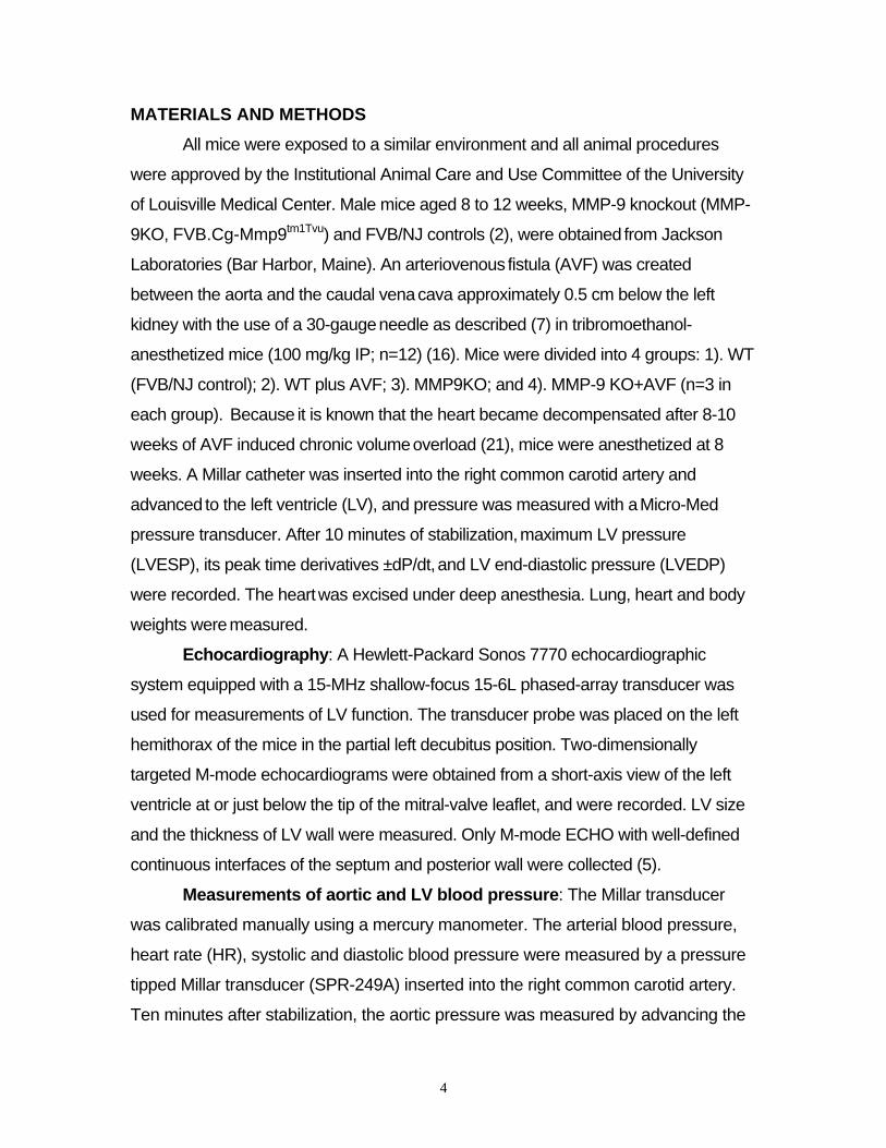
MATERIALS AND METHODS All mice were exposed to a similar environment and all animal procedures
were approved by the Institutional Animal Care and Use Committee of the University
of Louisville Medical Center. Male mice aged 8 to 12 weeks, MMP-9 knockout (MMP-
9KO, FVB.Cg-Mmp9tm1Tvu) and FVB/NJ controls (2), were obtained from Jackson
Laboratories (Bar Harbor, Maine). An arteriovenous fistula (AVF) was created
between the aorta and the caudal vena cava approximately 0.5 cm below the left
kidney with the use of a 30-gauge needle as described (7) in tribromoethanol-
anesthetized mice (100 mg/kg IP; n=12) (16). Mice were divided into 4 groups: 1). WT
(FVB/NJ control); 2). WT plus AVF; 3). MMP9KO; and 4). MMP-9 KO+AVF (n=3 in
each group). Because it is known that the heart became decompensated after 8-10
weeks of AVF induced chronic volume overload (21), mice were anesthetized at 8
weeks. A Millar catheter was inserted into the right common carotid artery and
advanced to the left ventricle (LV), and pressure was measured with a Micro-Med
pressure transducer. After 10 minutes of stabilization, maximum LV pressure
(LVESP), its peak time derivatives ±dP/dt, and LV end-diastolic pressure (LVEDP)
were recorded. The heart was excised under deep anesthesia. Lung, heart and body
weights were measured.
Echocardiography: A Hewlett-Packard Sonos 7770 echocardiographic
system equipped with a 15-MHz shallow-focus 15-6L phased-array transducer was
used for measurements of LV function. The transducer probe was placed on the left
hemithorax of the mice in the partial left decubitus position. Two-dimensionally
targeted M-mode echocardiograms were obtained from a short-axis view of the left
ventricle at or just below the tip of the mitral-valve leaflet, and were recorded. LV size
and the thickness of LV wall were measured. Only M-mode ECHO with well-defined
continuous interfaces of the septum and posterior wall were collected (5).
Measurements of aortic and LV blood pressure: The Millar transducer
was calibrated manually using a mercury manometer. The arterial blood pressure,
heart rate (HR), systolic and diastolic blood pressure were measured by a pressure
tipped Millar transducer (SPR-249A) inserted into the right common carotid artery.
Ten minutes after stabilization, the aortic pressure was measured by advancing the
4

catheter into the LV. The catheter was connected to a pressure transducer (Micro-
Med Corp.) positioned at the level of the heart. Ten minutes after the insertion of the
catheter, LV variables were measured (4). LVESP, LVEDP, and -dP/dt, were
measured. At the end of experiment, the anesthetized mice were prepared for the
excision of the lungs and arrested heart.
MMPs activity: Zymography using 1% gelatin gels were performed on LV
tissue homogenates as described (29).
MMP-2 and MMP-9 expression by Q-RT-PCR: DNA-free total RNA was
isolated from tissue with TRIzol reagent (GIBCO-BRL) and RNA (1µg/µl) was used for
preparation of first strand cDNA by reverse transcriptase (RT). The RNA samples
were incubated (70˚C, 5 min) with 1µl oligoprimers in a final volume of 5µl. Samples
then were incubated in 15 µl of a reaction buffer. Expression level of the RNA was
determined from 2µl of each cDNA sample. For amplification of the housekeeping
gene, glycerol aldehyde phosphate dehydrogenase (mouse GAPDH: GenBank
accession number: NM-008084). Upstream (5’-TACATTTCCCCTTCCTTACT-3’) and
down stream (3’-CCACATTTGACGTCCAGAGA-5’) primers for MMP-2 and -9 were
synthesized based on the mouse MMP-2 and -9 mRNA sequences (GenBank).
Denaturation step was set for 30s at 94°C, annealing for 60s at 60 °C and extension
for 90s cycles at 72°C. G3PDH samples were taken after 30 cycles and MMPs
samples were taken after 35 cycles. PCR reactions were performed with a Thermal
cycler (Perkin-Elmer 9600) in 50µl of 1x PCR buffer, 1.5 mM MgCl 2 , each primer at
0.2µM, 200 µM dNTP, and 1 U of Taq DNA polymerase (Invitrogen). Amplification
mixtures were analyzed by 1% agarose gel electrophoresis.
Western blot analysis of PAR-1, ADAM-12, connexin-43, and caspase-3: LV tissue homogenates were prepared, analyzed on 10% SDS-PAGE, and
transferred to PVDF membranes. Twenty five micrograms of total protein were
loaded on to each lane. The membranes were blotted with anti-caspase-3, PAR-1,
ADAM-12, or connexin-43 monoclonal antibodies (Cell signaling and Chemicon) as
previously described (4). The secondary IgG-alkaline phosphatase was used for
detection. Actin blots were used as a loading control. The bands were scanned and
5

normalized with actin intensity. The gels were stained with Commassie blue for
protein.
TUNEL and CD-31 immunohistochemical labeling: To determine whether
CHF caused endothelial and endocardial injury, 5-µm serial heart tissue sections from
snap-frozen blocks were labeled with Terminal Deoxynucleotidyl Nick End Labeling
Kit (TUNEL, Upstate cell signaling solutions, Lake Placid, NY) according to the
instructions of the manufacturer for apoptosis identification by nicked DNA labeling.
Endothelial cells were marked using FITC-labeled anti-mouse CD31/PECAM-1
antibody (Southern Biotech, Birmingham, AL) on serial tissue sections from the same
frozen blocks. Quantitative analysis was performed using Image Pro program
(IMAGE-PRO Media Cybernetics, Silver Spring, MD, USA) and data was expressed
in marker area (pixels2) averaged form five randomly selected fields per slide.
Statistical Analysis Values are reported as mean ± SEM. Differences
between groups were compared by one-way analysis of variance (ANOVA) with
repeated measurements and multiple comparisons (consideration given to within-
sample variability of observations). A probability level of p<0.05 was used to
indicate statistical significance.
6

RESULTS: Hemodynamic variables: Mean arterial blood pressure (MAP) and heart
rate (HR) (67.9±0.8 mmHg, n=3 and 408.8±9 beats/min, n=3, respectively) were
not significantly different between all groups (12 animals). Although the body
weight (26.7±0.5 g) was not significantly different between all groups, the heart
weight was significantly increased in AVF mice compared to controls (WT and
MMP9KO groups) (Table 1) suggesting cardiac hypertrophy in mice with chronic
volume overload. The histological data revealed biventricular hypertrophy in AVF
mice. In addition, compared to WT, the focal fibrosis was also increased in left
and right ventricles in the animals from the AVF group (Figure 1). In contrast,
there was no hypertrophy or focal fibrosis in WT, MMP-9KO and MMP-9KO+AVF
mice.
In AVF animals, compared to controls, LVESP was decreased and LVEDP
was increased. In those animals, the -dP/dt normalized by MAP, was significantly
decreased, suggesting that LV diastolic relaxation is decreased in AVF animals
compared to controls (Table1). LV systolic dysfunction in AVF animals was
shown by LVESP/HW ratio (Table1). In contrast, both diastolic and systolic
dysfunctions were prevented in MMP9KO+AVF mice (Table1).
Role of MMP-9 in cardiac remodeling: Total MMP activity in heart tissue
was significantly increased in the AVF group compared to WT. The Q-RT-PCR
data also showed increased MMP-9 mRNA expression in heart tissue from AVF
animals (Figure 2). Although the basal levels of MMP-2 were higher in MMP-9 -/-
mice then in WT, there was a significant increase in MMP-2 expression in AVF
compared to both WT and MMP-9KO groups (Figure 2). Compared to WT, the
level of PAR-1 expression was significantly increased in AVF animals, but
ablation of MMP-9, in animals that underwent AVF, ameliorated the increase
(Figure 3). Similarly, the heart tissue expressions of ADAM-12 and connexin-43,
which significantly increased in the AVF group, were attenuated in
MMP9KO+AVF animals (Figures 4 and 5). These data suggest that AVF-induced
MMP-9 is involved in PAR-1, ADAM-12, and connexin-43 over expression.
7

Cardiac function: The LV dilatation and LV wall thickness measured by
echocardiography revealed that the LVwall/LVEDD ratio was significantly decreased
in AVF group compared to controls, and ablation of MMP-9 in MMP9KO+AVF
animals prevented this decrease (Figure 6). These data suggest a role of MMP-9
in cardiac dilatation in congestive heart failure.
Endothelial and myocyte apoptosis: There was significant induction of
caspase-3 in AVF hearts as compared to WT or MMP-9KO, with or without AVF
(Figure 7). There were higher numbers of TUNEL positive cells (Figure 8) in
heart tissue of AVF animals compared to controls. Although the general
morphological appearance of the MMP-9KO/AVF heart tissue (Figure 8, panel C)
may appear to be different from WT panel, there was no quantitative or
qualitative difference when compared with MMP-9KO alone (no AVF), or WT.
The increase in caspase-3 activation and TUNEL was prevented in
MMP9KO+AVF animals. The co-labeling of TUNEL and CD-31, as a marker for
endothelial cells in serial heart tissue sections, demonstrated that endothelial
cells underwent apoptosis (Figure 9). Based on TUNEL positive and CD-31
positive versus CD-31 negative cells, we estimated that 75% TUNEL positive
cells were endothelial origin (Table II). Besides those cells, there were other
TUNEL positive cells, suggesting that endothelial, as well as myocyte apoptosis,
is involved in this chronic heart failure model (Table II). The induction of caspase-
3 and in situ TUNEL in the LV of AVF suggest involvement of MMP-9 in
transduction of apoptosis.
8

DISCUSSION
We and others suggested the role of endocardial apoptosis in the cardiac
dysfunction in congestive heart failure (27, 31). However, the mechanism of this
phenomenon is still unknown (1, 18). The results of the present study show that
impaired cardiac function in chronic volume overload CHF is associated with
endothelial and myocyte apoptosis and is accompanied by endothelial-myocyte
uncoupling in response to MMP-9 activation. MMPs are potential mediators of
cardiac remodeling and progression to heart failure. Previously, we showed that
during the compensatory phase of CHF, MMP-2 is increased and MMP-9 is over
expressed (23). Others have suggested that targeted deletion of MMP genes in
mice attenuates cardiac remodeling in acute (6, 9, 26) and in LV pressure
overload heart failure models (10). In the present study we demonstrated
molecular and cellular mechanisms that mediate the pathogenesis of chronic
heart failure, and the involvement of MMPs in cardiac remodeling and
dysfunction.
The histological and gravimetrical data suggested that cardiac hypertrophy
in mice with chronic volume overload heart failure were associated with ECM
accumulation (Figure 1). We obtained histological data on MMP-9KO similar to
the data obtained by others (6) and found that there was no significant difference
in MMP-9KO (control and AVF). The total MMP-2 and MMP-9 expressions were
significantly increased in animals from the AVF group (Figure 2). We measured
total MMP and MMP-9 expression in MMP-9KO animals (control and AVF), and
found that there was no expression of MMP-9 in control and AVF. However,
there was increased MMP-2 expression (Figure 2). This finding is in part due to a
compensatory response to MMP-9 ablation (6). In addition, the lung weight from
the AVF animals was significantly higher then controls, suggesting pulmonary
edema (Table 1). A significant decrease in the LVwall/LVEDD ratio, changes in
LVESP and LVEDP, decrease in LV contraction and relaxation suggested that
cardiac function in those animals was also impaired (Table 1 and Figure 6). The
ablation of MMP-9 in animals with AVF significantly normalized LV parameters,
and clearly indicates that MMP-9 plays a crucial role in pathogenesis of chronic
9

heart failure. These results are consistent with previous reports that MMP-9
inhibition, or targeted deletion of the MMP-9 gene in mice, attenuates LV dilation
and decreases collagen accumulation in the infarcted area after experimental MI
(6) or after AVF (3) or after pressure overload (10).
Our hypothesis was that impaired cardiac function in CHF could be the
consequence of endothelial apoptosis in response to E-M uncoupling due to
ECM accumulation in the myocardium (Figure 10). We found that the levels of
caspase-3 activation and numbers of TUNEL positive cells in heart tissue of AVF
animals were significantly higher than in controls (Figures 7 and 8). Serial heart
tissue sections were immunochemically developed with CD-31 antibody as a
label for endothelial cells and TUNEL for apoptosis. This TUNEL/CD-31 co-
labeling showed that endothelial, as well as myocyte apoptosis, is involved in
pathogenesis of CHF (Figure 9). Previously, we reported that activated PAR-1
lead to the cardiac remodeling (21) and inhibition of MMPs prevented connexin-
43 degradation and ameliorated heart failure (13). In the present study, we found
that besides increased expression of PAR-1, ADAM-12 (a disintegrin and
metalloprotease), enzyme involved in cell adhesion and myoblast differentiation
and fusion (15), was also over expressed in AVF animals. This finding suggests
that increased activity of PAR-1 could lead activation of several proteolytic
events. It was shown previously that reduced Connexin-43 expression produces
uncoupling between myocytes (24). In our model, increased expression of
connexin-43 might be associated with E-M uncoupling due to a compensatory
response to cell disintegration by ADAM-12. However, further studies are needed
to confirm the relationship between PAR-1 and connexin-43 remodeling by
ADAM-12. The results of our experiments show that in animals from
MMP9KO+AVF, the increase in the levels of apoptosis was prevented (Figures 8
and 9). In addition, PAR-1, ADAM-12 and connexin-43 expressions were also
normalized (Figures 3-5).
Limitations: TUNEL detection alone is not enough to confirm apoptotic
death (12, 25, and 30). Therefore, we measured the levels of caspase-3, and
showed that a caspase-3 inhibitor ameliorated the endothelial cell apoptosis (22).
10

Here, we suggested that caspase-3 was significantly induced in AVF hearts as
compared with the sham or the MMP-9-/- mouse hearts with and without AVF
(Figure 7). These results suggested apoptosis in the AVF hearts, in part, due to
activation of caspase-3. The connection between PAR-1 and connexin-43 may not be strong.
However, in light of our human heart end-stage failure data, in which we showed
that connexin-43 synthesis and degradation both were increased, and MMPs
were activated in CHF (22). It is a logical extension of the idea that volume
overload stress activated the MMP that in turn activated PAR-1, leading to
induction of connexin-43 in AVF.
In summary, the results of the present study demonstrate that in chronic
volume overload model of CHF in mice, impaired cardiac function is associated
with endothelial and myocyte apoptosis. The mechanism of these phenomena
involves activation of MMP-9, leading to cardiac remodeling and E-M uncoupling.
Our study also shows that MMP-9 has a major role in ventricular remodeling
associated with endothelial and myocyte apoptosis (Figure 10).
11

REFERENCES 1. Braunwald, E., and Bristow, M.R. Congestive heart failure: fifty years of
progress. Circulation 102:IV14-IV23, 2000.
2. Brewer J, Kisselgof A, Martin T. Genetic Variability in Pulmonary
Physiological, Cellular, and Antibody Responses to Antigen in Mice. Am. J.
Respir. Crit. Care Med 160(4):1150-1156, 1999.
3. Chancey AL, Brower GL, Peterson JT, Janicki JS. Effects of matrix
metalloproteinase inhibition on ventricular remodeliung due to volume
overload. Circulation 105(16):1983-1988, 2002.
4. Cox MJ, Sood HS, Hunt MJ, Chandler D, Henegar JR, Aru GM, and Tyagi SC. Apoptosis in the left ventricle of chronic volume overload causes
endocardial endothelial dysfunction in rats. Am J Physiol 282:H1197-H1205,
2002.
5. De Simone G, Wallerson DC, Volpe M, Devereux RB. Echocardiographic
measurements of LV mass and volume in normal and hypertensive rats. Am J
Hyperten 3:688-696, 1999.
6. Ducharme A., Frantz S., Aikawa M., Rabkin E., Lindsey M., Rohde L., Schoen F., Kelly R., Werb Z., Libby P., Lee R. Targeted deletion of matrix
metalloproteinase-9 attenuates left ventricular enlargement and collagen
accumulation after experimental myocardial infarction. J Clin Invest 106
(1):55-62, 2000.
7. Garcia R, Diebold S. Simple, rapid, and effective method of producing
aortocaval shunts in the rats. Cardiovasc Res 24:430-432, 1990.
8. Goette A, Juenemann G, Peters B, Klein HU, Roessner A, Huth C, Rocken C. Determinants and consequences of atrial fibrosis in patients
undergoing open heart surgery. Cardiovasc. Res 54:390–396, 2002.
9. Heymans S , Luttun A, Nuyens D, Theilmeier G, Creemers E, Moons L, Dyspersin G, Cleutjens JP, Shipley M, Angellilo A, Levi M, Nube O, Baker A, Keshet E, Lupu F, Herbert J, Smits J, Shapiro S, Baes M, Borgers M, Collen D, Daemen MJ, Carmeliet P. Inhibition of plasminogen
activators or matrix metalloproteinases prevents cardiac rupture but impairs
12

therapeutic angiogenesis and causes cardiac failure. Nat. Med 5:1135-1142,
1999.
10. Heymans S, Lupu F, Terclavers S, Vanwetswinkel B, Herbert J, Baker A, Collen D, Carmeliet P, Moons L. Loss or Inhibition of uPA or MMP-9
Attenuates LV Remodeling and Dysfunction after Acute Pressure Overload in
Mice. Am J Pathol 166:15-25, 2005.
11. Hoppeler H, Kayar SR. Capillary and oxidative capacity of muscles. News in
Physiol Sci 3:113-116, 1988.
12. Hughes SE. Detection of apoptosis using in situ markers for DNA strand
breaks in the failing human heart. Fact or epiphenomenon? J Pathol
201(2):181-186, 2003.
13. Hunt MJ, Aru GM, Hayden MR, Moore CK, Hoit BD, and Tyagi SC. Induction of oxidative stress and disintegrin metalloproteinase in human heart
end-stage failure. Am J Physiol Lung Cell Mol Physiol 283(2):L239-L245,
2002.
14. Jessup M. and Brozena S. Heart failure. N Engl J Med 348:2007–2018,
2003.
15. Kawaguchi N, Xu X, Tajima R, Kronqvist P, Sundberg C, Loechel F, Albrechtsen R and Wewer U. ADAM 12 Protease Induces Adipogenesis in
Transgenic Mice. Am J Pathol 160:1895-1903, 2002.
16. Kiatchoosakun S, Kirkpatrick D, Hoit BD. Effects of tribromoethanol
anesthesia on echocardiographic assessment of left ventricular function in
mice. Comp Med 51:26-29, 2001.
17. Kumar D., Jugdutt B. Apoptosis and oxidants in the heart. J Lab Clin Med
142(5):288-297, 2003
18. Lee R. Matrix Metalloproteinase Inhibition and the Prevention of Heart
Failure. Trends in Cardiovascular Medicine, 11(5):202-205, 2001.
19. Mebazaa A, Wetzel R, Cherian M, Abraham M. Comparison between
endothelial and great vessel endothelial cells: morphology, growth, and
prostaglandin release, Am J Physiol 268:H250-H259, 1995.
13

20. Michel J-B, Salzmann J-L, Nlom MO, Bruneval P, Barres D, Camilleri J.P,
Morphometric analysis of collagen network and plasma perfused capillary bed
in the myocardium of rats during evolution of cardiac hypertrophy, Bas Res
Cardiol 81:142-154, 1986.
21. Moshal KS, Tyagi N, Henderson B, Ovechkin AV, Tyagi SC. Protease
activated receptor and endothelial-myocyte uncoupling in chronic heart
failure, Am J Physiol Heart Circ Physiol 288(6):H2770-7, 2005. 22. Mujumdar VS, Aru GM, Tyagi SC. Induction of oxidative stress by
homocyst(e)ine impairs endothelial function, J Cell Biochem 82(3):491-500,
2001.
23. Mujumdar VS, Tyagi SC. Temporal regulation of extracellular matrix
components in transition from compensatory hypertrophy to decompensatory
heart failure. J Hypertens 17:261-270, 1999.
24. Poelzing S and Rosenbaum D. Altered Connexin-43 expression produces
arrhythmia substrate in heart failure. Am J Physiol Heart Circ Physiol
287:H1762-H1770, 2004
25. Rodriguez M. and Schaper J. Apoptosis: measurement and technical
issues. J Mol Cell Cardiol 38(1):15-20, 2005
26. Romanic A, Harrison S, Bao W, Burns-Kurtis C, Pickering S, Gu J, Mao J, Sathe G, Ohlstein E, Yue T. Myocardial protection from
ischemia/reperfusion injury by targeted deletion of matrix metalloproteinase-9.
Cardiovasc Res 54(3):549-58, 2002.
27. Tyagi SC. Dynamic ECM remodeling in the heart failure: cardiac
hypertrophy, dilatation and fibrosis. Pathophysiology 4:227-234, 1997.
28. Tyagi SC, Haas SJ, Kumar SG, Reddy HK, Voelker DJ, Hayden MR, Demmy TL, Schmaltz RA & Curtis JJ. Post-transcriptional regulation of extracellular
matrix metalloproteinase in human heart end-stage failure secondary to ischemic
cardiomyopathy, J Mol Cell Cardiol 28:1415-1428, 1996.
29. Tyagi SC, Matsubara L, and Weber KT. Direct extraction and estimation of
callagenase(s) activity by zymography in microquantities of rat myocardium
and uterus. Clin Biochem 26:191-198, 1993.
14

30. Yaoita H, Ogawa K, Maehara K, Maruyama Y. Apoptosis in relevant clinical
situations: contribution of apoptosis in myocardial infarction. Cardiovasc Res
45(3):730-641, 2000.
31. Ziori C, Zioris C., Kyriakidis M. Apoptosis in Heart Failure. Hellenic J
Cardiol 44: 56-70, 2003.
15

Table I:
Gravimetric and hemodynamic data
WT (n=3)
MMP9KO (n=3)
AVF (n=3)
MMP9KO+AVF (n=3)
Body weight, g 27.3 ± 0.4 26.2±0.5 27.1±0.6 26.3±0.3
Heart weight, g 0.15 ± 0.012 0.16±0.013 0.232±0.025* 0.18±0.014
Lungs weight, g 0.32 ± 0.04 0.31±0.05 0.54±0.07* 0.39±0.06
Heart rate, b/min 409.2 ± 9.5 428.3 ±8.9 398.5 ±10.1 399.2 ±7.4
LVEDP, mmHg 1.2 ± 0.6 1.4 ±1.0 8.4 ±2.9* 2.6 ±0.8
LVESP, mmHg 112.7 ± 1.6 110.9 ±2.0 96.4 ±1.8* 99.6 ±1.3
LVESP/Heart Weight 746.7 ± 133.3
687.5 ± 153.9 414.4 ± 72* 559.7 ± 92.3
MAP, mmHg 69.8 ± 0.7 67.7 ± 0.6 71.8 ± 1.1 62.3 ± 0.7
-dP/dt, mmHg/s 5491 ± 201 4838 ± 288 3156 ± 243* 4391 ± 298
+dP/dt, mmHg/s 9050 ± 435 8236 ± 378 7666 ± 443 7997 ± 478
(-dP/dt)/MAP(sec-1) 81.7 ± 6.3 82.4 ± 3.2 40.4 ± 4.1* 68.9 ± 8.3
LVEDd (mm) 2.81 ± 0.08 2.78 ± 0.07 3.91 ± 0.11* 2.91 ± 0.08
LVEDs (mm) 1.69 ± 0.07 1.65 ± 0.07 2.79 ± 0.12* 1.83 ± 0.09
LVPWd (mm) 0.74 ± 0.03 0.72 ± 0.04 0.85 ± 0.05* 0.79 ± 0.05
LVPWs (mm) 1.12 ± 0.03 1.08 ± 0.03 1.29 ± 0.04* 1.08 ± 0.03
FS, % 39.86 ± 0.85 40.65 ± 1.1 28.65 ± 1.03* 37.45 ± 1.33 Values are means ± SEM; n=number of animals. LVEDP and LVESP, left
ventricular end-diastolic and end-systolic pressure, respectively; MAP, mean
arterial pressure; LVEDd and LVEDd, left ventricular end-diastolic and end-
systolic diameter, respectively; LVPWd and LVPWs, left ventricular end-diastolic
and end-systolic wall thickness, , respectively; FS, fractional shortening. *
indicates p<0.05. Note that there were no significant differences between the
data obtained from WT, MMP9KO and MMP9KO+AVF groups.
16

Table II:
Morphological data
WT (n=3)
MMP9KO (n=3)
AVF (n=3)
MMP9KO+AVF (n=3)
CD31 positive cells/field 2456 ± 232 2277 ± 256 1845 ± 296 1978 ± 273
TUNEL positive cells/field 103 ± 23 87 ± 26 978 ± 32* 198 ± 39†
CD31/TUNEL co- localized cells/field 63 ± 15 56 ± 18 645 ± 22* 135 ± 26†
Values are means ± SEM; n=number of animals. * indicates p<0.05 compared to
all groups. † indicates p<0.05 compared to WT or MMP9KO group.
17

Legend to Figures: Figure 1: Histological analysis of left ventricle (LV) at 8 wks after AVF.
Frozen tissue sections were labeled with trichrome for collagen. A, WT; B, AVF
mice. Arrows indicate deposition of collagen (blue). Note that the heart with AVF
is hypertrophic. Regions shown in inset boxes were observed using 20 X
magnifications. C, WT; D, AVF mice. Note greater collagen expression in over
loaded heart.
Figure 2: Effects of AVF on total MMP activity (A). MMP-9 (B) and MMP-2
gene expression (C) in the LV by Q-RT-PCR analysis; Mean±SEM; *, p<0.05
compared to all groups; †, p<0.05 compared to all groups except MMP9KO
group.
Figure 3: Comparison of effects of AVF on PAR-1 expression in LV by
Western blot analysis; Mean±SEM; n=3 in each group; *, p<0.05 compared to all
groups. ∧, p<0.05 compared to all groups except AVF. Note that increased PAR-
1 expression in AVF group was prevented in MMP9KO+AVF group.
Figure 4: Comparison of effects of AVF on ADAM-12 expression in LV by
Western blot analysis; Mean±SEM; n=3 in each group; *, p<0.05. Note that
increased ADAM-12 expression in AVF group was prevented in MMP9KO+AVF
group.
Figure 5: Comparison of effects of AVF on Connexin-43 expression in LV
by Western blot analysis; Mean±SEM; n=3 in each group; *, p<0.05. Note that
increased Connexin-43 expression in AVF group was prevented in
MMP9KO+AVF group.
Figure 6: Two-dimensional M-mode echocardiogram of LV (A, WT group;
B, AVF group; C, MMP9KO+AVF group) and comparison of effects of AVF on LV
function (D). Mean±SEM; *, p=0.005. Note that increase in LV end-diastolic
diameter (LVEDD) in AVF animal (B) was prevented in MMP9KO+AVF animal
(C). Figure 7: Comparison of effects of AVF on Caspase-3 expression in LV
by Western blot analysis; Mean±SEM; n=3 in each group; *, p<0.05. Note that
18

increased caspase-3 expression in AVF group was prevented in MMP9KO+AVF
group.
Figure 8: Effect of AVF on endocardial apoptosis (TUNEL assay) in LV
(A, WT group; B, AVF group; C, MMP9KO+AVF group). D, quantitative
presentation of TUNEL marker expression; n=3 in each group. Scale bars
indicate means ± SEM. * Indicates p<0.0025 compared to all groups.
Figure 9: Comparison of immunofluorescence staining for apoptotic cells
(TUNEL assay) in LV. A, in AVF group; B, in MMP9KO+AVF group. Regions
shown were enlarged 6X. Bright green spots show positive immunostaining for
apoptotic cells. Red spots show positive immunostaining for CD-31 (endothelial
marker). Note greater EE apoptosis in AVF group (A) compared to
MMP9KO+AVF group (B).
Figure 10: Overall hypothesis: stress and progression of CHF are
associated with activation of latent MMP which activates PAR receptors and
subsequent activate ADAM-12. This, in turn, induces MMP-9 and ADAM-12
dependent oxidized ECM accumulation, leading to E-M uncoupling and
endothelial apoptosis.
19

Figure 1

Figure 2

Figure 3

Figure 4

Figure 5

Figure 6

Figure 7

Figure 8

Figure 9

Figure 10