Short and Long Term Effects of (-)Epicatechin on Myocardial Ischemia Reperfusion Injury
Upload
independentCategory
view
0download
0
Effects of Metalloproteinase Inhibition in a Murine Model ofRenal Ischemia Reperfusion Injury
Katherine B. Novak, Hau D. Le, Emily R. Christison-Lagay, Vania Nose, Robert J. Doiron,Marsha A. Moses, and Mark PuderDepartment of Surgery [K.B.N., H.D.L., E.R.C.-L., R.J.D., M.A.M., M.P.], Children’s HospitalBoston, Harvard Medical School, Boston, MA 02115; Department of Pathology [V.N.], Brighamand Women’s Hospital, Boston, MA 02115
AbstractIschemia-reperfusion injury (IRI) is a leading cause of acute tubular necrosis and delayed graftfunction in transplanted organs. Upregulation of matrix metalloproteinases (MMPs) propagates themicro-inflammatory response that drives IRI. This study sought to determine the specific effectsof Marimastat (Vernalis, BB-2516), a broad spectrum MMP and tumor necrosis factor-α-converting enzyme inhibitor, on IRI-induced acute tubular necrosis. Mice were pre-treated withMarimastat or methylcellulose vehicle for 4 days prior to surgery. Renal pedicles were bilaterallyoccluded for 30 minutes and allowed to reperfuse for 24 hours. Baseline creatinine levels wereconsistent between experimental groups; however, post-IRI creatinine levels were four-fold higherin control mice (P<0.0001). The mean difference between the post-IRI histology grades ofMarimastat-treated and control kidneys was 1.57 (P=0.003), demonstrating more severe damage tocontrol kidneys. Post-IRI mean (±SEM) MMP-2 activity rose from baseline levels in control mice(3.62±0.99); however, pretreated mice presented only a slight increase in mean MMP-2 activity(1.57±0.72) (P<0.001). In conclusion, these data demonstrate that MMP inhibition is associatedwith a reduction of IRI in a murine model.
Ischemia-reperfusion injury (IRI) affects post-transplant renal function and largelydetermines short and long-term graft survival. Although some ischemia is unavoidable, IRIcontinues to be the leading cause of acute renal injury and delayed graft function intransplanted organs (1). The development of acute tubular necrosis (ATN), one of theleading causes of intrinsic acute renal injury, is a hallmark of IRI. IRI induces the release ofpro-inflammatory cytokines which, in turn, contribute to graft fibrosis (2,3). There is noclinically acceptable therapy that directly addresses the cellular damage induced by IRI(4,5). Due to its intrinsic nature, IRI remains a challenge in the field of organtransplantation.
Ischemia–reperfusion initiates a cascade of events that involve tubular epithelial injury,inflammation, and altered microvascular function (6). The initial ischemic event depletesATP reservoirs which subsequently impairs cellular processes such as protein synthesis,lipogenesis, and membrane transport. The succeeding biochemical abnormalities disrupt the
Copyright © 2009 International Pediatric Research Foundation, Inc. All rights reserved.
Corresponding author Mark Puder, MD, PhD 300 Longwood Ave, Fegan 3 Boston, MA 02115 Phone: 617-355-1838 Fax:617-730-0298 [email protected]., H.D.L. contributed equal work to this study
This is a PDF file of an unedited manuscript that has been accepted for publication. As a service to our customers we are providingthis early version of the manuscript. The manuscript will undergo copyediting, typesetting, and review of the resulting proof before itis published in its final citable form. Please note that during the production process errors may be discovered which could affect thecontent, and all legal disclaimers that apply to the journal pertain.
NIH Public AccessAuthor ManuscriptPediatr Res. Author manuscript; available in PMC 2012 June 02.
Published in final edited form as:Pediatr Res. 2010 March ; 67(3): 257–262. doi:10.1203/PDR.0b013e3181ca0aa2.
NIH
-PA Author Manuscript
NIH
-PA Author Manuscript
NIH
-PA Author Manuscript
cytoskeleton, damage cellular proteins and degrade DNA, leading to apoptosis and/ornecrosis of tubular epithelial cells (7). The cellular damage induces a vigorous inflammatoryresponse which further propagates post-ischemic tissue damage (8,9).
Matrix metalloproteinases (MMPs) are active components of inflammation that are alsoinvolved in other processes in which tissue remodeling maintains proper function such asembryonic development, angiogenesis and wound healing (10,11). Upregulation of MMPspropagates the micro-inflammatory response that drives IRI. Evidence of MMP activationhas been inferred microscopically from loss of junctional complexes and altered expressionand distribution of cellular adhesion molecules, suggesting a disruption of cell-cell and cell-extracellular matrix (ECM) interactions (12,13). MMPs are sequestered by neutrophils andare activated by tumor necrosis factor-alpha (TNF-α) which is upregulated in animal modelsof ischemic injury (14,15). TNF-α is a critical mediator of both the physiologic defensiveresponse to and pathogenesis of IRI, and exists in a biologically active, secreted form and inan inactive membrane-anchored precursor. Cleavage of the TNF-α proform into its solubleform is mediated by TNF-α-converting enzyme (TACE, also known as ADAM17 andCD156b), which belongs to the disintegrin and metalloproteinase family of zinc-metalloproteinases (16,17). The TACE-induced increase in TNF-α availability is thought topromote a more extensive inflammatory response which would directly impact the severityof tissue injury.
The degree and duration of MMPs elevation (protein or activity) has been shown toinfluence the extent of renal damage (11). Although IRI is a strong predictor of graftsurvival, no current therapies address the role of MMPs during kidney transplantation. Thepresent study was designed to determine the specific effects of pre-treatment withMarimastat (Vernalis, BB-2516), a broad spectrum MMP-inhibitor, on IRI-induced ATN.Marimastat is a synthetic, low molecular weight succinate peptidomimetic inhibitor thatcovalently binds to the Zn2+ ion in the active site of MMP, thus inhibiting its action.Marimastat has been used in multiple oncologic clinical trials in the adult population. Themost consistently reported toxicity concern is musculoskeletal pain that likely to occur aftertwo months of treatment (18,19). We previously demonstrated that Marimastat is capable ofdecreasing serum TNF-α receptor II levels (20). Although Marimastat is a broad-spectrumMMP inhibitor, this study focused on its ability to inhibit MMP-2 and MMP-9 (gelatinases),both of which play a role in ischemia-related inflammation. We propose that Marimastatwill reduce kidney IRI, predominantly through its role as an inhibitor of MMP-2 andMMP-9.
Materials & MethodsAnimal Model
5-7 week old male C57BL/6 mice (Jackson Laboratories, Bar Harbor, ME) were housed fiveanimals to a cage in a barrier room with ad libitum access to food and water. Animalprotocols complied with the NIH Animal Research Advisory Committee guidelines andwere approved by the Children’s Hospital Boston Institutional Animal Care and UseCommittee. To ensure that Marimastat (half life 10 hrs) reaches a steady state plasma level,ninety-six hours prior to surgery, the experimental group (n=7) received 100mg/kg ofMarimastat treatment (MT) in 100 μl of 0.45% methylcellulose vehicle (Sigma-Aldrich, St.Louis, MO) via orogastric gavage twice daily (21). Control mice (n=8) receivedmethylcellulose vehicle alone. A sham group (n=5) was given methylcellulose vehicle alone.A dose-dependent relationship between Marimastat and TNF-α showed complete inhibitionat 200 mg/kg (22).
Novak et al. Page 2
Pediatr Res. Author manuscript; available in PMC 2012 June 02.
NIH
-PA Author Manuscript
NIH
-PA Author Manuscript
NIH
-PA Author Manuscript
IRI was performed through a flank incision after the animals were anesthetized with 2-4%isofluorane inhalation. IRI was induced as previously described (23). The kidney wasisolated and a microvascular clamp (Roboz, Rockville, MD) was placed on the renal pediclefor 30 minutes, during which the kidney and clamp were gently placed back into theperitoneum. The contra-lateral kidney underwent the same procedure within 5 minutes.Treatment was continued for 24 hours post-operatively. Sham mice underwent bilateralflank incisions without clamping the renal pedicles.
Specimen Collection24 hours after reperfusion, the experiment was ended. Animals was anesthetized, blood wascollected and centrifuged at 4°C at 8000 rpm for 10 min to separate the serum. Creatinineand BUN levels were measured using a Hitachi 917 analyzer (Roche, Branford, CT).
At the time of sacrifice, the kidneys were excised, weighed and bisected transversely to theirlength. Approximately one half of each kidney was fixed in 10% phosphate-bufferedformalin at 4°C overnight, washed with phosphate buffered saline and stored in 70% ethanoland then embedded in paraffin. Tissues were cut into 5-μm sections and stained withhaematoxylin and eosin (H&E) or periodic acid-Schiff reagent (PAS). Histological sectionswere reviewed by a pathologist in blinded fashion and scored with a semiquantitative scale(Fig. 1) to evaluate the extent of tubular necrosis: 0, normal kidney; 1, normal kidney withfocal apoptosis; 2, focal tubular necrosis (TN) <50% of the cortical-medullary junction(CMJ); 3, TN in the CMJ; 4, TN in the CMJ with focal extension into the cortex; 5, TNextending through the cortex to the surface of the kidney. Histological assessment and thescoring parameters were modified from previously described methods, with an emphasis onthe location of TN (24,25).
ZymographyUrine samples were collected before bilateral IRI and 24 hours following IRI. Gelatinzymography was performed on urine samples as previously described (26,27) withmodifications to determine MMP activity in the urine. Fifteen microliter of each urinesample was mixed with 10 μl buffer consisting of 4% sodium dodecyl sulfate (SDS), 0.15 MTris (pH 6.8), 20% (v/v) glycerol, and 0.5% (w/v) bromphenol blue. Samples were loadedinto wells of a 10% SDS-PAGE gel containing 0.1% (w/v) gelatin (Bio-Rad Laboratories,Hercules, CA) on a mini gel apparatus. Gels were run at 200 V for 50 minutes then soakedin 2.5% TritonX-100 with gentle shaking for 30 minutes at ambient temperature. Afterincubating overnight at 37°C in substrate buffer (50 mM Tris-HCL buffer pH 8, 5 mMCaCl2, and 0.02% NaN3), gels were stained for 30 minutes in 0.5% Coomassie Blue R-250in acetic acid, ethanol, and water (1:3:6) and destained for 1 hour. As previously described,MMP levels were quantified by scoring the band strength of each type of MMP examined onthe zymogram on a scale of zero to seven, with zero indicating no detectable MMPs, andseven indicating strong bands (28).
Western Blot AnalysisWestern Blot analysis was performed on the lysate of the other half of each kidney. Proteinextraction was performed using an ActiveMotif extraction kit according to themanufacture’s protocol (ActiveMotif, Carlsbad, CA). Samples were normalized for proteincontent by the Bio-Rad protein assay (Bio-Rad Laboratories, Hercules, CA). Seventy-fivemicrograms of protein per sample were analyzed by SDS-PAGE. Membranes were thenprobed overnight at 4°C with either goat anti-mouse MMP-2 (1:500, R&D Systems), goatanti-mouse MMP-9 (1:500, R&D Systems), or rabbit polyclonical anti-human TACE (1:500,Novus Biologicals). The secondary antibodies used were either horseradish peroxidase(HRP)-linked donkey anti-goat IgG in a 1:5,000 dilution (Santa Cruz, CA), or ECL donkey
Novak et al. Page 3
Pediatr Res. Author manuscript; available in PMC 2012 June 02.
NIH
-PA Author Manuscript
NIH
-PA Author Manuscript
NIH
-PA Author Manuscript
anti-rabbit IgG, HRP-linked whole antibody in a 1:5,000 dilution (GE Healthcare,Piscataway, NJ). Equal protein loading was verified by incubating the same membrane withα-actin antibody in a 1:500 dilution (MS X Actin, Chemicon International, Temecula, CA).The probed proteins were developed with Pierce enhanced chemiluminescent substrate fordetection of HRP according to manufacturer’s instructions (Pierce, Rockford, IL).
Statistical AnalysisANOVA with Turkey-Kramer multiple comparisons test was performed (unless otherwiseindicated) using GraphPad InStat Version 3.05 (GraphPad Software, San Diego, CA).Results were obtained from two independent occasions and were expressed as mean ± SEM.For all experiments probabilities of error (P values) were included; values for P<0.05 wereregarded as significant.
ResultsHistology Grades
Histological scores were determined by the presence and location of necrosis within thekidney. Both kidneys from each mouse received a separate, blinded grade. MT kidneysreceived a mean (±SEM) histological score of 1.93±0.34. Control kidneys demonstrated asignificant degree of tubular damage and received a histological score of 3.50±20 (Fig. 2).The mean difference between MT and control kidneys was 1.57 (P=0.003).
The injury seen in MT mice was mild and consistently limited to the CMJ (85.6% of thetotal number of kidneys). Extension of necrosis into the cortex denoted more severe tubularinjury, frequently observed in control mice (56.3%). Injury severity increased in parallel tothe proximity of necrotic extension towards the outer cortical surface, and more than half ofcontrol mice had necrotic extension into the cortex. One of the 2 kidneys from 2 MT micehad focal necrotic extension into the cortex (grade 4); however, neither mouse showed asignificant increase in post-IRI plasma creatinine levels (0.0 and 0.4).
Serum CreatinineSerum creatinine levels were obtained from all experimental groups immediately prior toIRI and again 24-hours after IRI. An additional draw was taken prior to the first Marimastatdose and showed that Marimastat did not affect creatinine levels prior to IRI (data notshown). Baseline creatinine levels prior to IRI were consistent between experimental groups(P=0.788) (Fig. 3). Mean serum creatinine levels in mice pretreated with Marimastat(0.25±0.03) were not statistically different from baseline levels (0.20±0.00) (P=0.268). Incontrast, the post-IRI control mice demonstrated a significant increase to 1.01±0.29 from thebaseline levels of 0.21±0.01 (P=0.003). Post-IRI, the creatinine levels of control mice werefour-fold higher than in MT mice (P<0.0001).
Gelatin zymographyUrine was screened for MMP activity by gelatin zymography prior to bilateral IRI and 24hours following IRI. In contrast to blood-specimen collection, urine collection isnoninvasive and will not influence detectable MMP levels. The band strength visualized bygelatin zymography provided an estimation of relative activity of the gelatinases MMP-2and MMP-9 (Fig. 4A-B).
Active MMP-2 was undetectable in the pre-op samples of MT and control mice. MMP-2activity rose significantly from baseline levels 24 hours post-IRI in control mice (3.62±0.99)however MT mice demonstrated only a slight increase in MMP-2 activity (1.57±0.72) (Fig.4D). The post-IRI rise in MMP-2 activity between control and MT mice was significant
Novak et al. Page 4
Pediatr Res. Author manuscript; available in PMC 2012 June 02.
NIH
-PA Author Manuscript
NIH
-PA Author Manuscript
NIH
-PA Author Manuscript
(P<0.001). There was no statistically difference in MMP-9 activity between groups and timepoints (Fig. 4E).
Western BlotWestern blot analysis was conducted to determine the levels of MMP-2, MMP-9 and TACEin kidney tissue. In normal kidney, TACE is expressed constitutively as an 85 kDa protein.Another 120 kDa band has also been described under reducing conditions in the normalkidney (17). In normal kidneys, both bands were present (Fig. 5A). Twenty four hours afterIRI, control mice showed a decreased in expression of the 85 kDa band, whereas the 120kDa band was no longer present. Instead, a smaller, approximately 45 kDa band was noted.Mice in the MT group showed reduced expression of the 85 kDa band.
MMP-2 was expressed in normal kidneys (29) as a 65 kDa band and there was no change inits level in control and MT kidneys postoperatively (Fig. 5B). On the other hand, MMP-9(90 kDa band) was not expressed in normal kidney. IRI kidneys expressed MMP-9 in thecontrol and MT group after 24 hours (Fig. 5C).
DiscussionInflammation plays an important role in the progression from IRI to ATN and ultimatelycompromised renal function. Post-IR inflammation is a pivotal phase which heavily dictatesthe severity of tubular injury. Upregulation of MMPs propagates this microinflammatoryresponse (30). The limited research regarding MMP expression in post-IRI of the kidneyshas focused on its later role in tubulointerstitial fibrosis and chronic allograft nephropathy(31,32). This study is directed at MMP participation in earlier stages of injury.
MMP activity is most pronounced and their inhibition is most effective in the early stage ofdisease. Animal models studying the efficacy of MMP inhibition in IRI and various cancershave suggested that MMP inhibition initiated prior to or in the initial period of diseaseprogression significantly reduced the severity and degree of tissue damage (33,34). Lutz etal. demonstrated that MMP-2 inhibition reduced allograph nephropathy if initiated in theearly, prefibrotic stages of disease but produced more severe nephropathy once fibrosis wasestablished (35). In clinical trials, however, MMP inhibition has been typically implementedat more advanced stages of disease (36,37). The therapeutic modalities appropriate at theearly stages may be misdirected once significant injury has been established. The disparityof outcome between experimental models and clinical trials demonstrates that the point oftherapeutic intervention may be a critical factor determining the relevance and efficacy ofMMP inhibition.
The murine model of renal IRI used in this study represents the renal injury observed duringearly graft dysfunction in the transplanted kidney (23). Tubular dysfunction begins shortlyafter the onset of ischemia, initiating a dynamic interplay between pro-inflammatorymediators that will direct subsequent inflammation.
MMP activity is mediated by several mechanisms and at several levels. TNF-α plays a rolein the induction and propagation of inflammation by directly activating MMPs and initiatingan influx of neutrophils to the site of injury (9). Cleavage of the cell-associated TNF-αproform into its soluble, biologically-active form is mediated by TACE (16,17). Activationof TACE following ischemia-reperfusion has been shown to directly contribute to thepathogenesis of brain, lung, heart and liver tissue injury (38-40). The upstream inhibition ofTACE potentially abrogates the inflammatory cascade by reducing the amount of activatedTNF-α. Results obtained from western blot analysis showed a change in the pattern ofexpression of TACE between controls and mice that underwent IRI. Twenty four hours after
Novak et al. Page 5
Pediatr Res. Author manuscript; available in PMC 2012 June 02.
NIH
-PA Author Manuscript
NIH
-PA Author Manuscript
NIH
-PA Author Manuscript
IRI, a new band with molecular weight 45 kDa was identified with anti-TACE antibody.Although further investigation is warranted, this molecule could be the catalytic domain ofTACE that has been described elsewhere (41). When administered prior to IRI, Marimastatreduced IRI as evidenced by both histological and biochemical analysis. Morphologically,Marimastat diminished the scope and severity of ATN. Although apoptosis does contributeto ischemic injury, necrosis is more often associated with IRI. In contrast to apoptosis,which occurs under normal physiological conditions and requires ATP, necrosis does notrequire ATP to proceed and only occurs after gross cellular injury (42). The location ofnecrosis was an important consideration in the histological analysis because differentialmetabolic demands within the kidney may determine areas most affected by ischemia.Necrosis was most prominent in the CMJ, which is presumably due to its high metabolicactivity and its limited ability to sustain anaerobic metabolism amidst decreased oxygentension (43,44). Not only do cortical proximal tubules require very low oxygen extraction tosupport tubule metabolism, blood flow in the post-ischemic kidney is more concentrated inthe cortex rather than the medulla, which could explain the initiation and predominance ofinjury in the CMJ (44). Tubular necrosis is associated with elevated serum creatinine levelsas seen in control mice. In contrast, serum creatinine in Marimastat mice demonstrated anon-significant increase over baseline indicating that Marimastat reduced tubular necrosis.
Although MMP-2 is constitutively expressed in renal tissue, the absence of MMP-2 activityin urine samples at baseline of both MT and control mice might reflect its low urinary leveluntil IRI occurs. A recent study found that while MMP-2 activity increased 24 hours afterischemia, an increase in MMP-9 activity was not detected until 48-72 hours post ischemia(45). Since we only measured MMP levels 24 hours following IRI, our findings areconsistent with this study demonstrating that urine MMP-2 is a prominent initial participantin post-IRI.
Results from the western blot showed increased MMP-9 protein levels following renal IRI.Gelatin zymography, however, demonstrated that MMP-9 activity remained unchangedbetween groups and time points. MMP-9 has been shown to form a complex with othermolecules such as NGAL, which protects it from degredation (46) and is a marker of renalinjury (47). Therefore, it is likely that increased MMP-9 protein level was partially due to adecrease of MMP-9 degradation.
Inhibition of MMPs has become a widely studied topic because of their involvement inmany pathologic states. Pretreatment with MMP inhibitors may be applicable clinically. Itmay benefit those awaiting transplantation in which viable, fully functional organs areexpected to undergo transient ischemia, an important one being pediatric transplantation. Acrucial element of graft health and sustainability is the degree of ischemic injury, which maybe effectively reduced if MMP inhibition is initiated prior to insult. Pretreatment withMarimastat may be a preferable alternative to current therapies in pediatric transplantation,such as tetracyclines, due to its more favorable side effect profile. Despite being protectiveagainst ischemia reperfusion injury in a variety of tissues, the use of tetracyclines in thepediatric population is complicated by non-compliance, enamel discoloration, stuntedgrowth and nephrotoxicity (48,49). The period immediately following reperfusion is acritical point in the prevention of renal injury (6). In conclusion, as we have shown thaturine MMPs can be used as biomarkers to follow a disease process (50), we demonstratehere that prevention of MMP-2 activation may be a possible mechanism for renal post-transplant protection. Prophylactic MMP inhibition in both donor and recipient prior to andfollowing renal transplant may decrease post-transplant morbidity and foster graft function.
Novak et al. Page 6
Pediatr Res. Author manuscript; available in PMC 2012 June 02.
NIH
-PA Author Manuscript
NIH
-PA Author Manuscript
NIH
-PA Author Manuscript
AcknowledgmentsFinancial support: K.B.N was supported by the Children’s Hospital Surgical Foundation (Boston, MA). H.D.L wasthe recipient of the Joshua Ryan Rappaport Fellowship and supported by the Children’s Hospital SurgicalFoundation (Boston, MA). M.P. was supported by the National Institutes of Health (grant DK069621-05) and theChildren’s Hospital Surgical Foundation (Boston, MA).
Abbreviations
ATN acute tubular necrosis
CMJ cortico-medullary junction
EMC extracellular matrix
H&E haematoxylin and eosin
HRP horseradish peroxidase
IRI ischemia-reperfusion injury
MT Marimastat treatment
MMPs matrix metalloproteinases
TACE TNF-α-converting enzyme
TN tubular necrosis
TNF-α tumor necrosis factor–alpha
References1. Shoskes DA, Halloran PF. Delayed graft function in renal transplantation: etiology, management
and long-term significance. J Urol. 1996; 155:1831–1840. [PubMed: 8618268]
2. Paul LC. Chronic renal transplant loss. Kidney Int. 1995; 47:1491–1499. [PubMed: 7643517]
3. Azuma H, Nadeau K, Takada M, Mackenzie HS, Tilney NL. Cellular and molecular predictors ofchronic renal dysfunction after initial ischemia/reperfusion injury of a single kidney.Transplantation. 1997; 64:190–197. [PubMed: 9256172]
4. Star RA. Treatment of acute renal failure. Kidney Int. 1998; 54:1817–1831. [PubMed: 9853246]
5. Kelly KJ, Molitoris BA. Acute renal failure in the new millennium: time to consider combinationtherapy. Semin Nephrol. 2000; 20:4–19. [PubMed: 10651214]
6. Bonventre JV, Weinberg JM. Recent advances in the pathophysiology of ischemic acute renalfailure. J Am Soc Nephrol. 2003; 14:2199–2210. [PubMed: 12874476]
7. Padanilam BJ. Cell death induced by acute renal injury: a perspective on the contributions ofapoptosis and necrosis. Am J Physiol Renal Physiol. 2003; 284:F608–F627. [PubMed: 12620919]
8. Bonventre JV, Zuk A. Ischemic acute renal failure: an inflammatory disease? Kidney Int. 2004;66:480–485. [PubMed: 15253693]
9. Donnahoo KK, Shames BD, Harken AH, Meldrum DR. Review article: the role of tumor necrosisfactor in renal ischemia-reperfusion injury. J Urol. 1999; 162:196–203. [PubMed: 10379787]
10. Roy R, Zhang B, Moses MA. Making the cut: protease-mediated regulation of angiogenesis. ExpCell Res. 2006; 312:608–622. [PubMed: 16442099]
11. Lenz O, Elliot SJ, Stetler-Stevenson WG. Matrix metalloproteinases in renal development anddisease. J Am Soc Nephrol. 2000; 11:574–581. [PubMed: 10703682]
12. Bonventre JV. Mechanisms of ischemic acute renal failure. Kidney Int. 1993; 43:1160–1178.[PubMed: 8510397]
13. Sutton TA, Molitoris BA. Mechanisms of cellular injury in ischemic acute renal failure. SeminNephrol. 1998; 18:490–497. [PubMed: 9754601]
Novak et al. Page 7
Pediatr Res. Author manuscript; available in PMC 2012 June 02.
NIH
-PA Author Manuscript
NIH
-PA Author Manuscript
NIH
-PA Author Manuscript
14. Donnahoo KK, Meng X, Ayala A, Cain MP, Harken AH, Meldrum DR. Early kidney TNF-alphaexpression mediates neutrophil infiltration and injury after renal ischemia-reperfusion. Am JPhysiol. 1999; 277:R922–R929. [PubMed: 10484513]
15. Donnahoo KK, Meldrum DR, Shenkar R, Chung CS, Abraham E, Harken AH. Early renalischemia, with or without reperfusion, activates NFkappaB and increases TNF-alpha bioactivity inthe kidney. J Urol. 2000; 163:1328–1332. [PubMed: 10737538]
16. Moss ML, Jin SL, Milla ME, Bickett DM, Burkhart W, Carter HL, Chen WJ, Clay WC, DidsburyJR, Hassler D, Hoffman CR, Kost TA, Lambert MH, Leesnitzer MA, McCauley P, McGeehan G,Mitchell J, Moyer M, Pahel G, Rocque W, Overton LK, Schoenen F, Seaton T, Su JL, Warner J,Willard D, Becherer JD. Cloning of a disintegrin metalloproteinase that processes precursortumour-necrosis factor-alpha. Nature. 1997; 385:733–736. [PubMed: 9034191]
17. Black RA, Rauch CT, Kozlosky CJ, Peschon JJ, Slack JL, Wolfson MF, Castner BJ, Stocking KL,Reddy P, Srinivasan S, Nelson N, Boiani N, Schooley KA, Gerhart M, Davis R, Fitzner JN,Johnson RS, Paxton RJ, March CJ, Cerretti DP. A metalloproteinase disintegrin that releasestumour-necrosis factor-alpha from cells. Nature. 1997; 385:729–733. [PubMed: 9034190]
18. Bramhall SR, Hallissey MT, Whiting J, Scholefield J, Tierney G, Stuart RC, Hawkins RE,McCulloch P, Maughan T, Brown PD, Baillet M, Fielding JW. Marimastat as maintenance therapyfor patients with advanced gastric cancer: a randomised trial. Br J Cancer. 2002; 86:1864–1870.[PubMed: 12085177]
19. Rosenbaum E, Zahurak M, Sinibaldi V, Carducci MA, Pili R, Laufer M, DeWeese TL, EisenbergerMA. Marimastat in the treatment of patients with biochemically relapsed prostate cancer: aprospective randomized, double-blind, phase I/II trial. Clin Cancer Res. 2005; 11:4437–4443.[PubMed: 15958628]
20. Alwayn IP, Andersson C, Lee S, Arsenault DA, Bistrian BR, Gura KM, Nose V, Zauscher B,Moses M, Puder M. Inhibition of matrix metalloproteinases increases PPAR-alpha and IL-6 andprevents dietary-induced hepatic steatosis and injury in a murine model. Am J Physiol GastrointestLiver Physiol. 2006; 291:G1011–G1019. [PubMed: 16844679]
21. Rasmussen HS, McCann PP. Matrix metalloproteinase inhibition as a novel anticancer strategy: areview with special focus on batimastat and marimastat. Pharmacol Ther. 1997; 75:69–75.[PubMed: 9364582]
22. Tsuji F, Oki K, Okahara A, Suhara H, Yamanouchi T, Sasano M, Mita S, Horiuchi M. Differentialeffects between marimastat, a TNF-alpha converting enzyme inhibitor, and anti-TNF-alphaantibody on murine models for sepsis and arthritis. Cytokine. 2002; 17:294–300. [PubMed:12061836]
23. Weight SC, Furness PN, Nicholson ML. New model of renal warm ischaemia-reperfusion injuryfor comparative functional, morphological and pathophysiological studies. Br J Surg. 1998;85:1669–1673. [PubMed: 9876072]
24. McWhinnie DL, Thompson JF, Taylor HM, Chapman JR, Bolton EM, Carter NP, Wood RF,Morris PJ. Morphometric analysis of cellular infiltration assessed by monoclonal antibody labelingin sequential human renal allograft biopsies. Transplantation. 1986; 42:352–358. [PubMed:3094207]
25. Solez K, Racusen LC, Marcussen N, Slatnik I, Keown P, Burdick JF, Olsen S. Morphology ofischemic acute renal failure, normal function, and cyclosporine toxicity in cyclosporine-treatedrenal allograft recipients. Kidney Int. 1993; 43:1058–1067. [PubMed: 8510383]
26. Moses MA, Wiederschain D, Loughlin KR, Zurakowski D, Lamb CC, Freeman MR. Increasedincidence of matrix metalloproteinases in urine of cancer patients. Cancer Res. 1998; 58:1395–1399. [PubMed: 9537238]
27. Camphausen K, Moses MA, Beecken WD, Khan MK, Folkman J, O’Reilly MS. Radiation therapyto a primary tumor accelerates metastatic growth in mice. Cancer Res. 2001; 61:2207–2211.[PubMed: 11280788]
28. Chan LW, Moses MA, Goley E, Sproull M, Muanza T, Coleman CN, Figg WD, Albert PS,Menard C, Camphausen K. Urinary VEGF and MMP levels as predictive markers of 1-yearprogression-free survival in cancer patients treated with radiation therapy: a longitudinal study ofprotein kinetics throughout tumor progression and therapy. J Clin Oncol. 2004; 22:499–506.[PubMed: 14752073]
Novak et al. Page 8
Pediatr Res. Author manuscript; available in PMC 2012 June 02.
NIH
-PA Author Manuscript
NIH
-PA Author Manuscript
NIH
-PA Author Manuscript
29. Reponen P, Sahlberg C, Huhtala P, Hurskainen T, Thesleff I, Tryggvason K. Molecular cloning ofmurine 72-kDa type IV collagenase and its expression during mouse development. J Biol Chem.1992; 267:7856–7862. [PubMed: 1373140]
30. Winn RK, Ramamoorthy C, Vedder NB, Sharar SR, Harlan JM. Leukocyte-endothelial cellinteractions in ischemia-reperfusion injury. Ann N Y Acad Sci. 1997; 832:311–321. [PubMed:9704059]
31. Roberts IS, Brenchley PE. Mast cells: the forgotten cells of renal fibrosis. J Clin Pathol. 2000;53:858–862. [PubMed: 11127270]
32. Yamada M, Ueda M, Naruko T, Tanabe S, Han YS, Ikura Y, Ogami M, Takai S, Miyazaki M.Mast cell chymase expression and mast cell phenotypes in human rejected kidneys. Kidney Int.2001; 59:1374–1381. [PubMed: 11260398]
33. Matsumoto H, Koga H, Iida M, Tarumi K, Fujita M, Haruma K. Blockade of tumor necrosisfactor-alpha-converting enzyme improves experimental small intestinal damage by decreasingmatrix metalloproteinase-3 production in rats. Scand J Gastroenterol. 2006; 41:1320–1329.[PubMed: 17060126]
34. Bergers G, Javaherian K, Lo KM, Folkman J, Hanahan D. Effects of angiogenesis inhibitors onmultistage carcinogenesis in mice. Science. 1999; 284:808–812. [PubMed: 10221914]
35. Lutz J, Yao Y, Song E, Antus B, Hamar P, Liu S, Heemann U. Inhibition of matrixmetalloproteinases during chronic allograft nephropathy in rats. Transplantation. 2005; 79:655–661. [PubMed: 15785371]
36. Bramhall SR, Rosemurgy A, Brown PD, Bowry C, Buckels JA. Marimastat as first-line therapy forpatients with unresectable pancreatic cancer: a randomized trial. J Clin Oncol. 2001; 19:3447–3455. [PubMed: 11481349]
37. Coussens LM, Fingleton B, Matrisian LM. Matrix metalloproteinase inhibitors and cancer: trialsand tribulations. Science. 2002; 295:2387–2392. [PubMed: 11923519]
38. Gilles S, Zahler S, Welsch U, Sommerhoff CP, Becker BF. Release of TNF-alpha duringmyocardial reperfusion depends on oxidative stress and is prevented by mast cell stabilizers.Cardiovasc Res. 2003; 60:608–616. [PubMed: 14659806]
39. Goto T, Ishizaka A, Kobayashi F, Kohno M, Sawafuji M, Tasaka S, Ikeda E, Okada Y, MaruyamaI, Kobayashi K. Importance of tumor necrosis factor-alpha cleavage process in post-transplantation lung injury in rats. Am J Respir Crit Care Med. 2004; 170:1239–1246. [PubMed:15333331]
40. Tang ZY, Loss G, Carmody I, Cohen AJ. TIMP-3 ameliorates hepatic ischemia/reperfusion injurythrough inhibition of tumor necrosis factor-alpha-converting enzyme activity in rats.Transplantation. 2006; 82:1518–1523. [PubMed: 17164725]
41. Amour A, Slocombe PM, Webster A, Butler M, Knight CG, Smith BJ, Stephens PE, Shelley C,Hutton M, Knauper V, Docherty AJ, Murphy G. TNF-alpha converting enzyme (TACE) isinhibited by TIMP-3. FEBS Lett. 1998; 435:39–44. [PubMed: 9755855]
42. Nicotera P, Leist M, Ferrando-May E. Apoptosis and necrosis: different execution of the samedeath. Biochem Soc Symp. 1999; 66:69–73. [PubMed: 10989658]
43. Sheridan AM, Bonventre JV. Cell biology and molecular mechanisms of injury in ischemic acuterenal failure. Curr Opin Nephrol Hypertens. 2000; 9:427–434. [PubMed: 10926180]
44. Vetterlein F, Petho A, Schmidt G. Distribution of capillary blood flow in rat kidney duringpostischemic renal failure. Am J Physiol. 1986; 251:H510–H519. [PubMed: 3752265]
45. Basile DP, Fredrich K, Weihrauch D, Hattan N, Chilian WM. Angiostatin and matrixmetalloprotease expression following ischemic acute renal failure. Am J Physiol Renal Physiol.2004; 286:F893–F902. [PubMed: 15075185]
46. Yan L, Borregaard N, Kjeldsen L, Moses MA. The high molecular weight urinary matrixmetalloproteinase (MMP) activity is a complex of gelatinase B/MMP-9 and neutrophil gelatinase-associated lipocalin (NGAL). Modulation of MMP-9 activity by NGAL. J Biol Chem. 2001;276:37258–37265. [PubMed: 11486009]
47. Mishra J, Ma Q, Kelly C, Mitsnefes M, Mori K, Barasch J, Devarajan P. Kidney NGAL is a novelearly marker of acute injury following transplantation. Pediatr Nephrol. 2006; 21:856–863.[PubMed: 16528543]
Novak et al. Page 9
Pediatr Res. Author manuscript; available in PMC 2012 June 02.
NIH
-PA Author Manuscript
NIH
-PA Author Manuscript
NIH
-PA Author Manuscript
48. Fine RN, Tejani A. Growth following renal transplantation in children. Transplant Proc. 1998;30:1959–1960. [PubMed: 9723351]
49. Guyot C, Karam G, Soulillou JP. Pediatric renal transplantation without maintenance steroids.Transplant Proc. 1994; 26:97. [PubMed: 8109041]
50. Smith ER, Zurakowski D, Saad A, Scott RM, Moses MA. Urinary biomarkers predict brain tumorpresence and response to therapy. Clin Cancer Res. 2008; 14:2378–2386. [PubMed: 18413828]
Novak et al. Page 10
Pediatr Res. Author manuscript; available in PMC 2012 June 02.
NIH
-PA Author Manuscript
NIH
-PA Author Manuscript
NIH
-PA Author Manuscript
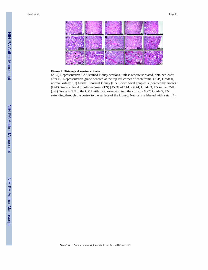
Figure 1. Histological scoring criteria(A-O) Representative PAS stained kidney sections, unless otherwise stated, obtained 24hrafter IR. Representative grade denoted at the top left corner of each frame. (A-B) Grade 0,normal kidney. (C) Grade 1, normal kidney (H&E) with focal apoptosis (denoted by arrow).(D-F) Grade 2, focal tubular necrosis (TN) (<50% of CMJ). (G-I) Grade 3, TN in the CMJ.(J-L) Grade 4, TN in the CMJ with focal extension into the cortex. (M-O) Grade 5, TNextending through the cortex to the surface of the kidney. Necrosis is labeled with a star (*).
Novak et al. Page 11
Pediatr Res. Author manuscript; available in PMC 2012 June 02.
NIH
-PA Author Manuscript
NIH
-PA Author Manuscript
NIH
-PA Author Manuscript
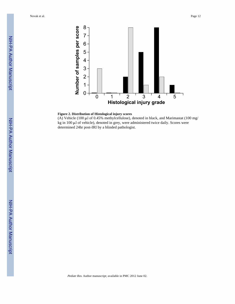
Figure 2. Distribution of Histological injury scores(A) Vehicle (100 μl of 0.45% methylcellulose), denoted in black, and Marimastat (100 mg/kg in 100 μl of vehicle), denoted in grey, were administered twice daily. Scores weredetermined 24hr post-IRI by a blinded pathologist.
Novak et al. Page 12
Pediatr Res. Author manuscript; available in PMC 2012 June 02.
NIH
-PA Author Manuscript
NIH
-PA Author Manuscript
NIH
-PA Author Manuscript
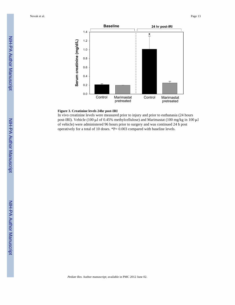
Figure 3. Creatinine levels 24hr post-IRIIn vivo creatinine levels were measured prior to injury and prior to euthanasia (24 hourspost-IRI). Vehicle (100 μl of 0.45% methylcellulose) and Marimastat (100 mg/kg in 100 μlof vehicle) were administered 96 hours prior to surgery and was continued 24 h postoperatively for a total of 10 doses. *P= 0.003 compared with baseline levels.
Novak et al. Page 13
Pediatr Res. Author manuscript; available in PMC 2012 June 02.
NIH
-PA Author Manuscript
NIH
-PA Author Manuscript
NIH
-PA Author Manuscript
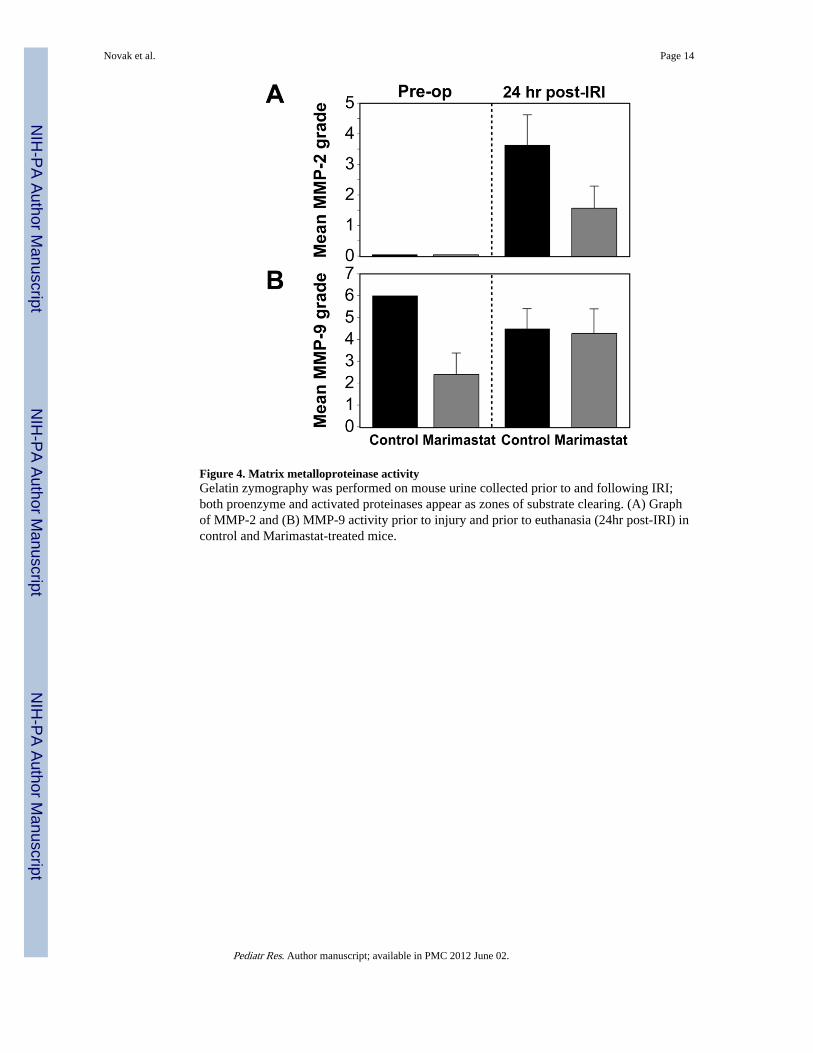
Figure 4. Matrix metalloproteinase activityGelatin zymography was performed on mouse urine collected prior to and following IRI;both proenzyme and activated proteinases appear as zones of substrate clearing. (A) Graphof MMP-2 and (B) MMP-9 activity prior to injury and prior to euthanasia (24hr post-IRI) incontrol and Marimastat-treated mice.
Novak et al. Page 14
Pediatr Res. Author manuscript; available in PMC 2012 June 02.
NIH
-PA Author Manuscript
NIH
-PA Author Manuscript
NIH
-PA Author Manuscript
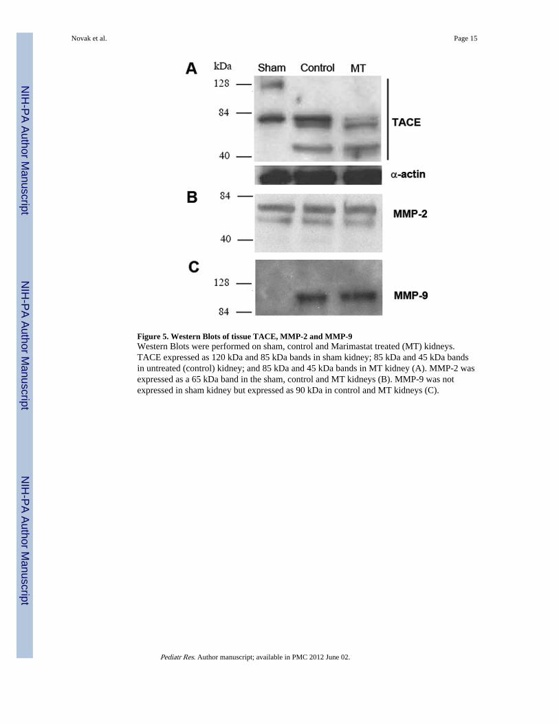
Figure 5. Western Blots of tissue TACE, MMP-2 and MMP-9Western Blots were performed on sham, control and Marimastat treated (MT) kidneys.TACE expressed as 120 kDa and 85 kDa bands in sham kidney; 85 kDa and 45 kDa bandsin untreated (control) kidney; and 85 kDa and 45 kDa bands in MT kidney (A). MMP-2 wasexpressed as a 65 kDa band in the sham, control and MT kidneys (B). MMP-9 was notexpressed in sham kidney but expressed as 90 kDa in control and MT kidneys (C).
Novak et al. Page 15
Pediatr Res. Author manuscript; available in PMC 2012 June 02.
NIH
-PA Author Manuscript
NIH
-PA Author Manuscript
NIH
-PA Author Manuscript
Copyright © 2022 FDOKUMEN