Wip1-Deficient Neutrophils Significantly Promote Intestinal Ischemia/Reperfusion Injury in Mice
REVIEW
The Contemporary Role of Antioxidant Therapy inAttenuating Liver Ischemia-Reperfusion Injury:
A ReviewGeorgios K. Glantzounis,1,2 Henryk J. Salacinski,1 Wenxuan Yang,1
Brian R. Davidson,1,2 and Alexander M. Seifalian1,2
Oxidative stress is an important factor in many patholog-ical conditions such as inflammation, cancer, ageing andorgan response to ischemia-reperfusion. Humans havedeveloped a complex antioxidant system to eliminate orattenuate oxidative stress. Liver ischemia-reperfusioninjury occurs in a number of clinical settings, includingliver surgery, transplantation, and hemorrhagic shockwith subsequent fluid resuscitation, leading to significantmorbidity and mortality. It is characterized by significantoxidative stress but accompanied with depletion of endog-enous antioxidants. This review has 2 aims: firstly, tohighlight the clinical significance of liver ischemia-reper-fusion injury, the underlying mechanisms and the mainpathways by which the antioxidants function, and sec-ondly, to describe the new developments that are ongoingin antioxidant therapy and to present the experimentaland clinical evidence about the role of antioxidants inmodulating hepatic ischemia-reperfusion injury. (LiverTranspl 2005;11:1031-1047.)
Liver ischemia-reperfusion (I/R) injury is well recog-nized as a significant cause of morbidity and mor-
tality in 2 principal settings. Firstly, it occurs in majorliver resections1 and transplantation2,3 where anoxic orischemic liver injury takes place. Secondly, it happens asa consequence of systemic hypoxia or with conditionsthat cause low blood flow to the liver resulting in insuf-ficient perfusion. The latter occurs in hemorrhagic, car-diogenic, or septic shock with subsequent fluid resusci-tation,4 in cardiovascular surgery with extracorporealcirculation,5 in laparoscopic surgery,6,7 and in abdom-inal compartment syndromes.8
Severe warm liver I/R can lead to liver or even tomultiple organ failure. The extent of hepatic injurycaused by I/R depends primarily on the condition of theliver prior to the ischemic insults and its duration.9
In liver surgery, new techniques have been applied toincrease the resectability rate of liver tumors such asdownstaging chemotherapy and portal vein emboliza-tion. However, these techniques can make the livermore susceptible to ischemic insults.10 Furthermore,the commonest primary liver cancer, hepatocellular car-cinoma usually develops on the background of liver
cirrhosis, which increases the risk of liver failure duringany subsequent surgery.11
In the field of liver transplantation, I/R injury isclosely related to the development of primary graft non-function (occurs in �5 % of grafts) and primary graftdysfunction (occurs in 10-30 % of grafts).12 Both con-ditions are associated with high rates of morbidity andmortality. I/R injury increases the incidence of subse-quent graft rejection.13
Another field where I/R injury affects outcome ishepatic resection or transplantation with steatotic liv-ers. It is reported that 25% of the western populationhas some degree of hepatic steatosis,14 which is theresult of the abnormal accumulation of triacylglycerolwithin the cytoplasm of hepatocytes, attributed to theeffects of alcohol excess, obesity, diabetes, or drugs.Furthermore, hepatic steatosis is associated with animpaired microcirculation,15,16 increased postoperativemorbidity and mortality, and poor graft function.14
It is obvious that liver I/R occurs in many diverseclinical settings and has a major impact on clinical out-
Abbreviations: I/R, ischemia-reperfusion; ROS, reactive oxygenspecies; RNS, reactive nitrogen species; ATP, adenosine triphosphate;XOR, xanthine oxidoreductase; TNF-�, tumor necrosis factor-�;O2, oxygen; OH, hydroxyl radical; NO, nitric oxide; H2O2, hydro-gen peroxide; ONOO�, peroxynitrite; HO, haem oxygenase; NOS,NO synthetase; iNOS, inducible NOS; GSH, glutathione; SOD,superoxide dismutase; NAC, N-acetylcysteine.
From the 1University Department of Surgery, Royal Free and Uni-versity College Medical School, University College London, London,NW3 2PF, UK, and the 2Hepatopancreaticobiliary and Liver Trans-plant Unit, Royal Free Hampstead NHS Trust, London, NW3 2QG,UK.
Received March 21, 2005; accepted May 3, 2005.Address reprint requests to: Professor Brian R. Davidson, Professor of
Hepatobiliary Surgery and Liver Transplantation, University Depart-ment of Surgery, Royal Free and University College Medical School,University College London, Rowland Hill Street, London, NW3 2PF,UK. Telephone: 020 7830 2901; FAX: 020 7472 6444; E-mail:[email protected]
Copyright © 2005 by the American Association for the Study ofLiver Diseases
Published online in Wiley InterScience (www.interscience.wiley.com).DOI 10.1002/lt.20504
1031Liver Transplantation, Vol 11, No 9 (September), 2005: pp 1031-1047
come. Reoxygenation of the ischemic liver causes thegeneration of numerous reactive oxygen species (ROS)and reactive nitrogen species (RNS). Although ROSand RNS in low concentrations have an important roleas mediators in normal cellular metabolism and signaltransduction,17,18 in higher concentrations they can bedamaging. In light of this, a complex defense network,called the antioxidant system, has been developed inmammals, to prevent or reduce the injury caused byhigh concentrations of ROS or RNS.
Free radical scavenging or administration of agentsthat enhance the endogenous antioxidant system couldreduce postischemic tissue injury and so be useful inclinical settings against hepatic I/R damage.
The aim of this review is to present the main mech-anisms by which antioxidants function and then thepotential use of exogenous antioxidants as a therapeuticstrategy in conditions associated with liver I/R injury.For information on other therapeutic strategies againstliver I/R injury, the reader is referred to recentreviews.19-23
Search Strategy Methods
All the studies were identified by PubMed, Web ofScience, and Embase searches between years 1966 and2004 with the following keywords: liver, hepatic, isch-aemia or ischemia, reperfusion, injury, antioxidant,pharmaceutical preconditioning. In vitro, experimentaland clinical studies included focused on the effect ofantioxidant therapy on liver I/R injury.
Ischemic Injury
When oxygen supply to cells becomes insufficient as adirect result of reduced blood flow or hypoxia, themitochondrial respiratory chain function alters and thereduction-oxidation (redox) state of the mitochondrialenzymes becomes reduced. This results in the inhibi-tion of the oxidative phosphorylation process with asubsequent reduction in adenosine triphosphate (ATP)synthesis.24 Reduction of cellular ATP causes distur-bances in membrane ion translocation by inhibition ofthe ATP-dependent sodium (Na�)/potassium (K�)ATPase, resulting in sodium influx and intracellularsodium accumulation with corresponding cell swellingand death.25
Intracellular calcium accumulation is also stronglyimplicated in the development of ischemic injury andthought to be a crucial step in the transition to irrevers-ible damage.26 The increased cytosolic calcium levelcauses activation of cell membrane phospholipases,
resulting in phospholipids degradation and cell mem-brane disruption.27 Prior to cell death, hepatocytes andother cells develop a state characterized by mitochon-drial permeability transition,28 lysosomal disruption,bleb formation and growth, cell swelling, and leakage ofsmall molecular mass solutes.29,30 Calcium also acti-vates xanthine oxidoreductase (XOR) which has a rolein oxygen free radical production following reperfu-sion31.
Although the basic mechanisms of ischemic injuryafter warm and cold liver ischemia are similar, there arealso significant differences. In liver transplantation theliver undergoes cold ischemic storage followed byrewarming ischemia and reperfusion.23 Cold ischemiais associated with reduced oxidative phosphorylation,lower cellular ATP levels, and increased glycolysis,32
while warm ischemia leads to greater oxidative stressand mitochondrial dysfunction.33,34 The main site ofinjury in cold ischemia are nonparenchymal cells(Kupffer, sinusoidal endothelial cells, Ito cells and bili-ary epithelium) whereas in warm ischemia are hepato-cytes.35
Reperfusion Injury
Although ischemia causes significant injury to tissueand cells, the injury during reperfusion is more severe. Acomplex network of hepatic and extrahepatic mecha-nisms is involved in the pathophysiology of hepatic I/Rinjury.
Experimental evidence shows that there are two dis-tinct phases of liver reperfusion injury. The early phasecovers the first 2 hours after reperfusion. During thisphase the main event is the activation of Kuppfercells.36 Complement activation and the recruitmentand activation of CD4� T lymphocytes are factors thatenable the activation of the Kuppfer cells.20,37
Kuppfer cell activation leads to structural changes,formation of vascular ROS and production of cytokinessuch as tumor necrosis factor-� (TNF-�) and interleu-kin 1.22,38. These ROS and cytokines have a directcytotoxic effect on endothelial cells, and hepatocytescan induce changes in cell membrane receptors in hepa-tocytes and release of cytokines. TNF-� acts as thecentral mediator in the hepatic response to I/R. Theproduction of TNF-� induces the expression of adhe-sion molecules on vascular endothelial cells and stimu-lates the production and release of neutrophil-attractingchemokines. The final result is the recruitment of neu-trophils. These activated neutrophils release ROS andproteases that are responsible for the induced oxidativestress during the late phase of reperfusion injury,39,40
1032 Glantzounis et al.

which is much more severe compared to that during theearly phase.
Recent evidence suggests that the T-lymphocytescan also be important mediators in short- and long-term liver I/R injury. Their role seems to be a multifac-torial one. There is evidence that systemic immunosup-pression attenuates hepatocellular injury followingI/R.41-43 The adherence of CD4� T-lymphocytes inhepatic sinusoids occurs during the early phase of reper-fusion and is mediated by TNF-� and interleukin 1.44
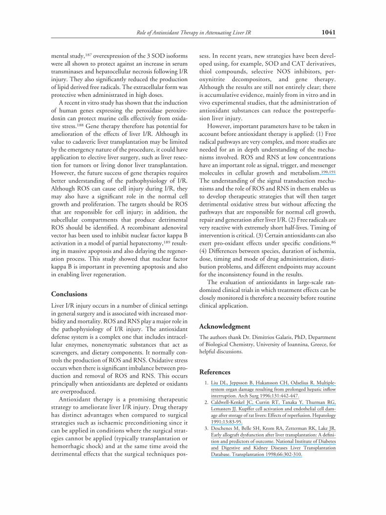
These T-lymphocytes can increase Kupffer cell activa-tion and can act as cellular mediators in polymorpho-nuclear cell recruitment through the release of sub-stances such as granulocyte colony stimulating factorand interferon �.45 The basic pathophysiologicalmechanisms in hepatic I/R injury are summarized inFigure 1.
Reactive Oxygen Species and I/R Injury
A “radical” is defined as any atom or biomolecule thatcontains unpaired electrons.46 These unpaired electronsinfluence the chemical reactivity, making the radicalmore reactive than the corresponding nonradical.Although oxygen (O2) is the most important biologicalmolecule for sustaining life, it is also the main source forfree radical formation due to its high availability.
The biologically relevant radicals are the superoxideanion (O2
•�), the hydroxyl radical (.OH) and nitricoxide (NO). Under normal conditions around 1 to 3%
of the oxygen that is metabolized in the mitochondria isconverted to the radical O2
•�.47 Some other species areintermediate in the metabolism of O2 or NO but arenot radicals, as they do not contain unpaired electrons.These intermediate species along with the radical spe-cies are called ROS and RNS, respectively. The mostrepresentative examples of nonradical ROS are hydro-gen peroxide (H2O2) and hypochlorous acid. The mostrepresentative of nonradical RNS is peroxynitrite(ONOO�)48. Peroxynitrite is formed when there issimultaneous production of nitric oxide with superox-ide anion (equation 1):
NO � O2• � 3 ONOO � (1)
Tissue toxicity from superoxide generation is based onits direct reactivity with numerous types of biologicalmolecules (typically lipids, DNA, RNA, cat-echolamines, and steroids) and from its dismutation toform H2O2.
49 Trace amounts of metals ions (princi-pally iron or copper) react with H2O2 in what is knownas the Fenton reaction to produce the toxic radical.OH.50 This radical can cleave covalent bonds in pro-teins and carbohydrates and destroy cell membranes.
Liver injury induced by I/R is caused, at least par-tially, by ROS and RNS. There is evidence that duringhepatic I/R there is generation and release of ROS andRNS with concomitant consumption of endogenousantioxidants and apoptotic or necrotic cell death.39,51-54
Although the exact sources of ROS generation in
Figure 1. Schematic presentation of pathophysiology of liver ischemia / reperfusion injury. IL-1�, interleukin 1�; EC,endothelial cells.
1033Role of Antioxidant Therapy in Attenuating Liver IR
liver I/R are still under investigation, the nicotinamideadenine dinucleotide phosphate oxidase, the xanthine/XOR system, and the mitochondria have been sug-gested to play key roles.55 Although XOR was regardedas the principal source of postischemic oxidant stress inthe liver, recent evidence suggests that XOR plays aminor role as compared to the mitochondria.56 Mito-chondria are the site of the production of large amountsof superoxide, under conditions of oxidative stress. It isthis stress that finally leads to the formation of mem-brane permeability transition pores and the breakdownof the mitochondrial membrane potential that cancause cellular death.57
ROS and RNS can have an important role in signaltransduction pathways coordinating the body’s inflam-matory response after liver I/R injury.20,39,58,59 They areinvolved as mediators in the production of substancesregulating liver blood flow60 and regeneration.61,62
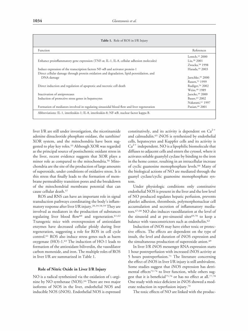
Transgenic mice with overexpression of antioxidantenzymes have decreased cellular ploidy during liverregeneration, suggesting a role for ROS in cell cyclecontrol.61 ROS also induce stress genes such as haemoxygenase (HO)-1.63 The induction of HO-1 leads toformation of the antioxidant biliverdin, the vasodilatorcarbon monoxide, and iron. The multiple roles of ROSin liver I/R are summarized in Table 1.
Role of Nitric Oxide in Liver I/R Injury
NO is a radical synthesized via the oxidation of L-argi-nine by NO synthetase (NOS).64 There are two majorisoforms of NOS in the liver, endothelial NOS andinducible NOS (iNOS). Endothelial NOS is expressed
constitutively, and its activity is dependent on Ca2�
and calmudulin.65 iNOS is synthesized by endothelialcells, hepatocytes and Kuppfer cells and its activity isCa2� independent. NO is a lipophilic biomolecule thatdiffuses to adjacent cells and enters the cytosol, where itactivates soluble guanylyl cyclase by binding to the ironin the heme center, resulting in an intracellular increaseof cyclic guanosine monophosphate levels.66 Many ofthe biological actions of NO are mediated through theguanyl cyclase/cyclic guanosine monophosphate sys-tem.
Under physiologic conditions only constitutiveendothelial NOS is present in the liver and the low levelof NO produced regulates hepatic perfusion, preventsplatelet adhesion, thrombosis, polymorphonuclear cellaccumulation and secretion of inflammatory media-tors.67,68 NO also induces vasodilatation at the level ofthe sinusoid and at pre-sinusoid sites69,70 to keep abalance with vasoconstrictors such as endothelin.62
Induction of iNOS may have either toxic or protec-tive effects. The effects are dependent on the type ofinsult, the level and duration of iNOS expression andthe simultaneous production of superoxide anion.49
In liver I/R iNOS messenger RNA expression starts1 hour postreperfusion with increased iNOS activity at5 hours postreperfusion.71 The literature concerningthe effect of iNOS in liver I/R injury is still ambivalent.Some studies suggest that iNOS expression has detri-mental effects72-74 to liver function, while others sug-gest that it is beneficial75,76 or has no effect at all.77,78
One study with mice deficient in iNOS showed a mod-erate reduction in reperfusion injury.79
The toxic effects of NO are linked with the produc-
Table 1. Role of ROS in I/R Injury
Function References
Enhance proinflammatory gene expression (TNF-�; IL-1, IL-8, cellular adhesion molecules)Lentch,22 2000Liu,38 2001
Induce expression of the transcription factors NF-�B and activator protein-1Zwacka,58 1998Harada,59 2003
Direct cellular damage through protein oxidation and degradation, lipid peroxidation, andDNA damage Jaeschke,39 2000
Direct induction and regulation of apoptotic and necrotic cell deathRauen,53 1999Rudiger,54 2002
Inactivation of antiproteasesWeiss,40 1989Jaescke,39 2000
Induction of protective stress genes in hepatocytes Bauer,63 2002
Formation of mediators involved in regulating sinusoidal blood flow and liver regenerationNakatani,61 1997Paxian,60 2001
Abbreviations: IL-1, interleukin-1; IL-8, interleukin-8; NF-�B, nuclear factor kappa B.
1034 Glantzounis et al.
tion of peroxynitrite, which is the product of O2.� and
NO. Peroxynitrite can cause cell injury through multi-ple pathways: initiation of lipid peroxidation, directinhibition of mitochondrial respiratory chain enzymes,inhibition of membrane Na�/ K� ATPase activity, oroxidative protein modification such as formation ofnitrotyrosine.80-82
However whether NO will act as a cytoprotective orcytotoxic agent depends on an number of factors, suchas NO-superoxide radical ratios, hepatic stores ofreduced glutathione, and the duration of ischemia.
The Antioxidant System
The body has developed major antioxidant defensemechanisms to protect it from damage from free radi-cals. An antioxidant is any substance that when presentat low concentrations, compared with those of an oxi-dizable substrate, significantly delays or prevents oxida-tion of the substrate.83 The endogenous antioxidantsmainly are small molecular weight substances that areable to prevent initiation of oxidative damage or to limitits propagation and enzymes that convert and detoxifyROS and RNS. Cellular redox balance is in normalcircumstances under tight control. However, whenROS and RNS are produced at levels that cannot becounteracted by endogenous antioxidant systems, animbalance takes place, called oxidative stress.84 Thiscondition can lead to the damage of lipids, proteins,carbohydrates, and nucleic acids. Hepatocytes tend tobe resistant to injury by ROS and RNS, since theycontain high intracellular concentrations of glutathione(GSH), superoxide dismutase (SOD), catalase, andlipid soluble antioxidants.
The antioxidant system is very important for theliving species and has allowed them to use O2 for energyproduction, without being exposed to the deleteriouseffects of O2. The composition of antioxidant defencesdiffers from tissue to tissue and from cell to cell in agiven tissue. Also, different organs contain differentconcentrations of antioxidants, and for this reason thereis variability in organ resistance to I/R. However, thereis evidence that the antioxidants operate as a balancedand coordinated system and each relies on the action ofthe others.85,86
Antioxidants are a heterogenous family of mole-cules. Several classifications have been used in the pasttaking into account the origin (natural or synthetic),nature (enzymatic or nonenzymatic), properties(hydrophilic or lipophilic), mechanism (catalyticalremoval of ROS, metal chelation, scavenging of ROS),and site of action (intracellular; membrane, and extra-
cellular). This review is presented according to the siteof action of the antioxidants.
Intracellular antioxidant defenses include the super-oxide dismutase; catalase; glutathione peroxidase andreductase enzymes, the tripeptide glutathione, thepolypeptide thioredoxine, the enzyme HO, and peroxi-dases of the peroxiredoxin family.
SOD catalyses the dismutation of superoxide tohydrogen peroxide and oxygen (equation 2):
2O2• � � 2H � ™3
SOD
H2O2 � O2 (2)
Three forms of SOD exist with different subcellularlocalizations. Those containing copper and zinc arelocated in the cytosol,87 manganese in the mitochon-dria,88 and the extracellular form usually located on theoutside of plasma membrane interacting with matrixcomponents.
The product of reaction 2, H2O2, is a weak oxidantand is relatively stable. However, unlike superoxide,H2O2 can rapidly diffuse across cell membranes, and inthe presence of transition metal ions it can be convertedto toxic hydroxyl radicals via Fenton chemistry (equa-tion 3):
Fe � 2 � H2O2 3 Fe � 3 � OH � OH � (3)
Three main systems can break down H2O2. One ofthem is the hemoprotein catalase. Catalase is present inall major body organs, especially concentrated in liver.It catalyzes the breakdown of hydrogen peroxide tooxygen and water (equation 4):
2H2O2 3 O2 � 2H2O (4)
The second system consists of the glutathione peroxi-dases. This group includes 4 different isoforms, such ascellular, gastrointestinal, extracellular, and phospholip-ids.89 They have a major role in removing hydrogenperoxide generated by superoxide dismutase with theoxidation of GSH to its oxidized form, glutathionedisulphide (GSSG) (equation 5):
2GSH � H2O2 3 GSSG � 2H2O (5)
Glutathione reductase is also an important enzyme inthis system. It expresses its action through the regener-ation of GSH from glutathione disulphide using nico-tinamide adenine dinucleotide phosphate.90
GSH is a tripeptide present in millimolar concentra-tions in virtually all cells. It is an important componentof the endogenous antioxidant system. GSH’s mainfunction is to act as a cosubstrate of glutathione perox-idase to reduce intracellularly generated peroxides.GSH also scavenges directly ROS and RNS. GSH is
1035Role of Antioxidant Therapy in Attenuating Liver IR
involved in many other metabolic processes includingprevention of oxidation of protein sulfhydryl groupsand chelation of copper ions. GSH is also present in theextracellular fluids in very small concentrations.51,91,92
The third system consists of peroxidases of the per-oxiredoxin family that reduce hydrogen peroxide andalkyl hydroperoxides to water and alcohol respectivelyusing reducing equivalents. These equivalents arederived specifically from thiol-containing donor mole-cules, such as thioredoxin. They are located in the cyto-plasm (peroxiredoxin I and II) and in mitochondria(peroxiredoxin III).93,94
Thioredoxin is a polypeptide especially concen-trated in the endoplasmic reticulum (thioredoxin 1),but it is also found in mitochondria (thioredoxin 2).Thioredoxin contains 2 adjacent sulfhydryl groups inits reduced form that are converted to a disulphide inthe oxidized form thioredoxin. In addition, it canundergo redox reactions with multiple proteins.86,95
HO is an enzyme found in the endoplasmic reticu-lum that catalyses the breakdown of haem to biliverdinwith the release of iron ions and carbon monoxide.Three isoforms of HO have been characterized: HO-1,which is highly inducible in conditions of inflammationand oxidative stress, and HO-2 and HO-3, which areconstitutively expressed.96 Induction of HO-1 protectsthe cell against oxidative injury by controlling intracel-lular levels of free heme (a prooxidant), producing biliv-erdin (an antioxidant), and improving microcirculationvia carbon monoxide release.63
Major extracellular antioxidant defenses include themetal-binding proteins.97 Free metals iron and coppercan promote free radical damage, accelerating lipid per-oxidation and catalyzing hydroxyl radical formation.The body is protected against these potentially adverseeffects by metal-binding proteins that ensure that thesemetals are maintained in a nonreactive state.98 Trans-ferrin and lactoferrin bind iron, while ceruloplasminand albumin bind copper. Haemoglobin and myoglo-bin are normally intracellular proteins. When these pro-teins are exposed to a large amount of oxidative stresssuch as H2O2, they are degraded, releasing both haemand iron ions that can then stimulate lipid peroxida-tion. Hemoglobin binding proteins known as hapto-globins and haem binding proteins such as hemopexindecrease the effectiveness of these substances in stimu-lating lipid peroxidation.99
In addition to the major protective role of the metal-binding proteins, various low-molecular-weight mole-cules that are synthesized in vivo have antioxidant prop-erties.100-102 The most important of these substances arebilirubin, melatonin, lipoic acid, coenzyme Q, uric acid
and the melamins. Recent evidence suggests that uricacid has an important role in the endogenous antioxi-dant system103,104 and that exogenous administrationof uric acid could have beneficial effects in situationsassociated with oxidative stress.105,106
A large number of dietary constituents exert antiox-idant effects in vivo. The most important are hydro-philic ascorbic acid (vitamin C) and lipophilic �-to-copherol (the most active form of vitamin E) that areimportant components of the human antioxidant sys-tem. Ascorbate is required in vivo as a cofactor for manyenzymes, of which the best known are proline hydrox-ylase and lysine hydroxylase, both involved in the bio-synthesis of collagen. Its main chemical property is itsability to act as a reducing agent. It can scavenge mostradicals such as O2
.�, .OH, peroxyl, thiyl, oxysulphurradicals, and peroxynitrite.98,107 The lipophilic �-to-copherol is a highly effective antioxidant when incor-porated in the lipid core of cell membranes. It has theability to scavenge intermediate peroxyl radicals andtherefore interrupt the chain reactions of lipid peroxi-dation.108
Carotenoids are a group of colored pigments that arewidespread in plant tissues. They serve as a precursor ofvitamin A and are the principal dietary source of vita-min A in humans. They exert their antioxidant action asfree-radical scavengers.109
Another group of antioxidants are the plant phenols.Plants contain a huge range of phenols, including toco-pherols, tocotrienols, flavonoids, anthocyanidins, andphenylpropanoids. They inhibit peroxidation by actingas chain-breaking peroxyl-radical scavengers. In addi-tion, they scavenge ROS and RNS such as .OH,ONOO�, and hypochlorous acid, and they act as metalchelators.110,111
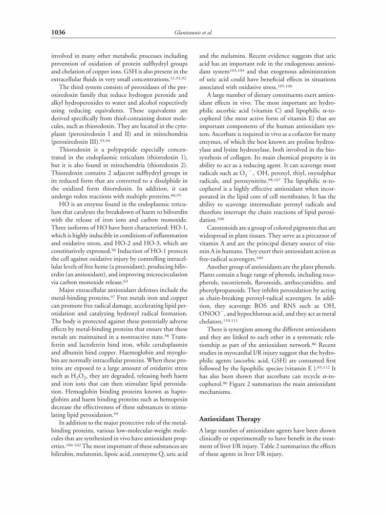
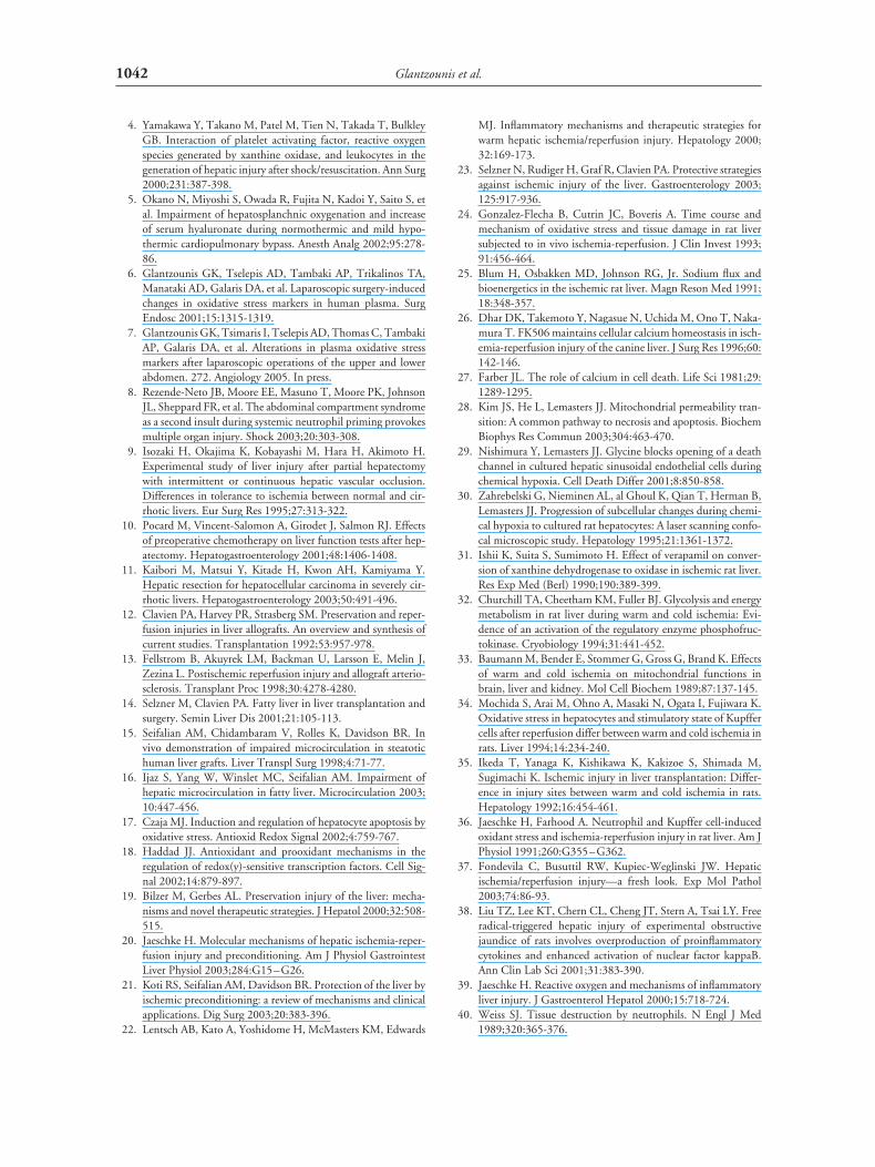
There is synergism among the different antioxidantsand they are linked to each other in a systematic rela-tionship as part of the antioxidant network.86 Recentstudies in myocardial I/R injury suggest that the hydro-philic agents (ascorbic acid, GSH) are consumed firstfollowed by the lipophilic species (vitamin E ).85,112 Ithas also been shown that ascorbate can recycle �-to-copherol.86 Figure 2 summarizes the main antioxidantmechanisms.
Antioxidant Therapy
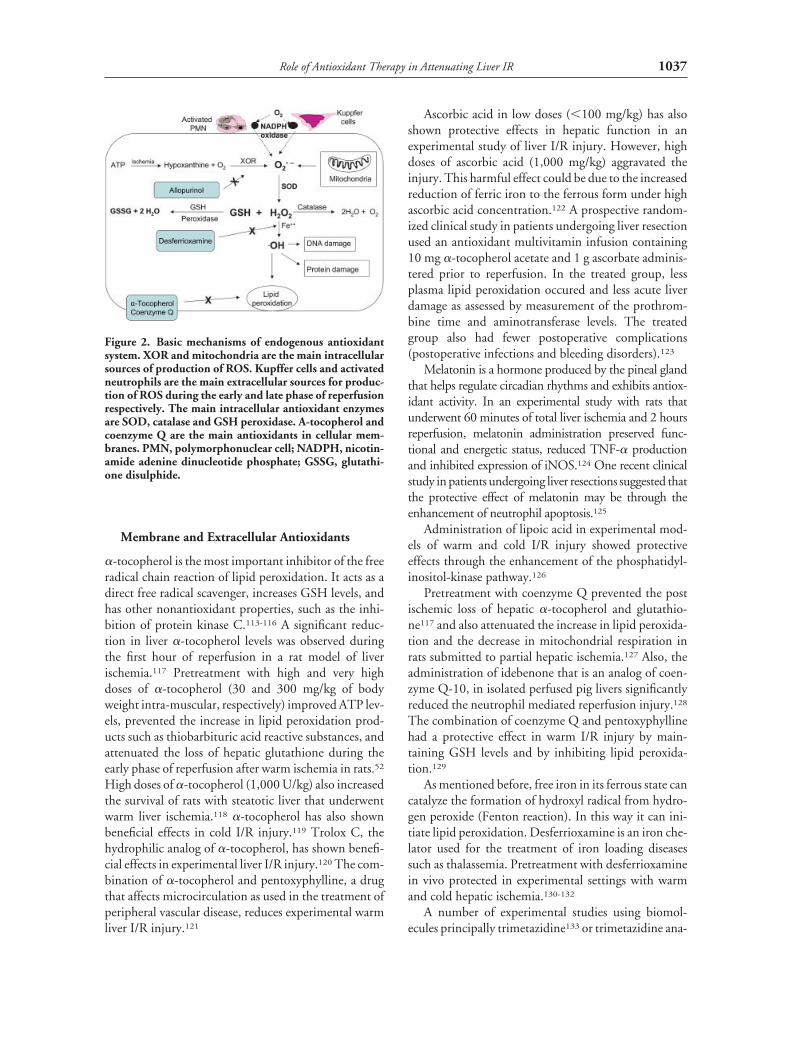
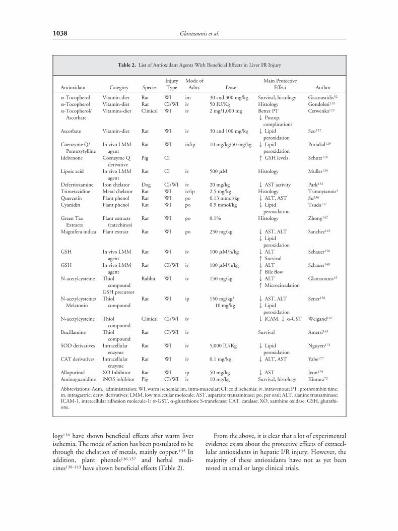
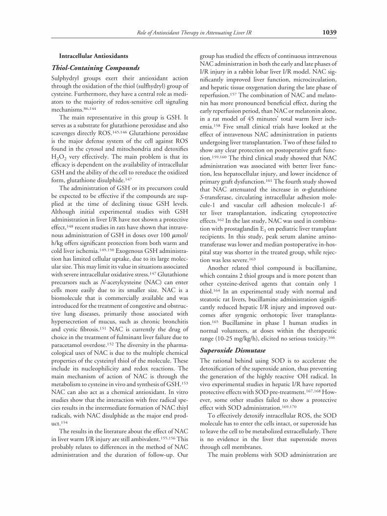
A large number of antioxidant agents have been shownclinically or experimentally to have benefit in the treat-ment of liver I/R injury. Table 2 summarizes the effectsof these agents in liver I/R injury.
1036 Glantzounis et al.
Membrane and Extracellular Antioxidants
�-tocopherol is the most important inhibitor of the freeradical chain reaction of lipid peroxidation. It acts as adirect free radical scavenger, increases GSH levels, andhas other nonantioxidant properties, such as the inhi-bition of protein kinase C.113-116 A significant reduc-tion in liver �-tocopherol levels was observed duringthe first hour of reperfusion in a rat model of liverischemia.117 Pretreatment with high and very highdoses of �-tocopherol (30 and 300 mg/kg of bodyweight intra-muscular, respectively) improved ATP lev-els, prevented the increase in lipid peroxidation prod-ucts such as thiobarbituric acid reactive substances, andattenuated the loss of hepatic glutathione during theearly phase of reperfusion after warm ischemia in rats.52
High doses of �-tocopherol (1,000 U/kg) also increasedthe survival of rats with steatotic liver that underwentwarm liver ischemia.118 �-tocopherol has also shownbeneficial effects in cold I/R injury.119 Trolox C, thehydrophilic analog of �-tocopherol, has shown benefi-cial effects in experimental liver I/R injury.120 The com-bination of �-tocopherol and pentoxyphylline, a drugthat affects microcirculation as used in the treatment ofperipheral vascular disease, reduces experimental warmliver I/R injury.121
Ascorbic acid in low doses (�100 mg/kg) has alsoshown protective effects in hepatic function in anexperimental study of liver I/R injury. However, highdoses of ascorbic acid (1,000 mg/kg) aggravated theinjury. This harmful effect could be due to the increasedreduction of ferric iron to the ferrous form under highascorbic acid concentration.122 A prospective random-ized clinical study in patients undergoing liver resectionused an antioxidant multivitamin infusion containing10 mg �-tocopherol acetate and 1 g ascorbate adminis-tered prior to reperfusion. In the treated group, lessplasma lipid peroxidation occured and less acute liverdamage as assessed by measurement of the prothrom-bine time and aminotransferase levels. The treatedgroup also had fewer postoperative complications(postoperative infections and bleeding disorders).123
Melatonin is a hormone produced by the pineal glandthat helps regulate circadian rhythms and exhibits antiox-idant activity. In an experimental study with rats thatunderwent 60 minutes of total liver ischemia and 2 hoursreperfusion, melatonin administration preserved func-tional and energetic status, reduced TNF-� productionand inhibited expression of iNOS.124 One recent clinicalstudy in patients undergoing liver resections suggested thatthe protective effect of melatonin may be through theenhancement of neutrophil apoptosis.125
Administration of lipoic acid in experimental mod-els of warm and cold I/R injury showed protectiveeffects through the enhancement of the phosphatidyl-inositol-kinase pathway.126
Pretreatment with coenzyme Q prevented the postischemic loss of hepatic �-tocopherol and glutathio-ne117 and also attenuated the increase in lipid peroxida-tion and the decrease in mitochondrial respiration inrats submitted to partial hepatic ischemia.127 Also, theadministration of idebenone that is an analog of coen-zyme Q-10, in isolated perfused pig livers significantlyreduced the neutrophil mediated reperfusion injury.128
The combination of coenzyme Q and pentoxyphyllinehad a protective effect in warm I/R injury by main-taining GSH levels and by inhibiting lipid peroxida-tion.129
As mentioned before, free iron in its ferrous state cancatalyze the formation of hydroxyl radical from hydro-gen peroxide (Fenton reaction). In this way it can ini-tiate lipid peroxidation. Desferrioxamine is an iron che-lator used for the treatment of iron loading diseasessuch as thalassemia. Pretreatment with desferrioxaminein vivo protected in experimental settings with warmand cold hepatic ischemia.130-132
A number of experimental studies using biomol-ecules principally trimetazidine133 or trimetazidine ana-
Figure 2. Basic mechanisms of endogenous antioxidantsystem. XOR and mitochondria are the main intracellularsources of production of ROS. Kupffer cells and activatedneutrophils are the main extracellular sources for produc-tion of ROS during the early and late phase of reperfusionrespectively. The main intracellular antioxidant enzymesare SOD, catalase and GSH peroxidase. A-tocopherol andcoenzyme Q are the main antioxidants in cellular mem-branes. PMN, polymorphonuclear cell; NADPH, nicotin-amide adenine dinucleotide phosphate; GSSG, glutathi-one disulphide.
1037Role of Antioxidant Therapy in Attenuating Liver IR
logs134 have shown beneficial effects after warm liverischemia. The mode of action has been postulated to bethrough the chelation of metals, mainly copper.135 Inaddition, plant phenols136,137 and herbal medi-cines138-143 have shown beneficial effects (Table 2).
From the above, it is clear that a lot of experimentalevidence exists about the protective effects of extracel-lular antioxidants in hepatic I/R injury. However, themajority of these antioxidants have not as yet beentested in small or large clinical trials.
Table 2. List of Antioxidant Agents With Beneficial Effects in Liver I⁄R Injury
Antioxidant Category SpeciesInjuryType
Mode ofAdm. Dose
Main ProtectiveEffect Author
�-Tocopherol Vitamin-diet Rat WI im 30 and 300 mg/kg Survival, histology Giacoustidis52
�-Tocopherol Vitamin-diet Rat CI/WI iv 50 IU/Kg Histology Gondolesi119
�-Tocopherol/Ascorbate
Vitamins-diet Clinical WI iv 2 mg/1,000 mg Better PT2 Postop.
complications
Cerwenka123
Ascorbate Vitamin-diet Rat WI iv 30 and 100 mg/kg 2 Lipidperoxidation
Seo122
Coenzyme Q/Pentoxyfylline
In vivo LMMagent
Rat WI in/ip 10 mg/kg/50 mg/kg 2 Lipidperoxidation
Portakal129
Idebenone Coenzyme Qderivative
Pig CI 1 GSH levels Schutz128
Lipoic acid In vivo LMMagent
Rat CI iv 500 �M Histology Muller126
Deferrioxamine Iron chelator Dog CI/WI iv 20 mg/kg 2 AST activity Park132
Trimetazidine Metal chelator Rat WI iv/ip 2.5 mg/kg Histology Tsimoyiannis3
Quercetin Plant phenol Rat WI po 0.13 mmol/kg 2 ALT, AST Su136
Cyanidin Plant phenol Rat WI po 0.9 mmol/kg 2 Lipidperoxidation
Tsuda137
Green TeaExtracts
Plant extracts(catechines)
Rat WI po 0.1% Histology Zhong142
Magnifera indica Plant extract Rat WI po 250 mg/kg 2 AST, ALT2 Lipid
peroxidation
Sanchez143
GSH In vivo LMMagent
Rat WI iv 100 �M/h/kg 2 ALT1 Survival
Schauer150
GSH In vivo LMMagent
Rat CI/WI iv 100 �M/h/kg 2 ALT1 Bile flow
Schauer149
N-acetylcysteine Thiolcompound
GSH precursor
Rabbit WI iv 150 mg/kg 2 ALT1Microcirculation
Glantzounis15
N-acetylcysteine/Melatonin
Thiolcompound
Rat WI ip 150 mg/kg/10 mg/kg
2 AST, ALT2 Lipid
peroxidation
Sener158
N-acetylcysteine Thiolcompound
Clinical CI/WI iv 2 ICAM,2 �-GST Weigand162
Bucillamine Thiolcompound
Rat CI/WI iv Survival Amersi165
SOD derivatives Intracellularenzyme
Rat WI iv 5,000 IU/Kg 2 Lipidperoxidation
Nguyen174
CAT derivatives Intracellularenzyme
Rat WI iv 0.1 mg/kg 2 ALT, AST Yabe177
Allopurinol XO Inhibitor Rat WI ip 50 mg/kg 2 AST Jeon179
Aminoguanidine iNOS inhibitor Pig CI/WI iv 10 mg/kg Survival, histology Kimura72
Abbreviations: Adm., administration; WI, warm ischemia; im, intra-muscular; CI, cold ischemia; iv, intravenous; PT, prothrombin time;in, intragastric; deriv, derivatives; LMM, low molecular molecule; AST, aspartate transaminase; po, per oral; ALT, alanine transaminase;ICAM-1, intercellular adhesion molecule-1; �-GST, �-glutathione S-transferase; CAT, catalase; XO, xanthine oxidase; GSH, glutathi-one.
1038 Glantzounis et al.
Intracellular Antioxidants
Thiol-Containing CompoundsSulphydryl groups exert their antioxidant actionthrough the oxidation of the thiol (sulfhydryl) group ofcysteine. Furthermore, they have a central role as medi-ators to the majority of redox-sensitive cell signalingmechanisms.86,144
The main representative in this group is GSH. Itserves as a substrate for glutathione peroxidase and alsoscavenges directly ROS.145,146 Glutathione peroxidaseis the major defense system of the cell against ROSfound in the cytosol and mitochondria and detoxifiesH2O2 very effectively. The main problem is that itsefficacy is dependent on the availability of intracellularGSH and the ability of the cell to rereduce the oxidizedform, glutathione disulphide.147
The administration of GSH or its precursors couldbe expected to be effective if the compounds are sup-plied at the time of declining tissue GSH levels.Although initial experimental studies with GSHadministration in liver I/R have not shown a protectiveeffect,148 recent studies in rats have shown that intrave-nous administration of GSH in doses over 100 �mol/h/kg offers significant protection from both warm andcold liver ischemia.149,150 Exogenous GSH administra-tion has limited cellular uptake, due to its large molec-ular size. This may limit its value in situations associatedwith severe intracellular oxidative stress.147 Glutathioneprecursors such as N-acetylcysteine (NAC) can entercells more easily due to its smaller size. NAC is abiomolecule that is commercially available and wasintroduced for the treatment of congestive and obstruc-tive lung diseases, primarily those associated withhypersecretion of mucus, such as chronic bronchitisand cystic fibrosis.151 NAC is currently the drug ofchoice in the treatment of fulminant liver failure due toparacetamol overdose.152 The diversity in the pharma-cological uses of NAC is due to the multiple chemicalproperties of the cysteinyl thiol of the molecule. Theseinclude its nucleophilicity and redox reactions. Themain mechanism of action of NAC is through themetabolism to cysteine in vivo and synthesis of GSH.153
NAC can also act as a chemical antioxidant. In vitrostudies show that the interaction with free radical spe-cies results in the intermediate formation of NAC thiylradicals, with NAC disulphide as the major end prod-uct.154
The results in the literature about the effect of NACin liver warm I/R injury are still ambivalent.155,156 Thisprobably relates to differences in the method of NACadministration and the duration of follow-up. Our
group has studied the effects of continuous intravenousNAC administration in both the early and late phases ofI/R injury in a rabbit lobar liver I/R model. NAC sig-nificantly improved liver function, microcirculation,and hepatic tissue oxygenation during the late phase ofreperfusion.157 The combination of NAC and melato-nin has more pronounced beneficial effect, during theearly reperfusion period, than NAC or melatonin alone,in a rat model of 45 minutes’ total warm liver isch-emia.158 Five small clinical trials have looked at theeffect of intravenous NAC administration in patientsundergoing liver transplantation. Two of these failed toshow any clear protection on postoperative graft func-tion.159,160 The third clinical study showed that NACadministration was associated with better liver func-tion, less hepatocellular injury, and lower incidence ofprimary graft dysfunction.161 The fourth study showedthat NAC attenuated the increase in �-glutathioneS-transferase, circulating intracellular adhesion mole-cule-1 and vascular cell adhesion molecule-1 af-ter liver transplantation, indicating cytoprotectiveeffects.162 In the last study, NAC was used in combina-tion with prostaglandin E1 on pediatric liver transplantrecipients. In this study, peak serum alanine amino-transferase was lower and median postoperative in-hos-pital stay was shorter in the treated group, while rejec-tion was less severe.163
Another related thiol compound is bucillamine,which contains 2 thiol groups and is more potent thanother cysteine-derived agents that contain only 1thiol.164 In an experimental study with normal andsteatotic rat livers, bucillamine administration signifi-cantly reduced hepatic I/R injury and improved out-comes after syngenic orthotopic liver transplanta-tion.165 Bucillamine in phase I human studies innormal volunteers, at doses within the therapeuticrange (10-25 mg/kg/h), elicited no serious toxicity.166
Superoxide DismutaseThe rational behind using SOD is to accelerate thedetoxification of the superoxide anion, thus preventingthe generation of the highly reactive .OH radical. Invivo experimental studies in hepatic I/R have reportedprotective effects with SOD pre-treatment.167,168 How-ever, some other studies failed to show a protectiveeffect with SOD administration.169,170
To effectively detoxify intracellular ROS, the SODmolecule has to enter the cells intact, or superoxide hasto leave the cell to be metabolized extracellularly. Thereis no evidence in the liver that superoxide movesthrough cell membranes.
The main problems with SOD administration are
1039Role of Antioxidant Therapy in Attenuating Liver IR
the short half-life (about 6 min) and the lack of uptakeof the intact protein into cells.147 Inadequate delivery totarget sites could be the cause for the mixed resultsreported to date in the literature.
To improve the intracellular availability of SOD,new derivatives have been developed. These include theconjugation of SOD with carbohydrate structures thatfacilitate the uptake into liver non-parenchymal cells.The techniques that have been developed are mannosy-lation, succinylation,171 and pegylation.172 Kupfferand sinusoidal endothelial cells have receptors thatrecognize and internalize ligands containing mannose,succinylated and pegylated proteins. Targeted SODderivatives showed beneficial effects in preventingexperimental warm liver I/R injury.173,174
Catalase
The results in the literature are ambivalent concerningthe role of catalase in liver I/R175,176 injury. The mainproblems are the same as with SOD, the short half-lifein plasma, and the difficulty of protein uptake into cells.To bypass these difficulties, catalase-targeted deriva-tives have been developed from conjugation with car-bohydrates. The initial results appear promising,177 andthe administration of a combination of both enzymes(catalase and SOD) has been tried and the results werefound to be good.178 Among the different combina-tions Man-SOD and Suc-catalase have shown the great-est efficacy in preventing liver injury. This combinationreduced significantly inter-cellular adhesion molecule-1expression and neutrophil infiltration.
Allopurinol
Allopurinol is an XO inhibitor. Low doses (5-10mg/kg) are sufficient to inhibit hepatic activities of xan-thine oxidase and xanthine dehydrogenase almost com-pletely, but these doses were not protective of liverI/R.147 On the other hand, an experimental studywhere allopurinol was administered intra-peritoneal,before ischemia, at high doses (50 mg/kg) showed aclear protective role in rats subjected to liver I/R in-jury.179 There is experimental evidence that allopurinolhas a protective role in acetaminophen-induced tox-icity.180 The most likely mechanism for this protectiveeffect is the prevention of mitochondrial oxidativestress. An alternative mechanism is the action as a freeradical scavenger and most probably as a scavenger ofperoxynitrite.180
Table 2 summarizes substances with extracellularand intracellular antioxidant action that have shownbeneficial effects in liver I/R injury.
Antioxidants That Modulate NO Metabolism
NO can have either beneficial or detrimental effects inliver I/R injury. Recent evidence has shown that duringthe reperfusion period endothelial NOS is downregu-lated in both hepatocytes and inflammatory cells duringthe late phase.181 The expression of iNOS is associatedwith evidence of ONOO� formation, although theexact role of peroxynitrite in liver I/R is not clear.20 Ithas been postulated that when the endogenous amountof SOD or GSH is not enough to inhibit ONOO�
formation, cellular damage can occur. Pharmacologicalintervention to block ONOO� formation could have aprotective role against the toxic effects of massiveONOO� production.49 This intervention could acteither at the level of the reactant (NO and O2
.� ) or theproduct (ONOO�). The blockage of O2
.� can be doneby the use of SOD or its derivatives. There are reports inthe literature that showed beneficial effects with use ofselective inhibitors of iNOS in both warm and coldliver I/R injury.72,182
Strategies that aim at decreasing the intrinsic life-time of ONOO� could be either competitive stoichio-metric trapping of ONOO� or catalysis of ONOO�
decomposition to benign products (for exampleisomerization to nitrate). Such ONOO� decomposi-tors have been developed and have as their base ironporphyrin complexes.49 The search for other redox-active complexes that will accomplish catalysis of theperoxynitrite isomerization to nitrate still continues.
Antioxidant Gene Therapy
Gene therapy has recently been applied as a therapeuticstrategy against I/R injury. Two categories of vectorsystems have been used: viral and nonviral. The advan-tages of virus-based systems include higher infectionefficiencies and the ability to encode multiple largegenes.183 The disadvantages include immunogenicityand vector production issues. For these reasons, nonvi-ral vector systems using lysosomes and DNA-proteincomplexes have been developed.
In liver I/R injury predominantly, to date, viral vec-tors, typically recombinant virus, have been used. Highdoses of mitochondrial SOD administered via viral vec-tor in rats prevented liver I/R injury via inactivation ofthe transcription factors nuclear factor kappa B andactivator protein-1.184 Experimental studies in a ratliver transplantation model with normal and steatoticlivers have shown that cytosolic and mitochondrialSOD markedly improved survival, whereas extracellu-lar SOD was not protective.185,186 In another experi-
1040 Glantzounis et al.
mental study,187 overexpression of the 3 SOD isoformswere all shown to protect against an increase in serumtransminases and hepatocellular necrosis following I/Rinjury. They also significantly reduced the productionof lipid derived free radicals. The extracellular form wasprotective when administrated in high doses.
A recent in vitro study has shown that the inductionof human genes expressing the peroxidase peroxire-doxin can protect murine cells effectively from oxida-tive stress.188 Gene therapy therefore has potential foramelioration of the effects of liver I/R. Although itsvalue to cadaveric liver transplantation may be limitedby the emergency nature of the procedure, it could haveapplication to elective liver surgery, such as liver resec-tion for tumors or living donor liver transplantation.However, the future success of gene therapies requiresbetter understanding of the pathophysiology of I/R.Although ROS can cause cell injury during I/R, theymay also have a significant role in the normal cellgrowth and proliferation. The targets should be ROSthat are responsible for cell injury; in addition, thesubcellular compartments that produce detrimentalROS should be identified. A recombinant adenoviralvector has been used to inhibit nuclear factor kappa Bactivation in a model of partial hepatectomy,189 result-ing in massive apoptosis and also delaying the regener-ation process. This study showed that nuclear factorkappa B is important in preventing apoptosis and alsoin enabling liver regeneration.
Conclusions
Liver I/R injury occurs in a number of clinical settingsin general surgery and is associated with increased mor-bidity and mortality. ROS and RNS play a major role inthe pathophysiology of I/R injury. The antioxidantdefense system is a complex one that includes intracel-lular enzymes, nonenzymatic substances that act asscavengers, and dietary components. It normally con-trols the production of ROS and RNS. Oxidative stressoccurs when there is significant imbalance between pro-duction and removal of ROS and RNS. This occursprincipally when antioxidants are depleted or oxidantsare overproduced.
Antioxidant therapy is a promising therapeuticstrategy to ameliorate liver I/R injury. Drug therapyhas distinct advantages when compared to surgicalstrategies such as ischaemic preconditioning since itcan be applied in conditions where the surgical strat-egies cannot be applied (typically transplantation orhemorrhagic shock) and at the same time avoid thedetrimental effects that the surgical techniques pos-
sess. In recent years, new strategies have been devel-oped using, for example, SOD and CAT derivatives,thiol compounds, selective NOS inhibitors, per-oxynitrite decompositors, and gene therapy.Although the results are still not entirely clear; thereis accumulative evidence, mainly from in vitro and invivo experimental studies, that the administration ofantioxidant substances can reduce the postreperfu-sion liver injury.
However, important parameters have to be taken inaccount before antioxidant therapy is applied: (1) Freeradical pathways are very complex, and more studies areneeded for an in depth understanding of the mecha-nisms involved. ROS and RNS at low concentrationshave an important role as signal, trigger, and messengermolecules in cellular growth and metabolism.190,191
The understanding of the signal transduction mecha-nisms and the role of ROS and RNS in them enables usto develop therapeutic strategies that will then targetdetrimental oxidative stress but without affecting thepathways that are responsible for normal cell growth,repair and generation after liver I/R. (2) Free radicals arevery reactive with extremely short half-lives. Timing ofintervention is critical. (3) Certain antioxidants can alsoexert pro-oxidant effects under specific conditions.86
(4) Differences between species, duration of ischemia,dose, timing and mode of drug administration, distri-bution problems, and different endpoints may accountfor the inconsistency found in the results.
The evaluation of antioxidants in large-scale ran-domized clinical trials in which treatment effects can beclosely monitored is therefore a necessity before routineclinical application.
Acknowledgment
The authors thank Dr. Dimitrios Galaris, PhD, Departmentof Biological Chemistry, University of Ioannina, Greece, forhelpful discussions.
References1. Liu DL, Jeppsson B, Hakansson CH, Odselius R. Multiple-
system organ damage resulting from prolonged hepatic inflowinterruption. Arch Surg 1996;131:442-447.
2. Caldwell-Kenkel JC, Currin RT, Tanaka Y, Thurman RG,Lemasters JJ. Kupffer cell activation and endothelial cell dam-age after storage of rat livers: Effects of reperfusion. Hepatology1991;13:83-95.
3. Deschenes M, Belle SH, Krom RA, Zetterman RK, Lake JR.Early allograft dysfunction after liver transplantation: A defini-tion and predictors of outcome. National Institute of Diabetesand Digestive and Kidney Diseases Liver TransplantationDatabase. Transplantation 1998;66:302-310.
1041Role of Antioxidant Therapy in Attenuating Liver IR
4. Yamakawa Y, Takano M, Patel M, Tien N, Takada T, BulkleyGB. Interaction of platelet activating factor, reactive oxygenspecies generated by xanthine oxidase, and leukocytes in thegeneration of hepatic injury after shock/resuscitation. Ann Surg2000;231:387-398.
5. Okano N, Miyoshi S, Owada R, Fujita N, Kadoi Y, Saito S, etal. Impairment of hepatosplanchnic oxygenation and increaseof serum hyaluronate during normothermic and mild hypo-thermic cardiopulmonary bypass. Anesth Analg 2002;95:278-86.
6. Glantzounis GK, Tselepis AD, Tambaki AP, Trikalinos TA,Manataki AD, Galaris DA, et al. Laparoscopic surgery-inducedchanges in oxidative stress markers in human plasma. SurgEndosc 2001;15:1315-1319.
7. Glantzounis GK, Tsimaris I, Tselepis AD, Thomas C, TambakiAP, Galaris DA, et al. Alterations in plasma oxidative stressmarkers after laparoscopic operations of the upper and lowerabdomen. 272. Angiology 2005. In press.
8. Rezende-Neto JB, Moore EE, Masuno T, Moore PK, JohnsonJL, Sheppard FR, et al. The abdominal compartment syndromeas a second insult during systemic neutrophil priming provokesmultiple organ injury. Shock 2003;20:303-308.
9. Isozaki H, Okajima K, Kobayashi M, Hara H, Akimoto H.Experimental study of liver injury after partial hepatectomywith intermittent or continuous hepatic vascular occlusion.Differences in tolerance to ischemia between normal and cir-rhotic livers. Eur Surg Res 1995;27:313-322.
10. Pocard M, Vincent-Salomon A, Girodet J, Salmon RJ. Effectsof preoperative chemotherapy on liver function tests after hep-atectomy. Hepatogastroenterology 2001;48:1406-1408.
11. Kaibori M, Matsui Y, Kitade H, Kwon AH, Kamiyama Y.Hepatic resection for hepatocellular carcinoma in severely cir-rhotic livers. Hepatogastroenterology 2003;50:491-496.
12. Clavien PA, Harvey PR, Strasberg SM. Preservation and reper-fusion injuries in liver allografts. An overview and synthesis ofcurrent studies. Transplantation 1992;53:957-978.
13. Fellstrom B, Akuyrek LM, Backman U, Larsson E, Melin J,Zezina L. Postischemic reperfusion injury and allograft arterio-sclerosis. Transplant Proc 1998;30:4278-4280.
14. Selzner M, Clavien PA. Fatty liver in liver transplantation andsurgery. Semin Liver Dis 2001;21:105-113.
15. Seifalian AM, Chidambaram V, Rolles K, Davidson BR. Invivo demonstration of impaired microcirculation in steatotichuman liver grafts. Liver Transpl Surg 1998;4:71-77.
16. Ijaz S, Yang W, Winslet MC, Seifalian AM. Impairment ofhepatic microcirculation in fatty liver. Microcirculation 2003;10:447-456.
17. Czaja MJ. Induction and regulation of hepatocyte apoptosis byoxidative stress. Antioxid Redox Signal 2002;4:759-767.
18. Haddad JJ. Antioxidant and prooxidant mechanisms in theregulation of redox(y)-sensitive transcription factors. Cell Sig-nal 2002;14:879-897.
19. Bilzer M, Gerbes AL. Preservation injury of the liver: mecha-nisms and novel therapeutic strategies. J Hepatol 2000;32:508-515.
20. Jaeschke H. Molecular mechanisms of hepatic ischemia-reper-fusion injury and preconditioning. Am J Physiol GastrointestLiver Physiol 2003;284:G15–G26.
21. Koti RS, Seifalian AM, Davidson BR. Protection of the liver byischemic preconditioning: a review of mechanisms and clinicalapplications. Dig Surg 2003;20:383-396.
22. Lentsch AB, Kato A, Yoshidome H, McMasters KM, Edwards
MJ. Inflammatory mechanisms and therapeutic strategies forwarm hepatic ischemia/reperfusion injury. Hepatology 2000;32:169-173.
23. Selzner N, Rudiger H, Graf R, Clavien PA. Protective strategiesagainst ischemic injury of the liver. Gastroenterology 2003;125:917-936.
24. Gonzalez-Flecha B, Cutrin JC, Boveris A. Time course andmechanism of oxidative stress and tissue damage in rat liversubjected to in vivo ischemia-reperfusion. J Clin Invest 1993;91:456-464.
25. Blum H, Osbakken MD, Johnson RG, Jr. Sodium flux andbioenergetics in the ischemic rat liver. Magn Reson Med 1991;18:348-357.
26. Dhar DK, Takemoto Y, Nagasue N, Uchida M, Ono T, Naka-mura T. FK506 maintains cellular calcium homeostasis in isch-emia-reperfusion injury of the canine liver. J Surg Res 1996;60:142-146.
27. Farber JL. The role of calcium in cell death. Life Sci 1981;29:1289-1295.
28. Kim JS, He L, Lemasters JJ. Mitochondrial permeability tran-sition: A common pathway to necrosis and apoptosis. BiochemBiophys Res Commun 2003;304:463-470.
29. Nishimura Y, Lemasters JJ. Glycine blocks opening of a deathchannel in cultured hepatic sinusoidal endothelial cells duringchemical hypoxia. Cell Death Differ 2001;8:850-858.
30. Zahrebelski G, Nieminen AL, al Ghoul K, Qian T, Herman B,Lemasters JJ. Progression of subcellular changes during chemi-cal hypoxia to cultured rat hepatocytes: A laser scanning confo-cal microscopic study. Hepatology 1995;21:1361-1372.
31. Ishii K, Suita S, Sumimoto H. Effect of verapamil on conver-sion of xanthine dehydrogenase to oxidase in ischemic rat liver.Res Exp Med (Berl) 1990;190:389-399.
32. Churchill TA, Cheetham KM, Fuller BJ. Glycolysis and energymetabolism in rat liver during warm and cold ischemia: Evi-dence of an activation of the regulatory enzyme phosphofruc-tokinase. Cryobiology 1994;31:441-452.
33. Baumann M, Bender E, Stommer G, Gross G, Brand K. Effectsof warm and cold ischemia on mitochondrial functions inbrain, liver and kidney. Mol Cell Biochem 1989;87:137-145.
34. Mochida S, Arai M, Ohno A, Masaki N, Ogata I, Fujiwara K.Oxidative stress in hepatocytes and stimulatory state of Kupffercells after reperfusion differ between warm and cold ischemia inrats. Liver 1994;14:234-240.
35. Ikeda T, Yanaga K, Kishikawa K, Kakizoe S, Shimada M,Sugimachi K. Ischemic injury in liver transplantation: Differ-ence in injury sites between warm and cold ischemia in rats.Hepatology 1992;16:454-461.
36. Jaeschke H, Farhood A. Neutrophil and Kupffer cell-inducedoxidant stress and ischemia-reperfusion injury in rat liver. Am JPhysiol 1991;260:G355–G362.
37. Fondevila C, Busuttil RW, Kupiec-Weglinski JW. Hepaticischemia/reperfusion injury—a fresh look. Exp Mol Pathol2003;74:86-93.
38. Liu TZ, Lee KT, Chern CL, Cheng JT, Stern A, Tsai LY. Freeradical-triggered hepatic injury of experimental obstructivejaundice of rats involves overproduction of proinflammatorycytokines and enhanced activation of nuclear factor kappaB.Ann Clin Lab Sci 2001;31:383-390.
39. Jaeschke H. Reactive oxygen and mechanisms of inflammatoryliver injury. J Gastroenterol Hepatol 2000;15:718-724.
40. Weiss SJ. Tissue destruction by neutrophils. N Engl J Med1989;320:365-376.
1042 Glantzounis et al.
41. Matsuda T, Yamaguchi Y, Matsumura F, Akizuki E, Okabe K,Liang J, et al. Immunosuppressants decrease neutrophil che-moattractant and attenuate ischemia/reperfusion injury of theliver in rats. J Trauma 1998;44:475-484.
42. Shen XD, Ke B, Zhai Y, Amersi F, Gao F, Anselmo DM, et al.CD154-CD40 T-cell costimulation pathway is required in themechanism of hepatic ischemia/reperfusion injury, and itsblockade facilitates and depends on heme oxygenase-1 medi-ated cytoprotection. Transplantation 2002;74:315-319.
43. Suzuki S, Toledo-Pereyra LH, Rodriguez FJ, Cejalvo D. Neu-trophil infiltration as an important factor in liver ischemia andreperfusion injury. Modulating effects of FK506 and cyclospor-ine. Transplantation 1993;55:1265-1272.
44. Clavien PA, Harvey PR, Sanabria JR, Cywes R, Levy GA,Strasberg SM. Lymphocyte adherence in the reperfused ratliver: Mechanisms and effects. Hepatology 1993;17:131-142.
45. Zwacka RM, Zhang Y, Halldorson J, Schlossberg H, Dudus L,Engelhardt JF. CD4(�) T-lymphocytes mediate ischemia/reperfusion-induced inflammatory responses in mouse liver.J Clin Invest 1997;100:279-289.
46. Halliwell B, Gutteridge JMC. Free Radicals in Biology andMedicine. 3rd ed. New York: Oxford University Press; 1999.
47. Nohl H, Gille L, Kozlov A, Staniek K. Are mitochondria aspontaneous and permanent source of reactive oxygen species?Redox Rep 2003;8:135-141.
48. Ischiropoulos H, Zhu L, Beckman JS. Peroxynitrite formationfrom macrophage-derived nitric oxide. Arch Biochem Biophys1992;298:446-451.
49. Cuzzocrea S, Riley DP, Caputi AP, Salvemini D. Antioxidanttherapy: A new pharmacological approach in shock, inflamma-tion, and ischemia/reperfusion injury. Pharmacol Rev 2001;53:135-159.
50. Sutton HC, Winterbourn CC. On the participation of higheroxidation states of iron and copper in Fenton reactions. FreeRadic Biol Med 1989;6:53-60.
51. Bilzer M, Paumgartner G, Gerbes AL. Glutathione protects therat liver against reperfusion injury after hypothermic preserva-tion. Gastroenterology 1999;117:200-210.
52. Giakoustidis D, Papageorgiou G, Iliadis S, Kontos N, Kosto-poulou E, Papachrestou A, et al. Intramuscular administrationof very high dose of alpha-tocopherol protects liver from severeischemia/reperfusion injury. World J Surg 2002;26:872-877.
53. Rauen U, Polzar B, Stephan H, Mannherz HG, de Groot H.Cold-induced apoptosis in cultured hepatocytes and liver endo-thelial cells: mediation by reactive oxygen species. FASEB J1999;13:155-168.
54. Rudiger HA, Clavien PA. Tumor necrosis factor alpha, but notFas, mediates hepatocellular apoptosis in the murine ischemicliver. Gastroenterology 2002;122:202-210.
55. Fan C, Zwacka RM, Engelhardt JF. Therapeutic approaches forischemia/reperfusion injury in the liver. J Mol Med 1999;77:577-592.
56. Jaeschke H. Xanthine oxidase-induced oxidant stress duringhepatic ischemia-reperfusion: Are we coming full circle after 20years? Hepatology 2002;36:761-763.
57. Nieminen AL, Byrne AM, Herman B, Lemasters JJ. Mitochon-drial permeability transition in hepatocytes induced byt-BuOOH: NAD(P)H and reactive oxygen species. Am JPhysiol 1997;272:C1286–C1294.
58. Zwacka RM, Zhang Y, Zhou W, Halldorson J, Engelhardt JF.Ischemia/reperfusion injury in the liver of BALB/c mice acti-
vates AP-1 and nuclear factor kappaB independently of Ikap-paB degradation. Hepatology 1998;28:1022-1030.
59. Harada N, Iimuro Y, Nitta T, Yoshida M, Uchinami H, NishioT, et al. Inactivation of the small GTPase Rac1 protects the liverfrom ischemia/reperfusion injury in the rat. Surgery 2003;134:480-491.
60. Paxian M, Rensing H, Rickauer A, Schonhofen S, Schmeck J,Pannen BH, et al. Kupffer cells and neutrophils as paracrineregulators of the heme oxygenase-1 gene in hepatocytes afterhemorrhagic shock. Shock 2001;15:438-445.
61. Nakatani T, Inouye M, Mirochnitchenko O. Overexpressionof antioxidant enzymes in transgenic mice decreases cellularploidy during liver regeneration. Exp Cell Res 1997;236:137-146.
62. Pannen BH. New insights into the regulation of hepatic bloodflow after ischemia and reperfusion. Anesth Analg 2002;94:1448-1457.
63. Bauer M, Bauer I. Heme oxygenase-1: Redox regulation androle in the hepatic response to oxidative stress. Antioxid RedoxSignal 2002;4:749-758.
64. Moncada S, Higgs A. The L-arginine-nitric oxide pathway.N Engl J Med 1993;329:2002-2012.
65. Vasquez-Vivar J, Kalyanaraman B, Martasek P, Hogg N, Mas-ters BS, Karoui H, et al. Superoxide generation by endothelialnitric oxide synthase: The influence of cofactors. Proc NatlAcad Sci U S A 1998;95:9220-9225.
66. Schmidt HH, Lohmann SM, Walter U. The nitric oxide andcGMP signal transduction system: regulation and mechanismof action. Biochim Biophys Acta 1993;1178:153-175.
67. Gauthier TW, Davenpeck KL, Lefer AM. Nitric oxide attenu-ates leukocyte-endothelial interaction via P-selectin in splanch-nic ischemia-reperfusion. Am J Physiol 1994;267:G562–G568.
68. Mittal MK, Gupta TK, Lee FY, Sieber CC, Groszmann RJ.Nitric oxide modulates hepatic vascular tone in normal rat liver.Am J Physiol 1994;267:G416–G422.
69. McCuskey RS. Morphological mechanisms for regulatingblood flow through hepatic sinusoids. Liver 2000;20:3-7.
70. Ming Z, Han C, Lautt WW. Nitric oxide mediates hepaticarterial vascular escape from norepinephrine-induced constric-tion. Am J Physiol 1999;277:G1200–G1206.
71. Hur GM, Ryu YS, Yun HY, Jeon BH, Kim YM, Seok JH, et al.Hepatic ischemia/reperfusion in rats induces iNOS gene tran-scription by activation of NF-kappaB. Biochem Biophys ResCommun 1999;261:917-922.
72. Kimura H, Katsuramaki T, Isobe M, Nagayama M, Meguro M,Kukita K, et al. Role of inducible nitric oxide synthase in pigliver transplantation. J Surg Res 2003;111:28-37.
73. Meguro M, Katsuramaki T, Nagayama M, Kimura H, Isobe M,Kimura Y, et al. A novel inhibitor of inducible nitric oxidesynthase (ONO-1714) prevents critical warm ischemia-reper-fusion injury in the pig liver. Transplantation 2002;73:1439-1446.
74. Serracino-Inglott F, Habib NA, Mathie RT. Hepatic ischemia-reperfusion injury. Am J Surg 2001;181:160-166.
75. Hsu CM, Wang JS, Liu CH, Chen LW. Kupffer cells protectliver from ischemia-reperfusion injury by an inducible nitricoxide synthase-dependent mechanism. Shock 2002;17:280-285.
76. Wang Y, Lawson JA, Jaeschke H. Differential effect of 2-ami-noethyl-isothiourea, an inhibitor of the inducible nitric oxidesynthase, on microvascular blood flow and organ injury in
1043Role of Antioxidant Therapy in Attenuating Liver IR
models of hepatic ischemia-reperfusion and endotoxemia.Shock 1998;10:20-25.
77. Hines IN, Kawachi S, Harada H, Pavlick KP, Hoffman JM,Bharwani S, et al. Role of nitric oxide in liver ischemia andreperfusion injury. Mol Cell Biochem 2002;234-235:229-237.
78. Rivera-Chavez FA, Toledo-Pereyra LH, Dean RE, Crouch L,Ward PA. Exogenous and endogenous nitric oxide but notiNOS inhibition improves function and survival of ischemicallyinjured livers. J Invest Surg 2001;14:267-273.
79. Lee VG, Johnson ML, Baust J, Laubach VE, Watkins SC,Billiar TR. The roles of iNOS in liver ischemia-reperfusioninjury. Shock 2001;16:355-360.
80. Doulias PT, Barbouti A, Galaris D, Ischiropoulos H. SIN-1-induced DNA damage in isolated human peripheral blood lym-phocytes as assessed by single cell gel electrophoresis (cometassay). Free Radic Biol Med 2001;30:679-685.
81. Hon WM, Lee KH, Khoo HE. Nitric oxide in liver diseases:friend, foe, or just passerby? Ann N Y Acad Sci 2002;962:275-295.
82. Szabo C. Multiple pathways of peroxynitrite cytotoxicity. Toxi-col Lett 2003;140-141:105-112.
83. Gutteridge JM. Lipid peroxidation and antioxidants as biomar-kers of tissue damage. Clin Chem 1995;41:1819-1828.
84. Sies H. Oxidative Stress II Oxidants and Antioxidants. London:Academic Press; 1991.
85. Marczin N, El Habashi N, Hoare GS, Bundy RE, Yacoub M.Antioxidants in myocardial ischemia-reperfusion injury: Ther-apeutic potential and basic mechanisms 1. Arch Biochem Bio-phys 2003;420:222-236.
86. Vertuani S, Angusti A, Manfredini S. The antioxidants andpro-antioxidants network: An overview. Curr Pharm Des 2004;10:1677-1694.
87. Sherman L, Dafni N, Lieman-Hurwitz J, Groner Y. Nucleotidesequence and expression of human chromosome 21-encodedsuperoxide dismutase mRNA. Proc Natl Acad Sci U S A 1983;80:5465-5469.
88. Ho YS, Crapo JD. Isolation and characterization of comple-mentary DNAs encoding human manganese-containing super-oxide dismutase. FEBS Lett 1988;229:256-260.
89. Dufaure JP, Lareyre JJ, Schwaab V, Mattei MG, Drevet JR.Structural organization, chromosomal localization, expressionand phylogenetic evaluation of mouse glutathione peroxidaseencoding genes. C R Acad Sci III 1996;319:559-568.
90. Meister A. Glutathione metabolism and its selective modifica-tion. J Biol Chem 1988;263:17205-17208.
91. Hanna PM, Mason RP. Direct evidence for inhibition of freeradical formation from Cu(I) and hydrogen peroxide by gluta-thione and other potential ligands using the EPR spin-trappingtechnique. Arch Biochem Biophys 1992;295:205-213.
92. Halliwell B, Gutteridge J. Antioxidant defences. In: HalliwellB, Gutteridge JMC, eds. Free Radicals in Biology and Medi-cine. Oxford: Oxford University Press; 1999:105-245.
93. Kang SW, Chae HZ, Seo MS, Kim K, Baines IC, Rhee SG.Mammalian peroxiredoxin isoforms can reduce hydrogen per-oxide generated in response to growth factors and tumor necro-sis factor-alpha. J Biol Chem 1998;273:6297-6302.
94. Seo MS, Kang SW, Kim K, Baines IC, Lee TH, Rhee SG.Identification of a new type of mammalian peroxiredoxin thatforms an intramolecular disulfide as a reaction intermediate.J Biol Chem 2000;275:20346-20354.
95. Hirota K, Nakamura H, Masutani H, Yodoi J. Thioredoxin
superfamily and thioredoxin-inducing agents. Ann N Y AcadSci 2002;957:189-199.
96. Guo X, Shin VY, Cho CH. Modulation of heme oxygenase intissue injury and its implication in protection against gastroin-testinal diseases. Life Sci 2001;69:3113-3119.
97. Betteridge DJ. What is oxidative stress? Metabolism 2000;49:3-8.
98. Halliwell B, Gutteridge JM. The antioxidants of human extra-cellular fluids. Arch Biochem Biophys 1990;280:1-8.
99. Gutteridge JM. Iron and oxygen: a biologically damaging mix-ture. Acta Paediatr Scand Suppl 1989;361:78-85.
100. Ames BN, Cathcart R, Schwiers E, Hochstein P. Uric acidprovides an antioxidant defense in humans against oxidant- andradical-caused aging and cancer: A hypothesis. Proc Natl AcadSci U S A 1981;78:6858-6862.
101. Frei B, Stocker R, Ames BN. Antioxidant defenses and lipidperoxidation in human blood plasma. Proc Natl Acad Sci U S A1988;85:9748-9752.
102. Layton ME, Wood JG, Yan ZY, Forster J. Ischemia/reperfusionalters uric acid and ascorbic acid levels in liver. J Surg Res1996;64:1-5.
103. Glantzounis G, Tsimoyiannis E, Kappas AM, Galaris DA. Uricacid and oxidative stress. Curr Pharm Des 2005. In press.
104. Reyes AJ, Leary WP. The increase in serum uric acid induced bydiuretics could be beneficial to cardiovascular prognosis inhypertension: A hypothesis. J Hypertens 2003;21:1775-1777.
105. Chamorro A, Planas AM, Muner DS, Deulofeu R. Uric acidadministration for neuroprotection in patients with acute brainischemia. Med Hypotheses 2004;62:173-176.
106. Waring WS, Convery A, Mishra V, Shenkin A, Webb DJ,Maxwell SR. Uric acid reduces exercise-induced oxidative stressin healthy adults. Clin Sci (Lond) 2003;105:425-430.
107. Birlouez-Aragon I, Tessier FJ. Antioxidant vitamins and degen-erative pathologies. A review of vitamin C. J Nutr Health Aging2003;7:103-109.
108. Traber MG. Determinants of plasma vitamin E concentrations.Free Radic Biol Med 1994;16:229-239.
109. Liebler DC, McClure TD. Antioxidant reactions of beta-caro-tene: Identification of carotenoid-radical adducts. Chem ResToxicol 1996;9:8-11.
110. Rice-Evans CA, Miller NJ, Paganga G. Structure-antioxidantactivity relationships of flavonoids and phenolic acids. FreeRadic Biol Med 1996;20:933-956.
111. Shahidi F, Wanasundara PK. Phenolic antioxidants. Crit RevFood Sci Nutr 1992;32:67-103.
112. Haramaki N, Stewart DB, Aggarwal S, Ikeda H, Reznick AZ,Packer L. Networking antioxidants in the isolated rat heart areselectively depleted by ischemia-reperfusion. Free Radic BiolMed 1998;25:329-339.
113. Brigelius-Flohe R, Kelly FJ, Salonen JT, Neuzil J, Zingg JM,Azzi A. The European perspective on vitamin E: Currentknowledge and future research. Am J Clin Nutr 2002;76:703-716.
114. Calfee-Mason KG, Spear BT, Glauert HP. Vitamin E inhibitshepatic NF-kappaB activation in rats administered the hepatictumor promoter, phenobarbital. J Nutr 2002;132:3178-3185.
115. Masaki H, Okano Y, Ochiai Y, Obayashi K, Akamatsu H,Sakurai H. alpha-tocopherol increases the intracellular glutathi-one level in HaCaT keratinocytes. Free Radic Res 2002;36:705-709.
116. Ricciarelli R, Zingg JM, Azzi A. The 80th anniversary of vita-
1044 Glantzounis et al.
min E: Beyond its antioxidant properties. Biol Chem 2002;383:457-465.
117. Marubayashi S, Dohi K, Yamada K, Kawasaki T. Changes inthe levels of endogenous coenzyme Q homologs, alpha-toco-pherol, and glutathione in rat liver after hepatic ischemia andreperfusion, and the effect of pretreatment with coenzymeQ10. Biochim Biophys Acta 1984;797:1-9.
118. Soltys K, Dikdan G, Koneru B. Oxidative stress in fatty livers ofobese Zucker rats: Rapid amelioration and improved toleranceto warm ischemia with tocopherol. Hepatology 2001;34:13-18.
119. Gondolesi GE, Lausada N, Schinella G, Semplici AM, VidalMS, Luna GC, et al. Reduction of ischemia-reperfusion injuryin parenchymal and nonparenchymal liver cells by donor treat-ment with DL-alpha-tocopherol prior to organ harvest. Trans-plant Proc 2002;34:1086-1091.
120. Eum HA, Lee SH, Lee SM. Trolox C ameliorates hepatic drugmetabolizing dysfunction after ischemia/reperfusion. ArchPharm Res 2002;25:940-945.
121. Vardareli E, Saricam T, Koken T, Degirmenci I, Aral E, Ereno-glu E. The effect of alpha-tocopherol and pentoxyfilline onischemia-reperfusion induced liver injury in rats. Hepatogas-troenterology 1998;45:1505-1508.
122. Seo MY, Lee SM. Protective effect of low dose of ascorbic acidon hepatobiliary function in hepatic ischemia/reperfusion inrats. J Hepatol 2002;36:72-77.
123. Cerwenka H, Khoschsorur G, Bacher H, Werkgartner G, ElShabrawi A, Quehenberger F, et al. Normothermic liver isch-emia and antioxidant treatment during hepatic resections. FreeRadic Res 1999;30:463-469.
124. Rodriguez-Reynoso S, Leal C, Portilla E, Olivares N, Muniz J.Effect of exogenous melatonin on hepatic energetic status dur-ing ischemia/reperfusion: Possible role of tumor necrosis factor-alpha and nitric oxide. J Surg Res 2001;100:141-149.
125. Chen JC, Ng CJ, Chiu TF, Chen HM. Altered neutrophilapoptosis activity is reversed by melatonin in liver ischemia-reperfusion. J Pineal Res 2003;34:260-264.
126. Muller C, Dunschede F, Koch E, Vollmar AM, Kiemer AK.Alpha-lipoic acid preconditioning reduces ischemia-reperfu-sion injury of the rat liver via the PI3-kinase/Akt pathway. Am JPhysiol Gastrointest Liver Physiol 2003;285:G769–G778.
127. Genova ML, Bonacorsi E, D’Aurelio M, Formiggini G, NardoB, Cuccomarino S, et al. Protective effect of exogenous coen-zyme Q in rats subjected to partial hepatic ischemia and reper-fusion. Biofactors 1999;9:345-349.
128. Schutz E, Wieland E, Hensel A, Niedmann PD, Dreiss A,Armstrong VW, et al. Suppression of leukocyte-enhanced coldischemia/reperfusion injury of liver endothelium with thebenzoquinone antioxidant idebenone. Clin Biochem 1997;30:619-624.
129. Portakal O, Inal-Erden M. Effects of pentoxifylline and coen-zyme Q10 in hepatic ischemia/reperfusion injury. Clin Bio-chem 1999;32:461-466.
130. Omar R, Nomikos I, Piccorelli G, Savino J, Agarwal N. Pre-vention of postischaemic lipid peroxidation and liver cell injuryby iron chelation. Gut 1989;30:510-514.
131. Bailey SM, Reinke LA. Antioxidants and gadolinium chlorideattenuate hepatic parenchymal and endothelial cell injuryinduced by low flow ischemia and reperfusion in perfused ratlivers. Free Radic Res 2000;32:497-506.
132. Park K, Chung KY, Sung SH, Kim BR, Kim YS. Protectiveeffect of desferrioxamine during canine liver transplantation:
significance of peritransplant liver biopsy. Transplant Proc2003;35:117-119.
133. Tsimoyiannis EC, Moutesidou KJ, Moschos CM, KarayianniM, Karkabounas S, Kotoulas OB. Trimetazidine for preventionof hepatic injury induced by ischaemia and reperfusion in rats.Eur J Surg 1993;159:89-93.
134. Elimadi A, Sapena R, Settaf A, Le Louet H, Tillement J, MorinD. Attenuation of liver normothermic ischemia—reperfusioninjury by preservation of mitochondrial functions withS-15176, a potent trimetazidine derivative. Biochem Pharma-col 2001;62:509-516.
135. Tselepis A, Doulias P, Lourida E, Glantzounis G, TsimoyiannisE, Galaris D. Trimetazidine protects low-density lipoproteinsfrom oxidation and cultured cells exposed to H(2)O(2) fromDNA damage. Free Radic Biol Med 2001;30:1357-1364.
136. Su JF, Guo CJ, Wei JY, Yang JJ, Jiang YG, Li YF. Protectionagainst hepatic ischemia-reperfusion injury in rats by oral pre-treatment with quercetin. Biomed Environ Sci 2003;16:1-8.
137. Tsuda T, Horio F, Kato Y, Osawa T. Cyanidin 3-O-beta-D-glucoside attenuates the hepatic ischemia-reperfusion injurythrough a decrease in the neutrophil chemoattractant produc-tion in rats. J Nutr Sci Vitaminol (Tokyo) 2002;48:134-141.
138. Nagai T, Egashira T, Kudo Y, Yamanaka Y, Shimada T. Atten-uation of dysfunction in the ischemia-reperfused liver by gly-cyrrhizin. Jpn J Pharmacol 1992;58:209-218.
139. Chiu JH, Wang JC, Lui WY, Wu CW, Hong CY. Effect ofmagnolol on in vitro mitochondrial lipid peroxidation andisolated cold-preserved warm-reperfused rat livers. J Surg Res1999;82:11-16.
140. Singh AK, Mani H, Seth P, Gaddipati JP, Kumari R, BanuadhaKK, et al. Picroliv preconditioning protects the rat liver againstischemia-reperfusion injury. Eur J Pharmacol 2000;395:229-239.
141. Topp S, Knoefel WT, Schutte A, Brilloff S, Rogiers X, Gun-dlach M. Ginkgo biloba (EGB 761) improves microcirculationafter warm ischemia of the rat liver. Transplant Proc 2001;33:979-981.
142. Zhong Z, Froh M, Connor HD, Li X, Conzelmann LO, MasonRP, et al. Prevention of hepatic ischemia-reperfusion injury bygreen tea extract. Am J Physiol Gastrointest Liver Physiol 2002;283:G957–G964.
143. Sanchez GM, Rodriguez HM, Giuliani A, Nunez Selles AJ,Rodriguez NP, Leon Fernandez OS. et al. Protective effect ofMangifera indica L. extract (Vimang) on the injury associatedwith hepatic ischaemia reperfusion. Phytother Res 2003;17:197-201.
144. Moran LK, Gutteridge JM, Quinlan GJ. Thiols in cellularredox signalling and control 1. Curr Med Chem 2001;8:763-772.
145. Bilzer M, Lauterburg BH. Effects of hypochlorous acid andchloramines on vascular resistance, cell integrity, and biliaryglutathione disulfide in the perfused rat liver: Modulation byglutathione. J Hepatol 1991;13:84-89.
146. Winterbourn CC, Metodiewa D. The reaction of superoxidewith reduced glutathione. Arch Biochem Biophys 1994;314:284-290.
147. Jaeschke H. Reactive oxygen and ischemia/reperfusion injury ofthe liver. Chem Biol Interact 1991;79:115-136.
148. Cho WH, Kim DG, Murase N, Mischinger HJ, Todo S, StarzlTE. Comparison of superoxide dismutase, allopurinol, coen-zyme Q10, and glutathione for the prevention of warm isch-emic injury. Transplantation 1990;50:353-355.
1045Role of Antioxidant Therapy in Attenuating Liver IR
149. Schauer RJ, Kalmuk S, Gerbes AL, Leiderer R, Meissner H,Schildberg FW, et al. Intravenous administration of glutathi-one protects parenchymal and non-parenchymal liver cellsagainst reperfusion injury following rat liver transplantation.World J Gastroenterol 2004;10:864-870.
150. Schauer RJ, Gerbes AL, Vonier D, Meissner H, Michl P, Lei-derer R, et al. Glutathione protects the rat liver against reper-fusion injury after prolonged warm ischemia. Ann Surg 2004;239:220-231.
151. Boman G, Backer U, Larsson S, Melander B, Wahlander L.Oral acetylcysteine reduces exacerbation rate in chronic bron-chitis: Report of a trial organized by the Swedish Society forPulmonary Diseases. Eur J Respir Dis 1983;64:405-415.
152. Cotgreave IA. N-acetylcysteine: Pharmacological consider-ations and experimental and clinical applications. Adv Pharma-col 1997;38:205-227.
153. Winniford MD, Kennedy PL, Wells PJ, Hillis LD. Potentia-tion of nitroglycerin-induced coronary dilatation by N-acetyl-cysteine. Circulation 1986;73:138-142.
154. Moldeus P, Cotgreave IA, Berggren M. Lung protection by athiol-containing antioxidant: N-acetylcysteine. Respiration1986;50(Suppl 1):31-42.
155. Chavez-Cartaya R, Jamieson NV, Ramirez P, Marin J, Pino-Chavez G. Free radical scavengers to prevent reperfusion injuryfollowing experimental warm liver ischaemia. Is there a realphysiological benefit? Transpl Int 1999;12:213-221.
156. Koeppel TA, Thies JC, Lehmann T, Gebhard MM, HerfarthC, Otto G, et al. Improvement of hepatic microhemodynamicsby N-acetylcysteine after warm ischemia. Eur Surg Res 1996;28:270-277.
157. Glantzounis GK, Yang W, Koti RS, Mikhailidis DP, SeifalianAM, Davidson BR. Continuous infusion of N-acetylcysteinereduces liver warm ischaemia-reperfusion injury. Br J Surg2004;91:1330-1339.
158. Sener G, Tosun O, Sehirli AO, Kacmaz A, Arbak S, Ersoy Y, etal. Melatonin and N-acetylcysteine have beneficial effects dur-ing hepatic ischemia and reperfusion. Life Sci 2003;72:2707-2718.
159. Bromley PN, Cottam SJ, Hilmi I, Tan KC, Heaton N, Gins-burg R, et al. Effects of intraoperative N-acetylcysteine inorthotopic liver transplantation. Br J Anaesth 1995;75:352-354.
160. Steib A, Freys G, Collin F, Launoy A, Mark G, Boudjema K.Does N-acetylcysteine improve hemodynamics and graft func-tion in liver transplantation? Liver Transpl Surg 1998;4:152-157.
161. Thies JC, Teklote J, Clauer U, Tox U, Klar E, Hofmann WJ, etal. The efficacy of N-acetylcysteine as a hepatoprotective agentin liver transplantation. Transpl Int 1998;11(Suppl 1):S390–S392.
162. Weigand MA, Plachky J, Thies JC, Spies-Martin D, Otto G,Martin E, et al. N-acetylcysteine attenuates the increase inalpha-glutathione S-transferase and circulating ICAM-1 andVCAM-1 after reperfusion in humans undergoing liver trans-plantation. Transplantation 2001;72:694-698.
163. Bucuvalas JC, Ryckman FC, Krug S, Alonso MH, BalistreriWF, Kotagal U. Effect of treatment with prostaglandin E1 andN-acetylcysteine on pediatric liver transplant recipients: A sin-gle-center study. Pediatr Transplant 2001;5:274-278.
164. Matsuno H, Sugiyama E, Muraguchi A, Nezuka T, Kubo T,Matsuura K, et al. Pharmacological effects of SA96 (bucil-lamine) and its metabolites as immunomodulating drugs—the
disulfide structure of SA-96 metabolites plays a critical role inthe pharmacological action of the drug. Int J Immunopharma-col 1998;20:295-304.
165. Amersi F, Nelson SK, Shen XD, Kato H, Melinek J, Kupiec-Weglinski JW, et al. Bucillamine, a thiol antioxidant, preventstransplantation-associated reperfusion injury. Proc Natl AcadSci U S A 2002;99:8915-8920.
166. Horwitz LD. Bucillamine: A potent thiol donor with multipleclinical applications. Cardiovasc Drug Rev 2003;21:77-90.
167. Atalla SL, Toledo-Pereyra LH, MacKenzie GH, Cederna JP.Influence of oxygen-derived free radical scavengers on ischemiclivers. Transplantation 1985;40:584-590.
168. Nauta RJ, Tsimoyiannis E, Uribe M, Walsh DB, Miller D,Butterfield A. Oxygen-derived free radicals in hepatic ischemiaand reperfusion injury in the rat. Surg Gynecol Obstet 1990;171:120-125.
169. Lambotte L, d’Udekem Y, Amrani M, Taper H. Free radicalsand liver ischemia-reperfusion injury. Transplant Proc 1988;20:977.
170. Minor T, Chung CW, Yamamoto Y, Obara M, Saad S, Issel-hard W. Evaluation of antioxidant treatment with superoxidedismutase in rat liver transplantation after warm ischemia. EurSurg Res 1992;24:333-338.
171. Yabe Y, Nishikawa M, Tamada A, Takakura Y, Hashida M.Targeted delivery and improved therapeutic potential of cata-lase by chemical modification: Combination with superoxidedismutase derivatives. J Pharmacol Exp Ther 1999;289:1176-1184.
172. Veronese FM, Caliceti P, Schiavon O, Sergi M. Polyethyleneglycol-superoxide dismutase, a conjugate in search of exploita-tion. Adv Drug Deliv Rev 2002;54:587-606.
173. Fujita T, Furitsu H, Nishikawa M, Takakura Y, Sezaki H,Hashida M. Therapeutic effects of superoxide dismutase deriv-atives modified with mono- or polysaccharides on hepaticinjury induced by ischemia/reperfusion. Biochem Biophys ResCommun 1992;189:191-196.
174. Nguyen WD, Kim DH, Alam HB, Provido HS, KirkpatrickJR. Polyethylene glycol-superoxide dismutase inhibits lipid per-oxidation in hepatic ischemia/reperfusion injury. Crit Care1999;3:127-130.
175. Nordstrom G, Saljo A, Hasselgren PO. Studies on the possiblerole of oxygen-derived free radicals for impairment of proteinand energy metabolism in liver ischemia. Circ Shock 1988;26:115-126.
176. Tanaka J, Malchesky PS, Omokawa S, Goldcamp JB, HarasakiH, Vogt DP, et al. Effects of prostaglandin I2, superoxidedismutase, and catalase on ischemia-reperfusion injury in livertransplantation. ASAIO Trans 1990;36:M600–M603.
177. Yabe Y, Koyama Y, Nishikawa M, Takakura Y, Hashida M.Hepatocyte-specific distribution of catalase and its inhibitoryeffect on hepatic ischemia/reperfusion injury in mice. FreeRadic Res 1999;30:265-274.
178. Yabe Y, Kobayashi N, Nishihashi T, Takahashi R, NishikawaM, Takakura Y, et al. Prevention of neutrophil-mediatedhepatic ischemia/reperfusion injury by superoxide dismutaseand catalase derivatives. J Pharmacol Exp Ther 2001;298:894-899.
179. Jeon BR, Yeom DH, Lee SM. Protective effect of allopurinol onhepatic energy metabolism in ischemic and reperfused rat liver.Shock 2001;15:112-117.
180. Knight TR, Kurtz A, Bajt ML, Hinson JA, Jaeschke H. Vascu-lar and hepatocellular peroxynitrite formation during acet-
1046 Glantzounis et al.
aminophen toxicity: role of mitochondrial oxidant stress. Toxi-col Sci 2001;62:212-220.
181. Serracino-Inglott F, Virlos IT, Habib NA, Williamson RC,Mathie RT. Differential nitric oxide synthase expression duringhepatic ischemia-reperfusion. Am J Surg 2003;185:589-595.
182. Isobe M, Katsuramaki T, Hirata K, Kimura H, Nagayama M,Matsuno T. Beneficial effects of inducible nitric oxide synthaseinhibitor on reperfusion injury in the pig liver. Transplantation1999;68:803-813.
183. Engelhardt JF. Redox-mediated gene therapies for environ-mental injury: Approaches and concepts. Antioxid Redox Sig-nal 1999;1:5-27.
184. Zwacka RM, Zhou W, Zhang Y, Darby CJ, Dudus L, Halldor-son J, et al. Redox gene therapy for ischemia/reperfusion injuryof the liver reduces AP1 and NF-kappaB activation. Nat Med1998;4:698-704.
185. Lehmann TG, Wheeler MD, Schoonhoven R, Bunzendahl H,Samulski RJ, Thurman RG. Delivery of Cu/Zn-superoxidedismutase genes with a viral vector minimizes liver injury andimproves survival after liver transplantation in the rat. Trans-plantation 2000;69:1051-1057.
186. Lehmann TG, Wheeler MD, Froh M, Schwabe RF, Bunzen-
dahl H, Samulski RJ, et al. Effects of three superoxide dismutasegenes delivered with an adenovirus on graft function after trans-plantation of fatty livers in the rat. Transplantation 2003;76:28-37.
187. Wheeler MD, Katuna M, Smutney OM, Froh M, Dikalova A,Mason RP, et al. Comparison of the effect of adenoviral deliveryof three superoxide dismutase genes against hepatic ischemia-reperfusion injury. Hum Gene Ther 2001;12:2167-2177.
188. Shau H, Merino A, Chen L, Shih CC, Colquhoun SD. Induc-tion of peroxiredoxins in transplanted livers and demonstrationof their in vitro cytoprotection activity. Antioxid Redox Signal2000;2:347-354.
189. Iimuro Y, Nishiura T, Hellerbrand C, Behrns KE, Schoon-hoven R, Grisham JW, et al. NFkappaB prevents apoptosis andliver dysfunction during liver regeneration. J Clin Invest 1998;101:802-811.
190. Gutteridge JM, Halliwell B. Free radicals and antioxidants inthe year 2000. A historical look to the future. Ann N Y Acad Sci2000;899:136-147.
191. Olthoff KM. Molecular pathways of regeneration andrepair after liver transplantation. World J Surg 2002;26:831-837.
1047Role of Antioxidant Therapy in Attenuating Liver IR
Copyright © 2022 FDOKUMEN