STUDIES ON ENDOTHELIAL REACTIONS.
13
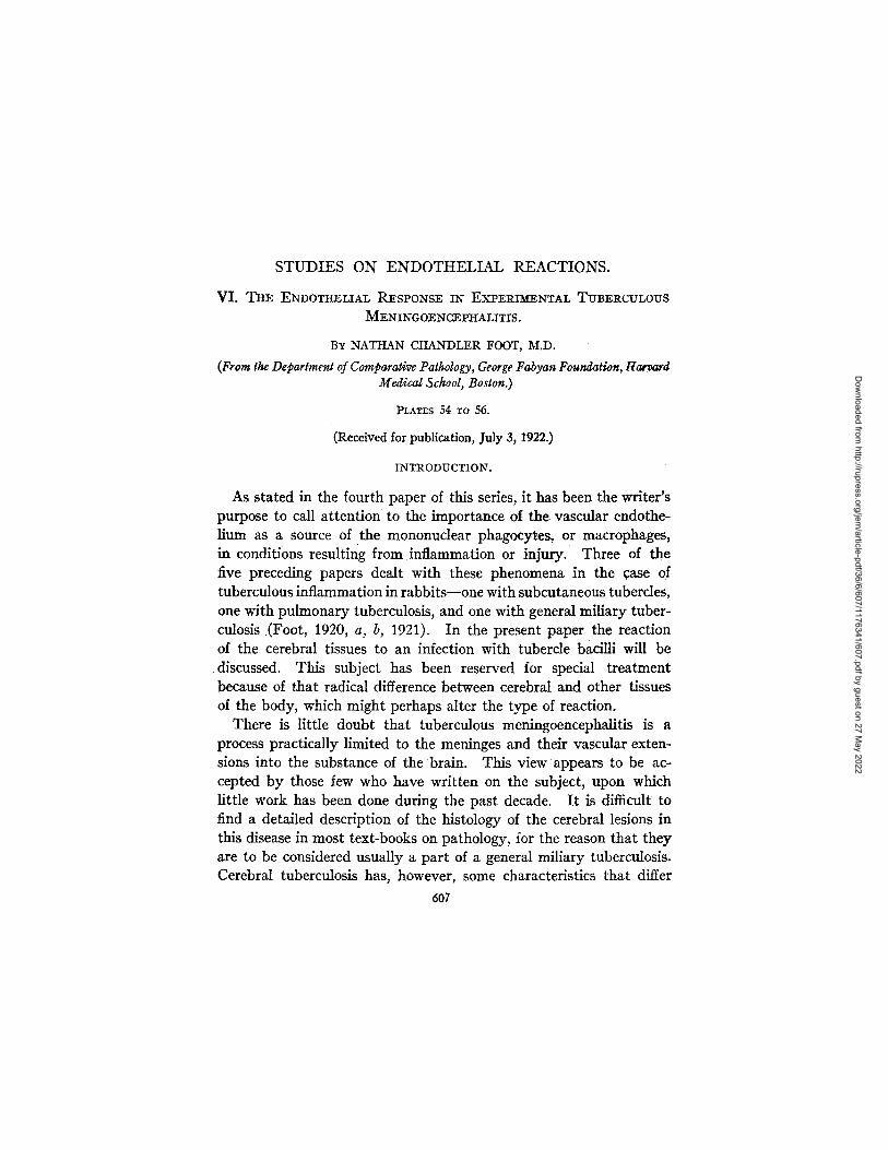
STUDIES ON ENDOTHELIAL REACTIONS. Vl. THE ENDOTHELIAL RESPONSE II~ EX.eEI~IX~EN:CAL TUBERCULOUS MENINGOE N CEPIf_ALITI S. BY NATHAN CHANDLER FOOT, M.D. (From the Department of ComparativePathology, GeorgeFabyan Foundation, Harvard Medical School, Boston.) :PLATES 54 To 56. (Receivedfor publication, July 3, 1922.) INTR ODUCTI ON. As stated in the fourth paper of this series, it has been the writer's purpose to call attention to the importance of the vascular endothe- lium as a source of the mononuclear phagocytes, or macrophages, in conditions resulting from inflammation or injury. Three of the five preceding papers dealt with these phenomena in the case of tuberculous inflammation in rabbits--one with subcutaneous tubercles, one with pulmonary tuberculosis, and one with general miliary tuber- culosis (Foot, 1920, a, b, 1921). In the present paper the reaction of the cerebral tissues to an infection with tubercle bacilli will be discussed. This subject has been reserved for special treatment because of that radical difference between cerebral and other tissues of the body, which might perhaps alter the type of reaction. There is little doubt that tuberculous meningoencephalitis is a process practically limited to the meninges and their vascular exten- sions into the substance of the brain. This view appears to be ac- cepted by those few who have written on the subject, upon which little work has been done during the past decade. It is difficult to find a detailed description of the histology of the cerebral lesions in this disease in most text-books on pathology, for the reason that they are to be considered usually a part of a general miliary tuberculosis. Cerebral tuberculosis has, however, some characteristics that differ 6O7 Downloaded from http://rupress.org/jem/article-pdf/36/6/607/1176341/607.pdf by guest on 27 May 2022
-
Upload
khangminh22 -
Category
Documents
-
view
0 -
download
0
Transcript of STUDIES ON ENDOTHELIAL REACTIONS.
STUDIES ON ENDOTHELIAL REACTIONS.
Vl. THE ENDOTHELIAL RESPONSE II~ EX.eEI~IX~EN:CAL TUBERCULOUS
MENINGOE N CEPIf_ALITI S.
BY NATHAN CHANDLER FOOT, M.D.
(From the Department of Comparative Pathology, George Fabyan Foundation, Harvard Medical School, Boston.)
:PLATES 54 To 56.
(Received for publication, July 3, 1922.)
INTR ODUCTI ON.
As stated in the fourth paper of this series, it has been the writer's purpose to call attention to the importance of the vascular endothe- lium as a source of the mononuclear phagocytes, or macrophages, in conditions resulting from inflammation or injury. Three of the five preceding papers dealt with these phenomena in the case of tuberculous inflammation in rabbits--one with subcutaneous tubercles, one with pulmonary tuberculosis, and one with general miliary tuber- culosis (Foot, 1920, a, b, 1921). In the present paper the reaction of the cerebral tissues to an infection with tubercle bacilli will be discussed. This subject has been reserved for special treatment because of that radical difference between cerebral and other tissues of the body, which might perhaps alter the type of reaction.
There is little doubt that tuberculous meningoencephalitis is a process practically limited to the meninges and their vascular exten- sions into the substance of the brain. This view appears to be ac- cepted by those few who have written on the subject, upon which little work has been done during the past decade. I t is difficult to find a detailed description of the histology of the cerebral lesions in this disease in most text-books on pathology, for the reason that they are to be considered usually a part of a general miliary tuberculosis. Cerebral tuberculosis has, however, some characteristics that differ
6O7
Dow
nloaded from http://rupress.org/jem
/article-pdf/36/6/607/1176341/607.pdf by guest on 27 May 2022
608 ENDOTHELIAL REACTIONS. VI
rather strikingly from the tuberculosis of other organs. It shows a well marked tendency to limit itself to the mesodermal elements of the brain, a far greater lymphocytic reaction, many large mononu- clear ceils circulating in the vessels of the affected area, and some- times fewer giant ceils are found here than in tubercles of other organs.
Perhaps the most detailed treatment of the process is to be found in Ranke's (1908) article, in which he gives an excellent historical sketch of the development of knowledge on this subject, tracing it from 1829 when it was first described by Abercrombie. Experimental work was not begun until Kure, working in Nissl's laboratory in 1898, produced tuberculous meningoencephalitis by drawing cotton threads impregnated with tubercle bacilli through the brains of dogs. This method elicited a mixed reaction in which there was, at first, a foreign body to be dealt with and later the infection resulting from the multiplication of the bacilli which had to be overcome. Delille improved on these experiments by injecting extracts of tubercle bacilli in ether, chloroform, and xylol. The tubercles produced in this way were found to be independent of the brain tissue, except where they encroached directly upon its substance.
Ranke's article concludes with a description of three cases of tuberculous meningitis with necropsy and a very careful report on the histopathological exami- nation of the lesions, replete with morphological details based upon the study of preparations stained in a great variety of ways. In the course of this description he discusses large mononuclcar cells of varying morphology that are first noted free in the blood of the pial vessels. These cells were first discussed by Tigges in 1863 and interpreted as exfoliated endothelial cells by Sawada in 1901, Diamond taking a similar view as to their origin a t about the same time. Ranke, after describing them exhaustively and with the greatest minuteness of detail, leaves the question of their origin open for further investigation and future decision. This is particularly interesting as Farrar (1908), in an article published in the same volume and issued from the same laboratory, describes similar cells in connection with experiments on the reaction to elder pith discs inserted into the cerebral cortex of rabbits, ascribing their origin to the pial endothelium and also tracing the grid cells from the vascular endothelium of the meninges.
Gehry (1909), writing in the following year, takes up the question of the origin of these macrophages and, after discussing them at length, categorically states that they are derivatives of the fixed tissues. As this term is loosely employed in the German literature, it may mean the vascular endothelium, the adventitial connective tissue, or the mesothelinm. All these writers are agreed that the brain tissue proper does not respond to foreign bodies, nor to tuberculous inflammation, except secondarily--when it is invaded by the agent in question and by the mesodermal reaction to its presence. In such areas the nerve cells die off, to be replaced by the multiplication of their satellite glia cells, which act as mere stop- gaps. The glia cells form clusters, or nests, and multiply usually by amitotic
Dow
nloaded from http://rupress.org/jem
/article-pdf/36/6/607/1176341/607.pdf by guest on 27 May 2022
NATHAN CHANDLER FOOT 609
division, although mitotic figures are sometimes found. Rod cells are found in the neighborhood of tuberculous lesions in man; the matter of their origin is fully discussed by Uyematsu (1920) in an article on diffuse cerebral sclerosis. He believes them to be modified glia cells, while Alzheimer and Nissl have considered them to be endothelial in origin. Farrar (1908) states that they do not occur in the rabbit's brain.
Shorter discussions of tuberculous meningitis will be found in the section on vascular lesions of the brain and cord by Nonne and Luce (1904) in the Handbuch der pathologischen Anatomic des Nervensystems. The fact that cerebral tuber- culosis is taken up under the heading of vascular diseases speaks for itself. Lugaro, in the same text-book, gives a good description of the glia cell and its manifold variations in type. According to him, it is the phagocyte of the ectoder- mal brain tissue, usually multiplying by direct division; it may form giant cells, spider cells or astrocytes, and may also phagocytose and replace ganglion cells, etc. In recent years an article by Friedenwald and Greenfeld (1915) brings the subject of cerebral tuberculosis more nearly to date---but their description of the pathological histology, of the lesions is rather meager.
From this review it will be seen tha t the main question left for decision is the origin of the mononuclear macrophages tha t circulate in the meningeal vessels and through the lymph spaces of the pia- arachnoid in tuberculous meningitis. Tha t the tubercles are formed in, or near vessels and tha t their epithelioid cells are probably of en- dothelial origin has been fairly well established. In order to decide this point, if possible, the following experiment was performed.
EXPERI~KENTAL.
Five adult rabbits were anesthetized in turn and a 0.5 cm. opening trephined in the right parietal bone at a point midway between the coronal and lambdoid sutures, about 1 cm. to the right of the sagittal suture. A few drops of a suspension containing in all 0.2 rag. of a 1 month culture of bovine tubercle bacilli on glycerol agar were then injected into the right hemisphere through a 24 gauge needle 1 cm. in length. The same strain of bacilli used in the preceding experi- ments of the series was employed here. The suspension was pre- pared as before; a weighed amount of bacilli was rubbed up in sterile salt solution and, after thorough shaking, diluted until 1 cc. equalled 1 rag. of bacteria. Of this suspension 0.2 cc. was injected through the needle, the latter being held as nearly vertical as possible, and
Dow
nloaded from http://rupress.org/jem
/article-pdf/36/6/607/1176341/607.pdf by guest on 27 May 2022
610 ENDOTHELIAL REACTIONS. VI
inser ted to its full length, releasing the bacilli a t the same d e p t h
in each brain . The re was some leakage f rom the needle s tab af ter
w i t hd ra wi ng the ins t rumen t , b u t this was no t w i t h o u t its advan tages .
T h e w o u n d was then closed w i t h o u t replacing the bone disc.
All the rabb i t s were given 10 cc. of 1 per cent aqueous N i a g a r a blue
(2 B recrystal l ized) , in t raper i toneal ly , and 5 cc. of Higgins ' wa te r -
p roof d rawing ink and distilled water , 50 per cent , in t ravenous ly .
These dyes were adminis te red thereaf te r twice a week, unt i l the
rabb i t s b e c a m e too sick for fu r the r inject ions.
One rabbit became sluggish and lost its appetite at the end of the 2nd week and was killed on the 14th day by injecting Zenker's solution into the right common carotid, under full anesthesia. Nothing was found on gross examination at necropsy, but the use of Niagara blue produces such an intense bluing of the tissues that an early general miliary tuberculosis was overlooked. The other four rabbits became very sick 5 days later, the 19th of the experiment; all were weak and apathetic and all but one had temperatures of 102-102.6°F. by rectum, that one having but 99.9 ° . The next day the rabbit with the lowest temperature was found dead, after paralysis in its fore legs. I t showed an early general miliary tuberculosis and the lower lobe of the right lung was almost completely consoli- dated and covered with a fibrinous exudate, later ascertained to be an almost com- plete infarction. The brain showed suspicious sandy nodules about the site of injection of the tubercle bacilli.
The third rabbit was killed the same day, by injecting Zenker's solution as before. I t had a definite right-sided weakness for 2 or 3 days preceding death. The brain showed a well developed basal tuberculous meningitis and extensive tuberculous involvement of the brain substance around the injection wound, with edema and swelling of the right hemisphere and right choroid plexus. This was the only brain that showed much gross staining with the Niagara blue, which was confined to the meninges. The internal organs, although they showed a millary tuberculosis under the microscope, were grossly negative.
The fourth rabbit became progressively weaker, and developed a somewhat inconspicuous left-sided paresis. On the 23rd day of the experiment it was killed in the same manner as were the others; necropsy showed basal meningeal tuber- culosis, tubercles along the needle wound, and general miliary tuberculosis.
The fifth rabbit, a strong buck, developed a tendency to turn to the left on the 21st day. After becoming very weak it was killed on the 26th day of the experi- ment, by an injection of Zenker's fluid into the right common carotid, under ether. At necropsy the findings were similar to those of the preceding four, except that there was more edema of the right temporal lobe near the base.
I t is interesting to note the failure of the blue stain to take in any of the brains, with one exception, in which the meninges were distinctly blue. The lesions were not grossly dyed.
Dow
nloaded from http://rupress.org/jem
/article-pdf/36/6/607/1176341/607.pdf by guest on 27 May 2022

NATHAN" CHANDLER FOOT 611
In all cases the brain, liver, lungs, spleen, and kidneys were hardened in Zenker's fluid, embedded in paraffin, and cut to 5 microns. Sections were stained with eosin-methylene blue, Mayer's aqueous carmalum, phosphotungstic acid-hema- toxylin, and Ziehl-Neelson carbolfuchsin, with Delafield's hematoxylin as a counterstain.
Microscopic Examination.
Each rabbit shows a tuberculous meningitis, the interlobar meninges and choroid plexus are also involved, the vessels running into the brain from the meninges are surrounded near their origin by tubercles and, deeper in the cortex, by a dense zone of lymphocytes and plasma cells. The vascular lesions are more marked in some animals than in others, according to the severity of the infection. The needle puncture is filled with masses of debris, polymorphonuclear leucocytes, and caseous material, surrounded by a zone of actively proliferating endothelial cells in close proximity to one another, which penetrate the surrounding nerve tissue in the form of long, ray-like extensions. Outside of this tuberculous tissue is a zone of degenerated nerve tissue, in which one finds edema, many polymorphonuclears and altered glia cells, and the remnants of nerve cells, but no ink-bearing endothe- lial macrophages.
In general all the tubercles are more or less deeply pigmented with ink granules; there is little or no Niagara blue, except in the form of coarse granules in the discrete macrophages of the meningeal lymphatics, cells which also contain ink. The changes in the brain tissue are typical and conform to those discussed in the Introduction: degeneration of nerve cells, increase in the number of glia cells, with- out evidence of mitosis, and polymorphonuclear infiltration of the most affected regions. There are often glia cell nests, or nebulm; the satellite, or Irabant cells fill the spaces left by the death of the ganglion cells. Apparently the glia cells perform those functions that are elsewhere effected by the endothelial cells, for ink-bearlng macrophages are not found at any distance from the tubercles.
The free mononuclear cells discussed so extensively by Ranke and Gehry are found in large numbers in the veins and capillaries of the meninges, less frequently in the arte'ries. Fully a third to a half of their number contain ink granules, which indicates their endothelial
Dow
nloaded from http://rupress.org/jem
/article-pdf/36/6/607/1176341/607.pdf by guest on 27 May 2022
612 ENDOTHELIAL REACTIONS. V]
origin. These cells are frequently found in mitosis while free in the lumen of the blood vessel; three such figures were encountered in the course of half an hour's search, while counting ink-bearing macro- phages (Fig. 1).
There is no need for a detailed description of the lesions; that they contain ink, wherever they may be situated, is the main fact to emphasize; hence they are composed chiefly of cells of endothelial origin (Figs. 2 and 3). The reasons for considering any ink-bearing cell as endothelial already have been discussed sufficiently in the preceding papers. Thus the cerebral tubercle conforms in its histo- genesis to tuberculosis of other organs. The more interesting point for consideration is the question as to the locus of origin of these endothelial cells. In this case the local activity of the capillary en- dothelium in the neighborhood of the lesions is not nearly as marked as it was in the lesions of other organs studied in the preceding ex- periments. Thickening of the capillary endothelium and an increase in the number of its nuclei are evident here and there, especially where the brain tissue has been more extensively destroyed, but on the whole these changes are not at all marked, nor are mitoses found to occur in the sessile endothelial cells of these vessels. I t will be recalled that these lining cells were frequently found in mitosis in the other experiments and that this usually occurred near the lesions.
Ink is not found in the ependymal cells of the ventricles or choroid plexus (Fig. 4). Where tubercles occur, they are always beneath the ependyma and covered by it, and they contain the usual amount of ink. The ependymal cells are, therefore, very similar, in their lack of response to the tubercle bacilli and injected pigments, to the meso- dermal cells of the peritoneum. I t was always the subperitoneal, perivascular tissue that responded in the cases of tuberculous peri- tonitis studied in the previous experiments.
Whence, then, come the endothelial cells that go to form the numer- ous tubercles in these brains? Ranke and Gehry called attention to circulating mononuclears. These cells are very abundant in all five of the rabbits studied; furthermore, many of them are fixed in the act of mitosis, proving that they are actively proliferating. Much has been written about them, little definitely proved; they probably come from other organs more richly supplied with capillaries than
Dow
nloaded from http://rupress.org/jem
/article-pdf/36/6/607/1176341/607.pdf by guest on 27 May 2022

NATHAN CHANDLER I~OOT 613
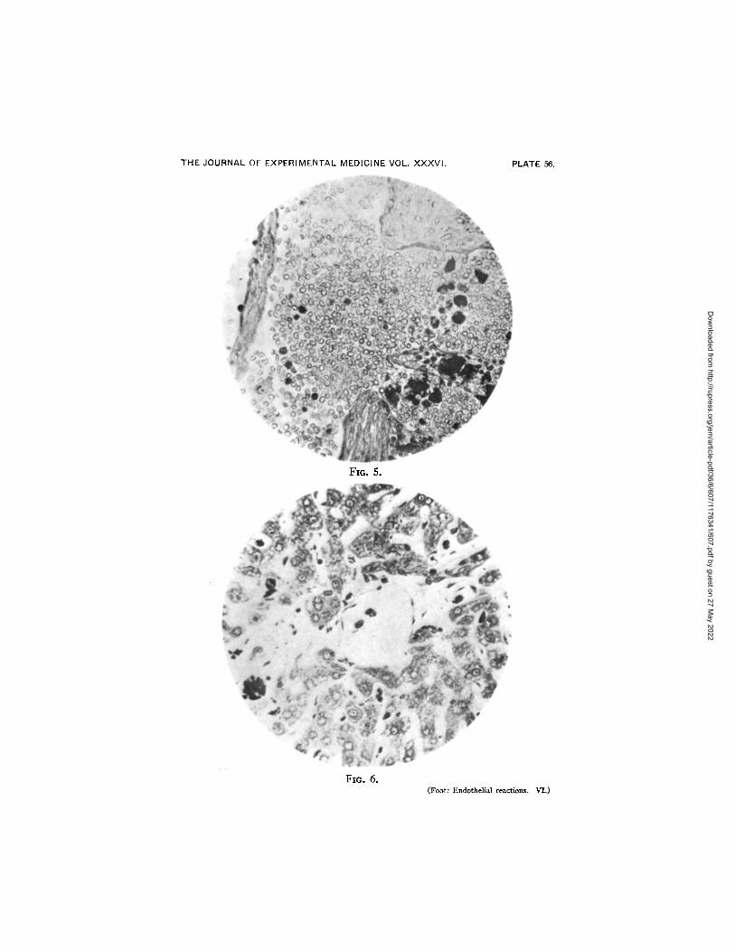
is the brain. Mallory has long maintained the endothelial origin of cir- culating mononuclears and more recently McJunkin (1919) has reached a similar conclusion. Downey (1916-17), Evans (1915), and Simp- son (1922) suggest that they proceed largely from the reticuloendothe- lium of the hematopoietic organs, particularly the spleen. A glance at the sections from the spleen (Fig. 5) and the liver (Fig. 6) of the animals of this experimental series is sufficient to make it clear that pigmented endothelial cells are leaving the capillary walls and entering the veins, to be thrown into the circulation in numbers somewhat larger than would be the case following the mere injection of ink (Simpson's "shower of macrophages"). In the lung they are also found in the larger vessels, and the pulmonary endothelium is rather swollen and active in places, but it must be remembered that here, as well as in the other organs, an active miliary tuberculosis is in progress. Although not much increased in number over what one finds in control animals without infection, they show a process not seen in controls--the phenomenon of mitotic division while free in the blood stream.
Supplementary Experiment.--In order to rule out at least one of the hematopoiefic organs as the sole source of these circulating macro- phages, the spleen was removed from four rabbits, which were then inoculated with tubercle bacilli of the same strain and in the same manner as the preceding series of animals. The cultures were younger (2 weeks) so that a smaller dose was administered, in order to decrease the toxicity. Only 0.16 rag. was given in these cases. Following the operation, which was performed under ether anesthesia, 5 cc. of Higgins' ink and distilled water in equal parts were injected into an ear vein 1, 5, and 8 days afterwards; the dose was repeated in two of the rabbits on the 12th day. These two died almost immediately with symptoms suggesting cerebral embolism. Necropsy showed no gross lesions, beyond an unusually light-colored liver and almost jet- black lungs. Microscopic examination showed that death was due to cerebral emboli of ink, which were scattered all through the precap- illaries of the cortex and were plugged quite solid with the ink. There were occasional ink emboli in the lungs, but not sufficient to give death from this cause. The remaining two rabbits went uninjected for 2 weeks, when another intravenous administration of 5 cc. of the
Dow
nloaded from http://rupress.org/jem
/article-pdf/36/6/607/1176341/607.pdf by guest on 27 May 2022
614 ENDOTHELIAL REACTIONS. VI
ink mixture was given, this time uneventfully. Another injection, 1 week later, killed the third rabbit in precisely the same way the other two had been overwhelmed and necropsy gave similar findings. The fourth rabbit was allowed to go uninjected for 4 weeks and was then killed by introducing Zenker's fluid into the right common carotid under anesthesia.
The glass pestle used in triturating the bacilli for the suspension injected into these splenectomized animals apparently was not suffi- ciently cooled after flaming, for the bacilli were killed. Very good local reactions (Hodenpyl tubercles) were, however, obtained; they differed from those of the preceding experiment merely in so far as they were localized, did not spread to other organs, and were not accompanied by generalized symptoms. Despite the fact that the spleen had been removed, the tubercles were formed and the macro- phages were found in apparently undiminished numbers lying free in the meningeal vessels. An examination of the liver and bone marrow revealed no increased production of macrophages, indeed they seemed to contain less ink than in the case of control animals, and there was no undue activity of their endothelial elements. The lungs, on the other hand, showed changes so striking that they call for another experiment to determine their cause and to follow the course of their development. This will be described in another article. The proliferation of the pulmonary endothelium, the swelling of its component cells, and the. increased amount of contained ink in the organ are most remarkable.
We may conclude, then, that cerebral tubercles are produced some- what differently from those of other organs, in that the endothelial cells that form them are evidently derived only in part from the local capillaries; the circulating macrophages showing much more prolifera. tive activity than the sessile endothelial cells of these capillaries prob- ably form a more important source of supply. Hence it appears that a much greater proportion of the endothelial cells of cerebral tubercles is derived from the circulating blood than is the case in tubercles of other organs. This conforms with the older French theories on the histogenesis of the tubercle.
Dow
nloaded from http://rupress.org/jem
/article-pdf/36/6/607/1176341/607.pdf by guest on 27 May 2022
NATHAN CHANDLER I~OOT 615
CONCLUSIONS.
1. Experimental cerebral and meningeal tubercles in the rabbit are formed from ceils of endothelial origin.
2. These cells are derived apparently from other sources than the neighboring capillary endothelium alone.
3. The circulating macrophages, 'which, in this case, are capable of multiplying by mitosis while still free in the blood, are drawn upon in the formation of the cerebral tubercle.
4. Splenectomy has not materially decreased the available supply of circulating macrophages in this experiment.
5. While these cells may originate in the endothelium of the liver and bone marrow, the lung appears to play a much more important r61e in this respect than has been hitherto suspected.
BIBLIOGRAPHY.
Downey, H., Anat. Rec., 1916-17, xi, 350. Evans, H. M., Am. J. Physiol., 1915, xxx-¢ii, 243. Farrar, C. B., Hist. u. histopath. Arb. Grosshirnrinde, 1908, ii, 1. Foot, N. C., J. Exp. Med., 1920, a, xxxii, 513. Foot, N. C., J. Exp. Med., 1920, b, xxxii, 533. Foot, N. C., J. Exp. Med., 1921, xxxiii, 271. Friedenwald, E. B., and Greenfeld, W., Am. J. Dis. Ckild., 1915, ix, 508. Gehry, K., Arch. Psychiat., 1909, xlv, 59. Lugaro, E., in Flatau, E., Jacobsohn, L., and Minor, L., Handbuch der patholo-
gischen Anatomic des Nervensystems, Berlin, 1904, i, 189. Mallory, F. B., The principles of pathologic histology, Philadelphia and London,
1918. McJunkln~ F. A., Am. J. Anat., 1919, xxv, 27. Nonne, M., and Luce, H. M., in Flatau, E., Jacobsohn, L., and Minor, L., Hand-
buch der pathologischen Anatomic des Nervensystems, Berlin, 1904, i, 200. Ranke, O., Hist. u. histopath. Arb. Grosshirnrinde, 1908, ii, 252. Simpson, M. E., J. Med. Research, 1922, xliii, 77. Uyematsu, S., J. Nero. and MenL Dis., 1920, 1i, 514.
Dow
nloaded from http://rupress.org/jem
/article-pdf/36/6/607/1176341/607.pdf by guest on 27 May 2022
6 1 6 ENDOTHELIAL REACTIONS. VI
EXPLANATION OF PLATES.
PLATE 54.
FXG. 1. Macrophages circulating in a meningeal vessel. Some are clumped, others discrete; one of these cells is seen in mitosis at the center of the photograph. X 400.
FIG. 2. Cerebral tubercle. The ink granules are so coarse that most of them are readily confused with nuclear material in the photograph. Several of the epithe- lioid cells near the periphery show finer granules. × 335.
PLATE 55.
FIG. 3. A meningeal tubercle. The ink shows to better advantage. X 335. FIG. 4. Tubercle in the choroid plexus. Note that carbon is not seen in the
ependymal layer, which remains aloof from the process going on beneath it. > 335.
PLATE 56.
FIG. 5. Group of macrophages leaving the spleen, one of them in mitosis, most of them heavily laden with ink. X 335.
FIG. 6. Macrophages in an hepatic vein and in the sinusoids of the liver, in one place clumped into a mass. X 335.
Dow
nloaded from http://rupress.org/jem
/article-pdf/36/6/607/1176341/607.pdf by guest on 27 May 2022

THE JOURNAL OF EXPERIMENTAL MEDICINE VOL. XXXVI . PLATE 54.
FIO. I.
FIG. 2. (Foot: Endothelial reactions. V].)
Dow
nloaded from http://rupress.org/jem
/article-pdf/36/6/607/1176341/607.pdf by guest on 27 May 2022

THE JOURNAL OF EXPERIMENTAL MEDICINE VOL. XXXVI j PLATE 55.
FIO. 3.
Fro. 4. (Foot: Endothelh~l reactions. VI.)
Dow
nloaded from http://rupress.org/jem
/article-pdf/36/6/607/1176341/607.pdf by guest on 27 May 2022
THE JOURNAL OF EXPERIMENTAL MEDICINE VOL. X X X V I . PLATE 56,
Fro. 5.
Fmo 6. (Foot: Endothelial reactions. VI.)
Dow
nloaded from http://rupress.org/jem
/article-pdf/36/6/607/1176341/607.pdf by guest on 27 May 2022