Warming-induced increase in carbon uptake leads to earlier ...
Upload
independentCategory
view
2download
0
Earlier onset of tumoral angiogenesis in matrix metalloproteinase-19 deficient
mice
Maud Jost,1 Alicia R. Folgueras,2 Françoise Frérart,1 Alberto M. Pendas,2,3 Silvia
Blacher,1 Xavier Houard,1 Sarah Berndt,1 Carine Munaut,1 Didier Cataldo,1 Jesus
Alvarez,2 Laurence Melen-Lamalle,1 Jean-Michel Foidart,1,4 Carlos López-Otín,2 and
Agnès Noël1*
1Laboratory of Tumor and Developmental Biology, Center for Experimental Cancer
Research (CECR), Center for Biomedical Integrative Genoproteomics (CBIG),
University of Liège, Tour de Pathologie (B23), B-4000 Sart-Tilman, Belgium,
2Departamento de Bioquímica y Biología Molecular, Facultad de Medicina, Instituto
Universitario de Oncología, Universidad de Oviedo, 33006-Oviedo, Spain, 3Centro
Investigacion Cancer, Salamanca, Spain, 4Department of Gynecology, CHU, 4000
Liège.
Running title: MMP-19 and tumoral angiogenesis
Corresponding author:
A. NOEL
Laboratory of Tumor and Developmental Biology
University of Liège, Tour de Pathologie (B23)
Sart-Tilman, B-4000 Liège
Tel : +32-4-366.25.69 ; Fax: +32-4-366.29.36
E-mail: [email protected]
2
Abstract
Among matrix metalloproteinases (MMPs), MMP-19 displays unique structural
features and tissue distribution. In contrast to most MMPs, MMP-19 is expressed in
normal human epidermis and downregulated during malignant transformation and
dedifferentiation. The contribution of MMP-19 during tumor angiogenesis is presently
unknown. In an attempt to give new insights into MMP-19 in vivo functions, angiogenic
response of mutant mice lacking Mmp-19 was analysed after transplantation of murine
malignant PDVA keratinocytes and after injection of matrigel supplemented with b-
FGF. In situ hybridization and immunohistochemical analysis revealed that MMP-19 is
produced by host mesenchymal cells, but not by endothelial capillary cells or CD11b-
positive inflammatory cells. Based on a new computer-assisted method of
quantification, we provide evidence that host Mmp-19 deficiency was associated with
an increased early angiogenic response. In addition, increased tumor invasion was
observed in Mmp-19-/- mice. We conclude that, in contrast to most MMPs which
promote tumor progression, Mmp-19 is a negative regulator of early steps of tumor
angiogenesis and invasion. These data highlight the requirement to understand the
individual functions of each MMP in order to improve anti-cancer strategies.

3
Introduction
Matrix metalloproteinases (MMPs) are a family of structurally related zinc-
dependent neutral endopeptidases that play major roles in tissue remodeling occurring
in a variety of physiological processes such as embryonic development, angiogenesis
and wound healing (1, 2). They are main effectors of pathological extracellular matrix
destruction in many diseases such as arthritis, atherosclerosis, age-related macular
degeneration, tumor invasion and metastasis (3-7). MMPs contribute to the fine tuning
of diverse biological processes through limited proteolysis of specific targets including
not only matrix components, but also growth factors, chemokines/cytokines and cell
surface receptors (8-11). More than 20 different human MMPs have been identified (12)
and classified into different subfamilies according to their primary structure, domain
organization, cellular localization and substrate specificity (11, 13, 14). Produced as
latent forms, they are either secreted in the extracellular medium or associated to the
cell membrane (Membrane-type MMPs or MT-MMPs) (7, 11, 14).
Among MMPs, MMP-19 displays unique structural features and tissue distribution.
Human MMP-19 cDNA was initially cloned from liver and mammary gland and was
also identified as an autoantigen in inflamed rheumatoid synovium (15, 16). MMP-19
presents the typical domain organization of soluble MMPs, including a signal sequence,
a propeptide maintaining enzyme latency, a catalytic domain with the typical zinc
binding motif, a linker region, and a C-terminal fragment with sequence similarity to
hemopexin (15). However, MMP-19 displays several structural features distinctive of
the diverse MMP subfamilies including (i) an unique insertion of glutamic acid residues
within the linker region, (ii) an unusual latency motif in propeptide domain, (iii) an
additional cysteine residue in catalytic region, (iv) a C-terminal extension lacking
4
sequence similarity to equivalent regions in other human MMPs (15, 17, 18). The
catalytic domain of MMP-19 is capable of degrading components of basement
membrane (laminin, type IV collagen, nidogen), connective tissue (fibronectin, type I
gelatine) and cartilage (cartilage oligomeric matrix protein and aggrecan), but does not
degrade triple-helical type I collagen (19, 20). In contrast to most other MMPs, MMP-
19 is expressed in human mammary or skin epithelial cells under normal quiescent
conditions and down regulated in invasive carcinomas (21-23).
Evidence for MMP-19 involvement in tissue remodeling events such as those
occurring during adipogenesis and tumor progression have been provided by the recent
generation of Mmp-19-deficient mice (24). Although lack of Mmp-19 did not affect
mice viability, fertility and development, it led to a diet-induced obesity and a decreased
susceptibility to skin tumors induced by chemical carcinogens. A role of MMP-19
during angiogenesis is suggested by its expression in endothelial cells of synovial
capillaries following injury and inflammation (3, 25). However, the ex vivo sprouting of
endothelial cells from aortic rings was not affected by Mmp-19 deficiency (24).
Therefore, the contribution of MMP-19 during angiogenic processes remains
controversial. In order to give new insights into Mmp-19 functions in vivo, the
angiogenic response of mutant mice lacking Mmp-19 was analysed after transplantation
of malignant murine PDVA keratinocytes and after injection of matrigel supplemented
with b-FGF. In contrast to other single or double MMP-deficient mice studied until
now, Mmp-19-deficient mice exhibited an early onset of angiogenesis and tumor
invasion.

5
Materials and Methods
Mmp-19–null mice. Mice genetically deficient in Mmp-19 (Mmp-19-/-) were
generated by replacing a portion of 1kb of the promoter and exons 1 and 2 of the gene
with a phosphoglycerate kinase-neomycin fusion gene and by homologous
recombination (24). Homozygous Mmp-19 (Mmp-19-/-) mice and their corresponding
WT (Mmp-19+/+) were littermates deriving from interbreeding of heterozygotes with a
mixed background of C57Bl6/129Ola. When applying the transplantation chamber
assay to mice with different genetic background, we previously demonstrated that the
extent of tumor invasion and vascularization was similar in all WT mice, independently
to the number of backcrosses in C57Bl6 mice (26). Mice experimentation was done in
accordance to guidelines of the University of Liège regarding the care and use of
laboratory animals.
Transplantation assay in mice. PDVA cells were generated by in vitro carcinogen
treatment (DMA) of cultured keratinocytes issued from B10LP mice (27). PDVA cells
were grown in modified Eagle’s minimal essential medium containing a 4-fold
concentration of amino acids and vitamins (Gibco Laboratories, Grant Island, NY), 10%
fetal calf serum (Gibco) and antibiotics in a humidified incubator at 37 °C, 5% CO2.
Cells (2 x 10⁵) were seeded on a collagen gel (4 mg/mL of type I collagen isolated from
rat tail tendons) inserted in Teflon rings (Renner GmbH, Dannstadt, Germany). After
24h of culture, cell-coated collagen gels were covered with a silicone transplantation
chamber (Renner GmbH) and implanted in toto onto dorsal muscle fascia of 6-8 weeks
old mice according to the procedure previously described (28, 29). At different time
points, tumor transplants were resected, embedded in Tissue Tek (Miles Laboratories
Inc., Naperville, IL) and frozen in liquid nitrogen for cryostat sectioning. Two hours
6
prior to sacrifice, mice were intraperitonally injected with 200 µl of BrdU/BrdC (65
µM, Acros Organics, Geel, Belgium). Each experimental group contained at least 6
animals. Tumor angiogenesis and invasion in Mmp-19-/- and WT mice were evaluated
in three independent sets of experiment.
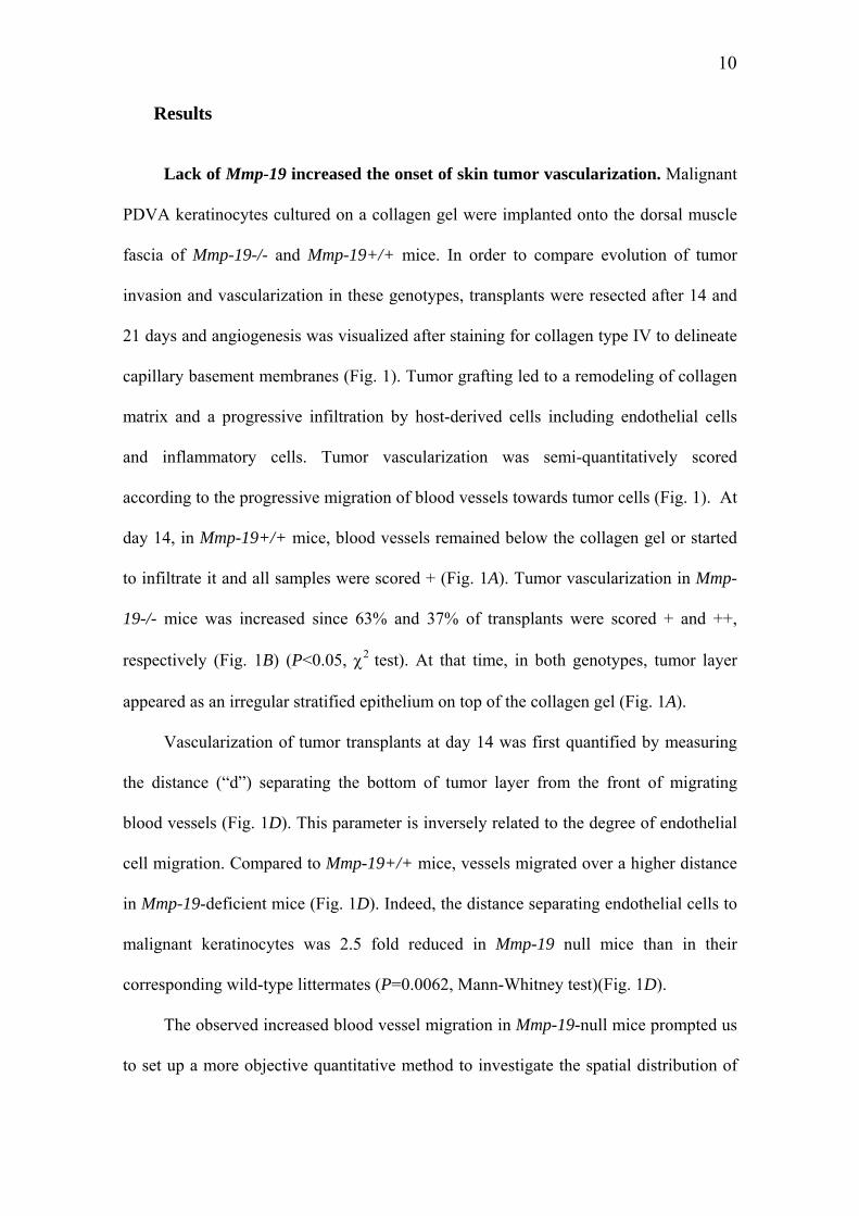
Histological analysis. Cryostat sections were fixed in acetone at –20°C and in
80% methanol at 4°C. For immunofluorescence labeling, the following antibodies were
used: anti-type IV collagen Ab (rabbit polyclonal Ab; diluted 1/100), anti-keratin Ab
(polyclonal guinea pig Ab; diluted 1/20, Sigma-Aldrich, St Louis, MO), anti-BrdU-
FITC (monoclonal mouse, diluted 1/3.5, Becton Dickinson, San Diego, CA), anti-hinge
region of MMP-19 (rabbit anti-human, diluted 1/20, Sigma-Aldrich; rabbit anti-human,
diluted 1/500, Abcam, Cambridge, MA), anti- MMP-19 prodomain (rabbit anti-human,
diluted 1/500, Abcam), anti-CD11b/TRITC (rat anti-mouse, diluted 1/50, Pharmingen,
San Diego, CA), anti α-smooth muscle actin/FITC (α-SMA) (monoclonal mouse
antibody, diluted 1/200, Sigma-Aldrich). When double immunofluorescence-labelings
were performed, after an incubation for 1 h with primary antibodies, sections were
washed with phosphate buffered saline (PBS) and then incubated for 30 min with
fluorescein-isothiocyanate (FITC)- or tetramethyl-rhodamine isothiocyanate (TRITC)-
conjugated appropriate secondary antibodies: swine anti-rabbit (diluted 1/40, Dako,
Glostrup, Denmark,), mouse anti-guinea pig (diluted 1/40, Sigma-Aldrich) or goat anti-
rat (diluted 1/100, Molecular Probes, Carlsbad, CA). After 3 washes in PBS, coverslips
were mounted with Aqua Polymount (Polysciences, Warrington, FL) and specific
labeling was observed using an inverted microscope equipped with epifluorescence
optics. Sections were counterstained in blue with bisbenzimide. At all times after
grafting, collagen type IV labelings were codistributed with endothelial cells recognized
by the anti-mouse PECAM immunostaining (data not shown).

7
Apoptosis was studied by terminal deoxynucleotidyl transferase (TDT)-mediated
deoxyuridine triphosphate (dUTP) nick-end labeling (TUNEL). Cryostat sections fixed
in 4% paraformaldehyde for 20 min and in methanol for 5 min were stained for
apoptosis following manufacturer’s instructions (Roche Diagnostics, Mannheim,
Germany).
For quantitative measurement of proliferating cells, automatic computer-assisted
image analysis was performed on images obtained after bisbenzimide staining and
immunolabeling of BrdU positive cells. The ratio between the surface of bisbenzimide
staining and the surface of specific immunostaining was measured by using a software
Aphelion 3.2 from Adsis.
In situ hybridization: Either sense or antisense 35S-uridine triphosphate-labeled
RNA probes were synthesized from linearized cDNA fragment (1600 bp) of mouse
Mmp-19 gene cloned into EcoRV site of pcDNA3. Cryostat sections were hybridized
with 35S-labeled Mmp-19 riboprobes and then exposed to photographic emulsion at 4°C
for 6 days. Sections were developed, fixed, cleared and counterstained with 0.02%
Toluidine Blue. Bright field and dark field images were captured with a SPOT digital
camera.
Scoring of tumor invasion and vascularization. For semi-quantitative analysis
of tumor vascularization, the following scoring was used: +: vessels below the collagen
gel or infiltrating the collagen gel without reaching the malignant epithelial layer; ++:
blood vessels in close apposition to the epithelial layer, and +++: blood vessels
intermingled with invasive epithelial tumor sprouts (29).
Morphometric measurements of tumor cell invasion (average distance of

8
invasion) and tumor vascularization (endothelial cell migration) were performed by
using a computer-assisted image analysis system (Olympus Micro Image version 3.0 for
Windows 95/NT, Olympus Optical CO. Europe GmBH) (30). Angiogenesis was
quantified by measuring the distance (“d”) separating tumor cells from the front of
migrating blood vessels. Therefore, the distance “d” is inversely related to the degree of
endothelial cell migration. At least five measurements of distance (“d”) were performed
in the central part of each tumor and the mean values are reported.
Quantitative measurement of tumor invasion and angiogenesis by computer-
assisted image processing. For quantitative measurements, automatic computer-
assisted image analysis was performed on images obtained after double
immunostainings of keratinocytes (in green) and vessels (in red). The software
Aphelion 3.2 from Adsis was used on a PC. Images were first digitized in 760 x 570
pixels with 256 grey levels. Tumor and vessels images were processed separately. For
tumor images, histogram equalization was first performed in order to optimize the
contrast. Then, tumor images were binarized/segmented automatically using the entropy
of the histogram of the grey level intensities (31). For vessel images, vessels were
binarized/segmented using an automatic threshold transformation that maximizes the
global average contrast of edges (32). The upper boundary of the tumor was then
automatically detected using a hit or miss transformation with an appropriate
neighborhood configuration. A grid was constructed with the successive dilations (n=1,
2, 3….) of this upper boundary, with a vertical line as structuring element. Tumor and
vessel densities were determined on each interval of the grid, and the results drawn in
function of the distance to the upper limit of the tumor. The largest distance of tumor
invasion gives the thickness of the tumor. Quantification was performed on each mouse
in all independent assays. To compare the different distributions, the analysis of

9
variance was performed and results were considered significantly different when the p-
value was less than 0.05.
Matrigel plug assay. A total of 0.5 ml of Matrigel (10mg/ml) mixed with 250 ng
of basic Fibroblast Growth Factor (bFGF) (R&D Systems, Mineapolis, MN) and 20U of
heparin (Leo Pharma, Ballerup, Denmark) (33) was subcutaneously injected in the
abdominal midline region of 8 week-old Mmp-19 null and wild-type mice. After 7 days,
animals were euthanized and Matrigel implants were harvested, frozen on dry ice and
lyophilized overnight. Dry plugs were weight and suspended in 0.4ml of 0.1% saponin
(Calbiochem, La Jolla, CA) for 1 h at 4°C, disrupted by vigorous pipetting and
centrifuged at 15,000g for 15 min, at 4°C to remove particulates. Concentration of
haemoglobin in the supernatant was determined after dilution in Drapkin’s solution by
measuring the absorbance at 560 nm (Drabkin reagent kit 525, Sigma). A standard
curve was performed by using purified haemoglobin (Sigma). The angiogenic index
corresponds to µg of haemoglobin per mg of Matrigel.
Statistical analysis. All experimental data are reported as mean ± SEM, and
statistical analysis was performed by χ2 test or Mann-Whitney test. P< 0.05 was
considered as significant.
10
Results
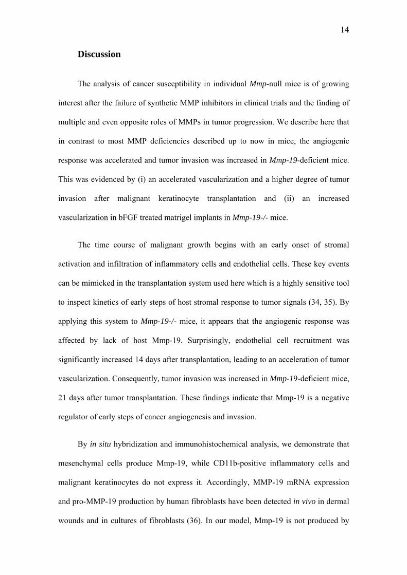
Lack of Mmp-19 increased the onset of skin tumor vascularization. Malignant
PDVA keratinocytes cultured on a collagen gel were implanted onto the dorsal muscle
fascia of Mmp-19-/- and Mmp-19+/+ mice. In order to compare evolution of tumor
invasion and vascularization in these genotypes, transplants were resected after 14 and
21 days and angiogenesis was visualized after staining for collagen type IV to delineate
capillary basement membranes (Fig. 1). Tumor grafting led to a remodeling of collagen
matrix and a progressive infiltration by host-derived cells including endothelial cells
and inflammatory cells. Tumor vascularization was semi-quantitatively scored
according to the progressive migration of blood vessels towards tumor cells (Fig. 1). At
day 14, in Mmp-19+/+ mice, blood vessels remained below the collagen gel or started
to infiltrate it and all samples were scored + (Fig. 1A). Tumor vascularization in Mmp-
19-/- mice was increased since 63% and 37% of transplants were scored + and ++,
respectively (Fig. 1B) (P<0.05, χ2 test). At that time, in both genotypes, tumor layer
appeared as an irregular stratified epithelium on top of the collagen gel (Fig. 1A).
Vascularization of tumor transplants at day 14 was first quantified by measuring
the distance (“d”) separating the bottom of tumor layer from the front of migrating
blood vessels (Fig. 1D). This parameter is inversely related to the degree of endothelial
cell migration. Compared to Mmp-19+/+ mice, vessels migrated over a higher distance
in Mmp-19-deficient mice (Fig. 1D). Indeed, the distance separating endothelial cells to
malignant keratinocytes was 2.5 fold reduced in Mmp-19 null mice than in their
corresponding wild-type littermates (P=0.0062, Mann-Whitney test)(Fig. 1D).
The observed increased blood vessel migration in Mmp-19-null mice prompted us
to set up a more objective quantitative method to investigate the spatial distribution of

11
blood vessels in the remodeled matrix. With this aim, an original method based on
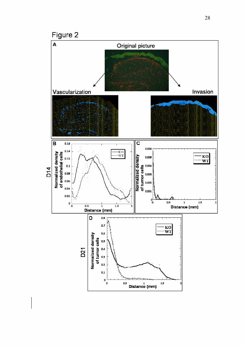
computer-assisted image analysis was developed (Fig. 2). The density of vessels picked
already at a distance of around 0.25 mm from the upper boundary of tumor layer in
Mmp-19-/- mice (Fig. 2B). In sharp contrast, the maximal density of endothelial cells
was observed at a distance of around 0.75 mm in Mmp-19+/+ mice. A computer-
assisted method of image analysis was also set up to quantify tumor cell invasion (Fig.
2A). At day 14 time point, no invasion was observed (Fig. 2C). This was expected since
in the transplantation chamber assay, infiltration of vessels through the collagen gel
towards tumor cells always precedes recognizable tumor cell invasion (34). Therefore,
at early time point, Mmp-19-deficient mice exhibited an acceleration of blood vessel
infiltration in the remodeled matrix.
Lack of Mmp-19 increased tumor invasion
At day 21, once blood vessels have reached tumor cell layers, malignant
keratinocytes formed tumor sprouts that invaded downwards the remodeled host tissue
and were intermingled with closely apposed new vessels (Fig. 1A). Such vascularization
pattern scored +++ was observed in about 60% (10/17) of Mmp-19-/- mice and only
22% (4/18) of Mmp-19+/+ mice (P = 0.027, χ2 test) (Fig. 1C).
At this time point, the distance (“d”) separating tumor layer from the front of
recruited blood vessels (Fig. 1) can not be measured since in more than 70% of tumor
transplants, vessels have reached the tumor layer (tumors scored ++ or +++). Therefore,
for quantitative assessment, our original method of image analysis (Fig. 2) was applied
to determine the malignant keratinocyte density as a function of the distance to the top
of tumor transplant (Fig. 2D). In wild-type mice, keratinocyte density decreased
abruptly with the distance to the top of tumor layer (Fig. 2D). Only few keratinocytes

12
were observed at a distance higher than 0.5 mm from the top of tumor layer. In sharp
contrast, in Mmp-19 null mice, tumor cell density decreased more slowly and numerous
malignant keratinocytes migrated over a distance of 0.5 mm. The maximal distance of
keratinocyte migration was 1.2 mm and 1.8 mm in Mmp-19+/+ and Mmp-19-/- mice,
respectively (Fig. 2D). Therefore, a significant increase of tumor invasion was observed
in the absence of host Mmp-19.
This tumor promoting effect observed in absence of Mmp-19 was not related to a
modification of tumor cell proliferation rate as assessed by BrdU incorporation. Indeed,
quantitative assessment performed by computer-assisted image analysis revealed that
the percentage of proliferating cells was similar in both genotypes, 14 and 21 days after
tumor transplantation (Fig. 3). Furthermore, TUNEL stainings for apoptotic cells
indicated that the extent of apoptosis was identical and always low in cancer cell layers
as well as in stromal strands of transplants resected from WT mice and Mmp-19-/- mice
(data not shown).
Mmp-19 is produced by host stromal cells
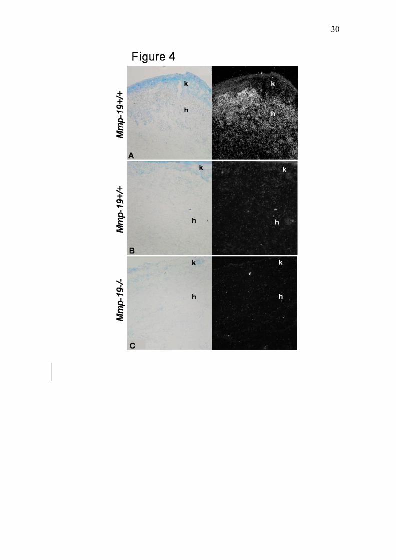
As a first step in determining the cellular source of Mmp-19, in situ
hybridization was performed on tumors transplanted into Mmp-19 proficient and
deficient mice. Hybridization signals for Mmp-19 mRNAs were found in the stroma of
WT mice (Fig. 4A), but not in that of KO mice (Fig. 4C). No positive signal was
detected after treatment with sense riboprobe used as negative controls (Fig. 4B).
Immunohistochemical staining of tumor transplants confirmed the stromal production of
Mmp-19 (Fig. 5A). This protease was produced by mesenchymal cells (Fig. 5A), but not
by inflammatory cells positive for CD11b staining (Fig. 5C). Interestingly, Mmp-19
was not associated with capillaries newly formed in the remodelled collagen matrix

13
(data not shown). In contrast, Mmp-19 staining was detected in large vascular structures
present deeply in the host tissue, below tumor transplants (Fig. 5B). These vessels were
positive for α-smooth muscle actin and correspond to quiescent mature vessels.
Lack of Mmp-19 increased angiogenesis in Matrigel plug assay
To further investigate the impact of Mmp-19 deficiency on angiogenesis in vivo
and to determine whether the angiogenic response was dependent upon the matrix
encountered by endothelial cells during their migration, the Matrigel plug assay was
applied to mutant mice. Matrigel supplemented with bFGF was sub-cutaneously
injected into the abdomen of wild type and mutant mice (n = 8) and harvested after 7
days. Quantitative analysis of angiogenesis was performed by measuring haemoglobin
content in implants giving quantitative information on functional vessels. In accordance
to results obtained in transplantation chamber assay, the angiogenic response in Mmp-
19 deficient mice was 6-fold increased as compared to that detected in wild type mice
(Fig. 6) (P< 0.05) and was therefore independent on the type of matrix used (type I
collagen versus a reconstituted basement membrane).
14
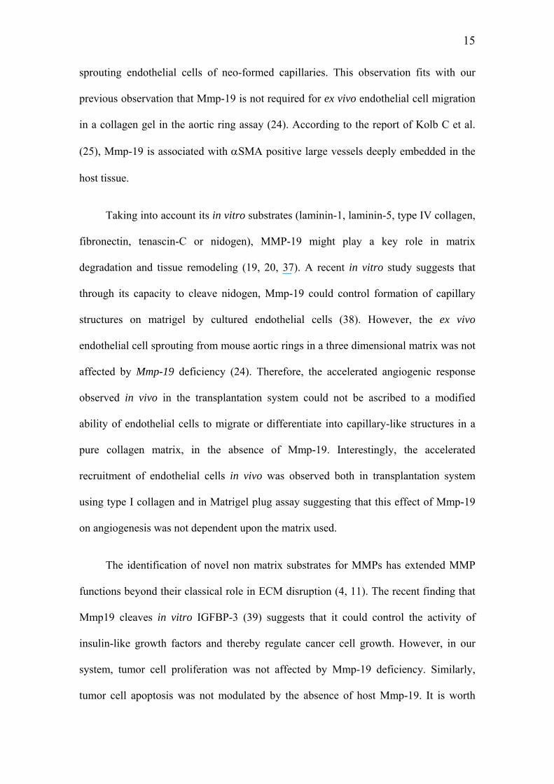
Discussion
The analysis of cancer susceptibility in individual Mmp-null mice is of growing
interest after the failure of synthetic MMP inhibitors in clinical trials and the finding of
multiple and even opposite roles of MMPs in tumor progression. We describe here that
in contrast to most MMP deficiencies described up to now in mice, the angiogenic
response was accelerated and tumor invasion was increased in Mmp-19-deficient mice.
This was evidenced by (i) an accelerated vascularization and a higher degree of tumor
invasion after malignant keratinocyte transplantation and (ii) an increased
vascularization in bFGF treated matrigel implants in Mmp-19-/- mice.
The time course of malignant growth begins with an early onset of stromal
activation and infiltration of inflammatory cells and endothelial cells. These key events
can be mimicked in the transplantation system used here which is a highly sensitive tool
to inspect kinetics of early steps of host stromal response to tumor signals (34, 35). By
applying this system to Mmp-19-/- mice, it appears that the angiogenic response was
affected by lack of host Mmp-19. Surprisingly, endothelial cell recruitment was
significantly increased 14 days after transplantation, leading to an acceleration of tumor
vascularization. Consequently, tumor invasion was increased in Mmp-19-deficient mice,
21 days after tumor transplantation. These findings indicate that Mmp-19 is a negative
regulator of early steps of cancer angiogenesis and invasion.
By in situ hybridization and immunohistochemical analysis, we demonstrate that
mesenchymal cells produce Mmp-19, while CD11b-positive inflammatory cells and
malignant keratinocytes do not express it. Accordingly, MMP-19 mRNA expression
and pro-MMP-19 production by human fibroblasts have been detected in vivo in dermal
wounds and in cultures of fibroblasts (36). In our model, Mmp-19 is not produced by
15
sprouting endothelial cells of neo-formed capillaries. This observation fits with our
previous observation that Mmp-19 is not required for ex vivo endothelial cell migration
in a collagen gel in the aortic ring assay (24). According to the report of Kolb C et al.
(25), Mmp-19 is associated with αSMA positive large vessels deeply embedded in the
host tissue.
Taking into account its in vitro substrates (laminin-1, laminin-5, type IV collagen,
fibronectin, tenascin-C or nidogen), MMP-19 might play a key role in matrix
degradation and tissue remodeling (19, 20, 37). A recent in vitro study suggests that
through its capacity to cleave nidogen, Mmp-19 could control formation of capillary
structures on matrigel by cultured endothelial cells (38). However, the ex vivo
endothelial cell sprouting from mouse aortic rings in a three dimensional matrix was not
affected by Mmp-19 deficiency (24). Therefore, the accelerated angiogenic response
observed in vivo in the transplantation system could not be ascribed to a modified
ability of endothelial cells to migrate or differentiate into capillary-like structures in a
pure collagen matrix, in the absence of Mmp-19. Interestingly, the accelerated
recruitment of endothelial cells in vivo was observed both in transplantation system
using type I collagen and in Matrigel plug assay suggesting that this effect of Mmp-19
on angiogenesis was not dependent upon the matrix used.
The identification of novel non matrix substrates for MMPs has extended MMP
functions beyond their classical role in ECM disruption (4, 11). The recent finding that
Mmp19 cleaves in vitro IGFBP-3 (39) suggests that it could control the activity of
insulin-like growth factors and thereby regulate cancer cell growth. However, in our
system, tumor cell proliferation was not affected by Mmp-19 deficiency. Similarly,
tumor cell apoptosis was not modulated by the absence of host Mmp-19. It is worth

16
noting that proteolytic processing of some bioactive molecules such as growth factors
and/or cytokines/chemokines could also indirectly contribute to micro-environment
modifications promoting or inhibiting endothelial cell recruitment during angiogenesis
onset. The assumption that MMPs are in general pro-angiogenic has been challenged by
the finding that some of them could suppress neovascularization by generating
angiogenic inhibitors (40). In this context, Mmp-19 could exert its anti-angiogenic
effect through inactivation of angiogenic/chemotactic factors or production/maturation
of angiogenic inhibitors. In addition, Mmp-19 production in large quiescent host vessels
present below tumor transplants, but not in growing capillaries suggests a yet unknown
functional role of this protease in maintenance of blood vessel stability. Its absence
could destabilize endothelial cell-mural cell interactions, thereby initiating active
sprouting events and endothelial cell migration. This hypothesis could explain the
transient effect of Mmp-19 deficiency at angiogenesis onset.
The present results do not negate the possibility that Mmp-19 might have dual
functions during cancer progression. In a model of methylcholanthrene-induced
chemical carcinogenesis, we previously showed that Mmp-19-/- mice develop less
fibrosarcomas and with a longer latency period than wild-type littermates (24). These
apparently paradoxical results may reflect different roles of Mmp-19 in the evolution of
various cancer types (carcinoma versus fibrosarcoma), as well as throughout different
steps of cancer progression. The down-regulation and disappearance of MMP-19
production observed during neoplastic progression in breast and skin carcinomas (21-
23) are consistent with our data demonstrating a control of early steps of skin carcinoma
evolution by Mmp-19. Altogether these data based on clinical and experimental studies
suggest that MMP-19 negatively regulates early stages of tumor cell invasion, but
cancer cells could become less sensitive to MMP-19 activity once tumor develops.

17
Temporal differences in effect of proteases on tumor growth and conversion to
aggressive tumors have also been reported for Mmp-9 (41) and Mmp-11 (42).
By applying the same transplantation chamber assay into different single or double
Mmp-deficient mice (43), we previously reported that tumor invasion and angiogenesis
were both impaired by the combined deficiency in Mmp-2 and Mmp-9 demonstrating
that concomitant production of gelatinases is required for tumor invasion and
vascularization. Therefore, although Mmp-2 and Mmp-9 are viewed as positive
regulators of tumor angiogenesis (4, 44-46), Mmp-19 could function in an opposite
manner, slowing-down the angiogenic process. These unexpected data are in
accordance with the emerging anti-tumor properties of some MMPs. In this context,
skin tumor susceptibility was increased in mice deficient for Mmp-8 (47) or Mmp-3
(48). Furthermore, our results support the view that MMPs act as sophisticated
modulators rather than simple inducers or suppressors and highlight the functional
complexity of MMP family during cancer progression. Some MMPs appear to have
dual role in cancer progression by promoting angiogenesis and generating angiogenesis
inhibitors (6). For instance, although MMP-7 and MMP-9 are both able to generate
angiostatin (40), MMP-7 facilitates tumor progression in mouse models (49) and MMP-
9 promotes tumor angiogenesis (43-46). Increased expression of some MMPs may both
confer increased tumor cell invasiveness and paradoxically, lead to production of
molecules that limit tumor growth. Altogether, these data point out the need to
determine both spatial and temporal significance of individual MMP during cancer
progression in order to design more rational MMP inhibitors.
18
Acknowledgments
We thank I. Dasoul, P. Gavitelli, F. Olivier and G. Roland for their excellent technical
assistance. This work was supported by grants from the Communauté Française de
Belgique (Actions de Recherches Concertées), the European Union (FP5 and FP6), the
Fonds National de la Recherche Scientifique (FNRS, Belgium), the Fédération Belge
Contre le Cancer, the Centre Anticancéreux près l'Université de Liège, the FB
Assurances, the Fondation Léon Frédéricq (University of Liège), the D.G.T.R.E. from
the “Région Wallonne”, the Interuniversity Attraction Poles Programme- Belgian
Science Policy (Brussels, Belgium), the Comision Interministerial de Ciencia y
Tecnologia (to C. L.-O). M.J. and S.B are recipients of a grant from FNRS-Télévie.
D.C. is a scientific research worker from the FNRS (Belgium).
19
References
1. Nagase H, Woessner JF. Matrix metalloproteinases. J Biol Chem 1999;274:21491-4.
2. Vu TH, Werb Z. Matrix metalloproteinases: effectors of development and normal physiology.
Genes Dev 2000;14:2123-33.
3. Konttinen YT, Ainola M, Valleala H et al. Analysis of 16 different matrix metalloproteinases
(MMP-1 to MMP-20) in the synovial membrane: different profiles in trauma and rheumatoid
arthritis. An Rheum Dis 1999;58:691-7.
4. Folgueras AR, Pendas AM, Sanchez LM, Lopez-Otin C. Matrix metalloproteinases in cancer:
from new functions to improved inhibition strategies. Int J Dev Biol 2004;48:411-24.
5. Noel A, Maillard C, Rocks N et al. Membrane associated proteases and their inhibitors in
tumour angiogenesis. J Clin Pathol 2004;57:577-84.
6. Handsley MM, Edwards DR. Metalloproteinases and their inhibitors in tumor angiogenesis.
Int J Canc 2005;115:849-60.
7. Sounni NE, Noel A. Membrane type-matrix metalloproteinases and tumor progression.
Biochimie 2005;87:329-42.
8. Sternlicht MD, Werb Z. How matrix metalloproteinases regulate cell behavior. Annu Rev
Cell Dev Biol 2001;17:463-516.
9. McQuibban GA, Gong JH, Tam EM, McCulloch CAG, Clark-Lewis I, Overall CM.
Inflammation dampened by gelatinase A cleavage of monocyte chemoattractant protein-3.
Science 2000;289:1202-6.
10. McQuibban GA, Butler GS, Gong JH et al. Matrix metalloproteinase activity inactivates the
CXC chemokine stromal cell-derived factor-1. J Biol Chem 2001;276:43503-8.
11. Egeblad M, Werb Z. New functions for the matrix metalloproteinases in cancer progression.
Nat Rev Cancer 2002;2:161-74.
12. Puente XS, Lopez-Otin C. A genomic analysis of rat proteases and protease inhibitors.
Genome Res 2004;14:609-22.

20
13. Lopez-Otin C, Overall CM. Protease degradomics: A new challenge for proteomics. Nat
Rev Mol Cell Biol 2002;3:509-19.
14. Zucker S, Pei DQ, Cao J, Lopez-Otin C. Membrane type-matrix metalloproteinases (MT-
MMP). Cell Surface Proteases 2003;54:1-74.
15. Pendas AM, Knauper V, Puente XS et al. Identification and characterization of a novel
human matrix metalloproteinase with unique structural characteristics, chromosomal location,
and tissue distribution. J Biol Chem 1997;272:4281-6.
16. Sedlacek R, Mauch S, Kolb B et al. Matrix metalloproteinase MMP-19 (RASI 1) is
expressed on the surface of activated peripheral blood mononuclear cells and is detected as an
autoantigen in rheumatoid arthritis. Immunobiology 1998;198:408-23.
17. Yang MZ, Kurkinen M. Cloning and characterization of a novel matrix metalloproteinase
(MMP), CMMP, from chicken embryo fibroblasts - CMMP, Xenopus XMMP, and human
MMP19 have a conserved unique cysteine in the catalytic domain. J Biol Chem
1998;273:17893-900.
18. Mueller MS, Mauch S, Sedlacek R. Structure of the human MMP-19 gene. Gene
2000;252:27-37.
19. Stracke JO, Fosang AJ, Last K et al. Matrix metalloproteinases 19 and 20 cleave aggrecan
and cartilage oligomeric matrix protein (COMP). Febs Letters 2000;478:52-6.
20. Stracke JO, Hutton M, Stewart M et al. Biochemical characterization of the catalytic domain
of human matrix metalloproteinase 19 - Evidence for a role as a potent basement membrane
degrading enzyme. J Biol Chem 2000;275:14809-16.
21. Djonov V, Hogger K, Sedlacek R, Laissue J, Draeger A. MMP-19: cellular localization of a
novel metalloproteinase within normal breast tissue and mammary gland tumours. J Pathol
2001;195:147-55.
22. Impola U, Toriseva M, Suomela S et al. Matrix metalloproteinase-19 is expressed by
proliferating epithelium but disappears with neoplastic dedifferentiation. Int J Canc
2003;103:709-16.

21
23. Impola U, Jeskanen L, Ravanti L et al. Expression of matrix metalloproteinase (MMP)-7
and MMP-13 and loss of MMP-19 and p16 are associated with malignant progression in chronic
wounds. Br J Dermatol 2005;152:720-6.
24. Pendas AM, Folgueras AR, Llano E et al. Diet-induced obesity and reduced skin cancer
susceptibility in matrix metalloproteinase 19-deficient mice. Mol Cell Biol 2004;24:5304-13.
25. Kolb C, Mauch S, Krawinkel U, Sedlacek R. Matrix metalloproteinase-19 in capillary
endothelial cells: Expression in acutely, but not in chronically, inflamed synovium. Exp Cell
Res 1999;250:122-30.
26. Bajou K, Masson V, Gerard RD et al. The plasminogen activator inhibitor PAI-1 controls in
vivo tumor vascularization by interaction with proteases, not vitronectin: Implications for
antiangiogenic strategies. J Cell Biol 2001;152:777-84.
27. Fusenig, N. E. Growth and differentiation characteristics of transformed keratinocytes from
mouse and human skin in vitro and in vivo. Breitkreutz, D., Dzarlieva, R. T., Boukamp, P.,
Bohnert A., and Tilgen, W. J Invest Dermatol 1983;81, 168s-75s.
28. Bajou K, Noel A, Gerard RD et al. Absence of host plasminogen activator inhibitor 1
prevents cancer invasion and vascularization. Nat Med 1998;4:923-8.
29. Bajou K, Masson V, Gerard RD et al. The plasminogen activator inhibitor PAI-1 controls in
vivo tumor vascularization by interaction with proteases, not vitronectin: Implications for
antiangiogenic strategies. J Cell Biol 2001;152:777-84.
30. Bajou K, Maillard C, Jost M et al. Host-derived plasminogen activator inhibitor-1 (PAI-1)
concentration is critical for in vivo tumoral angiogenesis and growth. Oncogene 2004;23:6986-
90.
31. Kapur, J. N., Sahoo, P. K, and Wong, A. K. C. A New Method for Gray-Level Picture
Thresholding Using the Entropy of the Histogram. Computer Vision, Graphics, and Image
Processing 29. 1985.
32. Kohler, R. A Segmentation System Based on Thresholding. Computer Graphics and Image
Processing 1985;15, 319-38.

22
33. Passaniti A, Taylor RM, Pili R et al. Methods in Laboratory Investigation - A Simple,
Quantitative Method for Assessing Angiogenesis and Antiangiogenic Agents Using
Reconstituted Basement-Membrane, Heparin, and Fibroblast Growth-Factor. Laboratory
Investigation 1992;67:519-28.
34. Mueller MM, Fusenig NE. Tumor-stroma interactions directing phenotype and progression
of epithelial skin tumor cells. Differentiation 2002;70:486-97.
35. Mueller MM, Fusenig NE. Friends or foes - Bipolar effects of the tumour stroma in cancer.
Nat Rev Cancer 2004;4:839-49.
36. Hieta N, Impola U, Lopez-Otin C, Saarialho-Kere U, Kahari VM. Matrix metalloproteinase-
19 expression in dermal wounds and by fibroblasts in culture. J Invest Dermatol 2003;121:997-
1004.
37. Sadowski T, Dietrich S, Koschinsky F et al. Matrix metalloproteinase 19 processes the
laminin 5 gamma 2 chain and induces epithelial cell migration. CMLS 2005;62:870-80.
38. Titz B, Dietrich S, Sadowski T, Beck C, Petersen A, Sedlacek R. Activity of MMP-19
inhibits capillary-like formation due to processing of nidogen-1. CMLS 2004;61:1826-33.
39. Sadowski T, Dietrich S, Koschinsky F, Sedlacek R. Matrix metalloproteinase 19 regulates
insulin-like growth factor-mediated proliferation, migration, and adhesion in human
keratinocytes through proteolysis of insulin-like growth factor binding protein-3. Mol Biol Cell
2003;14:4569-80.
40. Pozzi A, Moberg PE, Miles LA, Wagner S, Soloway P, Gardner HA. Elevated matrix
metalloprotease and angiostatin levels in integrin alpha 1 knockout mice cause reduced tumor
vascularization. Proc Natl Acad Sci U S A 2000;97:2202-7.
41. Coussens LM, Tinkle CL, Hanahan D, Werb Z. MMP-9 supplied by bone marrow-derived
cells contributes to skin carcinogenesis. Cell 2000;103:481-90.
42. Andarawewa KL, Boulay A, Masson W et al. Dual stromelysin-3 function during natural
mouse mammary tumor virus-ras tumor progression. Cancer Res 2003;63:5844-9.
43. Masson V, de la Ballina LR, Munaut C et al. Contribution of host MMP-2 and MMP-9 to
23
promote tumor vascularization and invasion of malignant keratinocytes. FASEB J 2005;19:234-
6.
44. Bergers G, Brekken R, McMahon G et al. Matrix metalloproteinase-9 triggers the
angiogenic switch during carcinogenesis. Nat Cell Biol 2000;2:737-44.
45. Chantrain CF, Shimada H, Jodele S et al. Stromal matrix metalloproteinase-9 regulates the
vascular architecture in neuroblastoma by promoting pericyte recruitment. Cancer Res
2004;64:1675-86.
46. Coussens LM, Tinkle CL, Hanahan D, Werb Z. MMP-9 supplied by bone marrow-derived
cells contributes to skin carcinogenesis. Cell 2000;103:481-90.
47. Balbin M, Fueyo A, Tester AM et al. Loss of collagenase-2 confers increased skin tumor
susceptibility to male mice. Nat Genet 2003;35:252-7.
48. Andarawewa KL, Boulay A, Masson W et al. Dual stromelysin-3 function during natural
mouse mammary tumor virus-ras tumor progression. Cancer Res 2003;63:5844-9.
49. Rudolph-Owen LA, Chan R, Muller WJ, Matrisian LM. The matrix metalloproteinase
matrilysin influences early-stage mammary tumorigenesis. Cancer Res 1998;58:5500-6.

24
Legends of figures:
Figure 1. A: Histological analysis of tumor transplants: Immunofluorescence
labeling of malignant keratinocytes and vessels in tumors transplanted into Mmp-19+/+
or Mmp-19-/- mice observed after 14 (D14) or 21 (D21) days. Malignant cells (anti-
keratin Ab in green) and vessels (anti-collagen type IV Ab in red) are visualized. The
dotted line delineates the front of endothelial cell migration. Scores and morphometric
parameter (d) used to quantify tumor invasion and angiogenesis are indicated (original
magnification x200). B-C: Semi-quantitative evaluation of tumor vascularization in
Mmp-19+/+ (black) or Mmp-19-/- mice (white). Tumors were scored as illustrated in
panel A and in Materials and Methods. n = number of animals per group. D:
Quantification of tumor vascularization in tumors transplanted for 14 days in Mmp-
19+/+ (black) or Mmp-19-/- (white) mice. “d” = distance separating tumor cell layer
from the front of migrating blood vessels as defined in panel A (see Materials and
Methods) (** P≤0.01, Mann-Whitney test) (n = 8). k: malignant keratinocytes; c:
collagen gel; h: host tissue.
Figure 2. Quantification of tumor vascularization and invasion in tumor transplants
based on computer-assisted image analysis. A: Image processing of a typical transplant
section. Malignant keratinocytes (in green) and vessels (in red) (original image) were
immunostained according to the procedure described in Materials and Methods. Images
are binarized with extraction of vessels (Vascularization) or of tumor cells (Invasion).
Tumor vascularization and tumor invasion were determined by using a grid obtained by
successive dilatation of the tumor upper border. B-D: determination of vessel density
(“normalized density of endothelial cells”) (B) and tumor cell density (“normalized
density of tumor cells”) (C, D) as a function of the distance to the upper boundary of

25
tumor layer. The maximal distance of tumor cell invasion corresponds to tumor
thickness (D). Malignant keratinocytes were transplanted into Mmp-19+/+ (WT) and
Mmp-19-/- (KO) mice for 14 days (D14) or 21 days (D21).
Figure 3. BrdU immunostaining of tumor transplants. Malignant keratinocytes
were transplanted for 14 days (D14) (A) or 21 days (D21) (B) into Mmp-19+/+ (black)
or Mmp-19-/- (white) mice. Before sacrifice, mice were injected with BrdU and
immunostaining was performed on tumor sections. The percentage of positive cells was
determined by a computer-assisted method described in Material and Methods.
Figure 4. In situ hybridization of tumor transplants. Sections of malignant
keratinocytes transplanted into Mmp-19+/+ (A, B) and Mmp-19-/- (C) mice for 21 days
were hybridized with 35S-labeled anti-sense Mmp-19 riboprobe (A, C) or sense Mmp-19
riboprobe (B) used as negative control. Bright fields (left) and dark fields (right) images
were both captured. Stromal cells in host tissue (h), but not malignant keratinocytes (k)
were positively stained for Mmp-19 mRNAs (original magnification x100).
Figure 5. Immunostaining of tumors transplanted into wild type mice. Sections are
counterstained with bisbenzimide (blue staining). Host stromal cells are positive for
Mmp-19 immunostaining (in green) (original magnification x200) (A). Double
immunostainings reveal that Mmp-19 (in red) is not present in newly formed vessels in
collagen matrix, but is associated to vascular structures stained with α-smooth muscle
actin (α-SMA) antibody (in green) and localized deeply in host tissue (original
magnification x400) (B). Mmp-19 (in green) does not co-localize with CD11b-positive
inflammatory cells (in red) (original magnification x400) (C). Higher magnification is
shown in insert (C).

26
Figure 6. Increased vascularization in Matrigel Plug containing bFGF and
implanted in Mmp-19-/- mice. Functional vascularization was quantified by measuring
haemoglobin concentration in Matrigel plug implanted into Mmp-19+/+ and Mmp-19-/-
mice. * P≤ 0.05.

27
28

29
30

31

32
Copyright © 2022 FDOKUMEN