Endothelial cell-derived interleukin-6 regulates tumor growth
Upload
khangminh22Category
view
0download
0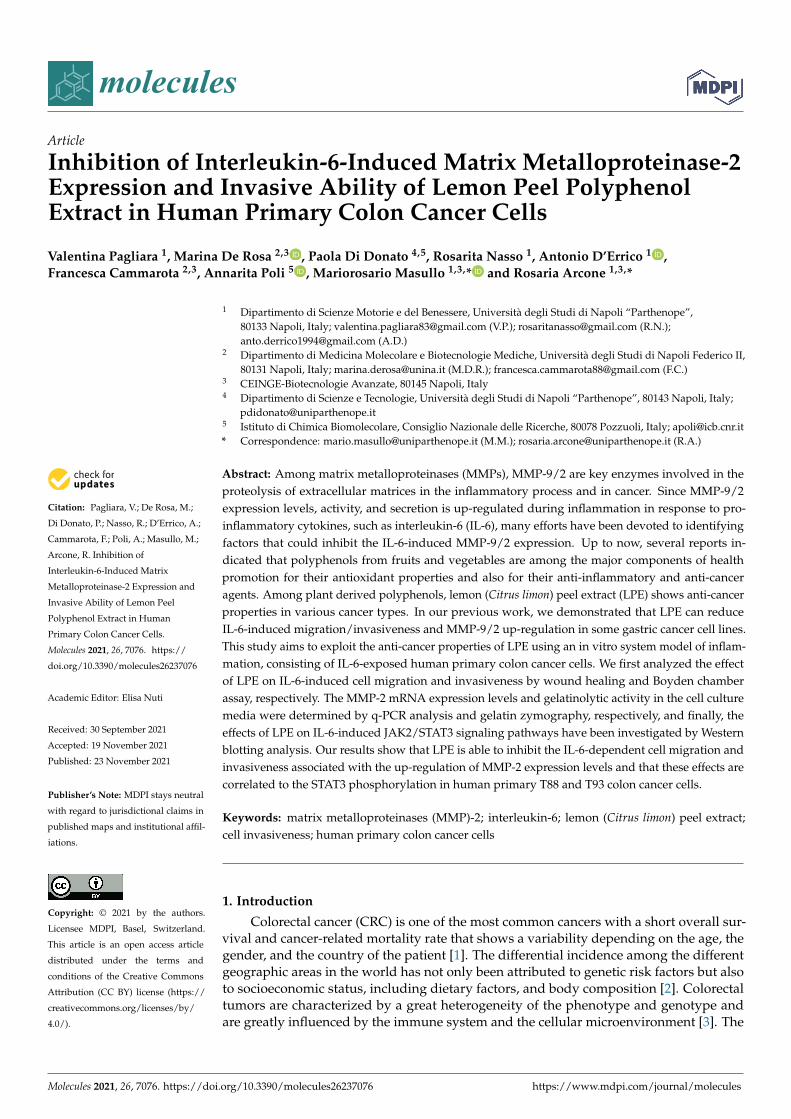
molecules
Article
Inhibition of Interleukin-6-Induced Matrix Metalloproteinase-2Expression and Invasive Ability of Lemon Peel PolyphenolExtract in Human Primary Colon Cancer Cells
Valentina Pagliara 1, Marina De Rosa 2,3 , Paola Di Donato 4,5, Rosarita Nasso 1, Antonio D’Errico 1 ,Francesca Cammarota 2,3, Annarita Poli 5 , Mariorosario Masullo 1,3,* and Rosaria Arcone 1,3,*
�����������������
Citation: Pagliara, V.; De Rosa, M.;
Di Donato, P.; Nasso, R.; D’Errico, A.;
Cammarota, F.; Poli, A.; Masullo, M.;
Arcone, R. Inhibition of
Interleukin-6-Induced Matrix
Metalloproteinase-2 Expression and
Invasive Ability of Lemon Peel
Polyphenol Extract in Human
Primary Colon Cancer Cells.
Molecules 2021, 26, 7076. https://
doi.org/10.3390/molecules26237076
Academic Editor: Elisa Nuti
Received: 30 September 2021
Accepted: 19 November 2021
Published: 23 November 2021
Publisher’s Note: MDPI stays neutral
with regard to jurisdictional claims in
published maps and institutional affil-
iations.
Copyright: © 2021 by the authors.
Licensee MDPI, Basel, Switzerland.
This article is an open access article
distributed under the terms and
conditions of the Creative Commons
Attribution (CC BY) license (https://
creativecommons.org/licenses/by/
4.0/).
1 Dipartimento di Scienze Motorie e del Benessere, Università degli Studi di Napoli “Parthenope”,80133 Napoli, Italy; [email protected] (V.P.); [email protected] (R.N.);[email protected] (A.D.)
2 Dipartimento di Medicina Molecolare e Biotecnologie Mediche, Università degli Studi di Napoli Federico II,80131 Napoli, Italy; [email protected] (M.D.R.); [email protected] (F.C.)
3 CEINGE-Biotecnologie Avanzate, 80145 Napoli, Italy4 Dipartimento di Scienze e Tecnologie, Università degli Studi di Napoli “Parthenope”, 80143 Napoli, Italy;
[email protected] Istituto di Chimica Biomolecolare, Consiglio Nazionale delle Ricerche, 80078 Pozzuoli, Italy; [email protected]* Correspondence: [email protected] (M.M.); [email protected] (R.A.)
Abstract: Among matrix metalloproteinases (MMPs), MMP-9/2 are key enzymes involved in theproteolysis of extracellular matrices in the inflammatory process and in cancer. Since MMP-9/2expression levels, activity, and secretion is up-regulated during inflammation in response to pro-inflammatory cytokines, such as interleukin-6 (IL-6), many efforts have been devoted to identifyingfactors that could inhibit the IL-6-induced MMP-9/2 expression. Up to now, several reports in-dicated that polyphenols from fruits and vegetables are among the major components of healthpromotion for their antioxidant properties and also for their anti-inflammatory and anti-canceragents. Among plant derived polyphenols, lemon (Citrus limon) peel extract (LPE) shows anti-cancerproperties in various cancer types. In our previous work, we demonstrated that LPE can reduceIL-6-induced migration/invasiveness and MMP-9/2 up-regulation in some gastric cancer cell lines.This study aims to exploit the anti-cancer properties of LPE using an in vitro system model of inflam-mation, consisting of IL-6-exposed human primary colon cancer cells. We first analyzed the effectof LPE on IL-6-induced cell migration and invasiveness by wound healing and Boyden chamberassay, respectively. The MMP-2 mRNA expression levels and gelatinolytic activity in the cell culturemedia were determined by q-PCR analysis and gelatin zymography, respectively, and finally, theeffects of LPE on IL-6-induced JAK2/STAT3 signaling pathways have been investigated by Westernblotting analysis. Our results show that LPE is able to inhibit the IL-6-dependent cell migration andinvasiveness associated with the up-regulation of MMP-2 expression levels and that these effects arecorrelated to the STAT3 phosphorylation in human primary T88 and T93 colon cancer cells.
Keywords: matrix metalloproteinases (MMP)-2; interleukin-6; lemon (Citrus limon) peel extract;cell invasiveness; human primary colon cancer cells
1. Introduction
Colorectal cancer (CRC) is one of the most common cancers with a short overall sur-vival and cancer-related mortality rate that shows a variability depending on the age, thegender, and the country of the patient [1]. The differential incidence among the differentgeographic areas in the world has not only been attributed to genetic risk factors but alsoto socioeconomic status, including dietary factors, and body composition [2]. Colorectaltumors are characterized by a great heterogeneity of the phenotype and genotype andare greatly influenced by the immune system and the cellular microenvironment [3]. The
Molecules 2021, 26, 7076. https://doi.org/10.3390/molecules26237076 https://www.mdpi.com/journal/molecules
Molecules 2021, 26, 7076 2 of 12
degradation of the extracellular matrix (ECM) represents a crucial step in the invasion andmigration of cancer cells, and this process is strictly dependent on the activity of numerousenzymes. Matrix metalloproteinases (MMPs) are the major enzymes responsible for thedegradation of collagen and proteins in the ECM [4,5]. Among the different MMPs, MMP-9and MMP-2 belong to a family of zinc-dependent endo-peptidases which are secreted bystromal and tumor cells as inactive zymogens, and then activated following the cleavage ofthe pro-domain peptide into their active form [5]. MMP-2 and MMP-9, known as gelatinaseA and B, are critically involved in tumor invasion and metastasis and their expressionhas been associated with poor overall survival in patients with colon cancer [6,7]. A closecorrelation has been demonstrated between the inflammatory state and increased expres-sion of MMPs, which is caused by proinflammatory cytokines, including interleukin-6(IL-6) [8,9]. Interleukin-6 (IL-6) is a major inflammatory cytokine also involved in cancerand autoimmune diseases [10]. Numerous evidences have shown that IL-6 induces the mi-gration and invasiveness of different types of tumor cells [11–14] and therefore representsa prognostic factor related to cell survival [15]. In fact, elevated levels of IL-6 have beendetected in the plasma of gastric and colon cancer patients and in gastric and colon cancercell lines, confirming the role of IL-6 as a predictor of poor prognoses and as correlated totumor aggressiveness [8,16,17]. The molecular mechanism activated by IL-6 involves theactivation of JAK/STAT3 signaling [18], which leads to the transcriptional activation of nu-merous target genes through the action of STAT3, which then results in tumor proliferationand/or survival. Furthermore, STAT3 also regulates the expression of factors that promoteangiogenesis and cell invasiveness, including MMPs [19–21]. In this context, IL-6 behavesas a pro-metastatic agent as described in the human gastric cancer AGS and MKN-28 celllines [22–24]. Among the various factors involved in the onset of CRC, lifestyle and diet arealso considered [25]. Polyphenols in fruit and vegetables are among the main componentsof health for their antioxidant properties and also for their anti-inflammatory and antitumoragents [26–28]. In our previous studies, we have shown that the polyphenols of plant origincontained in the extract from the flesh of Annurca apples and lemon (Citrus limon) peelextracts (LPE) are able to reduce the migration/invasiveness induced by IL-6 or on theup-regulation of MMP-9/2 in MKN-28 and AGS cancer cells [24,29].
In light of this previous evidence, in this study, we have analyzed the effects ofLPE using primary human colon T88 and T93 cancer cells, which have been previouslycharacterized [30–32]. These primary colon cancer cells were isolated and establishedfrom the tumor tissues of two patients affected by sporadic colon adenocarcinoma. TheT93 cell line exhibited a chromosomal instability (CIN) phenotype and the T88 cell lineexhibited a microsatellite instability (MSI) phenotype. It has been previously demonstratedthat these cells both underwent EMT from epithelial adenocarcinoma cells; indeed, theysimultaneously expressed epithelial and mesenchymal markers, such as cytokeratin and E-cadherin, and Vimentin and N-cadherin, respectively, together with high levels expressionof EMT-associated transcription factors, stemness markers, and the Cyclooxygenase-2(Cox-2) enzyme [30–32].
The protective effect of LPE against the increase in cell migration and invasivenessinduced by IL-6 was evaluated by pretreatments with LPE before exposure to IL-6. Theeffects of LPE on IL-6-induced JAK2/STAT3 signaling pathways were also analyzed. Ourresults indicate that LPE is capable of inhibiting rIL-6-dependent migration and invasive-ness associated with up-regulation of MMP-2 expression levels in human colon carcinomacell lines T88 and T93.
2. Results2.1. The LPE Pretreatment Reduces the rIL-6-Induced Migration and Invasiveness of HumanPrimary T88 and T93 Colon Cancer Cells
Since several reports demonstrated that IL-6 enhances the migration ability andinvasiveness of gastric and colon cancer cells [8,12,20,23,24,33], we investigated the effectof the treatment with rIL-6 on either migration or invasive ability in human primary T88and T93 colon cancer cells. In addition, to explore whether LPE could counteract the
Molecules 2021, 26, 7076 3 of 12
rIL-6-dependent effects, T88 and T93 cells were first pretreated with LPE for 6 h and thenstimulated with rIL-6 for a further 24 h. To avoid serum interference with LPE components,all the treatments were performed in serum-free media and, at the end of the incubation,the treated cells were compared with the untreated cells that were kept in serum-freeconditions. The cell migration was evaluated by the wound-healing assay that is shownin Figure 1. As displayed in this figure, in both cells, the exposure to rIL-6 alone led toa significant reduction in the wound area (Panel A, B, images b, f, j) compared to thatof untreated cells; this result clearly demonstrates that rIL-6 enhances the cell migrationability. In contrast, neither serum deprivation (Panel A, B, images a, e, i) nor treatmentwith LPE alone (Panel A, B, images c, g, k) affected the wound area, compared to that ofuntreated cells (Panel A, B, images a, e, i) in both T88 and T93 cells. Quantitative analysisof the wound area (Panel C, D) shows that in both cells, the exposure to rIL-6 inducesminimal reduction in the wound area at 12 h, whereas ~40% reduction is observed at24 h, compared to that of cells kept in serum-free conditions or exposed to LPE alone.It is noteworthy that the pre-treatment with LPE is able to inhibit the rIL-6-induced cellmigration, as demonstrated by the wound area that is similar to that of control cells, eitherin T88 (Panel A, images d, h, l) or in T98 (Panel B, images d, h, l). A maximal effect (~30%)was observed at 24 h and was compared with the wound area of cells stimulated with rIL-6alone.
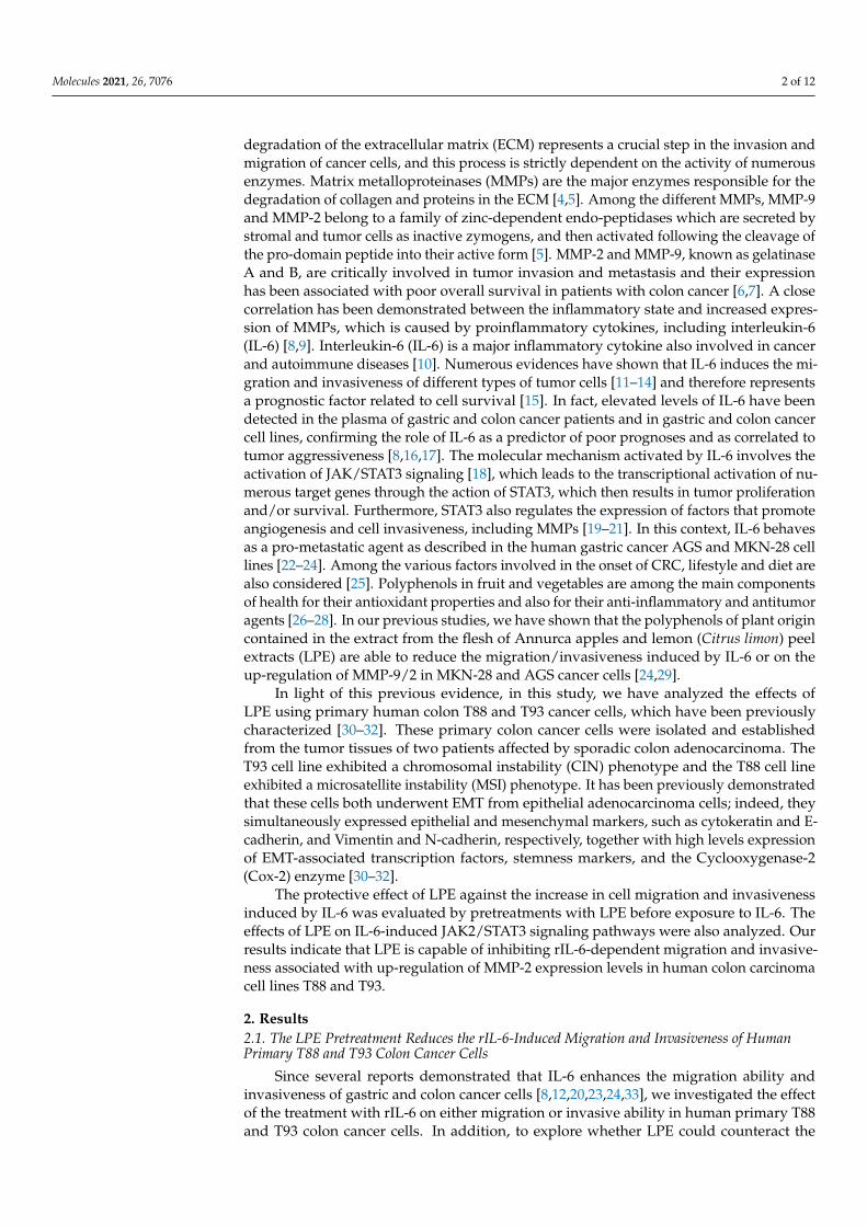
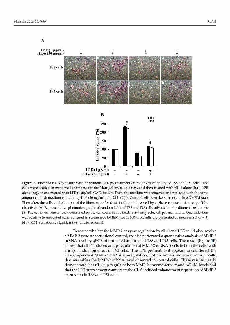
To evaluate the effect of rIL-6 alone or after LPE pretreatments on T88 and T93 cellinvasiveness, we performed a Matrigel assay. Figure 2, Panel A reveals that, in both cells,rIL-6 induces an ~2-fold (Panel B) increase in the number of cells that had invaded throughthe membrane (Panel A, images b, f), compared to those found in untreated cells (Panel A,images a, e). The treatment with LPE alone for 24 h (Panel A, images c, g) slightly decreasesthe cell invasiveness (Panel B), compared to untreated cells. The LPE pre-incubation beforerIL-6 exposure is able to reduce the cytokine-induced increase of cell invasiveness in bothT88 and T93 cells (Panel A, images c, g); in fact, an ~2.5-fold decrease of invasive abilityis observed in cells pretreated with LPE compared to that of cells exposed to rIL-6 alone(Panel B).
2.2. The LPE Pretreatment Reduces the rIL-6-Induced Up-Regulation of MMP-2 Activity andmRNA Expression Levels in T88 and T93 Cells
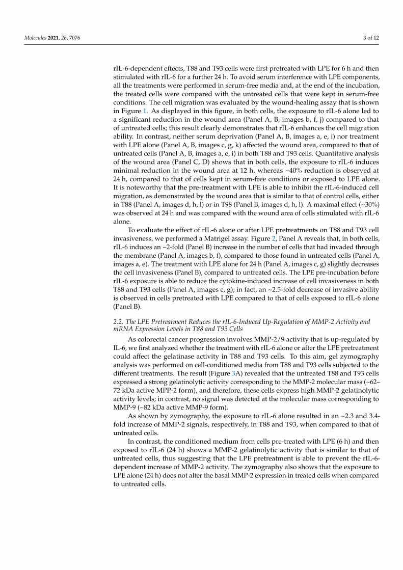
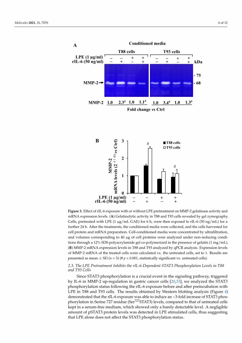
As colorectal cancer progression involves MMP-2/9 activity that is up-regulated byIL-6, we first analyzed whether the treatment with rIL-6 alone or after the LPE pretreatmentcould affect the gelatinase activity in T88 and T93 cells. To this aim, gel zymographyanalysis was performed on cell-conditioned media from T88 and T93 cells subjected to thedifferent treatments. The result (Figure 3A) revealed that the untreated T88 and T93 cellsexpressed a strong gelatinolytic activity corresponding to the MMP-2 molecular mass (~62–72 kDa active MPP-2 form), and therefore, these cells express high MMP-2 gelatinolyticactivity levels; in contrast, no signal was detected at the molecular mass corresponding toMMP-9 (~82 kDa active MMP-9 form).
As shown by zymography, the exposure to rIL-6 alone resulted in an ~2.3 and 3.4-fold increase of MMP-2 signals, respectively, in T88 and T93, when compared to that ofuntreated cells.
In contrast, the conditioned medium from cells pre-treated with LPE (6 h) and thenexposed to rIL-6 (24 h) shows a MMP-2 gelatinolytic activity that is similar to that ofuntreated cells, thus suggesting that the LPE pretreatment is able to prevent the rIL-6-dependent increase of MMP-2 activity. The zymography also shows that the exposure toLPE alone (24 h) does not alter the basal MMP-2 expression in treated cells when comparedto untreated cells.

Molecules 2021, 26, 7076 4 of 12Molecules 2021, 26, x FOR PEER REVIEW 4 of 14
Figure 1. Effect of rIL-6 exposure with or without LPE pretreatment on the cell migration of T88 and T93 cells by wound healing assay. Representative images of (A) T88 and (B) T93 cells captured at time 0, 12, and 24 h by a phase-contrast microscope (10 × objective). Cells kept in serum-free DMEM (a,e,i) were treated with rIL-6 alone (b,f,j), LPE alone (c,g,k), or pre-treated with LPE (1 μg/mL GAE) for 6 h and then the medium was removed and replaced with the same amount of fresh medium containing rIL-6 (50 ng/mL) for 12 h or 24 h. Quantitative analysis of the wound area of T88 (C) and T93 cells (D) at time 0, 12, and 24 h. For each treatment, the data shows the wound area at the indicated time in comparison to that of the open wound at time 0, set at 100%. Results are presented as mean ± SD (n = 3) (* p < 0.05, statistically significant vs. untreated cells).
12 h
C
Wou
nd a
rea
(%)
D
Wou
nd a
rea
(%)
*
* *
*
T93 cells T88 cells
T88 cells
+ _ rIL-6 (50 ng/ml) LPE (1 µg/ml) _ _ + _ + +
A
T93 cells B
_ rIL-6 (50 ng/ml) LPE (1 µg/ml) _ + +
0 h
24 h
12 h
0 h
24 h
+ _ rIL-6 (50 ng/ml) LPE (1 µg/ml) + _ +
+ _ _ rIL-6 (50 ng/ml)
LPE (1 µg/ml)
a b c d
e f g h
100 µm
i j k l
+ _ + _
a b c d
e f g h
100 µm
i j k l
+ _ + _ + + _ _ 0
20 40 60 80
100 120
0 h 12 h 24 h
0 20 40 60 80
100 120
0 h 12 h 24 h
Figure 1. Effect of rIL-6 exposure with or without LPE pretreatment on the cell migration of T88 andT93 cells by wound healing assay. Representative images of (A) T88 and (B) T93 cells captured attime 0, 12, and 24 h by a phase-contrast microscope (10× objective). Cells kept in serum-free DMEM(a,e,i) were treated with rIL-6 alone (b,f,j), LPE alone (c,g,k), or pre-treated with LPE (1 µg/mLGAE) for 6 h and then the medium was removed and replaced with the same amount of freshmedium containing rIL-6 (50 ng/mL) for 12 h or 24 h. Quantitative analysis of the wound area ofT88 (C) and T93 cells (D) at time 0, 12, and 24 h. For each treatment, the data shows the wound areaat the indicated time in comparison to that of the open wound at time 0, set at 100%. Results arepresented as mean ± SD (n = 3) (* p < 0.05, statistically significant vs. untreated cells).
Molecules 2021, 26, 7076 5 of 12
Molecules 2021, 26, x FOR PEER REVIEW 5 of 14
To evaluate the effect of rIL-6 alone or after LPE pretreatments on T88 and T93 cell invasiveness, we performed a Matrigel assay. Figure 2, Panel A reveals that, in both cells, rIL-6 induces an ~2-fold (Panel B) increase in the number of cells that had invaded through the membrane (Panel A, images b, f), compared to those found in untreated cells (Panel A, images a, e). The treatment with LPE alone for 24 h (Panel A, images c, g) slightly decreases the cell invasiveness (Panel B), compared to untreated cells. The LPE pre-incubation before rIL-6 exposure is able to reduce the cytokine-induced increase of cell invasiveness in both T88 and T93 cells (Panel A, images c, g); in fact, an ~2.5-fold de-crease of invasive ability is observed in cells pretreated with LPE compared to that of cells exposed to rIL-6 alone (Panel B).
Figure 2. Effect of rIL-6 exposure with or without LPE pretreatment on the invasive ability of T88 and T93 cells. The cells were seeded in trans-well chambers for the Matrigel invasion assay, and then treated with rIL-6 alone (b,f), LPE alone (c,g), or pre-treated with LPE (1 μg/mL GAE) for 6 h. Then, the medium was removed and replaced with the same amount of fresh medium containing rIL-6 (50 ng/mL) for 24 h (d,h). Control cells were kept in serum-free DMEM (a,e). Thereafter, the cells at the bottom of the filters were fixed, stained, and observed by a phase-contrast microscope (10 × objective). (A) Representative photomicrographs of random fields of T88 and T93 cells sub-jected to the different treatments. (B) The cell invasiveness was determined by the cell count in five fields, randomly selected, per membrane. Quantification was relative to untreated cells, cultured in serum-free DMEM, set at 100%. Results are presented as mean ± SD (n = 3) (§ p < 0.01, statistically significant vs. untreated cells).
2.2. The LPE Pretreatment Reduces the rIL-6-Induced Up-Regulation of MMP-2 Activity and mRNA Expression Levels in T88 and T93 Cells
B
Inva
sive
abili
ty(%
)
+_rIL-6 (50 ng/ml)LPE (1 µg/ml) _ _ +_ +
+
§
§
T88 cells
T93 cells
A
+_rIL-6 (50 ng/ml)LPE (1 µg/ml) _ _ +
++
0
50
100
150
200
250T88T93
a b c d
e f g h
100 µm
_
Figure 2. Effect of rIL-6 exposure with or without LPE pretreatment on the invasive ability of T88 and T93 cells. Thecells were seeded in trans-well chambers for the Matrigel invasion assay, and then treated with rIL-6 alone (b,f), LPEalone (c,g), or pre-treated with LPE (1 µg/mL GAE) for 6 h. Then, the medium was removed and replaced with the sameamount of fresh medium containing rIL-6 (50 ng/mL) for 24 h (d,h). Control cells were kept in serum-free DMEM (a,e).Thereafter, the cells at the bottom of the filters were fixed, stained, and observed by a phase-contrast microscope (10×objective). (A) Representative photomicrographs of random fields of T88 and T93 cells subjected to the different treatments.(B) The cell invasiveness was determined by the cell count in five fields, randomly selected, per membrane. Quantificationwas relative to untreated cells, cultured in serum-free DMEM, set at 100%. Results are presented as mean ± SD (n = 3)(§ p < 0.01, statistically significant vs. untreated cells).
To assess whether the MMP-2 enzyme regulation by rIL-6 and LPE could also involvea MMP-2 gene transcriptional control, we also performed a quantitative analysis of MMP-2mRNA level by qPCR of untreated and treated T88 and T93 cells. The result (Figure 3B)shows that rIL-6 induced an up-regulation of MMP-2 mRNA levels in both the cells, witha major induction effect in T93 cells. The LPE pretreatment appears to counteract therIL-6-dependent MMP-2 mRNA up-regulation, with a similar reduction in both cells,that resembles the MMP-2 mRNA level observed in control cells. These results clearlydemonstrate that rIL-6 up-regulates both MMP-2 enzyme activity and mRNA levels andthat the LPE pretreatment counteracts the rIL-6-induced enhancement expression of MMP-2expression in T88 and T93 cells.
Molecules 2021, 26, 7076 6 of 12Molecules 2021, 26, x FOR PEER REVIEW 7 of 14
Figure 3. Effect of rIL-6 exposure with or without LPE pretreatment on MMP-2 gelatinase activity and mRNA expression levels. (A) Gelatinolytic activity in T88 and T93 cells revealed by gel zy-mography. Cells, pretreated with LPE (1 μg/mL GAE) for 6 h, were then exposed to rIL-6 (50 ng/mL) for a further 24 h. After the treatments, the conditioned media were collected, and the cells harvested for cell protein and mRNA preparation. Cell-conditioned media were concentrated by ultrafiltration, and volumes corresponding to 40 μg of cell proteins were analyzed under non-reducing conditions through a 12% SDS-polyacrylamide gel co-polymerized in the presence of gelatin (1 mg/mL). (B) MMP-2 mRNA expression levels in T88 and T93 analyzed by qPCR analysis. Expression levels of MMP-2 mRNA of the treated cells were calculated vs. the untreated cells, set to 1. Results are presented as mean ± SD (n = 3) (# p < 0.001, statistically significant vs. untreated cells).
2.3. The LPE Pretreatment Inhibits the rIL-6-Dependent STAT3 Phosphorylation Levels in T88 and T93 Cells
Since STAT3 phosphorylation is a crucial event in the signaling pathway, triggered by IL-6 in MMP-2 up-regulation in gastric cancer cells [20,33], we analyzed the STAT3 phosphorylation status following the rIL-6 exposure before and after preincubation with LPE in T88 and T93 cells. The results obtained by Western blotting analysis (Figure 4) demonstrated that the rIL-6 exposure was able to induce an ~3-fold increase of STAT3 phosphorylation in Serine 727 residue (Ser727STAT3) levels, compared to that of untreated cells kept in a serum-free medium, which showed only a barely detectable level. A neg-
_T88 cells
rIL-6 (50 ng/ml)
A
LPE (1 µg/ml) __+ _ +
+
MMP-2
+
- 68
MMP-2 1.0 2.3# 1.0 1.1#
- 75
_kDa
T93 cells
__+ _ +
++
1.0 3.4# 1.0 1.3#
Fold change vs Ctrl
Conditioned media
rIL-6 (50 ng/ml)LPE (1 µg/ml) _
++_ +
__ +
0
1
2
3 T88 cellsT93 cells
MM
P-2
mR
NA
leve
ls(2
-!! Ctvs
Ctr
l)
#
#
B
Figure 3. Effect of rIL-6 exposure with or without LPE pretreatment on MMP-2 gelatinase activity andmRNA expression levels. (A) Gelatinolytic activity in T88 and T93 cells revealed by gel zymography.Cells, pretreated with LPE (1 µg/mL GAE) for 6 h, were then exposed to rIL-6 (50 ng/mL) for afurther 24 h. After the treatments, the conditioned media were collected, and the cells harvested forcell protein and mRNA preparation. Cell-conditioned media were concentrated by ultrafiltration,and volumes corresponding to 40 µg of cell proteins were analyzed under non-reducing condi-tions through a 12% SDS-polyacrylamide gel co-polymerized in the presence of gelatin (1 mg/mL).(B) MMP-2 mRNA expression levels in T88 and T93 analyzed by qPCR analysis. Expression levelsof MMP-2 mRNA of the treated cells were calculated vs. the untreated cells, set to 1. Results arepresented as mean ± SD (n = 3) (# p < 0.001, statistically significant vs. untreated cells).
2.3. The LPE Pretreatment Inhibits the rIL-6-Dependent STAT3 Phosphorylation Levels in T88and T93 Cells
Since STAT3 phosphorylation is a crucial event in the signaling pathway, triggeredby IL-6 in MMP-2 up-regulation in gastric cancer cells [20,33], we analyzed the STAT3phosphorylation status following the rIL-6 exposure before and after preincubation withLPE in T88 and T93 cells. The results obtained by Western blotting analysis (Figure 4)demonstrated that the rIL-6 exposure was able to induce an ~3-fold increase of STAT3 phos-phorylation in Serine 727 residue (Ser727STAT3) levels, compared to that of untreated cellskept in a serum-free medium, which showed only a barely detectable level. A negligibleamount of pSTAT3 protein levels was detected in LPE stimulated cells, thus suggestingthat LPE alone does not affect the STAT3 phosphorylation status.

Molecules 2021, 26, 7076 7 of 12
Molecules 2021, 26, x FOR PEER REVIEW 8 of 14
ligible amount of pSTAT3 protein levels was detected in LPE stimulated cells, thus sug-gesting that LPE alone does not affect the STAT3 phosphorylation status.
Figure 4. Effect of rIL-6 exposure with or without LPE pretreatment on STAT3 phosphorylation levels in T88 and T93 cells. Western blotting analysis of phospho-STAT3 protein expression levels in cells treated with rIL-6 alone (50 ng/mL for 24 h) or after preincubation with LPE (1 μg/mL GAE for 6 h)) and then exposed to rIL-6 (50 ng/mL for 24 h). The relative phospho-STAT3/STAT3 protein fold change level in the treated cells was calculated vs. the untreated cells, set to 1, is shown under each lane. Results are presented as mean ± SD (n = 3) (# p < 0.001, statistically significant vs. un-treated cells).
This result clearly indicates that the LPE pretreatment is able to prevent the rIL-6-dependent STAT3 phosphorylation and, therefore, that the LPE effects are also mediated through the IL-6-induced STAT3 signaling pathway in T88 and T93 cells.
3. Discussion In this study, we demonstrated that the inflammatory cytokine rIL-6 affects the mi-
gration and the invasiveness of two human primary adenocarcinoma cells, namely, T88 and T93 cells [30–32], and that this effect is well-correlated with the up-regulation of MMP-2 enzyme activity and mRNA expression levels. These findings agree with the key role of this metalloproteinase in the degradation of the ECM components, a process in-volved in tumor metastasis and cancer.
It should be noted that, in our studies, the zymography analysis shows a very high MMP-2 gelatinolytic level, but not that of MMP-9, which does not appear as a detectable signal even in smaller bands derived from degraded products. In addition, we observe a strong correlation between MMP-2 gelatinolytic activity and mRNA expression levels among all the treatments groups.
This evidence is consistent with previous observations on gastrointestinal malig-nancy demonstrating that the overexpression of MMP-2 is related to the conversion from premalignant conditions to a malignant phenotype, whereas MMP-9 is overexpressed in premalignant polyps [34,35], suggesting that the latter is expressed in the early stage during the adenocarcinoma malignant transformation.
In this scenario, the presence of the inflammatory IL-6 cytokine induces a strong up-regulation of MMP-2 for both the protein and mRNA expression levels.
Furthermore, the data reported here show that LPE is able to inhibit the rIL-6-dependent cell migration and invasiveness in human primary T88 and T93 colon cancer cells [30–32] via the up-regulation of MMP-2 expression, and that the observed effects correlate with the STAT3 phosphorylation levels. The protective effects showed by plant polyphenols against different types of cancer [36] include their anti-inflammatory and antioxidant properties [26–29]. Our results are in agreement with those reported
T88 cell lysates
rIL-6 (50 ng/ml)LPE (1 µg/ml) _
__+ _ +
++
β-Tubulin
STAT3
pSTAT3(Ser727)
pSTAT3/STAT3 1.0 2.8# 1.0 1.8#
- 85
- 85
- 50
kDa
T93 cell lysates__
_+ _ +
++
1.0 3.2# 1.0 1.9#
Fold change vs Ctrl
Figure 4. Effect of rIL-6 exposure with or without LPE pretreatment on STAT3 phosphorylationlevels in T88 and T93 cells. Western blotting analysis of phospho-STAT3 protein expression levelsin cells treated with rIL-6 alone (50 ng/mL for 24 h) or after preincubation with LPE (1 µg/mLGAE for 6 h) and then exposed to rIL-6 (50 ng/mL for 24 h). The relative phospho-STAT3/STAT3protein fold change level in the treated cells was calculated vs. the untreated cells, set to 1, is shownunder each lane. Results are presented as mean ± SD (n = 3) (# p < 0.001, statistically significant vs.untreated cells).
This result clearly indicates that the LPE pretreatment is able to prevent the rIL-6-dependent STAT3 phosphorylation and, therefore, that the LPE effects are also mediatedthrough the IL-6-induced STAT3 signaling pathway in T88 and T93 cells.
3. Discussion
In this study, we demonstrated that the inflammatory cytokine rIL-6 affects the migra-tion and the invasiveness of two human primary adenocarcinoma cells, namely, T88 andT93 cells [30–32], and that this effect is well-correlated with the up-regulation of MMP-2enzyme activity and mRNA expression levels. These findings agree with the key role ofthis metalloproteinase in the degradation of the ECM components, a process involved intumor metastasis and cancer.
It should be noted that, in our studies, the zymography analysis shows a very highMMP-2 gelatinolytic level, but not that of MMP-9, which does not appear as a detectablesignal even in smaller bands derived from degraded products. In addition, we observea strong correlation between MMP-2 gelatinolytic activity and mRNA expression levelsamong all the treatments groups.
This evidence is consistent with previous observations on gastrointestinal malig-nancy demonstrating that the overexpression of MMP-2 is related to the conversion frompremalignant conditions to a malignant phenotype, whereas MMP-9 is overexpressed inpremalignant polyps [34,35], suggesting that the latter is expressed in the early stage duringthe adenocarcinoma malignant transformation.
In this scenario, the presence of the inflammatory IL-6 cytokine induces a strongup-regulation of MMP-2 for both the protein and mRNA expression levels.
Furthermore, the data reported here show that LPE is able to inhibit the rIL-6-dependent cell migration and invasiveness in human primary T88 and T93 colon cancercells [30–32] via the up-regulation of MMP-2 expression, and that the observed effectscorrelate with the STAT3 phosphorylation levels. The protective effects showed by plantpolyphenols against different types of cancer [36] include their anti-inflammatory andantioxidant properties [26–29]. Our results are in agreement with those reported previouslyon the anti-inflammatory effects of diet polyphenols that are exerted mainly through thedown-regulation of circulating IL-6 [37–39].
In our previous studies, we analyzed the effect of rIL-6 and LPE using the humangastric MKN-28 cancer cell line, and we found a similar LPE protective effect against
Molecules 2021, 26, 7076 8 of 12
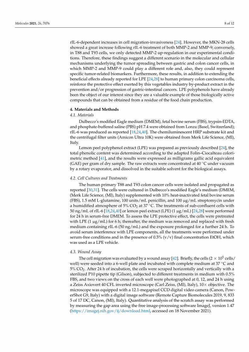
rIL-6-dependent increases in cell migration-invasiveness [24]. However, the MKN-28 cellsshowed a great increase following rIL-6 treatment of both MMP-2 and MMP-9; conversely,in T88 and T93 cells, we only detected MMP-2 up-regulation in our experimental condi-tions. Therefore, these findings suggest a different scenario in the molecular and cellularmechanisms underlying the tumor spreading between gastric and colon cancer cells, inwhich MMP-2 and MMP-9 could play a different role and, also, they could representspecific tumor-related biomarkers. Furthermore, these results, in addition to extending thebeneficial effects already reported for LPE [24,28] to human primary colon carcinoma cells,reinforce the protective effect exerted by this vegetables industry by-product extract in theprevention and/or progression of gastric-intestinal cancers. LPE polyphenols have alreadybeen the object of our interest since they are a valuable example of those biologically activecompounds that can be obtained from a residue of the food chain production.
4. Materials and Methods4.1. Materials
Dulbecco’s modified Eagle medium (DMEM), fetal bovine serum (FBS), trypsin-EDTA,and phosphate-buffered saline (PBS) pH 7.4 were obtained from Lonza (Basel, Switzerland);rIL-6 was produced as reported [18,24,40]. The chemiluminescent HRP substrate kit andthe centrifugal filter units (Amicon Ultra 10K) were obtained from Merk Life Science, (MI),Italy.
Lemon peel polyphenol extract (LPE) was prepared as previously described [24], thetotal phenolic content was determined according to the adapted Folin–Ciocalteau colori-metric method [41], and the results were expressed as milligrams gallic acid equivalent(GAE) per gram of dry sample. The raw extracts were concentrated at 40 ◦C under vacuumby a rotary evaporator, and dissolved in the suitable solvent for the biological assays.
4.2. Cell Cultures and Treatments
The human primary T88 and T93 colon cancer cells were isolated and propagated asreported [30,31]. The cells were cultured in Dulbecco’s modified Eagle’s medium (DMEM,(Merk Life Science, (MI), Italy) supplemented with 10% heat-inactivated fetal bovine serum(FBS), 1.5 mM L-glutamine, 100 units/mL penicillin, and 100 µg/mL streptomycin undera humidified atmosphere of 5% CO2 at 37 ◦C. The treatments of sub-confluent cells with50 ng/mL of rIL-6 [18,24,40] or lemon peel extract (LPE) (1 µg/mL) [24,28] were performedfor 24 h in serum-free DMEM. To assess the LPE protective effect, the cells were pretreatedwith LPE (1 µg/mL) for 6 h; thereafter, the medium was removed and replaced with freshmedium containing rIL-6 (50 ng/mL) and the exposure prolonged for a further 24 h. Toavoid serum interference with LPE components, all the treatments were performed underserum-free conditions and in the presence of 0.5% (v/v) final concentration EtOH, whichwas used as a LPE vehicle.
4.3. Wound Assay
The cell migration was evaluated by a wound assay [42]. Briefly, the cells (2 × 10⁵ cells/well) were seeded into a 6-well plate and incubated with complete medium at 37 ◦C and5% CO2. After 24 h of incubation, the cells were scraped horizontally and vertically with asterilized P10 pipette tip (Gilson), subjected to different treatments in medium with 0.5%FBS, and two views on the cross of each well were photographed at 0, 12, and 24 h usinga Zeiss Axiovert 40 CFL inverted microscope (Carl Zeiss, (MI), Italy), 10× objective. Themicroscope was equipped with a 12.1-megapixel CCD digital video camera (Canon, Pow-erShot G9, Italy) with a digital image software (Remote Capture Biomolecules 2019, 9, 8335 of 17 DC, Canon, (MI), Italy). Quantitative analysis of the scratch assay was performedby measuring the gap area using the free image-processing software ImageJ, version 1.47(https://imagej.nih.gov/ij/download.html, accessed on 18 November 2021).

Molecules 2021, 26, 7076 9 of 12
4.4. Matrigel Invasion Assay
The cell invasiveness was determined by cell invasion assay using a Boyden chambercoated with Matrigel (BioCoat Matrigel invasion chambers, cat. n. 354480, BD Bioscience,Bedford MA; 8 µm, BD Bioscience, (MI), Italy). The cell culture inserts were rehydratedand prepared as previously described [7,26]. Briefly, 2 × 10⁴ cells in 0.5 mL of DMEM with0.5% FBS were seeded in the upper chamber, and 750 µL medium with 5% FBS was placedin the lower chamber. After 24 h, cells in the upper chamber were removed with the cottonswab, and the cells at the bottom of the filters were fixed and stained with a Diff-Quickkit (cat. n. B4132-1A, Becton-Dickinson). After two washes with water, the inserts wereallowed to air dry, and phase-contrast images were captured as described above using anLD A-Plan 20 ×/0.30 Ph 1 objective. Cell invasive ability was determined by cell countingin five fields randomly selected per membrane.
4.5. RNA Extraction, Reverse Transcription (RT), and Quantitative Real-Time Polymerase ChainReaction (qPCR)
Total RNA was purified by the ultrapure TRizol reagent (Gibco BRL, Life TechnologyItalia, (MB), Italy) according to the manufacturer’s instructions. The concentration andpurity of RNA was determined spectrophotometrically by reading the absorbance at 260and 280 nm. Aliquots (1 µg) of total RNA were subjected to DNase I digestion (ThermoFisher Scientific, (MB), Italy) and reverse-transcribed using a SensiFAST TMcDNA synthesiskit (Cat. N. BIO-65054, Bioline, Italy) according to the manufacturer’s protocol. Real-timePCR was carried out using the PowerUP SYBR Green Master Mix (Thermo Fisher Scientific,(MB), Italy), the Quant Studio 7 Flex instrument (Thermo Fisher Scientific, Monza, Italy),and the fast gene-expression method with the following conditions: a first denaturationstep at 95 ◦C for 20 s, followed by 40 cycles at 95 ◦C for 1 s and 60 ◦C for 30; then, a meltingcurve analysis was performed, raising the temperature from 60 ◦C to 95 ◦C with a 0.5 ◦C/sincrease. Reactions were carried out in triplicate, and the 18S gene was used as an internalcontrol to normalize the variability in expression levels. The 2−∆∆CT (cycle threshold)method was used to calculate the results and mRNA expression levels were determined asfold-induction relative to control cells, set to 1. The human MMP-2 and 18S primers [24]were used for qPCR reactions that were carried out in triplicate; mRNA expression levelswere determined as fold-induction relative to control cells, set to 1.
4.6. Western Blotting Analysis
Cells were seeded in 6-well plates (2 × 10⁵ cells/well) and subjected to differenttreatments. Whole-cell protein extracts were prepared by lysing cells in 50 mM Tris-HClpH 8.0, 150 mM NaCl, 0.5% sodium deoxycholate, 0.1% SDS, 1 mM EDTA, 1% Igepal,1× protease inhibitor (cat. n. 11836153001, Roche Applied Science, Monza (MB), Italy),and a phosphatase-inhibitor cocktail (cat. n. 524627, Calbiochem, (MI), Italy). Proteinconcentration was determined by a colorimetric assay [43]. The culture medium washarvested and contaminating cells and debris were removed by centrifugation at 6000× gfor 20 min at 4 ◦C. The cell supernatant was then concentrated by approximately 20-foldby centrifugation at 5000× g using Ultra-4, PLGC Ultracell-PL Membrane, 10 kDa cut-off(cat. n. UFC 801024, Merck Millipore, (MI), Italy), at 4 ◦C. Whole-cell protein extracts(40 µg) and the appropriate volumes of concentrated conditioned media (correspondingto 30 µg total cell proteins) were heated at 95 ◦C for 5 min in Laemmli denaturing bufferin the presence of 2 M urea, and then loaded onto 12% reducing SDS-PAGE [44]. Afterelectrophoresis, proteins were transferred to a PVDF membrane (GE Healthcare LifeSciences, (MI), Italy) that was incubated with a Ponceau S solution for protein staining;the membrane was then exposed to the primary antibody as reported [45]. The followingantibodies were used: rabbit polyclonal antibody raised against phospho-STAT3 (Ser727) (1:1000, Cat. N. 9134, Cell Signaling Technology, Euroclone, (MI), Italy); STAT3(1:1000, Cat. N. 9139, Cell Signaling Technology, Euroclone, (MI), Italy); beta-tubulin IIIantibody (Cat. N. T2200, Sigma Aldrich, (MI), Italy) that was used as protein loading
Molecules 2021, 26, 7076 10 of 12
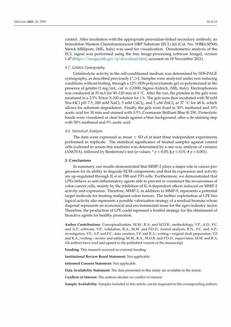
control. After incubation with the appropriate peroxidase-linked secondary antibody, anImmobilon Western Chemiluminescent HRP Substrate (ECL) kit (Cat. No. WBKLS0500,Merck Millipore, (MI), Italy) was used for visualization. Densitometric analysis of theECL signal was performed using the free image-processing software ImageJ, version1.47 (https://imagej.nih.gov/ij/download.html, accessed on 18 November 2021).
4.7. Gelatin Zymography
Gelatinolytic activity in the cell-conditioned medium was determined by SDS-PAGEzymography, as described previously [7,26]. Samples were analyzed under non-reducingconditions without boiling, through a 12% SDS-polyacrylamide gel co-polymerized in thepresence of gelatin (1 mg/mL, cat. n. G1890, Sigma-Aldrich, (MI), Italy). Electrophoresiswas conducted at 35 mA for 90–120 min at 4 ◦C. After the run, the proteins in the gels wererenatured in a 2.5% Triton X-100 solution for 1 h. The gels were then incubated with 50 mMTris-HCl pH 7.5, 200 mM NaCl, 5 mM CaCl2, and 5 µM ZnCl2 at 37 ◦C for 48 h, whichallows for substrate degradation. Finally, the gels were fixed in 30% methanol and 10%acetic acid for 30 min and stained with 0.5% Coomassie Brilliant Blue R-250. Proteolyticbands were visualized as clear bands against a blue background, after a de-staining stepwith 50% methanol and 5% acetic acid.
4.8. Statistical Analysis
The data were expressed as mean ± SD of at least three independent experimentsperformed in triplicate. The statistical significance of treated samples against controlcells (cultured in serum-free medium) was determined by a one-way analysis of variance(ANOVA), followed by Bonferroni’s test (p-values: * p < 0.05; § p < 0.01; # p < 0.001).
5. Conclusions
In summary, our results demonstrated that MMP-2 plays a major role in cancer pro-gression for its ability to degrade ECM components, and that its expression and activityare up-regulated through IL-6 in T88 and T93 cells. Furthermore, we demonstrated thatLPEs behave as anti-inflammatory agents able to prevent or counteract the invasiveness ofcolon cancer cells, mainly by the inhibition of IL-6-dependent effects induced on MMP-2activity and expression. Therefore, MMP-2, in addition to MMP-9, represents a potentialtarget molecule for treating malignant colon tumors. The further exploitation of LPE bio-logical activity also represents a possible valorization strategy of a residual biomass whosedisposal represents an economical and environmental issue for the agro-industry sector.Therefore, the production of LPE could represent a fruitful strategy for the obtainment ofbioactive agents for healthy promotion.
Author Contributions: Conceptualization, M.M., R.A. and M.D.R.; methodology, V.P., A.D., F.C.and A.P.; software, V.P.; validation, R.A., M.M. and P.D.D.; formal analysis, R.N., F.C. and A.P.;investigation, V.P., A.P. and F.C.; data curation, V.P. and R.A.; writing—original draft preparation, V.P.and R.A.; writing—review and editing, M.M., R.A., M.D.R. and P.D.D.; supervision, M.M. and R.A.All authors have read and agreed to the published version of the manuscript.
Funding: This research received no external funding.
Institutional Review Board Statement: Not applicable.
Informed Consent Statement: Not applicable.
Data Availability Statement: The data presented in this study are available in the article.
Conflicts of Interest: The authors declare no conflict of interest.
Sample Availability: Samples included in this article, can be requested to the corresponding authors.
Molecules 2021, 26, 7076 11 of 12
References1. Macrae, F.A. Colorectal Cancer: Epidemiology, Risk Factors, and Protective Factors. Available online: https://www.uptodate.
com/contents/colorectal-cancer-epidemiology-risk-factors-and-protective-factors (accessed on 24 May 2021).2. Thanikachalam, K.; Khan, G. Colorectal Cancer and Nutrition. Nutrients 2019, 11, 164. [CrossRef]3. De Rosa, M.; Rega, D.; Costabile, V.; Duraturo, F.; Niglio, A.; Izzo, P.; Pace, U.; Delrio, P. The biological complexity of colorectal
cancer: Insights into biomarkers for early detection and personalized care. Ther. Adv. Gastroenterol. 2016, 9, 861–886. [CrossRef]4. Jabłonska-Trypuc, A.; Matejczyk, M.; Rosochacki, S. Matrix metalloproteinases (MMPs), the main extracellular matrix (ECM)
enzymes in collagen degradation, as a target for anticancer drugs. J. Enzyme Inhib. Med. Chem. 2016, 31, 177–183. [CrossRef][PubMed]
5. Cui, N.; Hu, M.; Khalil, R.A. Biochemical and Biological Attributes of Matrix Metalloproteinases. Prog. Mol. Biol. Transl. Sci. 2017,147, 1–73. [CrossRef] [PubMed]
6. Sier, C.F.; Kubben, F.J.; Ganesh, S.; Heerding, M.M.; Griffioen, G.; Hanemaaijer, R.; van Krieken, J.H.; Lamers, C.B.; Verspaget,H.W. Tissue levels of matrix metalloproteinases MMP-2 and MMP-9 are related to the overall survival of patients with gastriccarcinoma. Br. J. Cancer 1996, 74, 413–417. [CrossRef]
7. Pagliara, V.; Adornetto, A.; Mammì, M.; Masullo, M.; Sarnataro, D.; Pietropaolo, C.; Arcone, R. Protease Nexin-1 affects migrationand invasion of C6 glioma cells through regulation of urokinase Plasminogen Activator and Matrix Metalloprotease-9/2. Biochim.Biophys. Acta 2014, 26, 2631–2644. [CrossRef]
8. Yang, T.; Zhang, J.; Zhou, J.; Zhu, M.; Wang, L.; Yan, L. Resveratrol inhibits Interleukin-6 induced invasion of human gastriccancer cells. Biomed. Pharmacother. 2018, 99, 766–773. [CrossRef]
9. Shiozaki, A.; Shimizu, H.; Ichikawa, D.; Konishi, H.; Komatsu, S.; Kubota, T.; Fujiwara, H.; Okamoto, K.; Iitaka, D.; Nakashima, S.;et al. Claudin 1 mediates tumor necrosis factor alpha-induced cell migration in human gastric cancer cells. World J. Gastroenterol.2014, 21, 17863–17876. [CrossRef]
10. Vainer, N.; Dehlendorff, C.; Johansen, J.S. Systematic literature review of IL-6 as a biomarker or treatment target in patients withgastric, bile duct, pancreatic and colorectal cancer. Oncotarget 2018, 9, 29820–29841. [CrossRef]
11. Kossakowska, A.E.; Edwards, D.R.; Prusinkiewicz, C.; Zhang, M.C.; Guo, D.; Urbanski, S.J.; Grogan, T.; Marquez, L.A.; Janowska-Wieczorek, A. Interleukin-6 regulation of matrix metalloproteinase (MMP-2 and MMP-9) and tissue inhibitor of metalloproteinase(TIMP-1) expression in malignant non-Hodgkin’s lymphomas. Blood 1999, 15, 2080–2089. [CrossRef]
12. Wang, Y.; Li, L.; Guo, X.; Jin, X.; Sun, W.; Zhang, X.; Xu, R.C. Interleukin-6 signaling regulates anchorage-independent growth,proliferation, adhesion and invasion in human ovarian cancer cells. Cytokine 2012, 59, 228–236. [CrossRef]
13. Wang, X.; Lee, S.O.; Xia, S.; Jiang, Q.; Luo, J.; Li, L.; Yeh, S.; Chang, C. Endothelial cells enhance prostate cancer metastasis viaIL-6→androgen receptor→TGF-β→MMP-9 signals. Mol. Cancer Ther. 2013, 12, 1026–1037. [CrossRef]
14. Castellana, B.; Aasen, T.; Moreno-Bueno, G.; Dunn, S.E.; Ramón y Caja, S. Interplay between YB-1 and IL-6 promotes themetastatic phenotype in breast cancer cells. Oncotarget 2015, 6, 38239–38256. [CrossRef]
15. Ashizawa, T.; Okada, R.; Suzuki, Y.; Takagi, M.; Yamazaki, T.; Sumi, T.; Aoki, T.; Ohnuma, S.; Aoki, T. Clinical significance ofinterleukin-6 (IL-6) in the spread of gastric cancer: Role of IL-6 as a prognostic factor. Gastric Cancer 2005, 8, 124–131. [CrossRef]
16. Taniguchi, K.; Karin, M. IL-6 and related cytokines as the critical lynchpins between inflammation and cancer. Semin. Immunol.2014, 26, 54–74. [CrossRef]
17. Turano, M.; Cammarota, F.; Duraturo, F.; Izzo, P.; De Rosa, M. A Potential Role of IL-6/IL-6R in the Development and Managementof Colon Cancer. Membranes 2021, 24, 312. [CrossRef]
18. Pagliara, V.; Parafati, M.; Adornetto, A.; White, M.C.; Masullo, M.; Grimaldi, M.; Arcone, R. Dibutyryl cAMP- or Interleukin-6-induced astrocytic differentiation enhances mannose binding lectin (MBL)-associated serine protease (MASP)-1/3 expression inC6 glioma cells. Arch. Biochem. Biophys. 2018, 653, 39–49. [CrossRef]
19. Hirano, T.; Ishihara, K.; Hibi, M. Roles of STAT3 in mediating the cell growth, differentiation and survival signals relayed throughthe IL-6 family of cytokine receptors. Oncogene 2000, 19, 2548–2556. [CrossRef]
20. Luo, J.; Yan, R.; He, X.; He, J. Constitutive activation of STAT3 and cyclin D1 overexpression contribute to proliferation, migrationand invasion in gastric cancer cells. Am. J. Transl. Res. 2017, 9, 5671–5677.
21. Johnson, D.E.; O’Keefe, R.A.; Grandis, J.R. Targeting the IL-6/JAK/STAT3 signalling axis in cancer. Nat. Rev. Clin. Oncol. 2018, 15,234–248. [CrossRef]
22. Wang, C.; Wu, W.K.; Liu, X.; To, K.F.; Chen, G.G.; Yu, J.; Ng, E.K. Increased serum chemerin level promotes cellular invasivenessin gastric cancer: A clinical and experimental study. Peptides 2014, 51, 131–138. [CrossRef]
23. Zhao, G.; Zhu, G.; Huang, Y.; Zheng, W.; Hua, J.; Yang, S.; Zhuang, J.; Ye, J. IL-6 mediates the signal pathway of JAK-STAT3-VEGF-C promoting growth, invasion and lymphangiogenesis in gastric cancer. Oncol. Rep. 2016, 35, 1787–1795. [CrossRef][PubMed]
24. Pagliara, V.; Nasso, R.; Di Donato, P.; Finore, I.; Poli, A.; Masullo, M.; Arcone, R. Lemon Peel Polyphenol Extract ReducesInterleukin-6-Induced Cell Migration, Invasiveness, and Matrix Metalloproteinase-9/2 Expression in Human Gastric Adenocarci-noma MKN-28 and AGS Cell Lines. Biomolecules 2019, 9, 833. [CrossRef] [PubMed]
25. Nistal, E.; Fernández-Fernández, N.; Vivas, S.; Olcoz, J.L. Factors determining colorectal cancer: The role of the intestinalmicrobiota. Front. Oncol. 2015, 5, 220. [CrossRef] [PubMed]

Molecules 2021, 26, 7076 12 of 12
26. Arcone, R.; Palma, M.; Pagliara, V.; Graziani, G.; Masullo, M.; Nardone, G. Green tea polyphenols affect invasiveness of humangastric MKN-28 cells by inhibition of LPS or TNF-α induced Matrix Metalloproteinase-9/2. Biochim. Open 2016, 3, 56–63.[CrossRef] [PubMed]
27. Fresco, P.; Borges, F.; Diniz, C.; Marques, M.P. New insights on the anticancer properties of dietary polyphenols. Med. Res. Rev.2006, 26, 747–766. [CrossRef] [PubMed]
28. Di Donato, P.; Taurisano, V.; Tommonaro, G.; Pasquale, V.; Jiménez, J.M.S.; de Pascual, S.; Poli, T.A.; Nicolaus, B. BiologicalProperties of Polyphenols Extracts from Agro Industry’s Wastes. Waste Biomass Valorization 2018, 9, 1567–1578. [CrossRef]
29. Nasso, R.; Pagliara, V.; D’Angelo, S.; Rullo, R.; Masullo, M.; Arcone, R. Annurca Apple Polyphenol Extract Affects Acetyl-Cholinesterase and Mono-Amine Oxidase In Vitro Enzyme Activity. Pharmaceuticals 2021, 14, 62. [CrossRef]
30. Turano, M.; Costabile, V.; Cerasuolo, A.; Duraturo, F.; Liccardo, R.; Delrio, P.; Pace, U.; Rega, D.; Dodaro, C.A.; Milone, M.; et al.Characterisation of mesenchymal colon tumour-derived cells in tumourspheres as a model for colorectal cancer progression. Int.J. Oncol. 2018, 53, 2379–2396. [CrossRef]
31. Cammarota, F.; Conte, A.; Aversano, A.; Muto, P.; Ametrano, G.; Riccio, P.; Turano, M.; Valente, V.; Delrio, P.; Izzo, P.; et al.Lithium chloride increases sensitivity to photon irradiation treatment in primary mesenchymal colon cancer cells. Mol. Med. Rep.2020, 3, 1501–1508. [CrossRef]
32. Costabile, V.; Duraturo, F.; Delrio, P.; Rega, D.; Pace, U.; Liccardo, R.; Rossi, G.B.; Genesio, R.; Nitsch, L.; Izzo, P.; et al. Lithiumchloride induces mesenchymal-to-epithelial reverting transition in primary colon cancer cell cultures. Int. J. Oncol. 2015, 46,1913–1923. [CrossRef] [PubMed]
33. Lin, M.T.; Lin, B.R.; Chang, C.C.; Chu, C.Y.; Su, H.J.; Chen, S.T.; Jeng, Y.M.; Kuo, M.L. IL-6 induces AGS gastric cancer cell invasionvia activation of the c-Src/RhoA/ROCK signaling pathway. Int. J. Cancer 2007, 120, 2600–2608. [CrossRef] [PubMed]
34. Parsons, S.L.; Watson, S.A.; Collins, H.M.; Griffin, N.R.; Clarke, P.A.; Steele, R.J. Gelatinase (MMP-2 and -9) expression ingastrointestinal malignancy. Br. J. Cancer 1998, 78, 1495–1502. [CrossRef] [PubMed]
35. Wei, D.; Li, H.; Zhang, Y.; Yang, H.; Guo, M.; Li, L.; Liu, T. Matrix metalloproteinase 2 promotes cell growth and invasion incolorectal cancer. Acta Biochim. Biophys. Sin. 2011, 43, 840–848. [CrossRef]
36. Farinetti, A.; Zurlo, V.; Manenti, A.; Coppi, F.; Mattioli, A.V. Mediterranean diet and colorectal cancer: A systematic review.Nutrition 2017, 44, 83–88. [CrossRef] [PubMed]
37. Fung, T.T.; McCullough, M.L.; Newby, P.K.; Manson, J.E.; Meigs, J.B.; Rifai, N.; Willett, W.C.; Hu, F.B. Diet-quality scores andplasma concentrations of markers of inflammation and endothelial dysfunction. Am. J. Clin. Nutr. 2005, 82, 163–173. [CrossRef]
38. Nichenametla, S.N.; Taruscio, T.G.; Barney, D.L.; Exon, J.H. A review of the effects and mechanisms of polyphenolics in cancer.Crit. Rev. Food Sci. Nutr. 2006, 46, 161–183. [CrossRef] [PubMed]
39. Rose-John, S. IL-6 Trans-Signaling via the Soluble IL-6 Receptor: Importance for the Pro-Inflammatory Activities of IL-6. Int. J.Biol. Sci. 2012, 8, 1237–1247. [CrossRef]
40. Arcone, R.; Pucci, P.; Zappacosta, F.; Fontaine, V.; Malorni, A.; Marino, G.; Ciliberto, G. Single-step purification and structuralcharacterization of human interleukin-6 produced in Escherichia coli from a T7 RNA polymerase expression vector. Eur. J.Biochem. 1991, 198, 541–547. [CrossRef]
41. Singleton, V.L.; Rossi, J.A. Colorimetry of total phenolics with phosphomolybdic phosphotungstic acid reagents. Am. J. Enol.Viticult. 1965, 16, 144–158.
42. Tam, J.C.; Lau, K.M.; Liu, C.L.; To, M.H.; Kwok, H.F.; Lai, K.K.; Lau, C.P.; Ko, C.H.; Leung, P.C.; Fung, K.P.; et al. The in vivo andin vitro diabetic wound healing effects of a 2-herb formula and its mechanisms of action. J. Ethnopharmacol. 2011, 134, 831–838.[CrossRef] [PubMed]
43. Bradford, M.M. A rapid and sensitive method for the quantification of microgram quantities of proteins utilizing the principle ofprotein dye binding. Ann. Biochem. 1976, 72, 248–254. [CrossRef]
44. Laemmli, U.K. Cleavage of structural proteins during the assembly of the head of bacteriophage T4. Nature 1970, 227, 680–685.[CrossRef] [PubMed]
45. Adornetto, A.; Pagliara, V.; Di Renzo, G.; Arcone, R. Polychlorinated biphenyls impair dibutyryl cAMP-induced astrocyticdifferentiation in rat C6 glial cell line. FEBS Open Bio 2013, 29, 459–466. [CrossRef]
Copyright © 2022 FDOKUMEN