Critical role of extracellular vesicles in modulating the cellular effects of cytokines
Interleukin 7 Plays a Role in T Lymphocyte ApoptosisInhibition Driven by Mesenchymal Stem Cell withoutFavoring Proliferation and Cytokines SecretionMarilia Normanton1,5, Heliene Alvarenga1,5, Nelson Hamerschlak2, Andreza Ribeiro3, Andrea Kondo4,
Luiz Vicente Rizzo1, Luciana Cavalheiro Marti1,5*
1Hospital Israelita Albert Einstein, Instituto Israelita de Ensino e Pesquisa Albert Einstein (IIEP-AE), Sao Paulo, SP, Brasil, 2Hospital Israelita Albert Einstein, Bone Marrow
Transplant Program, Sao Paulo, SP, Brasil, 3Hospital Israelita Albert Einstein, Oncology Department, Sao Paulo, SP, Brasil, 4Hospital Israelita Albert Einstein, Blood Bank
Department, Sao Paulo, SP, Brasil, 5 Programa de Pos-graduacao em Alergia e Imunopatologia, Faculdade de Medicina da Universidade de Sao Paulo (FMUSP), Sao Paulo,
SP, Brasil
Abstract
Since 2004, when a case report describing the use of human mesenchymal stem cells (hMSCs) infusion as a therapy forGVHD after bone marrow transplantation, a new perspective in MSC function emerged. Since then hMSCsimmunomodulatory potential became the target of several studies. Although great progress has been made in ourunderstanding of hMSCs, their effect on T cell remains obscure. Our study has confirmed the already described effect ofhMSCs on lymphocytes proliferation and survival. We also show that the impairment of lymphocyte proliferation andapoptosis is contact-independent and occurs in a prostaglandin-independent manner. A potential correlation between IL-7and hMSCs effect is suggested, as we observed an increase in IL-7 receptors (CD127) on lymphocyte membrane in MSCpresence. Additionally, blocking IL-7 in hMSCs-lymphocytes co-cultures increased lymphocytes apoptosis and we also havedemonstrated that hMSCs are able to produce this interleukin. Moreover, we found that during Th1/Th17 differentiationin vitro, hMSCs presence leads to Th1/Th17 cells with reduced capacity of INF-y and IL-17 secretion respectively, regardlessof having several pro-inflammatory cytokines in culture. We did not confirm an increment of Treg in these cultures, but areduced percentage of INF-y/IL-17 secreting cells was observed, suggesting that the ratio between anti and pro-inflammatory cells changed. This changed ratio is very important to GvHD therapy and links hMSCs to an anti-inflammatoryrole. Taken together, our findings provide important preliminary results on the lymphocyte pathway modulated by MSCsand may contribute for developing novel treatments and therapeutic targets for GvHD and others autoimmune diseases.
Citation: Normanton M, Alvarenga H, Hamerschlak N, Ribeiro A, Kondo A, et al. (2014) Interleukin 7 Plays a Role in T Lymphocyte Apoptosis Inhibition Driven byMesenchymal Stem Cell without Favoring Proliferation and Cytokines Secretion. PLoS ONE 9(9): e106673. doi:10.1371/journal.pone.0106673
Editor: Derya Unutmaz, New York University, United States of America
Received May 30, 2014; Accepted July 30, 2014; Published September 3, 2014
Copyright: � 2014 Normanton et al. This is an open-access article distributed under the terms of the Creative Commons Attribution License, which permitsunrestricted use, distribution, and reproduction in any medium, provided the original author and source are credited.
Data Availability: The authors confirm that all data underlying the findings are fully available without restriction. All relevant data are within the paper and itsSupporting Information files.
Funding: Fundacao de Amparo a Pesquisa do Estado de Sao Paulo (FAPESP) funded this project through the grant number 2011/02027-4. The funders had norole in study design, data collection and analysis, decision to publish, or preparation of the manuscript.
Competing Interests: The authors have declared that no competing interests exist.
* Email: [email protected]
Introduction
The immunomodulatory activity of mesenchymal stem cells is at
the basis of their therapeutic use in graft versus host disease
(GVHD) which was first reported by Le Blanc et al. in 2004. After
receiving human mesenchymal stem cells (hMSCs) infusions, the
patient had significant clinical improvement without showing
adverse reactions [1]. Since then, several studies have addressed
the potential use of hMSCs to down regulate immune responses.
Mesenchymal stem cells (MSCs), also known as mesenchymal
stromal cells, are adult non-hematopoietic stem cells that were first
described as plastic-adherent fibroblast-like cells capable of self-
renewal and of differentiation into other cell type [2,3].
In 2006, the International Society for Cellular Therapy
proposed a set of phenotypic and functional criteria to define
MSCs [4], which isolation is possible from a variety of sources [5–
7]. However, the bone marrow still is the most studied source of
hMSCs, mainly because it is the one used in cell therapy clinical
trials. Although MSC immunomodulation occurs by distinct cell
signaling pathways in humans and in animal models, an effector
activity common to mouse, monkey and hMSCs is the inhibition
of lymphocyte proliferation. Several causes of this inhibition have
been described such as depletion of tryptophan by indoleamine-
2,3-dioxigenase (IDO), up-regulation of prostaglandins secretion,
oxidative stress or reactive oxygen species and nitrogen radicals
(mainly in animal models) or, in humans, the action of IFN-c [8,9].
The proliferative responses of T cells, whether stimulated by
alloantigen [10–12], antigenic peptides [13], mitogens [10,14] or
by anti-CD3/CD28 antibodies, were suppressed in the presence of
MSCs suggesting non-specificity of mechanisms.
However, as bone marrow derived MSCs reside in hematopoi-
etic stem cell niches they primarily affect the differentiation and
maintenance of hematopoietic stem cells. Most likely MSCs
influence the survival of their neighbor cells, either by contact-
dependent interactions or by secreting soluble molecules [15,16].
PLOS ONE | www.plosone.org 1 September 2014 | Volume 9 | Issue 9 | e106673
Recently it was shown that MSCs secrete multiple cytokines,
promote angiogenesis and have distinct effects on chemotaxis and
apoptosis [17].
Among the cytokines that affect T cell proliferation, interleukin-
7 (IL-7), is a cytokine secreted by stromal cells that is critical for the
development, homeostatic proliferation and survival of naıve T
cells. The absence of IL7 or any of its proximal signaling
components leads to a severe combined immunodeficiency
(SCID). IL-7 is detected by a two-part receptor on lymphocytes,
consisting by a c-chain that is shared by multiple cytokines and a
more specific receptor IL-7Ra. IL-7R signals through the Jak/
STAT and PI3K/Akt signaling pathways, both of which are
known to have effects on cell survival, growth and metabolism.
Although, the specific role and mechanism by which IL-7R may
influence each of these processes in vivo, has not been fully
determined [18–20].
Given that relatively little is known on the mechanisms that
underlie the control of GVHD and T cell proliferation by MSCs in
humans, we undertook the present investigation. To this end we
studied the effect of hMSCs on phytohemagglutinin-stimulated
lymphocytes cultures. Phytohemagglutinin (PHA) is a mitogen that
stimulates very robust activation and proliferation of T cells. We
evaluated the cytokine secretion patterns by lymphocytes when in
contact with MSCs in culture, their effect on activation-induced
lymphocyte cell death (AICD) rates, and the participation of
prostaglandins and IL-7 in the death-signaling pathway. Because
Th1/Th-17 and INF-c/IL-17 participate in GVHD-inflammation
[21] and given the beneficial effect of hMSCs infusion at reducing
the intensity of GVHD, we also investigated whether MSCs would
influence Th1 and Th17 differentiation or INF-c and IL-
17 secretion.
Briefly, we found that hMSCs suppression of lymphocyte
proliferation and apoptosis did not depend on cell-contact and was
not mediated by prostaglandins. In addition, depletion of IL-7 in
co-cultures increased apoptosis suggesting that T cell survival is
promoted by this cytokine. Furthermore, hMSCs reduced INF-cand IL-17 synthesis and the frequency of Th1/Th17 cells in co-
culture with no increment of Treg cells. Our findings pave the way
to a better understanding on how hMSCs act on lymphocytes and
may contribute for further applications of these powerful
therapeutic cells.
Materials and Methods
Cells isolationhMSCs were obtained from health bone marrow donors, by
washing the cells trapped in the dischargeable collection filter as
previously described by Deus et al [22]. The lymphocytes were
obtained from health donor’s peripheral blood following the
signing of informed consent (Approved by Hospital Albert Einstein
Ethical Committee -10/1412; CAAE: 06592712.4.0000.0071).
Peripheral blood cells were diluted 1:3 (vol/vol) with Phosphate
Buffered Saline (PBS). Next, the suspension were transferred to a
50 mL conical tube containing 20 mL of Ficoll-Paque 1.077
density (GE Healthcare, United Kingdom) and centrifuged
(30 min/500 g) at 22uC. Then, the cells from the interface were
collected, ressuspended and centrifuged again (5 min/500 g). The
supernatant was discarded and cells ressuspended with DMEM-
LG, supplemented with 10% FBS, 1% antibiotic-antimycotic and
2 mM L-glutamine, in order to achieve 16106 cells/mL. In order
to deplete monocytes the cells were placed in a petri dish
previously coated with human albumin 20% (Sandoz, Germany),
next the cells were incubated for 1 hour in humidified 5% CO2
incubators at 37uC, the non-adherent cells were removed, and the
efficiency of depletion was verified by flow cytometry. The
mentioned culture media, supplements and PBS were bought from
Gibco (Carlsbad, CA).
Bone marrow collection filters were washed with 20 mL
DMEM-LG; the cells were diluted 1:3 (vol/vol) with Phosphate
Buffered Saline (PBS). Next, the suspension were transferred to a
50 mL conical tube containing 20 mL of Ficoll-Paque 1.077
density (GE Healthcare, United Kingdom) and centrifuged
(30 min/500 g) at 22uC. Then, the cells from the interface were
collected, ressuspended and centrifuged again (5 min/500 g). The
supernatant was discarded and cells ressuspended with DMEM-
LG, supplemented with 10% FBS, 1% antibiotic-antimycotic,
2 mM L-glutamine, in order to achieve 16105 cells/mL. The
mentioned culture media, supplements and PBS were bought from
Gibco (Carlsbad, CA). Next, cells (5 mL) were cultivated into
25 cm2 flasks for 48 hours and maintained in humidified 5% CO2
incubators at 37uC to favor the attachment of the hMSCs to the
flask bottom. The non-adherent cells were removed, the adherent
layer was washed twice with DMEM-LG, than maintained in
culture until the 4th passage.
hMSCs were characterized by flow cytometry and checked for
the ability to differentiate in osteocytes, chondrocytes or adipocytes
as previously published by our group and briefly described below
[22].
Mesenchymal stem cell characterization by flowcytometric expression markers
Cells from passage four were used to analysis of cell surface
markers. The cells were washed with PBS, and then detached from
the plastic with TryPLE (Gibco Carlsbad, CA). Next, the cells
were stained for CD106-FITC (clone: 51-10C9), CD73-PE (clone:
AD2), CD34-PE (clone: My10), CD105-PE-CF594 (clone: 266),
CD90-PE-Cy7 (clone: SE10), CD29-APC (clone: MAR04), CD14-
Alexa 700 (clone: M5E2) from BD Pharmingen (San Diego – CA),
CD44-PerCPCy5 (clone: G44-26), HLA-DR-APC-H7 (clone:
G46-6) from Biosciences (San Jose – CA), CD45-V500 (clone:
H130) and CD31-V450 (clone: WM59) from Biolegend (San
Diego – CA), and for fluorescence minus one (FMO). After
staining, the tubes were incubated at room temperature for
30 minutes, followed by a wash step; the cell pellet was
ressuspended and measurements were performed using FAC-
SARIA equipment (BD Biosciences).
The human mesenchymal stem cells (hMSCs) used in our assays
fulfilled this identity criteria established by the International
Society for Cell Therapy (ISCT), as shown by Figure S1.
Mesenchymal stem cell characterization by adipocyte,chondrocyte and osteocyte differentiation
After the establishment of hMSCs cultures on the fourth
passage, the cells were differentiated into adipocytes, osteoblasts,
and chondrocytes.
Adipogenesis was induced by addition of an adipogenic medium
to hMSCs culture, this medium was comprised by Alpha-MEM
supplemented with 10% FBS, 1 mm dexamethasone, 100 mg/mL
3-Isobutyl-1-methylxanthine, 10 mg/mL insulin and 100 mM
indomethacin. The adipogenic medium was changed every other
day for 3 weeks. After 3 weeks in culture the cells were stained to
evidence lipid droplets formation. The cells were fixed in 4%
paraformaldehyde for 30 minutes, washed, dehydrated in 60%
isopropanol for 2 to 5 minutes, and stained with 0.5% Oil Red O
(O-0625) in 100% isopropanol previously diluted in water.
Osteogenesis was induced by addition of an osteogenic medium
which was comprised by Alpha-MEM supplemented with 10%
IL-7 Plays a Role in MSCs-Driven Apoptosis Inhibition of T Lymphocytes
PLOS ONE | www.plosone.org 2 September 2014 | Volume 9 | Issue 9 | e106673
FBS, 1 mm dexamethasone, 2 mg/mL ascorbic acid and 10 mm b-
glycerophosphate. The osteogenic medium was changed every
other day for 3 weeks. After 3 weeks in culture the cells were
stained with Alizarin Red to evidence the calcium deposition.
Next, the cells were fixed in 4% paraformaldehyde for 30 minutes,
washed with distilled water, stained with Alizarin Red (2 g in
100 mL of distillated water) pH 4.2 (A5533) for 5 to 10 minutes
and thoroughly washed.
Chondrogenesis was induced by addition of a chondrogenic
medium which was comprised by Alpha-MEM supplemented with
10% FBS, 1 mm dexamethasone, 2 ug/mL ascorbic acid,
6,25 ug/mL insulin, and 10 ng/mL TGF-b. Chondrogenic
medium was changed every other day for 3 weeks. After 3 weeks
in culture the cells were stained with toluidine blue for evidence
the proteoglycans enriched matrix. For toluidine blue, the cells
were fixed with ethanol 70% for 1 minute, ethanol 90% for
1 minute and absolute ethanol for 1 minute, then toluidine blue
was added (1 g toluidine blue, 1 g sodium borate/100 mL of
water) (198161).
The mentioned culture media and FBS were bought from
Gibco (Carlsbad, CA) and the other reagents were all from Sigma
(St Louis, MO).
The human mesenchymal stem cells (hMSCs) used in our assays
fulfilled this identity criteria established by the International
Society for Cell Therapy (ISCT), as shown by Figure S2.
Cell culturehMSCs at 4th passage were transferred to a 6-well plate and
maintained in culture until reaching 80% confluence. The
lymphocytes were suspended in Xvivo15 (Cambrex, Walkersville,
MD), supplemented with 1% human pooled serum AB (Life
Technologies, Carlsbad, CA) and 1% antibiotic-antimycotic
(Gibco, Carlsbad, CA) then stimulated with 1 mg of phytohemag-
glutinin (PHA)/16106 lymphocytes. Approximately 16106 lym-
phocytes were added to 16105 hMSCs. The co-cultures were
incubated for 24, 48 and/or 72 hours before assessing for
lymphocyte apoptosis/proliferation. Lymphocytes were also co-
cultivated with hMSCs and PHA separated by transwell 0.2 mm -
anopore membrane (Nunc Kamstrup, Denamark). Control
cultures of lymphocytes without PHA stimulation were also
included in all experiments.
Mixed leucocyte reaction (MLR)Dendritic cells were derived from monocytes selected for CD14
by magnetic selection column (Myltenyi Biotec, Bergisch Glad-
bach, Germany). The CD14+ cell population was dispensed into
six-well plates containing X-vivo 15 medium (Cambrex) supple-
mented with antibiotic-antimycotics (Gibco). To generate imma-
ture dendritic cells (DCs), the cells were cultured in the presence of
20 ng/mL recombinant IL-4 (rIL-4) and 50 ng/ml rGM-CSF
(both from R&D Systems, Minneapolis, MN) for six days
(indicated as D6 cells). Mature DCs were obtained after 24 h
(D7) after stimulation of the immature DC culture with 10 ng/mL
rTNF-a (R&D System) plus 0.01 mmol/L of PGE2 (Sigma) [23].
In order to set up the MLR, matured DCs were irradiated at
1,500 rads and co-cultured with allogeneic lymphocytes at the
concentration of 1 DC:100 Lymphocytes (16104:16106 respec-
tively) in presence or absence of 16105 allogeneic hMSCs. The
proliferation rates were determined after four days of co-culture.
Lymphocyte proliferation was measured by KI-67 nuclear staining
on CD3+ gated population. The lymphocytes were gated based on
SSC vs FSC, followed by CD3 gating; the selected events were
analyzed in a second plot for KI-67 expression. The proliferation
negative control was lymphocytes without allogeneic DCs
stimulation.
Treatment of cultures with Indomethacin, interleukin 7 oranti-IL-7 antibody
Co-cultures of PHA-stimulated lymphocytes and hMSCs were
treated with different concentrations of Indomethacin (5, 25, 50
and 100 ng/mL) in order to verify if the blocking of prostaglandin
synthesis would interfere with the anti-proliferative effects of
hMSCs. Co-cultures of PHA-stimulated lymphocytes and hMSCs
were also treated with different concentrations of polyclonal
neutralizing goat anti-human IL-7 antibody (10 and 20 ul) (R&D
Systems) or with rIL-7 (10 and 20 ng/mL, R&D systems), in order
to verify a possible activity of this cytokine on the anti-apoptotic
effect of hMSCs on lymphocytes.
Th1 and Th17 differentiationNaive T lymphocytes derived from healthy volunteers periph-
eral blood, (CD4, CD45RA), were separated by indirect cell
targeting by using Naive CD4+ T Cell Isolation Kit II (Miltenyi
biotec) according manufactures instruction. Naıve T cells (80%
purity) were stimulated with CD3/CD28 microspheres (Life
Technologies), and cultured for 5 days, treated with 10 ng/mL
of IL1b, IL6, IL23 and 5 ng/mL of TGFb (R&D Systems) for Th1
and with monoclonal antibodies (mAb) anti-IFN-y (Clone:
25723.11) and anti-IL2 (Clone: 5344.111) at 1:50 dilution bought
from BD Biosciences for Th17. This protocol of naıve T cells to
Th1/Th17 differentiation was carried out in presence or absence
of hMSCs.
T-cell apoptosis and proliferation assayApoptosis and proliferation were measured by flow cytometric
assays. Lymphocytes were stained according to manufactures
instructions, using two different combinations: First combination
was Annexin V-FITC, propidium iodide-PI and CD3-APC (clone:
HIT3a) from BD Pharmingen, (CA, San Diego). The second
combination used was KI-67-FTIC (clone: MOPC-21), Caspase-3
activated-PE (clone: C92-605), CD4-APC (clone: RPA-T4) from
BD Pharmingen and CD3-Per CP Cy5.5 (clone: SK7) from BD
Biosciences. The corresponding isotype controls were used as
control for both staining protocols.
Briefly, for testing apoptosis the lymphocytes were ressuspen-
dend with Annexin-V binding buffer (BD Biosciences) and stained
for Annexin V, PI and CD3 for 15 minutes; the samples were
acquired for flow cytometric analysis within 30 minutes after
finishing incubation. For proliferation and apoptosis, staining for
surface markers as CD3 and CD4 was performed and the cells
were incubated for 30 minutes following fixation using BD FACS
Lysing solution (BD Biosciences). Next, the cells were permeabi-
lized using BD FACS Permeabilization solution 2 (BD Biosciences)
and the intracellular staining was performed. After washing, the
cell suspension was submitted to FACS analysis.
T-cell BrdU proliferation assayLymphocytes were stimulated with 1 mg of phytohemagglutinin
(PHA)/16106 lymphocytes. Approximately 16106 lymphocytes
were added to 16105 hMSCs or cultured alone. These cultures
were incubated for 48, and then pulsed with 10 mM of BrdU (BD
Pharmingen)/16106 lymphocytes for another 24 hours. The
lymphocytes were first stained for surface antigens such as CD3-
PerCP Cy5.5, CD4-PE, then fixed and permeabilized. Next the
cells were treated for 1 hour with DNase in order to expose the
incorporated BrdU. Next, the staining with anti-BrdU-FITC was
IL-7 Plays a Role in MSCs-Driven Apoptosis Inhibition of T Lymphocytes
PLOS ONE | www.plosone.org 3 September 2014 | Volume 9 | Issue 9 | e106673
performed following manufactures’ instructions of FITC BrdU
flow kit (BD Pharmingen).
Lymphocyte proliferation was measured by BrdU-FITC stain-
ing on CD3+ gated population. The proliferation negative controls
were lymphocytes without PHA stimulation.
We also have used BD Trucount tubes (BD Biosciences) to
determine the absolute number of proliferating T lymphocyte.
The cells absolute number was calculated by using the following
formula:
number of events in the T cell gate
number of events in absolute count bead region
|number of beads per test
test volume~absolute count of cells
Intracellular IL-7 detection on mesenchymal stem cellsTo determine whether hMSCs produce IL-7 when in co-
cultivation with lymphocytes, the cultures were maintained for
24 hours followed by BFA addition for 4 hours. hMSCs were
stained with anti-CD105-PE (clone: 43A3; Biolegend – San Diego
- CA). After 30 minutes of incubation the cells were washed, next
fixed with lyses buffer (lyses buffer – BD Biosciences, San Diego,
CA) followed by permeabilization (permeabilization solution 2–
BD Biosciences). Next, we proceed the intracellular staining with
polyclonal goat anti-human IL-7 antibody, followed by the
secondary anti-goat FITC, the same were performed using isotype
control.
Dendritic cells characterizationDendritic cells were characterized by flow cytometry regarding
there phenotype and maturation status according to the intensity
of CD14, CD209, CD80, CD83 and CD86 expression as showed
in Figure S3. The staining of the cells was performed using
commercially available monoclonal antibodies (mAbs) according
to the manufacturer’s instructions. Briefly, the cells were stained
for surface markers with the selected mAbs and incubated in the
dark for 30 min at room temperature, followed by flow cytometry
acquisition. The mAbs used to evaluate DC and its maturation
were as follows: CD14-FITC (clone: M5E2), CD80-PE (clone:
L307.4), CD83-PE (clone: HB15e), CD86-PE (clone: 2331), and
CD209-PE (clone: DCN46) all purchased from BD Pharmingen,
HLA-DR-PerCP-Cy5.5 (clone: L243) from BD Biosciences and
corresponding isotype control.
Th17 cell and regulatory T cell characterizationT cells in co-culture were characterized by flow cytometry assay
and stained according to manufactures instruction. Briefly, for
Th17 cells, the staining for surface markers CD4-APC (clone:
RPA-T4), CD8-PE (clone: RPA-T8), CD45RA-FITC (clone:
HI100), CD45RO-PE (clone: UCHL1), CD3-APC (clone: HIT3a)
or CD3-Per CP-Cy5.5 (clone: SK7) from BD Pharmingen, CD4-
APC-Cy7 (clone: SK3) and CD45 Per CP-Cy5.5 (clone: 2D1)
from BD Biosciences, CCR5-FITC (clone: 45502) and CCR6-PE
(clone: 53103) from R&D System, was performed and cells were
incubated for 30 minutes following fixation using BD FACS
Lysing solution (BD Biosciences). Next, cells were permeabilized
using BD FACS Permeabilization solution II (BD Biosciences) and
the intracellular staining was performed with Tbet-Per CP-Cy5.5
(clone: 4-46), anti-IL17A-FITC (clone: N49-653) from BD
Pharmingen, RORct-PE (clone: AFKJS-9) from eBiosciences,
IFN-c-FITC (clone: 25723.11) from BD Biosciences and the
corresponding isotype control.
For Treg cells, staining for surface markers CD45-PE-Cy7
(clone: HI-30), CD3-APC-Cy7 (clone: SK-7), CD4-FITC (clone:
RPA-T4), CD25-APC (Clone: M-A251) and CD127-PercP-Cy5.5
(clone: HIL-7R-M21), was performed and cells were incubated for
30 minutes following fixation/permeabilization using Foxp3
staining Kit (BD Biosciences, San Jose, CA) and intracellular
staining FoxP3-PE (clone: 259D/C7). FMO control was per-
formed for CD25-APC and Foxp3-PE.
T-cell surface expression of CD127CD127 was measured by flow cytometric assays. Lymphocytes
were stained according to manufacture instructions for CD3-FITC
(clone: HIT3a), CD127-PercP-Cy5.5 (clone: HIL-7R-M21), CD4-
APC (clone: RPA-T4) from BD Pharmingen and corresponding
isotype controls. Briefly, the staining for surface markers CD3,
CD4 and CD127 was performed and cells were incubated for
30 minutes followed by fixation using BD FACS Lysing solution
BD Biosciences.
FACS analysisAnalysis of stained cells was performed using the BD
FACSARIA equipment (BD Biosciences) and at least 10,000
events were acquired. The results were analyzed by FACSDIVA
and/or FlowJo software (Treestar, Eugene, OR).
Statistical AnalysisGraphPad Prisma Program was used to perform the statistical
analyses. Statistical significance was calculated by Student’s t-test
or ANOVA with Bonferroni correction for multiple comparisons;
p,0.05 was considered significant.
Results
Mesenchymal stem cell reduces the proliferation of PHAand allogeneic DC stimulated T lymphocytes, and thissuppression is not contact-dependent
CD3+ T cells PHA-stimulated and co-cultivated with hMSCs
show 15% less cells expressing KI-67 compared with similarly
stimulated cultures without hMSCs (Figure 1A), in absolute
numbers, the cells expressing KI-67 decreased almost 50% from
(3.0610561.36105) up to (1.7610560.96105) (Figure 1B). Sup-
pression of T cell proliferation by hMSCs (of the order of 50%)
was also observed in allo DC-stimulated lymphocyte cultures
(Figure 1C) compared to allogeneic cultures in the absence of
hMSCs, showing that hMSCs play a role in T lymphocytes clonal
expansion, either to a specific or nonspecific stimulus response.
The flow cytometry approach and data that originated the figures
are exemplified in Figure S4.
hMSCs were maintained separated from lymphocytes in
transwell cultures or allowed direct contact with lymphocytes in
culture wells. PHA-stimulated proliferation was significantly
reduced in both culture settings showing that the suppressive
effect on lymphocytes exerted by hMSCs did not require contact
between these cell populations (Figure 1D).
To confirm that the cells have not only a low cycling percentage
by lower expression of KI-67 but also a reduced number of
division in hMSCs presence, we also performed a proliferation
assay by other technique, measuring BrdU incorporation, and we
found that a reduction on T cell division by hMSCs presence (of
the order of 50%) was also observed in both percentage and
absolute number of cells (Figures 1E–L).
IL-7 Plays a Role in MSCs-Driven Apoptosis Inhibition of T Lymphocytes
PLOS ONE | www.plosone.org 4 September 2014 | Volume 9 | Issue 9 | e106673

Figure 1. hMSCs reduces proliferation of stimulated lymphocytes in a contact-independent manner. (A–B) A significant proliferationreduction of PHA stimulated lymphocytes (CD3) (38.58612.39%) assessed by expression of KI-67 was observed in presence of hMSCs (CD3: hMSC)(24.39612.39%), the same was observed when absolute number KI-67 expressing cells was evaluated decreasing from (3.0610561.36105) up to(1.7610560.96105) (n = 8). (C) A significant proliferation reduction of DC stimulated lymphocytes (CD3: DC) (12.9562.23%) assessed by expression ofKI-67 was also observed in presence of hMSCs (CD3:DC:hMSC) (6.562.23%). (n = 8). (D) There was significant reduction of lymphocytes proliferationassessed by expression of KI-67, when in presence of hMSCs (CD3: hMSC) (16.1864.99%) and with transwell (CD3:T:hMSC) (12.6760.83%) if comparedto the control (CD3) (37.6766.63) (n = 6). (E–G) Figures showing the cells after 72 hs of culture (E) Lymphocytes without stimulation (F) LymphocytesPHA stimulated (G) Lymphocytes PHA stimulated in co-cultures with hMSCs (H–J) Figures showing the (%) of BrdU (H) Lymphocytes withoutstimulation (I) Lymphocytes PHA stimulated (J) Lymphocytes PHA stimulated in co-culture with hMSCs (K) A significant proliferation reduction of PHAstimulated lymphocytes (CD3) (7.8961.05%) assessed by BrdU incorporation was observed in presence of hMSCs (CD3:hMSC) (3.3761.12%), (L) thesame was observed when absolute number of BrdU expressing cells were evaluated in absence (0.6610560.26105) or presence of hMSCs(0.2610560.16105) (n = 6). Significant p-values showed in the graphic.doi:10.1371/journal.pone.0106673.g001
IL-7 Plays a Role in MSCs-Driven Apoptosis Inhibition of T Lymphocytes
PLOS ONE | www.plosone.org 5 September 2014 | Volume 9 | Issue 9 | e106673
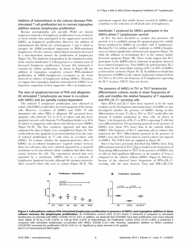
Addition of Indomethacin to the cultures decrease PHA-stimulated T cell proliferation but in contrast tryptophanaddition restores lymphocytes proliferation
Because prostaglandins and specially PGE2 are known
suppressor molecules of lymphocyte proliferation it was of interest
to check whether they participated in the suppression exerted by
hMSCs on lymphocyte proliferation. However, the addition of
indomethacin that blocks the cyclooxygenase 1 and 2, failed to
abrogate the hMSCs-mediated suppression in PHA-stimulated
lymphocytes. On the contrary, indomethacin treatment decreased
in a dose-dependent manner the proliferation of lymphocytes
(Figure 2A). The depletion of tryptophan by the stimulated activity
of the enzyme indoleamine 2 3-dioxygenase is a common cause of
decreased lymphocyte proliferation, because this aminoacid is
essential for cellular proliferation. As shown in Figure 2B, the
addition of tryptophan completely restored PHA-stimulated
proliferation in hMSCs-lymphocytes co-cultures to the levels
observed in cultures of lymphocytes lacking hMSCs. Therefore,
we suggest that tryptophan depletion determined by hMSCs is an
important component of their suppressive effect on lymphocytes.
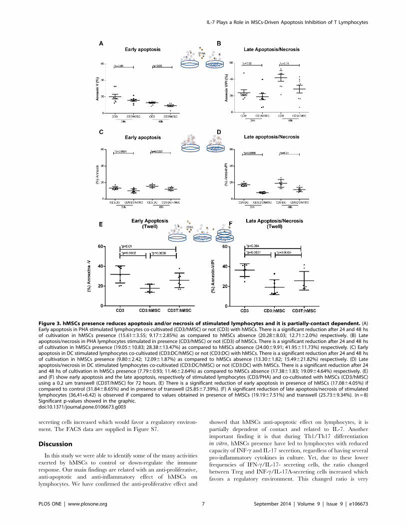
The rates of apoptosis/necrosis of PHA and allogeneic-DC-stimulated T lymphocytes are lower in co-culturewith hMSCs and are partially contact-dependent
The reduced T lymphocyte proliferation rates observed in
culture with hMSCs could reflect increased apoptosis of the former
cells. However, co-cultures of hMSCs and CD3+ T cells
stimulated with either PHA or allogeneic DC presented fewer
apoptotic cells (Annexin V+) at 24 h of culture and also fewer
apoptotic/necrotic cells (Annexin V+/Propidium Iodide +) at 48 h
of culture in comparison with cultures that did not receive hMSCs
(Figure 3A–D). The flow cytometry approach and data that
originated the data in Figure 3 are exemplified in Figure S5. The
results indicate that apoptosis or necrosis induction is not the cause
of reduced proliferation by T cells when in co-culture with
hMSCs. To evaluate whether the apoptosis-protective effect of
hMSCs on co-cultured lymphocytes required contact between
these two cell types, they were cultured separated by a transwell
membrane or in conventional culture conditions that allow direct
contact between the cells. The experiments showed that even
separated by a membrane, hMSCs led to a reduction of
lymphocytes apoptosis/necrosis, although the strongest protective
effect occurred when cells were in contact (Figure 3E–F). The
experiment suggests that soluble factors secreted by hMSCs also
contribute to the reduction of cell death rates of lymphocytes.
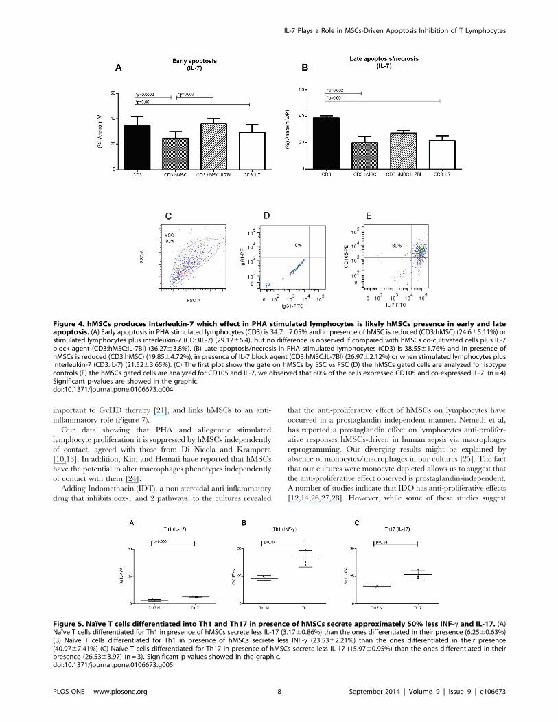
Interleukin 7 produced by hMSCs participates in thehMSCs-driven T Lymphocytes survival
As IL-7 has been described as cytokine that promotes cell
survival, it is a candidate among the apoptosis-protective soluble
factors produced by hMSCs in co-culture with T lymphocytes.
Blocking IL-7 by adding anti-IL-7 antibody to hMSCs/lympho-
cytes co-cultures significantly enhanced early apoptosis (Figure 4A)
while the addition of recombinant IL-7 to the cultures further
decreased apoptosis (Figure 4B). These results suggest that IL-7
participates in the hMSCs-driven reduction of apoptosis observed
in co-cultured lymphocytes. That hMSCs do indeed produce IL-7
was shown by the intracellular staining of IL-7 in 80% of hMSCs
as detected by cytometry (Figure 4C–E). In addition we found that
the presence of hMSCs in the cultures moderately enhanced (from
37610% to 44610%) the frequency of T lymphocytes expressing
the IL-7 receptor, CD127 (data not shown).
The presence of hMSCs in Th1 or Th17 lymphocytedifferentiation cultures results in lower frequencies ofcells and modifies the relative frequency of T regulatoryand IFN-c/IL-17- secreting cells
Since IFN-c and IL-17 have been reported to be the main
cytokines in the development and maintenance of GvHD, we also
investigated whether the presence of hMSCs during in vitrodifferentiation of naıve T cells to Th1 or Th17 would affect the
amount of cytokine production by these cells. As shown in
Figure 5, the frequencies of IL-17- or IFN-c expressing T cells that
were differentiated by Th1-promoting protocols in the presence of
hMSCs were about 50% lower than in the controls without
hMSCs. The frequency of IL-17–expressing cells in cultures that
underwent the Th17 differentiation protocol in the presence of
hMSCs were also 40% lower than in control cultures not exposed
to hMSCs. The FACS data are supplied in Figure S6.
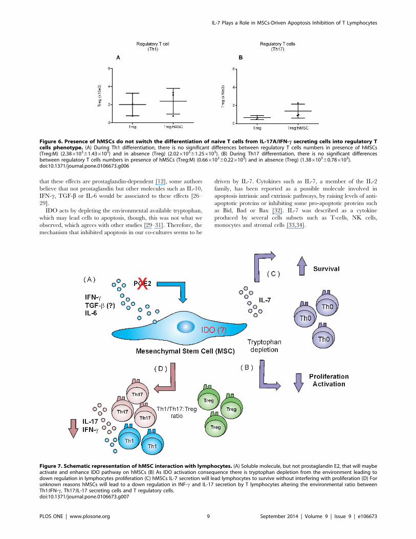
Since it has been previously described that hMSCs favor Treg
differentiation instead of Th17 [36] we looked at the frequencies of
Treg during differentiation to Th17 in the presence of hMSCs, but
we did not find significant differences in the number of Treg as
compared to the cultures without hMSCs (Figure 6). However,
because of the observed lower frequencies of IFN-c/IL-17-
secreting cells the ratio between Treg and INF-y/IL-17A-
Figure 2. Indomethacin reduces PHA stimulated lymphocytes proliferation in presence of hMSCs and tryptophan addition to thesecultures recovery the lymphocytes proliferation. (A) Proliferation control (CD3) (21.3563.62%) is enhanced if compared to stimulatedlymphocytes co-cutivated with hMSCs (CD3:M) (16.7562.81). In addition, we observed that CD3:hMSC have their proliferation even more reducedwhen added of 50 ng (11.9962.92%), or 100 ng (8.9262.32%) of indomethacin. (B) Proliferation control (CD3) (21.4363.38%) is enhanced ifcompared with stimulated lymphocytes co-cutivated with hMSCs (CD3:M) (7.5561.66). However, tryptophan addition to hMSCs condition(CD3:M:TRY) recovered the proliferation (20.3365.03) (n = 4). Significant p-values showed in the graphic.doi:10.1371/journal.pone.0106673.g002
IL-7 Plays a Role in MSCs-Driven Apoptosis Inhibition of T Lymphocytes
PLOS ONE | www.plosone.org 6 September 2014 | Volume 9 | Issue 9 | e106673
secreting cells increased which would favor a regulatory environ-
ment. The FACS data are supplied in Figure S7.
Discussion
In this study we were able to identify some of the many activities
exerted by hMSCs to control or down-regulate the immune
response. Our main findings are related with an anti-proliferative,
anti-apoptotic and anti-inflammatory effect of hMSCs on
lymphocytes. We have confirmed the anti-proliferative effect and
showed that hMSCs anti-apoptotic effect on lymphocytes, it is
partially dependent of contact and related to IL-7. Another
important finding it is that during Th1/Th17 differentiation
in vitro, hMSCs presence have led to lymphocytes with reduced
capacity of INF-c and IL-17 secretion, regardless of having several
pro-inflammatory cytokines in culture. Yet, due to these lower
frequencies of IFN-c/IL-17- secreting cells, the ratio changed
between Treg and INF-c/IL-17A-secreting cells increased which
favors a regulatory environment. This changed ratio is very
Figure 3. hMSCs presence reduces apoptosis and/or necrosis of stimulated lymphocytes and it is partially-contact dependent. (A)Early apoptosis in PHA stimulated lymphocytes co-cultivated (CD3/hMSC) or not (CD3) with hMSCs. There is a significant reduction after 24 and 48 hsof cultivation in hMSCs presence (15.6163.55; 9.1762.85%) as compared to hMSCs absence (20.2868.03; 12.7162.0%) respectively. (B) Lateapoptosis/necrosis in PHA lymphocytes stimulated in presence (CD3/hMSC) or not (CD3) of hMSCs. There is a significant reduction after 24 and 48 hsof cultivation in hMSCs presence (19.05610.83; 28.38613.47%) as compared to hMSCs absence (24.0069.91; 41.95611.73%) respectively. (C) Earlyapoptosis in DC stimulated lymphocytes co-cultivated (CD3:DC/hMSC) or not (CD3:DC) with hMSCs. There is a significant reduction after 24 and 48 hsof cultivation in hMSCs presence (9.8062.42; 12.0961.87%) as compared to hMSCs absence (13.3061.82; 15.49621.82%) respectively. (D) Lateapoptosis/necrosis in DC stimulated lymphocytes co-cultivated (CD3:DC/hMSC) or not (CD3:DC) with hMSCs. There is a significant reduction after 24and 48 hs of cultivation in hMSCs presence (7.7960.93; 11.4662.64%) as compared to hMSCs absence (17.3861.83; 19.0964.64%) respectively. (E)and (F) show early apoptosis and the late apoptosis, respectively of stimulated lymphocytes (CD3/PHA) and co-cultivated with hMSCs (CD3/hMSC)using a 0.2 um transwell (CD3T/hMSC) for 72 hours. (E) There is a significant reduction of early apoptosis in presence of hMSCs (17.0864.05%) ifcompared to control (31.8468.65%) and in presence of transwell (25.8567.39%). (F) A significant reduction of late apoptosis/necrosis of stimulatedlymphocytes (36,41+6.42) is observed if compared to values obtained in presence of hMSCs (19.1967.51%) and transwell (25.7369.34%). (n = 8)Significant p-values showed in the graphic.doi:10.1371/journal.pone.0106673.g003
IL-7 Plays a Role in MSCs-Driven Apoptosis Inhibition of T Lymphocytes
PLOS ONE | www.plosone.org 7 September 2014 | Volume 9 | Issue 9 | e106673
important to GvHD therapy [21], and links hMSCs to an anti-
inflammatory role (Figure 7).
Our data showing that PHA and allogeneic stimulated
lymphocyte proliferation it is suppressed by hMSCs independently
of contact, agreed with those from Di Nicola and Krampera
[10,13]. In addition, Kim and Hemati have reported that hMSCs
have the potential to alter macrophages phenotypes independently
of contact with them [24].
Adding Indomethacin (IDT), a non-steroidal anti-inflammatory
drug that inhibits cox-1 and 2 pathways, to the cultures revealed
that the anti-proliferative effect of hMSCs on lymphocytes have
occurred in a prostaglandin independent manner. Nemeth et al,
has reported a prostaglandin effect on lymphocytes anti-prolifer-
ative responses hMSCs-driven in human sepsis via macrophages
reprogramming. Our diverging results might be explained by
absence of monocytes/macrophages in our cultures [25]. The fact
that our cultures were monocyte-depleted allows us to suggest that
the anti-proliferative effect observed is prostaglandin-independent.
A number of studies indicate that IDO has anti-proliferative effects
[12,14,26,27,28]. However, while some of these studies suggest
Figure 4. hMSCs produces Interleukin-7 which effect in PHA stimulated lymphocytes is likely hMSCs presence in early and lateapoptosis. (A) Early apoptosis in PHA stimulated lymphocytes (CD3) is 34.767.05% and in presence of hMSC is reduced (CD3:hMSC) (24.665.11%) orstimulated lymphocytes plus interleukin-7 (CD:3IL-7) (29.1266.4), but no difference is observed if compared with hMSCs co-cultivated cells plus IL-7block agent (CD3:hMSC:IL-7Bl) (36.2763.8%). (B) Late apoptosis/necrosis in PHA stimulated lymphocytes (CD3) is 38.5561.76% and in presence ofhMSCs is reduced (CD3:hMSC) (19.8564.72%), in presence of IL-7 block agent (CD3:hMSC:IL-7Bl) (26.9762.12%) or when stimulated lymphocytes plusinterleukin-7 (CD3:IL-7) (21.5263.65%). (C) The first plot show the gate on hMSCs by SSC vs FSC (D) the hMSCs gated cells are analyzed for isotypecontrols (E) the hMSCs gated cells are analyzed for CD105 and IL-7, we observed that 80% of the cells expressed CD105 and co-expressed IL-7. (n = 4)Significant p-values are showed in the graphic.doi:10.1371/journal.pone.0106673.g004
Figure 5. Naıve T cells differentiated into Th1 and Th17 in presence of hMSCs secrete approximately 50% less INF-c and IL-17. (A)Naıve T cells differentiated for Th1 in presence of hMSCs secrete less IL-17 (3.1760.86%) than the ones differentiated in their presence (6.2560.63%)(B) Naıve T cells differentiated for Th1 in presence of hMSCs secrete less INF-y (23.5362.21%) than the ones differentiated in their presence(40.9767.41%) (C) Naıve T cells differentiated for Th17 in presence of hMSCs secrete less IL-17 (15.9760.95%) than the ones differentiated in theirpresence (26.5363.97) (n = 3). Significant p-values showed in the graphic.doi:10.1371/journal.pone.0106673.g005
IL-7 Plays a Role in MSCs-Driven Apoptosis Inhibition of T Lymphocytes
PLOS ONE | www.plosone.org 8 September 2014 | Volume 9 | Issue 9 | e106673
that these effects are prostaglandin-dependent [12], some authors
believe that not prostaglandin but other molecules such as IL-10,
IFN-c, TGF-b or IL-6 would be associated to these effects [26–
29].
IDO acts by depleting the environmental available tryptophan,
which may lead cells to apoptosis, though, this was not what we
observed, which agrees with other studies [29–31]. Therefore, the
mechanism that inhibited apoptosis in our co-cultures seems to be
driven by IL-7. Cytokines such as IL-7, a member of the IL-2
family, has been reported as a possible molecule involved in
apoptosis intrinsic and extrinsic pathways, by raising levels of anti-
apoptotic proteins or inhibiting some pro-apoptotic proteins such
as Bid, Bad or Bax [32]. IL-7 was described as a cytokine
produced by several cells subsets such as T-cells, NK cells,
monocytes and stromal cells [33,34].
Figure 6. Presence of hMSCs do not switch the differentiation of naive T cells from IL-17A/IFN-c secreting cells into regulatory Tcells phenotype. (A) During Th1 differentiation, there is no significant differences between regulatory T cells numbers in presence of hMSCs(Treg:M) (2.38610361.436103) and in absence (Treg) (2.02610361.256103). (B) During Th17 differentiation, there is no significant differencesbetween regulatory T cells numbers in presence of hMSCs (Treg:M) (0.66610360.226103) and in absence (Treg) (1.38610360.786103).doi:10.1371/journal.pone.0106673.g006
Figure 7. Schematic representation of hMSC interaction with lymphocytes. (A) Soluble molecule, but not prostaglandin E2, that will maybeactivate and enhance IDO pathway on hMSCs (B) As IDO activation consequence there is tryptophan depletion from the environment leading todown regulation in lymphocytes proliferation (C) hMSCs IL-7 secretion will lead lymphocytes to survive without interfering with proliferation (D) Forunknown reasons hMSCs will lead to a down regulation in INF-c and IL-17 secretion by T lymphocytes altering the environmental ratio betweenTh1:IFN-c, Th17:IL-17 secreting cells and T regulatory cells.doi:10.1371/journal.pone.0106673.g007
IL-7 Plays a Role in MSCs-Driven Apoptosis Inhibition of T Lymphocytes
PLOS ONE | www.plosone.org 9 September 2014 | Volume 9 | Issue 9 | e106673
In fact, we found that IL-7 blockage in lymphocytes:hMSCs co-
culture enhances significantly early apoptosis while addition of IL-
7 to control cells impairs early and late apoptosis. This inhibition
effect was similar to what was observed in hMSCs presence.
Supporting our findings, IL-7 has been described as maintaining
the cell viability by repressing cell death factors and/or activating
a life factor [33,35].
In addition, we observed that in co-culture the differentiation
into Th1 and Th17 cells show a reduced capacity of INF-c and IL-
17 secretion, but there was not a significant increase in Treg
differentiation in those co-cultures. Ghannam et al, also reported a
reduced differentiation of Th17 in hMSCs presence, but to these
authors, naıve cells were differentiating into a Treg phenotype
instead to Th17 [36]. While we measured the amount of Treg cells
considering the whole cell phenotype and FoxP3 protein
expression, Ghannam et al measured FoxP3 by mRNA expres-
sion, which might explain why they obtained different results [36].
Taken together, our finds provide important preliminary results
on the lymphocyte pathway modulated by hMSCs (Figure 7).
Importantly we suggest that survival signals driven by IL-7
cytokine may play a prominent role on the apoptosis process. It
remains unclear how IL-7 acts on CD3 T cells survival in our co-
cultures and whether other molecules are involved with this
process. Our findings pave the way to a better understanding on
how hMSCs act on lymphocytes and may contribute for
developing novel treatments and therapeutic targets for GvHD.
Supporting Information
Figure S1 Human mesenchymal stem cell (hMSC)phenotyping: Isotype control showed in red, markers staining
showed in blue. Cells were first gated on SSC vs FSC, than
analyzed for each maker. Less than 1% of hMSCs expressed
CD106, CD34, CD45, CD31, CD14 and HLA-DR. At least 93%
of the analyzed hMSCs, expressed CD29, CD44, CD73, CD90
and CD105.
(TIF)
Figure S2 hMSCs differentiation in 3 mesodermallineages: (A) Staining with Alizarin red the osteoblast differen-
tiation control (B) Staining with Alizarin red the osteoblast
differentiation, evidencing the calcium deposits (C) Staining with
Toluidine blue the condroblast differentiation control (D) Staining
with Toluidine blue the condroblast differentiation, evidencing the
presence of proteoglycans (E) Staining with Oil red the adipocytes
differentiation control (F) Staining with Oil red the adipocytes
differentiation, evidencing the presence of intracellular lipid drops.
(60x).
(TIF)
Figure S3 Dendritic cells differentiation and matura-tion: (A–E) CD14, CD209, CD80, CD83 and CD86 expression
in M, iDC and mDC.
(TIF)
Figure S4 PHA stimulated T lymphocytes proliferation.(A) Gate on forward and side scatter (B) Gate selection of CD3
positive cells. (C) T lymphocytes without stimulus, control for KI-
67 staining,. (D) PHA stimulated T lymphocytes proliferation
(51.7%) in absence of hMSCs (E) PHA stimulated T lymphocytes
proliferation (27.5%) in presence of hMSCs.
(TIF)
Figure S5 PHA stimulated T lymphocytes apoptosis/necrosis. (A) Control – T Lymphocytes stimulated with PHA
stained only with Annexin-V (B) Control – T Lymphocytes
stimulated with PHA stained only with propidium iodide (PI) (C) T
Lymphocytes stimulated with PHA in absence of hMSCs, show
late apoptosis/necrosis (39.5%) represented by cells that are
double positive for PI/AnnexinV and the early apoptosis cells
(31.2%%) represented by the single positive cell (Annexin-V). (D)
Effect of hMSCs on lymphocytes apoptosis, result for late
apoptosis/necrosis (15.9%) and the early apoptosis cells (14.3%).
(TIF)
Figure S6 Graphic representation of gate strategy oflymphocytes cytokines production. (A–B) Naive lymphocyte
differentiation into Th1 in absence of hMSCs, gate on IFN-cintracellular (38%) and in hMSCs presence, gate on IFN-cintracellular (21%) (C–D) Naive lymphocyte differentiation into
Th1 in absence of hMSCs, gate on IL-17A intracellular (6%) and
in hMSCs presence, gate on IL-17A intracellular production (3%)
(E–F) Naive lymphocyte differentiation into Th17, gate strategy of
double positive cells for RORyt and IL-17A in absence of hMSCs
(31.1%) and (16.6%) in hMSCs presence.
(TIF)
Figure S7 Graphic representation of gate strategy ofregulatory T cells. In (A) Gate strategy of stimulated
lymphocytes by scatter and CD45, (B) Gate on CD3 positive
population (73%), (C) Gate strategy of double positive cells for
CD3 and CD4 (55%), (D) Gate strategy in high CD25 (23.7%), (E)
and low expression for CD127 (79.8%) and in (E) Gate strategy of
double positive population for CD3 and FoxP3 expression (100%),
(G–H) Fluorescence minus one (FMO) control for FoxP3 and
CD25.
(TIF)
Acknowledgments
We are thankful to FAPESP that have funded this project thought the
grant number 2011/02027-4. We are also grateful to Prof. Ises de Almeida
Abrahamson for reviewing this article and to Hospital Israelita Albert
Einstein for all the support received.
Author Contributions
Conceived and designed the experiments: LCM MN. Performed the
experiments: LCM MN HA. Analyzed the data: LCM MN. Contributed
reagents/materials/analysis tools: LCM AR NH LVR AK. Contributed to
the writing of the manuscript: LCM MN. Suggestions: LVR.
References
1. Le Blanc K, Rasmusson I, Sundberg B, Gotherstrom C, Hassan M, et al. (2004)
Treatment of severe acute graft-versus-host disease with third party haploiden-
tical mesenchymal stem cells. Lancet; 363(9419): 1439–41.
2. Friedenstein AJ, Deriglasova UF, Kulagina NN, Panasuk AF, Rudakowa SF, et
al. (1974) Precursors for fibroblasts in different populations of hematopoietic cells
as detected by the in vitro colony assay method. Experimental hematology; 2 (2):
83–92.
3. Pittenger MF, Mackay AM, Beck SC, Jaiswal RK, Douglas R, et al. (1999)
Multilineage potential of adult human mesenchymal stem cells. Science; 284
(5411): 143–7.
4. Dominici M, Le Blanc K, Mueller I, Slaper-Cortenbach I, Marini F, et al. (2006)
Minimal criteria for defining multipotent mesenchymal stromal cells. The
International Society for Cellular Therapy position statement. Cytotherapy; 8(4):
315–7.
5. Prockop DJ (1997) Marrow stromal cells as stem cells for nonhematopoietic
tissues. Science; 276(5309): 71–4.
6. Beyer Nardi N and da Silva Meirelles L (2006) Mesenchymal stem cells:
isolation, in vitro expansion and characterization. Handbook of experimental
pharmacology; (174): 249–82.
7. Ding DC, Shyu WC, Lin SZ. (2011) Mesenchymal stem cells. Cell
transplantation; 20(1): 5–14.
IL-7 Plays a Role in MSCs-Driven Apoptosis Inhibition of T Lymphocytes
PLOS ONE | www.plosone.org 10 September 2014 | Volume 9 | Issue 9 | e106673
8. Ren G, Su J, Zhang L, Zhao X, Ling W, et al. (2009) Species variation in the
mechanisms of mesenchymal stem cell-mediated immunosuppression. Stem
cells; 27(8): 1954–62.
9. Bernardo ME and Fibbe WE (2013) Mesenchymal stromal cells: sensors and
switchers of inflammation. Cell stem cell; 13(4): 392–402.
10. Di Nicola M, Carlo-Stella C, Magni M, Milanesi M, Longoni PD, et al. (2002)
Human bone marrow stromal cells suppress T-lymphocyte proliferation induced
by cellular or nonspecific mitogenic stimuli. Blood; 99(10): 3838–43.
11. Meisel R, Zibert A, Laryea M, Gobel U, Daubener W, et al. (2004) Human
bone marrow stromal cells inhibit allogeneic T-cell responses by indoleamine
2,3-dioxygenase-mediated tryptophan degradation. Blood; 103(12): 4619–21.
12. Aggarwal S and Pittenger MF (2005) Human mesenchymal stem cells modulate
allogeneic immune cell responses. Blood; 105(4): 1815–22.
13. Krampera M, Glennie S, Dyson J, Scott D, Laylor R, et al. (2003) Bone marrow
mesenchymal stem cells inhibit the response of naive and memory antigen-
specific T cells to their cognate peptide. Blood; 101(9): 3722–9.
14. Sato K, Ozaki K, Oh I, Meguro A, Hatanaka K, et al. (2007) Nitric oxide plays
a critical role in suppression of T-cell proliferation by mesenchymal stem cells.
Blood; 109(1): 228–34.
15. Mendez-Ferrer S, Michurina TV, Ferraro F, Mazloom AR, Macarthur BD, et
al. (2010) Mesenchymal and haematopoietic stem cells form a unique bone
marrow niche. Nature; 466(7308): 829–34.
16. Rodriguez-Pardo VM and Vernot JP (2013) Mesenchymal stem cells promote a
primitive phenotype CD34+c-kit+ in human cord blood-derived hematopoietic
stem cells during ex vivo expansion. Cellular & molecular biology letters; 18(1):
11–33.
17. Boomsma RA and Geenen DL (2012) Mesenchymal stem cells secrete multiple
cytokines that promote angiogenesis and have contrasting effects on chemotaxis
and apoptosis. PloS one; 7(4): e35685.
18. Tan JT, Dudl E, LeRoy E, Murray R, Sprent J, et al. (2001) IL-7 is critical for
homeostatic proliferation and survival of naive T cells. Proceedings of the
National Academy of Sciences of the United States of America; 98(15): 8732–7.
19. Khaled AR and Durum SK (2002) Lymphocide: cytokines and the control of
lymphoid homeostasis. Nature reviews. Immunology; 2(11): 817–30.
20. Jacobs SR, Michalek RD, Rathmell JC (2010) IL-7 is essential for homeostatic
control of T cell metabolism in vivo. J Immuno; 184(7): 3461–9.
21. Normanton M and Marti LC (2013) Current data on IL-17 and Th17 cells and
implications for graft versus host disease. Einstein; 11(2): 237–46.
22. Deus GC, Normanton M, Hamerschlak N, Kondo AT, Ribeiro AA, et al. (2012)
Isolation and characterization of mesenchymal stem cells obtained from reusable
and disposable bone marrow collection filters. Einstein; 10(3): 296–301.
23. Marti LC, Pavon L, Severino P, Sibov T, Guilhen D, et al. (2013) Vascular
endothelial growth factor-A enhances indoleamine 2,3-dioxygenase expression
by dendritic cells and subsequently impacts lymphocyte proliferation. Mem Inst
Oswaldo Cruz; 109(1): 70–79.24. Kim J and Hematti P (2009) Mesenchymal stem cell-educated macrophages: a
novel type of alternatively activated macrophages. Experimental hematology;
37(12): 1445–53.25. Nemeth K, Leelahavanichkul A, Yuen PS, Mayer B, Parmelee A, et al. (2009)
Bone marrow stromal cells attenuate sepsis via prostaglandin E(2)-dependentreprogramming of host macrophages to increase their interleukin-10 production.
Nature medicine; 15(1): 42–9.
26. Meisel R, Zibert A, Laryea M, Gobel U, Daubener W, et al. (2004) Humanbone marrow stromal cells inhibit allogeneic T-cell responses by indoleamine
2,3-dioxygenase mediated tryptophan degradation. Blood; 103: 4619–21.27. Ryan JM, Barry F, Murphy JM, Mahon BP (2007) Interferon-gamma does not
break, but promotes the immunosuppressive capacity of adult humanmesenchymal stem cells. Clinical and experimental immunology; 149(2): 353–
63.
28. Francois M, Romieu-Mourez R, Li M, Galipeau J (2012) Human MSCsuppression correlates with cytokine induction of indoleamine 2,3-dioxygenase
and bystander M2 macrophage differentiation. Molecular therapy: the journal ofthe American Society of Gene Therapy; 20(1): 187–95.
29. Benvenuto F, Ferrari S, Gerdoni E, Gualandi F, Frassoni F, et al (2007) Human
mesenchymal stem cells promote survival of T cells in a quiescent state. Stemcells; 25(7): 1753–60.
30. Ramasamy R, Lam EW, Soeiro I, Tisato V, Bonnet D, et al. (2007)Mesenchymal stem cells inhibit proliferation and apoptosis of tumor cells:
impact on in vivo tumor growth. Leukemia; 21(2): 304–10.31. Khubutiya MS, Vagabov AV, Temnov AA, Sklifas AN (2014) Paracrine
mechanisms of proliferative, anti-apoptotic and anti-inflammatory effects of
mesenchymal stromal cells in models of acute organ injury. Cytotherapy; 16(5):579–585.
32. Danial NN and Korsmeyer SJ (2004) Cell death: critical control points. Cell;116(2): 205–19.
33. Hofmeister R, Khaled AR, Benbernou N, Rajnavolgyi E, Muegge K, et al (1999)
Interleukin-7: physiological roles and mechanisms of action. Cytokine GrowthFactor Rev.; 10(1): 41–60.
34. Al-Rawi MA, Mansel RE, Jiang WG (2003) Interleukin-7 (IL-7) and IL-7receptor (IL-7R) signalling complex in human solid tumours. Histology and
histopathology; 18(3): 911–23.35. De Miguel MP, Fuentes-Julian S, Blazquez-Martınez A, Pascual CY, Aller MA,
etal. (2012) Immunosuppressive properties of mesenchymal stem cells: advances
and applications. Curr Mol Med.; 12(5): 574–91.36. Ghannam S, Pene J, Moquet-Torcy G, Jorgensen C, Yssel H (2013) Correction:
mesenchymal stem cells inhibit human th17 cell differentiation and function andinduce a T regulatory cell phenotype. Journal of immunology; 191(11): 5777.
IL-7 Plays a Role in MSCs-Driven Apoptosis Inhibition of T Lymphocytes
PLOS ONE | www.plosone.org 11 September 2014 | Volume 9 | Issue 9 | e106673
Copyright © 2022 FDOKUMEN