The capillarity of left ventricular tissue of rats subjected to coronary artery occlusion
Upload
mississippimedicalCategory
view
0download
0
Selective Matrix Metalloproteinase Inhibition WithDeveloping Heart Failure
Effects on Left Ventricular Function and Structure
Mary K. King, Mytsi L. Coker, Aaron Goldberg, James H. McElmurray III, Himali R. Gunasinghe,Rupak Mukherjee, Michael R. Zile, Timothy P. O’Neill, Francis G. Spinale
Abstract—The matrix metalloproteinases (MMPs) are an endogenous family of proteolytic enzymes implicated tocontribute to LV remodeling. However, broad-spectrum MMP inhibition (MMPi), particularly inhibition of interstitialcollagenase (MMP-1), may not be clinically applicable. This study examined the effects of selective MMPi (sparingMMP-1) in a model of developing congestive heart failure. Pigs were randomly assigned to 3 groups: (1) rapid pacingfor 3 weeks (240 bpm, n�10); (2) selective MMPi (20 mg/kg per day-PO;PGE7113313) and rapid pacing (n�12); and(3) controls (n�10). LV peak wall stress increased from controls with rapid pacing (140�6 versus 319�18 g/cm2;P�0.05) and was reduced with selective MMPi (208�9 g/cm2; P�0.05. Preload recruitable stroke work was reducedwith rapid pacing (4.3�0.4 versus 1.2�0.2 dyne · cm/mm Hg; P�0.05) and was increased with selective MMPi(2.6�0.3 dyne · cm/mm Hg; P�0.05). Plasma norepinephrine increased by 6-fold in the rapid pacing group (P�0.05)and was reduced from untreated values with selective MMPi (P�0.05). At the myocardial level, myocyte cross-sectionalarea was increased with selective MMPi but fibrillar collagen volume fraction remained unchanged relative to controlvalues. These results suggest that targeting a selective portfolio of myocardial MMP species for inhibition may providea more rational therapeutic strategy in the setting of congestive heart failure. (Circ Res. 2003;92:177-185.)
Key Words: left ventricular systolic function � myocardial stiffness � myocardial structure� matrix metalloproteinases � heart failure
Astructural milestone in the progression of congestiveheart failure (CHF) is alterations in left ventricular (LV)
geometry, commonly referred to as myocardial remodeling.The myocardial extracellular matrix contributes to the main-tenance of LV geometry, structural alignment of adjoiningmyocytes, as well as modulating transmembrane signalingpathways. A family of enzymes that contributes to extracel-lular collagen degradation and tissue remodeling is the matrixmetalloproteinases (MMPs).1–3 Increased expression and ac-tivity of MMPs within the LV myocardium occurs in bothpatients and animals with CHF.4–8 Orally active nonselectiveMMP inhibitors, termed as broad spectrum MMP inhibitors,have been used in several animal models of CHF anddemonstrated to attenuate the LV remodeling process.7–9
Therefore, modulating MMP activity represents a potentialtherapeutic target in the context of LV remodeling and CHF.However, long-term inhibition of all MMP species will likelyinterfere with normal tissue remodeling processes and cangive rise to undesirable systemic effects.10–12 Thus, selectivetargeting of MMP species that contribute to pathologicalmyocardial remodeling in developing CHF will likely hold
greater therapeutic potential. Whereas a number of MMPspecies are expressed within the human myocardium, not allMMPs are upregulated in end-stage CHF.6,13,14 Specifically,the abundance of interstitial collagenase-1, or MMP-1, issignificantly reduced in patients with cardiomyopathic dis-ease.6 However, it remains unknown whether inhibition ofMMP-1 is a fundamental requirement in order to alter themyocardial remodeling process during the initiation anddevelopment of CHF. Accordingly, the overall goal of thisstudy was to institute selective MMP inhibition that wouldeffectively spare MMP-1 inhibition in an animal model ofdeveloping CHF.
Materials and MethodsSelective MMP InhibitionPast studies have demonstrated that several classes of MMPs areincreased in CHF including the interstitial collagenase MMP-13,stromelysins such as MMP-3, and the gelatinases such as MMP-2and MMP-9.4–8,14 In order to identify an MMP inhibition dosageregimen that would provide acceptable plasma profiles with respectto inhibition of these species but effectively spare MMP-1 inhibition,pharmacokinetic studies were performed on 6 chronically instru-
Original received May 13, 2002; resubmission received November 1, 2002; revised resubmission December 4, 2002; accepted December 4, 2002.From the Division of Cardiothoracic Surgery (M.K.K., M.L.C., A.G., J.H.M., H.R.G., R.M., M.R.Z., F.G.S.), Medical University of South Carolina,
Charleston, SC; and Pharmaceutical Research Division (T.P.O’.N.), Procter and Gamble, Mason, Ohio.Correspondence to Francis G. Spinale, MD, PhD, Cardiothoracic Surgery, Medical University of South Carolina, Strom Thurmond Research Building,
770 MUSC Complex, Suite 625, Charleston, SC 29425.© 2003 American Heart Association, Inc.
Circulation Research is available at http://www.circresaha.org DOI: 10.1161/01.RES.0000052312.41419.55
177 by guest on February 18, 2016http://circres.ahajournals.org/Downloaded from

mented pigs using methods described previously.9 The MMP inhib-itor chosen for this study was PGE7113313 (Procter and Gamble),which based on initial in vitro assay systems, exhibited a 50%inhibitory concentration (IC50) for MMP-2 of 1.5 nmol/L, MMP-3 of13 nmol/L, MMP-8 of 1.9 nmol/L, MMP-9 of 1 nmol/L, andMMP-13 of 1.1 nmol/L.15 However, the IC50 of this compound forMMP-1 was greater than 4000 nmol/L. This MMP inhibitor did notinfluence TNF-� release in a THP-1 cell culture assay, nor affectedangiotensin-converting enzyme activity based on an enzymaticassay. Thus, this MMP inhibitor did not influence other metallopro-tease systems. Plasma MMP inhibitor levels were determined byhigh-performance chromatography and mass spectroscopy. Frominitial dose determination studies and pharmacokinetic profiles, itwas determined that a 20 mg/kg dose administered 3 times per day(TID) would result in the maintenance of plasma levels at approxi-mately 10-fold higher than the IC50 for the MMP species of interest,but would not approach MMP-1 IC50 levels.
Experimental Design and CHF Model PreparationFor these studies, the rapid pacing pig model was used because thispredictably causes progressive LV dilation, myocardial remodeling,and after 3 weeks of pacing, recapitulates the clinical features of theCHF phenotype.4,9 Weight-matched pigs (22 to 23 kg, HamboneFarms, Orangeburg, SC) were instrumented with an aortic accesscatheter and a modified atrial pacemaker (8329, Medtronic, Inc).4,9
The pigs were randomly assigned to 3 groups: (1) rapid pacing (240bpm) for 3 weeks (n�10); (2) concomitant MMP inhibition(PGE7113313 20 mg/kg-TID PO) and rapid pacing (n�12); and (3)sham controls, instrumentation with no rapid pacing (n�10). Drugtreatment was started 3 days before the initiation of pacing andcontinued for the entire 21-day pacing protocol. All animals de-scribed in these studies were treated and cared for in accordance withthe National Institutes of Health “Guide for the Care and Use ofLaboratory Animals” (National Research Council, Washington,1996).
LV Systolic and Diastolic FunctionLV size and function were measured at weekly intervals in all of thepigs entered in the protocol using simultaneous pressure-echocardi-ography as described previously.4,9 After the final set of LVechocardiographic studies on day 21, plasma samples were collectedfor norepinephrine and endothelin levels, peak/trough drug levels,and MMP-2 quantitation. The pigs were then anesthetized (sufenta-nyl-1�g/kg bolus and 0.5 �g/kg per hour infusion) and intubated. Amultilumened thermodilution catheter (7.5F, Baxter HealthcareCorp) was positioned in the pulmonary artery. A sternotomy wasperformed, and a vascular ligature was placed around the inferiorvena cava. A previously calibrated microtipped transducer (7.5 F,Millar Instruments Inc) was placed in the LV. Four piezoelectriccrystals (2 mm, Sonometrics) were positioned in the LV endocar-dium in order to provide an orthogonal myocardial dimension acrossthe short axis.9 LV preload was altered by sequential occlusion andrelease of the caval tape and the isochronal LV end-diastolicpressure-stroke work points subjected to linear regression in order tocompute the preload recruitable stroke work relation (PRSWR).16
LV myocardial velocity of circumferential fiber shortening, cor-rected for heart rate (Vcfc), was also computed from the digitized LVcrystal and pressure data.4 Indices of LV diastolic function weredetermined by computations of the regional LV chamber stiffnessconstant (Kc) and myocardial stiffness constant (Km).17 As an indexof LV active myocardial relaxation, the time constant of LVisovolumic relaxation, or Tau, was computed.17 The animals werethen deeply anesthetized (5% isoflurane), a sternotomy performed,and the LV removed. The circumflex coronary artery was cannulatedand the LV region served by this coronary perfusion fixed formorphometric assessment. Sections of the LV free wall served by theleft anterior descending coronary artery were snap frozen forbiochemistry. The region that was perfusion-fixed or frozen wasalternated with each preparation.
LV Myocardial Structure and MMPLevels/ZymographySections of perfusion-fixed LV myocardium were embedded, cut inthe circumferential orientation, and examined by light microscopy inorder to measure myocyte cross-sectional area.4 Perfusion-fixed LVsections were also subjected to immunohistochemical staining for thebasement membrane protein laminin. Briefly, LV sections wereincubated overnight at 4°C with an anti-laminin antibody (1:500dilution, rabbit anti-laminin AB19012, Chemicon), followed byincubation with an anti-rabbit anti-sera conjugated to horse-radishperoxidase (1:1000, Vector Laboratories). Positive staining wasvisualized by incubation in a 3�,3�-diaminobenzidine-hydrogen per-oxide substrate. Negative controls included substitution with nonim-mune primary anti-sera. LV myocardial samples were prepared forscanning electron microscopy and light microscopic examination ofthe collagen matrix. In addition to routine scanning electron micros-copy, perfusion-fixed LV myocardial samples were subjected topotassium-hydroxide/tannic acid maceration treatment in order toremove cellular constituents and provide a greater relief of thefibrillar collagen matrix. LV full thickness myocardial sections (0.5g) were subjected to biochemical assessment for hydroxyproline aswell as collagen solubility.18 The sections underwent acid hydrolysisand hydroxyproline measured spectrophotometrically (550 nm) afterreaction with Ehrlich’s reagent. Collagen solubility was determinedas the quotient of hydroxyproline obtained from salt extraction andthe total hydroxyproline from hydrolysis.
Relative LV MMP activity was examined by substrate-specificzymographic analysis as described previously.4,6 Before zymogra-phy, the myocardial extracts were incubated with trypsin (0.5�g/mL, type I;EC 3.4.21.4, 5 minutes at 37°C) in order to unfold theMMP enzyme and cleave the propeptide sequence in order to revealtotal MMP zymographic activity. The zymograms were quantitatedby image analysis (Gel Pro Analyzer, Media Cybernetics). Using a2-site ELISA sandwich format and internal standards, plasma andmyocardial MMP-2 levels were measured (RPN 2617, 2611 BIO-TRAK, Amersham Life Sciences).6
Data AnalysisIndices of LV function and systemic hemodynamics were initiallyexamined using multiway analysis of variance (ANOVA) in whichthe main effects were treatment and time. Pair-wise tests of groupmeans were compared using Bonferroni probabilities. For the bio-chemical and morphometrics data, the 3 treatment groups werecompared by ANOVA and Tukey’s procedure. All statistical proce-dures were performed using the BMDP statistical software package(BMDP Statistical Software Inc). Results are presented asmean�SEM. Values of P�0.05 were considered to be statisticallysignificant.
ResultsLV Size and FunctionWeekly changes in LV size and function obtained in theconscious, awake state for the rapid pacing groups aresummarized in Figure 1. In both rapid pacing groups, LVend-diastolic dimension and peak wall stress increased, andfractional shortening decreased in a time dependent manner.However, at week 3 of the pacing protocol, LV fractionalshortening was higher in with selective MMP inhibition whencompared with untreated pacing values. LV peak wall stresswas significantly lower in the selective MMP inhibitiongroup at 2 and 3 weeks of pacing when compared with theuntreated pacing group. A small but significant increase inLV mass occurred in the untreated rapid pacing group at 3weeks of pacing when compared with baseline values (Figure2). A significant and time-dependent increase in LV massoccurred in the selective MMP inhibition group.
178 Circulation Research February 7, 2003
by guest on February 18, 2016http://circres.ahajournals.org/Downloaded from

LV Systolic and Diastolic FunctionSystemic hemodynamics and pressures are summarized in theTable for the 3 groups. The changes in hemodynamicsobserved after 3 weeks of rapid pacing are consistent withpast reports and the CHF phenotype.4,9 In the selective MMPinhibition group, mean arterial pressure, cardiac output, andsystemic and pulmonary vascular resistance were similar tocontrol values. In the selective MMP inhibition group, LVsystolic pressure and dP/dt were increased from pacing-onlyvalues but remained reduced from control values.
The slope of the LV preload recruitable stroke work relation(PRSW) was reduced in both rapid pacing groups when com-pared with control values (Figure 3). In the selective MMPinhibition group, the slope of the PRSWR was increased fromrapid pacing–only values. The slope of the Vcfc-stress relationwas reduced from control values in the both rapid pacing groupswhen compared with controls (Figure 3) and was increased fromrapid pacing–only values in the selective MMP inhibition group.
LV end-diastolic pressure was increased and the timeconstant of isovolumic relaxation (�) was prolonged in therapid pacing group (Table). In the selective MMP inhibitiongroup, LV end-diastolic pressure was reduced from untreatedpacing values, and � was similar to control values. The LVchamber and myocardial stiffness constants were unchangedfrom control values with chronic rapid pacing (Figure 4). Inthe selective MMP inhibition group, LV myocardial stiffnesswas increased from both control and untreated pacing values.However, LV chamber stiffness was not significantly in-creased with selective MMP inhibition (P�0.19).
Plasma Drug Concentration, Neurohormones, andMMP-2 LevelsAfter the 21-day pacing protocol, peak plasma concentrationsof the MMP inhibitor (drawn 2 hours after dose) were455�120 nmol/L, and the trough values (drawn 8 hours after
Figure 1. LV size and function were seri-ally measured with each week of rapidpacing with no treatment (filled circles)and with selective MMP inhibition (opensquares). All measurements were per-formed with the pacemaker deactivatedand in the conscious state. LV fractionalshortening fell in a time-dependent man-ner with chronic rapid pacing in bothgroups (top), but was higher in the MMPinhibition group after 3 weeks of pacing.LV end-diastolic dimension significantlyincreased from baseline values after 1week of pacing and increased in a time-dependent manner in both pacinggroups (middle). LV peak systolic wallstress increased in both groups witheach week of pacing (bottom). However,LV systolic wall stress was lower in theMMP inhibition group when comparedwith untreated pacing values. Hatchedregion indicates the reference range forage-matched controls; mean�SEM.§P�0.05 vs baseline vales; �P�0.05 vspacing only values.
Figure 2. LV mass was computed by echo-cardiography with each week of the pacingprotocol. In the untreated pacing CHF group(filled circles), a small but significantincrease in LV mass was observed after 3weeks of pacing. In the selective MMP inhi-bition group (open circles), LV massincreased in a time-dependent manner.Hatched region indicates the referencerange for age-matched controls;mean�SEM. §P�0.05 vs baseline values;�P�0.05 vs pacing only values.
King et al Selective MMP Inhibition in Heart Failure 179
by guest on February 18, 2016http://circres.ahajournals.org/Downloaded from

dose) were 97�25 nmol/L. These plasma concentrationswere 10-fold lower than those necessary to inhibit MMP-1and were within the target range for selective MMP inhibi-tion. Plasma norepinephrine was increased from baselinecontrol values with chronic rapid pacing (208�40 versus1231�271 pg/mL; P�0.05, respectively) and was reducedfrom untreated values in the selective MMP inhibition group(568�74 pg/mL, P�0.05), but remained increased frombasal control values (P�0.05). Plasma endothelin increasedin the rapid pacing group compared with baseline values(11.12�1.25 versus 2.56�0.11 fmol/mL; P�0.05). In theselective MMP inhibition group, plasma endothelin wasreduced from untreated pacing values (4.89�0.93 fmol/mL;P�0.05), but remained increased from normal baseline val-ues (P�0.05). Baseline MMP-2 plasma levels were 90�33ng/mL (range 40 to 160 ng/mL). The change in plasmaMMP-2 levels were determined at the completion of the studyprotocol and are summarized in Figure 5. Plasma MMP-2levels increased in the untreated pacing CHF group anddecreased in the selective MMP inhibition group.
Myocardial Structure and CompositionThe measurements for LV myocyte cross-sectional areaapproximated a Gaussian distribution in all treatment groups.LV myocyte cross-sectional area decreased from controlvalues in the rapid pacing group (190�3 versus 210�4 �m2;P�0.05). In the MMP inhibition group, myocyte cross-sectional area increased from both control and the rapidpacing–only values (230�3 �m2; P�0.05). Representativepicro-Sirius–stained LV myocardial sections are shown inFigure 6. In the pacing-only group, morphometric analysis
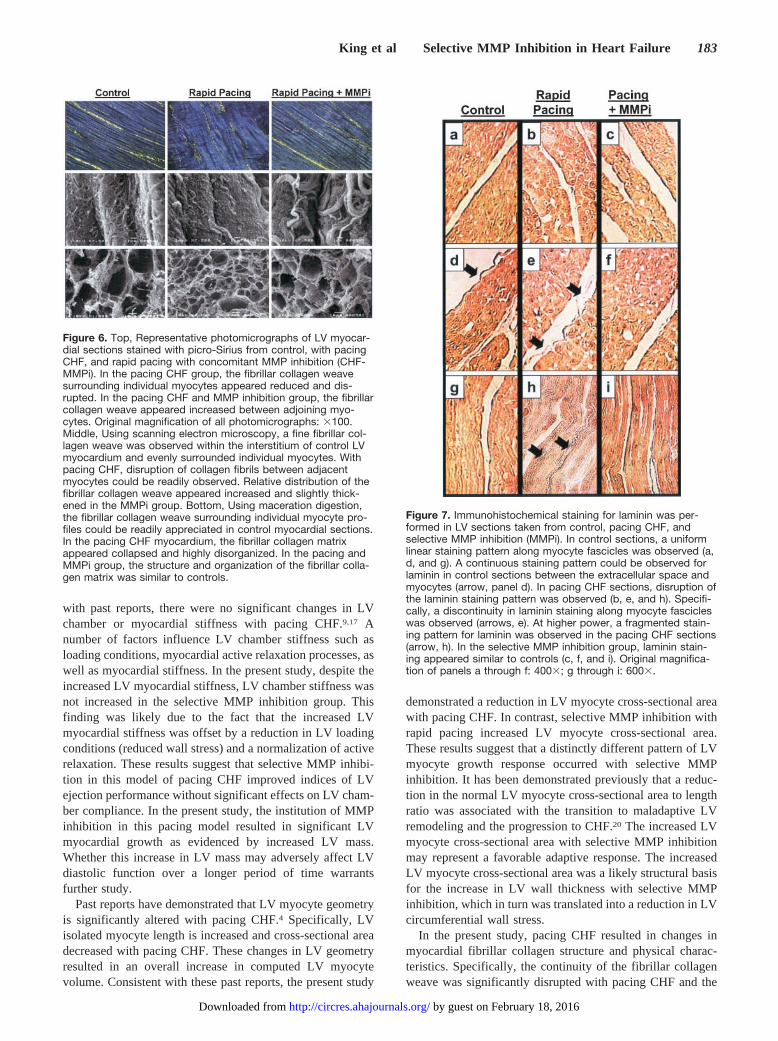
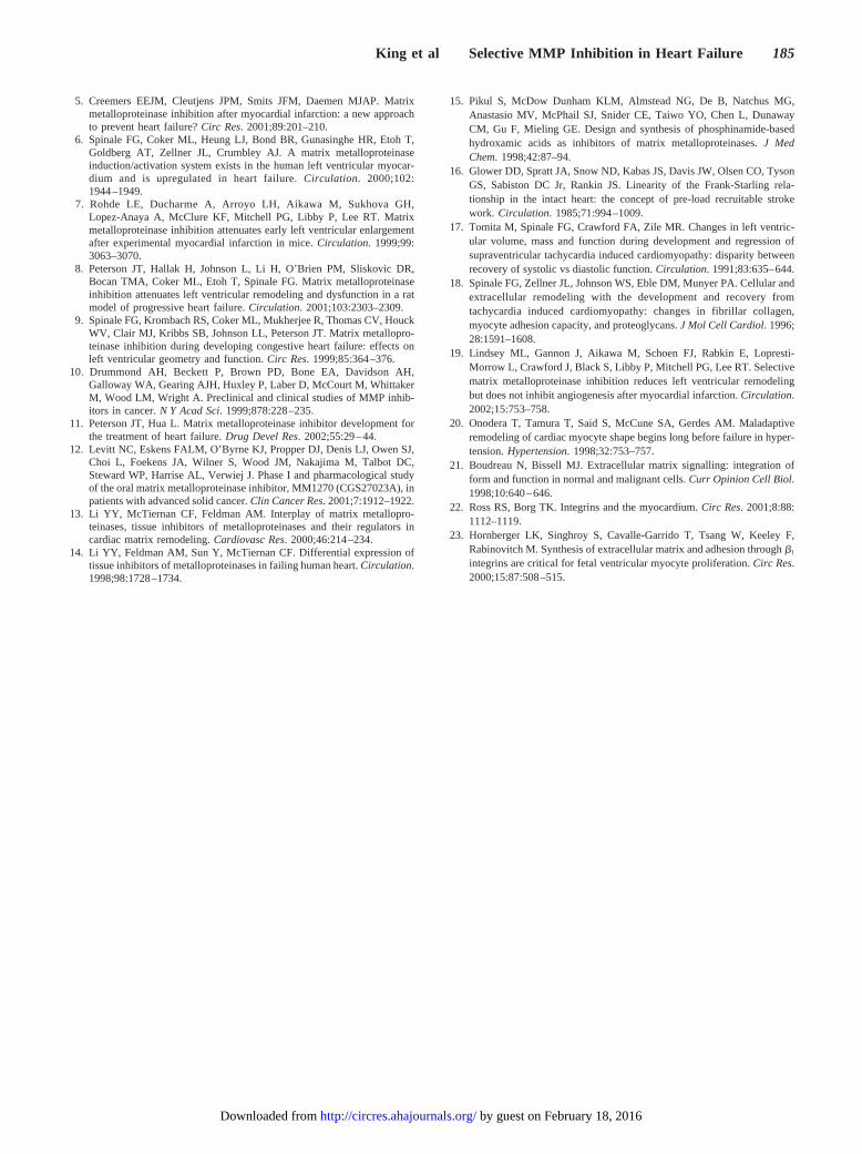
revealed a reduction in the relative content of fibrillarcollagen between myocytes (4.3�0.3 versus 6.2�0.2%;P�0.05). In control LV myocardial sections, immunohisto-chemical staining for laminin revealed a uniform linearstaining pattern along myocyte fascicles (Figure 7). In thepacing CHF sections, disruptions in the continuity of thelaminin staining pattern was observed. At higher power, amore fragmented staining pattern for laminin was observed inthe pacing CHF sections. In the selective MMP inhibitiongroup, laminin staining appeared similar to controls. Scan-ning electron microscopy revealed a disruption of the fibrillarcollagen weave in the pacing CHF group (Figure 6). In theselective MMP inhibition group, the relative content thefibrillar collagen weave was similar to control values(5.8�0.0.5%). Maceration digestion revealed a highly struc-tured fibrillar weave with clear profiles for individual myo-cytes (Figure 6). In marked contrast, cellular digestion ofpacing CHF samples revealed a disorganized, collapsed, andemphysematous fibrillar matrix. In the selective MMP inhi-bition group, the organization of the collagen matrix appearedsimilar to normal control myocardium. Myocardial hy-droxyproline content was 0.28�0.05 mg/g wet weight in thecontrol group and was unchanged either pacing group.However, the salt extractable fraction of hydroxyproline wassignificantly increased in the pacing CHF group comparedwith control (10.1�1.8 versus 4.96�0.94%; P�0.05) andremained increased in the selective MMP inhibition group(14.9�4.8%; P�0.05).
LV Myocardial MMP Activity and ContentMMP zymographic activity was increased in myocardialsamples from the pacing CHF group when compared with
Left Ventricular Function and Hemodynamics With Pacing Congestive HeartFailure: Effects of Matrix Metalloproteinase Inhibition
Control Rapid Pacing Rapid Pacing and MMPi
Heart rate, bpm 88�6 120�7* 119�6*
Cardiac output, L/min 2.79�0.16 1.71�0.19* 2.77�0.22†
LV pump function
Stroke volume, mL 33�2 14�2* 24�2*†
LV systolic, mm Hg 121�4 94�4* 109�3*†
LV diastolic, mm Hg 8�1 20�2* 14�2*†
Peak �dPdt, mm Hg/s 1898�75 1085�77* 1544�101*†
Peak �dPdt, mm Hg/s �1689�116 �1041�64* �1425�71*†
�, ms 36.6�1.9 49.0�3.7* 40.7�2.0†
Systemic pressures, mm Hg
Mean aortic 98�4 74�4* 86�3*†
Mean pulmonary artery 10�1 25�2* 16�1*†
Resistances, dyne � s � cm�5
Systemic 2900�174 4011�530* 2600�224†
Pulmonary 307�24 1319�204* 512�64†
LV mass/BW, g/kg 3.6�0.3 4.5�0.1* 6.1�0.3*†
Sample size, n 10 10 12
Rapid Pacing indicates 21 days of chronic rapid pacing, 240 bpm; Rapid Pacing and MMPi, MMPi(PGE 7113313) 20 mg/kg TID for the duration of rapid pacing; �, time constant of left ventricularisovolumic relaxation.
Values presented as mean�SEM. *P�0.05 vs Control; †P�0.05 vs Rapid Pacing only.
180 Circulation Research February 7, 2003
by guest on February 18, 2016http://circres.ahajournals.org/Downloaded from

control (13 390�1604 versus 8912�1443 pixels; P�0.05).However, in the selective MMP inhibition group, trypsin-ac-tivated MMP zymographic activity was similar to controlvalues (8291�917 pixels). By ELISA, LV myocardialMMP-2 concentration was 1578�205 ng/g in control sam-ples and was increased in the pacing CHF and selective MMPinhibition groups (2180�270 and 3178�548 ng/g, respec-tively; P�0.05).
DiscussionIncreased myocardial expression and activation of the MMPshave been demonstrated in patients with CHF and therebyhave been implicated to contribute to the LV remodelingprocess.2,6,13 Broad spectrum MMP inhibition has been dem-onstrated to modify the progression of LV remodeling andslow the CHF process in several animal model systems.5,7–9
Thus, selective targeting of myocardial MMPs that directlycontribute to the LV remodeling process constitutes animportant avenue of investigation. In past clinical reports ofend-stage CHF, MMP-1 levels were reduced, whereas in-creased levels of the gelatinases (MMP-2, MMP-9) andstromelysin (MMP-3) were observed.6 The present studyinstituted MMP inhibition during in the pacing pig model ofCHF, which would achieve plasma levels necessary to inhibita portfolio of MMP species, but would not directly affect
MMP-1 activity. Using this selective MMP inhibition strat-egy, the important findings of the present study were 2-fold.First, selective MMP inhibition improved LV function andsystemic hemodynamics and reduced the degree of neurohor-monal activation. Second, the structural basis for the effectsof MMP inhibition on LV geometry and function includedLV myocyte growth and improved integrity of the collagenmatrix.
A recent study demonstrated that MMP-1–sparing inhibi-tion favorably affected the early remodeling process inrabbits after myocardial infarction.19 The present study dem-onstrated that MMP-1 inhibition is not a fundamental require-ment for influencing the LV myocardial remodeling processin developing CHF. Selective MMP inhibition significantlyaffected the LV myocardial remodeling process as evidencedby increased posterior wall thickness and reduced chamberdimensions when compared with untreated pacing values.Because important determinants of LV wall stress are wallthickness and radius, LV peak wall stress was reduced in theMMP inhibition group when compared with untreated pacingCHF values. Thus, a contributory factor for the increased LVpump function observed with selective MMP inhibition wasthe reduction in LV wall stress. Two indices of LV ejectionperformance were evaluated: the preload recruitable strokework relation and the rate corrected velocity of circumferen-
Figure 3. Indices of LV ejection perfor-mance determined by alterations in pre-load in sham controls, after 3 weeks ofchronic rapid pacing, or with chronicrapid pacing and concomitant selectiveMMP inhibition (MMPi). Top, Slope of thepreload recruitable stroke work (PRSW)relation was reduced in both rapid pac-ing groups when compared with controlvalues. In the MMPi group, the slope ofthis relation was increased when com-pared with rapid pacing–only values.Bottom, Slope of the velocity of circum-ferential fiber shortening, corrected forheart rate (Vcfc) and end-systolic wallstress relation was reduced after 3weeks of rapid pacing. Slope of this rela-tion was increased from untreated pac-ing values in the MMPi group, butremained reduced from control values.*P�0.05 vs control; �P�0.05 vs rapidpacing only.
King et al Selective MMP Inhibition in Heart Failure 181
by guest on February 18, 2016http://circres.ahajournals.org/Downloaded from

tial fiber shortening-end-systolic stress relation. These indicesof LV ejection performance were reduced with pacing CHFand improved selective MMP inhibition. With selective MMPinhibition, the continuity of the myocardial fibrillar collagen
weave was improved from the untreated pacing CHF. There-fore, a structural mechanism for the improved LV systolicfunction with selective MMP inhibition was a stabilization ofintermyocyte collagen that in turn would improve the trans-duction of myocyte shortening into an overall LV ejection.
Another contributory factor for the improved LV functionwith selective MMP inhibition was the effects on neurohor-monal system activity. Plasma norepinephrine and endothelinlevels were reduced with selective MMP inhibition relative tountreated pacing CHF. The reduction in these bioactivemolecules suggests that selective MMP inhibition duringrapid pacing reduced the degree of neurohormonal stimula-tion, which in turn would provide favorable effects onvascular resistance and LV myocardial function. Moreover,this reduced neurohormonal activity with selective MMPinhibition would produce a secondary favorable effect on theLV remodeling process. A contributory factor for the reducedplasma norepinephrine and endothelin levels with selectiveMMP inhibition was the attenuation in the degree of hemo-dynamic compromise that invariably occurs in the pacingmodel of CHF.
Because changes in the myocardial collagen matrix havebeen implicated to influence LV compliance, an importantobjective of the present study was to examine LV stiffnesscharacteristics after selective MMP inhibition. Consistent
Figure 4. LV chamber stiffness (Kc; top) andmyocardial stiffness (Km; bottom) constantswere determined through the analysis of iso-chronal measurements of LV end-diastolicdimensions and pressures. In the rapid pac-ing–only group and in the concomitant selec-tive MMP inhibition group (MMPi), there wasno increase in chamber stiffness (P�0.20). Inthe rapid pacing–only group, LV myocardialstiffness was unchanged from control values,but was increased with MMP inhibition.*P�0.05 vs control.
Figure 5. Absolute change in plasma MMP-2 levels in the pacingCHF group and in the pacing and MMP inhibition group. PlasmaMMP-2 levels increased from baseline values in the pacing CHFgroup. In contrast, plasma MMP-2 levels were reduced from base-line and pacing CHF values in the MMP inhibition group. *P�0.05vs baseline, �P�0.05 vs untreated CHF values.
182 Circulation Research February 7, 2003
by guest on February 18, 2016http://circres.ahajournals.org/Downloaded from
with past reports, there were no significant changes in LVchamber or myocardial stiffness with pacing CHF.9,17 Anumber of factors influence LV chamber stiffness such asloading conditions, myocardial active relaxation processes, aswell as myocardial stiffness. In the present study, despite theincreased LV myocardial stiffness, LV chamber stiffness wasnot increased in the selective MMP inhibition group. Thisfinding was likely due to the fact that the increased LVmyocardial stiffness was offset by a reduction in LV loadingconditions (reduced wall stress) and a normalization of activerelaxation. These results suggest that selective MMP inhibi-tion in this model of pacing CHF improved indices of LVejection performance without significant effects on LV cham-ber compliance. In the present study, the institution of MMPinhibition in this pacing model resulted in significant LVmyocardial growth as evidenced by increased LV mass.Whether this increase in LV mass may adversely affect LVdiastolic function over a longer period of time warrantsfurther study.
Past reports have demonstrated that LV myocyte geometryis significantly altered with pacing CHF.4 Specifically, LVisolated myocyte length is increased and cross-sectional areadecreased with pacing CHF. These changes in LV geometryresulted in an overall increase in computed LV myocytevolume. Consistent with these past reports, the present study
demonstrated a reduction in LV myocyte cross-sectional areawith pacing CHF. In contrast, selective MMP inhibition withrapid pacing increased LV myocyte cross-sectional area.These results suggest that a distinctly different pattern of LVmyocyte growth response occurred with selective MMPinhibition. It has been demonstrated previously that a reduc-tion in the normal LV myocyte cross-sectional area to lengthratio was associated with the transition to maladaptive LVremodeling and the progression to CHF.20 The increased LVmyocyte cross-sectional area with selective MMP inhibitionmay represent a favorable adaptive response. The increasedLV myocyte cross-sectional area was a likely structural basisfor the increase in LV wall thickness with selective MMPinhibition, which in turn was translated into a reduction in LVcircumferential wall stress.
In the present study, pacing CHF resulted in changes inmyocardial fibrillar collagen structure and physical charac-teristics. Specifically, the continuity of the fibrillar collagenweave was significantly disrupted with pacing CHF and the
Figure 6. Top, Representative photomicrographs of LV myocar-dial sections stained with picro-Sirius from control, with pacingCHF, and rapid pacing with concomitant MMP inhibition (CHF-MMPi). In the pacing CHF group, the fibrillar collagen weavesurrounding individual myocytes appeared reduced and dis-rupted. In the pacing CHF and MMP inhibition group, the fibrillarcollagen weave appeared increased between adjoining myo-cytes. Original magnification of all photomicrographs: �100.Middle, Using scanning electron microscopy, a fine fibrillar col-lagen weave was observed within the interstitium of control LVmyocardium and evenly surrounded individual myocytes. Withpacing CHF, disruption of collagen fibrils between adjacentmyocytes could be readily observed. Relative distribution of thefibrillar collagen weave appeared increased and slightly thick-ened in the MMPi group. Bottom, Using maceration digestion,the fibrillar collagen weave surrounding individual myocyte pro-files could be readily appreciated in control myocardial sections.In the pacing CHF myocardium, the fibrillar collagen matrixappeared collapsed and highly disorganized. In the pacing andMMPi group, the structure and organization of the fibrillar colla-gen matrix was similar to controls.
Figure 7. Immunohistochemical staining for laminin was per-formed in LV sections taken from control, pacing CHF, andselective MMP inhibition (MMPi). In control sections, a uniformlinear staining pattern along myocyte fascicles was observed (a,d, and g). A continuous staining pattern could be observed forlaminin in control sections between the extracellular space andmyocytes (arrow, panel d). In pacing CHF sections, disruption ofthe laminin staining pattern was observed (b, e, and h). Specifi-cally, a discontinuity in laminin staining along myocyte fascicleswas observed (arrows, e). At higher power, a fragmented stain-ing pattern for laminin was observed in the pacing CHF sections(arrow, h). In the selective MMP inhibition group, laminin stain-ing appeared similar to controls (c, f, and i). Original magnifica-tion of panels a through f: 400�; g through i: 600�.
King et al Selective MMP Inhibition in Heart Failure 183
by guest on February 18, 2016http://circres.ahajournals.org/Downloaded from

degree of collagen solubility, reflecting the degree of collagencross-linking was reduced. Non–cross-linked collagen fibrilsare more susceptible to proteolytic cleavage by MMPs.1,3,13
With selective MMP inhibition, the degree of collagencross-linking was similar to pacing CHF values, suggestingthat MMP inhibition did not influence posttranslationalevents in collagen biosynthesis. Instead, selective MMPinhibition improved fibrillar collagen structure and architec-ture. These observations suggest that MMP inhibition pre-vented proteolysis of the more susceptible collagen matrixand thereby maintained a more normal extracellular architec-ture. Whereas the structure of the collagen matrix appearedimproved with selective MMP inhibition, the collagen fibrilsappeared thickened when compared with normal myocardialsamples. The physiological significance of this observationremains to be established. In a recent report using anMMP-1–sparing inhibitor in a rabbit model of myocardialinfarction, it was demonstrated that collagen turnover rateswere not affected.19 Consistent with this past report, thepresent study demonstrated that in this pacing model, theinstitution of selective MMP inhibition was not associatedwith a fibrotic response or significant collagen accumulation.
The dosing strategy used in this study resulted in theinhibition of MMP-2 and MMP-9, both of which degradeextracellular components associated with the LV myocytebasement membrane.1,3 Specifically, these MMPs degradelaminin, collagen IV, and fibronectin; all fundamental com-ponents necessary for maintaining the myocyte-fibrillar col-lagen interface. Using immunohistochemistry, the presentstudy demonstrated a disrupted staining pattern for lamininwith pacing CHF. Laminin is a large glycoprotein, is a majorcomponent of the extracellular matrix, and contributes to cellattachment and growth response.21 Thus, a loss of normallaminin continuity within the myocardial interstitium wouldlikely affect myocyte adhesion and alignment to the extracel-lular matrix. Significant alterations in extracellular myocytesupport and basement membrane adhesion capacity havebeen demonstrated with pacing CHF.18 In the present study,selective MMP inhibition attenuated the degree of focaldisruptions to the laminin architecture, which in turn wouldpotentially improve myocyte engagement to the extracellularmatrix. The attachment of the myocyte to the extracellularmatrix is achieved through the class of transmembraneproteins called integrins, and an important binding domain forthese integrins is laminin.22 Engagement of integrins to theextracellular matrix results in an important series of intracel-lular events, which regulate cell behavior.22,23 Thus, integrinengagement to the extracellular matrix likely constitutes animportant pathway by which physical stimuli are transducedinto intracellular signaling molecules and in turn influencemyocyte growth. While remaining speculative, one potentialmechanism by which selective MMP inhibition increased LVmass in the chronic pacing model was through a stabilizationof the extracellular matrix-integrin complex.
Preactivation of LV myocardial extracts with the serineprotease trypsin resulted in much higher MMP zymographicactivity in the untreated pacing group when compared withnormal values and is consistent with past reports.4,9 Using aquantitative immunoassay, MMP-2 myocardial levels were
increased in the pacing CHF group. In the selective MMPinhibition group, MMP-2 myocardial levels were increasedfrom normal values and were similar to untreated pacingvalues. In contrast, MMP zymographic activity after serineprotease activation was reduced to within normal values inthe selective MMP inhibition group. In this assay, proteaseactivation is performed before detergent treatment and elec-trophoretic separation. Thus, the reduction in recruitableMMP zymographic activity was likely due, at least in part, tothe presence of the MMP inhibitor in the myocardial extract.The in vivo plasma levels of the MMP inhibitor weresufficient for obtaining the desired profile of MMP speciesinhibition. In addition, the release of MMP-2 into the plasmawas decreased with selective MMP inhibition, suggestingreduced activation of this MMP species. Taken together,these in vitro and in vivo studies provide documentation thatthe desired pharmacological effect of selective MMP inhibi-tion was achieved. However, it must be recognized that theseare indirect measurements of the degree of in-vivo MMPinhibition achieved at the myocardial level.
Although the etiologies of clinical CHF are diverse, acommon end-point is LV remodeling and pump dysfunction.Chronic pacing in animals causes well defined, predictable,and progressive LV dilation, contractile dysfunction, andneurohormonal activation and therefore was chosen for thepresent study. However, extrapolation of the findings fromthis project to clinical forms of CHF should be done withcaution. Nevertheless, the present study demonstrated thatselective control of myocardial MMP activity favorablyinfluenced the LV myocardial remodeling process in devel-oping CHF. The use of broad-spectrum MMP inhibition inpatients with metastatic cancer have reported severe muscu-loskeletal pain necessitating treatment discontinuation.10–12
In contrast, initial clinical reports suggest that more selectiveMMP inhibition, in particular sparing MMP-1, was notassociated with this musculoskeletal syndrome.10 Taken to-gether, these past observations and the results from thepresent study suggest that a selective MMP inhibition strat-egy can be developed that will effectively retard the LVremodeling process in developing CHF without adverselyaffecting normal tissue structure and function. Thus, thedevelopment of strategies that selectively inhibit myocardialMMPs in CHF represent a potentially novel therapeutictarget.
AcknowledgmentsThis study was supported in part, by NIH grants HL59165, PO1HL48788-08, and a basic research grant from Procter and Gamble.
References1. Woessner JF, Nagase H. Introduction to the matrix metalloproteinases
(MMPs). In: Matrix metalloproteinases and TIMPs. Oxford, UK: OxfordUniversity Press; 2000:1–125.
2. Spinale FG. Matrix metalloproteinases: regulation and dysregulation inthe failing heart. Circ Res. 2002;90:520–530.
3. Nelson AR, Fingleton B, Rathenberg ML, Matrisian LM. Matrix metal-loproteinases: biological activity and clinical implications. J Clin Oncol.2000;18:1135–1149.
4. Spinale FG, Coker ML, Thomas CV, Walker JD, Mukherjee R, Hebbar L.Time dependent changes in matrix metalloproteinase activity andexpression during the progression of congestive heart failure: relation toventricular and myocyte function. Circ Res. 1998;82:482–495.
184 Circulation Research February 7, 2003
by guest on February 18, 2016http://circres.ahajournals.org/Downloaded from
5. Creemers EEJM, Cleutjens JPM, Smits JFM, Daemen MJAP. Matrixmetalloproteinase inhibition after myocardial infarction: a new approachto prevent heart failure? Circ Res. 2001;89:201–210.
6. Spinale FG, Coker ML, Heung LJ, Bond BR, Gunasinghe HR, Etoh T,Goldberg AT, Zellner JL, Crumbley AJ. A matrix metalloproteinaseinduction/activation system exists in the human left ventricular myocar-dium and is upregulated in heart failure. Circulation. 2000;102:1944–1949.
7. Rohde LE, Ducharme A, Arroyo LH, Aikawa M, Sukhova GH,Lopez-Anaya A, McClure KF, Mitchell PG, Libby P, Lee RT. Matrixmetalloproteinase inhibition attenuates early left ventricular enlargementafter experimental myocardial infarction in mice. Circulation. 1999;99:3063–3070.
8. Peterson JT, Hallak H, Johnson L, Li H, O’Brien PM, Sliskovic DR,Bocan TMA, Coker ML, Etoh T, Spinale FG. Matrix metalloproteinaseinhibition attenuates left ventricular remodeling and dysfunction in a ratmodel of progressive heart failure. Circulation. 2001;103:2303–2309.
9. Spinale FG, Krombach RS, Coker ML, Mukherjee R, Thomas CV, HouckWV, Clair MJ, Kribbs SB, Johnson LL, Peterson JT. Matrix metallopro-teinase inhibition during developing congestive heart failure: effects onleft ventricular geometry and function. Circ Res. 1999;85:364–376.
10. Drummond AH, Beckett P, Brown PD, Bone EA, Davidson AH,Galloway WA, Gearing AJH, Huxley P, Laber D, McCourt M, WhittakerM, Wood LM, Wright A. Preclinical and clinical studies of MMP inhib-itors in cancer. N Y Acad Sci. 1999;878:228–235.
11. Peterson JT, Hua L. Matrix metalloproteinase inhibitor development forthe treatment of heart failure. Drug Devel Res. 2002;55:29–44.
12. Levitt NC, Eskens FALM, O’Byrne KJ, Propper DJ, Denis LJ, Owen SJ,Choi L, Foekens JA, Wilner S, Wood JM, Nakajima M, Talbot DC,Steward WP, Harrise AL, Verwiej J. Phase I and pharmacological studyof the oral matrix metalloproteinase inhibitor, MM1270 (CGS27023A), inpatients with advanced solid cancer. Clin Cancer Res. 2001;7:1912–1922.
13. Li YY, McTiernan CF, Feldman AM. Interplay of matrix metallopro-teinases, tissue inhibitors of metalloproteinases and their regulators incardiac matrix remodeling. Cardiovasc Res. 2000;46:214–234.
14. Li YY, Feldman AM, Sun Y, McTiernan CF. Differential expression oftissue inhibitors of metalloproteinases in failing human heart. Circulation.1998;98:1728–1734.
15. Pikul S, McDow Dunham KLM, Almstead NG, De B, Natchus MG,Anastasio MV, McPhail SJ, Snider CE, Taiwo YO, Chen L, DunawayCM, Gu F, Mieling GE. Design and synthesis of phosphinamide-basedhydroxamic acids as inhibitors of matrix metalloproteinases. J MedChem. 1998;42:87–94.
16. Glower DD, Spratt JA, Snow ND, Kabas JS, Davis JW, Olsen CO, TysonGS, Sabiston DC Jr, Rankin JS. Linearity of the Frank-Starling rela-tionship in the intact heart: the concept of pre-load recruitable strokework. Circulation. 1985;71:994–1009.
17. Tomita M, Spinale FG, Crawford FA, Zile MR. Changes in left ventric-ular volume, mass and function during development and regression ofsupraventricular tachycardia induced cardiomyopathy: disparity betweenrecovery of systolic vs diastolic function. Circulation. 1991;83:635–644.
18. Spinale FG, Zellner JL, Johnson WS, Eble DM, Munyer PA. Cellular andextracellular remodeling with the development and recovery fromtachycardia induced cardiomyopathy: changes in fibrillar collagen,myocyte adhesion capacity, and proteoglycans. J Mol Cell Cardiol. 1996;28:1591–1608.
19. Lindsey ML, Gannon J, Aikawa M, Schoen FJ, Rabkin E, Lopresti-Morrow L, Crawford J, Black S, Libby P, Mitchell PG, Lee RT. Selectivematrix metalloproteinase inhibition reduces left ventricular remodelingbut does not inhibit angiogenesis after myocardial infarction. Circulation.2002;15:753–758.
20. Onodera T, Tamura T, Said S, McCune SA, Gerdes AM. Maladaptiveremodeling of cardiac myocyte shape begins long before failure in hyper-tension. Hypertension. 1998;32:753–757.
21. Boudreau N, Bissell MJ. Extracellular matrix signalling: integration ofform and function in normal and malignant cells. Curr Opinion Cell Biol.1998;10:640–646.
22. Ross RS, Borg TK. Integrins and the myocardium. Circ Res. 2001;8:88:1112–1119.
23. Hornberger LK, Singhroy S, Cavalle-Garrido T, Tsang W, Keeley F,Rabinovitch M. Synthesis of extracellular matrix and adhesion through �1
integrins are critical for fetal ventricular myocyte proliferation. Circ Res.2000;15:87:508–515.
King et al Selective MMP Inhibition in Heart Failure 185
by guest on February 18, 2016http://circres.ahajournals.org/Downloaded from

Gunasinghe, Rupak Mukherjee, Michael R. Zile, Timothy P. O'Neill and Francis G. SpinaleMary K. King, Mytsi L. Coker, Aaron Goldberg, James H. McElmurray III, Himali R.
Left Ventricular Function and StructureSelective Matrix Metalloproteinase Inhibition With Developing Heart Failure: Effects on
Print ISSN: 0009-7330. Online ISSN: 1524-4571 Copyright © 2002 American Heart Association, Inc. All rights reserved.is published by the American Heart Association, 7272 Greenville Avenue, Dallas, TX 75231Circulation Research
doi: 10.1161/01.RES.0000052312.41419.552003;92:177-185; originally published online December 19, 2002;Circ Res.
http://circres.ahajournals.org/content/92/2/177World Wide Web at:
The online version of this article, along with updated information and services, is located on the
http://circres.ahajournals.org//subscriptions/
is online at: Circulation Research Information about subscribing to Subscriptions:
http://www.lww.com/reprints Information about reprints can be found online at: Reprints:
document. Permissions and Rights Question and Answer about this process is available in the
located, click Request Permissions in the middle column of the Web page under Services. Further informationEditorial Office. Once the online version of the published article for which permission is being requested is
can be obtained via RightsLink, a service of the Copyright Clearance Center, not theCirculation Researchin Requests for permissions to reproduce figures, tables, or portions of articles originally publishedPermissions:
by guest on February 18, 2016http://circres.ahajournals.org/Downloaded from
Copyright © 2022 FDOKUMEN