Glycogen turnover and anaplerosis in preconditioned rat hearts
Upload
independentCategory
view
3download
0
apyw(p
s
Journal of the American College of Cardiology Vol. 57, No. 18, 2011© 2011 by the American College of Cardiology Foundation ISSN 0735-1097/$36.00
PRE-CLINICAL RESEARCH
Adenylyl Cyclase 6 Improves Calcium Uptakeand Left Ventricular Function in Aged Hearts
Tong Tang, PHD,*† H. Kirk Hammond, MD,*† Amy Firth, PHD,† Yuan Yang, MD,†Mei Hua Gao, PHD,*† Jason X.-J. Yuan, MD, PHD,† N. Chin Lai, PHD*†
San Diego and La Jolla, California
Objectives This study tested the hypothesis that activation of adenylyl cyclase 6 (AC6) expression in cardiac myocytes im-proves calcium uptake and left ventricular (LV) function in aging mice.
Background Aging hearts exhibit impaired �-adrenergic receptor signaling and LV dysfunction.
Methods Twenty-month-old mice with cardiac-directed and regulated AC6 expression were randomized into 2 groups, andAC6 expression was activated in 1 group (AC6-On) but not the other (AC6-Off). One month later, LV function andsarcoplasmic reticulum calcium uptake were assessed.
Results AC6 expression was associated with increased LV contractility, as reflected by ejection fraction(p � 0.02), rate of pressure development (p � 0.002), and slope of the LV end-systolic pressure-volume relation-ship (p � 0.04). No changes in LV weight to tibial length ratio, LV fibrosis, and expression of fetal genes (atrialnatriuretic factor, �-skeletal muscle actin, and �-myosin heavy chain) and collagens were observed betweenAC6-On and AC6-Off groups. However, LV samples from AC6-On mice showed increases in: isoproterenol-stimulated cAMP production (p � 0.04), cAMP-dependent protein kinase activity (p � 0.0004), phosphorylationof phospholamban (at Ser16 site; p � 0.04) and cardiac troponin I (at Ser23/24 sites; p � 0.01), velocity ofsarcoplasmic reticulum calcium uptake (p � 0.0001), and sarcoplasmic reticulum calcium-ATPase2a (SERCA2a)affinity for calcium (p � 0.0001). Finally, we found that AC6 expression increased sarcoplasmic reticulum cal-cium storage in cardiac myocytes isolated from 23-month-old rats. In contrast, AC6 expression in 7-month-oldmice did not change LV function and calcium uptake.
Conclusions These results indicate that activation of cardiac AC6 expression improves impaired function of aged heartsthrough improved calcium uptake. (J Am Coll Cardiol 2011;57:1846–55) © 2011 by the American College ofCardiology Foundation
Published by Elsevier Inc. doi:10.1016/j.jacc.2010.11.052
sAt
pioA
Cardiac senescence is associated with reduced left ventric-ular (LV) function (1–4) and impaired cardiac �-adrenergicreceptor (�AR) responsiveness (5). In humans, LV diastolicnd systolic functions in response to �AR stimulationrogressively decrease after the age of 20 years. At age 80ears, LV contractile reserve is less than one-half of what itas at age 20 years (6). In addition, congestive heart failure
CHF) is a common disease of the elderly (7–9), and olderatients with CHF have a particularly poor prognosis (8).Adenylyl cyclase (AC) is the effector molecule for �AR
ignaling (10), playing a pivotal role in contractile respon-
From the *VA San Diego Healthcare System, San Diego, California; and the†Department of Medicine, University of California San Diego, La Jolla, California.This work was supported by Grants-in-Aid from the American Heart AssociationWestern States Affiliate, NIH grants (5P01HL066941, HL081741, HL088426), anda VA Merit Review Award. Dr. Hammond is a founder, consultant, and equity holderfor Renova Therapeutics. All other authors have reported that they have norelationships to disclose. Douglas Mann, MD, acted as Guest Editor for this article.
tManuscript received September, 2010; revised manuscript received November 9,
2010, accepted November 9, 2010.
iveness, cardiac relaxation, and LV diastolic function (11).C catalyzes ATP to generate cAMP, a second messenger
hat is required for many intracellular events (12). Reduced�AR responsiveness in aging hearts occurs in the presenceof increased plasma catecholamine levels (13), underscoringthe abnormality in AC signaling (5,14). Indeed, impairedLV cAMP production is associated with decreased cardiacAC content in hearts from animals of advanced age (15,16).However, the precise mechanism by which AC regulatescardiac function in aging hearts is not known and is thefocus of the current investigation.
Cardiac-directed expression of AC type 6 (AC6) in-creases LV function in CHF (17,18), in which impaired�AR-AC signaling and impaired calcium uptake are
rominent (19). Increased cardiac cAMP production andmproved sarcoplasmic reticulum (SR) calcium uptake aref mechanistic importance for the beneficial effect ofC6 on failing hearts (18,20). In this study, we used a
ransgenic line with cardiac-directed tetracycline-regulated

ctiIdsitcTttawiSGS
R
Aa(2(ssvrAmEL2aAaTgamf
1847JACC Vol. 57, No. 18, 2011 Tang et al.May 3, 2011:1846–55 AC6 Function in Aged Hearts
AC6 expression (21,22) to test the hypothesis that activa-tion of cardiac AC6 expression improves LV function byincreasing cardiac cAMP production and correcting calciumuptake impairment in hearts from older mice.
Methods
Animals. The Animal Use and Care Committee of the VASan Diego Healthcare System, in accordance with NationalInstitutes of Health and Association for Assessment andAccreditation of Laboratory Animal Care guidelines, ap-proved this study. Twenty-month-old transgenic mice withcardiac myocyte–specific tetracycline-regulated (tet-off)AC6 expression (22) were used for echocardiography, invivo physiology, and biochemistry studies. Adenylyl cyclase6 transgene expression is completely suppressed until tetra-cycline is removed from the water supply, and chronictetracycline treatment did not affect cardiac function. Micewere randomized into 2 groups. AC6 expression was acti-vated (by removing tetracycline suppression) in one group(AC6-On) and was continuously suppressed by tetracyclinein the other group (AC6-Off). These mice were studied 1month after activation (or continued inactivation) of AC6transgene expression. For comparison, echocardiography, invivo physiology, and biochemistry studies were also per-formed on 7-month-old tetracycline-regulated AC6 mice.For calcium transients study, cardiac myocytes were isolatedfrom 23-month-old Sprague-Dawley rats (Harlan Labora-tories, Indianapolis, Indiana).Echocardiography. Echocardiography was performed un-der light anesthesia as previously reported (23). Data wereacquired and analyzed without knowledge of group identity.LV in vivo physiologic studies. A 1.4-F conductance-micromanometer catheter was used to measure LV pressureand volume to assess the LV end-systolic pressure-volumerelationship (ESPVR) as previously reported (18). Datawere acquired and analyzed without knowledge of groupidentity.Necropsy and LV fibrosis assessment. Body and LVweights (including septum) and tibial lengths were re-corded. A short-axis midwall LV ring was formalin fixedand paraffin embedded. The LV sections were stained withpicrosirius red, and collagen fractional area was quantifiedusing NIH ImageJ software (24).Biochemistry studies. Total RNA extraction and quanti-tative reverse transcriptase-polymerase chain reaction wereperformed as previously reported (24). LV samples werehomogenized and used for Western blotting as describedpreviously (23). AC activity, cAMP-dependent proteinkinase (PKA) activity, and caspase 3/7 activity in LVsamples were measured as reported (18).Calcium uptake. LV tissues were homogenized, and theATP-dependent initial rate of SR calcium uptake wasmeasured by a modified Millipore filtration technique as
reported (20). eCalcium transients. Cardiac myo-cytes were isolated from adultrats as previously described (25),plated on laminin-coated 25-mmglass coverslips, and infectedwith adenovirus encoding greenfluorescent protein or murine AC6(400 viral particles/cell). Fortyhours after infection, cells wereloaded with the calcium-sensitivefluorescent indicator Fura-2 AM(3 �M), and the intracellular cal-ium concentration was moni-ored using a digital fluorescencemaging system (Intracellularmaging, Cincinnati, Ohio), asescribed previously (23). To as-ess SR calcium load, caffeine-nduced calcium release was ini-iated by addition of 10 mMaffeine to Tyrode’s solution.he peak amplitude of calcium
ransients was calculated fromhe baseline and the transient risefter caffeine treatment. Dataere acquired and analyzed without knowledge of group
dentity.tatistical analysis. Results are shown as mean � SE.roup differences were compared using unpaired, 2-tailed
tudent t test. The null hypothesis was rejected when p � 0.05.
esults
C activity in aging hearts. To confirm that aging isssociated with decreased AC activity in the heart5,14,16,26), we measured LV cAMP production in 7- and0-month-old mice. There were a 43% reduction in basalp � 0.0001), a 56% reduction in isoproterenol-timulated (p � 0.04), and a 58% reduction in NKH477-timulated LV cAMP production (p � 0.0001) in 20-ersus 7-month-old mice (Fig. 1A). We also found a 59%eduction of mRNA expression of AC6, a major cardiacC isoform, in LV samples from 20- versus 7-month-oldouse hearts (Fig. 1B).chocardiography. Aging was associated with a decline inV ejection fraction (7-month-old [n � 13]: 80 � 3%;0-month-old [n � 17]: 57 � 10%; p � 0.0001). Both 7-nd 20-month-old mice with cardiac-directed and regulatedC6 expression were randomized, and AC6 expression was
ctivated in the AC6-On but not the AC6-Off group.here were no group differences (AC6-On vs. AC6-Offroup) in any of the echocardiographic measures beforectivation of AC6 expression. However, in 20-month-oldice, activation of AC6 expression increased LV ejection
raction (Table 1). These mice also showed reduced LV
Abbreviationsand Acronyms
AC � adenylyl cyclase
�AR � �-adrenergicreceptor
CHF � congestive heartfailure
cTnI � cardiac troponin I
ESPVR � end-systolicpressure-volumerelationship
LV � left ventricular
LV/TL � LV weight totibial length ratio
MMP � matrixmetalloproteinase
PKA � cAMP-dependentprotein kinase
PLN � phospholamban
SERCA2a � sarcoplasmicreticulum calcium-ATPase2a
SR � sarcoplasmicreticulum
nd-systolic dimension after AC6 e
xpression was activated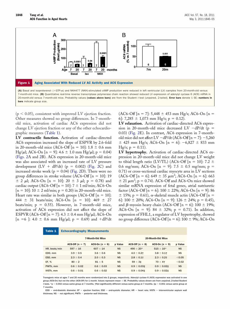
(wdi
c[H4baE[
(6Ls0o�
�sf�
1848 Tang et al. JACC Vol. 57, No. 18, 2011AC6 Function in Aged Hearts May 3, 2011:1846–55
(p � 0.05), consistent with improved LV ejection fraction.Other measures showed no group differences. In 7-month-old mice, activation of cardiac AC6 expression did notchange LV ejection fraction or any of the other echocardio-graphic measures (Table 1).LV contractile function. Activation of cardiac-directedAC6 expression increased the slope of ESPVR by 2.6-foldin 20-month-old mice (AC6-Off [n � 10]: 1.8 � 0.6 mmHg/�l; AC6-On [n � 9]: 4.6 � 1.0 mm Hg/�l; p � 0.04)Figs. 2A and 2B). AC6 expression in 20-month-old miceas also associated with an increased rate of LV pressureevelopment (LV � dP/dt) (p � 0.002) (Fig. 2C) andncreased stroke work (p � 0.04) (Fig. 2D). There were no
group differences in stroke volume (AC6-Off [n � 10]: 19� 2 �l; AC6-On [n � 10]: 20 � 3 �l; p � 0.78) andardiac output (AC6-Off [n � 10]: 7 � 1 ml/min; AC6-Onn � 10]: 10 � 2 ml/min; p � 0.20) in 20-month-old mice.eart rate was similar in both groups (AC6-Off [n � 10]:
44 � 31 beats/min; AC6-On [n � 10]: 469 � 27eats/min; p � 0.55). However, in 7-month-old mice,ctivation of AC6 expression did not affect the slope ofSPVR (AC6-Off [n � 7]: 4.3 � 0.4 mm Hg/�l; AC6-On
n �6 ]: 4.0 � 0.6 mm Hg/�l; p � 0.69) and �dP/dt
Figure 1 Aging Associated With Reduced LV AC Activity and A
(A) Basal and isoproterenol- (�GTP�s) and NKH477 (NKH)-stimulated cAMP pr7-month-old mice. (B) Quantitative real-time reverse transcriptase polymerase20-month-old versus 7-month-old mice. Probability values (values above bars)bars indicate group size.
Echocardiography MeasurementsTable 1 Echocardiography Measurements
7-Month-Old Mice
AC6-Off (n � 7) AC6-On (n � 6)
HR, beats/min 597 � 16 607 � 14
EDD, mm 3.9 � 0.5 3.6 � 0.6
ESD, mm 2.3 � 0.4 2.0 � 0.3
EF, % 80 � 2 81 � 5
PWTh, mm 0.6 � 0.02 0.6 � 0.03
IVSTh, mm 0.6 � 0.01 0.6 � 0.02
Transgenic mice at ages 7 and 20 months were randomized into 2 grgroup (AC6-On) but not the other (AC6-Off) for 1 month. Values represet tests. *p � 0.002 versus same group at 7 months. †Not significantly7 months.
EDD � end-diastolic diameter; EF � ejection fraction; ESD � end-systolithickness; NS � not significant; PWTh � posterior wall thickness.
AC6-Off [n � 7]: 5,448 � 453 mm Hg/s; AC6-On [n �]: 7,285 � 1,073 mm Hg/s; p � 0.12).V relaxation. Activation of cardiac-directed AC6 expres-
ion in 20-month-old mice decreased LV �dP/dt (p �.03) (Fig. 2E). In contrast, AC6 expression in 7-month-ld mice did not affect LV �dP/dt (AC6-Off [n � 7]: �5,260
425 mm Hg/s; AC6-On [n � 6]: �6,827 � 833 mmHg/s; p � 0.11).LV hypertrophy. Activation of cardiac-directed AC6 ex-pression in 20-month-old mice did not change LV weightto tibial length ratio (LV/TL) (AC6-Off [n � 10]: 7.2 �0.6 mg/mm; AC6-On [n � 9]: 7.5 � 0.5 mg/mm; p �0.71) or cross-sectional cardiac myocyte area in LV sections(AC6-Off [n � 6]: 649 � 35 �m2; AC6-On [n � 6]: 663
21 �m2; p � 0.74). AC6-Off and AC6-On mice showedimilar mRNA expression of fetal genes, atrial natriureticactor (AC6-Off [n � 6]: 100 � 22%; AC6-On [n � 9]: 86
15%; p � 0.61), �-skeletal muscle actin (AC6-Off [n �6]: 100 � 20%; AC6-On [n � 9]: 126 � 24%; p � 0.45),and �-myosin heavy chain (AC6-Off [n � 6]: 100 � 19%;AC6-On [n � 9]: 84 � 32%; p � 0.71). In addition,expression of FHL1, a regulator of LV hypertrophy, showedno group difference (AC6-Off [n � 6]: 100 � 9%; AC6-On
xpression
on were reduced in left ventricular (LV) samples from 20-month-old versusreaction showed reduced LV expression of adenylyl cyclase 6 (AC6) mRNA inm the Student t test (unpaired, 2-tailed). Error bars denote 1 SE; numbers in
20-Month-Old Mice
lue AC6-Off (n � 8) AC6-On (n � 9) p Value
S 490 � 20* 515 � 16* NS
S 4.0 � 0.2† 3.8 � 0.1† NS
S 2.8 � 0.1† 2.3 � 0.2† �0.05
S 59 � 3‡ 70 � 4† �0.02
S 0.9 � 0.03‡ 0.9 � 0.02‡ NS
S 0.9 � 0.04‡ 0.9 � 0.02‡ NS
spectively. Adenylyl cyclase 6 (AC6) expression was activated in onen � SE. Probability values shown are from unpaired, 2-tailed Student
nt versus same group at 7 months. ‡p � 0.001 versus same group at
C6 E
oductichainare fro
p Va
N
N
N
N
N
N
oups, rent meadiffere
c diameter; HR � heart rate; IVSTh � interventricular septum wall

ccAeAcO
6
a
1849JACC Vol. 57, No. 18, 2011 Tang et al.May 3, 2011:1846–55 AC6 Function in Aged Hearts
[n � 9]: 113 � 12%; p � 0.47). Similarly, activation ofardiac AC6 expression in 7-month-old mice did nothange LV/TL (AC6-Off [n � 7]: 6.4 � 0.4 mg/mm;C6-On [n � 6]: 6.2 � 0.3 mg/mm; p � 0.71) or mRNA
xpression of fetal genes and FHL1. Although activation ofC6 expression in 7-month-old mice did not change
ross-sectional cardiac myocyte area in LV sections (AC6-ff [n � 6]: 489 � 21 �m2; AC6-On [n � 6]: 465 � 27
�m2; p � 0.50), aging was associated with an increase(7-month-old [n � 6]: 489 � 21 �m2; 20-month-old [n � 6]:49 � 35 �m2; p � 0.003).
Calcium uptake. We compared ATP-dependent initial SRcalcium uptake in LV homogenates from 20-month-oldAC6-Off and AC6-On mice. Activation of AC6 increasedthe velocity of calcium uptake in the presence of 0.2 �Mnd 2 �M calcium (Fig. 3A), the approximate intracellular
calcium concentration range in cardiac myocytes (27). AC6expression also increased sarcoplasmic reticulum calcium-ATPase (SERCA2a) affinity for calcium (Fig. 3B). In-creased calcium uptake and SERCA2a affinity for cal-cium would be expected to increase cardiac contractile
Figure 2 Cardiac-Directed AC6 Expression Increased LV Functi
(A) LV pressure-volume loops, generated by inferior vena caval occlusion, are shovolume relationship (ESPVR) in AC6-On mice compared with AC6-Off mice. (C) AC6expression increased stroke work in hearts from 20-month-old mice. (E) There waare from the Student t test (unpaired, 2-tailed). Error bars denote 1 SE; numbers
function as we observed. In contrast, activation of AC6
expression in 7-month-old mice did not change thevelocity of calcium uptake (Fig. 3C) and SERCA2aaffinity for calcium (Fig. 3D).
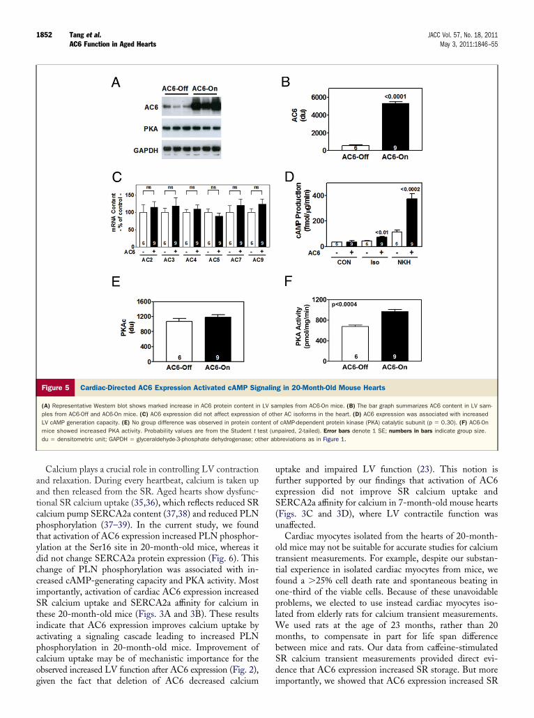
In addition, we isolated cardiac myocytes from 23-month-old rats, infected these cells with adenovirusencoding AC6, and measured the intracellular calciumconcentration under basal and caffeine-stimulated condi-tions (Fig. 4A). There was no group difference in basalintracellular calcium concentration in cardiac myocytes(Fig. 4B). However, there was a 1.5-fold increase in thepeak amplitude of caffeine-stimulated calcium transientsassociated with AC6 expression in cardiac myocytes fromaging rats (Fig. 4C). These results indicated that AC6expression increased calcium storage in aging cardiacmyocytes.AC6 expression and cAMP production. In 20-month-oldmice, cardiac-directed AC6 transgene expression was asso-ciated with a 10-fold increase in LV AC6 protein content(Figs. 5A and 5B). LV expression of other cardiac ACisoforms (AC2, AC3, AC4, AC5, AC7, and AC9) wasunchanged in these mice (Fig. 5C). Increased AC6 protein
20-Month-Old Mice
) Activation of AC6 expression increased the slope of the end-systolic pressure-ice showed increased �dP/dt compared with AC6-Off mice. (D) Activation of AC6bstantial increase in �dP/dt in AC6-On mouse hearts. Probability values shown
indicate group size. Abbreviations as in Figure 1.
on in
wn. (B-On m
s a suin bars
content was associated with a 1.7-fold increase in

a(bDstAiPplAei(cpfAPspm
1
hch
1850 Tang et al. JACC Vol. 57, No. 18, 2011AC6 Function in Aged Hearts May 3, 2011:1846–55
isoproterenol-stimulated cAMP production (p � 0.01) and3-fold increase in forskolin-stimulated cAMP production
p � 0.002) in LV samples from 20-month-old mice, andasal LV cAMP production was not changed (Fig. 5D).espite unchanged protein expression of PKA catalytic
ubunit (Fig. 5E), PKA activity was increased after activa-ion of AC6 expression (Fig. 5F), indicating a role ofC6-increased cAMP production in regulating PKA activ-
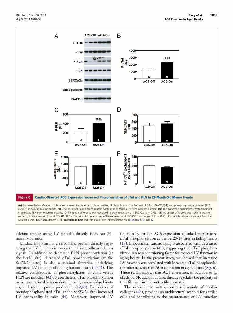
ty in the aging heart.rotein phosphorylation. We compared PKA-inducedhosphorylation of cardiac troponin I (cTnI) and phospho-
amban (PLN) in LV homogenates from 20-month-oldC6-Off and AC6-On mice. Activation of cardiac AC6
xpression in these mice was associated with a 1.9-foldncrease in phosphorylation of cTnI at the Ser23/24 sitesp � 0.01) (Figs. 6A and 6B). No change was found in totalTnI protein content. There was also a 1.9-fold increase inhosphorylation of PLN at the Ser16 site in LV samplesrom 20-month-old AC6-On mice compared with that ofC6-Off mice (p � 0.04; Figs. 6A and 6C), although totalLN protein content was unaltered. Western blottinghowed no group differences in protein content of proteinhosphatase 1 (AC6-Off [n � 6]: 1,152 � 195 densito-etric units (du); AC6-On [n � 9]: 1,153 � 277 du; p �
1.00) or protein phosphatase 2A (AC6-Off [n � 6]: 743 �
Figure 3 Activation of Cardiac AC6 Expression Increased Calci
(A) Cardiac-directed AC6 expression improved sarcoplasmic reticulum (SR) calciummice (n � 6 for each group). (B) Cardiac-directed AC6 expression increased SR ca(C) AC6 expression did not affect SR calcium uptake in the presence of 0.2 �M (for each group). (D) AC6 expression did not affect SERCA2a affinity for calcium int test (unpaired, 2-tailed). Error bars denote 1 SE; numbers in bars indicate group
04 du; AC6-On [n � 9]: 604 � 65 du; p � 0.25), 2
indicating a selective role of AC6-increased PKA activityin regulating phosphorylation of cTnI and PLN. Inaddition, we found no group differences in expression ofSR calcium pump SERCA2a (Fig. 6D), the calcium-binding protein calsequestrin (Fig. 6E), or Na�-Ca2�
exchanger 1 (Fig. 6F).Extracellular matrix. Extracellular matrix remodeling, aprocess that involves changes of collagen synthesis, degra-dation, and cross-linking, leads to increased number andsize of collagen fibers within interstitial spaces. There wassubstantial collagen deposition in LV sections from 20-month-old mice compared with those from 7-month-oldmice (collagen fractional area: 7-month-old [n � 7]: 3.1 �1.1%; 20-month-old [n � 6]: 15.4 � 2.5%; p � 0.001);
owever, activation of cardiac AC6 expression did nothange collagen fractional area in 20-month-old mouseearts (AC6-Off [n � 6]: 15.4 � 2.5%; AC6-On [n � 9]:
14.4 � 2.0%; p � 0.77).We compared mRNA content of type I and III collagens,
2 major constituents of the cardiac extracellular matrix, inLV samples from 20-month-old AC6-Off and AC6-Onmice. Activation of AC6 expression did not change mRNAexpression of collagen I�1 (AC6-Off [n � 6]: 100 � 14%;AC6-On [n � 9]: 129 � 19%; p � 0.30) or III�1(AC6-Off [n � 6]: 100 � 29%; AC6-On [n � 9]: 85 �
ptake in 20-Month-Old But Not 7-Month-Old Mice
ke in the presence of 0.2 and 2 �M calcium in LV samples from 20-month-old-ATPase2a (SERCA2a) affinity for calcium in LV samples from 20-month-old mice.33) and 2 �M calcium (p � 1.00) in LV samples from 7-month-old mice (n � 6mples from 7-month-old mice (p � 0.72). Probability values are from the StudentAbbreviations as in Figure 1.
um U
uptalcium
p � 0.LV sasize.
9%; p � 0.73). Quantitative reverse transcriptase-

A(1r2[Te11CttAnp1i2a[dsm�
D
Tctwabmsn
tfm
1851JACC Vol. 57, No. 18, 2011 Tang et al.May 3, 2011:1846–55 AC6 Function in Aged Hearts
polymerase chain reaction also showed that AC6 expressiondid not change the mRNA content of matrix metallopro-teinase 2 (MMP2) (AC6-Off [n � 6]: 100 � 14%;
C6-On [n � 9]: 135 � 20%; p � 0.22) or MMP8AC6-Off [n � 6]: 100 � 16%; AC6-On [n � 9]: 87 �4%; p � 0.55). Similar levels of periostin mRNA, aegulator of cardiac fibrosis, were seen in LV samples from0-month-old AC6-Off and AC6-On mice (AC6-Offn � 6]: 100 � 16%; AC6-On [n � 9]: 121 � 19%; p � 0.44).here were no group differences in mRNA expression of
lastin (AC6-Off [n � 6]: 100 � 10%; AC6-On [n � 9]:21 � 18%; p � 0.69) or fibronectin (AC6-Off [n � 6]:00 � 17%; AC6-On [n � 9]: 89 � 19%; p � 0.39).ardiac myocyte apoptosis. Terminal deoxynucleotidyl
ransferase-mediated dUTP nick-end labeling was usedo evaluate apoptosis. In 20-month-old mouse hearts,C6 expression did not change the apoptosis rate (dataot shown). AC6-Off and AC6-On mice showed com-arable LV mRNA expression of Bcl2 (AC6-Off [n � 6]:00 � 27%; AC6-On [n � 9]: 104 � 18%; p � 0.89), anmportant apoptosis inhibitor. Increased AC6 expression in0-month-old mouse hearts did not change LV caspase 3/7ctivity (AC6-Off [n � 6]: 40.3 � 2.4 U/mg; AC6-Onn � 9]: 37.6 � 2.0 U/mg; p � 0.41). Finally, no groupifference in expression of superoxidase dismutase 2 waseen after activation of AC6 expression in 20-month-oldice (AC6-Off [n � 6]: 100 � 6%; AC6-On [n � 9]: 93
Figure 4 Increased Calcium Storage in Cardiac Myocytes From
(A) Representative calcium transients recorded from cardiac myocytes stimulatedprotein (CON) (left panel); cardiac myocyte infected with adenovirus encoding AC6cium concentration in cardiac myocytes. Data were derived from 67 CON cells andof caffeine-stimulated calcium transients recorded from cardiac myocytes. Data wefrom the Student t test (unpaired, 2-tailed). Error bars denote 1 SE. Abbreviations
6%; p � 0.46).
iscussion
he most important finding of this study is that activation ofardiac AC6 expression improved aging-impaired LV con-ractile function and relaxation. These beneficial effectsere associated with increases in cAMP production, PKA
ctivity, and phosphorylation of PLN and cTnI. Moreover,oth SR calcium uptake and SR calcium storage in cardiacyocytes were increased following activation of AC6 expres-
ion. Improved SR calcium uptake appears to be the mecha-ism by which AC6 expression evokes these beneficial effects.Results from our previous studies (17,18,28,29) indicate
hat increased cardiac AC6 has beneficial effects in theailing heart. However, increased expression of other ele-ents of the �AR signaling pathway (e.g., �1AR, G�s,
PKA) is associated with pronounced deleterious effects onthe heart, including precipitation of heart failure, fibrosis,and increased mortality (30–33). These results indicate thatAC6 is unique among �AR signaling elements in regulatingcardiac function. In clinical settings, increased age is asso-ciated with reduced LV function (1–4), and elderly patientshave an increased prevalence of heart failure (7–9). In thisstudy, we tested the hypothesis that activation of AC6expression increases function of the aging heart. We as-sessed LV contractile function by 3 different methods: LVejection fraction, �dP/dt, and slope of ESPVR (34). Eachof these measures led to the same conclusion that increasedexpression of cardiac AC6 improves LV contractile function
onth-Old Rats Following AC6 Gene Transfer
affeine. Cardiac myocyte infected with adenovirus encoding green fluorescentpanel). (B) The bar graph shows no group difference in basal intracellular cal-6 cells (blinded; p � 0.40). (C) AC6 expression increased the peak amplitude
ived from 41 CON cells and 51 AC6 cells (blinded). Probability values shown areFigure 1.
23-M
with c(right77 ACre deras in
in the aging heart.
1852 Tang et al. JACC Vol. 57, No. 18, 2011AC6 Function in Aged Hearts May 3, 2011:1846–55
Calcium plays a crucial role in controlling LV contractionand relaxation. During every heartbeat, calcium is taken upand then released from the SR. Aged hearts show dysfunc-tional SR calcium uptake (35,36), which reflects reduced SRcalcium pump SERCA2a content (37,38) and reduced PLNphosphorylation (37–39). In the current study, we foundthat activation of AC6 expression increased PLN phosphor-ylation at the Ser16 site in 20-month-old mice, whereas itdid not change SERCA2a protein expression (Fig. 6). Thischange of PLN phosphorylation was associated with in-creased cAMP-generating capacity and PKA activity. Mostimportantly, activation of cardiac AC6 expression increasedSR calcium uptake and SERCA2a affinity for calcium inthese 20-month-old mice (Figs. 3A and 3B). These resultsindicate that AC6 expression improves calcium uptake byactivating a signaling cascade leading to increased PLNphosphorylation in 20-month-old mice. Improvement ofcalcium uptake may be of mechanistic importance for theobserved increased LV function after AC6 expression (Fig. 2),
Figure 5 Cardiac-Directed AC6 Expression Activated cAMP Sig
(A) Representative Western blot shows marked increase in AC6 protein content inples from AC6-Off and AC6-On mice. (C) AC6 expression did not affect expressionLV cAMP generation capacity. (E) No group difference was observed in protein conmice showed increased PKA activity. Probability values are from the Student t tdu � densitometric unit; GAPDH � glyceraldehyde-3-phosphate dehydrogenase; ot
given the fact that deletion of AC6 decreased calcium
uptake and impaired LV function (23). This notion isfurther supported by our findings that activation of AC6expression did not improve SR calcium uptake andSERCA2a affinity for calcium in 7-month-old mouse hearts(Figs. 3C and 3D), where LV contractile function wasunaffected.
Cardiac myocytes isolated from the hearts of 20-month-old mice may not be suitable for accurate studies for calciumtransient measurements. For example, despite our substan-tial experience in isolated cardiac myocytes from mice, wefound a �25% cell death rate and spontaneous beating inone-third of the viable cells. Because of these unavoidableproblems, we elected to use instead cardiac myocytes iso-lated from elderly rats for calcium transient measurements.We used rats at the age of 23 months, rather than 20months, to compensate in part for life span differencebetween mice and rats. Our data from caffeine-stimulatedSR calcium transient measurements provided direct evi-dence that AC6 expression increased SR storage. But more
g in 20-Month-Old Mouse Hearts
mples from AC6-On mice. (B) The bar graph summarizes AC6 content in LV sam-er AC isoforms in the heart. (D) AC6 expression was associated with increasedf cAMP-dependent protein kinase (PKA) catalytic subunit (p � 0.30). (F) AC6-Onpaired, 2-tailed). Error bars denote 1 SE; numbers in bars indicate group size.breviations as in Figure 1.
nalin
LV saof othtent oest (unher ab
importantly, we showed that AC6 expression increased SR
1853JACC Vol. 57, No. 18, 2011 Tang et al.May 3, 2011:1846–55 AC6 Function in Aged Hearts
calcium uptake using LV samples directly from our 20-month-old mice.
Cardiac troponin I is a sarcomeric protein directly regu-lating the LV function in concert with intracellular calciumsignals. In addition to decreased PLN phosphorylation (atthe Ser16 site), decreased cTnI phosphorylation (at theSer23/24 sites) is also a seminal alteration underlyingimpaired LV function of failing human hearts (40,41). Therelative contributions of phosphorylation of cTnI versusPLN are not clear (42). Nevertheless, cTnI phosphorylationincreases maximal tension development, cross-bridge kinet-ics, and systolic power production (42,43). Expression ofpseudophosphorylated cTnI at the Ser23/24 sites increased
Figure 6 Cardiac-Directed AC6 Expression Increased Phosphor
(A) Representative Western blots show marked increase in protein content of(Ser16) in AC6-On mouse hearts. (B) The bar graph summarizes protein content oof phospho-PLN from Western blotting. (D) No group difference was observed in pcontent of calsequestrin (p � 0.37). (F) AC6 expression did not change mRNA exStudent t test. Error bars denote 1 SE; numbers in bars indicate group size. Abbre
LV contractility in mice (44). Moreover, improved LV
function by cardiac AC6 expression is linked to increasedcTnI phosphorylation at the Ser23/24 sites in failing hearts(18). Importantly, cardiac aging is associated with decreasedcTnI phosphorylation (45), suggesting that cTnI phosphor-ylation is also a contributing factor for reduced LV function inaging hearts. In the present study, we showed that increasedLV function was correlated with increased cTnI phosphoryla-tion after activation of AC6 expression in aging hearts (Fig. 6).These results suggest that AC6 expression, in addition to itseffects on SR calcium uptake, directly regulates the property ofthin filament in the contractile apparatus.
The extracellular matrix, composed mainly of fibrillarcollagens (46), provides an architectural scaffold for cardiac
n of cTnI and PLN in 20-Month-Old Mouse Hearts
ho–cardiac troponin I (cTnI) (Ser23/24) and phospho-phospholamban (PLN)pho-cTnI from Western blotting. (C) The bar graph summarizes protein contentcontent of SERCA2a (p � 0.61). (E) No group difference was seen in proteinn of Na�-Ca2� exchanger 1 (p � 0.27). Probability values shown are from the
ns as in Figures 1, 3, and 5.
ylatio
phospf phos
roteinpressioviatio
cells and contributes to the maintenance of LV function

1854 Tang et al. JACC Vol. 57, No. 18, 2011AC6 Function in Aged Hearts May 3, 2011:1846–55
(47,48). In this study, we confirmed that cardiac aging isassociated with increased collagen deposition. However,activation of AC6 expression in 20-month-old mice did notchange LV collagen deposition, indicating that the im-proved LV function does not likely reflect a change in LVcollagen content. Additional support for this notion is ourfinding that AC6 expression did not change expression oftype I and III collagens, the major cardiac collagen isoformscontributing 75% to 80% (type I) and 15% to 20% (type III)of total collagen content (49). Expression of MMPs andperiostin was not changed by activation of AC6 expression,although aging is associated with increased expression ofthese important extracellular matrix proteins (50,51). Inaddition, our data showed that increased cardiac AC6expression in 20-month-old mice was not associated withchanges in apoptosis or reactive oxygen species. Takentogether, our results suggest that activation of AC6 expres-sion improves LV function through increased SR calciumuptake.
Whether LV hypertrophy in aged mice affected ourresults is an important consideration. Although posterolat-eral and septal wall thicknesses at the mid-papillary musclelevel were increased (0.3 mm) in aged mice (Table 1), theechocardiographic estimate of LV mass, which considersmore than a single plane, showed no difference betweenmice age 20 months versus 7 months (data not shown).Cardiac myocyte cross-sectional area was increased, but the13% increase in LV/TL did not reach statistical signifi-cance. The disparity between cross-sectional area and LVmass at necropsy vis-à-vis LV hypertrophy in 7- versus20-month-old mice may reflect LV hypertrophy associatedwith concomitant aging-related increases in cardiac myocyteapoptosis (52) or the limitations of assessing cardiac myo-cyte cross-sectional area in hearts not obtained after dia-stolic arrest. Similarly, others have documented that, in agedrats, despite increased myocyte cross-sectional area (52), LVmass and LV/TL were not increased (53). Our studyshowed that activation of cardiac AC6 expression, whichhad no independent effect on LV hypertrophy, improvedaging-impaired LV contractile function and relaxation.Improved SR calcium uptake appears to be of mechanisticimportance for these beneficial effects.
Gene manipulation has recently been promoted to probedisease mechanism and test treatments designed to attenu-ate the progression of aging-related diseases. Transgeniclines with increased or decreased expression of a candidategene typically are used, and if deterioration of heart functionis prevented, the candidate gene (or deletion of this gene) issaid to have “rescued” the heart. However, a superiorstrategy, and one used in the present study, is to activategene expression only when the adverse effect of aging ispresent. We accomplished this by activating AC6 expres-sion in the setting of declining heart function in aged mice.Although additional studies will be required to determine
whether activation of cardiac AC6 expression reduces mor-tality, our current data suggest a therapeutic potential forincreased AC6 expression in aging hearts.
Conclusions
Activation of cardiac-directed AC6 expression improves LVfunction in aged hearts. This is associated with increasedcAMP production and PKA activity and phosphorylation ofPLN and cTnI. Improved SR calcium uptake in cardiacmyocytes appears to be of mechanistic importance for thesebeneficial effects.
Reprint requests and correspondence: Dr. Tong Tang, VA SanDiego Healthcare System (111A), 3350 La Jolla Village Drive, SanDiego, California 92161. E-mail: [email protected].
REFERENCES
1. Gerstenblith G. Echocardiographic assessment of a normal adult agingpopulation. Circulation 1977;56:273–8.
2. Fraticelli A, Josephson R, Danziger R, Lakatta EG, Spurgeon H.Morphological and contractile characteristics of rat cardiac myocytesfrom maturation to senescence. Am J Physiol Heart Circ Physiol1989;257:H259–65.
3. Chiamvimonvat N. Diastolic dysfunction and the aging heart. J MolCell Cardiol 2002;34:607–10.
4. Dai DF, Santana LF, Vermulst M, et al. Overexpression of catalasetargeted to mitochondria attenuates murine cardiac aging. Circulation2009;119:2789–97.
5. White M, Roden R, Minobe W, et al. Age-related changes inbeta-adrenergic neuroeffector systems in the human heart. Circulation1994;90:1225–38.
6. Lakatta EG, Sollott SJ. The “heartbreak” of older age. Mol Interv2002;2:431–46.
7. Lakatta EG. Age-associated cardiovascular changes in health: impacton cardiovascular disease in older persons. Heart Failure Rev 2002;7:29–49.
8. Lakatta EG, Levy D. Arterial and cardiac aging: major shareholders incardiovascular disease enterprises: part II: the aging heart in health:links to heart disease. Circulation 2003;107:346–54.
9. Marian AJ. Beta-adrenergic receptors signaling and heart failure inmice, rabbits and humans. J Mol Cell Cardiol 2006;41:11–3.
10. Rockman HA, Koch WJ, Lefkowitz RJ. Seven-transmembrane-spanning receptors and heart function. Nature 2002;415:206–12.
11. Feldman AM. Adenylyl cyclase: a new target for heart failure thera-peutics. Circulation 2002;105:1876–8.
12. Katsushika S, Chen L, Kawabe J, et al. Cloning and characterizationof a sixth adenylyl cyclase isoform: types V and VI constitute asubgroup within the mammalian adenylyl cyclase family. Proc NatlAcad Sci U S A 1992;89:8774–8.
13. Rowe JW, Troen BR. Sympathetic nervous system and aging in man.Endocr Rev 1980;1:167–79.
14. Narayanan N, Tucker L. Autonomic interactions in the aging heart:age-associated decrease in muscarinic cholinergic receptor mediatedinhibition of beta-adrenergic activation of adenylate cyclase. MechAgeing Dev 1986;34:249–59.
15. Shu Y, Scarpace PJ. Forskolin binding sites and G-protein immuno-reactivity in rat hearts during aging. J Cardiovasc Pharmacol 1994;23:188–93.
16. Tobise K, Ishikawa Y, Holmer SR, et al. Changes in type VI adenylylcyclase expression correlate with a decreased capacity for cAMPgeneration in the aging ventricle. Circ Res 1994;74:596–603.
17. Roth DM, Gao MH, Lai NC, et al. Cardiac-directed adenylyl cyclaseexpression improves heart function in murine cardiomyopathy. Circu-lation 1999;99:3099–102.
18. Lai NC, Tang T, Gao MH, et al. Activation of cardiac adenylyl cyclaseexpression increases function of the failing ischemic heart in mice.
J Am Coll Cardiol 2008;51:1490–7.
3
4
4
4
4
4
4
4
4
4
4
5
5
5
5
1855JACC Vol. 57, No. 18, 2011 Tang et al.May 3, 2011:1846–55 AC6 Function in Aged Hearts
19. Mann DL, Bristow MR. Mechanisms and models in heart failure: thebiomechanical model and beyond. Circulation 2005;111:2837–49.
20. Tang T, Gao MH, Roth DM, Guo T, Hammond HK. Adenylylcyclase type VI corrects cardiac sarcoplasmic reticulum calcium uptakedefects in cardiomyopathy. Am J Physiol Heart Circ Physiol 2004;287:H1906–12.
21. Fishman GI. Timing is everything in life: conditional transgeneexpression in the cardiovascular system. Circ Res 1998;82:837–44.
22. Gao MH, Bayat H, Roth DM, et al. Controlled expression ofcardiac-directed adenylylcyclase type VI provides increased contractilefunction. Cardiovasc Res 2002;56:197–204.
23. Tang T, Gao MH, Lai NC, et al. Adenylyl cyclase type 6 deletiondecreases left ventricular function via impaired calcium handling.Circulation 2008;117:61–9.
24. Tang T, Lai NC, Hammond HK, et al. Adenylyl cyclase 6 deletionreduces LV hypertrophy, dilation, dysfunction and fibrosis in pressure-overloaded female mice. J Am Coll Cardiol 2010;55:1476–86.
25. Tang T, Lai NC, Roth DM, et al. Adenylyl cyclase type V deletionincreases basal left ventricular function and reduces left ventricularcontractile responsiveness to beta-adrenergic stimulation. Basic ResCardiol 2006;101:117–26.
26. O’Connor SW, Scarpace PJ, Abrass IB. Age-associated decrease ofadenylate cyclase activity in rat myocardium. Mech Ageing Dev1981;16:91–5.
27. Bers DM. Calcium fluxes involved in control of cardiac myocytecontraction. Circ Res 2000;87:275–81.
28. Lai NC, Roth DM, Gao MH, et al. Intracoronary adenovirusencoding adenylyl cyclase VI increases left ventricular function in heartfailure. Circulation 2004;110:330–6.
29. Roth DM, Bayat H, Drumm JD, et al. Adenylyl cyclase increasessurvival in cardiomyopathy. Circulation 2002;105:1989–94.
30. Gaussin V, Tomlinson JE, Depre C, et al. Common genomic responsein different mouse models of beta-adrenergic-induced cardiomyopa-thy. Circulation 2003;108:2926–33.
31. Antos CL, Frey N, Marx SO, et al. Dilated cardiomyopathy andsudden death resulting from constitutive activation of protein kinaseA. Circ Res 2001;89:997–1004.
32. Engelhardt S, Hein L, Wiesmann F, Lohse MJ. Progressive hyper-trophy and heart failure in beta1-adrenergic receptor transgenic mice.Proc Natl Acad Sci U S A 1999;96:7059–64.
33. Iwase M, Uechi M, Vatner DE, et al. Cardiomyopathy induced bycardiac Gs� overexpression. Am J Physiol 1997;272:H585–9.
34. Hess OM, Carroll JD. Clinical assessment of heart failure. In: LibbyP, Bonow RO, Mann DL, Zipes DP, editors. Braunwald’s HeartDisease: A Textbook of Cardiovascular Medicine. 8th edition. Phila-delphia, PA: Saunders, 2008;561–81.
35. Orchard CH, Lakatta EG. Intracellular calcium transients and devel-oped tensions in rat heart muscle. A mechanism for the negativeinterval-strength relationship. J Gen Physiol 1985;86:637–51.
36. Froehlich JP, Lakatta EG, Beard E, Spurgeon HA, Weisfeldt ML,Gerstenblith G. Studies of sarcoplasmic reticulum function andcontraction duration in young and aged rat myocardium. J Mol CollCardiol 1978;10:427–38.
37. Taffet GE, Tate CA. CaATPase content is lower in cardiac sarcoplas-
mic reticulum isolated from old rats. Am J Physiol Heart Circ Physiol1993;264:H1609–14.em
38. Xu A, Narayanan N. Effects of aging on sarcoplasmic reticulumCa2�-cycling proteins and their phosphorylation in rat myocardium.Am J Physiol Heart Circ Physiol 1998;275:H2087–94.
9. Jiang MT, Narayanan N. Effects of aging on phospholamban phos-phorylation and calcium transport in rat cardiac sarcoplasmic reticu-lum. Mech Ageing Dev 1990;54:87–101.
0. Messer AE, Jacques AM, Marston SB. Troponin phosphorylation andregulatory function in human heart muscle: dephosphorylation ofSer23/24 on troponin I could account for the contractile defect inend-stage heart failure. J Mol Cell Cardiol 2007;42:247–59.
1. MacLennan DH, Kranias EG. Phospholamban: a crucial regulator ofcardiac contractility. Nat Rev Mol Cell Biol 2003;4:566–77.
2. Metzger JM, Westfall MV. Covalent and noncovalent modification ofthin filament action: the essential role of troponin in cardiac muscleregulation. Circ Res 2004;94:146–58.
3. Solaro RJ. Modulation of cardiac myofilament activity by proteinphosphorylation. In: Fozzarad H, Solaro RJ, editors. Handbook ofPhysiology, Volume 1, the Heart. New York, NY: Oxford UniversityPress, 2002;264–300.
4. Takimoto E, Soergel DG, Janssen PM, Stull LB, Kass DA, MurphyAM. Frequency- and afterload-dependent cardiac modulation in vivoby troponin I with constitutively active protein kinase A phosphory-lation sites. Circ Res 2004;94:496–504.
5. Maejima Y, Adachi S, Ito H, Hirao K, Isobe M. Induction ofpremature senescence in cardiomyocytes by doxorubicin as a novelmechanism of myocardial damage. Aging Cell 2008;7:125–36.
6. Jugdutt BI. Extracellular matrix and cardiac remodeling. In: VillarrealF, editor. Interstitial Fibrosis in Heart Failure. New York, NY:Springer, 2004;23–55.
7. Lindsey ML, Borg TK. Understanding the role of the extracellularmatrix in cardiovascular development and disease: where do we gofrom here? J Mol Cell Cardiol 2010;48:431–2.
8. Tomasek JJ, Gabbiani G, Hinz B, Chaponnier C, Brown RA.Myofibroblasts and mechano-regulation of connective tissue remodel-ling. Nat Rev Mol Cell Biol 2002;3:349–63.
9. Legrice I, Pope A, Smaill B. The architecture of the heart: myocyteorganization and the cardiac extracellular matrix. In: Villarreal F,editor. Interstitial Fibrosis in Heart Failure. New York, NY: Springer,2004;3–21.
0. Lindsey ML, Goshorn DK, Squires CE, et al. Age-dependent changesin myocardial matrix metalloproteinase/tissue inhibitor of metallopro-teinase profiles and fibroblast function. Cardiovasc Res 2005;66:410–9.
1. Borg TK, Markwald R. Periostin: more than just an adhesionmolecule. Circ Res 2007;101:230–1.
2. Anversa P, Hiler B, Ricci R, Guideri G, Olivetti G. Myocyte cell lossand myocyte hypertrophy in the aging rat heart. J Am Coll Cardiol1986;8:1441–8.
3. Raya TE, Gaballa M, Anderson P, Goldman S. Left ventricularfunction and remodeling after myocardial infarction in aging rats.Am J Physiol Heart Circ Physiol 1997;273:H2652–8.
Key Words: adenylyl cyclase y calcium uptake y cardiac aging y
xtracellular matrix remodeling y left ventricular function y transgenicouse.Copyright © 2022 FDOKUMEN