Effects of Off-Pump and On-Pump Coronary-Artery Bypass Grafting at 1 Year

T h e n e w e ngl a nd j o u r na l o f m e dic i n e
n engl j med 364;17 nejm.org april 28, 2011 1607
original article
Coronary-Artery Bypass Surgery in Patients with Left Ventricular Dysfunction
Eric J. Velazquez, M.D., Kerry L. Lee, Ph.D., Marek A. Deja, M.D., Ph.D., Anil Jain, M.D., George Sopko, M.D., M.P.H., Andrey Marchenko, M.D., Ph.D.,
Imtiaz S. Ali, M.D., Gerald Pohost, M.D., Sinisa Gradinac, M.D., Ph.D., William T. Abraham, M.D., Michael Yii, M.S., F.R.C.S., F.R.A.C.S.,
Dorairaj Prabhakaran, M.D., D.M., Hanna Szwed, M.D., Paolo Ferrazzi, M.D., Mark C. Petrie, M.D., Christopher M. O’Connor, M.D.,
Pradit Panchavinnin, M.D., Lilin She, Ph.D., Robert O. Bonow, M.D., Gena Roush Rankin, M.P.H., R.D., Robert H. Jones, M.D.,
and Jean-Lucien Rouleau, M.D., for the STICH Investigators*
The authors’ affiliations are listed in the Appendix. Address reprint requests to Dr. Velazquez at Duke Clinical Research Institute, Rm. 0311 Terrace Level, 2400 Pratt St., Durham, NC 27705, or at [email protected].
* A complete list of investigators partici-pating in the hypothesis 1 component of the Surgical Treatment for Ischemic Heart Failure (STICH) trial is provided in the Supplementary Appendix, available at NEJM.org.
This article (10.1056/NEJMoa1100356) was published on April 4, 2011, at NEJM.org.
N Engl J Med 2011;364:1607-16.Copyright © 2011 Massachusetts Medical Society.
A bs tr ac t
Background
The role of coronary-artery bypass grafting (CABG) in the treatment of patients with coronary artery disease and heart failure has not been clearly established.
Methods
Between July 2002 and May 2007, a total of 1212 patients with an ejection fraction of 35% or less and coronary artery disease amenable to CABG were randomly as-signed to medical therapy alone (602 patients) or medical therapy plus CABG (610 patients). The primary outcome was the rate of death from any cause. Major second-ary outcomes included the rates of death from cardiovascular causes and of death from any cause or hospitalization for cardiovascular causes.
Results
The primary outcome occurred in 244 patients (41%) in the medical-therapy group and 218 (36%) in the CABG group (hazard ratio with CABG, 0.86; 95% confidence interval [CI], 0.72 to 1.04; P = 0.12). A total of 201 patients (33%) in the medical-therapy group and 168 (28%) in the CABG group died from an adjudicated cardio-vascular cause (hazard ratio with CABG, 0.81; 95% CI, 0.66 to 1.00; P = 0.05). Death from any cause or hospitalization for cardiovascular causes occurred in 411 pa-tients (68%) in the medical-therapy group and 351 (58%) in the CABG group (haz-ard ratio with CABG, 0.74; 95% CI, 0.64 to 0.85; P<0.001). By the end of the follow-up period (median, 56 months), 100 patients in the medical-therapy group (17%) underwent CABG, and 555 patients in the CABG group (91%) underwent CABG.
Conclusions
In this randomized trial, there was no significant difference between medical ther-apy alone and medical therapy plus CABG with respect to the primary end point of death from any cause. Patients assigned to CABG, as compared with those assigned to medical therapy alone, had lower rates of death from cardiovascular causes and of death from any cause or hospitalization for cardiovascular causes. (Funded by the National Heart, Lung, and Blood Institute and Abbott Laboratories; STICH ClinicalTrials.gov number, NCT00023595.)
The New England Journal of Medicine Downloaded from NEJM Media Center by DIANE STRIAR on April 22, 2011. Embargo lifted April 27, 2011 at 5pm ET.
Copyright © 2011 Massachusetts Medical Society. All rights reserved.

T h e n e w e ngl a nd j o u r na l o f m e dic i n e
n engl j med 364;17 nejm.org april 28, 20111608
It is estimated that 5.8 million patients in the United States1 and 15 million in Europe2
have heart failure. Coronary artery disease is the most common substrate for heart failure in industrialized nations.3 However, the role of coro-nary-artery bypass grafting (CABG) in the treat-ment of patients with coronary artery disease and heart failure has not been clearly established.
In three landmark clinical trials in the 1970s, a total of 2234 patients with chronic stable an-gina were randomly assigned to undergo CABG or receive medical therapy alone.4-6 The findings from these trials led to recommendations sup-porting the use of CABG to relieve disabling symptoms of angina, particularly among high-risk subgroups with extensive coronary artery disease.7,8 These trials excluded patients with severe left ventricular dysfunction (patients with an ejection fraction of <35%). A meta-analysis of the trials showed that 7.2% of the patients who underwent randomization had an ejection frac-tion of 40% or less, and only 4.0% had primary symptoms of heart failure rather than angina.9Furthermore, these trials predate the major de-velopments in medical therapy and cardiac sur-gery that have led to the current guidelines.10-13
More recently, observational analyses supporting a benefit of CABG14 and the proliferation of contemporary evidence-based medical and de-vice-associated therapies have led to substantial clinical uncertainty regarding the incremental benefits of CABG relative to its risks in patients with ischemic cardiomyopathy.15,16
We designed the Surgical Treatment for Is-chemic Heart Failure (STICH) trial to evaluate the role of cardiac surgery in the treatment of patients with coronary artery disease and left ventricular systolic dysfunction. A major hypoth-esis of the trial was that CABG plus intensive medical therapy based on current guidelines, as compared with medical therapy alone, would reduce mortality.
Me thods
Study Design
The design of the STICH trial has been described previously.17,18 We conducted a multicenter, non-blinded, randomized study at 127 clinical sites in 26 countries. The trial protocol (available with the full text of this article at NEJM.org) was de-signed by several of the authors and was ap-proved by the principal investigator and the eth-
ics committee at each participating center. The trial was sponsored by the National Heart, Lung, and Blood Institute (NHLBI). Additional support was provided by Abbott Laboratories, which had no role in the conduct or reporting of the trial. An executive committee met weekly and moni-tored the daily conduct of the trial. An indepen-dent data and safety monitoring committee was appointed by the NHLBI. The Duke Clinical Re-search Institute coordinated all aspects of global trial operations, site management and monitor-ing, data collection, statistical analyses, and re-porting. The authors reviewed the data, partici-pated in the analyses and wrote the manuscript, and assume responsibility for the completeness and accuracy of the data and the analyses and for the fidelity of the study to the trial protocol.
Study Patients
Eligible patients had coronary artery disease that was amenable to CABG and an ejection fraction of 35% or less, as determined at each enrolling site. Details of the enrollment criteria are pro-vided in Table 1 in the Supplementary Appendix, available at NEJM.org. All patients provided writ-ten informed consent, as approved by the local institutional review board.
After initial determination of overall eligibil-ity for the trial, patients were assessed to deter-mine whether they were potential candidates for any of three possible therapeutic options: medi-cal therapy alone, medical therapy plus CABG, or medical therapy plus CABG and surgical ven-tricular reconstruction. Patients were eligible for medical therapy alone if they did not have a ste-notic lesion leading to loss of 50% or more of the diameter of the left main coronary artery and if they did not have Canadian Cardiovascu-lar Society class III or IV angina while receiving medical therapy. (The Canadian Cardiovascular Society angina classification ranges from class 0, which indicates no symptoms, to class IV, which indicates angina at any level of physical exertion.) Patients were eligible for surgical ventricular re-construction if they had dominant anterior left ventricular akinesia or dyskinesia. As noted above, all patients were eligible for CABG.
On the basis of these eligibility criteria, pa-tients were assigned by the enrolling physician to one of three trial strata. Stratum A included pa-tients who were eligible for either medical therapy alone or medical therapy plus CABG, stratum B included patients who were eligible for any of the
The New England Journal of Medicine Downloaded from NEJM Media Center by DIANE STRIAR on April 22, 2011. Embargo lifted April 27, 2011 at 5pm ET.
Copyright © 2011 Massachusetts Medical Society. All rights reserved.

CABG in Patients with Left Ventricular Dysfunction
n engl j med 364;17 nejm.org april 28, 2011 1609
three treatment options, and stratum C included patients who were eligible for either medical therapy plus CABG or medical therapy plus CABG and surgical ventricular reconstruction (Fig. 1 in the Supplementary Appendix). Patients were then randomly assigned to one of the treatment options for which they were eligible. As a result, all the patients in stratum A and some of the patients in stratum B were randomly assigned to either medi-cal therapy alone or medical therapy plus CABG (the hypothesis 1 component of the STICH trial). All the patients in stratum C and some of the pa-tients in stratum B were randomly assigned to ei-ther medical therapy plus CABG or medical therapy plus CABG and surgical ventricular reconstruc-tion (the hypothesis 2 component of the STICH trial). The results of the hypothesis 2 comparison have been reported previously.19 The results of the hypothesis 1 comparison are reported here.
Study Procedures
During the initial evaluation, information was ob-tained on demographic factors and on clinical characteristics, including current medications and prior diagnostic and other cardiovascular proce-dures, and a physical examination was performed. Patients were randomly assigned to medical ther-apy alone or to medical therapy plus CABG by means of an investigator-initiated telephone call to an interactive voice-response system.
A lead cardiologist at each center was re-sponsible for recommending the most appro-priate medications and devices for the treat-ment of heart failure and coronary artery disease on the basis of current guidelines. Adherence to treatment guidelines was emphasized in the care of all patients and was monitored by a medical therapy committee. Cardiac surgery was performed by surgeons who had provided data on at least 25 patients with an ejection fraction of 40% or less in whom they had performed CABG and among whom the operative death rate was 5% or less. CABG was to be performed within 14 days after randomization. A surgical therapy committee monitored the conduct of sur-gery during the enrollment period. All patients underwent follow-up evaluations at the time of discharge or at 30 days, every 4 months for the first year, and every 6 months thereafter.
Study Outcomes
The primary outcome was the rate of death from any cause. Prespecified secondary outcomes in-
cluded the rate of death from cardiovascular causes and the rate of death from any cause or hospitalization for cardiovascular causes. The causes of death and of selected secondary out-comes were adjudicated according to prespeci-fied criteria by an independent clinical events committee whose members were unaware of the treatment assignments (see the definition of events in the Supplementary Appendix).
Statistical Analysis
We originally estimated that with a sample of 2000 patients who would be followed for an aver-age of approximately 3 years, the study would have 90% power to detect a 25% reduction in mor-tality with CABG as compared with medical ther-apy alone, assuming a 3-year mortality of 25% in the medical-therapy group. Because enrollment was slower than expected, we modified the design so that the sample size was reduced and the dura-tion of the follow-up period was increased corre-spondingly. The final study design specified that a sufficient number of patients had to be enrolled and the follow-up period had to be long enough that 400 deaths would occur; given this require-ment, we estimated that we would need to enroll approximately 1200 patients, with an average fol-low-up period of 5 years. These numbers allowed for as much as a 20% treatment crossover from medical therapy to CABG without reducing the study’s power to an unacceptable level. Random-ization was performed with the use of permuted blocks and was stratified according to clinical site and stratum (A or B, as described above).
All major comparisons between the treatment groups were performed according to the inten-tion-to-treat principle. Two-sided significance testing was used for all statistical tests. Cumula-tive event rates were calculated according to the Kaplan–Meier method,20 with all event or censor-ing times measured from the time of randomiza-tion. The significance of differences in mortality between the treatment groups was assessed with the use of the log-rank test, with adjustment for randomization stratum. Relative risks were ex-pressed as hazard ratios with associated confi-dence intervals and were derived from the Cox proportional-hazards model.21,22 To characterize the time-dependent nature of the relative risks of the groups according to the treatment to which they had been randomly assigned, hazard ratios (and confidence intervals) were examined within time intervals of clinical importance: random-
The New England Journal of Medicine Downloaded from NEJM Media Center by DIANE STRIAR on April 22, 2011. Embargo lifted April 27, 2011 at 5pm ET.
Copyright © 2011 Massachusetts Medical Society. All rights reserved.

T h e n e w e ngl a nd j o u r na l o f m e dic i n e
n engl j med 364;17 nejm.org april 28, 20111610
Table 1. Baseline Characteristics of the Patients.*
VariableMedical Therapy
(N = 602)CABG
(N = 610)
Age — yr
Median 59 60
Interquartile range 53–67 54–68
Female sex — no. (%) 75 (12) 73 (12)
Race or ethnic group — no. (%)†
White 402 (67) 389 (64)
Hispanic, Latino, or nonwhite 200 (33) 221 (36)
Body-mass index‡
Median 27 27
Interquartile range 24–30 24–30
Medical history — no. (%)
Previous myocardial infarction 472 (78) 462 (76)
Hyperlipidemia 370 (61) 360 (59)
Hypertension 370 (61) 358 (59)
Diabetes 238 (40) 240 (39)
Previous percutaneous coronary intervention 74 (12) 82 (13)
Chronic renal insufficiency 45 (7) 49 (8)
Previous stroke 41 (7) 51 (8)
Previous CABG 14 (2) 22 (4)
Current smoker 122 (20) 130 (21)
Current CCS angina class§
0 225 (37) 217 (36)
I 91 (15) 96 (16)
II 260 (43) 265 (43)
III 23 (4) 25 (4)
IV 3 (<1) 7 (1)
Current NYHA class
I 74 (12) 65 (11)
II 307 (51) 319 (52)
III 205 (34) 207 (34)
IV 16 (3) 19 (3)
Systolic blood pressure — mm Hg
Median 120 120
Interquartile range 110 –130 110 –130
Pulse — beats/min
Median 72 74
Interquartile range 65–80 66–82
6-Minute walk distance — ft¶
Median 1115 1145
Interquartile range 840–1345 863–1320
* CABG denotes coronary-artery bypass grafting, and NYHA New York Heart Association.† Race or ethnic group was self-reported.‡ The body-mass index is the weight in kilograms divided by the square of the height in meters.§ The Canadian Cardiovascular Society (CCS) angina classification ranges from class 0, which indicates no symptoms, to
class IV, which indicates angina at any level of physical exertion.¶ To convert the values for the 6-minute walk distance to meters, multiply by 0.305.
The New England Journal of Medicine Downloaded from NEJM Media Center by DIANE STRIAR on April 22, 2011. Embargo lifted April 27, 2011 at 5pm ET.
Copyright © 2011 Massachusetts Medical Society. All rights reserved.
CABG in Patients with Left Ventricular Dysfunction
n engl j med 364;17 nejm.org april 28, 2011 1611
ization through 30 days, 31 through 365 days, 366 days through 2 years, and any time after 2 years. As prespecified in the study protocol, comparisons of the treatment groups were also performed with adjustment for key baseline fac-tors (in addition to randomization stratum). The Cox model was also used to assess the consis-tency of treatment effects by testing for interac-tions between treatment and prespecified base-line characteristics.
To aid in an assessment of the effect of treat-ment crossovers, secondary as-treated and per-protocol analyses were also performed. The as-treated comparison was analyzed with the Cox model in which CABG was treated as a time-dependent covariate.
Ten interim analyses of the data were per-formed and were reviewed by the independent data and safety monitoring committee. Interim treatment comparisons for the primary outcome were monitored with the use of two-sided symmet-ric O’Brien–Fleming boundaries generated with the Lan–DeMets alpha-spending-function approach to group-sequential testing.23,24 A significance
level of 0.04 was required for the primary out-come at the final analysis to adjust for the in-terim analyses. The final clinical assessment for each patient was performed in the 4-month pe-riod leading up to November 30, 2010, which was the cutoff date for analyses of all reported out-comes.
R esult s
Study Population
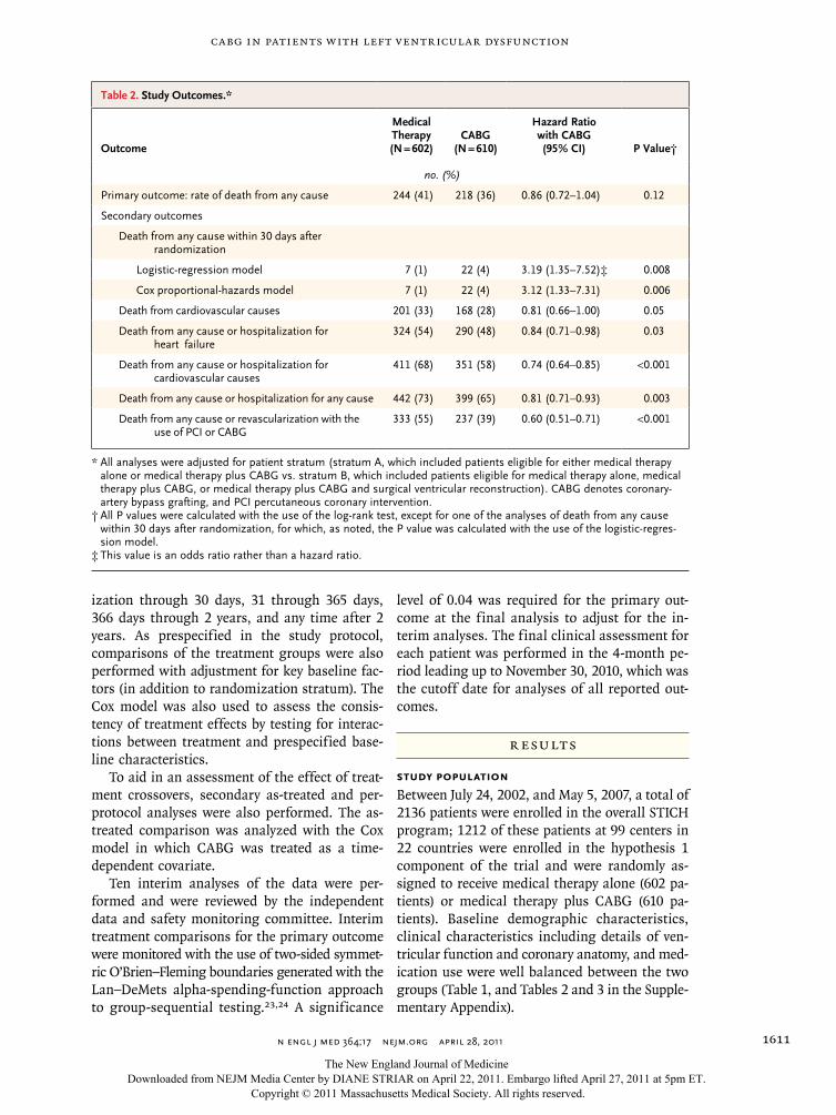
Between July 24, 2002, and May 5, 2007, a total of 2136 patients were enrolled in the overall STICH program; 1212 of these patients at 99 centers in 22 countries were enrolled in the hypothesis 1 component of the trial and were randomly as-signed to receive medical therapy alone (602 pa-tients) or medical therapy plus CABG (610 pa-tients). Baseline demographic characteristics, clinical characteristics including details of ven-tricular function and coronary anatomy, and med-ication use were well balanced between the two groups (Table 1, and Tables 2 and 3 in the Supple-mentary Appendix).
Table 2. Study Outcomes.*
Outcome
Medical Therapy(N = 602)
CABG (N = 610)
Hazard Ratio with CABG(95% CI) P Value†
no. (%)
Primary outcome: rate of death from any cause 244 (41) 218 (36) 0.86 (0.72–1.04) 0.12
Secondary outcomes
Death from any cause within 30 days after randomization
Logistic-regression model 7 (1) 22 (4) 3.19 (1.35–7.52)‡ 0.008
Cox proportional-hazards model 7 (1) 22 (4) 3.12 (1.33–7.31) 0.006
Death from cardiovascular causes 201 (33) 168 (28) 0.81 (0.66–1.00) 0.05
Death from any cause or hospitalization for heart failure
324 (54) 290 (48) 0.84 (0.71–0.98) 0.03
Death from any cause or hospitalization for cardiovascular causes
411 (68) 351 (58) 0.74 (0.64–0.85) <0.001
Death from any cause or hospitalization for any cause 442 (73) 399 (65) 0.81 (0.71–0.93) 0.003
Death from any cause or revascularization with the use of PCI or CABG
333 (55) 237 (39) 0.60 (0.51–0.71) <0.001
* All analyses were adjusted for patient stratum (stratum A, which included patients eligible for either medical therapy alone or medical therapy plus CABG vs. stratum B, which included patients eligible for medical therapy alone, medical therapy plus CABG, or medical therapy plus CABG and surgical ventricular reconstruction). CABG denotes coronary- artery bypass grafting, and PCI percutaneous coronary intervention.
† All P values were calculated with the use of the log-rank test, except for one of the analyses of death from any cause within 30 days after randomization, for which, as noted, the P value was calculated with the use of the logistic-regres-sion model.
‡ This value is an odds ratio rather than a hazard ratio.
The New England Journal of Medicine Downloaded from NEJM Media Center by DIANE STRIAR on April 22, 2011. Embargo lifted April 27, 2011 at 5pm ET.
Copyright © 2011 Massachusetts Medical Society. All rights reserved.

T h e n e w e ngl a nd j o u r na l o f m e dic i n e
n engl j med 364;17 nejm.org april 28, 20111612
Study Treatments
Of the 610 patients randomly assigned to CABG, 555 (91%) underwent CABG before the end of the study; the median time to the CABG procedure was 10 days (interquartile range, 5 to 16), with a maximum of 177 days. The surgery was performed electively in 529 of these patients (95%) and with urgency in 26 patients (5%). The median time in the intensive care unit was 52.4 hours (interquar-tile range, 40.8 to 94.6), the median duration of intubation was 16.5 hours (interquartile range, 11.1 to 22.4), and the median length of stay in the hospital was 9.0 days (interquartile range, 7.0 to 13.0). A concurrent mitral-valve operation was per-formed in 63 patients (11%). Of the 553 patients in the CABG group who underwent CABG and for whom data on arterial and venous conduits were available, 505 (91%) received at least one arterial conduit and 473 (86%) received one or more ve-nous conduits. Overall, 484 patients (87%) in the CABG group who underwent CABG had two or more distal anastomoses placed during surgery.
Of the 602 patients randomly assigned to medi-cal therapy alone, 100 (17%) underwent CABG before the end of the follow-up period. The me-dian time to the CABG procedure was 142 days (interquartile range, 19 to 576), with a maximum of 2402 days. The most common indication for crossover to CABG was progressive symptoms (40%), followed by acute decompensation (27%), patient’s or family’s decision (28%), and physi-cian’s decision (5%).
Adherence to guideline-based use of medica-tions was high throughout the study period, with-out significant differences between the treatment groups (Table 3 in the Supplementary Appendix). Postrandomization procedures are summarized in Table 4 in the Supplementary Appendix.
Follow-up
Final follow-up status was ascertained between August 1, 2010, and November 30, 2010, for 1207 of the 1212 patients (99.6%) who underwent ran-domization. During this last follow-up period, 5 patients could not be evaluated; the median time from randomization to the date of the last contact for these 5 patients, which was the date on which follow-up data were censored, was 40 months. The median length of follow-up was 56 months (interquartile range, 48 to 68), with a minimum of 12 months and a maximum of 100 months.
Outcomes
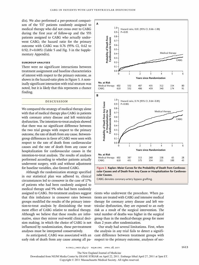
The primary outcome occurred in 244 of the 602 patients (41%) assigned to medical therapy alone and in 218 of the 610 patients (36%) assigned to CABG (hazard ratio with CABG, 0.86; 95% confi-dence interval [CI], 0.72 to 1.04; P = 0.12) (Table 2and Fig. 1). A total of 201 patients (33%) assigned to medical therapy and 168 (28%) assigned to CABG died from a cardiovascular cause (hazard ratio with CABG, 0.81; 95% CI, 0.66 to 1.00; P = 0.05) (Table 2 and Fig. 2A). Death from any cause or hospitalization for cardiovascular causes occurred in 411 patients (68%) in the medical-therapy group and 351 (58%) in the CABG group (hazard ratio with CABG, 0.74; 95% CI, 0.64 to 0.85; P<0.001) (Table 2 and Fig. 2B). The results with respect to all other secondary clinical out-comes also favored CABG, except for 30-day mor-tality (Table 2). The results of models adjusted for covariates, including those with CABG as a time-dependent covariate, are shown in Table 5 in the Supplementary Appendix.
We performed an as-treated analysis compar-ing the 592 patients who were treated with medical therapy throughout the first year after randomization and the 620 patients who under-went CABG either because they were randomly assigned to CABG or because they crossed over to CABG during year 1 of the follow-up period; the hazard ratio for the primary outcome with CABG was 0.70 (95% CI, 0.58 to 0.84; P<0.001) (Table 5 and Fig. 2 in the Supplementary Appen-
Prob
abili
ty o
f Dea
th fr
om A
ny C
ause
1.0
0.8
0.9
0.7
0.6
0.4
0.3
0.1
0.5
0.2
0.00 1 2 3 4 5 6
Years since Randomization
Hazard ratio, 0.86 (95% CI, 0.72–1.04)P=0.12
No. at RiskMedical therapyCABG
602610
532532
487486
435459
312340
154174
8091
Medical therapy
CABG
Figure 1. Kaplan–Meier Curves for the Probability of Death from Any Cause.
CABG denotes coronary-artery bypass grafting.
The New England Journal of Medicine Downloaded from NEJM Media Center by DIANE STRIAR on April 22, 2011. Embargo lifted April 27, 2011 at 5pm ET.
Copyright © 2011 Massachusetts Medical Society. All rights reserved.
CABG in Patients with Left Ventricular Dysfunction
n engl j med 364;17 nejm.org april 28, 2011 1613
dix). We also performed a per-protocol compari-son of the 537 patients randomly assigned to medical therapy who did not cross over to CABG during the first year of follow-up and the 555 patients assigned to CABG who actually under-went CABG; the hazard ratio for the primary outcome with CABG was 0.76 (95% CI, 0.62 to 0.92; P = 0.005) (Table 5 and Fig. 3 in the Supple-mentary Appendix).
Subgroup Analyses
There were no significant interactions between treatment assignment and baseline characteristics of interest with respect to the primary outcome, as shown in the hazard-ratio plots in Figure 3. A nom-inally significant interaction with trial stratum was noted, but it is likely that this represents a chance finding.
Discussion
We compared the strategy of medical therapy alone with that of medical therapy plus CABG in patients with coronary artery disease and left ventricular dysfunction. The intention-to-treat analysis showed that there was no significant difference between the two trial groups with respect to the primary outcome, the rate of death from any cause. Between-group differences in favor of CABG were seen with respect to the rate of death from cardiovascular causes and the rate of death from any cause or hospitalization for cardiovascular causes in the intention-to-treat analysis. The results of analyses performed according to whether patients actually underwent surgery, with and without adjustment for baseline variables, also favored CABG.
Although the randomization strategy specified in our statistical plan was adhered to, clinical circumstances led to crossover in the case of 17% of patients who had been randomly assigned to medical therapy and 9% who had been randomly assigned to CABG. Per-treatment analyses suggest that this imbalance in crossover rates between groups modified the results of the primary inten-tion-to-treat analysis by diminishing the treat-ment effect of CABG relative to medical therapy. Although we believe that these results are infor-mative, since they mirror real-world clinical deci-sion making, in which the choice of CABG is not influenced by randomization, these per-treatment analyses must be interpreted conservatively.
As anticipated, CABG was associated with an early risk of death from any cause among all pa-
tients who underwent the procedure. When pa-tients are treated with CABG and intensive medical therapy for coronary artery disease and left ven-tricular dysfunction, they are exposed to an early risk as a result of the surgical intervention. The total number of deaths was higher in the surgical group than in the medical-therapy group for more than 2 years after randomization.
Our study had several limitations. First, when the analysis in any trial fails to detect a signifi-cant difference between treatment groups with respect to the primary outcome, analyses of sec-
Prob
abili
ty o
f Dea
th fr
omC
ardi
ovas
cula
r C
ause
s
1.0
0.8
0.9
0.7
0.6
0.4
0.3
0.1
0.5
0.2
0.00 1 2 3 4 5 6
Years since Randomization
B
A
Hazard ratio, 0.81 (95% CI, 0.66–1.00)P=0.05
No. at RiskMedical therapyCABG
602610
532532
487486
435459
312340
154174
8091
Medical therapy
CABG
Prob
abili
ty o
f Dea
th fr
om A
ny C
ause
or H
ospi
taliz
atio
n fo
rC
ardi
ovas
cula
r C
ause
s
1.0
0.8
0.9
0.7
0.6
0.4
0.3
0.1
0.5
0.2
0.00 1 2 3 4 5 6
Years since Randomization
Hazard ratio, 0.74 (95% CI, 0.64–0.85)P<0.001
No. at RiskMedical therapyCABG
602610
387431
315375
260334
158221
65100
2843
Medical therapy
CABG
Figure 2. Kaplan–Meier Curves for the Probability of Death from Cardiovas-cular Causes and of Death from Any Cause or Hospitalization for Cardiovas-cular Causes.
CABG denotes coronary-artery bypass grafting.
The New England Journal of Medicine Downloaded from NEJM Media Center by DIANE STRIAR on April 22, 2011. Embargo lifted April 27, 2011 at 5pm ET.
Copyright © 2011 Massachusetts Medical Society. All rights reserved.

T h e n e w e ngl a nd j o u r na l o f m e dic i n e
n engl j med 364;17 nejm.org april 28, 20111614
ondary outcomes showing a benefit must inevi-tably be considered to be somewhat provisional. Second, the between-group difference with re-spect to death from cardiovascular causes, which
is presumably the mortality outcome most likely to be influenced by CABG, had a P value of 0.05, which would not have been significant if it had been adjusted for multiple testing. Given the re-
All subjects
Age
≥65 yr
<65 yr
Sex
Male
Female
Race or ethnic group
Hispanic, Latino, or nonwhite
White
Region
Poland
United States
Canada
Western Europe
Other
Current NYHA class
I or II
III or IV
LVEF (best available)
≤27%
>27%
Stratum
A
B
Baseline diabetes
No
Yes
CCS angina class
0, I, or II
III or IV
No. of vessels with ≥50% stenosis
1 or 2
3
≥50% Stenosis of LM or ≥75% stenosis of PLAD
No
Yes
Mitral regurgitation
None or trace
Mild (≤2+)
Moderate or severe (3+ or 4+)
No. of SubjectsSubgroupP Value forInteraction
1212
396
816
1064
148
421
791
319
120
123
87
563
765
447
612
600
1061
151
734
478
1154
58
478
733
373
838
435
554
220
0.41
0.61
0.09
0.39
0.83
0.20
0.01
0.56
0.30
0.23
0.40
0.59
0.50 1.00 2.00 4.00
Medical TherapyBetter
CABGBetter
Hazard Ratio (95% CI)
0.86 (0.72–1.04)
0.77 (0.59–1.01)0.97 (0.69–1.36)
0.98 (0.73–1.32)0.79 (0.62–0.99)
0.84 (0.70–1.01)1.26 (0.57–2.79)
0.92 (0.70–1.22)0.83 (0.65–1.05)
0.97 (0.73–1.29)
0.77 (0.60–0.98)
0.94 (0.78–1.15)
0.48 (0.28–0.81)
0.82 (0.66–1.02)0.97 (0.69–1.35)
0.92 (0.63–1.35)
0.84 (0.63–1.12)
0.73 (0.54–0.97)
0.87 (0.69–1.11)
1.38 (0.77–2.47)
0.87 (0.48–1.60)
0.95 (0.68–1.33)
0.81 (0.47–1.40)
0.96 (0.77–1.19)
0.68 (0.49–0.95)
0.75 (0.42–1.31)0.87 (0.72–1.06)
0.80 (0.63–1.01)
0.25
0.93 (0.70–1.23)
Figure 3. Hazard Ratio for Death from Any Cause, According to Subgroup.
Stratum A included patients eligible for either medical therapy alone or medical therapy plus coronary-artery bypass grafting (CABG), and stratum B included patients eligible for medical therapy alone, medical therapy plus CABG, or medical therapy plus CABG and surgical ventricular reconstruction. The Canadian Cardiovascular Society (CCS) an-gina classification ranges from class 0, which indicates no symptoms, to class IV, which indicates angina at any level of physical exertion. LM denotes left main coronary artery, LVEF left ventricular ejection fraction, NYHA New York Heart Association, and PLAD proximal left anterior descending artery.
The New England Journal of Medicine Downloaded from NEJM Media Center by DIANE STRIAR on April 22, 2011. Embargo lifted April 27, 2011 at 5pm ET.
Copyright © 2011 Massachusetts Medical Society. All rights reserved.

CABG in Patients with Left Ventricular Dysfunction
n engl j med 364;17 nejm.org april 28, 2011 1615
sults of other secondary analyses, we believe that the lack of an unequivocally significant between-group difference in the outcome of death from any given cause was more likely to have been due to limited power and limited duration of follow-up than to a true lack of benefit of CABG. Finally, since the trial was not blinded (and could not realistically have been blinded), the nonfatal outcomes (e.g., hospitalization due to various causes) could have been influenced by the clini-cian’s knowledge of the treatment the patient received.
Clinical decision making for patients with coronary artery disease associated with left ven-tricular dysfunction has been constrained by the absence of randomized, comparative data on re-vascularization. In fact, small subgroup analyses of the previous randomized trials of CABG in-volving patients with chronic stable angina dis-suaded many clinicians from pursuing diagnostic assessments for coronary artery disease or revas-cularization in patients presenting with heart failure.8,9 This approach continues to be sup-ported by guidelines.10-13 In the absence of direct evidence, attention has shifted toward the detec-tion of acutely and chronically underperfused but viable myocardium (i.e., myocardium that exhib-its stunning or hibernation), a condition for which functional recovery may be achieved with reperfusion.25 Imaging strategies15,26 and clinical
factors are routinely used to select patients who have the highest likelihood of functional recov-ery, as well as those in whom CABG should be avoided. In another report in this issue of the Journal, STICH investigators provide additional information on the interaction between viable myocardium and treatment effect.27
In summary, the STICH trial compared medi-cal therapy alone with medical therapy plus CABG in patients with coronary artery disease and left ventricular dysfunction. There was no significant difference between the two study groups with respect to the primary end point of the rate of death from any cause. The rates of death from cardiovascular causes and of death from any cause or hospitalization for cardiac causes were lower among patients assigned to CABG than among those assigned to medical therapy.
Supported by grants (U01HL69015 and U01HL69013) from the National Heart, Lung, and Blood Institute and by Abbott Laboratories.
Dr. Velazquez reports receiving consulting fees from Novar-tis, Gilead, and Boehringer Ingelheim Pharmaceuticals; Dr. Abraham, consulting fees from Medtronic, St. Jude Medical, Biotronik, and CardioMEMS; and Dr. Rouleau, consulting fees and grant support from Novartis. No other potential conflict of interest relevant to this article was reported.
Disclosure forms provided by the authors are available with the full text of this article at NEJM.org.
We thank Vanessa Moore, Doreen Bain, Anthony Doll, and Elizabeth Cook of the Duke Clinical Research Institute for their assistance with the preparation of the manuscript, and all the patients in the STICH trial for their participation in the study.
appendixThe authors’ affiliations are as follows: Division of Cardiovascular Medicine (E.J.V., C.M.O.), Departments of Biostatistics and Bioinfor-matics (K.L.L.) and Surgery (R.H.J.), and Duke Clinical Research Institute (L.S., G.R.R.), Duke University Medical Center, Durham, NC; Second Department of Cardiac Surgery, Medical University of Silesia, Katowice (M.A.D.), and National Institute of Cardiology, Warsaw (H.S.) — both in Poland; SAL Hospital and Medical Institute, Ahmedabad (A.J.), and Center for Chronic Disease Control, New Delhi (D.P.) — both in India; Division of Cardiovascular Sciences, National Heart, Lung, and Blood Institute, Bethesda, MD (G.S.); Research Institute of Circulation Pathology, Novosibirsk, Russia (A.M.); Division of Cardiac Surgery, Dalhousie University and the Queen Eliza-beth II Health Sciences Centre, Halifax, NS, Canada (I.S.A.); Keck School of Medicine, Viterbi School of Engineering, University of Southern California, Los Angeles (G.P.); Dedinje Cardiovascular Institute, University of Belgrade School of Medicine, Belgrade, Serbia (S.G.); Division of Cardiovascular Medicine, Ohio State University, Columbus (W.T.A.); Departments of Cardiothoracic Surgery and Surgery, University of Melbourne, St. Vincent’s Hospital, Melbourne, VIC, Australia (M.Y.); Dipartimento Cardiovascolare Clinico e di Ricerca, Ospedali Riuniti Bergamo, Bergamo, Italy (P.F.); Scottish National Advanced Heart Failure Service, Golden Jubilee National Hospital, Glasgow, United Kingdom (M.C.P.); Siriraj Hospital, Mahidol University, Bangkok, Thailand (P.P.); Division of Cardiology, Northwestern University, Chicago (R.O.B.); and Montreal Heart Institute, Montreal (J.-L.R.).
1. Lloyd-Jones D, Adams RJ, Brown TM, et al. Heart disease and stroke statistics — 2010 update: a report from the Ameri-can Heart Association. Circulation 2010; 121(7):e46-e215.2. Dickstein K, Cohen-Solal A, Filippatos G, et al. ESC guidelines for the diagnosis and treatment of acute and chronic heart failure 2008: the Task Force for the Diag-nosis and Treatment of Acute and Chronic Heart Failure 2008 of the European Soci-ety of Cardiology: developed in collabora-
tion with the Heart Failure Association of the ESC (HFA) and endorsed by the Euro-pean Society of Intensive Care Medicine (ESICM). Eur Heart J 2008;29:2388-442.3. Gheorghiade M, Sopko G, De Luca L, et al. Navigating the crossroads of coro-nary artery disease and heart failure. Cir-culation 2006;114:1202-13.4. Coronary Artery Surgery Study (CASS): a randomized trial of coronary artery bypass surgery: survival data. Cir-culation 1983;68:939-50.
5. The Veterans Administration Coro-nary Artery Bypass Surgery Cooperative Study Group. Eleven-year survival in the Veterans Administration randomized tri-al of coronary bypass surgery for stable angina. N Engl J Med 1984;311:1333-9.6. Varnauskas E. Twelve-year follow-up of survival in the randomized European Coronary Surgery Study. N Engl J Med 1988;319:332-7.7. Passamani E, Davis KB, Gillespie MJ, Killip T. A randomized trial of coronary
References
The New England Journal of Medicine Downloaded from NEJM Media Center by DIANE STRIAR on April 22, 2011. Embargo lifted April 27, 2011 at 5pm ET.
Copyright © 2011 Massachusetts Medical Society. All rights reserved.
n engl j med 364;17 nejm.org april 28, 20111616
CABG in Patients with Left Ventricular Dysfunction
artery bypass surgery: survival of patients with a low ejection fraction. N Engl J Med 1985;312:1665-71.8. Alderman EL, Fisher LD, Litwin P, et al. Results of coronary artery surgery in patients with poor left ventricular func-tion (CASS). Circulation 1983;68:785-95.9. Yusuf S, Zucker D, Peduzzi P, et al. Ef-fect of coronary artery bypass graft sur-gery on survival: overview of 10-year results from randomised trials by the Coronary Artery Bypass Graft Surgery Trialists Col-laboration. Lancet 1994;344:563-70. [Er-ratum, Lancet 1994;344:1446.]10. Hunt SA, Abraham WT, Chin MH, et al. 2009 Focused update incorporated into the ACC/AHA 2005 Guidelines for the Diagnosis and Management of Heart Failure in Adults: a report of the Ameri-can College of Cardiology Foundation/American Heart Association Task Force on Practice Guidelines developed in col-laboration with the International Society for Heart and Lung Transplantation. J Am Coll Cardiol 2009;53(15):e1-e90. [Erra-tum, J Am Coll Cardiol 2009;54:2464.]11. Eagle KA, Guyton RA, Davidoff R, et al. ACC/AHA 2004 guideline update for coronary artery bypass graft surgery: a re-port of the American College of Cardiol-ogy/American Heart Association Task Force on Practice Guidelines (Committee to Up-date the 1999 Guidelines for Coronary Artery Bypass Graft Surgery). Circulation 2004;110(14):e340-437. [Erratum, Circu-lation 2005;111:2014.]12. Fraker TD Jr, Fihn SD, Gibbons RJ, et al. 2007 Chronic angina focused update
of the ACC/AHA 2002 guidelines for the management of patients with chronic stable angina: a report of the American College of Cardiology/American Heart Association Task Force on Practice Guide-lines Writing Group to develop the fo-cused update of the 2002 guidelines for the management of patients with chronic stable angina. J Am Coll Cardiol 2007; 50:2264-74.13. Wijns W, Kolh P, Danchin N, et al. Guidelines on myocardial revasculariza-tion: the Task Force on Myocardial Revas-cularization of the European Society of Cardiology (ESC) and the European As-sociation for Cardio-Thoracic Surgery (EACTS). Eur Heart J 2010;31:2501-55.14. O’Connor CM, Velazquez EJ, Gardner LH, et al. Comparison of coronary artery bypass grafting versus medical therapy on long-term outcome in patients with ische-mic cardiomyopathy (a 25-year experience from the Duke Cardiovascular Disease Da-tabank). Am J Cardiol 2002;90:101-7.15. Cleland JG, Calvert M, Freemantle N, et al. The Heart Failure Revascularisation Trial (HEART). Eur J Heart Fail 2011; 13:227-33.16. Chareonthaitawee P, Gersh BJ, Araoz PA, Gibbons RJ. Revascularization in se-vere left ventricular dysfunction: the role of viability testing. J Am Coll Cardiol 2005;46:567-74.17. Velazquez EJ, Lee KL, O’Connor CM, et al. The rationale and design of the Sur-gical Treatment for Ischemic Heart Fail-ure (STICH) trial. J Thorac Cardiovasc Surg 2007;134:1540-7.
18. Jones RH, White H, Velazquez EJ, et al. STICH (Surgical Treatment for Is chemic Heart Failure) trial enrollment. J Am Coll Cardiol 2010;56:490-8.19. Jones RH, Velazquez EJ, Michler RE, et al. Coronary bypass surgery with or without surgical ventricular reconstruc-tion. N Engl J Med 2009;360:1705-17.20. Kaplan EL, Meier P. Nonparametric estimation from incomplete observations. J Am Stat Assoc 1958;53:457-81.21. Cox DR. Regression models and life-tables. J R Stat Soc [B] 1972;34:187-220.22. Breslow N. Covariance analysis of censored survival data. Biometrics 1974; 30:89-99.23. Lan KKG, DeMets DL. Discrete se-quential boundaries for clinical trials. Biometrika 1983;70:659-63.24. O’Brien PC, Fleming TR. A multiple testing procedure for clinical trials. Bio-metrics 1979;35:549-56.25. Braunwald E, Rutherford JD. Revers-ible ischemic left ventricular dysfunction: evidence for the “hibernating myocardi-um.” J Am Coll Cardiol 1986;8:1467-70.26. Beanlands RS, Nichol G, Huszti E, et al. F-18-fluorodeoxyglucose positron emission tomography imaging-assisted management of patients with severe left ventricular dys-function and suspected coronary disease: a randomized, controlled trial (PARR-2). J Am Coll Cardiol 2007;50:2002-12.27. Bonow RO, Maurer G, Lee KL, et al. Myocardial viability and survival in ische-mic left ventricular dysfunction. N Engl J Med 2011;364:1617-25.Copyright © 2011 Massachusetts Medical Society.
an nejm app for iphone
The NEJM Image Challenge app brings a popular online feature to the smartphone. Optimized for viewing on the iPhone and iPod Touch, the Image Challenge app lets
you test your diagnostic skills anytime, anywhere. The Image Challenge app randomly selects from 300 challenging clinical photos published in NEJM, with a new image added each week. View an image, choose your answer,
get immediate feedback, and see how others answered. The Image Challenge app is available at the iTunes App Store.
The New England Journal of Medicine Downloaded from NEJM Media Center by DIANE STRIAR on April 22, 2011. Embargo lifted April 27, 2011 at 5pm ET.
Copyright © 2011 Massachusetts Medical Society. All rights reserved.
Copyright © 2022 FDOKUMEN