Cerebral lesions in patients undergoing coronary artery bypass grafting in relation to asymptomatic...
7
Original Article Cerebral Lesions in Patients Undergoing Coronary Artery Bypass Grafting in Relation to Asymptomatic Carotid and Vertebral Artery Stenosis Sebastian Wiberg, MD, 1 Mikkel Schoos, MD, PhD, 1 Henrik Sillesen, DMSc, 2 Carsten Thomsen, DMSc, 3 Christian Hassager, DMSc, 1 Daniel Steinbrüchel, DMSc, 4 Torben Schroeder, DMSc, 5 Peter Clemmensen, DMSc, 1,6 and Henning Kelbæk, DMSc 7 1 Department of Cardiology, Copenhagen University Hospital, Copenhagen, Denmark 2 Department of Vascular Surgery, Copenhagen University Hospital, Copenhagen, Denmark 3 Department of Radiology, Copenhagen University Hospital, Copenhagen, Denmark 4 Department of Thoracic Surgery, Copenhagen University Hospital, Copenhagen, Denmark 5 Centre for Clinical Education, Copenhagen University Hospital, Copenhagen, Denmark 6 Department of Internal medicine, Nykoebing F Hospital, Nykoebing, Denmark 7 Department of Cardiology, Roskilde Hospital, Roskilde, Denmark Received: June 12, 2014; Accepted: November 1, 2014 Corresponding author: Sebastian Wiberg, MD. Department of Cardiology, Rigshospitalet, Blegdamsvej 9, DK-2100 Copenha- gen East, Denmark Tel: +45 26 92 40 30 E-mail: [email protected] Objectives: Carotid artery stenosis (CAS) and vertebral artery stenosis (VAS) are associated with cerebral infarction after coronary artery bypass graft surgery (CABG). It remains unclear whether this association is causal. We investigated the associations between neurologically asymptomatic CAS and VAS and the occurrence of subclinical cerebral lesions after CABG verified by magnetic resonance imaging. Methods: CABG patients were included and CAS and VAS were identified by magnetic resonance angiography. Cere- bral magnetic resonance imaging was performed to identify new post-operative subclinical cerebral lesions. The associa- tions between CAS/VAS post-operative cerebral lesions were investigated. Results: Forty-six patients were included in the study. 13% had significant CAS and 11% had significant VAS. Thirty-five percent had new cerebral infarction postoperatively. We found a significant association between the presence of cerebral vessel stenosis and acute cerebral infarction (67% vs. 27%, p = 0.047). However none of the patients with stenosis had isolated cerebral lesions in the ipsilateral vascu- lar territory. Conclusion: Asymptomatic CAS and VAS is common in CABG patients and is associated with an increased risk of postoperative cerebral infarction. Our study suggests that asymptomatic CAS and VAS primarily are risk markers rather than causal factors for cerebral infarction after CABG. Keywords: Carotid stenosis, cerebral infarction Introduction Ischemic stroke remains one of the most feared complica- tions to cardiac surgery. The risk of developing stroke after cardiac surgery is somewhere between 2% and 6%. 1–4) It is well known that many patients undergoing coronary artery bypass grafting (CABG) have stenosis of their internal carotid arteries due to atherosclerosis, and it is evident that both the presence and severity of this disease increase the risk of stroke. 5,6) The natural history of asymptomatic, carotid stenosis under best medical treatment is poorly understood, and it remains unclear whether asymptomatic carotid steno- sis primarily is an etiological factor for stroke or simply a marker of increased risk. 7–9) This makes the treatment of patients with asymptomatic, significant carotid artery stenosis undergoing cardiac surgery a controversial issue, often attracting polarised opinions. 7,10–13) The differing interpretations have led to varying guidelines regarding whether to screen CABG patients for carotid stenosis and in turn whether to treat asymptomatic patients with carotid endarterectomy, carotid artery stenting or simply best medical treatment. 14–17) Symptomatic vertebral artery stenosis is a major predictor of recurrent ischemic stroke in the posterior circulation. 18) In addition asymptomatic patients with vertebral artery stenosis are reported to have a higher risk of ischemic stroke than patients without this disease, however, investigation and treatment of vertebral artery territory stroke has received much less attention than that of stroke in the carotid territory. 19) The consequence of vertebral artery stenosis among CABG patients is unknown. Annals of Vascular Diseases Vol. 8, No. 1 (2015) 7 Ann Vasc Dis Vol. 8, No. 1; 2015; pp 7–13 Online March 9, 2015 ©2015 Annals of Vascular Diseases doi:10.3400/avd.oa.14-00073
-
Upload
independent -
Category
Documents
-
view
0 -
download
0
Transcript of Cerebral lesions in patients undergoing coronary artery bypass grafting in relation to asymptomatic...

Original Article
Cerebral Lesions in Patients Undergoing Coronary Artery Bypass Grafting in Relation to Asymptomatic Carotid and Vertebral Artery Stenosis
Sebastian Wiberg, MD,1 Mikkel Schoos, MD, PhD,1 Henrik Sillesen, DMSc,2 Carsten Thomsen, DMSc,3 Christian Hassager, DMSc,1 Daniel Steinbrüchel, DMSc,4 Torben Schroeder, DMSc,5 Peter Clemmensen, DMSc,1,6 and Henning Kelbæk, DMSc7
1Department of Cardiology, Copenhagen University Hospital, Copenhagen, Denmark2Department of Vascular Surgery, Copenhagen University Hospital, Copenhagen, Denmark3Department of Radiology, Copenhagen University Hospital, Copenhagen, Denmark4Department of Thoracic Surgery, Copenhagen University Hospital, Copenhagen, Denmark5Centre for Clinical Education, Copenhagen University Hospital, Copenhagen, Denmark6Department of Internal medicine, Nykoebing F Hospital, Nykoebing, Denmark7Department of Cardiology, Roskilde Hospital, Roskilde, Denmark
Received: June 12, 2014; Accepted: November 1, 2014Corresponding author: Sebastian Wiberg, MD. Department of Cardiology, Rigshospitalet, Blegdamsvej 9, DK-2100 Copenha-gen East, DenmarkTel: +45 26 92 40 30E-mail: [email protected]
Objectives: Carotid artery stenosis (CAS) and vertebral artery stenosis (VAS) are associated with cerebral infarction after coronary artery bypass graft surgery (CABG). It remains unclear whether this association is causal. We investigated the associations between neurologically asymptomatic CAS and VAS and the occurrence of subclinical cerebral lesions after CABG verifi ed by magnetic resonance imaging.Methods: CABG patients were included and CAS and VAS were identifi ed by magnetic resonance angiography. Cere-bral magnetic resonance imaging was performed to identify new post-operative subclinical cerebral lesions. The associa-tions between CAS/VAS post-operative cerebral lesions were investigated. Results: Forty-six patients were included in the study. 13% had signifi cant CAS and 11% had signifi cant VAS. Thirty-fi ve percent had new cerebral infarction postoperatively. We found a signifi cant association between the presence of cerebral vessel stenosis and acute cerebral infarction (67% vs. 27%, p = 0.047). However none of the patients with stenosis had isolated cerebral lesions in the ipsilateral vascu-lar territory.Conclusion: Asymptomatic CAS and VAS is common in CABG patients and is associated with an increased risk of
postoperative cerebral infarction. Our study suggests that asymptomatic CAS and VAS primarily are risk markers rather than causal factors for cerebral infarction after CABG.
Keywords: Carotid stenosis, cerebral infarction
IntroductionIschemic stroke remains one of the most feared complica-tions to cardiac surgery. The risk of developing stroke after cardiac surgery is somewhere between 2% and 6%.1–4) It is well known that many patients undergoing coronary artery bypass grafting (CABG) have stenosis of their internal carotid arteries due to atherosclerosis, and it is evident that both the presence and severity of this disease increase the risk of stroke.5,6)
The natural history of asymptomatic, carotid stenosis under best medical treatment is poorly understood, and it remains unclear whether asymptomatic carotid steno-sis primarily is an etiological factor for stroke or simply a marker of increased risk.7–9) This makes the treatment of patients with asymptomatic, signifi cant carotid artery stenosis undergoing cardiac surgery a controversial issue, often attracting polarised opinions.7,10–13) The differing interpretations have led to varying guidelines regarding whether to screen CABG patients for carotid stenosis and in turn whether to treat asymptomatic patients with carotid endarterectomy, carotid artery stenting or simply best medical treatment.14–17)
Symptomatic vertebral artery stenosis is a major predictor of recurrent ischemic stroke in the posterior circulation.18) In addition asymptomatic patients with vertebral artery stenosis are reported to have a higher risk of ischemic stroke than patients without this disease, however, investigation and treatment of vertebral artery territory stroke has received much less attention than that of stroke in the carotid territory.19) The consequence of vertebral artery stenosis among CABG patients is unknown.
Annals of Vascular Diseases Vol. 8, No. 1 (2015) 7
Ann Vasc Dis Vol. 8, No. 1; 2015; pp 7–13 Online March 9, 2015©2015 Annals of Vascular Diseases doi:10.3400/avd.oa.14-00073
05_01_oa_00073_Wiberg.indd 705_01_oa_00073_Wiberg.indd 7 2015/03/12 11:412015/03/12 11:41

Wiberg S, et al.
The purpose of this study was to examine the prevalence of acute subclinical cerebral infarction as well as the prevalence of signifi cant, asymptomatic carotid and ver-tebral artery stenosis in a population undergoing CABG. In addition this study sought to establish whether the presence of asymptomatic carotid and vertebral artery stenosis primarily is useful for risk prediction or for estab-lishing causality for acute subclinical cerebral infarction after CABG.
Material and Methods
The study population was derived from all patients under-going CABG at our hospital from February to August 2012. The participants were recruited on the day of admission. Inclusion criteria were indication for CABG and age above 18 years. Exclusion criteria were stroke or transient isch-emic attack (TIA) within the past 6 months in order to rule out patients with symptomatic carotid or vertebral artery stenosis. Other exclusion criteria were indication for carotid endarterectomy (CEA), simultaneous valve intervention and contraindications to magnetic resonance imaging (MRI) such as implanted cardiac pacemakers. In addition patients with claustrophobia and severe nausea were excluded. All participants provided written informed consent and the study was approved by the National Committee on Health Research Ethics, reference H-4-2011-092.
The study design was cross sectional. Contrast enhanced magnetic resonance angiography (CE-MRA) was per-formed between three and seven days after surgery in order to assess the prevalence of carotid and vertebral stenosis in the population. Concomitant to the CE-MRA the participants underwent cerebral magnetic resonance diffusion-weighted imaging (DWI) between three and seven days after surgery, which was used to assess the occurrence of per- or postoperative acute infarction in the population.
The MRI studies were obtained with a 3.0-Tesla (Siemens Verio) whole-body scanner using a standard-ized protocol. Comparison of CE-MRA and DWI was used to identify associations between carotid and verte-bral artery stenosis and subclinical cerebral infarction in the individual patients.
Contrast-enhanced magnetic resonance angiographyCE-MRA is established as an accurate non-invasive method for measuring degrees of carotid stenosis.20,21) 15 ml of Gadolinium was used as the contrast agent. Two patients had allergy to Gadolinium contrast and had Time-of-Flight MRA sequences instead of CE-MRA (Fig. 1). The CE-MRA images were blinded and presented the principal investigator minimum a month after completion of the
scans. The degree of carotid artery stenosis was assessed by measurements of the smallest transverse diameter at the level of the stenosis compared to the diameter of the distal uninvolved vessel where the arterial walls are parallel.
Stenosis
diameter of distal univolved arterysmallest transvers
=
−ee diameter
diameter of distal univolved artery× 100%
Non-signifi cant stenosis was defi ned as a stenosis diameter of <50% and signifi cant stenosis was defi ned as stenosis diameter >50%.22,23) Vertebral artery stenosis was assessed in a similar fashion.
Cerebral magnetic resonance diffusion-weighted imagingCerebral DWI is established as a highly accurate modality for identifying regions of cerebral ischemia and for distin-guishing the acute from the non-acute cerebral infarction. Acute infarction can be distinguished from non-acute infarction for up to 10 days.24,25) The DWI scans were used to establish the presence and volume of existing infarcts and to distinguish acute from non-acute cerebral infarction among the participants. The DWI scans were obtained on the same scanner as the CE-MRA. The DWI images from the cerebral MRIs were interpreted by one of the principal investigators blinded to the results of the CE-MRA studies. The images were analysed for the presence, number and volume of acute DWI lesions. Acute lesions smaller than 0.2 cm3 were reported as <0.2 cm3 due to the limited spa-tial resolution of MRI. Acute lesion volumes greater than 0.2 cm3 were reported in their actual size. (Fig. 1)
Surgical ProceduresCardiopulmonary bypass was established by aortic can-nulation and a two-stage venous cannula in the right atrium. Patients were fully heparinised (activated clotting time >480 s). Cardiac arrest was achieved with cold blood cardioplegia and extracorporeal circulation was per-formed under normothermia. Surgical access to the heart was gained through a median sternotomy in all of the patients. The left internal mammary artery and great saphenous vein were harvested. If veins were of poor quality a radial artery from the non-dominant hand was used. The left internal mammary artery was anastomosed whenever indicated to the left anterior descending coro-nary artery.
Standard anaesthetic techniques were used and intra operative monitoring was in accordance with guidelines. The surgical volume of patients and their clinical out-come are comparable to other high-volume heart centres.
Standard guidelines for anti-coagulation therapy were used. Before elective CABG anti-coagulation therapy was
8 Annals of Vascular Diseases Vol. 8, No. 1 (2015)
05_01_oa_00073_Wiberg.indd 805_01_oa_00073_Wiberg.indd 8 2015/03/12 11:412015/03/12 11:41
Asymptomatic Carotid Stenosis in CABG-Patients
level of 5% statistical signifi cance (two tailed) was assumed throughout.
Categorical variables were presented as percentages and comparison between participants and non-participants was analyzed with the χ2 statistic. If the expected cell count was <5 the Fisher’s exact test was used. Continu-ous variables were presented as mean +/– standard devi-ation if normally distributed.
Results
Baseline characteristicsA total of 298 patients underwent CABG during the inclusion period. Of these patients 46 were included as participants in the study. The participants were com-pared to the non-participants in order to evaluate possi-ble selection bias in this non-consecutive series. However, the groups were well balanced. (Table 1) Five patients (9%) suffered from preoperative atrial fi brillation. Addi-tional baseline data for the participants are presented in Table 2.
paused for 1 week. If the patient suffered from unstable angina or had stenosis of the left mainstem, acetylsali-cylic acid and low-molecular-weight heparin was contin-ued until the evening before CABG. In case of treatment with fondaparinux this was paused 48 h before CABG. Treatment with warfarin due to atrial fi brillation was paused 3 days prior to CABG. During surgery 3.5 mg/kg of unfractionated heparin was given before cardiopul-monary bypass was initiated resulting in an activated clotting time above 480 s. After cardiopulmonary bypass was fi nished protamine sulfate was given until the acti-vated clotting time was below 125 s. In the postoperative period of immobilization 4000 international units of enoxaparin was given once daily. Lifelong postoperative treatment with acetylsalicylic acid as well as patients’ previous anti-coagulation therapy was restarted on the second day after CABG.
Statistical AnalysisStatistical analyses were performed using the SAS Enter-prise Guide statistical software version 4.3. A nominal
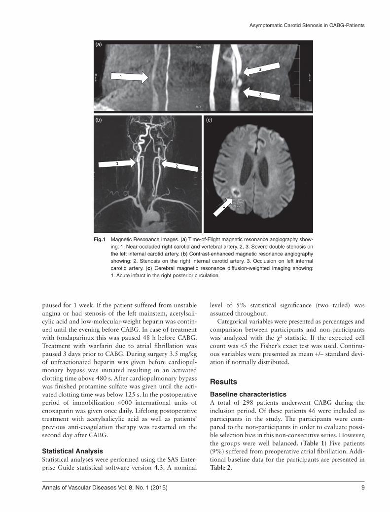
Fig.1 Magnetic Resonance Images. (a) Time-of-Flight magnetic resonance angiography show-ing: 1. Near-occluded right carotid and vertebral artery. 2, 3. Severe double stenosis on the left internal carotid artery. (b) Contrast-enhanced magnetic resonance angiography showing: 2. Stenosis on the right internal carotid artery. 3. Occlusion on left internal carotid artery. (c) Cerebral magnetic resonance diffusion-weighted imaging showing: 1. Acute infarct in the right posterior circulation.
(a)
(b) (c)
Annals of Vascular Diseases Vol. 8, No. 1 (2015) 9
05_01_oa_00073_Wiberg.indd 905_01_oa_00073_Wiberg.indd 9 2015/03/12 11:412015/03/12 11:41

Wiberg S, et al.
Table 1 Baseline characteristics of participating vs. non-participating patients
Participants Non-participants p
46 252
Male Gender (%) 91.3 82.9 0.150Age (years) 67.0 ± 9.0 67.5 ± 9.8 0.765Dyslipidemia (%) 73.9 83.6 0.115Hypertension (%) 82.2 72.0 0.152Family history (%) 58.5 58.4 0.987Smoker (%) 60.9 58.2 0.732Diabetes (%) 22.2 27.5 0.406Renal Failure (%) 4.4 6.4 0.257NYHA Class 0–2 (%) 84.2 72.7NYHA Class >2 (%) 15.8 27.3 0.412CCS Class 0–2 (%) 70.7 73.1CCS Class >2 (%) 29.3 26.9 0.086Previous Stroke or TIA (%) 6.5 13.6 0.185Previous MI (%) 54.4 52.6 0.826
MI: Myocardial infarction; TIA: Transient ischemic attack; NYHA: New York Heart Association functional classifi cation; CCS: Canadian Cardiovascular Society grading of angina pectoris
Table 2 Baseline characteristics of participants
n (%)
Referal diagnosis Stable angina 21 (47)Unstable angina 2 (4)
NSTEMI 10 (22)STEMI 8 (18)
Arrhythmia 1 (2)Heart Failure 3 (7)
Institio 1 (2)Preoperative atrial fi brillation 5 (9)Number of coronary lesions 1 2 (4)
2 6 (13)3 16 (35)4 16 (35)5 3 (7)6 2 (4)7 1 (2)
Coronary vessel disease 1-VD 2 (4)2-VD 19 (42)3-VD 25 (54)
Left ventricular ejection fraction (%) >49 26 (57)30–49 15 (33)
<30 5 (11)Left main stem disease 16 (36)
VD: Vessel disease
Patient outcomesPostoperatively one participant (no. 94) suffered a clinical stroke (day 0) with light paresis of the upper left extrem-ity. Description of the cerebral MRI-scan for this patient can be viewed in Table 3. The paresis was resolving on day 2. There was no development of neurological symp-toms among the other participants.
CE-MRAA total of 11 out of 46 participants (24%) had signs of carotid artery plaque (i.e., all carotid stenoses visible on CE-MRA). Three of the participants had bilaterally dis-tributed plaques. Six participants (13%) had signifi cant
carotid artery stenosis (i.e., carotid stenosis diameter >50%), and fi ve participants (11%) had signifi cant verte-bral artery stenosis.
DWI 16 out of 46 participants (35%) had evidence of acute cerebral infarction ranging from <0.2 cm3 to 3.7 cm3 (Fig. 1). Twelve of the participants with acute cerebral infarction (25%) had multiple lesions, and in 10 partic-ipants (22%), the multiple lesions occurred in more than one vascular territory. Eight of the participants with multiple lesions had bilaterally distributed lesions (Table 3).
10 Annals of Vascular Diseases Vol. 8, No. 1 (2015)
05_01_oa_00073_Wiberg.indd 1005_01_oa_00073_Wiberg.indd 10 2015/03/12 11:412015/03/12 11:41

Asymptomatic Carotid Stenosis in CABG-Patients
More than one third of the patients (39%) had peri- or postoperative atrial fi brillation during their admission. However, the incidence of cerebral infarction was identi-cal in the group with versus without atrial fi brillation (33% vs. 36%, p = 0.869). This suggests that peri- or postoperative atrial fi brillation is not a risk factor for cerebral infarction after CABG. The reason for this fi nd-ing can well be, that patients during admission are treated rapidly as soon as atrial fi brillation is diagnosed, and that thrombus thus does not form in the atrium (Table 4).
Among patients with stenosis of the cerebral vessels and acute cerebral infarction, none had isolated cerebral lesions in the vascular territory of the ipsilateral stenotic artery, which should have been the case, had the carotid stenosis been the etiological factor for the infarction. The only patient with isolated infarction in the medial circu-lation did not have any signs of carotid artery stenosis and the six patients with isolated infarction in the poste-rior circulation did not have vertebral artery stenosis.
Finally, in the fi ve patients with multiple cerebral lesions, where one of the infarct locations could be caused by emboli from the stenotic artery (termed ‘partial’ in Table 3) all had additional cerebral lesions with no asso-ciation between the location of the stenotic artery and the vascular territory. Thus, asymptomatic carotid and vertebral artery stenosis primarily seem to be markers for increased infarction risk rather than ethological factors (Table 3).
There are a number of limitations to this study. Pri-marily, the study was slightly underpowered, yielding a strong yet insignifi cant trend regarding the association of isolated asymptomatic carotid stenosis and post-operative cerebral infarction. Never the less this study is the larg-est of its kind. The inclusion of the participants was
Association between carotid and vertebral stenosis and DWI verified cerebral lesionsFour out of six participants (67%) with signifi cant carotid artery stenosis had acute cerebral infarction in contrast to 12 out of 40 participants (30%) without signifi cant carotid artery stenosis (p = 0.163).
Four out of fi ve participants (80%) with vertebral artery stenosis had acute cerebral infarction, whereas 12 out of 41 participants (29%) without vertebral artery stenosis developed acute cerebral infarction (p = 0.043) (Table 4).
Discussion
In this study population, which was very similar to the background population of consecutively operated CABG patients (Table 1), we found that approximately one in three CABG patients (33%) developed DWI-verifi ed acute subclinical cerebral infarctions (range, 1 to 12 lesions) during or shortly after on-pump CABG with normo ther-mic circulatory arrest. One patient (2%) suffered a clini-cal stroke. The rate of subclinical infarction remains unchanged since 200226,27) which is highly clinically rele-vant since subclinical infarctions are associated with cog-nitive decline.28)
Two in three patients (67%) with either asymptomatic signifi cant carotid or vertebral artery stenosis developed subclinical cerebral infarction compared to only a quarter of the patients (27%) without stenoses. This confi rms that stenosis of the cerebral vessels is an important risk factor for cerebral infarction after CABG. On the other hand two in three patients with subclinical cerebral infarction (63%) did not have either carotid artery or vertebral artery stenosis, confi rming that many other factors contribute to the development of cerebral infarction after CABG.
Table 3 Overview of Participants with DWI identifi ed acute cerebral infarcts
Participant no.No. of
infarctsTotal Infarct
volume (cm3)Vascular territories
Right/Left side
Carotid StenosisVertebral stenosis
Corresponding Stenosis/Infarct
160 1 <0.2 Posterior Left None None No194 1 0.3 Posterior Left Right None No224 1 <0.2 Medial Left None None No244 1 <0.2 Posterior Right None None No240 2 0.2 Anterior, Posterior Right None Right Partial297 2 <0.2 Posterior Right None None No265 2 <0.2 Medial, Posterior Both None None No268 2 3.7 Posterior Right None None No79 3 0.8 Posterior Both None None No238 3 <0.2 Medial, Posterior Right None Left Partial81 4 0.3 All territories Both Occlusion, Right Right Partial148 5 0.3 Medial, Posterior Both Occlusion, Left None Partial58 7 0.7 Medial, Posterior Both None None No298 8 2.5 All territories Both Right Occlusion Left Partial206 10 0.3 All territories Both None None No94∗ 12 2.1 All territories Both None None Partial
DWI: Cerebral magnetic resonance diffusion-weighted imaging; ∗: Patient suffered a clinical stroke
Annals of Vascular Diseases Vol. 8, No. 1 (2015) 11
05_01_oa_00073_Wiberg.indd 1105_01_oa_00073_Wiberg.indd 11 2015/03/12 11:412015/03/12 11:41

Wiberg S, et al.
6) Naylor AR, Mehta Z, Rothwell PM, et al. Carotid artery disease and stroke during coronary artery bypass: a critical review of the literature. Eur J Vasc Endovasc Surg 2002; 23: 283-94.
7) Li Y, Walicki D, Mathiesen C, et al. Strokes after cardiac surgery and relationship to carotid stenosis. Arch Neurol 2009; 66: 1091-6.
8) Ghosh J, Murray D, Khwaja N, et al. The infl uence of asymptomatic signifi cant carotid disease on mortality and morbidity in patients undergoing coronary artery bypass surgery. Eur J Vasc Endovasc Surg 2005; 29: 88-90.
9) Baiou D, Karageorge A, Spyt T, et al. Patients undergoing cardiac surgery with asymptomatic unilateral carotid ste-noses have a low risk of peri-operative stroke. Eur J Vasc Endovasc Surg 2009; 38: 556-9.
10) Perler BA. Managing patients with clinically signifi cant cardiac and carotid artery occlusive disease. Arch Surg 2009; 144: 998-9.
11) Borger MA. Preventing stroke during coronary bypass: are we focusing on the wrong culprit? J Card Surg 2005; 20: 58-9.
12) Akins CW, Hilgenberg AD, Vlahakes GJ, et al. Late results of combined carotid and coronary surgery using actual versus actuarial methodology. Ann Thorac Surg 2005; 80: 2091-7.
13) Ricotta JJ, Wall LP, Blackstone E. The infl uence of concur-rent carotid endarterectomy on coronary bypass: a case-controlled study. J Vasc Surg 2005; 41: 397-401.
14) Eagle KA, Guyton RA, Davidoff R, et al. ACC/AHA 2004 guideline update for coronary artery bypass graft surgery: summary article. A report of the American College of Car-diology/American Heart Association Task Force on Prac-tice Guidelines (Committee to Update the 1999 Guidelines for Coronary Artery Bypass Graft Surgery). J Am Coll Cardiol 2004; 44: e213-e310.
15) Chaturvedi S, Bruno A, Feasby T, et al. Carotid endarterectomy–an evidence-based review: report of the Therapeutics and Technology Assessment Subcommittee of the American Academy of Neurology. Neurology 2005; 65: 794-801.
16) Wolff T, Guirguis-Blake J, Miller T, et al. Screening for carotid artery stenosis: an update of the evidence for the U.S. Preventive Services Task Force. Ann Intern Med 2007; 147: 860-70.
17) Brott TG, Halperin JL, Abbara S, et al. 2011 ASA/ACCF/AHA/AANN/AANS/ACR/ASNR/CNS/SAIP/SCAI/SIR/SNIS/SVM/SVS guideline on the management of patients
Table 4 The association between stenotic arteries and DWI-verifi ed cerebral infarction
CE-MRA results DWI lesions present DWI lesion absent p
Carotid stenosis present 4 (67%) 2 (33%)Carotid stenosis absent 12 (30%) 28 (70%) 0.163Vertebral artery stenosis present 4 (80%) 1 (20%)Vertebral artery stenosis absent 12 (29%) 29 (71%) 0.043Carotid stenosis or vertebral stenosis present 6 (67%) 3 (33%)Carotid stenosis and vertebral stenosis absent 10 (27%) 27 (73%) 0.047Peri- and postoperative atrial fi brillation present 6 (33%) 12 (67%)Peri- and postoperative atrial fi brillation absent 10 (36%) 18 (64%) 0.869
CE-MRA: Contrast-enhanced magnetic resonance angiography; DWI: Cerebral magnetic resonance diffusion-weighted imaging
non-consecutive; however the baseline characteristics were similar between participants and all CABG patients. The CE-MRA scans were performed postoperatively. Prefera-bly, all CE-MRA scans should have been completed before surgery and DWI scans after surgery. However, since carotid plaque morphology does not change signifi cantly over a one year period, the impact is small.29)
Conclusion
Development of subclinical cerebral infarction after on-pump CABG is frequent and occurs in approximately one third of patients. Asymptomatic, signifi cant carotid artery stenosis is present in 13% of CABG patients. Carotid and vertebral artery stenoses are associated with an increased risk of sub-clinical cerebral infarction after CABG surgery. However, the lack of connection between the location of the cere-bral vessel disease and subsequent post-operative cerebral infarction suggests that the atherosclerotic burden in the arteries supplying the brain primarily is useful for risk pre-diction rather than establishing causality.
Disclosure statement
All authors have no confl icts of interest.
References
1) Hogue CW Jr, Murphy SF, Schechtman KB, et al. Risk fac-tors for early or delayed stroke after cardiac surgery. Circu-lation 1999; 100: 642-7.
2) Tarakji KG, Sabik JF 3rd, Bhudia SK, et al. Temporal onset, risk factors, and outcomes associated with stroke after cor-onary artery bypass grafting. JAMA 2011; 305: 381-90.
3) Bucerius J, Gummert JF, Borger MA, et al. Stroke after car-diac surgery: a risk factor analysis of 16,184 consecutive adult patients. Ann Thorac Surg 2003; 75: 472-8.
4) Kanemitsu S, Tanabe S, Ohue K, et al. Improve morbidity and mortality in coronary artery bypass graft surgery for severe atherosclerosis. Ann Vasc Dis 2011; 4: 93-8.
5) Nicolaides AN, Kakkos SK, Kyriacou E, et al. Asymptom-atic internal carotid artery stenosis and cerebrovascular risk stratifi cation. J Vasc Surg 2010; 52: 1486-96.
12 Annals of Vascular Diseases Vol. 8, No. 1 (2015)
05_01_oa_00073_Wiberg.indd 1205_01_oa_00073_Wiberg.indd 12 2015/03/12 11:412015/03/12 11:41

Asymptomatic Carotid Stenosis in CABG-Patients
22) Eliasziw M, Rankin RN, Fox AJ, et al. Accuracy and prognostic consequences of ultrasonography in identify-ing severe carotid artery stenosis. North American Symp-tomatic Carotid Endarterectomy Trial (NASCET) Group. Stroke 26: 1747-52.
23) Moneta GL, Edwards JM, Chitwood RW, et al. Correla-tion of North American Symptomatic Carotid Endarterec-tomy Trial (NASCET) angiographic defi nition of 70% to 99% internal carotid artery stenosis with duplex scanning. J Vasc Surg 1993; 17: 152-7.
24) Singer MB, Chong J, Lu D, et al. Diffusion-weighted MRI in acute subcortical infarction. Stroke 1998; 29: 133-6.
25) Wityk RJ, Goldsborough MA, Hillis A, et al. Diffusion- and perfusion-weighted brain magnetic resonance imag-ing in patients with neurologic complications after cardiac surgery. Arch Neurol 2001; 58: 571-6.
26) Restrepo L, Wityk RJ, Grega MA, et al. Diffusion- and perfusion-weighted magnetic resonance imaging of the brain before and after coronary artery bypass grafting sur-gery. Stroke 2002; 33: 2909-15.
27) Bendszus M, Reents W, Franke D, et al. Brain damage after coronary artery bypass grafting. Arch Neurol 2002; 59: 1090-5.
28) Barber PA, Hach S, Tippett LJ, et al. Cerebral ischemic lesions on diffusion-weighted imaging are associated with neurocognitive decline after cardiac surgery. Stroke 2008; 39: 1427-33.
29) Kwee RM, Truijman MT, van Oostenbrugge RJ, et al. Longitudinal MRI study on the natural history of carotid artery plaques in symptomatic patients. PLoS One 2012; 7: e42472.
with extracranial carotid and vertebral artery disease: executive summary: a report of the American College of Cardiology Foundation/American Heart Association Task Force on Practice Guidelines, and the American Stroke Association, American Association of Neuroscience Nurses, American Association of Neurological Surgeons, American College of Radiology, American Society of Neu-roradiology, Congress of Neurological Surgeons, Society of Atherosclerosis Imaging and Prevention, Society for Cardiovascular Angiography and Interventions, Society of Interventional Radiology, Society of NeuroInterventional Surgery, Society for Vascular Medicine, and Society for Vascular Surgery Developed in Collaboration With the American Academy of Neurology and Society of Cardio-vascular Computed Tomography. Catheter Cardiovasc Interv. 2013; 81: E76-123.
18) Gulli G, Marquardt L, Rothwell PM, et al. Stroke risk after posterior circulation stroke/transient ischemic attack and its relationship to site of vertebrobasilar stenosis: pooled data analysis from prospective studies. Stroke 2013; 44: 598-604.
19) Compter A, van der Worp HB, Algra A, et al. Prevalence and prognosis of asymptomatic vertebral artery origin ste-nosis in patients with clinically manifest arterial disease. Stroke 2011; 42: 2795-800.
20) King-Im JM, Young V, Gillard JH. Carotid-artery imag-ing in the diagnosis and management of patients at risk of stroke. Lancet Neurol 2009; 8: 569-80.
21) Petrou M, Foerster BR. Relative roles of magnetic resonance angiography and computed tomographic angiography in evaluation of symptomatic carotid stenosis: a critically appraised topic. Semin Roentgenol 2009; 44: 184-7.
Annals of Vascular Diseases Vol. 8, No. 1 (2015) 13
05_01_oa_00073_Wiberg.indd 1305_01_oa_00073_Wiberg.indd 13 2015/03/12 11:412015/03/12 11:41