Patient-specific hemodynamic analysis of small internal carotid artery-ophthalmic artery aneurysms
7
Aneurysm Patient-specific hemodynamic analysis of small internal carotid artery-ophthalmic artery aneurysms Aichi Chien, PhD a, ⁎ , Satoshi Tateshima, MD a , James Sayre, PhD b , Marcelo Castro, PhD c , Juan Cebral, PhD c , Fernando Viñuela, MD a a Division of Interventional Neuroradiology, David Geffen School of Medicine, University of California, Los Angeles, CA 90095, USA b Department of Biostatistics, School of Public Health, University of California, Los Angeles, CA 90095, USA c Department of Computational Sciences, George Mason University, Fairfax, VA 22030, USA Received 25 July 2008; accepted 31 December 2008 Abstract Background: Prophylactic treatment of unruptured small brain aneurysms is still controversial due to the low risk of rupture. Distinguishing which small aneurysms are at risk for rupture has become important for treatment. Previous studies have indicated a variety of hemodynamic properties that may influence aneurysm rupture. This study uses hemodynamic principles to evaluate these in the context of ruptured and unruptured small aneurysms in a single location. Methods: Eight small internal carotid artery-ophthalmic artery (ICA-Oph) aneurysms (b10 mm) were selected from the University of California, Los Angeles, database. We analyzed rupture-related hemodynamic characteristics including flow patterns, wall shear stress (WSS), and flow impingement using previously developed patient-specific computational fluid dynamics software. Results: Most ruptured aneurysms had complicated flow patterns in the aneurysm domes, but all of the unruptured cases showed a simple vortex. A reduction in flow velocity between the parent artery and the aneurysm sac was found in all the cases. Inside the aneurysms, the highest flow velocities were found either at the apex or neck. We also observed a trend of higher and more inhomogeneous WSS distribution within ruptured aneurysms (10.66 ± 5.99 Pa) in comparison with the unruptured ones (6.31 ± 6.47 Pa) (P b .01). Conclusion: A comparison of hemodynamic properties between ruptured and unruptured small ICA- Oph aneurysms found that some hemodynamic properties vary between small aneurysms although they are similar in size and share the same anatomical location. In particular, WSS may be a useful hemodynamic factor for studying small aneurysm rupture. © 2009 Published by Elsevier Inc. Keywords: Cerebral aneurysm; Hemodynamics; Flow analysis; Wall shear stress 1. Introduction In general, prophylactic treatment of unruptured brain aneurysms remains controversial [9,11,14,25-27]. Interna- tional studies have shown that the risk of aneurysm rupture increases as the aneurysm size increases, supporting treatment of larger aneurysms [27]. On the other hand, many small aneurysms are observed conservatively due to the lower risk of rupture compared with the risk of morbidity and mortality related to treatments. Recent advancements in medical imaging technology have helped the early detection of unruptured brain aneurysms, and more small aneurysms are found before rupture [2,14]. Among conservatively observed small brain aneurysms, some grow over time with a corresponding increase in the risk of rupture; however, reports have also shown that certain small aneurysms rupture without evidence of any growth [8,13,25]. Although they are Available online at www.sciencedirect.com Surgical Neurology xx (2009) xxx – xxx www.surgicalneurology-online.com Abbreviations: CFD, computational fluid dynamics; ICA-Oph, internal carotid artery-ophthalmic artery; SD, standard deviation; WSS, wall shear stress. ⁎ Corresponding author. Tel.: +1 310 794 7921; fax: +1 310 206 5958 E-mail address: [email protected] (A. Chien). 0090-3019/$ – see front matter © 2009 Published by Elsevier Inc. doi:10.1016/j.surneu.2008.12.013 ARTICLE IN PRESS
-
Upload
independent -
Category
Documents
-
view
0 -
download
0
Transcript of Patient-specific hemodynamic analysis of small internal carotid artery-ophthalmic artery aneurysms

Available online at www.sciencedirect.com
Surgical Neurology xx (2009) xxx–xxxwww.surgicalneurology-online.com
ARTICLE IN PRESS
Aneurysm
Patient-specific hemodynamic analysis of small internal carotidartery-ophthalmic artery aneurysms
Aichi Chien, PhDa,⁎, Satoshi Tateshima, MDa, James Sayre, PhDb, Marcelo Castro, PhDc,Juan Cebral, PhDc, Fernando Viñuela, MDa
aDivision of Interventional Neuroradiology, David Geffen School of Medicine, University of California, Los Angeles, CA 90095, USAbDepartment of Biostatistics, School of Public Health, University of California, Los Angeles, CA 90095, USA
cDepartment of Computational Sciences, George Mason University, Fairfax, VA 22030, USA
Received 25 July 2008; accepted 31 December 2008
Abstract Background: Prophylactic treatment of unruptured small brain aneurysms is still controversial due
Abbreviations: CFcarotid artery-ophthalm
⁎ Corresponding aE-mail address: ai
0090-3019/$ – see frodoi:10.1016/j.surneu.2
to the low risk of rupture. Distinguishing which small aneurysms are at risk for rupture has becomeimportant for treatment. Previous studies have indicated a variety of hemodynamic properties thatmay influence aneurysm rupture. This study uses hemodynamic principles to evaluate these in thecontext of ruptured and unruptured small aneurysms in a single location.Methods: Eight small internal carotid artery-ophthalmic artery (ICA-Oph) aneurysms (b10 mm)were selected from the University of California, Los Angeles, database. We analyzed rupture-relatedhemodynamic characteristics including flow patterns, wall shear stress (WSS), and flowimpingement using previously developed patient-specific computational fluid dynamics software.Results: Most ruptured aneurysms had complicated flow patterns in the aneurysm domes, but all ofthe unruptured cases showed a simple vortex. A reduction in flow velocity between the parent arteryand the aneurysm sac was found in all the cases. Inside the aneurysms, the highest flow velocitieswere found either at the apex or neck. We also observed a trend of higher and more inhomogeneousWSS distribution within ruptured aneurysms (10.66 ± 5.99 Pa) in comparison with the unrupturedones (6.31 ± 6.47 Pa) (P b .01).Conclusion: A comparison of hemodynamic properties between ruptured and unruptured small ICA-Oph aneurysms found that some hemodynamic properties vary between small aneurysms althoughthey are similar in size and share the same anatomical location. In particular, WSS may be a usefulhemodynamic factor for studying small aneurysm rupture.© 2009 Published by Elsevier Inc.
Keywords: Cerebral aneurysm; Hemodynamics; Flow analysis; Wall shear stress
1. Introduction
In general, prophylactic treatment of unruptured brainaneurysms remains controversial [9,11,14,25-27]. Interna-tional studies have shown that the risk of aneurysm ruptureincreases as the aneurysm size increases, supporting
D, computational fluid dynamics; ICA-Oph, internalic artery; SD, standard deviation;WSS,wall shear stress.uthor. Tel.: +1 310 794 7921; fax: +1 310 206 [email protected] (A. Chien).
nt matter © 2009 Published by Elsevier Inc.008.12.013
treatment of larger aneurysms [27]. On the other hand,many small aneurysms are observed conservatively due tothe lower risk of rupture compared with the risk of morbidityand mortality related to treatments. Recent advancements inmedical imaging technology have helped the early detectionof unruptured brain aneurysms, and more small aneurysmsare found before rupture [2,14]. Among conservativelyobserved small brain aneurysms, some grow over time with acorresponding increase in the risk of rupture; however,reports have also shown that certain small aneurysms rupturewithout evidence of any growth [8,13,25]. Although they are

2 A. Chien et al. / Surgical Neurology xx (2009) xxx–xxx
ARTICLE IN PRESS
similar in diameter, it appears that a subpopulation of smallbrain aneurysms possess a different rupture risk than others.Identification of small brain aneurysms at high risk forrupture is crucial for evaluating the risks of inaction andtreatment [9,15].
Intraaneurysmal hemodynamics has been intensivelystudied to understand the etiology and natural history ofbrain aneurysms. In the last decade, a variety ofintraaneurysmal hemodynamic research has been publishedthat shows the importance of intraaneurysmal flowcharacteristics [3,4,6,12,19,20,22,24]. Using patient-speci-fic angiographic data for flow simulation, recent flowanalysis studies have suggested that certain hemodynamicparameters, such as flow pattern, flow impingement, andwall shear stress can be used to evaluate the risk ofaneurysm rupture [5,19,20,24].
The objective of this research is to use patient-specifichemodynamic simulation to study small aneurysm rupture.Because hemodynamic results are sensitive to aneurysmanatomical location [1,16], to minimize its influence, wecompared flow characteristics between ruptured and unrup-tured small aneurysms at the same anatomical location.
2. Methods
2.1. Case selection
To include as many cases as possible from a singlelocation, we studied the internal carotid artery-ophthalmicartery (ICA-Oph) aneurysm, the most common aneurysmlocation in our database [17]. A total of 276 patients withICA-Oph aneurysms who underwent endovascular treatmentin the Division of Interventional Neuroradiology, Universityof California, Los Angeles, Medical Center, from 1996 to2008 (May) were screened. Patients having angiographicimages acquired with sufficient 3-dimensional geometricaldetail were selected. A total of 32 in our database satisfiedthese criteria, and 12 aneurysms met the criteria for smallaneurysms, with the greatest diameter less than 10 mm.Because vasospasm may greatly affect the intraaneurysmalhemodynamics, 4 aneurysms with angiographic evidence ofvasospasm in the parent artery were excluded [10,21].Ultimately, 4 ruptured and 4 unruptured small ICA-Ophaneurysms were included in this study.
2.2. Image collection
Aneurysmal 3-dimensional rotational angiography wasobtained using a Philips Integris unit (Philips MedicalSystems, Best, The Netherlands) before the embolizationprocedure and then transferred to the Philips Integrisworkstation for 3-dimensional voxel generation. Because itis difficult to collect images of ruptured aneurysms before theevent of rupture, images of ruptured aneurysms wereacquired within 24 hours of rupture. Using those images tomodel ruptured aneurysms, we assumed limited arterial andhemodynamic changes in the 24 hours after rupture.
2.3. Computational fluid dynamics simulation
Image-based computational fluid dynamics (CFD) soft-ware developed by researchers at George Mason University(Fairfax, VA) was used for the hemodynamic simulation[5,28]. For each aneurysm, the 3-dimensional voxel dataobtained from rotational angiography was first transferredto a Dell 490 workstation, and the computational modelwas constructed semiautomatically through segmentation,surface generation, and 3-dimensional grid generation.Normal pulsatile flow conditions measured from a healthysubject using magnetic resonance phase contrast measure-ment were imposed on the model [5]. The unsteadyincompressible Navier-Stokes equations were implementedand solved under the Newtonian fluid assumption. Bloodwas assumed to have uniform viscosity of 0.004 Pa·s.Because information about the aneurysm wall elasticproperties is not yet obtainable, the rigid and no-slipboundary condition was assumed for the aneurysm wall inthe current simulation [5,19].
Although studies have suggested the importance ofincorporating the entire circle of Willis into hemodynamicanalysis, this type of simulation has been unable to modelsmall vessels such as ophthalmic arteries [1,5]. Torealistically analyze the hemodynamic properties in ICA-Oph aneurysms, models focusing on the internal carotidartery segment incorporating ophthalmic arteries were used.This approach assumed that the flow in the ophthalmic arteryhas more influence on a ICA-Oph aneurysm than the flow incircle of Willis because this part of the ICA is not within thecircle of Willis [1,5].
2.4. Results analysis
The hemodynamic results at the peak of the pulsatile flowtime point were carefully examined. The qualitative flowcharacteristics, such as flow patterns, locations of flowimpingement, and impingement size, were evaluated by bothAC and ST for each aneurysm. Flow pattern was categorizeddepending on whether the flow in the aneurysm formed asingle vortex or multiple associated vortices. The flowimpingement location was defined as the position where theinflow jet contacted the aneurysmal wall. It was classified asneck, body, or apex. The impingement size was consideredlarge if the area of impingement was more than half thereference region. For example, if the impingement locationwas at the neck of the aneurysm, then the impingement sizewas considered large if the impingement area was larger thanhalf of the neck. Otherwise, the impingement size wasrecorded as small. Detailed examples of different hemody-namic characteristics can be found in Cebral et al [4].
Values of flow velocity and wall shear stress (WSS) werealso compared between ruptured and unruptured cases. Tostudy hemodynamic value changes due to the formation ofthe aneurysm, hemodynamic results from parent arteriesand aneurysm sacs were compared [19]. We obtainedhemodynamic results from 6 different locations as indicated

Fig. 1. Schematic representation of aneurysm regions for the quantitativecomparisons. Regions in the parent artery were defined evenly spaced at theproximal end of aneurysm sac (P1), center of the aneurysm sac (P2), anddistal end of the aneurysm sac (P3). Regions in the aneurysm sac weredefined as planes leveled at the neck (A1), middle (A2), and top (A3) of theaneurysm. The distance between these planes is approximately one third ofthe distance from the neck to the apex of aneurysm.
Fig. 2. Blood flow patterns in ruptured aneurysms (top, case 1-4) and unruptured aneblood flow pattern with multiple vortices. The rest of the aneurysms had a single vaneurysm. Cases 3 and 7 had blood flow impingement at the body of the aneurys
3A. Chien et al. / Surgical Neurology xx (2009) xxx–xxx
ARTICLE IN PRESS
in Fig. 1. Three different regions in the parent artery wereselected—one at the proximal end of the aneurysm dome(P1), the center of the aneurysm dome (P2), and at the distalend of the aneurysm dome (P3). Likewise, 3 regions in theaneurysm were selected at the neck of aneurysm (A1), at themiddle region of the aneurysm (A2), and at the top of theaneurysms (A3).
2.5. Statistical analysis
Quantitative results are expressed as mean value and SD.Multivariate tests were performed for comparisons with theruptured and unruptured aneurysm data using the SPSS 13.0statistical software (SPSS Inc, Chicago, IL). Statisticalsignificance was indicated at the 1% level.
3. Results
3.1. Blood flow characteristics
Flow patterns at the peak of pulsatile flow are shown inFig. 2. Cases 1 to 4 and cases 5 to 8 are ruptured andunruptured aneurysms, respectively. All of the unrupturedaneurysms had blood inflow into the aneurysm forming asingle vortex. In the ruptured cases, only one had a singlevortex (case 2); the other aneurysms had blood inflow withmultiple associated vortices (Table 1). Five aneurysms hadblood flow impinging at the neck (3 ruptured and 2unruptured), 2 at the body (1 ruptured and 1 unruptured),and 1 at the apex of the aneurysm (unruptured). Overall, half
urysms (bottom, case 5-8). Among the aneurysms, cases 1, 3, and 4 showed aortex. Cases 1, 2, and 4 to 6 had blood flow impingement at the neck of them. Case 8 had blood flow impingement at the dome of the aneurysm.

Table 1Blood flow characteristics in ruptured and unruptured aneurysms
Ruptured Unruptured Total
n 4 4 8Flow directionSingle vortex 1 4 5Multiple vortices 3 0 3Impingement locationNeck 3 2 5Body 1 1 2Apex 0 1 1Impingement sizeSmall 2 2 4Large 2 2 4WSS pattern in aneurysm regionHigh at aneurysm dome 4 0 4Low at aneurysm dome 0 4 4
4 A. Chien et al. / Surgical Neurology xx (2009) xxx–xxx
ARTICLE IN PRESS
of the aneurysms showed a small blood flow impingementsize. We did not find a distinguishable difference inimpingement location and size between the ruptured andunruptured cases.
3.2. Wall shear stress distribution
Fig. 3 shows the WSS distribution of aneurysms. Wefound that in the ruptured aneurysms (cases 1-4), the WSSwas higher and distributed more inhomogeneously in theaneurysm sacs. Around the neck of most ruptured aneur-ysms, there was an area of WSS with magnitude similar tothe parent artery, which then broadened from the neck to the
Fig. 3. Wall shear stress distributions in ruptured aneurysms (top, case 1-4) and unrufound in the ruptured aneurysms. In unruptured aneurysms, large areas of low WS
body of the aneurysm. In contrast, in the unrupturedaneurysms (cases 5-8), the WSS throughout the aneurysmsacs was much lower than the WSS at the parent arteries.Unruptured aneurysms also usually had a border of lowWSS(dark blue) around the neck. Local, low WSS was observedat some bulb regions (cases 3 and 4), but some high WSSbulbs were also seen (cases 1 and 4).
3.3. Quantitative comparison
Fig. 4 shows the flow velocity at different areas of eachaneurysm. Fig. 4A shows the flow velocity in the parentartery, and Fig. 4B shows the flow velocity in the aneurysmsacs. In general, the blood flow velocity was high beforeentering the aneurysm (P1). Then, flow velocity in the parentartery decreased at the aneurysm region (P2). Distal to theaneurysm, the high flow velocity in the parent arteriesreturned (P3). In Fig. 4A and B, a reduction in flow velocitybetween the parent artery and the aneurysm sac is visible inall cases. The highest flow velocity inside an aneurysm waseither at the apex (A3) or the neck (A1) of the aneurysms.Notably, the aneurysms with the highest velocity at theaneurysm apex (A3) were ruptured aneurysms (cases 1-3).The average flow velocities in ruptured aneurysms were63.20 ± 29.89 cm/s in parent arteries and 38.56 ± 18.38 cm/sin aneurysm sacs. The average flow velocities in unrupturedaneurysms were 51.98 ± 18.64 cm/s in parent arteries and35.46 ± 24.59 cm/s in aneurysm sacs. On this scale, therewas no significant difference in flow velocity betweenruptured and unruptured aneurysms.
ptured aneurysms (bottom, case 5-8). Inhomogeneous and higher WSS wereS (dark blue) were found at the aneurysm sacs.
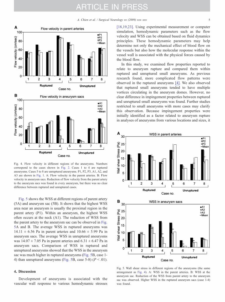
Fig. 4. Flow velocity in different regions of the aneurysms. Numberscorrespond to the cases shown in Fig. 2. Cases 1 to 4 are rupturedaneurysms. Cases 5 to 8 are unruptured aneurysms. P1, P2, P3, A1, A2, andA3 are shown in Fig. 1. A: Flow velocity in the parent arteries. B: Flowvelocity in aneurysm sacs. Reduction of flow velocity from the parent arteryto the aneurysm sacs was found in every aneurysm, but there was no cleardifference between ruptured and unruptured cases.
5A. Chien et al. / Surgical Neurology xx (2009) xxx–xxx
ARTICLE IN PRESS
Fig. 5 shows the WSS at different regions of parent artery(5A) and aneurysm sac (5B). It shows that the highest WSSarea near an aneurysm is usually the proximal region in theparent artery (P1). Within an aneurysm, the highest WSSoften occurs at the neck (A1). The reduction of WSS fromthe parent artery to the aneurysm sac can be observed in Fig.5A and B. The average WSS in ruptured aneurysms was14.11 ± 6.56 Pa in parent arteries and 10.66 ± 5.99 Pa inaneurysm sacs. The average WSS in unruptured aneurysmswas 14.07 ± 7.05 Pa in parent arteries and 6.31 ± 6.47 Pa inaneurysm sacs. Comparison of WSS in ruptured andunruptured aneurysms showed that the WSS in the aneurysmsac was much higher in ruptured aneurysms (Fig. 5B, case 1-4) than unruptured aneurysms (Fig. 5B, case 5-8) (P b .01).
Fig. 5. Wall shear stress in different regions of the aneurysms (the samearrangement as Fig. 4). A: WSS in the parent arteries. B: WSS at theaneurysm sac. Reduction of the WSS from parent artery to the aneurysmsac was observed. Higher WSS in the ruptured aneurysm sacs (case 1-4)was found.
4. Discussion
Development of aneurysms is associated with thevascular wall response to various hemodynamic stresses
[18,19,23]. Using experimental measurement or computersimulation, hemodynamic parameters such as the flowvelocity and WSS can be obtained based on fluid dynamicsprinciples. These hemodynamic parameters may helpdetermine not only the mechanical effect of blood flow onthe vessels but also how the molecular response within thevessel wall is associated with the physical forces caused bythe blood flow.
In this study, we examined flow properties reported torelate to aneurysm rupture and compared them withinruptured and unruptured small aneurysms. As previousresearch found, more complicated flow patterns wereobserved in the ruptured aneurysms [4]. We also observedthat ruptured small aneurysms tended to have multiplevortices circulating in the aneurysm domes. However, noclear difference in impingement properties between rupturedand unruptured small aneurysms was found. Further studiesrestricted to small aneurysms with more cases may clarifythis observation. Because impingement properties wereinitially identified as a factor related to aneurysm rupturein analyses of aneurysms from various locations and sizes, it

6 A. Chien et al. / Surgical Neurology xx (2009) xxx–xxx
ARTICLE IN PRESS
is possible that they may not be particularly strong indicatorsof the risk of small aneurysm rupture [4].
Blood flow in the vascular system causes a frictional forceacting on the vessel wall, WSS, which elicits strongphysiologic responses on a molecular scale [7,18]. In thisstudy, we analyzed the blood flow before and after enteringthe aneurysm sac, and the WSS around the aneurysm area, tocharacterize the flow changes around the aneurysms and thecorresponding frictional force. We found the same flowvelocity changes around the aneurysm in all cases and noclear difference between ruptured and unruptured aneur-ysms. On the other hand, although for all the cases, the WSSin the aneurysm sac was lower than the WSS in the parentartery on average, the difference was less pronounced inruptured aneurysms. Ruptured aneurysms had WSS of75.33% lower than the parent artery, whereas unrupturedaneurysms had WSS of 41.19% lower than the parent artery.Further studies incorporating molecular response in theaneurysm wall may help to explain the influence of differentWSS levels. Still, the present study suggests that amongrupture-related hemodynamic properties, WSS may be auseful hemodynamic parameter to evaluate the risk ofrupture in small brain aneurysms.
4.1. Limitations
We performed hemodynamic analysis on 8 aneurysmcases in a single location. Because of the limited number ofcases, the present study can only show the trend ofhemodynamic properties in small aneurysms. A study withmore cases, for example, combining databases from differentcenters, is needed to confirm our observation. Moreover,because aneurysms in other locations may have differentflow characteristics, analyses for small aneurysms in otherlocations may also be needed.
5. Conclusion
Hemodynamic analyses of small aneurysms enabled us tocompare the detailed blood flow characteristics betweenruptured and unruptured cases. We found that not allaneurysms that are small share the same hemodynamicproperties. Among the characteristics investigated, the valueof WSS in an aneurysm sac was the only hemodynamicparameter that showed statistical differences betweenruptured and unruptured aneurysms.
References
[1] Alnaes MS, Isaksen J, Mardal KA, et al. Computation of hemody-namics in the circle of Willis. Stroke 2007;38:2500-5.
[2] Brisman JL, Song JK, Newell DW. Cerebral aneurysms. N Engl J Med2006;355:928-39.
[3] Burleson AC, Strother CM, Turitto VT. Computer modeling ofintracranial saccular and lateral aneurysms for the study of theirhemodynamics. Neurosurgery 1995;37:774-82 [discussion 782-4].
[4] Cebral JR, Castro MA, Burgess JE, et al. Characterization of cerebralaneurysms for assessing risk of rupture by using patient-specific
computational hemodynamics models. AJNR Am J Neuroradiol 2005;26:2550-9.
[5] Cebral JR, Castro MA, Soto O, et al. Blood-flowmodels of the circle ofWillis from magnetic resonance data. J Eng Math 2003;47:369-86.
[6] Cebral JR, Lohner R. Efficient simulation of blood flow past complexendovascular devices using an adaptive embedding technique. IEEETrans Med Imaging 2005;24:468-76.
[7] Cheng C, Tempel D, van Haperen R, et al. Atherosclerotic lesion sizeand vulnerability are determined by patterns of fluid shear stress.Circulation 2006;113:2744-53.
[8] Dhaliwal S, Sylvester L, Pope W, et al. The natural history ofunruptured cerebral aneurysms studied longitudinally using helical CTangiography. Chicago (Ill): Radiological Society of North America;2003. p. 284.
[9] Donnan GA, Davis SM. Patients with small, asymptomatic, unrupturedintracranial aneurysms and no history of subarachnoid hemorrhageshould be treated conservatively. Stroke 2005;36:407.
[10] Humphrey JD. Vascular adaptation and mechanical homeostasis attissue, cellular, and sub-cellular levels. Cell Biochem Biophys 2008;50:53-78.
[11] Johnston SC, Gress DR, Kahn JG. Which unruptured cerebralaneurysms should be treated? A cost-utility analysis. Neurology1999;52:1806-15.
[12] Jou LD, Quick CM, Young WL, et al. Computational approach toquantifying hemodynamic forces in giant cerebral aneurysms. AJNRAm J Neuroradiol 2003;24:1804-10.
[13] Juvela S, Porras M, Heiskanen O. Natural history of unrupturedintracranial aneurysms: a long-term follow-up study. J Neurosurg1993;79:174-82.
[14] Juvela S, Porras M, Poussa K. Natural history of unrupturedintracranial aneurysms: probability of and risk factors for aneurysmrupture. J Neurosurg 2000;93:379-87.
[15] Kataoka K, Taneda M, Asai T, et al. Difference in nature of rupturedand unruptured cerebral aneurysms. Lancet 2000;355:203.
[16] Kayembe KN, Sasahara MHazama F. Cerebral aneurysms andvariations in the circle of Willis. Stroke 1984;15:846-50.
[17] Murayama Y, Nien YL, Duckwiler G, et al. Guglielmi detachable coilembolization of cerebral aneurysms: 11 years' experience. J Neurosurg2003;98:959-66.
[18] Papaioannou TG, Karatzis EN, Vavuranakis M, et al. Assessment ofvascular wall shear stress and implications for atherosclerotic disease.Int J Cardiol 2006;113:12-8.
[19] Shojima M, Oshima M, Takagi K, et al. Magnitude and role of wallshear stress on cerebral aneurysm: computational fluid dynamic studyof 20 middle cerebral artery aneurysms. Stroke 2004;35:2500-5.
[20] Shojima M, Oshima M, Takagi K, et al. Role of the bloodstreamimpacting force and the local pressure elevation in the rupture ofcerebral aneurysms. Stroke 2005;36:1933-8.
[21] Soustiel JF, Levy E, Bibi R, et al. Hemodynamic consequences ofcerebral vasospasm on perforating arteries: a phantom model study.Stroke 2001;32:629-35.
[22] Steinman DA, Milner JS, Norley CJ, et al. Image-based computationalsimulation of flow dynamics in a giant intracranial aneurysm. AJNRAm J Neuroradiol 2003;24:559-66.
[23] Tateshima S, Murayama Y, Villablanca JP, et al. In vitro measurementof fluid-induced wall shear stress in unruptured cerebral aneurysmsharboring blebs. Stroke 2003;34:187-92.
[24] Tateshima S, Murayama Y, Villablanca JP, et al. Intraaneurysmal flowdynamics study featuring an acrylic aneurysm model manufacturedusing a computerized tomography angiogram as a mold. J Neurosurg2001;95:1020-7.
[25] Tsutsumi K, Ueki K, Morita A, et al. Risk of rupture from incidentalcerebral aneurysms. J Neurosurg 2000;93:550-3.
[26] Wermer MJ, van der Schaaf IC, Algra A, et al. Risk of rupture ofunruptured intracranial aneurysms in relation to patient andaneurysm characteristics: an updated meta-analysis. Stroke 2007;38:1404-10.

7A. Chien et al. / Surgical Neurology xx (2009) xxx–xxx
ARTICLE IN PRESS
[27] Wiebers DO, Whisnant JP, Huston III J, et al. Unruptured intracranialaneurysms: natural history, clinical outcome, and risks of surgical andendovascular treatment. Lancet 2003;362:103-10.
[28] Yim P, Demarco K, Castro MA, et al. Characterization of shear stresson the wall of the carotid artery using magnetic resonance imaging andcomputational fluid dynamics. Stud Health Technol Inform 2005;113:412-42.
Commentary
The authors have presented a very interesting andpotentially clinically valuable technique for the study ofintracranial aneurysms. Using their methods, they foundstatistically significant differences inwall shear stress betweenruptured and unruptured ophthalmic aneurysms and identifiedother differences that did not quite reach statistical signifi-cance. The technique, which involves modeling of hemody-namic parameters from high-resolution angiograms, ispotentially clinically important because we currently do nothave methods for segregating small aneurysms into groupswith higher or lower rupture probability.
We believe that aneurysm diameter is the most importantdeterminant of rupture probability and that the potential forrupture increases as the third power of diameter. However,although most small aneurysms do not rupture, a smallfraction of them do. Perhaps analysis of wall shear stress orother hemodynamic parameters will help determine whichaneurysms of a given small diameter are more likely torupture and deserve prophylactic treatment.
Obviously, the authors need to improve their techniquesso that they can study larger groups of aneurysms.Furthermore, it would be valuable if their databases includedsome cases that were studied both before and after rupturebecause with the current data it is difficult to exclude the
possibility that differences in wall shear stress were a resultof rupture rather than causative. However, the techniques andapproach presented in this article deserve careful considera-tion and have the potential to significantly add to our abilityto decide which small lesions should be treated.
Phillip Dickey, MDKailasnath Purushothaman, PhD
Yale School of MedicineNew Haven, CT 06510, USA
This article compared the hemodynamic properties forruptured and unruptured aneurysms by simulating the
hemodynamics of small (b10 mm) internal carotid artery-ophthalmic artery aneurysms using an image-based compu-ter fluid dynamics software. They found a higher and moreinhomogeneous wall shear stress distribution in the rupturedaneurysms when compared to the unruptured ones. Thisgives us more information about how to predict whichaneurysms will rupture—at least evidence based more thanjust the size of the aneurysm.As with all mathematical models, assumptions are madeso as to represent as accurately as possible the biologicsystem properties. The authors have addressed some of theseconcerns in their Methods section. The article also uses asmall sample size, which subjects its conclusions to somelimitations. In general, however, this article lays thegroundwork for further studies for looking at how hemody-namic parameters can be used to predict which aneurysmsare at higher risks for rupture.
Kern H. Guppy, MD, PhDDivision of NeurosurgeryKaiser Permanente/UCSF
Sacramento, CA 95825, USA