Hemodynamic Disorders, Thromboembolic Disease, and Shock
100
-
Upload
khangminh22 -
Category
Documents
-
view
0 -
download
0
Transcript of Hemodynamic Disorders, Thromboembolic Disease, and Shock


EDEMA
Edema: is increase fluid in interstitial space
(collection in body cavities called hydrothorax o,
hydropericardium and hydroperitoneum (ascites).

Pathophysiology of edema:
Vascular hydrostatic pressure &plasma colloid osmotic
pressure control the movement of fluid between
vascular & interstitial spaces .
The outflow of fluid from arteriolar end is balanced
by the inflow at the venular end and small amount
drain by lymphatics.


Factors regulate edema:
1- Increase capillary hydrostatic pressure or decrease colloid osmotic pressure.
2- Effect of inflammatory mediators on vascular permeability.
3- Lymphatic obstruction can impair fluid drainage &cause edema.

Edematous fluid in hydrodynamic disturbances is transudates with specific gravity 1.012 while inflammatory edema due to increased vascular permeability is exudates (1.020).
Increased hydrostatic pressure:
Regional increases in hydrostatic pressure can result from a focal impairment in venous return e.g (deep venous thrombosis DVT) with edema of the affected limb.

Generalized increase in venous pressure leading to
systemic edema occur in congestive heart failure in
which reduced cardiac output causes reduced renal
perfusion & trigger of rennin angiotensin aldosterone
axis causing sodium & water retention by kidney in
order to increase intravascular volume & improve
cardiac output & renal perfusion .
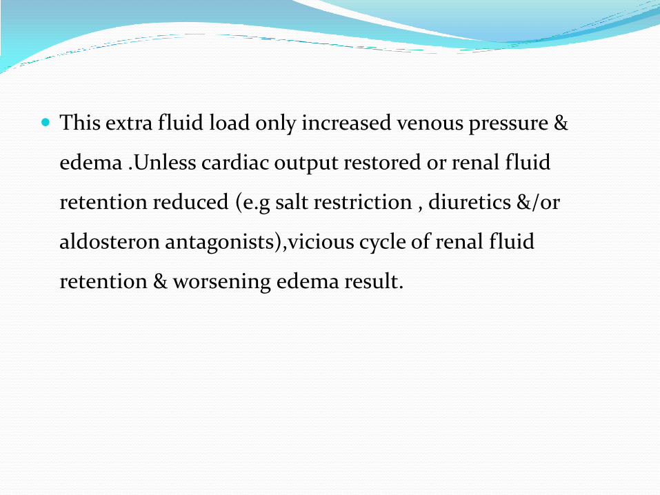
This extra fluid load only increased venous pressure &
edema .Unless cardiac output restored or renal fluid
retention reduced (e.g salt restriction , diuretics &/or
aldosteron antagonists),vicious cycle of renal fluid
retention & worsening edema result.

Mechanism of edema formation in heart failure , renal failure and hepatic failure

Reduced plasma osmotic pressure: (reduced plasma proteins) Result from:
Increased loss of albumin as in nephrotic syndrome.
Reduced protein synthesis as in cirrhosis, malnutrition.
Protein losing enteropathy(intestinal diseases with malabsorption)
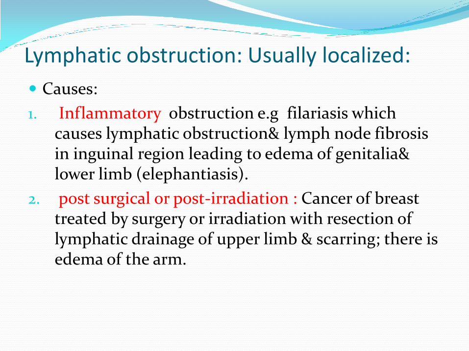
Lymphatic obstruction: Usually localized:
Causes:
1. Inflammatory obstruction e.g filariasis which causes lymphatic obstruction& lymph node fibrosis in inguinal region leading to edema of genitalia& lower limb (elephantiasis).
2. post surgical or post-irradiation : Cancer of breast treated by surgery or irradiation with resection of lymphatic drainage of upper limb & scarring; there is edema of the arm.


3. Neoplastic : In CA breast, infiltration & obstruction of superficial lymphatics will cause edema of breast skin (peau-de-orange) due to accentuation of depression in the skin at site of hair follicles.


Morphology of edema: Microscopically: Clearing & separation of extra cellular
matrix.
Any organ or tissue can be involved, but edema is most commonly seen in subcutaneous tissues, the lungs, and the brain
In most cases the distribution is influenced by gravity and is termed dependent edema (e.g., the legs when standing, the sacrum when recumbent). Finger pressure over substantially edematous subcutaneous tissue displaces the interstitial fluid and leaves a depression, a sign called pitting edema


Special forms of edema: 1. Pulmonary edema
Seen in:
1. Left sided heart failure (dependent distribution in lung)
2. Respiratory distress syndrome.
3. Pulmonary infection.
4. Hypersensitivity reaction.
The lung is 2-3 times their normal weight, cut section frothy, blood tinged fluid represent mixture of air, edematous fluid & extravasated RBCs.

2. Edema of brain
Localized: due to abscess, neoplasm &trauma.
Generalized: due to encephalitis, hypertension crises, venous outflow obstruction &trauma
Grossly: Swollen with narrowed sulci & distended gyri.

Clinical correlation of edema:
a. Subcutaneous edema in cardiac failure & renal failure can impair wound healing or clearance of infection.
b. Pulmonary edema can cause death by interfering with normal ventilatory function by:
(1) fluid collects in alveolar space& impair oxygen diffusion).
(2)Edematous fluid in alveolar spaces is favorable environment for bacterial infection.

Clinical consequnces of edema-continue
c. Brain edema if severe causes death due to brain herniated through foramen magnum which causes compression of vital centers & also due to compression of vascular supply of brain.

Hyperemia & Congestion
Hyperemia and congestion both stem from locally increased blood volumes

Hyperemia is an active process in which arteriolar dilation (e.g., at sites of inflammation or in skeletal muscle during exercise) leads to increased blood flow
Affected tissues turn red (erythema) because of the engorgement of vessels with oxygenated blood.
Hyperemia could be :
1. Local or General
2. Physiological or Pathological

Localized hyperemia: could be
A. physiological: e.g flushing, exercise, after meal.
B. Pathological : e.g site of inflammation .
Generalized hyperemia: could be
A. Physiological : e.g hot weather .
B. Pathological: e.g:
Fever, Hyperthyroidism, Arteriovenous shunt

*Congestion: is passive process from impair outflow from tissue (venous obstruction).
Congested tissues take on a dusky reddish-blue color (cyanosis) due to red cell stasis and the accumulation of deoxygenated hemoglobin.
As a result of the increased volumes and pressures, congestion commonly leads to edema.
long-standing chronic passive congestion, the lack of blood flow causes chronic hypoxia
Capillary rupture in chronic congestion can also cause small hemorrhagic foci; subsequent catabolism of extravasated red cells can leave residual clusters of hemosiderin-laden macrophages.

1- Generalized systemic congestion e.g right ventricular failure in which there is decrease venous return to the heart with accumulation of blood in the venous side.
Clinically:
A- Dilated veins e.g increase JVP (jugular venous pressure)
B- Edema (limbs, ascites, hepatospleenomegaly) .

Morphology of generalized congestion:
Liver: will show chronic venous congestion causing a grossly mottled appearance similar to the nut meg hence the name Nut meg liver.
Mic.:
Congestion of the central venule.
Necrosis of the surrounding hepatocytes because of pressure &hypoxia .
The peripheral liver cells are normal or show fatty change.

Liver with chronic passive congestion and hemorrhagic necrosis

2- Pulmonary venous congestion: e.g left ventricular failure where there is accumulation of blood in the surrounding capillaries, RBCs will escape to the alveolar space & engulfed by macrophages (heart failure cells) which cause cough with blood stained sputum.
3- Localized venous congestion: e.g DVT of leg (edema, cold, cyanosis).

HEMORRHAGE
Is extravasation of blood into the extravascular
space
It is either external or internal (encased within
tissue( hematoma) which may be: Insignificant
(bruises) or Massive (retroperitoneal hematoma)
from rupture of dissecting aortic aneurysm &cause
death.

Petechiae: Minute hemorrhage in skin ,mucous membranes & serosal surfaces(1-2 mm)
Slightly larger (≥3 mm) hemorrhages are called purpura.
Ecchymosis: 1-2 cm subcutaneous hematoma

Causes of Hemorrhage:
1. Trauma: e.g penetrating wounds to the heart & large vessels----- rapid blood loss.
2. Abnormalities of blood vessel wall like:
Inflammatory lesions may lead to weakening of arteries &may cause aneurysmal dilatation &rupture.
Neoplastic invasion e.g. carcinoma of tongue with invasion of lingual arteries.
Other vascular diseases e.g atheroma, aneurysm.

3- High pressure within blood vessel e.g systemic HT leading to hemorrhage at sites of arterial weakness or increase venous pressure in varicose veins e.g. legs ,esophageal hemorrhage .

Effects of acute hemorrhage:
Depend on volume &rate of blood loss: as following
a. the effect is slight if less than 20% of blood volume is lost, while sudden loss of 33% of blood volume leads to death.
b. Gradual loss (within 24 hour) of more than 50% blood volume is not necessarily fatal but it is serious.
C. chronic blood loss can lead to iron-deficiency anemia

In human 60-70% of blood volume contained in veins &venules, so constriction of these vessels will increase venous return to the heart.
Arteriolar constriction is selective i.e there is decrease blood flow to skin, salivary glands, liver, spleen & kidneys while blood is preserved to the brain, heart, skeletal muscles &diaphragm.
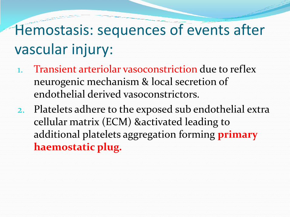
Hemostasis: sequences of events after vascular injury: 1. Transient arteriolar vasoconstriction due to reflex
neurogenic mechanism & local secretion of endothelial derived vasoconstrictors.
2. Platelets adhere to the exposed sub endothelial extra cellular matrix (ECM) &activated leading to additional platelets aggregation forming primary haemostatic plug.

3. Tissue factors release at site of injury & with platelets factors, there will be activation of coagulation cascade leading to fibrin deposition .Thrombin activated in coagulation cascade causing further platelets recruitment &secondary hemostasis( secondary hemostatic plug)
4. Polymerized fibrin & platelets aggregation form solid permanent plug to prevent any further hemorrhage. At this stage, counter-regulatory mechanisms set into motion

COMPONENTS OF NORMAL HEMOSTASIS:
1. Normal endothelium
2. Normal platelets
3. Normal coagulation cascade

The endothelium has antithrombotic and prothrombotic properties which are:
(a)Antiplatelets effect
(b)Anticoagulant properties
(c) fibrinolytic properties
(d)Prothrombotic properties

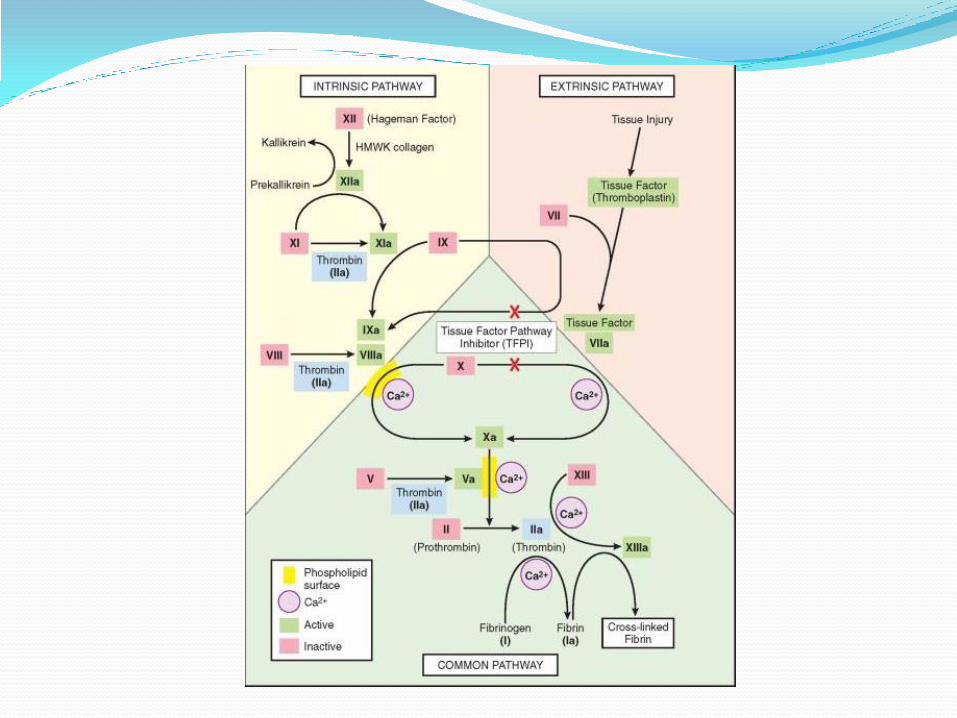
THROMBOSIS: formation of solid or semisolid mass from blood
constituents within the cardiovascular system during life.
Pathogenesis: (virchow’s triad):
1. Endothelial injury
2. Alteration of blood flow
3. Hypercoagulability
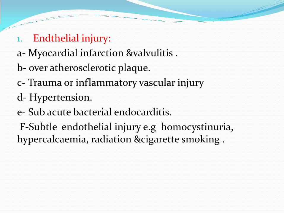
1. Endthelial injury:
a- Myocardial infarction &valvulitis .
b- over atherosclerotic plaque.
c- Trauma or inflammatory vascular injury
d- Hypertension.
e- Sub acute bacterial endocarditis.
F-Subtle endothelial injury e.g homocystinuria, hypercalcaemia, radiation &cigarette smoking .

2- Alteration of blood flow: either
a- Turbulance: Important in arterial & cardiac thrombosis by causing endothelial injury & local pockets of stasis.
b- Stasis: Is major factors in venous thrombosis .

Both turbulence &stasis cause
1- Disruption of laminar blood flow & bring platelets in contact with endothelium.
2- Prevent dilution of clotting factors by fresh flowing blood.
3- Retard flow of clotting factor inhibitors & permit build up of thrombus.
4- Promote endothelial activation leading to local thrombosis, adhesion of leukocytes & other effects of endothelium.

3- Hypercoagulablity Divided into primary & secondary disorders.
Primary disorders:
1- Mutation in factor V which found in up to 12% of Caucasian population. 60 % in DVT because mutant V is resistant to anticoagulant effect of active protien C leading to functional deficiency of protien C.
2- Inherited mutation of antithrombin III, protien C&S leading to venous thrombosis & recurrent thromboembolism in adolescent& early adult life.

Secondary disorders:
1- Heart failure or trauma causing vascular endothelial injury or stasis.
2- Oral contraceptive pills because of increase hepatic synthesis of coagulation factors& decrease synthesis of antithrombin III.
3- Dissemenated tumors because of release of procoagulant tumor products.

4- Advancing age because of increase platelets aggregation & decrease PGI2 by endothelium.
5- Smoking.
6- Obesity
7- Lupus anticoagulants which are antibodies directed against anionic phospholipids & high frequency of venous & arterial thrombosis .Those patients may have SLE or thrombosis is the only clinical manifestation.

Lecture 3

Morphology of thrombi Depends on:
Site of origin & circumstances leading to their development.
The area of attachment of thrombus to the underlying vessel firmest at point of origin (Arterial thrombi grow in retrograde direction from attachment, venous thrombi in direction of blood flow .
Both types of thrombi propagate in the direction of heart, the propagating portion is poorly attached and prone to dislodgment and embolus formation

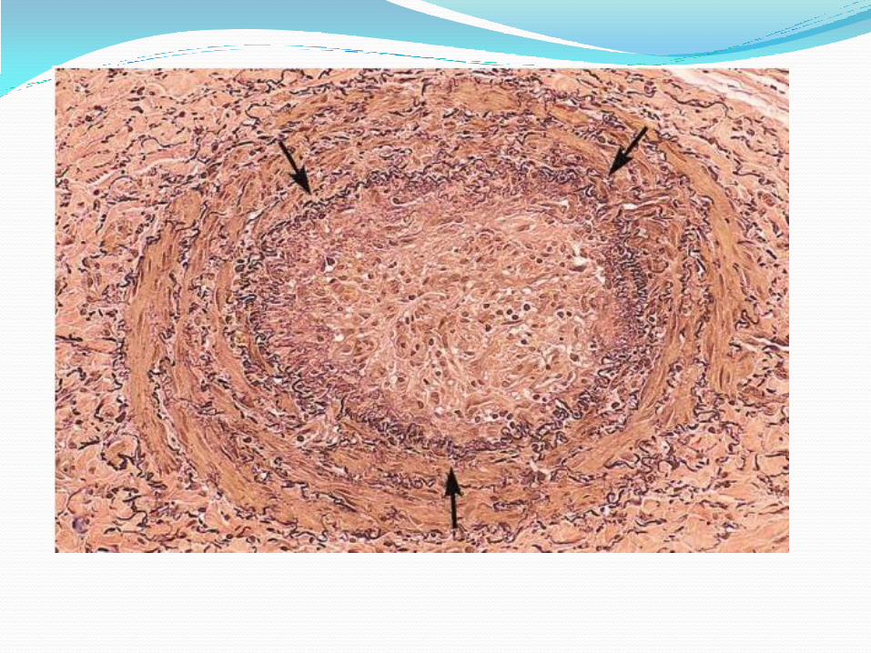
Thrombi often have grossly and microscopically apparent laminations called lines of Zahn; these represent pale platelet and fibrin deposits alternating with darker red cell–rich layers.
Such laminations signify that a thrombus has formed in flowing blood( during life to differentiate from clots occurring after death )

1-cardiac and aortic lumen thrombi Thrombi occurring in heart chambers or in the aortic
lumen are designated mural thrombi.
Cardiac causes include:
1. Abnormal cardiac muscle movement: in infarction or with arrhythmias turbulence and thrombus formation
2. Endocardial (endothelial ) injury in myocarditis
Aortic causes include complicated atherosclerotic plaque and aneurysmal dilation

2. Arterial thrombi: (white thrombi)
Are frequently occlusive.
Most common sites are coronary, cerebral &femoral arteries, superimposed on atherosclerotic plaques.
Firmly adheres to arterial wall.
Grayish white &composed of platelets, fibrin, RBCs °enerated WBCs.
3. Venous thrombi: (red thrombi) Almost invariably occlusive because they form in
slowly moving blood, with the thrombus forming a long cast of the lumen
They contain more RBCs and relatively less platelets and known as red or stasis thrombi.
Soft, gelatinous.
90% affect veins of lower limbs.

4. POST MORTEM THROMBUS:
Confused with venous (red) thrombus.
They are gelatinous with dark red dependent portion
where RBCs settled by gravity with yellow ( chicken fat
) upper portion
Not attached to arterial wall.
While red thrombi are:
More firm.
Almost always have point of attachment & transaction
reveals vague strands of pale gray fibrin.(lines of Zahn)

Fate of thrombus: 1- Dissolution: Activation of fibrinolytic pathway may lead
to shrinkage & total lysis of recent thrombus
older thrombi undergo fibrin polymerization & become
more resistant to proteolysis.
This phenomenon explains why therapeutic
administration of fibrinolytic agents such as t-PA (e.g., in
the setting of acute coronary thrombosis) is generally
effective only when given in the first few hours of a
thrombotic episode.)

2- Organization & recanalization: Ingrowth of
endothelial cells, smooth muscle cells & fibroblasts
into fibrin rich thrombus will create conduits from one
end of thrombus to other & re-establish continuity of
original lumen.
3- Propagation: Thrombus accumulates more
platelets & fibrin &lead to obstruction.
4- Embolization: Thrombus dislodges & transported
to other sites.

Dissemenated Intravascular Coagulation (DIC):
characterized by
1- Occur as obstetrical complication &in advanced malignancies .There is sudden or insidious onset of widespread fibrin thrombi in the microcirculation which cause circulatory insufficiency especially in brain, lung, heart &kidneys.
2- Concurrent consumption of platelets & coagulation proteins (consumption coagulopathy)
3- Fibrinolytic mechanism is activated & eventually serious bleeding disorder arises.

Embolism:
An embolus is a detached intravascular solid, liquid, or gaseous mass that is carried by blood to a site distant from point of origin.
90% originate from thrombus
Types of emboli according to the constituents :
1. Solid 2. Liquid 3. gaseous

Solid emboli: (examples of solid emboli)
1. Fragments detached from a thrombus , this is called thromboembolic phenomenon.
2. Fragments of an ulcerated atherosclerotic plaque.
3. fragments of tumor.
4. Droplets of fat.
Fluid emboli:
E.g. Amniotic fluid embolus.
Gaseous emboli:
e.g. air embolism.

Types of emboli according to the site:
1. Arterial 2.venous
3. Paradoxical (emboli that arise from the right side
and cross to the left side as in cases of : patent ductus
arteriosus, septal defects.

I: Arterial Embolism: Sites:
1- Atrial thrombus in atrial fibrillation.
2- Ventricular thrombus.
3- Vegetative thrombus.
4- Atherosclerotic thrombus.
Effects of arterial embolism: Vascular occlusion leading to ischemia &hypoxia which is either: Reversible or irreversible (infarction) & affect any organ like kidney, heart brain….etc .

II: Venous embolism: Sites:
1- Deep vein thrombosis of the lower limbs in 95%.
2- Pelvic vein thrombosis.
3- Thrombosis of the right side of the heart & IVC.

Effects of venous emboli: The emboli pass from systemic veins to the right side of the heart then to the pulmonary arteries leading to pulmonary embolism which is serious, important &common clinical problem.
pulmonary embolism is the major cause of death in 15% of hospitalized patients .The effect depend on:
Size of embolus.
State of pulmonary circulation.

1- Obstruction of pulmonary trunk or both of its branches
(saddle shape embolus) lead to acute right sided heart
failure(cor pulmonale) & asphyxia .
2- Obstruction of medium size arteries leading to pulmonary
hemorrhage if the bronchial circulation is good but causes
infarction in the setting of left sided heart failure
3- Occlusion of small arteries will cause either:
• No symptoms
• Hemorrhage or infarction
• Pulmonary hypertension if multiple over period of time


Fat embolism: (causes):
1. Fracture of long bones in which there is an injury to the fatty marrow.
2. Injury to the adipose tissue.
3. Burn
4. Fatty liver
5. Acute pancreatitis.
It is characterized by pulmonary insufficiency, neurological manifestation, anemia &thrombocytopenia & it is fatal in 10% of cases.


Pathogenesis:
1-Fat microemboli cause occlusion of cerebral & pulmonary microvasculatures.
2- Free fatty acids released from fat globules leading to local toxic injury to endothelium.

Air Embolism It means the presence of air bubbles in the circulation.
Causes:
neck wounds
cardiothoracic injury
venous or arterial catheterization
caisson syndrome.
laparoscopic procedure
Outcome (results):
Small volume of air is harmless
100 ml will acute distress
300 ml will be fatal.

Amniotic Fluid Embolism It is a rare obstetrical complication with a mortality
rate of 80% , it is unpredictable, unpreventable event.
Signs and symptoms:
sudden onset of Dyspnea , cyanosis , shock , convulsions , coma.

Pathogenesis:
o amniotic fluid or Fetal epithelial cells and fat gain entry to maternal pulmonary circulation through tear in placenta
o mechanical obstruction of microvasculature
o Vasoconstriction of the pulmonary vessels caused by certain vasoactive substances in the amniotic fluid.
o Thrombogenic factors which mayintravascular coagulation


Ischemia:
Defined as deficient blood supply to the tissue which is either:
1. complete deficiency : caused by obstruction by a thrombus or an embolus infarction.
2.partial deficiency : caused by an atheroma or spasm tissue damage and fibrous tissue formation.
Tissue susceptibility to ischemia: depends on
Blood supply and collateral circulation.
Tissue metabolism.

Blood supply and collateral circulation :
Tissue with double blood supply resist ischemia e.g liver, lung.
Tissues that possess a collateral circulation resist ischemia e.g brain, lower limbs, bowel.
Organs that have end arteries are susceptible to ischemia e.g kidney, spleen , heart (coronaries are end arteries)

Tissue metabolism:
Supporting tissues: as fibrous tissue, fatty t., cartilage and bone are less susceptible to ischemia because of their low metabolic rate .
parenchymal tissue: are so susceptible to ischemia because of their high metabolic rate e.g brain, heart.

Infarction Area of ischemic necrosis caused by occlusion of either
arterial supply or venous drainage in particular tissue.
90 % results from thrombotic or embolic events & almost all result from arterial occlusion.
Other mechanisms:
1- Local vasospasm
2- Swelling of an atheroma
3- Secondary to hemorrhage within plaque
4- Extrinsic compression of vessels e.g by tumor.

Types of infarction: 1. Red (hemorrhagic)
2. White (anemic)
Red Infarct: occur with:
1- Venous occlusion like ovarian torsion.
2- Loose tissues e.g. lung that allow blood to collect in infarcted zone.

3- Tissues with dual circulation e.g. lung & small intestines.
4- Tissues that previously congested because of sluggish venous outflow.
5- When flow re-established to a site of previous arterial occlusion & necrosis (fragmentation of occlusive embolus).

White Infarct: occur with:
1- Arterial occlusion.
2- Solid organs (heart, spleen, kidneys) where solidity of tissues limits the amount of hemorrhage into ischemic necrosis .The few of extravasated RBCs lysed &hemoglobin released which remain in form of hemosidrin, while in spongy organs, the hemorrhage is too extensive.


Histologically:
* The infracted area shows ischemic coagulative necrosis.
* If infarction occur minutes or hours before death, no demonstrable histological changes.
* Inflammatory response begin within few hours along margin & becomes well defined in 1-2 days caused by necrotic tissues then gradual degradation of dead tissues with phagocytosis by inflammatory cells.
* Reparative response begin in margin & most infarction replaced by scar tissues

Factors Influence Development of an Infarct: (1) Nature of Vascular Supply:
Availability of alternative blood supply e.g. lung have dual pulmonary & bronchial arterial blood supply. Obstruction of pulmonary arterioles not cause infarction in intact bronchial circulation, liver with portal vein & hepatic artery, hand & forearm with radial & ulnar artery. In contrast renal & splenic circulation are end arterial & obstruction cause infarction.

(2)Rate of Development of Occlusion:
Slowly developing occlusion less likely to cause infarction because provide time for alternative pathway to develop e.g. small inter arterial anastamosis connect the three coronary arteries, so if one coronary artery slowly occluded (by atherosclerotic plaque), the flow in this collateral circulation increase & prevent infarction.

(3) Vulnerability to hypoxia:
Neurons undergo irreversible damage when deprived of their blood supply 3-4 minutes, myocardial cells 20-30 minutes, fibroblasts within myocardium remain viable after many hours of ischemia.

4) Oxygen content of blood:
(Partial obstruction in anemic patient may lead to infarction, while it is without effect in normal conditions

Shock:
It is a state in which the supply of blood to the tissues is inadequate to meet the metabolic demands
is characterized by systemic hypotension due either to reduced cardiac output or to reduced effective circulating blood volume

Classification: 1- Hypovolemic shock: In which there is a real
decrease in blood volume.
Causes:
Hemorrhage.
Fluid loss as in severe vomiting, diarrhea & burns.
Mechanism of development: is inadequate blood or plasma volume with resultant low cardiac output

2- Cardiogenic shock: There is relative decrease in blood volume (pooling of blood).
Causes:
Myocardial infarction.
Rupture of the heart.
Pulmonary embolism.
Arrhythmias.
Cardiac temponade.
• Mechanism of development: Failure of myocardial pump due to intrinsic myocardial damage or extrinsic pressure or obstruction to outflow with reduced output
3- Septic shock: Causes:
Overwhelming bacterial infection by gram positive or (gram negative septicemia or endotoxic shock) or fungi
Septic shock is associated with severe hemodynamic and hemostatic derangements, With a mortality rate near 20%

Pathogenesis of septic shock 1. Role of inflammatory mediators: Various microbial
cell wall constituents engage receptors on inflammatory cells with extensive release of mediators that activate endothelial cells and coagulation system procoagulant state
2. Role of endothelial cells: injury results in
(1) Thrombosis –> DIC in half of patients
(2) increased vascular permeability edema
(3) vasodilation , through release of vasoactive substances pooling of blood

3. Metabolic abnormalities. Septic patients exhibit insulin resistance and hyperglycemia( increase stress hormones) later in the course adrenal insufficiency
4. Organ dysfunction. Systemic hypotension, interstitial edema, and small vessel thrombosis all decrease the delivery of oxygen and nutrients to the tissues
4- Neurogenic shock:
Causes:
Anesthesia & spinal cord injury .
o Mechanism: peripheral vasodilatation due to loss of vascular tone

Stages of shock: 1- Non progressive phase: which is the compensatory
phase .In this stage a compensatory mechanisms operate to maintain cardiac output &blood pressure near normal levels .The compensatory mechanisms include:
a- Arteriolar constriction leading to increase blood pressure.
b- Increase heart rate &cardiac output.
c- Retention of fluid through increase secretion of ADH & activation of rennin angiotensin aldosteron axis to retain fluid.
2- Progressive phase :When an additional factor is added like extensive burn complicated by bacterial infection .
In this stage ,despite the compensatory mechanisms ,there is progressive decline in blood pressure &cardiac output .
Clinically observed as increase in respiratory rate &decrease in urine output reflecting pulmonary &renal hypoperfusion .

3. Irreversible Phase: result from irreversible injury to the cell membrane as manifested by dysfunction of sodium-potassium pump & defect in cell membrane so cell contents go to outside .
The reduction in blood flow to the vital organs such as brain, heart, kidney lead to ischemic cell death in these organs .

Clinical course:
In hypovolemic &cardiogenic shock: patient present with hypotension, weak rapid pulse, tachypnea, cool &cyanotic skin.
In septic shock: the skin may initially be warm &flushed because of peripheral vasodilatation .

Prognosis:
In hypovolemic shock: 80-90% of young patients survive.
Cardiogenic shock associated with extensive myocardial infarction & in gram-negative shock, 75% died .

Thank you