Design of a Bioreactor to Mimic Hemodynamic Shear Stresses ...
Upload
khangminh22Category
view
0download
0
Med Intensiva. 2012;36(2):77---88
www.elsevier.es/medintensiva
ORIGINAL
Respiratory and hemodynamic changes during lung recruitment
maneuvering through progressive increases and decreases in PEEP
level�
M.I. Monge García ∗, A. Gil Cano, M. Gracia Romero, J.C. Díaz Monrové
Servicio de Cuidados Intensivos y Urgencias, Unidad de Investigación Experimental, Hospital del SAS Jerez, Spain
Received 27 June 2011; accepted 29 August 2011
KEYWORDSMechanicalventilation;Acute respiratorydistress syndrome;Lung recruitmentmaneuver;Positiveend-expiratorypressure;Cardiac output;Preload;Hemodynamicmonitoring
Abstract
Objective: To evaluate the respiratory and hemodynamic changes during lung recruitment
maneuvering (LRM) through stepwise increases and decreases in PEEP level.
Design and setting: A retrospective study in a 17-bed ICU was carried out.
Patients: Twenty-one patients with acute respiratory failure and bilateral pulmonary infiltra-
tion.
Intervention: LRM was carried out, consisting of stepwise increases in PEEP (4 cmH2O every
3 min), with fixed ventilation pressure, until reaching a maximal value of 36 cmH2O PEEP
(ascending branch), followed by progressive decreases in PEEP (2 cmH2O every 3 min) until
establishing the open-lung PEEP at the value associated to maximum respiratory compli-
ance (Crs) (descending branch). Continuous hemodynamic monitoring was performed using an
esophageal echodoppler probe.
Results: Crs gradually decreased in the ascending branch of the LRM, and progressively
increased surpassing the initial value after establishing the open-lung PEEP in the descending
branch, reducing the ventilation pressure and increasing the SpO2/FiO2 ratio. Hemodynamic
changes primarily consisted of a fall in cardiac output and left ventricular preload, together
with an increased heart rate and cardiac contractility. At comparable levels of PEEP and mean
airway pressure, these changes were more pronounced during the descending branch of the
LRM.
Conclusions: (1) LRM increased Crs, improving oxygenation and decreasing ventilation pressure;
(2) the main hemodynamic consequence was the drop in cardiac output and left ventricular
preload; and (3) the unequal hemodynamic derangement in both branches, at the same level of
PEEP and mean airway pressure, showed that, along with intrathoracic pressure, other factors
such as Crs and hypercapnia may have influenced the hemodynamic consequences of this type
of LRM.
© 2011 Elsevier España, S.L. and SEMICYUC. All rights reserved.
� Please cite this article as: Monge García MI, et al. Cambios respiratorios y hemodinámicos durante una maniobra de reclutamientopulmonar mediante incrementos y decrementos progresivos de PEEP. Med Intensiva. 2012:36:77---88.
∗ Corresponding author.E-mail address: [email protected] (M.I. Monge García).
2173-5727/$ – see front matter © 2011 Elsevier España, S.L. and SEMICYUC. All rights reserved.

78 M.I. Monge García et al.
PALABRAS CLAVEVentilación mecánica;Síndrome de distrésrespiratorio agudo;Reclutamientopulmonar;Presión al final de laespiración;Gasto cardiaco;Precarga;Monitorizaciónhemodinámica
Cambios respiratorios y hemodinámicos durante una maniobra de reclutamiento
pulmonar mediante incrementos y decrementos progresivos de PEEP
Resumen:
Objetivo: Estudiar los cambios respiratorios y hemodinámicos durante una maniobra de reclu-
tamiento pulmonar (MRP) mediante incrementos y decrementos progresivos de PEEP.
Diseno y ámbito: Estudio retrospectivo en una UCI de 17 camas.
Pacientes: Un total de 21 pacientes con insuficiencia respiratoria aguda e infiltrados pulmonares
bilaterales.
Intervención: MRP consistente en incrementos progresivos de PEEP (4 cmH2O cada 3 minutos),
con presión de ventilación fija, hasta alcanzar un valor máximo de 36 cmH2O de PEEP (rama
ascendente), seguida de decrementos progresivos (2 cmH2O cada 3 minutos) hasta establecer
la PEEP de apertura en el valor asociado a la máxima distensibilidad del sistema respiratorio
(Dsr) (rama descendente). La monitorización hemodinámica se realizó de forma continua con
una sonda ecodoppler esofágica.
Resultados: La Dsr disminuyó gradualmente en la rama ascendente de la MRP y aumentó de
forma progresiva superando el valor inicial al establecer la PEEP de apertura en la rama descen-
dente, reduciéndose la presión de ventilación y aumentando la relación SpO2/FiO2. Los cambios
hemodinámicos consistieron fundamentalmente en una disminución del gasto cardiaco y de la
precarga del ventrículo izquierdo, junto con un aumento de la frecuencia y de la contractilidad
cardiaca. A niveles equiparables de PEEP y presión media en vía aérea, estos cambios fueron
más intensos durante la rama descendente.
Conclusiones: (1) La realización de la MRP incrementó la Dsr mejorando la oxigenación y
disminuyendo la presión de ventilación; (2) la principal consecuencia hemodinámica fue la
disminución del gasto cardiaco y de la precarga ventricular izquierda; (3) la afectación hemodi-
námica desigual en ambas ramas, a niveles equiparables de PEEP y presión media en vía
aérea, puso de manifiesto que, junto a la presión intratorácica, otros factores como la Dsr
y la hipercapnia pudieron influir en las consecuencias hemodinámicas en este tipo de MRP.
© 2011 Elsevier España, S.L. y SEMICYUC. Todos los derechos reservados.
Introduction
‘‘Open lung’’ mechanical ventilation is a protective ven-tilation modality that aims to minimize lung collapse,prevent cyclic alveolar aperture and closure, and reducepulmonary overdistension.1---3 Its implementation is mainlybased on three interventions: (1) the early applicationof lung recruitment maneuvering (LRM); (2) the use of apositive end-expiratory pressure (PEEP) level sufficient tostabilize and keep open the maximum possible number ofalveolar units (the so-called ‘‘open-lung PEEP’’); and (3)ventilation with as low a pulmonary distension pressure aspossible.4,5 However, in order to reach these objectives itis necessary to at least temporarily use very high intratho-racic pressures, and for this reason application of the aboveventilatory strategy remains the subject of debate.6,7
The advocators of open lung ventilation justify its usein terms of the harmful effects of lung collapse uponthe evolution of the lesions associated to mechanical ven-tilation, favored by the use of small tidal volumes.8 Incontrast, the critics of the technique point out that in addi-tion to the absence of solid confirmation of its benefits interms of lessened patient mortality, the application of highintrathoracic pressures is not risk-free, with non-negligiblehemodynamic alterations and the appearance of barotraumaphenomena.6,9
In September 2003 we introduced an open lung mechani-cal ventilation protocol based on the use of LRM followedby the application of open-lung PEEP. The aim of this
ventilatory strategy was to recruit the maximum possiblenumber of alveoli, ventilating with maximum respiratorysystem compliance (Crs), and thus with the lowest lung ven-tilation pressure.2,10 The recent debate published in thisjournal on the use of this ventilation mode6,7 has promptedus to present some of the most relevant data which we havecollected during these years of experience. Specifically, thepresent retrospective analysis has two aims: (1) to investi-gate the respiratory and hemodynamic effects of LRM in agroup of patients subjected to careful monitoring; and (2)to establish the incidence and form of presentation of baro-trauma, and describe the main characteristics and evolutionof the patients who develop this complication. While thefirst of these two points constitutes the main objective of thepresent study, the second point, related to lung barotrauma,is dealt with in another part of this issue.11 Some of theresults of the present study have been previously reportedin abstract form.12
Patients and methods
Patients
During the period between September 2003 and January2011, a total of 100 patients were ventilated with our openlung ventilation protocol due to severe hypoxemic acuterespiratory failure, defined by the incapacity to maintainoxygen saturation measured by pulsioxymetry (SpO2) > 90%
Respiratory and hemodynamic changes during lung recruitment maneuvering 79
Lung recruitment protocol
PEEP60
50
40
30
20
10
Ascending branch Descending branch0
5
18202224242012 16
36
32
27
24
2119
17
21
26
30
33
37
4243
3 min
15 c
mH
2O
2828 3232 36 16
10
15
20
25
30
35
40
45
Co
mp
lian
ce (
ml/cm
/cm
H2O
)
Open-lu
ng P
EE
P
0
Pre
ssu
re level (c
mH
2O
)
Ventilation pressure
Respiratory compliance
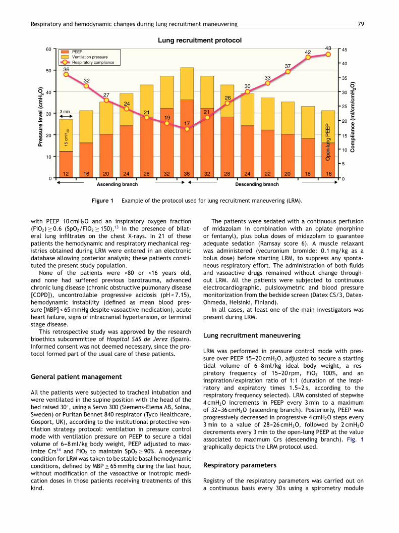
Figure 1 Example of the protocol used for lung recruitment maneuvering (LRM).
with PEEP 10 cmH2O and an inspiratory oxygen fraction(FiO2) ≥ 0.6 (SpO2/FiO2 ≥ 150),13 in the presence of bilat-eral lung infiltrates on the chest X-rays. In 21 of thesepatients the hemodynamic and respiratory mechanical reg-istries obtained during LRM were entered in an electronicdatabase allowing posterior analysis; these patients consti-tuted the present study population.
None of the patients were >80 or <16 years old,and none had suffered previous barotrauma, advancedchronic lung disease (chronic obstructive pulmonary disease[COPD]), uncontrollable progressive acidosis (pH < 7.15),hemodynamic instability (defined as mean blood pres-sure [MBP] < 65 mmHg despite vasoactive medication), acuteheart failure, signs of intracranial hypertension, or terminalstage disease.
This retrospective study was approved by the researchbioethics subcommittee of Hospital SAS de Jerez (Spain).Informed consent was not deemed necessary, since the pro-tocol formed part of the usual care of these patients.
General patient management
All the patients were subjected to tracheal intubation andwere ventilated in the supine position with the head of thebed raised 30◦, using a Servo 300 (Siemens-Elema AB, Solna,Sweden) or Puritan Bennet 840 respirator (Tyco Healthcare,Gosport, UK), according to the institutional protective ven-tilation strategy protocol: ventilation in pressure controlmode with ventilation pressure on PEEP to secure a tidalvolume of 6---8 ml/kg body weight, PEEP adjusted to max-imize Crs14 and FiO2 to maintain SpO2 ≥ 90%. A necessarycondition for LRM was taken to be stable basal hemodynamicconditions, defined by MBP ≥ 65 mmHg during the last hour,without modification of the vasoactive or inotropic medi-cation doses in those patients receiving treatments of thiskind.
The patients were sedated with a continuous perfusionof midazolam in combination with an opiate (morphineor fentanyl), plus bolus doses of midazolam to guaranteeadequate sedation (Ramsay score 6). A muscle relaxantwas administered (vecuronium bromide: 0.1 mg/kg as abolus dose) before starting LRM, to suppress any sponta-neous respiratory effort. The administration of both fluidsand vasoactive drugs remained without change through-out LRM. All the patients were subjected to continuouselectrocardiographic, pulsioxymetric and blood pressuremonitorization from the bedside screen (Datex CS/3, Datex-Ohmeda, Helsinki, Finland).
In all cases, at least one of the main investigators waspresent during LRM.
Lung recruitment maneuvering
LRM was performed in pressure control mode with pres-sure over PEEP 15---20 cmH2O, adjusted to secure a startingtidal volume of 6---8 ml/kg ideal body weight, a res-piratory frequency of 15---20 rpm, FiO2 100%, and aninspiration/expiration ratio of 1:1 (duration of the inspi-ratory and expiratory times 1.5---2 s, according to therespiratory frequency selected). LRM consisted of stepwise4 cmH2O increments in PEEP every 3 min to a maximumof 32---36 cmH2O (ascending branch). Posteriorly, PEEP wasprogressively decreased in progressive 4 cmH2O steps every3 min to a value of 28---26 cmH2O, followed by 2 cmH2Odecrements every 3 min to the open-lung PEEP at the valueassociated to maximum Crs (descending branch). Fig. 1graphically depicts the LRM protocol used.
Respiratory parameters
Registry of the respiratory parameters was carried out ona continuous basis every 30 s using a spirometry module

80 M.I. Monge García et al.
connected to the input of the tracheal tube and integratedwith the bedside monitor of the patient (MCOVX, Datex-Ohmeda, Helsinki, Finland). The data were entered on alaptop computer with S/5 Collect software, version 4.0(Datex-Ohmeda, Helsinki, Finland). Crs was calculated asthe exhaled tidal volume/(peak pressure−PEEP), taking intoaccount that the inspiratory time was sufficiently prolongedfor the inspiratory flow to reach baseline before the end ofinspiration (zero flow), so that the peak pressure equaledthe plateau pressure.15 The exclusion of air trapping andauto-PEEP was ensured through careful observation of theexpiratory flow tracing.
Hemodynamic monitorization
Hemodynamic monitorization was carried out using anesophageal echodoppler system (Hemosonic 100, ArrowIntl., Everett., USA) inserted through the oral cavity untilobtaining the best Doppler signal and adequate visualiza-tion of the anterior and posterior walls of the aorta.16,17 Thisdevice allows beat-by-beat evaluation of blood flow in theascending aorta, based on the simultaneous measurementof aortic diameter and flow velocity by means of two trans-ducers in the distal tip of the catheter (5 MHz for Dopplerand 10 MHz for the M-mode). Cardiac output (CO) was cal-culated assuming that the arterial flow in the descendingaorta represents 70% of the CO. The device also allows usto obtain other parameters related to preload (correctedleft ventricle ejection time [LVETc])18 and cardiac contractil-ity (maximum acceleration of aortic flow [Accel]).19 Cardiacpower, a measure of the hydraulic efficiency of the heart,was calculated as MBP × CO/451.20 The hemodynamic datawere registered and stored every 10 s using specific soft-ware (Hemosoft, version 1.0. Arrow Intl., Everett., USA),with posterior analysis.
Blood gases
In 15 of the 21 patients studied, central venous blood sam-ples were collected for blood gas analysis at the start of themaneuver, after reaching maximum PEEP and after estab-lishing open-lung PEEP.
Statistical analysis
The statistical analysis was carried out using MedCalc 11.1.7(MedCalc Software, Mariakerke, Belgium). The results wereexpressed as the mean ± standard deviation or the median(interquartile range). Normal distribution of the data wasassessed using the D’Agostino---Pearson test. Use was madeof the mean of each of the hemodynamic and respiratoryparameters with each modification of PEEP for statisti-cal purposes. Comparison of the hemodynamic, respiratoryand blood gas data during the initial phase of the maneu-ver (preMRP phase), maximum PEEP (maxPEEP phase) andPEEP level at maximum respiratory compliance (bestPEEPphase) was made using analysis of variance (ANOVA) forrepeated measures with Bonferroni correction. We selectedfour statistically comparable PEEP levels of the ascend-ing and descending branches to evaluate the differences
in respiratory and hemodynamic behavior between the twobranches. Each pair of values was compared using the Stu-dent t-test for paired samples. Statistical significance wasconsidered for p < 0.05.
Results
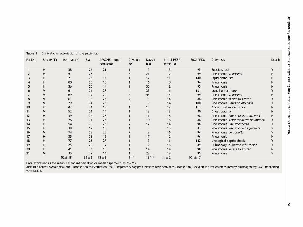
The principal patients characteristics are summarized inTable 1. In most cases, the cause of respiratory failurewas lung infection (67%). At the time of LRM, a total of12 patients were receiving vasoactive or inotropic medica-tion (noradrenalin in 9 cases, noradrenalin plus dobutaminein 2, and dobutamine alone in one patient). The meannoradrenalin and dobutamine dose was 0.24 ± 0.09 and6.91 ± 0.43 �g kg−1 min−1, respectively. The respiratory fre-quency and tidal volume at the start of maneuveringwas 16.6 ± 2.2 rpm and 6.9 ± 1.1 ml/kg body ideal weight,respectively.
Effects of recruitment upon respiratory function
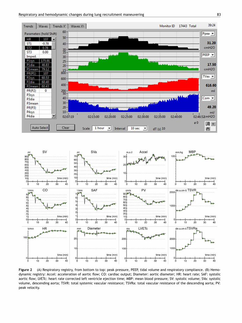
The evolution of the main respiratory parameters during LRMis reported in Table 2. Fig. 2A in turn shows the evolution ofthe respiratory parameters in one of the patients analyzed.
The maximum PEEP level reached during maneu-vering was 35.1 ± 2.1 cmH2O, with a maximum peakpressure of 54.2 ± 3.7. The open-lung PEEP value was16.8 ± 2.7 cmH2O, and was higher in obese patients (bodymass index > 30 kg/m2): 19.6 cmH2O (17.8---20.4) versus15.7 cmH2O (14.8---17.5; p < 0.05). At maximum PEEP level,Crs decreased 52% with respect to the initial value (95%CI:48.4---56.2%), while at the end of LRM it increased 23 ± 15.2%(95%CI: 15.91---30.14%). In 16 patients (76%) this increase was≥15% with respect to the basal value (15.3---56.3%), and inonly 6 patients was it ≥30% (33.8---56.3%).
In the 15 patients subjected to central venous blood gasanalysis, LRM induced or increased hypercapnia with res-piratory acidosis during the maximum PEEP phase. Afterestablishing the open-lung PEEP value, these alterationswere maintained with respect to the initial values (Table 3),while the SpO2/FiO2 ratio increased significantly (Table 2).
Effects of recruitment maneuvering upon patienthemodynamics
The hemodynamic effects during LRM are detailed in Table 4.The hemodynamic registry of one of the patients is shown inFig. 2B.
During the maximum PEEP phase, a decrease wasrecorded in CO, systolic volume (SV) and LVETc (23 ± 18%,26 ± 18% and 16 ± 13%, respectively) that persisted at theend of LRM despite the increase in heart rate and Accel.The MBP increased temporarily coinciding with the maxi-mum PEEP phase, while cardiac power decreased during thisLRM stage. Only one patient showed increased CO during themaneuver (patient 13).
Resp
irato
ry
and
hem
odyn
am
ic
change
s
durin
g
lung
recru
itment
maneuve
ring
81
Table 1 Clinical characteristics of the patients.
Patient Sex (M/F) Age (years) BMI APACHE II upon
admission
Days on
MV
Days in
ICU
Initial PEEP
(cmH2O)
SpO2/FiO2 Diagnosis Death
1 H 38 26 21 1 5 13 95 Septic shock Y
2 H 51 28 10 3 21 12 99 Pneumonia S. aureus N
3 H 21 26 12 1 12 11 140 Lipid embolism N
4 H 80 25 10 1 16 10 94 Pneumonia N
5 H 36 26 14 1 36 12 95 Pneumonia N
6 M 61 31 27 4 33 16 131 Lung hemorrhage Y
7 M 69 37 20 4 43 14 99 Pneumonia S. aureus N
8 M 61 33 22 2 3 14 88 Pneumonia varicella zoster Y
9 M 79 24 23 8 9 14 100 Pneumonia Candida albicans Y
10 H 42 21 18 1 13 12 112 Abdominal septic shock N
11 M 52 21 14 1 13 13 80 Chest trauma N
12 H 39 34 22 1 11 16 98 Pneumonia Pneumocystis jiroveci N
13 H 76 31 28 1 10 16 88 Pneumonia Acinetobacter baumannii Y
14 H 54 29 23 7 17 14 98 Pneumonia Pneumococcus Y
15 H 38 17 16 1 8 15 83 Pneumonia Pneumocystis jiroveci Y
16 M 74 23 25 7 8 16 94 Pneumonia Legionella Y
17 H 35 33 15 1 17 12 96 Pneumonia N
18 H 77 25 27 1 3 16 142 Urological septic shock Y
19 H 25 23 9 1 9 16 89 Pulmonary leukemic infiltration Y
20 H 41 26 15 1 14 14 98 Pneumonia Varicella zoster N
21 M 35 39 14 1 28 18 95 Pneumonia Y
52 ± 18 28 ± 6 18 ± 6 11---4 139---18 14 ± 2 101 ± 17
Data expressed as the mean ± standard deviation or median (percentiles 25---75).APACHE: Acute Physiological and Chronic Health Evaluation; FiO2: inspiratory oxygen fraction; BMI: body mass index; SpO2: oxygen saturation measured by pulsioxymetry; MV: mechanicalventilation.

82 M.I. Monge García et al.
Table 2 Respiratory changes during pulmonary recruitment maneuvering.
preRM maxPEEP bestPEEP
PEEP, cmH2O 14.1 ± 2.1 35.1 ± 2.1†† 16.8 ± 2.7†
Tidal volume, ml 437.6 ± 87.1 231.3 ± 59.9†† 488.5 ± 93.2**
Compliance, ml/cmH2O 26.5 ± 7.8 12.4 ± 3.9†† 32.4 ± 10.6††
Peak pressure, cmH2O 31.3 ± 3.9 54.2 ± 3.7†† 32.8 ± 4.2†
Mean pressure, cmH2O 21.9 ± 2.8 43.9 ± 2.7†† 24.3 ± 3.3†
Ventilation pressure, cmH2O 17.2 ± 2.9 19.2 ± 2.9†† 15.9 ± 3.4*
SpO2/FiO2, % 101.2 ± 17.7 171.2 ± 57††
Data expressed as the mean ± standard deviation.*P < 0.05 vs preMR, **P < 0.01 vs preMR, †P < 0.001 vs preMR, ††P ≤ 0.0001 vs preMR. bestPEEP: PEEP at maximum respiratory compliance;FiO2: inspiratory oxygen fraction; maxPEEP: maximum PEEP reached during recruitment maneuvering; PEEP: positive end-expirationpressure; PreRM: basal values, before start of recruitment maneuvering; SpO2: oxygen saturation measured by pulsioxymetry.
Table 3 Venous blood gas measurements during pulmonary
recruitment maneuvering.
PreRM maxPEEP bestPEEP
pH 7.24 ± 0.09 7.08 ± 0.11† 7.12 ± 0.13*
PvCO2, mmHg 61.6 ± 13.7 95.8 ± 23† 85.8 ± 22.4**
HCO3, mEq/l 25.2 ± 5.6 26.8 ± 5.9* 26.8 ± 5.7*
n = 15. Data expressed as the mean ± standard deviation.*P < 0.01 vs preRM; **P < 0.001 vs preRM; †P ≤ 0.0001 vs preRM.bestPEEP: PEEP at maximum respiratory compliance; max-PEEP: maximum PEEP reached during recruitment maneuvering;PreRM: basal values, before start of recruitment maneuvering;PvCO2: venous CO2 pressure.
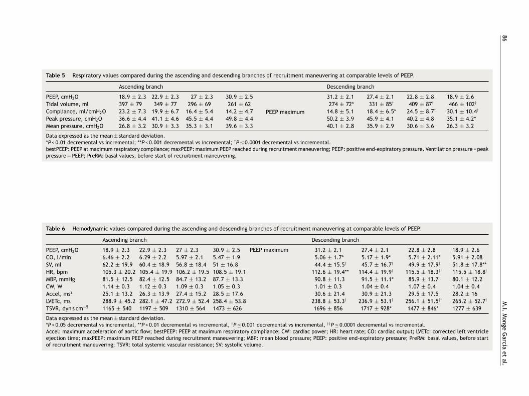
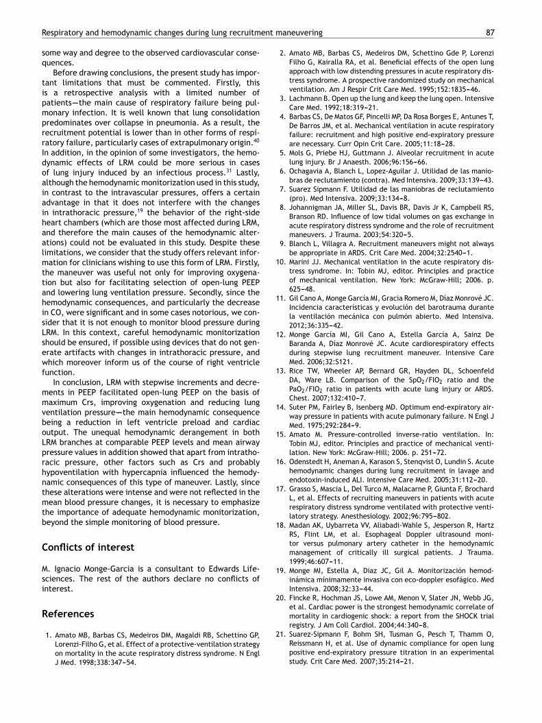
Comparison of the ascending and descendingbranches of recruitment maneuvering
On contrasting comparable PEEP levels in both LRMbranches, with similar mean airway pressures, the Crs valuewas always greater over the descending branch (Fig. 3 andTable 5). From the hemodynamic perspective, the modifi-cation of LVETc, SV and CO was more pronounced over thedescending branch, while heart rate was greater during thisLRM stage (Figs. 4 and 5, and Table 6).
Discussion
In this study, lung recruitment maneuvering (LRM) compris-ing an ascending branch with progressive (stepwise) PEEPincrements followed by a descending branch until reachingmaximum respiratory system compliance (Crs) conditionedrespiratory changes characterized by an important decreasein Crs at maximum PEEP level and an increase at the endof LRM with values higher than at the start, together withimprovement of the SpO2/FiO2 ratio and a decrease in ven-tilation pressure. The hemodynamic changes fundamentallyconsisted of a decrease in CO, SV and LVETc, together withan increase in heart rate and contractility. At comparablePEEP levels and mean airway pressure, significant differ-ences were observed between the two LRM branches, withmore marked modification of left ventricle preload and COover the descending branch.
The increase in Crs following LRM and its variations inthe form of stepwise PEEP increments and decrements havebeen demonstrated in different clinical and experimentalstudies,21---24 and have been carefully detailed by Hickling ina theoretical mathematical model.25 According to the obser-vations of this author, the changes in Crs during this kindof maneuver depend on: (a) the interaction between thenumber of alveoli that remain stable and inflated over the
Table 4 Hemodynamic changes during pulmonary recruitment maneuvering.
preRM maxPEEP bestPEEP
CO, l/min 6.78 ± 2.38 5.11 ± 1.77†† 6.02 ± 2.17*
SV, ml 64.8 ± 20.6 47.1 ± 16.3†† 52.6 ± 18.2†
HR, bpm 105.7 ± 19.7 110.1 ± 19.5 115.6 ± 18.9†
MBP, mmHg 79.2 ± 9 89.2 ± 12.7** 77.5 ± 11.7
CW, W 1.17 ± 0.38 1 ± 0.34* 1.03 ± 0.41
Accel, ms2 24.4 ± 12.1 29.5 ± 19.6 28.4 ± 15.7**
LVETc, ms 293.3 ± 39.9 246.3 ± 54.7†† 265.9 ± 51.5††
TSVR, dyn s cm−5 1093 ± 484 1599 ± 623†† 1268 ± 620
Data expressed as the mean ± standard deviation.*P < 0.05 vs preRM, **P < 0.01 vs preRM, †P < 0.001 vs preRM, ††P ≤ 0.0001 vs preRM.Accel: maximum acceleration of aortic flow; bestPEEP: PEEP at maximum respiratory compliance; CW: cardiac power; HR: heart rate;CO: cardiac output; LVETc: corrected left ventricle ejection time; maxPEEP: maximum PEEP reached during recruitment maneuvering;MBP: mean blood pressure; PreRM: basal values, before start of recruitment maneuvering; TSVR: total systemic vascular resistance; SV:systolic volume.
Respiratory and hemodynamic changes during lung recruitment maneuvering 83
Figure 2 (A) Respiratory registry, from bottom to top: peak pressure, PEEP, tidal volume and respiratory compliance. (B) Hemo-
dynamic registry: Accel: acceleration of aortic flow; CO: cardiac output; Diameter: aortic diameter; HR: heart rate; SAF: systolic
aortic flow; LVETc: heart rate corrected left ventricle ejection time; MBP: mean blood pressure; SV: systolic volume; SVa: systolic
volume, descending aorta; TSVR: total systemic vascular resistance; TSVRa: total vascular resistance of the descending aorta; PV:
peak velocity.

84 M.I. Monge García et al.
45
40
35
30
25
20
Respirato
ry c
om
plia
nc (
ml/cm
H2O
)
pre
RM
bestP
EE
P
15
10
10 15 20
∗
∗
∗
∗
25 30 35 40
PEEP (cmH2O)
Ascending branch (incremental PEEP)Descending branch (decremental PEEP)
5
Figure 3 Evolution of respiratory compliance during lung recruitment maneuvering at comparable levels of PEEP. Each level is
represented by its mean value and standard deviation.
10
6
Card
iac o
utp
ut
(l/m
in)
4
10 15 20
∗
∗∗
∗
25 30 35 40
PEEP (cmH2O)
Ascending branch (incremental PEEP)
Descending branch (decremental PEEP)
2
8
pre
RM
bestP
EE
P
Figure 4 Evolution of cardiac output during lung recruitment maneuvering at comparable levels of PEEP. Each level is represented
by its mean value and standard deviation.
entire respiratory cycle; (b) the transalveolar pressure anddegree of overdistension reached; and (c) the number ofinitially collapsed alveoli but which become inflated in thecourse of the inspiratory period (so-called ‘‘tidal recruit-ment’’26). In our patients, the changes in Crs during LRMwere similar to those predicted by the mathematical modelof Hickling, with the exception that in our study the start-ing PEEP had been previously established according to themaximum Crs, though without prior opening maneuver. Forthis reason it is probable that in addition to the numberof aerated stable alveoli and the transalveolar pressurereached, a non-negligible component of tidal recruitmentcould influence the Crs value before performing LRM. Like-wise, the stepwise decrease in Crs with each PEEP incrementafter starting the maneuver could have been related tothe gradual increase in transalveolar pressure and the
successive decrease in tidal recruitment; accordingly, onreaching the upper transalveolar pressure limit, the lungoverdistension value was maximum and tidal recruitmentminimum or inexistent----this resulting in Crs at its lowestlevel. Lastly, during the descending phase, the stepwisedecrease in transalveolar pressure gave rise to a gradualincrease in Crs until reaching its maximum value, which inaccordance to previous observations should coincide withthe reinitiation of lung collapse.21,27 It is very likely thatthe explanation for the observed differences in Crs betweenboth branches of the maneuver, at comparable PEEP levels,is the existence of a larger number of permanently aeratedalveoli over the descending branch of the maneuver, whichwould support the efficacy of LRM.21,25
Together with an improvement in the SpO2/FiO2 ratio,another consequence of LRM was that the Crs increment

Respiratory and hemodynamic changes during lung recruitment maneuvering 85
350
300
250
200
LV
ET
c,
Co
rrecte
d l
eft
ven
tric
le e
jecti
on
tim
e (
ms)
pre
RM
be
stP
EE
P
15010 15 20
∗
∗∗
∗
∗
25 30 35 40
PEEP (cmH2O)
Ascending branch (incremental PEEP)
Descending branch (decremental PEEP)
Figure 5 Evolution of heart rate corrected left ventricle ejection time (LVETc) at comparable levels of PEEP. Each level is
represented by its mean value and standard deviation.
allowed a reduction of ventilation pressure. The importanceof this measure during open lung ventilation has alreadybeen underscored by Amato et al.,1,2 who established avalue of <20 cmH2O as one of the main objectives in theirventilation strategy. Conceptually, the relationship betweenplateau pressure, PEEP and tissue distension can be repre-sented by a simple lever model in which PEEP representsthe point of support or fulcrum, plateau pressure is theapplied force, the degree of tissue stretch is the displace-ment reached, and ventilation pressure is the length of thelever torque arm.10 According to this model, on increas-ing the PEEP level without increasing plateau pressure----asoccurred in our patients----LRM decreases the ventilationpressure and consequently also the length of the lever armand tissue stretch, thereby exerting a favorable influenceupon the course of lung damage.2,3
Although the effects and depth of the hemodynamicdeterioration of LRM appear to be related to a series offactors, including the basal cardiovascular situation,28---30
the origin and time elapsed from lung injury,17 the typeof maneuver employed31 and its duration,29 etc., themarked but generally transient drop in CO is the hemody-namic effect most often reported in different clinical17,32,33
and experimental studies.16,21,28,29,31 In contrast, however,some authors have observed no significant hemodynamicalterations.22,34 On the other hand, the cardiovascular con-sequences of LRM fundamentally have been attributed to adecrease in venous return and cardiac preloadm,28,32 as wellas to an increase in right ventricle afterload22,28----the rela-tive importance of each of these mechanisms being lesser orgreater depending on the predominant effect of LRM upontranspulmonary or pleural pressure.35
The results of our study are consistent with the hemo-dynamic alterations described, with a decrease in CO (ofup to 58% in some cases) and left ventricle preload, asassessed from the monitorization of LVETc. However, sinceright ventricle function was not evaluated, we cannot besure whether these alterations were fundamentally due tochanges in preload or in postload of the right ventricle.Nevertheless, the unequal behavior of Crs over both LRMbranches at a comparable PEEP level and mean pressuresuggests that transpulmonary pressure was also different in
each stage of the maneuver, and therefore that the effectupon right ventricle afterload could be the most relevantfactor over the ascending branch, while the decrease invenous return and right ventricle preload as a result ofincreased transmission to the pleural space could be themost important mechanism over the descending branch ofLRM.
In accordance with the observations of previous stud-ies, our results also confirm the scant usefulness of bloodpressure as an exclusive parameter for monitoring the hemo-dynamic consequences of LRM.16,22,31,33,34 In effect, duringthe end stage of the ascending branch, MBP increased sig-nificantly, behaving in a way opposite to CO, and thereforewithout reflecting the global effect of LRM upon the car-diocirculatory system. Moreover, and curiously, despite theincrease in MBP, cardiac power (a parameter assessing theefficiency of the heart as a hydraulic pump, combining simul-taneous measurement of flow and pressure)36 decreasedsignificantly over the ascending branch. However, since thisparameter appears to be influenced not only by contractil-ity but also by the changes in preload and heart rate,36 wedo not know the precise significance of its evolution dur-ing LRM, since there were marked changes in these latterparameters during maneuvering. Nevertheless, since accel-eration of aortic flow and heart rate increased during themaneuver, we suspect that the changes in cardiac powercould be related mainly to worsened preload, and thereforethat heart function of the patients was preserved.
On the other hand, although blood gas measurementswere not obtained in each PEEP step or in all patients, itcan be assumed that progressive hypoventilation with hyper-capnia was an accompanying phenomenon during LRM thatcould have influenced the observed hemodynamic effects.Although there is some controversy regarding the principalhemodynamic effects of acute hypercapnia, they appear tocomprise the generation of a hyperdynamic state (high CO,elevated heart rate and lessened systemic vascular resis-tances), as well as an increase in pulmonary artery pressurethat could negatively affect right ventricle function.37---39 Itis therefore very probable that all these mechanisms wereimplicated in the end hemodynamic effect of LRM, andthat the combined result of these factors contributed in
86
M.I.
Monge
García
et
al.
Table 5 Respiratory values compared during the ascending and descending branches of recruitment maneuvering at comparable levels of PEEP.
Ascending branch Descending branch
PEEP, cmH2O 18.9 ± 2.3 22.9 ± 2.3 27 ± 2.3 30.9 ± 2.5
PEEP maximum
31.2 ± 2.1 27.4 ± 2.1 22.8 ± 2.8 18.9 ± 2.6
Tidal volume, ml 397 ± 79 349 ± 77 296 ± 69 261 ± 62 274 ± 72* 331 ± 85† 409 ± 87† 466 ± 102†
Compliance, ml/cmH2O 23.2 ± 7.3 19.9 ± 6.7 16.4 ± 5.4 14.2 ± 4.7 14.8 ± 5.1 18.4 ± 6.5* 24.5 ± 8.7† 30.1 ± 10.4†
Peak pressure, cmH2O 36.6 ± 4.4 41.1 ± 4.6 45.5 ± 4.4 49.8 ± 4.4 50.2 ± 3.9 45.9 ± 4.1 40.2 ± 4.8 35.1 ± 4.2*
Mean pressure, cmH2O 26.8 ± 3.2 30.9 ± 3.3 35.3 ± 3.1 39.6 ± 3.3 40.1 ± 2.8 35.9 ± 2.9 30.6 ± 3.6 26.3 ± 3.2
Data expressed as the mean ± standard deviation.*P < 0.01 decremental vs incremental; **P < 0.001 decremental vs incremental; †P ≤ 0.0001 decremental vs incremental.bestPEEP: PEEP at maximum respiratory compliance; maxPEEP: maximum PEEP reached during recruitment maneuvering; PEEP: positive end-expiratory pressure. Ventilation pressure = peakpressure − PEEP; PreRM: basal values, before start of recruitment maneuvering.
Table 6 Hemodynamic values compared during the ascending and descending branches of recruitment maneuvering at comparable levels of PEEP.
Ascending branch Descending branch
PEEP, cmH2O 18.9 ± 2.3 22.9 ± 2.3 27 ± 2.3 30.9 ± 2.5 PEEP maximum 31.2 ± 2.1 27.4 ± 2.1 22.8 ± 2.8 18.9 ± 2.6
CO, l/min 6.46 ± 2.2 6.29 ± 2.2 5.97 ± 2.1 5.47 ± 1.9 5.06 ± 1.7* 5.17 ± 1.9* 5.71 ± 2.11* 5.91 ± 2.08
SV, ml 62.2 ± 19.9 60.4 ± 18.9 56.8 ± 18.4 51 ± 16.8 44.4 ± 15.5† 45.7 ± 16.7† 49.9 ± 17.9† 51.8 ± 17.8**
HR, bpm 105.3 ± 20.2 105.4 ± 19.9 106.2 ± 19.5 108.5 ± 19.1 112.6 ± 19.4** 114.4 ± 19.9† 115.5 ± 18.3†† 115.5 ± 18.8†
MBP, mmHg 81.5 ± 12.5 82.4 ± 12.5 84.7 ± 13.2 87.7 ± 13.3 90.8 ± 11.3 91.5 ± 11.1* 85.9 ± 13.7 80.1 ± 12.2
CW, W 1.14 ± 0.3 1.12 ± 0.3 1.09 ± 0.3 1.05 ± 0.3 1.01 ± 0.3 1.04 ± 0.4 1.07 ± 0.4 1.04 ± 0.4
Accel, ms2 25.1 ± 13.2 26.3 ± 13.9 27.4 ± 15.2 28.5 ± 17.6 30.6 ± 21.4 30.9 ± 21.3 29.5 ± 17.5 28.2 ± 16
LVETc, ms 288.9 ± 45.2 282.1 ± 47.2 272.9 ± 52.4 258.4 ± 53.8 238.8 ± 53.3† 236.9 ± 53.1† 256.1 ± 51.5†† 265.2 ± 52.7†
TSVR, dyn s cm−5 1165 ± 540 1197 ± 509 1310 ± 564 1473 ± 626 1696 ± 856 1717 ± 928* 1477 ± 846* 1277 ± 639
Data expressed as the mean ± standard deviation.*P < 0.05 decremental vs incremental, **P < 0.01 decremental vs incremental, †P ≤ 0.001 decremental vs incremental, ††P ≤ 0.0001 decremental vs incremental.Accel: maximum acceleration of aortic flow; bestPEEP: PEEP at maximum respiratory compliance; CW: cardiac power; HR: heart rate; CO: cardiac output; LVETc: corrected left ventricleejection time; maxPEEP: maximum PEEP reached during recruitment maneuvering; MBP: mean blood pressure; PEEP: positive end-expiratory pressure; PreRM: basal values, before startof recruitment maneuvering; TSVR: total systemic vascular resistance; SV: systolic volume.
Respiratory and hemodynamic changes during lung recruitment maneuvering 87
some way and degree to the observed cardiovascular conse-quences.
Before drawing conclusions, the present study has impor-tant limitations that must be commented. Firstly, thisis a retrospective analysis with a limited number ofpatients----the main cause of respiratory failure being pul-monary infection. It is well known that lung consolidationpredominates over collapse in pneumonia. As a result, therecruitment potential is lower than in other forms of respi-ratory failure, particularly cases of extrapulmonary origin.40
In addition, in the opinion of some investigators, the hemo-dynamic effects of LRM could be more serious in casesof lung injury induced by an infectious process.31 Lastly,although the hemodynamic monitorization used in this study,in contrast to the intravascular pressures, offers a certainadvantage in that it does not interfere with the changesin intrathoracic pressure,19 the behavior of the right-sideheart chambers (which are those most affected during LRM,and therefore the main causes of the hemodynamic alter-ations) could not be evaluated in this study. Despite theselimitations, we consider that the study offers relevant infor-mation for clinicians wishing to use this form of LRM. Firstly,the maneuver was useful not only for improving oxygena-tion but also for facilitating selection of open-lung PEEPand lowering lung ventilation pressure. Secondly, since thehemodynamic consequences, and particularly the decreasein CO, were significant and in some cases notorious, we con-sider that it is not enough to monitor blood pressure duringLRM. In this context, careful hemodynamic monitorizationshould be ensured, if possible using devices that do not gen-erate artifacts with changes in intrathoracic pressure, andwhich moreover inform us of the course of right ventriclefunction.
In conclusion, LRM with stepwise increments and decre-ments in PEEP facilitated open-lung PEEP on the basis ofmaximum Crs, improving oxygenation and reducing lungventilation pressure----the main hemodynamic consequencebeing a reduction in left ventricle preload and cardiacoutput. The unequal hemodynamic derangement in bothLRM branches at comparable PEEP levels and mean airwaypressure values in addition showed that apart from intratho-racic pressure, other factors such as Crs and probablyhypoventilation with hypercapnia influenced the hemody-namic consequences of this type of maneuver. Lastly, sincethese alterations were intense and were not reflected in themean blood pressure changes, it is necessary to emphasizethe importance of adequate hemodynamic monitorization,beyond the simple monitoring of blood pressure.
Conflicts of interest
M. Ignacio Monge-Garcia is a consultant to Edwards Life-sciences. The rest of the authors declare no conflicts ofinterest.
References
1. Amato MB, Barbas CS, Medeiros DM, Magaldi RB, Schettino GP,Lorenzi-Filho G, et al. Effect of a protective-ventilation strategyon mortality in the acute respiratory distress syndrome. N EnglJ Med. 1998;338:347---54.
2. Amato MB, Barbas CS, Medeiros DM, Schettino Gde P, LorenziFilho G, Kairalla RA, et al. Beneficial effects of the open lungapproach with low distending pressures in acute respiratory dis-tress syndrome. A prospective randomized study on mechanicalventilation. Am J Respir Crit Care Med. 1995;152:1835---46.
3. Lachmann B. Open up the lung and keep the lung open. IntensiveCare Med. 1992;18:319---21.
4. Barbas CS, De Matos GF, Pincelli MP, Da Rosa Borges E, Antunes T,De Barros JM, et al. Mechanical ventilation in acute respiratoryfailure: recruitment and high positive end-expiratory pressureare necessary. Curr Opin Crit Care. 2005;11:18---28.
5. Mols G, Priebe HJ, Guttmann J. Alveolar recruitment in acutelung injury. Br J Anaesth. 2006;96:156---66.
6. Ochagavia A, Blanch L, Lopez-Aguilar J. Utilidad de las manio-bras de reclutamiento (contra). Med Intensiva. 2009;33:139---43.
7. Suarez Sipmann F. Utilidad de las maniobras de reclutamiento(pro). Med Intensiva. 2009;33:134---8.
8. Johannigman JA, Miller SL, Davis BR, Davis Jr K, Campbell RS,Branson RD. Influence of low tidal volumes on gas exchange inacute respiratory distress syndrome and the role of recruitmentmaneuvers. J Trauma. 2003;54:320---5.
9. Blanch L, Villagra A. Recruitment maneuvers might not alwaysbe appropriate in ARDS. Crit Care Med. 2004;32:2540---1.
10. Marini JJ. Mechanical ventilation in the acute respiratory dis-tress syndrome. In: Tobin MJ, editor. Principles and practiceof mechanical ventilation. New York: McGraw-Hill; 2006. p.625---48.
11. Gil Cano A, Monge García MI, Gracia Romero M, Díaz Monrové JC.Incidencia características y evolución del barotrauma durantela ventilación mecánica con pulmón abierto. Med Intensiva.2012;36:335---42.
12. Monge García MI, Gil Cano A, Estella García A, Sainz DeBaranda A, Díaz Monrové JC. Acute cardiorespiratory effectsduring stepwise lung recruitment maneuver. Intensive CareMed. 2006;32:S121.
13. Rice TW, Wheeler AP, Bernard GR, Hayden DL, SchoenfeldDA, Ware LB. Comparison of the SpO2/FIO2 ratio and thePaO2/FIO2 ratio in patients with acute lung injury or ARDS.Chest. 2007;132:410---7.
14. Suter PM, Fairley B, Isenberg MD. Optimum end-expiratory air-way pressure in patients with acute pulmonary failure. N Engl JMed. 1975;292:284---9.
15. Amato M. Pressure-controlled inverse-ratio ventilation. In:Tobin MJ, editor. Principles and practice of mechanical venti-lation. New York: McGraw-Hill; 2006. p. 251---72.
16. Odenstedt H, Aneman A, Karason S, Stenqvist O, Lundin S. Acutehemodynamic changes during lung recruitment in lavage andendotoxin-induced ALI. Intensive Care Med. 2005;31:112---20.
17. Grasso S, Mascia L, Del Turco M, Malacarne P, Giunta F, BrochardL, et al. Effects of recruiting maneuvers in patients with acuterespiratory distress syndrome ventilated with protective venti-latory strategy. Anesthesiology. 2002;96:795---802.
18. Madan AK, Uybarreta VV, Aliabadi-Wahle S, Jesperson R, HartzRS, Flint LM, et al. Esophageal Doppler ultrasound moni-tor versus pulmonary artery catheter in the hemodynamicmanagement of critically ill surgical patients. J Trauma.1999;46:607---11.
19. Monge MI, Estella A, Diaz JC, Gil A. Monitorización hemod-inámica mínimamente invasiva con eco-doppler esofágico. MedIntensiva. 2008;32:33---44.
20. Fincke R, Hochman JS, Lowe AM, Menon V, Slater JN, Webb JG,et al. Cardiac power is the strongest hemodynamic correlate ofmortality in cardiogenic shock: a report from the SHOCK trialregistry. J Am Coll Cardiol. 2004;44:340---8.
21. Suarez-Sipmann F, Bohm SH, Tusman G, Pesch T, Thamm O,Reissmann H, et al. Use of dynamic compliance for open lungpositive end-expiratory pressure titration in an experimentalstudy. Crit Care Med. 2007;35:214---21.
88 M.I. Monge García et al.
22. Gernoth C, Wagner G, Pelosi P, Luecke T. Respiratory andhaemodynamic changes during decremental open lung positiveend-expiratory pressure titration in patients with acute respi-ratory distress syndrome. Crit Care. 2009;13:R59.
23. Lambermont B, Ghuysen A, Janssen N, Morimont P, Hartstein G,Gerard P, et al. Comparison of functional residual capacity andstatic compliance of the respiratory system during a positiveend-expiratory pressure (PEEP) ramp procedure in an experi-mental model of acute respiratory distress syndrome. Crit Care.2008;12:R91.
24. Hodgson CL, Tuxen DV, Bailey MJ, Holland AE, Keating JL, PilcherD, et al. A positive response to a recruitment maneuver withPEEP titration in patients with ARDS, regardless of transientoxygen desaturation during the maneuver. J Intensive Care Med.2011;26:41---9.
25. Hickling KG. Best compliance during a decremental, but notincremental, positive end-expiratory pressure trial is relatedto open-lung positive end-expiratory pressure: a mathematicalmodel of acute respiratory distress syndrome lungs. Am J RespirCrit Care Med. 2001;163:69---78.
26. Terragni PP, Rosboch G, Tealdi A, Corno E, Menaldo E, Davini O,et al. Tidal hyperinflation during low tidal volume ventilation inacute respiratory distress syndrome. Am J Respir Crit Care Med.2007;175:160---6.
27. Borges JB, Okamoto VN, Matos GF, Caramez MP, Arantes PR,Barros F, et al. Reversibility of lung collapse and hypoxemia inearly acute respiratory distress syndrome. Am J Respir Crit CareMed. 2006;174:268---78.
28. Nielsen J, Nilsson M, Freden F, Hultman J, Alstrom U, Kjaer-gaard J, et al. Central hemodynamics during lung recruitmentmaneuvers at hypovolemia, normovolemia and hypervolemia.A study by echocardiography and continuous pulmonary arteryflow measurements in lung-injured pigs. Intensive Care Med.2006;32:585---94.
29. Odenstedt H, Lindgren S, Olegard C, Erlandsson K, Lethvall S,Aneman A, et al. Slow moderate pressure recruitment maneuverminimizes negative circulatory and lung mechanic side effects:evaluation of recruitment maneuvers using electric impedancetomography. Intensive Care Med. 2005;31:1706---14.
30. Fougeres E, Teboul JL, Richard C, Osman D, Chemla D, MonnetX. Hemodynamic impact of a positive end-expiratory pressuresetting in acute respiratory distress syndrome: importance ofthe volume status. Crit Care Med. 2010;38:802---7.
31. Lim SC, Adams AB, Simonson DA, Dries DJ, Broccard AF,Hotchkiss JR, et al. Transient hemodynamic effects of recruit-ment maneuvers in three experimental models of acute lunginjury. Crit Care Med. 2004;32:2378---84.
32. Nielsen J, Ostergaard M, Kjaergaard J, Tingleff J, BerthelsenPG, Nygard E, et al. Lung recruitment maneuver depressescentral hemodynamics in patients following cardiac surgery.Intensive Care Med. 2005;31:1189---94.
33. Toth I, Leiner T, Mikor A, Szakmany T, Bogar L, Molnar Z. Hemo-dynamic and respiratory changes during lung recruitment anddescending optimal positive end-expiratory pressure titrationin patients with acute respiratory distress syndrome. Crit CareMed. 2007;35:787---93.
34. Villagra A, Ochagavia A, Vatua S, Murias G, Del Mar Fer-nandez M, Lopez Aguilar J, et al. Recruitment maneuversduring lung protective ventilation in acute respiratory dis-tress syndrome. Am J Respir Crit Care Med. 2002;165:165---70.
35. Jardin F, Vieillard-Baron A. Right ventricular function andpositive pressure ventilation in clinical practice: from hemo-dynamic subsets to respirator settings. Intensive Care Med.2003;29:1426---34.
36. Pinsky MR. Functional hemodynamic monitoring: applied phys-iology at the bedside. In: Vincent JL, editor. Yearbook ofintensive care and emergency medicine. Heidelberg: Springer-Verlag; 2002. p. 534---51.
37. Thorens JB, Jolliet P, Ritz M, Chevrolet JC. Effects of rapidpermissive hypercapnia on hemodynamics, gas exchange, andoxygen transport and consumption during mechanical ventila-tion for the acute respiratory distress syndrome. Intensive CareMed. 1996;22:182---91.
38. Carvalho CR, Barbas CS, Medeiros DM, Magaldi RB, Lorenzi FilhoG, Kairalla RA, et al. Temporal hemodynamic effects of per-missive hypercapnia associated with ideal PEEP in ARDS. Am JRespir Crit Care Med. 1997;156:1458---66.
39. Mekontso Dessap A, Charron C, Devaquet J, Aboab J, Jardin F,Brochard L, et al. Impact of acute hypercapnia and augmentedpositive end-expiratory pressure on right ventricle function insevere acute respiratory distress syndrome. Intensive Care Med.2009;35:1850---8.
40. Rocco PR, Zin WA. Pulmonary and extrapulmonary acute res-piratory distress syndrome: are they different? Curr Opin CritCare. 2005;11:10---7.
Copyright © 2022 FDOKUMEN