Hemodynamic Disorders, Thromboembolism, and Shock
60
Hemodynamic Disorders, Thromboembolism, and Shock -I- Dr Lina Haffar Pr of Pathology 1
-
Upload
khangminh22 -
Category
Documents
-
view
0 -
download
0
Transcript of Hemodynamic Disorders, Thromboembolism, and Shock

Hemodynamic Disorders, Thromboembolism, and Shock -I-
Dr Lina Haffar
Pr of Pathology
1

Hemodynamic Disorders, Thromboembolism, and Shock
• Hyperemia and Congestion• Edema • Increased Hydrostatic Pressure • Reduced Plasma Osmotic
Pressure • Lymphatic Obstruction • Sodium and Water Retention • Hemorrhage• Hemostasis and
Thrombosis• Normal Hemostasis • Thrombosis • Disseminated Intravascular
Coagulation (DIC)
• Embolism• Pulmonary Thromboembolism • Systemic Thromboembolism • Fat Embolism• Amniotic Fluid Embolism • Air Embolism • Infarction• Shock• Pathogenesis of Septic Shock • Stages of Shock
2

HYPEREMIA AND CONGESTION
• Hyperemia and congestion both refer to an increase in blood volume within a tissue, but have different underlying mechanisms.
• Hyperemia is an active process resulting from arteriolar dilation and increased blood inflow, as occurs at sites of inflammation or in exercisingskeletal muscle.
• Hyperemic tissues are redder than normal because of engorgement with oxygenated blood.
3

HYPEREMIA AND CONGESTION
• Congestion is a passive process resulting from impaired outflow of venous blood from a tissue.
• It can occur systemically, as in cardiacfailure, or locally as a consequence of an isolated venous obstruction.
4

HYPEREMIA AND CONGESTION
• Congested tissues have an abnormal blue-red color ( cyanosis) that stems from the accumulation of deoxygenated hemoglobin in the affected area.
• In longstanding chronic congestion , inadequate tissue perfusion and persistent hypoxia may lead to parenchymal cell death and secondary tissue fibrosis, and the elevated intravascular pressures may cause edema or sometimes rupture capillaries, producing focal hemorrhages.
5

• M O R P H O L O G Y
• Cut surfaces of hyperemic or congestedtissues feel wet and typically ooze blood.
• On microscopic examination :
• acute pulmonary congestion is marked by blood-engorged alveolar capillaries with edema and intra-alveolar hemorrhage.
• In chronic pulmonary congestion , thickened and fibrotic septa , and alveolar spaces contain numerous macrophages laden -hemosiderin , siderophages (“heart failure cells”) (phagocytosed red cells ) .
6
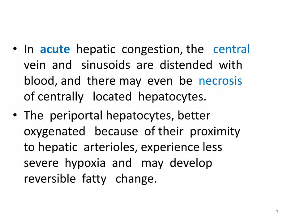
• In acute hepatic congestion, the centralvein and sinusoids are distended with blood, and there may even be necrosisof centrally located hepatocytes.
• The periportal hepatocytes, better oxygenated because of their proximity to hepatic arterioles, experience less severe hypoxia and may developreversible fatty change.
7

• In chronic passive congestion of the liver, the central regions of the hepatic lobules, viewed ongross examination, are red-brownand are accentuated against thesurrounding zones of uncongested tan, sometimes fattyliver (nutmeg liver)
• Microscopic findings include centrilobular hepatocyte necrosis, hemorrhage, and hemosiderin-laden macrophages .
8

EDEMA• Approximately 60 % of body weight is water, two-thirds
of which is intracellular. • Most of the remaining water is found in extracellular
compartments in the form of interstitial fluid; • only 5% of the body’s water is in blood plasma. • Edema is an accumulation of interstitial fluid within
tissues. • Extravascular fluid can also collect in body cavities and
such accumulations are often referred to collectively as effusions.
• Examples include effusions in the pleural cavity (hydrothorax ), the pericardial cavity (hydropericardium), or the peritoneal cavity (hydro -peritoneum, or ascites ).
9
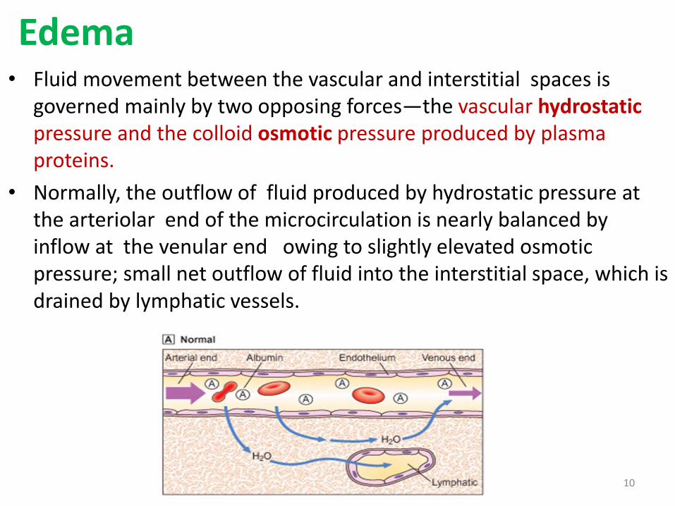
Edema• Fluid movement between the vascular and interstitial spaces is
governed mainly by two opposing forces—the vascular hydrostaticpressure and the colloid osmotic pressure produced by plasma proteins.
• Normally, the outflow of fluid produced by hydrostatic pressure at the arteriolar end of the microcirculation is nearly balanced by inflow at the venular end owing to slightly elevated osmotic pressure; small net outflow of fluid into the interstitial space, which is drained by lymphatic vessels.
10

• Either increased hydrostatic pressure or diminished colloid osmotic pressure causes increased movement of water into the interstitium
• Excess edema fluid is removed by lymphatic drainage and is returned to the bloodstream by way of the thoracic duct.
11

• Increased Hydrostatic Pressure :
• Mainly caused by disorders that impair venous return.
• Local increases in intravascular pressure caused by deep venous thrombosis in lower extremity can cause edema restricted to the distal portion of the affected leg.
12

• Generalized increases in venous pressure, with resultant systemic edema, occur most commonly in congestive heart failure.
• Fig. 4.3 illustrates the interlocking mechanisms that underlie generalized edema resulting from cardiac, renal, and hepatic failure.
• Several factors increase venous hydrostatic pressure in patients with congestive heart failure .
13

• Reduced Plasma Osmotic Pressure
• Reduction of plasma albumin concentrations leads to decreased colloid osmotic pressure of the blood and loss of fluid from the circulation.
• Therefore, conditions in which albumin is either lost from the circulation or synthesized in inadequate amounts are common causes of reduced plasma osmotic pressure.
• Nephrotic syndrome is the most important cause of albumin loss from the blood.
14

15

16

• Lymphatic Obstruction
• Edema may result from lymphatic obstruction that compromises resorption of fluid from interstitial spaces.
• Impaired lymphatic drainage and consequent lymphedema usually results from a localized obstruction caused by an inflammatory or neoplastic condition.
• For example, the parasitic infection filariasis can cause massive edema of the lower extremity and external genitalia (so-called “elephantiasis”) by producing inguinal lymphatic and lymph node fibrosis.
17

• Lymphatic Obstruction• Infiltration and obstruction of superficial
lymphatics by breast cancer may cause edema of the overlying skin; the characteristic finely pitted appearance of the skin of the affected breast is called peau d’orange (orange peel).
• Lymphedema also may occur as a complication of therapy.
• One relatively common setting for this clinical entity is in women with breast cancer who undergo axillary lymph node resection and/or irradiation, both of which can disrupt and obstruct lymphatic drainage, resulting in severe lymphedema of the arm.
18

• M O R P H O L O G Y• Edema is easily recognized on gross
inspection;• microscopic examination shows clearing
and separation of the extracellular matrix(ECM) elements.
• Edema most commonly is encountered in subcutaneous tissues, lungs, and brain.
• Edema typically is most pronounced in the legs with standing and the sacrum with recumbency, a relationship termed dependent edema.
•
19

• M O R P H O L O G Y
• Finger pressure over edematous subcutaneoustissue displaces the interstitial fluid, leaving a finger-shapeddepression; this appearance is called pitting edema.
20
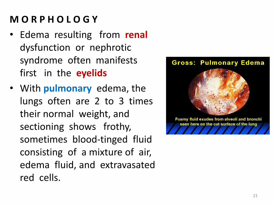
M O R P H O L O G Y
• Edema resulting from renaldysfunction or nephrotic syndrome often manifests first in the eyelids
• With pulmonary edema, the lungs often are 2 to 3 times their normal weight, and sectioning shows frothy, sometimes blood-tinged fluid consisting of a mixture of air,edema fluid, and extravasatedred cells.
21

M O R P H O L O G Y
• Brain edema can be localized ( abscess or tumor) or generalized, depending on the nature of injury. (Tortesky skull )
• With generalized edema, the sulci اثالم are narrowed as the gyri تالفيف swell and become flattened against the skull.
22

S U M M A RY
• EDEMA
• • Edema results from the movement of fluid from thevasculature into the interstitial spaces; the fluid may be protein poor (transudate) or protein rich (exudate).
• Edema may be caused by:
• • Increased hydrostatic pressure (e.g., heart failure)
• • Increased vascular permeability (e.g., inflammation)
• • Decreased colloid osmotic pressure resulting from reduced plasma albumin
• • Decreased synthesis (e.g., liver disease, proteinmalnutrition)
• • Increased loss (e.g., nephrotic syndrome)
• • Lymphatic obstruction (e.g., inflammation or neoplasia)23

HEMORRHAGE
• Hemorrhage, defined as the extravasation of blood from vessels, is most often the result of damage to blood vessels or defective clot formation.
• Hemorrhage may be external or accumulate
within a tissue as a hematoma, which ranges in
significance from mild (e.g., a bruise) to fatal
(e.g., a massive retroperitoneal hematoma
resulting from rupture of a dissecting aortic
aneurysm) .
24

HEMORRHAGE
- Large bleeds into body cavitiesare described variously according to location :• hemothorax , • hemopericardium, • hemoperitoneum ,• hemarthrosis (in joints). • Extensive hemorrhages can
occasionally result in jaundice from the massive breakdown of red cells and hemoglobin.
25

• I • Petechiae are minute (1 to 2 mm in diameter) hemorrhages into skin, mucous membranes, or serosal surfaces .
• causes include low platelet counts (thrombocytopenia),
• defective platelet function, and
• loss of vascular wall support, as in vitamin C deficiency .
26

• II • Purpura are slightly larger (3 to 5 mm) hemorrhages.
• Purpura can result from the same disorders that cause petechiae, as well as
• trauma,
• vascular inflammation (vasculitis), and
• increased vascular fragility.
27

• III • Ecchymoses are larger (1 to 2 cm) subcutaneous hematomas (called bruises).
• Extravasated red cells are phagocytosed and degraded by macrophages; the characteristic color changes of a bruise result from the enzymatic conversion of hemoglobin (red-blue color) to bilirubin (blue-green color) and eventually hemosiderin (golden-brown).
28
HEMORRHAGE
• The clinical significance of any particular hemorrhage depends on the volume of blood that is lost and the rate of bleeding.
• Rapid loss of up to 20% of the blood volume, or slow losses of even larger amounts, may have little impact in healthy adults; greater losses, however, can cause hemorrhagic (hypovolemic) shock .
29

HEMORRHAGE
• The site of hemorrhage also is important; bleeding that would be trivial in the subcutaneous tissues can cause death if located in the brain .
• Finally, chronic or recurrent external blood loss (e.g., due to peptic ulcer or menstrual bleeding) frequently culminates in iron deficiency anemia as a consequence of a loss of iron in hemoglobin. By contrast, iron is efficiently recycled from phagocytosed red cells, so internal bleeding (e.g., a hematoma) does not lead to iron deficiency.
30

HEMOSTASIS AND THROMBOSIS
• Normal hemostasis اإلرقاء comprises a series of regulated processes that culminate in the formation of a blood clot that limits bleeding from an injured vessel.
• The pathologic counterpart of hemostasis Is thrombosis, the formation of blood clot (thrombus) within non-traumatized, intact vessels.
31

HEMOSTASIS AND THROMBOSIS
• Normal Hemostasis• Hemostasis is a precisely orchestrated process
involving platelets, clotting factors, and endothelium that occurs at the site of vascular injury and culminates in the formation of a blood clot, which serves to prevent or limit the extent of bleeding.
• The general sequence of events leading to hemostasis at a site of vascular injury is :
32

I • Arteriolar vasoconstriction occurs immediately and markedly reduces blood flow to the injured area .• It is mediated by reflex neurogenic mechanisms
and augmented by the local secretion of factors such as endothelin, a potent endothelium-derived vasoconstrictor.
• This effect is transient
33

• Primary hemostasis: • the formation of the platelet plug.
• Disruption of the endothelium exposes subendothelialvon Willebrand factor (vWF) and collagen, which promote platelet adherence and activation.
• Within minutes the secreted products recruit additional platelets, which undergo aggregation to form a primary hemostatic plug .
34

• Secondary hemostasis: deposition of fibrin.
• Vascular injury exposes tissue factor at the site of injury.
• Tissue factor is normally expressed by subendothelial cells in the vessel wall, such as smooth muscle cells and fibroblasts.
• Tissue factor binds and activates factor VII setting in motion a cascade of reactions that culminates in thrombin generation.
• Thrombin cleaves circulating fibrinogen into insoluble fibrin, creating a fibrin meshwork, and also is a potent activator of platelets
35

• Clot stabilization and resorption.
• Polymerized fibrin and platelet aggregates undergo contraction to form a solid, permanent plug that prevents further hemorrhage.
• At this stage, counterregulatory mechanisms (e.g., tissue plasminogen activator, t-PA , made by endothelial cells) are set into motion that limit clotting to the site of injury and eventually lead to clot resorption and tissue repair.
36
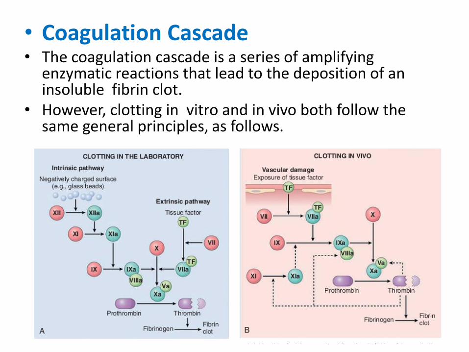
• Coagulation Cascade• The coagulation cascade is a series of amplifying
enzymatic reactions that lead to the deposition of an insoluble fibrin clot.
• However, clotting in vitro and in vivo both follow the same general principles, as follows.
37

S UMMARY
• COAGULATION FACTORS• • Coagulation occurs via the sequential enzymatic
conversion of a cascade of circulating and locally synthesized proteins.
• • Tissue factor elaborated at sites of injury is themost important initiator of the coagulation cascade in vivo.
• • At the final stage of coagulation, thrombin converts fibrinogen into insoluble fibrin that contributes to formation of the definitive hemostatic plug.
38

Thrombosis
• The primary abnormalities that lead to intravascular thrombosis are
• ( 1) endothelial injury,
• ( 2) stasis or turbulent blood flow, and
• (3) hypercoagulability of the blood
• (the so-called “Virchow triad”)
39

• 1- Endothelial Injury• Endothelial injury leading to platelet activation almost
inevitably underlies thrombus formation in the heart and the arterial circulation, where the high rates of blood flow impede clot formation.
• Notably, cardiac and arterial clots are typically rich in platelets, and it is believed that platelet adherence and activation is a necessary prerequisite for thrombus formation
• This insight provides part of the reasoning behind the use of aspirin and other platelet inhibitors in coronary artery disease and acute myocardial infarction.
40

• 2- Abnormal Blood Flow
• Turbulence (chaotic blood flow) contributes to arterial and cardiac thrombosis by causing endothelial injury or dysfunction, as well as by forming countercurrents and local pockets of stasis.
41

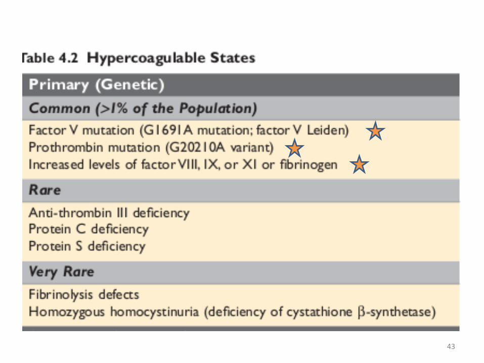
• 3- Hypercoagulability• Hypercoagulability refers to an abnormally high
tendency of the blood to clot, and is typically caused by alterations in coagulation factors.
• The alterations of the coagulation pathways that predispose affected persons to thrombosis can be divided into
• primary (genetic) and secondary (acquired) disorders
42
43

44
• M O R P H O L O G Y - I
• Thrombi can develop anywhere in the cardiovascular system.
• Arterial or cardiac thrombi typically arise at sites of endothelial injury or turbulence;
• Venous thrombi characteristically occur at sites of stasis.
• Thrombi are focally attached to the underlyingvascular surface and tend to propagate toward the heart; thus, arterial thrombi grow in a retrograde direction from the point of attachment, whereas venous thrombi extend in the direction of blood flow.
• The propagating portion of a thrombus tends to be poorly attached and therefore prone to fragmentation and migration through the blood as an embolus.
45

• MORPHOLOGY II • Migration through the blood as an embolus.• Thrombi can have grossly (and microscopically) apparent
laminations called lines of Zahn; these represent pale platelet and fibrin layers alternating with darker redcell–rich layers.
• Such lines are significant in that they are only found in thrombi that form in flowing blood; their presence cantherefore usually distinguish antemortem thrombosis from the bland nonlaminated clots that form in thepostmortem state.
• Although thrombi formed in the “low-flow” venous system superficially resemble postmortem clots, careful evaluation generally shows ill-defined laminations.
46

• MORPHOLOGY - III
• Thrombi in heart chambers or in the aortic lumen are designated as mural thrombi.
• Abnormal myocardial contraction(arrhythmias, dilated cardiomyopathy,or myocardial infarction) or endomyocardial injury ( myocarditis,catheter trauma) promote cardiac mural thrombi, whereas ulcerated atherosclerotic plaques and aneurysmal dilation promote aortic thrombosis .
47

• MORPHOLOGY - IV
• Arterial thrombi
• are frequently occlusive.
• They are typically rich in platelets, as theprocesses underlying their development
• (e.g., endothelial injury) lead to platelet activation.
• Although usually superimposed on a ruptured atherosclerotic plaque, other vascular injuries (vasculitis, trauma) can also be underlying causes.
48

• MORPHOLOGY - V
• Venous thrombi (phlebothrombosis)
• are almost invariably occlusive; they frequently propagate some distance toward the heart, forming a long cast within the vessel lumen that is give rise to emboli.
• Because these thrombi form in the slow venous circulation, they tend to contain more enmeshed redcells, leading to red, or stasis, thrombi.
• The veins of the lower extremities are most commonlyaffected (90% of venous thromboses); venous thrombi also can occur in the upper extremities, ovarian and periuterine veins
49

• MORPHOLOGY - VI
• At autopsy, postmortem clots can sometimes be mistaken for venous thrombi.
• However, the former are gelatinous and because of red cell settling they have a dark red dependent
portion and a yellow “chicken fat” upper portion;
they also are usually not attached to the underlying
vessel wall.
• By contrast, red thrombi typically are firm, focally attached to vessel walls, and they contain gray strands of deposited fibrin.
50

• MORPHOLOGY - VII
• Thrombi on heart valves are called vegetations.
• Bacterial or fungal bloodborne infections can cause valve damage, leading to the development of large thrombotic masses (infective endocarditis)
• Sterile vegetations also can develop onnoninfected valves in hypercoagulable states—the lesions called “nonbacterial thrombotic endocarditis”
• Less commonly, sterile, verrucous endocarditis (Libman-Sacks endocarditis) can occur in the setting of systemic lupus Erythematosus SLE .
51

Fate of the Thrombus
• If a patient survives an initial thrombotic event, during days to weeks the thrombus evolves through some combination of the following four processes:
I • Propagation. The thrombus enlarges through the accretion of additional platelets and fibrin, increasing the odds of vascular occlusion or embolization.
II • Embolization. Part or all of the thrombus is dislodged and transported elsewhere in the vasculature.
52

III • Dissolution.
If a thrombus is newly formed, activation of fibrinolytic
factors may lead to its rapid shrinkage and complete
dissolution.
With older thrombi, extensive fibrin polymerization
renders the thrombus substantially more resistant to
plasmin-induced proteolysis, and lysis is ineffectual.
• This resistance to lysis has clinical significance, as therapeutic administration of fibrinolytic agents ( in the setting of acute coronary thrombosis) generally is not effective unless administered within a few hours of thrombus formation.
53
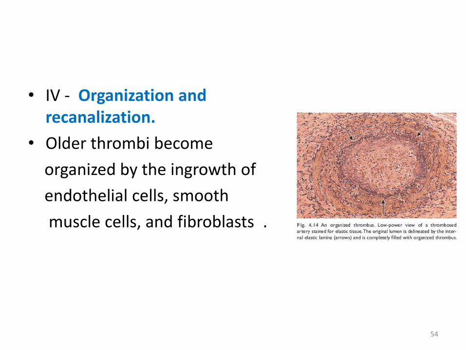
• IV - Organization and recanalization.
• Older thrombi become
organized by the ingrowth of
endothelial cells, smooth
muscle cells, and fibroblasts .
54

Venous Thrombosis ( Phlebothrombosis )
• Most venous thrombi occur in the superficial or the deep veins of the leg.
• Superficial venous thrombi usually arise in the saphenous system, particularly in the setting of varicosities; they can be painful and can cause local congestion and swelling from impaired venous outflow, predisposing the overlying skin to the development of infections and varicose ulcers.
55

Arterial and Cardiac Thrombosis
• Atherosclerosis is a major cause of arterial thromboses because it is associated with the loss of endothelial integrity and with abnormal blood flow .
• Myocardial infarction can predispose to cardiac mural thrombi by causing dyskinetic myocardial contraction and endocardial injury , and rheumatic heart disease may engender atrial mural thrombi by causing atrial dilation and fibrillation.
56

Disseminated Intravascular Coagulation (DIC)
• DIC is widespread thrombosis within the microcirculation that may be of sudden or insidious onset. ( renal failure ) .
• It may be seen in disorders ranging from obstetric complications to advanced malignancy.
• The widespread micro-vascular thrombosis consumes platelets and coagulation proteins (hence the synonym consumptive coagulopathy), and at the same time, fibrinolytic mechanisms are activated.
57

S U M M A RY• THROMBOSIS• Thrombus development is usually related to one or more components of Virchow’s triad :
• • endothelial injury (e.g., by toxins, hypertension, inflammation, or metabolic products)
• • abnormal blood flow, stasis , or turbulence (e.g., resulting from aneurysms, atherosclerotic plaque)
• • hypercoagulability: either primary (e.g., factor V Leiden, increased prothrombin synthesis, anti-thrombin III deficiency) or secondary (e.g., bed rest, tissue damage, malignancy)
58
S U M M A RY
• THROMBOSIS• Fate of thrombi
• • Thrombi may propagate, resolve, become organized, or embolize
• • Thrombosis causes tissue injury by local vascular occlusion or by distal embolization.
59

60