Adult advanced life support - European Resuscitation Council ...
Upload
khangminh22Category
view
1download
0
Medicina Intensiva 45 (2021) 532---540
http://www.medintensiva.org/
ORIGINAL
Hemodynamic resuscitation with fluids bolus and
norepinephrine increases severity of lung damage in
an experimental model of septic shock
P. Guijo Gonzalez a,b,c,∗, M.A. Gracia Romero a, A. Gil Cano a, M. Garcia Rojod,M. Cecconie, I.M. Monge Garcia a
a Intensive Care Medicine Department, Hospital del SAS Jerez, Jerez de la Frontera, Spainb Research Group in Critical Disorders (GREPAC), Institut Hospital del Mar d’Investigacions Mèdiques (IMIM), Barcelona, Spainc Critical Care Department, Hospital del Mar, Barcelona, Spaind Derpartment of Pathology, Hospital Universitario Puerta del Mar, Cadiz, Spaine Department of Anaesthesia and Intensive Care, IRCCS Instituto Clínico Humanitas, Humanitas University, Milan, Italy
Received 11 November 2019; accepted 18 May 2020
Available online 24 July 2020
KEYWORDS
Septic shock;Acute respiratorydistress syndrome;Endotoxemia;Resuscitation;Fluid
Abstract
Objective: Hemodynamic resuscitation is considered a cornerstone of the initial treatment of
septic shock. However, there is growing concern about its side effects. Our objective was
to assess the relationship between fluid administration and norepinephrine infusion and the
development of lung injury.
Design: Randomized in vivo study in rabbits.
Setting: University animal research laboratory.
Patients: Eighteen New Zealand rabbits. Control group (SHAM, n = 6), Sepsis group with or
without hemodynamic resuscitation (ETX-R, n = 6; ETX-NR, n = 6).
Interventions: Sepsis was induced by intravenous lipopolysaccharide administration and ani-
mals were followed-up for 4 h. Hemodynamic resuscitation with Ringer lactate (20 mL·kg−1)
was administered and later norepinephrine was initiated 3 h after sepsis induction. At the end,
the left lung was excised.
Main variables of interest: An indwelling arterial catheter and an esophageal Doppler were
placed. Lung mechanics were monitored with side stream spirometry. Lung damage was ana-
lyzed by histopathological examination.
∗ Corresponding author.
E-mail address: pedro [email protected] (P. Guijo Gonzalez).
https://doi.org/10.1016/j.medin.2020.05.011
0210-5691/© 2020 Elsevier Espana, S.L.U. y SEMICYUC. All rights reserved.

Medicina Intensiva 45 (2021) 532---540
Results: The SHAM group did not show hemodynamic or respiratory changes. Lipopolysaccha-
ride administration aimed an increase in cardiac output and arterial hypotension. In the ETX-NR
group, animals remained hypotensive until the end of the experiment. Resuscitation with fluids
and norepinephrine reversed arterial hypotension. Compared to the ETX-NR group, the remain-
ing lung of the ETX-R group showed greater accumulation of neutrophils and reactive type-II
pneumocytes, thicker alveolar wall, alveolar hemorrhage and non-aerated pulmonary areas.
Lung injury score was larger in the ETX-R group.
Conclusions: In our experimental study, following a strategy with bolus fluids and late nore-
pinephrine used in the early phase of endotoxic septic shock has a negative influence on the
development of lung injury.
© 2020 Elsevier Espana, S.L.U. y SEMICYUC. All rights reserved.
PALABRAS CLAVE
Shock séptico;Síndrome de distrésrespiratorio agudo;Endotoxemia;Resucitación;Fluidos
La resucitación hemodinámica con bolos de fluidos y noradrenalina incrementa la
severidad del dano pulmonar en un modelo experimental de shock séptico
Resumen
Objetivo: La resucitación hemodinámica es considerada piedra angular en el tratamiento inicial
del shock séptico. Sin embargo, existe creciente preocupación sobre sus efectos indeseables.
Nuestro objetivo fue evaluar la relación entre la administración de fluidos e infusión de nora-
drenalina y el desarrollo de lesión pulmonar.
Diseno: Estudio aleatorizado en animales vivos.
Ámbito: Laboratorio universitario de investigación.
Participantes: Dieciocho conejos de raza New Zealand White. Grupo control (SHAM, n = 6),
grupo séptico con o sin resucitación hemodinámica (ETX-R, n = 6; ETX-NR, n = 6).
Intervención: La sepsis fue inducida tras administración intravenosa de lipopolisacárido, y los
animales fueron seguidos durante 4 h. La resucitación hemodinámica mediante suero Ringer
lactato (20 ml·kg-1) y posterior noradrenalina fue iniciada a las 3 h de ser inducida la sepsis. Al
final del estudio, el pulmón izquierdo fue extraído.
Principales variables de interés: Fueron empleados catéter arterial y doppler esofágico. La
mecánica pulmonar fue monitorizada con sensor de flujo. El dano pulmonar fue analizado
mediante examen histopatológico.
Resultados: El grupo control no mostró cambios hemodinámicos ni respiratorios. La adminis-
tración del lipopolisacárido produjo un incremento del gasto cardíaco e hipotensión arterial.
En el grupo ETX-NR, los animales permanecieron hipotensos hasta el final del estudio. La
resucitación con fluidos y noradrenalina revirtió la hipotensión arterial. Comparados con el
grupo ETX-NR, en el grupo ETX-R el estudio histopatológico mostró mayor acumulación de neu-
trófilos, así como mayor presencia de neumocitos activados tipo II, engrosamiento de la pared
alveolar, hemorragia alveolar y zonas pulmonares no aireadas. La escala final de dano pulmonar
fue mayor en el grupo ETX-R.
Conclusiones: En nuestro estudio experimental, la estrategia basada en la administración de
fluidos y posterior infusión de noradrenalina en la fase precoz del shock séptico tiene una
influencia negativa sobre el desarrollo de la lesión pulmonar.
© 2020 Elsevier Espana, S.L.U. y SEMICYUC. Todos los derechos reservados.
Introduction
Severe sepsis and septic shock are the most frequent causesof admission in intensive care units, with a high mortalityrate around 30%.1 Recently reviewed definitions character-ize sepsis as life-threatening organ dysfunction, while septicshock is a subset of sepsis in which particularly profound cir-culatory, cellular and metabolic abnormalities substantiallyincrease mortality.2 Hemodynamic resuscitation, by meansof fluid bolus (30 mL·kg−1) and subsequent norepinephrine
(NE), has once again been considered by the Surviving Sep-sis Campaign (SSC) as a cornerstone of the initial treatmentof septic shock.3 The rationale for these recommendationsis that, in the early phase of severe sepsis and septic shock,restoring intravascular volume and maintaining end-organperfusion are the top priorities.3,4 However, the optimalstrategy of hemodynamic resuscitation in the early hoursof severe sepsis and septic shock is still controversial. Giventhe evidence of harm associated with positive fluid balanceaccumulated in septic patients from hospital admission to
533

P. Guijo Gonzalez, M.A. Gracia Romero, A. Gil Cano et al.
Intensive Care Unit (ICU) discharge,5---7 there is a growingconcern about the undesirable effects of this strategy, suchas acute respiratory distress syndrome (ARDS).8---10
ARDS is a devastating complication of sepsis that influ-ences its clinical management and outcomes.11,12 On theone hand, studies have established a strong relation-ship between fluid administration and the appearance ofARDS.13---18 On the other hand, the cardiovascular effectsof NE on the development of lung injury have not beenadequately investigated. NE, due to its ability to increasevenous return and myocardial contraction in septic shockpatients, increases pulmonary flow and perfusion,19---22 whichwould likely increase the severity of lung injury.
Because up to 40% of sepsis patients develop ARDS,23
and given that sepsis is a systemic inflammation state withhigh capillary permeability, it is possible that the effectsof hemodynamic resuscitation in the early phase of sep-tic shock could lead to enhaced pulmonary edema andincreased the risk for ARDS development. The purpose ofthis experimental study is to investigate the effects of bolusfluid administration and infusion of NE on the developmentof lung injury. We hypothesized that this strategy used in theearly phase of septic shock leads to lung injury development.
Patients and methods
This study was approved by the Ethics Committee of theUniversity of Cadiz (license 07-9604) and the Junta deAndalucía. Animal care and use procedures conformedto national and European Union regulations and guide-lines (Spanish Royal Decree 53/2013 and EU Directive2010/63/EU).
Anesthesia and instrumentation
Eighteen New-Zealand rabbits (weight 2.51 ± 0.13 kg) wereanesthetized with an intramuscular dose of xylazinehydrochloride (10 mg·kg−1) and ketamine (40 mg·kg−1). Theadequacy of anesthesia throughout the experiment wasassessed by the absence of any significant blood pres-sure and/or heart rate change, either spontaneous orin response to a noxious stimulus (tail clamping). Therabbits were tracheotomized, intubated and mechanicallyventilated (Servo 900c; Siemens-Elema, Solna, Sweden)in a volume-controlled mode, with a tidal volume of8 mL·kg−1, PEEP of 0 cmH2O, inspiratory-to-expiratory ratioof 1:2, inspired oxygen fraction of 0.6, and a respira-tory rate adjusted to maintain an end-tidal CO between35---45 mmHg. The anesthesia was maintained with a contin-uous intravenous infusion of ketamine (15---20 mg·kg−1
·h−1),midazolam (1---3 mg·kg−1
·h−1) and muscular blockade withrocuronium bromide (1 mg·kg−1
·h−1). Ringer’s lactate solu-tion (6 mL·kg−1
·h−1) was administered as maintenance fluidtherapy.
Hemodynamic monitoring
A pediatric esophageal Doppler probe (KDP72; CardioQCombi, Deltex Medical, Chichester, UK) was introduced intothe esophagus until the optimal outline and maximal peak
velocity of aortic blood waveform was obtained. Consecu-tive transesophageal Doppler measurements for 60 secondsat the beginning and after 1, 2, 3 and 4 h (end of experiment)were completed and averaged to calculate variables as theheart rate (HR), stroke volume (SV) and cardiac index (CI).Systolic, diastolic and mean arterial pressure, were contin-uously measured by an indwelling femoral artery catheterconnected to a pressure transducer (TruWave®, EdwardsLifesciences LLC, Irvine, CA, USA).
Respiratory monitoring
Continuous non-invasive measurements of flow, pressure andvolume in the animal’s airway were obtained using a spirom-etry monitor (Datex-Ohmeda M-COV, Helsinki, Finland) witha neonatal flow sensor (Patient Spirometry Kit 8004382, GEHealthcare, Helsinki, Finland) connected directly to the ani-mal’s tracheal tube. Dynamic compliance of the respiratorysystem (Cdyn) and the peak pressure (Ppeak) were measuredand averaged during a period of 60 s at the beginning andafter 2, 3 and 4 h (end of experiment).
Experimental protocol
After 5 min of stabilization and baseline measurementsof hemodynamic and lung mechanics, the animals wereassigned using a computer-generated random sequence tothree groups (6 animals each): a sham-operated group(SHAM), a non-resuscitated septic group (ETX-NR), anda septic group with hemodynamic resuscitation (ETX-R).In septic animals, a purified endotoxin-lipopolysaccharide(ETX) prepared from Escherichia coli 055:B5 (Sigma Chemi-cal, St. Louis, MO) was intravenously infused over a periodof 10 min (1 mg·kg−1). The dose and rate of the ETX wasselected based on previous publications.24 However, todetermine the best way for ETX administration, we required8 animals. SHAM animals received an equivalent volumeof normal saline. Three hours after ETX infusion, animalsin the ETX-R group received a fluid bolus (Ringer’s lac-tate) of 20 mL·kg−1, and if the mean arterial pressure(MAP) was lower than the baseline measurement, NE infu-sion was started at 0.25 mcg·kg−1
·min−1. NE was increased0.10 mcg·kg−1
·min−1 every 3 min until reaching a MAP simi-lar to the baseline level. A schematic representation of theexperimental protocol is shown in Fig. 1. After completionof the study protocol, animals were euthanized by a lethaldose of chloride potassium.
Histological analysis
At the end of the experiment, the left lung was excisedand immersed in 10% formaldehyde for at least 24 h. Tissuesamples were dehydrated with graded alcohol, embeddedin paraffin, and cut in a series of 5 �m-thick slices thatwere stained with hematoxylin and eosin. After the histo-logical preparations were obtained, they were scanned toobtain digital preparations (3DHistech, Budapest, Hungary).An expert pathologist then evaluated these tissue sectionsin a blinded fashion using the following scoring system todetermine the degree of lung injury: 0, no damage; 1, mild
534

Medicina Intensiva 45 (2021) 532---540
Figure 1 Schematic representation of the study protocol.
damage (present in 1---3 areas of 1 mm2); 2, moderate dam-age (present in more than 3 areas of 1 mm2 and less than75% of the tissue section); 3, severe damage (present inmore than 75% of the tissue section). These scores used thecombined assessments of six parameters: accumulation ofneutrophils in alveolar or interstitial space, reactive typeII pneumocytes with atypical nuclei in the alveolus, alveo-lar congestion/collapse, alveolar wall thickening, alveolarhemorrhage and hyaline membrane formation, presenting ascore from 0 to 18 ranging from normal histology to maxi-mum damage.25,26,27
Statistical analysis
Data are expressed as the mean ± standard deviation (SD),unless otherwise stated. Normality of data was checkedby the Shapiro---Wilk test. We used a two-way analysis ofvariance (ANOVA) for repeated measures to determine thestatistical significance of group differences in the respira-tory and hemodynamic parameters at different time points.The Greenhouse---Geisser correction was used when violationof sphericity was detected by the Mauchly test. When statis-tical significance was indicated, it was further examined bya post hoc analysis (Bonferroni test). Baseline parametersand the differences within-subjects were evaluated usinga repeated-measures ANOVA. The data obtained from thehistopathological study were analyzed according to inten-sity and extension score using the non-parametric KruskalWallis test. The differences between each pair of 2 groupswere assessed by Mann---Whitney U test. A p value <0.05
was considered statistically significant, unless otherwiseindicated. Data were analyzed by using MedCalc Statisti-cal Software version 16.8 (MedCalc Software bvba, Ostend,Belgium) and SPSS (SPSS 21, SPPS Inc, Chicago, IL).
Results
In the 18 rabbits randomly allocated to three differentgroups, we found that each group had similar baselinecharacteristics for any of the hemodynamic and respira-tory variables measured, except for the Cdyn (SupplementaryFile, Table S1).
Hemodynamic changes
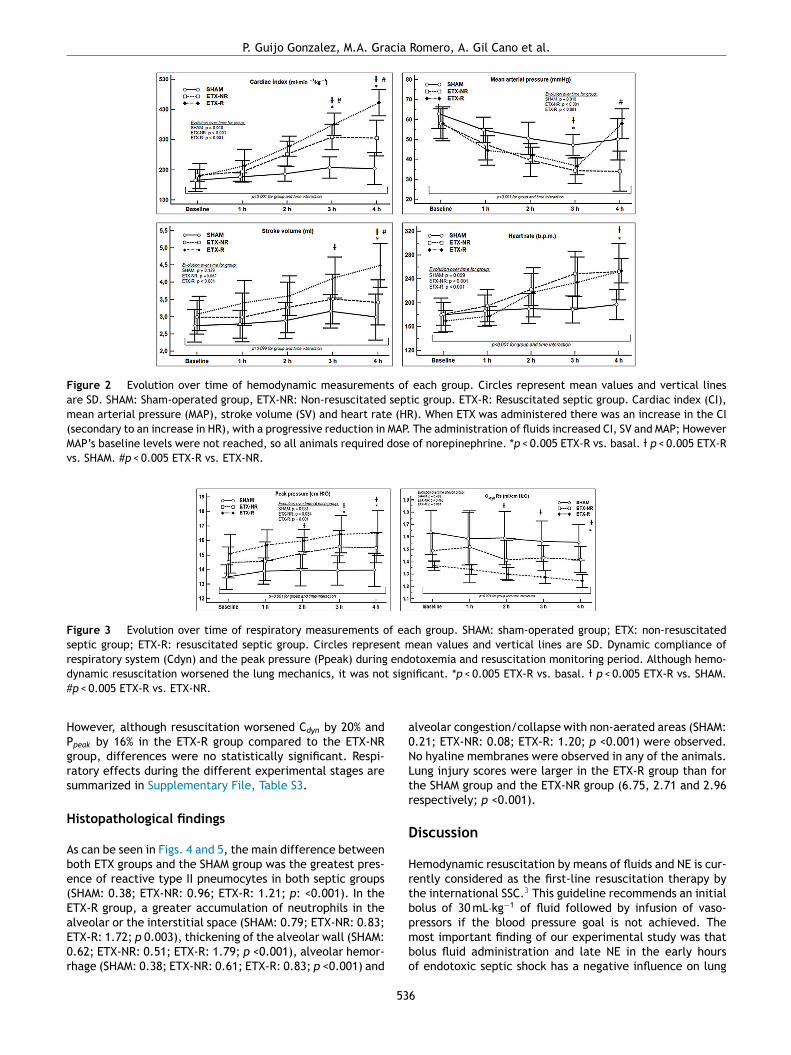
Infusion of ETX resulted in a hyperdynamic hemodynamicprofile with a progressive increase in CI (mostly secondaryto a higher HR) and a reduction in blood pressure. In the ETX-R group, although the administration of fluids increased CI,stroke volume (SV) and MAP by 20%, 11% and 18% respec-tively, MAP’s baseline levels were not reached, so NE wasnecessary in all cases (Fig. 2). Hemodynamic effects duringdifferent experimental stages are detailed in SupplementaryFile, Table S2.
Respiratory changes
As shown in Fig. 3, infusion of ETX caused a significancedecrease and increase in the Cdyn and Ppeak respectively.
535
P. Guijo Gonzalez, M.A. Gracia Romero, A. Gil Cano et al.
Figure 2 Evolution over time of hemodynamic measurements of each group. Circles represent mean values and vertical lines
are SD. SHAM: Sham-operated group, ETX-NR: Non-resuscitated septic group. ETX-R: Resuscitated septic group. Cardiac index (CI),
mean arterial pressure (MAP), stroke volume (SV) and heart rate (HR). When ETX was administered there was an increase in the CI
(secondary to an increase in HR), with a progressive reduction in MAP. The administration of fluids increased CI, SV and MAP; However
MAP’s baseline levels were not reached, so all animals required dose of norepinephrine. *p < 0.005 ETX-R vs. basal. p < 0.005 ETX-R
vs. SHAM. #p < 0.005 ETX-R vs. ETX-NR.
Figure 3 Evolution over time of respiratory measurements of each group. SHAM: sham-operated group; ETX: non-resuscitated
septic group; ETX-R: resuscitated septic group. Circles represent mean values and vertical lines are SD. Dynamic compliance of
respiratory system (Cdyn) and the peak pressure (Ppeak) during endotoxemia and resuscitation monitoring period. Although hemo-
dynamic resuscitation worsened the lung mechanics, it was not significant. *p < 0.005 ETX-R vs. basal. p < 0.005 ETX-R vs. SHAM.
#p < 0.005 ETX-R vs. ETX-NR.
However, although resuscitation worsened Cdyn by 20% andPpeak by 16% in the ETX-R group compared to the ETX-NRgroup, differences were no statistically significant. Respi-ratory effects during the different experimental stages aresummarized in Supplementary File, Table S3.
Histopathological findings
As can be seen in Figs. 4 and 5, the main difference betweenboth ETX groups and the SHAM group was the greatest pres-ence of reactive type II pneumocytes in both septic groups(SHAM: 0.38; ETX-NR: 0.96; ETX-R: 1.21; p: <0.001). In theETX-R group, a greater accumulation of neutrophils in thealveolar or the interstitial space (SHAM: 0.79; ETX-NR: 0.83;ETX-R: 1.72; p 0.003), thickening of the alveolar wall (SHAM:0.62; ETX-NR: 0.51; ETX-R: 1.79; p <0.001), alveolar hemor-rhage (SHAM: 0.38; ETX-NR: 0.61; ETX-R: 0.83; p <0.001) and
alveolar congestion/collapse with non-aerated areas (SHAM:0.21; ETX-NR: 0.08; ETX-R: 1.20; p <0.001) were observed.No hyaline membranes were observed in any of the animals.Lung injury scores were larger in the ETX-R group than forthe SHAM group and the ETX-NR group (6.75, 2.71 and 2.96respectively; p <0.001).
Discussion
Hemodynamic resuscitation by means of fluids and NE is cur-rently considered as the first-line resuscitation therapy bythe international SSC.3 This guideline recommends an initialbolus of 30 mL·kg−1 of fluid followed by infusion of vaso-pressors if the blood pressure goal is not achieved. Themost important finding of our experimental study was thatbolus fluid administration and late NE in the early hoursof endotoxic septic shock has a negative influence on lung
536

Medicina Intensiva 45 (2021) 532---540
Figure 4 Graphical representation of histopathological varia-
bles (mean value of intensity and extension) analyzed in each
group. Lung injury score 0---3. SHAM: sham-operated group;
ETX: non-resuscitated septic group; ETX-R: resuscitated sep-
tic group. Hemodynamic resuscitation resulted in greater score
of lung injury. *Indicate statistically significant difference from
SHAM, ETX-NR and ETX-R by the non-parametric Kruskal Wallis
test (p < 0.01).
injury development. When ETX was administered to induceseptic shock, an increase in reactive type II pneumocyteswas found, which suggests epithelial damage.28 However,when fluids and NE were administered to restore bloodpressure, they resulted in greater histopathological findingswith an increase in neutrophils infiltration, reactive typeII pneumocytes, alveolar congestion/collapse and alveolarwall thickening, and a larger lung injury score.
It can be assumed that increased pulmonary blood flow,due to administration of fluids (20 mL·kg−1) and NE infusion,combined with a capillary permeability disorder, were themechanisms responsible for the worsening of lung inflamma-tory damage initiated by endotoxemia. Although this is likelyto be the price to pay for adequate hemodynamic optimiza-tion, our findings provide valuable information about theconsequences and damaging effects of this strategy in septicshock.
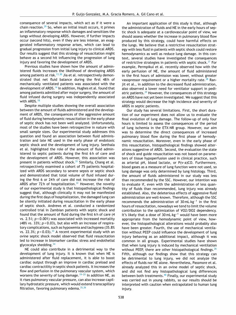
ARDS is a catastrophic form of lung injury characterizedby diffuse alveolar damage with severe inflammation andhigh permeability protein-rich edema.12 Recent studies sug-gest that the development of lung damage appears as a
Figure 5 Microscopic aspects of the lungs from SHAM group (A), and the ETX-R (B, C, D, E). (A) normal lung. (B) Neutrophils in
interstitium and alveolar wall. (C) Reactive Type II pneumocytes with hyperchromatic nuclei and nuclear membrane irregularity. (D)
Thickened alveolar walls with intramural neutrophils, macrophages and fibroblasts. (E) Area with pulmonary emphysema. (F) Gross
pathology surface of lung in the SHAM (left) and ETX-R (right) animals. Last one shows more intense damage, especially hemorrhagic
areas and lung edema. SHAM: sham-operated group; ETX: non-resuscitated septic group; ETX-R: resuscitated septic group (HE ×40).
537
P. Guijo Gonzalez, M.A. Gracia Romero, A. Gil Cano et al.
consequence of several impacts, which act as if it were achain reaction.29 So, when an initial insult occurs, it primesan inflammatory response which damages and sensitizes thelungs without developing ARDS. However, if further impactsoccur (second hits), even if they are less intense, an exag-gerated inflammatory response arises, which can lead togradual progression from initial lung injury to clinical ARDS.Our results suggest that this strategy of resuscitation mightbehave as a second hit influencing the progression of lunginjury and favoring the development of ARDS.
Previous studies have shown how the amount of admin-istered fluids increases the likelihood of developing ARDSamong patients at risk.13,30 Jia et al. retrospectively demon-strated that net fluid balance during the first 48 h ofmechanically ventilated patients was associated with thedevelopment of ARDS.14 In addition, Hughes et al. found thatamong patients admitted after major surgery, the amount offluid infused during surgery was independently associatedwith ARDS.18
Despite multiple studies showing the overall associationbetween the amount of fluids administered and the develop-ment of ARDS, the consequences of the aggressive amountof fluid during hemodynamic resuscitation in the early phaseof septic shock has not been well analyzed. Unfortunately,most of the studies have been retrospective in nature withsmall sample sizes. Our experimental study addresses thisquestion and found an association between fluid adminis-tration and late NE used in the early phase of endotoxicseptic shock and the development of lung injury. Seethalaet al. highlighted the role of the amount of fluid admin-istered to septic patients during the first 6 h of care andthe development of ARDS. However, this association waspresent in patients without shock.31 Similarly, Chang et al.retrospectively examined a cohort of 75 patients hospital-ized with ARDS secondary to severe sepsis or septic shockand demonstrated that total volume of fluid infused dur-ing the first 6 or 24 h of care did not increase the risk ofARDS after 72 h of hospitalization.32 However, the noveltyof our experimental study is that histopathological findingssuggest that, although clinically it may not be manifestedduring the first days of hospitalization, the damaged lung canbe silently initiated during resuscitation in the early phaseof septic shock. Andrews et al. conducted a randomizedcontrolled trial in Zambian patients with septic shock andfound that the amount of fluid during the first 6 h of care (4vs. 2.5 l; p < 0.001) was associated with increased mortality(48% vs. 33%; p < 0.03), but also with an increase of respira-tory complications, such as hypoxemia and tachypnea (35.8%vs. 22.3%; p < 0.03).33 A recent experimental study with anovine septic shock model observed that fluid resuscitationled to increase in biomarker cardiac stress and endothelialglycocalyx shedding.34
NE could also contribute in a detrimental way to thedevelopment of lung injury. It is known that when NE isadministered after fluid replacement, it is able to boostcardiac output through an improve in cardiac preload andcardiac contractility in septic shock patients. It increases theflow and perfusion in the pulmonary vascular system, whichworsens the severity of lung damage.19---20 In addition NE, asit rises pulmonary vascular pressure, can also increase capil-lary hydrostatic pressure, which would extend transcapillaryfiltration, favoring pulmonary edema.35,36
An important application of this study is that, althoughthe administration of fluids and NE in the early hours of sep-tic shock is adequate at a cardiovascular point of view, weshould assess whether the increase in pulmonary blood flowproduced by this strategy, could have harmful effects onthe lungs. We believe that a restrictive resuscitation strat-egy with less fluid in patients with septic shock could restorehemodynamics as well as reduce lung damage. In this con-text, several studies have investigated the consequencesof restrictive strategies in patients with septic shock.37 Forexample, Permpikul et al. recently observed how with theearly perfusion of NE, the amount of fluid administeredin the first hours of admission was lower, without greatervasopressor requirement or a higher mortality rate.38 Ran-jit et al., in addition to the decreased fluid administration,also observed a lower need for ventilator support in pedi-atric patients.39 However, the consequences of this strategyon ARDS have not yet been investigated. We suggest that thisstrategy would decrease the high incidence and severity ofARDS in septic patients.
Our study has several limitations. First, the short dura-tion of our experiment does not allow us to evaluate thefinal evolution of lung damage. The follow-up of only fourhours is limited to assess the consequences of the periodof lung ischemia in the ETX-NR group. However, our aimwas to determine the direct consequences of increasedpulmonary blood flow during the first phase of hemody-namic resuscitation. Moreover, even in the early phase ofthis resuscitation, histopathological findings showed alter-ations suggestive of ARDS. Second, the evaluation the stateof shock and guide resuscitation was not based on parame-ters of tissue hypoperfusion used in clinical practice, suchas arterial pH, blood lactate, or P(v-a)CO. Furthermore,blood gases as a measure of lung function were not used, solung damage was only determined by lung histology. Third,the amount of fluids administered in our study was lessthan recommended by the SSC. However, our intention wasto evaluate if, even with the administration of less quan-tity of fluids than recommended, lung injury was alreadyestablished. Also, the deleterious effects of aggressive fluidadministration are well-known. Therefore, although the SSCrecommends the administration of 30 mL·kg−1 in the firsthours of resuscitation, nowadays we tend to limit the volumecontribution to the optimization of VO2/DO2 dependency.It’s likely that a dose of 30 mL·kg−1 would have been moreappropriate from the hemodynamic point of view, how-ever, the histopathological alterations would probably alsohave been greater. Fourth, the use of mechanical ventila-tion without PEEP could influence the development of lunginjury behaving as an additional impact, although it wascommon in all groups. Experimental studies have shownthat when lung injury is induced by mechanical ventilationwithout PEEP, there are other histopathological findings.26
Fifth, although our findings show that this strategy canbe detrimental to lung injury, we did not analyze theeffects of fluids nor NE alone. Nevertheless, Passmore et al.already analyzed this in an ovine model of septic shock,and did not find any histopathological lung differencesbetween both treatments.40 Finally, our experimental studywas carried out in young rabbits, so our results should beinterpreted with caution when extrapolated to human lunginjury.
538

Medicina Intensiva 45 (2021) 532---540
Conclusions
Our experimental study shows that increasing pulmonaryblood flow with fluids and NE until reaching a MAP endpointin endotoxic septic shock has direct deleterious effects onlung damage. The relations between this strategy and theinflammatory lung injury were explained by histopathologicfindings. These findings suggest that once lung damage hasbeen initiated by endotexemia, the aggressive administra-tion of fluids and NE act as second hits. More studies arerequired to assess whether the prevention of these secondhits could result in a decrease in the incidence and severityof lung damage.
Contributions of the authors
P.G.G.: designed the study, participated in the experiments,acquired and interpreted the data, performed the statisticalanalysis, and draft the manuscript. M.I.M.G. and M.G.R.:participated in the experiments, acquired and interpretedthe data, performed the statistical analysis and helped todraft the manuscript. A.G.C.: designed the study, acquiredand interpreted the data, performed the statistical analysis,and helped to draft the manuscript. M.G.R.: Pathology study.M.C.: contributed in the conception and design of the study.All the authors approved the final manuscript, and have alsoensured that questions related to the accuracy or integrityof any part of the work are appropriately investigated andresolved.
Funding
This work was supported by the St George’s University ofLondon, UK, and performed at the Servicio Central de Exper-imentación y Producción Animal (SEPA) of the University ofCadiz, Spain.
Conflict of interests
M.I.M.G. is consultant to Edwards Lifescences and receivedhonoraria and/or travel expenses from Deltex Medical. M.C.has received honoraria and/ot travel expenses from EdwardsLifescences, LiDCO, Cheetah, Bmeye, Masimo and DeltexMedical. P.G.G., M.G.R. and A.G.C. have no relevant finan-cial relationships or conflicts of interest to disclose.
Acknowledgments
We thank Dr. Carlos Costela Villodres, from the Servicio Cen-tral de Experimentación y Producción Animal (SEPA) of theUniversity of Cádiz, for his valuable technical assistance.The work was performed at the Servicio Central de Exper-imentación y Producción Animal (SEPA) of the University ofCadiz, Spain.
Appendix A. Supplementary data
Supplementary data associated with this article can befound, in the online version, at doi:10.1016/j.medin.2020.05.011.
References
1. Fleischmann C, Scherag A, Adhikari NK, Hartog CS, Tsaganos T,
Schlattmann P, et al. Assessment of global incidence and mortal-
ity o hospital-treated sepsis: current estimates and limitations.
Am J Respir Crit Care Med. 2016;193:259---72.
2. Singer M, Deutschman CS, Seymour CW, Shankar-Hari M, Annane
D, Bauer M, et al. The third international consensus defini-
tions for sepsis and septic shock (Sepsis-3). JAMA. 2016;315:
801---10.
3. Rhodes A, Evans LE, Alhazzani W, Levy MM, Antonelli M, Ferrer
R, et al. Surviving sepsis campaign: International guidelines for
management of sepsis and septic shock. Intensive Care Med.
2017;43:304---77.
4. Rivers EP, Coba V, Visbal A, Whitmill M, Amponsah D.
Management of sepsis: early resuscitation. Clin Chest Med.
2008;29:689---704.
5. Tigabu BM, Davari M, Kebriaeezadeh A, Mojtahedzadeh M. Fluid
volume, fluid balance and patient outcome in severe sepsis and
septic shock: a systematic review. J Crit Care. 2018;48:153---9.
6. Marik PE, Linde-Zwirble WT, Bittner EA, Sahatjian J, Hansell
D. Fluid administration in severe sepsis and septic shock, pat-
terns and outcomes: an analysis of a large national database.
Intensive Care Med. 2017;43:625---32.
7. Boyd JH, Forbes J, Nakada TA, Walley KR, Russell JA. Fluid resus-
citation in septic shock: a positive fluid balance and elevated
central venous pressure are associated with increased mortality.
Crit Care Med. 2011;39:259---65.
8. Osborn TM. Severe sepsis and septic shock trials (ProCESS,
ARISE, ProMISe): what is optimal resuscitation? Crit Care Clin.
2017;33:323---44.
9. Lesur O, Delile E, Asfar P, Radermacher P. Hemodynamic support
in the early phase of septic shock: a review of challenges and
unanswered questions. Ann Intensive Care. 2018;8:102.
10. De Haro C, Martin-Loeches I, Torrents E, Artigas A. Acute res-
piratory distress syndrome: prevention and early recognition.
Ann Intensive Care. 2013;3:1.
11. Mikkelsen ME, Shah CV, Meyer NJ, Gaieski DF, Lyon S, Miltiades
AN, et al. The epidemiology of acute respiratory distress syn-
drome in patients presenting to the emergency department with
severe sepsis. Shock. 2013;40:375---81.
12. Cardinal-Fernández P, Correger E, Villanueva J, Rios F. Acute
respiratory distress: from syndrome to disease. Med Intensiva.
2016;40:169---75.
13. Zielinski MD, Jenkins D, Cotton BA, Inaba K, Vercruysse G, Coim-
bra R, et al. Adult respiratory distress syndrome risk factors
for injured patients undergoing damage control laparotomy:
AAST multicenter post hoc analysis. J Trauma Acute Care Surg.
2014;77:886---91.
14. Jia X, Malhotra A, Saeed M, Mark RG, Talmor D. Risk factors
for ARDS in patients receiving mechanical ventilation for >48 h.
Chest. 2008;133:853---61.
15. Brandstrup B, Tønnesen H, Beier-Holgersen R, Hjortsø E,
Ørding H, Lindorff-Larsen K, et al. Effects of intravenous fluid
restriction on postoperative complications: comparison of two
perioperative fluid regimens. A randomized assessor-blinded
multicenter trial. Ann Surg. 2003;238:641---8.
16. Corcoran T, Rhodes JE, Clarke S, Myles PS, Ho KM. perioperative
fluid management strategies in major surgery: a stratified meta-
analysis. Anesth Analg. 2012;114:640---51.
17. Bishop MH, Jorgens J, Shoemaker WC, Appel PL, Fleming A,
Williams D. The relationship between ARDS, pulmonary infiltra-
tion, fluid balance, and hemodynamics in critically ill surgical
patients. Am Surg. 1991;57:785---92.
18. Hughes CG, Weavind L, Banerjee A, Mercaldo ND, Schildcrout JS,
Pandharipande PP. Intraoperative risk factors for acute respira-
tory distress syndrome in critically ill patients. Anesth Analg.
2010;111:464---7.
539

P. Guijo Gonzalez, M.A. Gracia Romero, A. Gil Cano et al.
19. Hamzaoui O, Georger JF, Monnet X, Ksouri H, Maizel J, Richard
C, et al. Early administration of norepinephrine increases
cardiac preload and cardiac output in septic patients with life-
threatening hypotension. Crit Care. 2010;14:R142.
20. Hamzaoui O, Jozwiak M, Gefriaud T, Sztrymf B, Prat D, Jacobs F,
et al. Norepinephrine exerts an inotropic effect during the early
phase of human septic shock. Br J Anaesth. 2018;120:517---24.
21. Martin C, Perrin G, Saux P, Papazian L, Gouin F. Effects of nore-
pinephrine on right ventricular function in septic shock patients.
Int Care Med. 1994;20:444---7.
22. Persichini R, Silva S, Teboul JL, Jozwiak M, Chemla D, Richard
C, et al. Effects of norepinephrine on mean systemic pres-
sure and venous return in human septic shock. Crit Care Med.
2012;40:3146---53.
23. Iscimen R, Cartin-Ceba R, Yilmaz M, Khan H, Hubmayr RD, Afessa
B, et al. Risk factors for the development of acute lung injury in
patients with septic shock: an observational cohort study. Crit
Care Med. 2008;36:1518---22.
24. Pinsky MR, Rico P. Cardiac contractility is not depressed in
early canine endotoxic shock. Am J Respir Crit Care Med.
2000;161:1087---93.
25. Matute-Bello G, Downey G, Moore BB, Groshong SD, Matthay MA,
Slutsky AS, et al. An official American Thoracic Society workshop
report: features and measurements of experimental acute lung
injury in animals. Am J Respir Cell Mol Biol. 2011;44:725---38.
26. Rocco PR, Nieman GF:. ARDS: what experimental models have
taught us. Intensive Care Med. 2016;42:806---10.
27. Ficial M, Chilosi M. Update on acute respiratory distress
syndrome’s pathology. Recent insights into in vivo alveolar
regeneration. Shortness Breath. 2014;3:102---7.
28. Adamson IY, Bowden DH. The type 2 cell as progenitor of alve-
olar epithelial regeneration. A cytodynamic study in mice after
exposure to oxygen. Lab Invest. 1974;30:35---42.
29. Rotstein OD. Modeling the two-hit hypothesis for evaluating
strategies to prevent organ injury after shock/resuscitation. J
Trauma. 2003;54. S203---S6.
30. Acheampong A, Vincent JL. A positive fluid balance is an inde-
pendent prognostic factor in patients with sepsis. Crit Care.
2015;19:251.
31. Seethala RR, Hou PC, Aisiku IP, Frendl G, Park PK, Mikkelsen
ME, et al. Early risk factors and the role of fluid administra-
tion in developing acute respiratory distress syndrome in septic
patients. Ann Intensive Care. 2017;7:11.
32. Chang DW, Huynh R, Sandoval E, Han N, Coil CJ, Spellberg
BJ. Volume of fluids administered during resuscitation for
severe sepsis and septic shock and the development of the
acute respiratory distress syndrome. J Crit Care. 2014;29:
1011---5.
33. Andrews B, Semler MW, Muchemwa L, Kelly L, Lakhi S, Heim-
burger DC, et al. Effect of an early resuscitation protocol on
in-hospital mortality among adults with sepsis and hypotension:
a randomized clinical trial. JAMA. 2017;318:1233---40.
34. Byrne L, Obonyo NG, Diab SD, Dunster KR, Passmore MR, Boon
AC, et al. Unintended consequences: fluid resuscitation worsens
shock in an ovine model of endotoxemia. Am J Respir Crit Care
Med. 2018;198:1043---54.
35. Broccard AF, Hotchkiss JR, Kuwayama N, Olson DA, Jamal S,
Wangensteen DO, et al. Consequences of vascular flow on lung
injury induced by mechanical ventilation. Am J Respir Crit Care
Med. 1998;157:1935---42.
36. Anglade D, Corboz M, Menaouar A, Parker JC, Sanou S, Bayat
S, et al. Blood flow vs. venous pressure effects on filtra-
tion coefficient in oleic acid-injured lung. J Appl Physiol.
1998;84:1011---23.
37. Bai X, Yu W, Ji W, Lin Z, Tan S, Duan K, et al. Early versus delayed
administration of norepinephrine in patients with septic shock.
Crit Care. 2014;18:532.
38. Permpikul C, Tongyoo S, Viarasilpa T, Trainarongsakul T, Chakorn
T, Udompanturak S. Early use of norepinephrine in septic shock
resuscitation (CENSER): a randomized trial. Am J Respir Crit
Care Med. 2019;199:1097---105.
39. Ranjit S, Natraj R, Kandath SK, Kissoon N, Ramakrishnan B,
Marik PE. Early norepinephrine decreases fluid and ventilatory
requirements in pediatric vasodilatory septic shock. Indian J
Crit Care Med. 2016;20:561---9.
40. Passmore MR, Byrne L, Obonyo NG, See Hoe LE, Boon AC, Diab
SD, et al. Inflammation and lung injury in an ovine model of
fluid resuscitated endotoxemic shock. Respir Res. 2018;19:231.
540
Copyright © 2022 FDOKUMEN