Defective prelamin A processing and muscular and adipocyte alterations in Zmpste24...
6
letter 94 nature genetics • volume 31 • may 2002 The mouse ortholog of human FACE-1, Zmpste24, is a multi- spanning membrane protein widely distributed in mammalian tissues 1,2 and structurally related to Afc1p/ste24p, a yeast metalloproteinase involved in the maturation of fungal pheromones 3 . Disruption of the gene Zmpste24 caused severe growth retardation and premature death in homozygous-null mice. Histopathological analysis of the mutant mice revealed several abnormalities, including dilated cardiomyopathy, muscular dystrophy and lipodystrophy. These alter- ations are similar to those developed by mice deficient in A-type lamin 4 , a major component of the nuclear lamina 5 , and phenocopy most defects observed in humans with diverse con- genital laminopathies 6–8 . In agreement with this finding, Zmpste24-null mice are defective in the proteolytic processing of prelamin A. This deficiency in prelamin A maturation leads to the generation of abnormalities in nuclear architecture that probably underlie the many phenotypes observed in both mice and humans with mutations in the lamin A gene. These results indicate that prelamin A is a specific sub- strate for Zmpste24 and demonstrate the use- fulness of genetic approaches for identifying the in vivo substrates of proteolytic enzymes. To clarify the function of Zmpste24, and to identify in vivo substrates targeted by this proteinase, we generated Zmpste24- null mice (Fig. 1a). After heterozygote intercrossing, we obtained Zmpste24-null, heterozygous and wildtype mice in the expected mendelian ratio. We verified homozygosity with respect to the mutated allele by Southern blot (Fig. 1b), and lack of Zmpste24 Fig. 1 Generation of Zmpste24-deficient mice. a, Schematic representation of the wildtype Zmpste24 locus (WT), tar- geting vector and targeted allele (KO). Positions of restric- tion enzyme sites and probes used for Southern-blot analysis are shown. b, Southern-blot analysis of genomic DNA from three littermate progeny of Zmpste24 het- erozygote crosses. Probing of EcoRI-digested DNA revealed fragments of 6 kb and 4 kb for wildtype and dis- rupted alleles, respectively. Probing of BamHI-digested DNA revealed fragments of 10 kb and 8 kb for wildtype and disrupted alleles, respectively. c, Extracts of kidneys from wildtype and Zmpste24 –/– mice were examined with a monoclonal antibody directed against the C terminus of Zmpste24. d, Photograph of two littermate progeny of a Zmpste24 heterozygote cross, at 3 mo. e, Radiograph of a Zmpste24 –/– mouse at 3 mo, compared with a wildtype control. f, Cumulative plot of body weight versus age. Dots represent mean values, and error bars indicate s.e.m. Defective prelamin A processing and muscular and adipocyte alterations in Zmpste24 metalloproteinase–deficient mice Alberto M. Pendás 1 *, Zhongjun Zhou 2 *, Juan Cadiñanos 1 *, José M.P. Freije 1 , Jianming Wang 2 , Kjell Hultenby 3 , Aurora Astudillo 4 , Annika Wernerson 3 , Francisco Rodríguez 1 , Karl Tryggvason 2 & Carlos López-Otín 1 *These authors contributed equally to this work. 1 Departamento de Bioquímica y Biología Molecular, Facultad de Medicina, Instituto Universitario de Oncología, Universidad de Oviedo, 33006 Oviedo, Spain. 2 Division of Matrix Biology, Department of Medical Biochemistry and Biophysics, Karolinska Institutet, Stockholm, Sweden. 3 Department of Pathology, Karolinska Institutet, Huddinge University Hospital, Huddinge, Sweden. 4 Servicio de Anatomía Patológica, Hospital Central de Asturias, Oviedo, Spain. Correspondence should be addressed to C.L.-O. (e-mail: [email protected]). Published online: 1 April 2002, DOI: 10.1038/ng871 a b c d e f © 2002 Nature Publishing Group http://genetics.nature.com
-
Upload
independent -
Category
Documents
-
view
0 -
download
0
Transcript of Defective prelamin A processing and muscular and adipocyte alterations in Zmpste24...

letter
94 nature genetics • volume 31 • may 2002
The mouse ortholog of human FACE-1, Zmpste24, is a multi-spanning membrane protein widely distributed in mammaliantissues1,2 and structurally related to Afc1p/ste24p, a yeastmetalloproteinase involved in the maturation of fungalpheromones3. Disruption of the gene Zmpste24 caused severegrowth retardation and premature death in homozygous-nullmice. Histopathological analysis of themutant mice revealed several abnormalities,including dilated cardiomyopathy, musculardystrophy and lipodystrophy. These alter-ations are similar to those developed by micedeficient in A-type lamin4, a major componentof the nuclear lamina5, and phenocopy mostdefects observed in humans with diverse con-genital laminopathies6–8. In agreement withthis finding, Zmpste24-null mice are defective inthe proteolytic processing of prelamin A. Thisdeficiency in prelamin A maturation leads tothe generation of abnormalities in nucleararchitecture that probably underlie the manyphenotypes observed in both mice and humanswith mutations in the lamin A gene. Theseresults indicate that prelamin A is a specific sub-strate for Zmpste24 and demonstrate the use-fulness of genetic approaches for identifyingthe in vivo substrates of proteolytic enzymes.
To clarify the function of Zmpste24, and to identify in vivosubstrates targeted by this proteinase, we generated Zmpste24-null mice (Fig. 1a). After heterozygote intercrossing, we obtainedZmpste24-null, heterozygous and wildtype mice in the expectedmendelian ratio. We verified homozygosity with respect to themutated allele by Southern blot (Fig. 1b), and lack of Zmpste24
Fig. 1 Generation of Zmpste24-deficient mice. a, Schematicrepresentation of the wildtype Zmpste24 locus (WT), tar-geting vector and targeted allele (KO). Positions of restric-tion enzyme sites and probes used for Southern-blotanalysis are shown. b, Southern-blot analysis of genomicDNA from three littermate progeny of Zmpste24 het-erozygote crosses. Probing of EcoRI-digested DNArevealed fragments of 6 kb and 4 kb for wildtype and dis-rupted alleles, respectively. Probing of BamHI-digestedDNA revealed fragments of 10 kb and 8 kb for wildtypeand disrupted alleles, respectively. c, Extracts of kidneysfrom wildtype and Zmpste24–/– mice were examined witha monoclonal antibody directed against the C terminus ofZmpste24. d, Photograph of two littermate progeny of aZmpste24 heterozygote cross, at 3 mo. e, Radiograph of aZmpste24–/– mouse at 3 mo, compared with a wildtypecontrol. f, Cumulative plot of body weight versus age.Dots represent mean values, and error bars indicate s.e.m.
Defective prelamin A processing and muscularand adipocyte alterations in Zmpste24metalloproteinase–deficient mice
Alberto M. Pendás1*, Zhongjun Zhou2*, Juan Cadiñanos1*, José M.P. Freije1, Jianming Wang2, Kjell Hultenby3, Aurora Astudillo4, Annika Wernerson3, Francisco Rodríguez1, Karl Tryggvason2
& Carlos López-Otín1
*These authors contributed equally to this work.
1Departamento de Bioquímica y Biología Molecular, Facultad de Medicina, Instituto Universitario de Oncología, Universidad de Oviedo, 33006 Oviedo,Spain. 2Division of Matrix Biology, Department of Medical Biochemistry and Biophysics, Karolinska Institutet, Stockholm, Sweden. 3Department ofPathology, Karolinska Institutet, Huddinge University Hospital, Huddinge, Sweden. 4Servicio de Anatomía Patológica, Hospital Central de Asturias, Oviedo,Spain. Correspondence should be addressed to C.L.-O. (e-mail: [email protected]).
Published online: 1 April 2002, DOI: 10.1038/ng871
a b
c
d e
f
©20
02 N
atu
re P
ub
lish
ing
Gro
up
h
ttp
://g
enet
ics.
nat
ure
.co
m
letter
nature genetics • volume 31 • may 2002 95
protein by western blot (Fig. 1c). Heterozygous mice were mor-phologically indistinguishable from their wildtype littermates. Atbirth, Zmpste24-null mice were also indistinguishable from theirheterozygous or wildtype siblings and seemed healthy, until fourweeks of age. Subsequently, null mice could be distinguishedfrom wildtype littermates by size and weight. Within four to sixweeks, their growth rate was reduced; by seven weeks, theystopped growing, despite normal feeding habits. At this stage,null mice were 30% smaller and had gained 40% less weight thantheir wildtype or heterozygous littermates (Fig. 1d–f). After twomonths of age, homozygous null mice began to progressively loseweight, showing an abnormal posture characterized by ahunched position and scoliosis (Fig. 1e). They became lessmobile and began to slap and splay their hind paws while walk-ing. When lifted by their tails, they reflexively overextended theirhind limbs, trembled and were unable to bow upwards, unlikewildtype mice. In addition, most Zmpste24-null mice died pre-maturely, having an average lifespan of 20 weeks. We dissectedanimals between 14 and 20 weeks of age and found that the rela-tive weight of the heart was greater in the mutants than in theirlittermate controls (0.53% ± 0.028% versus 0.44% ± 0.014%,mean ± s.e.m.; P<0.05). The relative weight of the kidneys wasalso higher in the Zmpste24-null mice than in controls (P<0.05).
To analyze the molecular alterations underlying the observedabnormalities in Zmpste24-null mice, we first studied Zmpste24expression during mouse development and in adult tissues.Northern-blot analysis identified a transcript of 3.5 kb in all tis-sues, indicating widespread expression of this gene (data notshown). To dissect more precisely the spatial and temporalexpression pattern of Zmpste24 during mouse development, we
took advantage of the lacZ-neor fusion expressed under the con-trol of the Zmpste24 promoter as a consequence of the targetingstrategy. We saw extensive X-gal staining as early as embryonicday (E) 10.5, with the strongest activity being detected in theheart (Fig. 2a). We also carried out staining on tissue sectionsfrom mice at different stages of postnatal development, from 3 to24 weeks of age (Fig. 2b–h). The intensity of the X-gal stainingincreased with age. We observed strong signals in cardiomy-ocytes, proximal and distal tubules and collecting ducts of kid-ney, neuronal cells of brain and the basal layer of epidermis, aswell as hair follicles and fibroblasts of dermis. We also detectedstaining in pancreatic islets, smooth muscle cells of intestine, uri-nary bladder, blood vessels, seminiferous tubules of the testes,epithelial cells of the thyroid and skeletal muscle cells (Fig. 2 anddata not shown).
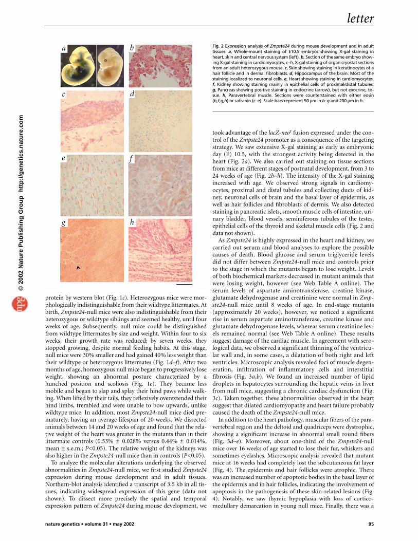
As Zmpste24 is highly expressed in the heart and kidney, wecarried out serum and blood analyses to explore the possiblecauses of death. Blood glucose and serum triglyceride levelsdid not differ between Zmpste24-null mice and controls priorto the stage in which the mutants began to lose weight. Levelsof both biochemical markers decreased in mutant animals thatwere losing weight, however (see Web Table A online). Theserum levels of aspartate aminotransferase, creatine kinase,glutamate dehydrogenase and creatinine were normal in Zmp-ste24-null mice until 8 weeks of age. In end-stage mutants(approximately 20 weeks), however, we noticed a significantrise in serum aspartate aminotransferase, creatine kinase andglutamate dehydrogenase levels, whereas serum creatinine lev-els remained normal (see Web Table A online). These resultssuggest damage of the cardiac muscle. In agreement with sero-logical data, we observed a significant thinning of the ventricu-lar wall and, in some cases, a dilatation of both right and leftventricles. Microscopic analysis revealed foci of muscle degen-eration, infiltration of inflammatory cells and interstitialfibrosis (Fig. 3a,b). We found an increased number of lipiddroplets in hepatocytes surrounding the hepatic veins in liverfrom null mice, suggesting a chronic cardiac dysfunction (Fig.3c). Taken together, these abnormalities observed in the heartsuggest that dilated cardiomyopathy and heart failure probablycaused the death of the Zmpste24-null mice.
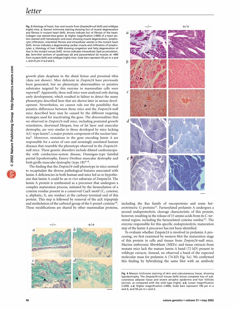
In addition to the heart pathology, muscular fibers of the para-vertebral region and the deltoid and quadriceps were dystrophic,showing a significant increase in abnormal small round fibers(Fig. 3d–e). Moreover, about one-third of the Zmpste24-nullmice over 16 weeks of age started to lose their fur, whiskers andsometimes eyelashes. Microscopic analysis revealed that mutantmice at 16 weeks had completely lost the subcutaneous fat layer(Fig. 4). The epidermis and hair follicles were atrophic. Therewas an increased number of apoptotic bodies in the basal layer ofthe epidermis and in hair follicles, indicating the involvement ofapoptosis in the pathogenesis of these skin-related lesions (Fig.4). Notably, we saw thymic hypoplasia with loss of cortico-medullary demarcation in young null mice. Finally, there was a
Fig. 2 Expression analysis of Zmpste24 during mouse development and in adulttissues. a, Whole-mount staining of E10.5 embryos showing X-gal staining inheart, skin and central nervous system (left). b, Section of the same embryo show-ing X-gal staining in cardiomyocytes. c–h, X-gal staining of organ cryostat sectionsfrom an adult heterozygous mouse. c, Skin showing staining in keratinocytes of ahair follicle and in dermal fibroblasts. d, Hippocampus of the brain. Most of thestaining localized to neuronal cells. e, Heart showing staining in cardiomyocytes.f, Kidney showing staining mainly in epithelial cells of proximal/distal tubules.g, Pancreas showing positive staining in endocrine (arrow), but not exocrine, tis-sue. h, Paravertebral muscle. Sections were counterstained with either eosin(b,f,g,h) or safranin (c–e). Scale bars represent 50 µm in b–g and 200 µm in h.
a b
c d
e f
g h
©20
02 N
atu
re P
ub
lish
ing
Gro
up
h
ttp
://g
enet
ics.
nat
ure
.co
m
letter
96 nature genetics • volume 31 • may 2002
growth plate dysplasia in the distal femur and proximal tibia(data not shown). Mice deficient in Zmpste24 have previouslybeen generated, but no phenotypic abnormalities or putativesubstrates targeted by this enzyme in mammalian cells werereported9. Apparently, these null mice were analyzed only duringearly development, which resulted in failure to detect the manyphenotypes described here that are shown later in mouse devel-opment. Nevertheless, we cannot rule out the possibility thatputative differences between those mice and the Zmpste24-nullmice described here may be caused by the different targetingstrategies used for inactivating the gene. The abnormalities thatwe observed in Zmpste24-null mice, including postnatal growthretardation, shortened lifespan, loss of fat layer and musculardystrophy, are very similar to those developed by mice lackingA/C-type lamin4, a major protein component of the nuclear lam-ina5. Moreover, mutations in the gene encoding lamin A areresponsible for a series of rare and seemingly unrelated humandiseases that resemble the phenotype observed in the Zmpste24-null mice. These genetic disorders include dilated cardiomyopa-thy with conduction-system disease, Dunnigan-type familialpartial lypodystrophy, Emery-Dreifuss muscular dystrophy andlimb girdle muscular dystrophy (type 1B)6–8.
The finding that the Zmpste24-null phenotype in mice seemedto recapitulate the diverse pathological features associated withlamin A deficiencies in both human and mice led us to hypothe-size that lamin A could be an in vivo substrate of Zmpste24. Thelamin A protein is synthesized as a precursor that undergoes acomplex maturation process, initiated by the farnesylation of acysteine residue present in a conserved CaaX motif (C, cysteine;a, aliphatic; X, any residue) at the carboxy-terminal end of thisprotein. This step is followed by removal of the aaX tripeptideand methylation of the carboxyl group of the S-prenyl-cysteine10.These modifications are shared by other mammalian proteins,
including the Ras family of oncoproteins and some het-erotrimeric G proteins11. Farnesylated prelamin A undergoes asecond endoproteolytic cleavage characteristic of this protein,however, resulting in the release of 15 amino acids from its C-ter-minal region, including the farnesylated cysteine residue12. Theenzyme responsible for this specific endoproteolytic maturationstep of the lamin A precursor has not been identified.
To evaluate whether Zmpste24 is involved in prelamin A pro-cessing, we first examined by western blot the maturation stageof this protein in cells and tissues from Zmpste24-null mice.Murine embryonic fibroblasts (MEFs) and tissue extracts frommutant mice lack the mature lamin A band (72 kD) present inwildtype extracts. Instead, we observed a band of the expectedmolecular mass for prelamin A (74 kD; Fig. 5a). We confirmedthis finding by hybridizing the same blot with an antibody
Fig. 3 Histology of heart, liver and muscle from Zmpste24-null (left) and wildtype(right) mice. a, Gomori trichrome staining showing foci of muscle degenerationand fibrosis in mutant heart (left). Arrows indicate foci of fibrosis of the heart.Collagen was stained blue-green. b, Higher magnification (×400) of a heart sec-tion stained with hematoxylin and eosin showing muscle degeneration, lympho-cytic infiltration, interstitial fibrosis and intracellular vesicles in the mutant heart(left). Arrow indicates a degenerating cardiac muscle and infiltration of lympho-cytes. c, Histology of liver (×400) showing congestion and fatty degeneration ofliver in the mutant mouse (left). Arrow indicates intracellular lipid accumulation.d,e, Semi-thin sections of quadriceps (d) and paravertebral (e) muscles at ×400from mutant (left) and wildtype (right) mice. Scale bars represent 50 µm in a andc, and 25 µm in b,d and e.
Fig. 4 Masson trichrome staining of skin and subcutaneous tissue, showinglypodystrophy. The Zmpste24-null mouse (left) shows complete loss of sub-cutaneous adipose tissue and severe atrophic epidermis and hair follicles(arrow), as compared with the wild type (right). a,b, Lower magnification(×200). c,d, Higher magnification (×400). Scale bars represent 100 µm in aand b, and 50 µm in c and d.
a
b
c
d
e
a b
c d
©20
02 N
atu
re P
ub
lish
ing
Gro
up
h
ttp
://g
enet
ics.
nat
ure
.co
m
letter
nature genetics • volume 31 • may 2002 97
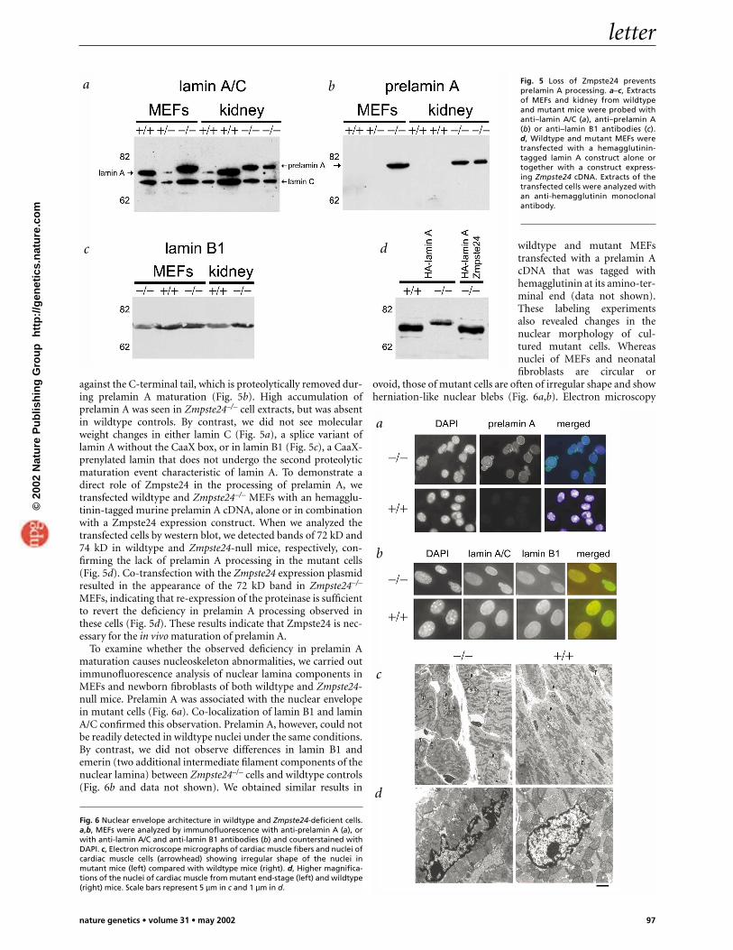
against the C-terminal tail, which is proteolytically removed dur-ing prelamin A maturation (Fig. 5b). High accumulation ofprelamin A was seen in Zmpste24–/– cell extracts, but was absentin wildtype controls. By contrast, we did not see molecularweight changes in either lamin C (Fig. 5a), a splice variant oflamin A without the CaaX box, or in lamin B1 (Fig. 5c), a CaaX-prenylated lamin that does not undergo the second proteolyticmaturation event characteristic of lamin A. To demonstrate adirect role of Zmpste24 in the processing of prelamin A, wetransfected wildtype and Zmpste24–/– MEFs with an hemagglu-tinin-tagged murine prelamin A cDNA, alone or in combinationwith a Zmpste24 expression construct. When we analyzed thetransfected cells by western blot, we detected bands of 72 kD and74 kD in wildtype and Zmpste24-null mice, respectively, con-firming the lack of prelamin A processing in the mutant cells(Fig. 5d). Co-transfection with the Zmpste24 expression plasmidresulted in the appearance of the 72 kD band in Zmpste24–/–
MEFs, indicating that re-expression of the proteinase is sufficientto revert the deficiency in prelamin A processing observed inthese cells (Fig. 5d). These results indicate that Zmpste24 is nec-essary for the in vivo maturation of prelamin A.
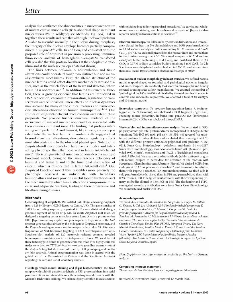
To examine whether the observed deficiency in prelamin Amaturation causes nucleoskeleton abnormalities, we carried outimmunofluorescence analysis of nuclear lamina components inMEFs and newborn fibroblasts of both wildtype and Zmpste24-null mice. Prelamin A was associated with the nuclear envelopein mutant cells (Fig. 6a). Co-localization of lamin B1 and laminA/C confirmed this observation. Prelamin A, however, could notbe readily detected in wildtype nuclei under the same conditions.By contrast, we did not observe differences in lamin B1 andemerin (two additional intermediate filament components of thenuclear lamina) between Zmpste24–/– cells and wildtype controls(Fig. 6b and data not shown). We obtained similar results in
wildtype and mutant MEFstransfected with a prelamin AcDNA that was tagged withhemagglutinin at its amino-ter-minal end (data not shown).These labeling experimentsalso revealed changes in thenuclear morphology of cul-tured mutant cells. Whereasnuclei of MEFs and neonatalfibroblasts are circular or
ovoid, those of mutant cells are often of irregular shape and showherniation-like nuclear blebs (Fig. 6a,b). Electron microscopy
Fig. 5 Loss of Zmpste24 preventsprelamin A processing. a–c, Extractsof MEFs and kidney from wildtypeand mutant mice were probed withanti–lamin A/C (a), anti–prelamin A(b) or anti–lamin B1 antibodies (c).d, Wildtype and mutant MEFs weretransfected with a hemagglutinin-tagged lamin A construct alone ortogether with a construct express-ing Zmpste24 cDNA. Extracts of thetransfected cells were analyzed withan anti-hemagglutinin monoclonalantibody.
Fig. 6 Nuclear envelope architecture in wildtype and Zmpste24-deficient cells.a,b, MEFs were analyzed by immunofluorescence with anti-prelamin A (a), orwith anti-lamin A/C and anti-lamin B1 antibodies (b) and counterstained withDAPI. c, Electron microscope micrographs of cardiac muscle fibers and nuclei ofcardiac muscle cells (arrowhead) showing irregular shape of the nuclei inmutant mice (left) compared with wildtype mice (right). d, Higher magnifica-tions of the nuclei of cardiac muscle from mutant end-stage (left) and wildtype(right) mice. Scale bars represent 5 µm in c and 1 µm in d.
a b
c d
a
b
c
d
©20
02 N
atu
re P
ub
lish
ing
Gro
up
h
ttp
://g
enet
ics.
nat
ure
.co
m
letter
98 nature genetics • volume 31 • may 2002
with toluidine blue following standard procedures. We carried out whole-mount embryo staining and histochemical analysis of β-galactosidasereporter activity in frozen sections as described17.
Electron microscopy. We killed mice by cervical dislocation and immedi-ately placed the heart in 2% glutaraldehyde and 0.5% paraformaldehydein 0.1 M sodium cacodylate buffer containing 0.1 M sucrose and 3 mMCaCl2, pH 7.4. We cut small pieces from the myocardium and stored themin the fixative overnight at 4 °C. We rinsed samples in 0.15 M sodiumcacodylate buffer containing 3 mM CaCl2 and post-fixed them in 2%OsO4 in 0.07 M sodium cacodylate buffer containing 3 mM CaCl2 for 2 h.Specimens were dehydrated and embedded in LX-112, and we examinedthem in a Tecnai 10 transmission electron microscope at 80 kV.
Evaluation of nuclear morphology in heart muscles. We defined normalnuclei as spool-shaped or rounded, and pathological nuclei as irregularand more elongated. We randomly took electron micrographs and blindlyselected counting areas at low magnification. We counted the number of‘pathological nuclei’ at ×4400 and divided by the total number of nuclei incontrols and knockouts, respectively. We analyzed a total of 60 wildtypeand 104 mutant nuclei.
Expression constructs. To produce hemagglutinin-lamin A (epitope-tagged at the N terminus), we subcloned a PCR fragment (BglII-XbaI)encoding mouse prelaminA in-frame into pcDNA3-HA (Invitrogen).Human FACE-1 cDNA was subcloned into pcDNA3.
Western blot and immunofluorescence analysis. We separated in SDS-polyacrylamide gels total protein extracts homogenized in SDS lysis buffercontaining Tris-HCl (60 mM, pH 6.8), 1% SDS, 8% glycerol. We trans-ferred proteins to nitrocellulose and incubated them overnight at 4 °Cwith the different primary antibodies polyclonal anti–prelamin A (sc-6214, Santa Cruz Biotechnology), polyclonal anti–lamin B1 (sc-6217,Santa Cruz Biotechnology), monoclonal anti-lamin A/C (Manlac-1, pro-vided by G. Morris), monoclonal anti–FACE-1 (Daiichi) and monoclonalanti-HA (Roche). We used a secondary antibody (rabbit anti-goat or goatanti-mouse) coupled to peroxidase for detection of the reaction withSupersignal Chemiluminiscent Substrate (Pierce). We derived MEFs fromembryos at E13.5 as previously described18 and transiently transfectedthem with Fugene-6 (Roche). For immunofluorescence, we fixed cells incold paraformaldehyde, rinsed them in PBS and permeabilized them with0.2% Triton X-100. Finally, we incubated cells with the corresponding pri-mary antibodies diluted in 15% FCS in PBS. The rhodamine and FITC-conjugated secondary antibodies were from Santa Cruz Biotechnology.We counterstained nuclei with DAPI.
AcknowledgmentsWe thank A.A. Ferrando, M. Serrano, D. Langenau, A. Fueyo, M. Balbín,G. Velasco, S. Cal, J.A. Uría and L.M. Sánchez for helpful comments; T.Look for support and advice; G. Morris, P. Crespo and K. Iwata forproviding reagents; F. Alvarez for help in biochemical analysis and T.Sánchez, M. Fernández, U. Mikkonen and J. Wilbertz for excellent technicalassistance. This work was supported by Comisión Interministerial deCiencia y Tecnología, Fondos Plan FEDER, European Union, The NovoNordisk Foundation, Swedish Medical Research Council and the SwedishCancer Foundation. J.C. is the recipient of a fellowship from GobiernoVasco (Spain). J.W. is a recipient of a Karolinska Institute Postdocfellowship. The Instituto Universitario de Oncología is supported by ObraSocial Cajastur-Asturias, Spain.
Note: Supplementary information is available on the Nature Geneticswebsite.
Competing interests statementThe authors declare that they have no competing financial interests.
Received 27 November 2001; accepted 12 March 2002.
analysis also confirmed the abnormalities in nuclear architectureof mutant cardiac muscle cells (83% abnormal shape in mutantnuclei versus 8% in wildtype; see Methods; Fig. 6c,d). Takentogether, these results indicate that although uncleaved prelaminA is able to assemble normally in the nucleus during interphase,the integrity of the nuclear envelope becomes partially compro-mised in Zmpste24–/– cells. In addition, and consistent with theproposed role of Zmpste24 in prelamin A processing, immuno-fluorescence analysis of hemagglutinin-Zmpste24 transfectedcells revealed that this protease localizes at the endoplasmic retic-ulum and at the nuclear envelope (data not shown).
The links between prelamin A accumulation and tissuealterations could operate through two distinct but not mutu-ally exclusive mechanisms. First, the altered structure of thenuclear lamina could affect directly mechanically stressed tis-sues, such as the muscle fibers of the heart and skeleton, wherelamin B1 is not expressed13. In addition to this structural func-tion, there is growing evidence that lamins are implicated inDNA replication, chromatin organization, regulation of tran-scription and cell division. These effects on nuclear dynamicsmay account for many of the clinical features and tissue-spe-cific alterations observed in human laminopathies14–16. Ourstudies of Zmpste24-deficient mice confirm and extend theseproposals. We provide further structural evidence of theoccurrence of marked nuclear abnormalities associated withthese diseases in mutant mice. The finding that proteins inter-acting with prelamin A and lamin A, like emerin, are incorpo-rated into the nuclear lamina in mutant cells suggests thatbeyond structural alterations, downstream regulatory effectsmust also contribute to the observed phenotypes. Notably, theZmpste24-null mice described here have a milder and later-acting phenotype than that observed in lamin A/C–deficientmice4. These Zmpste24-null mice can be considered a multipleknockout model, owing to the simultaneous deficiency oflamin A and lamin C and to the functional inactivation ofemerin, which is mislocalized in lamin A/C–null cells4. OurZmpste24 knockout model thus resembles more precisely thephenotype observed in individuals with hereditarylaminopathies and may provide a useful tool to further dissectthe mechanisms by which lamin alterations compromise mus-cular and adipocyte function, leading to these progressive andlife-threatening diseases.
MethodsGene targeting of Zmpste24. We isolated PAC clones enclosing Zmpste24from a 129 Sv library (HGMP Resource Centre, UK). This gene consists of1,475 bp of coding sequence, organized in 10 exons distributed along agenomic segment of 38 kb (Fig. 1a). To create Zmpste24-null mice, wedesigned a targeting vector to replace exons 2 and 3 with a promoter-lessIRES β-geo containing a splice acceptor sequence. Expression of the lacZreporter gene was thus driven by the endogenous Zmpste24 promoter, andthe Zmpste24 coding sequence was interrupted after codon 39. After elec-troporation of NotI-linearized targeting in 129 Ola embryonic stem cells,Southern-blot analysis of 120 neomycin-resistant colonies revealedhomologous recombination in six independent clones. We used two ofthese heterozygote clones to generate chimeric mice. Five highly chimericmales were bred to C57BL/6 females; two gave germline transmission ofthe Zmpste24 targeted allele, as confirmed by PCR genotyping and South-ern-blot analysis. Animal experimentation was done in accord with theguidelines of the Universidad de Oviedo and the Karolinska Institutetregarding the care and use of laboratory animals.
Histology, whole-mount staining and β-gal histochemistry. We fixedsamples with cold 4% paraformaldehyde in PBS, processed them into serialparaffin sections and stained them with hematoxylin and eosin or with theMasson’s trichromic staining. We stained epoxy semithin muscle sections
©20
02 N
atu
re P
ub
lish
ing
Gro
up
h
ttp
://g
enet
ics.
nat
ure
.co
m
letter
nature genetics • volume 31 • may 2002 99
1. Freije, J.M.P. et al. Identification and chromosomal location of two human genesencoding enzymes potentially involved in proteolytic maturation of farnesylatedproteins. Genomics 58, 270–280 (1999).
2. Tam, A. et al. Dual roles for Zmpste24p in yeast a-factor maturation: NH2-terminal proteolysis and COOH-terminal CAAX processing. J. Cell Biol. 142,635–649 (1998).
3. Boyartchuck, V.L., Ashby, M.N. & Rine, J. Modulation of ras and a-factor functionby carboxyl-terminal proteolysis. Science 275, 1796–1800 (1997).
4. Sullivan, T. et al. Loss of A-type lamin expression compromises nuclear envelopeintegrity leading to muscular dystrophy. J. Cell. Biol. 147, 913–920 (1999).
5. Fisher, D.Z., Chaudhary, N. & Blobel, G. cDNA sequencing of nuclear lamins A andC reveals primary and secondary structural homology to intermediate filamentproteins. Proc. Natl Acad. Sci. USA 83, 6450–6454 (1986).
6. Bonne, G. et al. Mutations in the gene encoding lamin A/C cause autosomaldominant Emery-Dreifuss muscular dystrophy. Nature Genet. 21, 285–288 (1999).
7. Fatkin, D. et al. Missense mutations in the rod domain of the lamin A/C gene ascauses of dilated cardiomyopathy and conduction-system disease. N. Engl. J. Med.341, 1715–1724 (1999).
8. Shackleton, S. et al. LMNA, encoding lamin A/C, is mutated in partiallipodystrophy. Nature Genet. 24, 153–156 (2000).
9. Leung, G.K. et al. Biochemical studies of Zmpste24-deficient mice. J. Biol. Chem.276, 29051–29058 (2001).
10. Sinensky, M. et al. The processing pathway of prelamin A. J. Cell Sci. 107, 61–67(1994).
11. Zhang, F.L. & Casey, P.J. Protein prenylation: molecular mechanisms andfunctional consequences. Annu. Rev. Biochem. 65, 241–269 (1996).
12. Weber, K., Plessmann, U. & Traub, P. Maturation of nuclear lamin A involves aspecific carboxy-terminal trimming, which removes the polyisoprenylation sitefrom the precursor; implications for the structure of the nuclear lamina. FEBSLett. 257, 411–414 (1989).
13. Manilal, S. et al. Distribution of emerin and lamins in the heart and implicationsfor Emery-Dreifuss muscular dystrophy. Hum. Mol. Genet. 8, 353–359 (1999).
14. Cohen, M., Lee, K.K., Wilson, K.L. & Gruenbaum, Y. Transcriptional repression,apoptosis, human disease and the functional evolution of the nuclear lamina.Trends Biochem. Sci. 26, 41–47 (2001).
15. Wilson, K.L., Zastrow, M.S. & Lee, K.L. Lamins and disease: insights into nuclearinfrastructure. Cell 104, 647–650 (2001).
16. Morris, G.E. & Manilal, S. Heart to heart: from nuclear proteins to Emery-Dreifussmuscular dystrophy. Hum. Mol. Genet. 8, 1847–1850 (1999).
17. Hogan, B., Beddington, R., Costantini, F. & Lacy, E. Manipulating the MouseEmbryo: a Laboratory Manual (Cold Spring Harbor Laboratory Press, Cold SpringHarbor, New York, 1994).
18. Palmero, I. & Serrano, M. Induction of senescence by oncogenic Ras. MethodsEnzymol. 333, 247–256 (2001).
©20
02 N
atu
re P
ub
lish
ing
Gro
up
h
ttp
://g
enet
ics.
nat
ure
.co
m