The effect of dehydroleucodine in adipocyte differentiation
18
The effect of dehydroleucodine in adipocyte differentiation. Adriana Galvis 1 , Adriana Marcano 1 , Chad Stefancin 1 , Nicole Villaverde 1 , Horacio A. Priestap 1 , Carlos E. Tonn 2 , Luis A. Lopez 3 , and Manuel A. Barbieri 1 1 Department of Biological Sciences, Florida International University, Miami, FL 33199, USA. 2 Cátedra de Química Orgánica, Facultad de Química, Bioquímica y Farmacia, Universidad Nacional de San Luis, Argentina. 3 Laboratory of Cytoskeleton and Cell Cycle, Instituto de Histología y Embriología, Facultad de Ciencias Medicas, Universidad Nacional de Cuyo, 5500 Mendoza, Argentina. Abstract Dehydroleucodine (DhL) is a sesquiterpene lactone of the guaianolide group with gastric citoprotective activity. Recent studies have also demonstrated that DhL inhibits the proliferation of vascular smooth muscle cells. In this study we examined the effect of DhL in the differentiation 3T3-L1 preadipocytes. The addition of DhL significantly inhibited the differentiation 3T3-L1 preadipocytes along with significant decrease in the accumulation of lipid content by a dramatic down regulation of the expression of adipogenic-specific transcriptional factors PPARγ and C- EBPα. However, phosphorylation of AMPKα, Erk1/2 and Akt1 was not inhibited by DhL treatment. Interestingly, we also found that 11,13-dihydro-dehydroleucodine, a derivative of DhL with inactivated α-methylene-γ-lactone function, also inhibited the differentiation 3T3-L1 preadipocytes. Taken together, these data suggest DhL has an important inhibitory effect in cellular pathways regulating adipocyte differentiation by modulating the PPARγ expression, which is known to play a pivotal role during adipogenesis. Keywords Dehydroleucodine; adipocytes; α-methylene-γ-lactone function; transcriptional factors 1. Introduction Obesity is a prevalent health risk in industrialized countries and is associated with multiple pathological disorders, including diabetes (Sartipy and Loskutoff, 2003), hypertension (Pi- Sunyer, 2002), cancer (Lagra et al., 2004), gallbladder disease (Galal, 2003), and aterosclerosis (Wofford et al., 1999). Obesity has also been associated with cancer and cardiovascular disease, which have reached epidemic proportions worldwide (Roberts et al., 2010; Yun, 2010). It has been reported that weight loss reduces lipid levels, blood pressure, and the incidence of type 2 diabetes mellitus (Sheard, 1993). © 2011 Elsevier B.V. All rights reserved. Corresponding author: M. Alejandro Barbieri; Department of Biological Sciences, Florida International University, 11220 SW 8 th Street, Miami, FL 33199, USA; Tel: 305-348-7536, Fax: 305-348-1986; [email protected]. Publisher's Disclaimer: This is a PDF file of an unedited manuscript that has been accepted for publication. As a service to our customers we are providing this early version of the manuscript. The manuscript will undergo copyediting, typesetting, and review of the resulting proof before it is published in its final citable form. Please note that during the production process errors may be discovered which could affect the content, and all legal disclaimers that apply to the journal pertain. NIH Public Access Author Manuscript Eur J Pharmacol. Author manuscript; available in PMC 2012 December 05. Published in final edited form as: Eur J Pharmacol. 2011 December 5; 671(1-3): 18–25. doi:10.1016/j.ejphar.2011.09.033. NIH-PA Author Manuscript NIH-PA Author Manuscript NIH-PA Author Manuscript
Transcript of The effect of dehydroleucodine in adipocyte differentiation

The effect of dehydroleucodine in adipocyte differentiation.
Adriana Galvis1, Adriana Marcano1, Chad Stefancin1, Nicole Villaverde1, Horacio A.Priestap1, Carlos E. Tonn2, Luis A. Lopez3, and Manuel A. Barbieri11Department of Biological Sciences, Florida International University, Miami, FL 33199, USA.2Cátedra de Química Orgánica, Facultad de Química, Bioquímica y Farmacia, UniversidadNacional de San Luis, Argentina.3Laboratory of Cytoskeleton and Cell Cycle, Instituto de Histología y Embriología, Facultad deCiencias Medicas, Universidad Nacional de Cuyo, 5500 Mendoza, Argentina.
AbstractDehydroleucodine (DhL) is a sesquiterpene lactone of the guaianolide group with gastriccitoprotective activity. Recent studies have also demonstrated that DhL inhibits the proliferation ofvascular smooth muscle cells. In this study we examined the effect of DhL in the differentiation3T3-L1 preadipocytes. The addition of DhL significantly inhibited the differentiation 3T3-L1preadipocytes along with significant decrease in the accumulation of lipid content by a dramaticdown regulation of the expression of adipogenic-specific transcriptional factors PPARγ and C-EBPα. However, phosphorylation of AMPKα, Erk1/2 and Akt1 was not inhibited by DhLtreatment. Interestingly, we also found that 11,13-dihydro-dehydroleucodine, a derivative of DhLwith inactivated α-methylene-γ-lactone function, also inhibited the differentiation 3T3-L1preadipocytes. Taken together, these data suggest DhL has an important inhibitory effect incellular pathways regulating adipocyte differentiation by modulating the PPARγ expression,which is known to play a pivotal role during adipogenesis.
KeywordsDehydroleucodine; adipocytes; α-methylene-γ-lactone function; transcriptional factors
1. IntroductionObesity is a prevalent health risk in industrialized countries and is associated with multiplepathological disorders, including diabetes (Sartipy and Loskutoff, 2003), hypertension (Pi-Sunyer, 2002), cancer (Lagra et al., 2004), gallbladder disease (Galal, 2003), andaterosclerosis (Wofford et al., 1999). Obesity has also been associated with cancer andcardiovascular disease, which have reached epidemic proportions worldwide (Roberts et al.,2010; Yun, 2010). It has been reported that weight loss reduces lipid levels, blood pressure,and the incidence of type 2 diabetes mellitus (Sheard, 1993).
© 2011 Elsevier B.V. All rights reserved.
Corresponding author: M. Alejandro Barbieri; Department of Biological Sciences, Florida International University, 11220 SW 8th
Street, Miami, FL 33199, USA; Tel: 305-348-7536, Fax: 305-348-1986; [email protected].
Publisher's Disclaimer: This is a PDF file of an unedited manuscript that has been accepted for publication. As a service to ourcustomers we are providing this early version of the manuscript. The manuscript will undergo copyediting, typesetting, and review ofthe resulting proof before it is published in its final citable form. Please note that during the production process errors may bediscovered which could affect the content, and all legal disclaimers that apply to the journal pertain.
NIH Public AccessAuthor ManuscriptEur J Pharmacol. Author manuscript; available in PMC 2012 December 05.
Published in final edited form as:Eur J Pharmacol. 2011 December 5; 671(1-3): 18–25. doi:10.1016/j.ejphar.2011.09.033.
NIH
-PA Author Manuscript
NIH
-PA Author Manuscript
NIH
-PA Author Manuscript

Animal models as well as in vitro systems have been used extensively in diabetes research.3T3-L1 preadipocytes have been a useful in vitro model for the study of obesity, due to anobservable accumulation of triglycerides upon differentiating in culture (Rosen andSpiegelman, 2000). Adipocyte differentiation is induced by the expression and/orphosphorylation of specific genes, such as AMPK, Akt1, Erk1/2, (Kemp et al., 2003; Rosenand Spiegelman, 2000; Rosen et al., 2000; Spiegelman et al., 1993), PPARγ and C-EBPα(Bost et al., 2005). It is anticipated that compounds, which inhibit adipocyte differentiationcould beneficially prevent and/or treat obesity. Thus, natural products such as crude aqueousand chloroform plant extracts as well as other pure active compounds that specifically targetand inhibit adipogenesis have been considered potentially promising treatments of obesity(Roberts et al., 2010).
Sesquiterpene lactones are a large and structurally diverse group of plant second metabolites(Heinrich et al., 1998) with distinctive biological activities, including gastric cytoprotectoreffects (Penissi et al., 1998), anti-migraine (Beekman et al., 1997), antiviral andantimicrobial activities (Hayashi et al., 1996; Perry and Foster, 1995), anti-tumor (Robles etal., 1995) and neurotoxic effect (Cheng et al., 1992).
Sesquiterpene lactones are also blockers of smooth muscle contractility (Hay et al., 1994)aromatase activity (Blanco et al., 1997) and NF-kappa B activation (Hehner et al., 1998;Lyss et al., 1998). Also, it has been found that sesquiterpene lactones inhibit the activationof cyclooxygenase and proinflammatory cytokines in macrophages (Hwang et al., 1996).Within the group of sesquiterpene lactones, helenalin, which occurs in the aerial portion ofArnica Montana L., was also found to block the hormonally induced Sky2 mRNA and Aktphosphorylation during early stages of adipocyte differentiation (Auld et al., 2006).Inhibitory activities have been principally linked to the α-methylene-γ-lactone function(Heinrich et al., 1998). However, the reduction of the α-methylene-γ-lactone limited itscytotoxicity effect without affecting the anti-proliferative and anti-aromatase activity(Blanco et al., 1997).
Dehydroleucodine (DhL) is a sesquiterpene lactone of the guaianolide group, which alsocontains a α-methylene-γ-lactone ring in its molecule. It was first isolated from Lidbeckiapectinata (Bohlmann and Zdero, 1972). The aerial parts of Artemisia douglasiana Besser arealso rich in DhL (Giordano et al., 1990). Chloroform extracts of the air-dried aerial parts ofArtemisia douglasiana showed significant gastric cytoprotective activity (Giordano et al.,1992). In addition, DhL inhibited cell proliferation (Polo et al., 2007) and growth ofTrypanosome cruzi in culture (Brengio et al., 2000). In this study, we investigated theeffects of both DhL and 11,13-dihydro-dehydroleucodine (DH-DhL) on the differentiationof 3T3-L1 preadipocytes, and its mechanism of action at the cellular and molecular levels.
2. Material and Methods2.1. Materials
DhL was isolated from Artemisia douglasiana as previously described (Giordano et al.,1990) and DH-DhL was obtained for DhL reduction as previously described (Giordano etal., 1992). 3T3-L1 cells were purchased from American Type Culture Collection (ATCC,Manassas, VA). Dulbecco's modified Eagle's medium high glucose (DMEM), penicillin/streptomycin and L-glutamine were purchased from Mediatech, Inc. (Manassas, VA). C-EBPα antibodies were from Santa Cruz Biotechnology, Inc. (Santa Cruz, CA). PPARγ,total (T)-Erk, phospho (P)-Erk, T-Akt, P-Akt, T-AMPKα and P-AMPKα antibodies werefrom Cell Signaling Technology (Boston, MA). Tubulin antibodies were from Sigma-Aldrich (St. Louis, MO). All secondary antibodies were purchased from Jackson
Galvis et al. Page 2
Eur J Pharmacol. Author manuscript; available in PMC 2012 December 05.
NIH
-PA Author Manuscript
NIH
-PA Author Manuscript
NIH
-PA Author Manuscript

ImmunoResearch Laboratories (West Grove, PA). All other chemicals were obtained fromSigma-Aldrich unless otherwise stated.
2.2. Cell culture and differentiation3T3-L1 cells were grown to confluence in Dulbecco's modified Eagle's medium (DMEM)high glucose supplemented with 10% fetal bovine serum (Invitrogen, Carlsbad, CA), 1%penicillin/ streptomycin and 1% L-glutamine (growth media) in a humidified atmosphere of5% CO2 at 37°C. Culture was fed every 48 hours, both for cell growth and differentiation.To trigger differentiation cells were exposed to induction medium (IM) (growth mediasupplemented with 670nM insulin, 65nM dexamethasone and 0.5mM 3-isobutyl-1-methylxanthine [IBMX]) for the first two days, then fed with post-differentiation media(DMEM high glucose supplemented with 5% fetal bovine serum, 1% penicillin/streptomycin and 1% L-glutamine) for the next seven days. Cells were exposed to DhLthroughout the entire differentiation process, unless otherwise indicated.
2.3. Cell viability assayCells were cultured on 6-well plates and treated with DhL as indicated for each experiment.Cell viability was measured using the MTT (3-[4,5-dimethylthiazol-2-yl]-2,5-diphenyltetrazolium bromide) colorimetric assay following the manufacturer's instructions (ATCC).In brief, MTT reagent was added to cells at a final concentration of 10μl/ml. Then cells wereincubated as indicated in the Fig. 1 at 37°C and 5% CO2. After incubation, the medium wasremoved, and formazan crystals were dissolved in detergent reagent. Optical density foreach condition was determined at 570 nm.
2.4. Oil Red O staining and microscopyOn the ninth day cells were fixed with 10% formalin (Thermo Fisher Scientific. Inc.,Pittsburgh, PA) in phosphate buffer saline (PBS) 1 hour at 4°C. Oil Red O (Allied Chemical,Morristown, NJ) stock solution (0.6g in 100ml of isopropanol) was diluted with 0.6 parts ofwater, filtered and added to the fixed cells for 15 minutes at room temperature. Cells werethen washed with water and analyzed in a Leica DM IRB inverted microscope. Photos oflipid droplets were taken using digital Leica DC 500 camera. To quantify the lipid droplets,Oil Red O was eluted with 100% isopropanol for 10 minutes at 37°C, collected, and itsoptical density was measured at 540 nm.
2.5. Triglyceride assayNine days 3T3-L1 adipocytes were washed with PBS, scraped and centrifuged. Pellets wereresuspended in PBS and then homogenized by sonication and the cell suspension wasassayed for total triglyceride (Triglyceride assay kit; Cayman Chemical Company, AnnArbor, MI) according to the method of (Mendez et al., 1986). Results were expressed astotal triglyceride per cellular protein (DC protein assay; Bio-Rad, Hercules, CA).
2.6. Western blot analysisTo prepare cell lysates, cell monolayers where washed with ice-cold lysis buffer (20mMTris-HCL pH7.5, 150mM NaCl, 1mM Na2 EDTA, 10% NP40 and 10% Na Deoxycholate)containing protease and phosphatase inhibitors. Lysates were collected by centrifugation andprotein concentrations were estimated by using BCA protein assay (Thermo FisherScientific, Inc., Pittsburgh, PA) following the manufacturer's instructions. Proteins wereresolved by SDS-PAGE and transferred to nitrocellulose membranes, blocked, probed withthe specific antibodies and then visualized by Western analysis. Relative levels of Erk1/2and Akt1 proteins were determined by densitometry using the ratio of P-Erk1/2 to T-Erk1/2
Galvis et al. Page 3
Eur J Pharmacol. Author manuscript; available in PMC 2012 December 05.
NIH
-PA Author Manuscript
NIH
-PA Author Manuscript
NIH
-PA Author Manuscript

and P-Akt1 to T-Akt1, respectively. Relative levels of PPARγ and C-EBPα weredetermined using the ratio of PPARγ to tubulin and C-EBPα to tubulin, respectively.
2.7. High-performance liquid chromatography analysisThe high-performance liquid chromatography (HPLC) equipment consisted of aSpectraSystem SMC1000 solvent delivery system, vacuum membrane degasser, P4000gradient pumps and AS3000 autosampler (Thermo Electro Corporation, San Jose, CA).Column effluent was monitored at 254 nm with Spectra System UV6000LP variablewavelength PDA detector and ChromQuest 4.1 software. DhL, (11S)DH-DhL and(11R)DH-DhL were separated using a C18 YMC column (A-302, 150 × 4.3 mm i.d., S-5μm, 12 nm; Waters) and the following solvents: A. acetonitrile; B. 0.1 % TFA in water.System 1: linear gradient 10% to 100 % A in 120 min; flow rate 1 ml/min. PreparativeHPLC was performed in the above equipment with a XTerra Prep MS C18 OBD column, 15μm, 19 × 50 mm (Waters) and the solvent system 25 % acetonitrile – 75 % 0.1 % TFA inwater (isocratic); flow rate 3 ml/min.
2.8. Gas chromatography (GC)/flame ionization detector (FID) and GC/mass spectrometry(MS) analysis
GC/FID analyses were performed on a Trace GC Ultra apparatus (Thermo ElectroCorporation, San Jose, CA) equipped with a flame ionization detector. The output wasrecorded using a ChromQuest version 4.1 data system. A DB-5MS capillary column (0.25mm i.d. × 30 m; film thickness 0.25 μm; J & W Scientific, Folsom, CA) was employed. Thetemperature was programmed 105 to 240°C at 3°C/min (linear increase) and then thetemperature was held at 240°C for 10 min. The injector temperature was 250°C with a splitratio of 1/20. The detector temperature was 270°C. Helium was used as gas carrier at 1 ml/min. DHL, (11S)DH-DHL and (11R)DH-DhL were dissolved in ethyl acetate and two μl ofthe solution were injected. GC/MS determinations were carried out in a Hewlett Packardmodel 6890 instrument coupled to a Q-Mass 910 quadrupole selective detector at 70 eV andequipped with a DB-5MS capillary column. Temperature program and other conditions wereas indicated above.
2.9. Compound identificationDehydroleucodine {(1S,6S,2R)-9,13-dimethyl-5-methylene-3oxatricyclo[8.3.0.0<2,6>]trideca-9,12-diene-4,11-dione, IUPAC nomenclature}. DhL(compound 1): HPLC (System 1), Rt 27.74 min; GC/MS, Rt 22.72 min; UV/PDA λmax 256nm; MS m/z (rel. int.), 244 (100) M+, 173 (18.2), 145 (19.8), 129 (18.9), 115 (18.5), 105(18.3), 91 (62.9), 79 (17.7), 77 (28.4), 65 (21.6), 53 (30.8). (1S,2S,5S,6S)-5,9,13-trimethyl-3-oxatricyclo[8.3.0.0<2,6>]trideca-9,12-diene-4,11-dione;11,13-dihydro-dehydroleucodine (IUPAC nomenclature), (11S)DH-DhL (compound 2): HPLC (System 1),Rt 28.16 min; GC/MS, Rt 21.87; UV/PDA λmax 257 nm; MS, m/z (rel. int.): 246 (100) M+,217 (26.7), 173 (37.6), 172 (31.2), 145 (26.2), 105 (23.2), 91 (60.7), 77 (26.2), 55 (24.3).(1S,2S,6S,5R)-5,9,13-trimethyl-3-oxatricyclo[8.3.0.0<2,6>]trideca-9,12-diene-4,11-dione,11,13-dihydro-dehydroleucodine (IUPAC nomenclature), (11R)DH-DhL (compound 3):HPLC (System 1), Rt 26.23 min; GC/MS, Rt 22.84; UV/PDA λmax 257 nm; MS, m/z (rel.int.): 246 (100) M+, 217 (31.5), 173 (35.5), 172 (33.1), 145 (27.5), 105 (26.1), 91 (67.1), 77(29.0), 55 (25.8).
2.10. Statistical analysisAll experiments were done in duplicates and they were repeated at least three times. Valuesare represented as the standard error of the mean (S.E.M.) of triplicates and the statistical
Galvis et al. Page 4
Eur J Pharmacol. Author manuscript; available in PMC 2012 December 05.
NIH
-PA Author Manuscript
NIH
-PA Author Manuscript
NIH
-PA Author Manuscript

significance was analyzed by One-way ANOVA or Student's test. Results with *P <0.05 and**P < 0.01 were considered as statistically significant.
3. Results3.1. DhL inhibits differentiation of preadipocytes
To examine the potential role of DhL on the differentiation of preadipocytes, cells wereincubated with induction media in the presence of various concentration of DhL. In Fig. 1A,we show that the addition of DhL inhibited the lipid content in a dose-dependent mannerwith an IC50 of 6 μM. Furthermore, our experimental conditions demonstrate that theaddition of DMSO (0.2%) exclusively does not hinder 3T3-L1 preadipocytes differentiation(Fig. 1A).
We were further interested in examining the effect of DhL on the viability of 3T3-L1preadipocytes. Cells were incubated with induction media in the presence of 8 μM DhL for24hrs (Day 1) or throughout the entire 9 days of differentiation (Day 9). Cell viability wasmeasured using the MTT colorimetric assay as described in Material and Methods. In Fig.1A, we show that the addition of DhL blocked the differentiation of 3T3-L1 preadipocyteswhereas cell viability was not affected as compared with DMSO or untreated cells (Fig. 1B).However, at higher DhL concentrations (>10μM), 3T3-L1 preadipocytes detached from theplate, which was coupled with a significant reduction in the MTT assay (data not shown).These results suggest that, up to a maximum concentration of 10 μM, DhL has a stronginhibitory activity of 3T3-L1 preadipocytes differentiation without significant effect on cellviability.
Adipocyte differentiation can be also monitored by formation of intracellular lipid droplets(Rosen and Spiegelman, 2000). As described above, 3T3-L1 preadipocytes were cultivatedand induced to differentiate into adipocytes with induction media in the absence or presenceof 8 μM DhL. At day 9, Oil Red O staining showed that an abundant number of lipiddroplets suggesting a significant lipid accumulation in untreated differentiated cells.However, lipid droplets were not observed in untreated non-differentiated cells (compareFig. 2A and B). More importantly, formation of lipid droplets was inhibited by 8 μM DhLtreatment (Fig. 2C). These observations were further supported with the quantitativemeasurement of lipid content by determining the absorbance at 540 nm (Fig. 1A). Inaddition, DhL treatment significantly inhibited adipogenic morphology (i.e., transition froma fibroblast-like shape to an increasingly rounded-up appearance with an accumulation ofcytoplasmic lipid droplets; compare Fig. 2, B and C).
Given that DhL inhibited differentiation to 3T3-L1 preadipocytes, we next consideredwhether DhL would inhibit triglyceride accumulation. Cells treated with induction mediumin the presence of 8 μM of DhL accumulated roughly 31±5% of the intracellular triglyceridecontained in controls (Fig. 2D). As expected, DMSO-treated cells did not affect theformation of triglyceride accumulation as compared with cells incubated with inductionmedia alone (Fig. 2D).
3.2. DhL inhibits key adipogenic transcriptional factorsAdipogenesis is a highly regulated process requiring coordinated expression and activationof key transcriptional factors and signaling molecules (Rosen and Spiegelman, 2000) Toinvestigate whether DhL affects the expression of PPARγ and C-EBPα, 3T3-L1preadipocytes were incubated with induction media in the absence or presence of 8 μM DhLas described in Fig. 1, and then harvested at day 9 for Western blot analysis using anti-PPARγ and C-EBPα antibodies. In Fig. 3A, we show that the addition of DhL clearlyattenuated the expression of PPARγ and C-EBPα. We also examined the effect of DhL on
Galvis et al. Page 5
Eur J Pharmacol. Author manuscript; available in PMC 2012 December 05.
NIH
-PA Author Manuscript
NIH
-PA Author Manuscript
NIH
-PA Author Manuscript

the phosphorylation of Akt1 and Erk1/2 proteins. To our surprise we found that the additionof DhL did not attenuate phosphorylation of Erk1/2 and Akt1 (Fig. 3B and C). Furthermore,we tested whether the addition of DhL affected the phosphorylation of AMPKα during 3T3-L1 preadipocytes differentiation. This result shows that phosphorylation of AMPKα (P-AMPKα) was not inhibited by the addition of DhL (Fig. 3D). In contrast, DhL furtherenhanced phosphorylation of AMPKα (Fig. 3D). Furthermore, we also found that the levelof total AMPKα appeared relatively constant in the presence or absence of DhL. Therefore,these results indicated that the level of phosphorylated AMPKα increased in the presence ofDhL. Interestingly, both PPARγ and C-EBPα are selectively expressed during thedifferentiation of 3T3-L1 preadipocytes. The levels of expression of these transcriptionfactors is undetectable in preadipocytes; however, expression increases 2 days afterinduction and they are expressed 5 days after the induction of differentiation (Rosen andSpiegelman, 2000). Consistent with these observations, we also observed that the expressionof PPARγ and C-EBPα was significant increased with the progression of the differentiationof 3T3-L1 preadipocyte (supplementary Fig 3A and B). However, DhL treatment at theconcentration of 8μM significantly blocked the expression of PPARγ and C-EBPα(supplementary Fig. 3A and B). In addition, the differentiation of 3T3-L1 preadipocyteprogressed with an increase of the phosphorylation status of AMPKα (supplementary Fig.3C). Treatment with DhL further significantly increased the phospho-status of AMPKα,suggesting that DhL also induced the activation of AMPKα (supplementary Fig. 3C). Takentogether, these data suggest that DhL selectively blocks the expression of PPARγ and C-EBPα and also increases the phosphorylation of AMPKα during the differentiation of 3T3-L1 preadipocytes.
It is also possible that the extent of inhibition is dependent on the timing of DhL addition.For this purpose, 8 μM DhL was added in discrete periods during differentiation (Fig. 4A).We observed a significant reduction in the differentiation of 3T3-L1 preadipocytes whencompared with DMSO-control cells upon early DhL addition, corresponding to days 1, or 3of treatment (Fig. 4A and B). However, this inhibitory effect was not apparent when DhLwas added on day 5, 7 or at day 8 post-induction. These results indicate that DhL may affectearly adipocyte gene expression during in vitro differentiation.
3.3. Effect of 11,13-dihydro-dehydroleucodine on the differentiation of 3T3-L1preadipocytes
Our results clearly show that the addition of DhL inhibited differentiation of 3T3-L1preadipocytes in a dose-dependent manner without a significant effect of cell toxicity (Fig.1). However, it has been postulated that the non-saturated α-methylene-γ-lactone functionof sesquiterpene lactones produces an unspecific toxic effect leading to cell death (Polo etal., 2007). Therefore, we then investigated the effect of DH-DhL, which is a derivative ofDhL lacking alkylating function, on the differentiation of 3T3-L1 preadipocytes.
DhL (Fig. 5A, compound 1) can be gently reduced with sodium borohydride to give thecorresponding 11,13-dihydro derivative (classical nomenclature; (Giordano et al., 1990).Analysis of the reaction product (DH-DhL) by GC show that two epimers (Fig. 5 B,compounds 2 and 3) are formed in different amounts (Fig. 5B) due to the generation of achiral center at C-11 of the molecule during reduction. The 11S-epimer (compound 2) is themajor reaction product (Giordano et al., 1992). It is accompanied by the minor 11R-epimer(compound 3), and by traces of unreacted DhL (compound 1) (Fig. 5B). In the followingthese epimers are denoted (11S)DH-DhL and (11R)DH-DhL, respectively, for simplicity.After separation by preparative HPLC, the epimers were obtained in pure form andconfirmed by GC analysis (Fig. 5, C and D). They were studied, in parallel with DhL, fortheir effect on the differentiation of 3T3-L1 preadipocytes.
Galvis et al. Page 6
Eur J Pharmacol. Author manuscript; available in PMC 2012 December 05.
NIH
-PA Author Manuscript
NIH
-PA Author Manuscript
NIH
-PA Author Manuscript

In Fig. 6A, we show that the addition of DH-DhL inhibited the differentiation of 3T3-L1preadipocytes. This inhibition was dose-dependent and required a higher concentration ofDH-DhL to produce a similar inhibitory effect as DhL. Thus, it required 10 times the DH-DhL concentration to achieve the same effect as DhL (Fig. 6A). Furthermore, we also foundthat 100 μM of DH-DhL did not affect cell viability (Control cells: 98±2% of viability vs.80 μM DH-DhL treated cells: 97±2% of viability). These results suggest that the reductionof the α-methylene-γ-lactone group of the DhL was not required to block the differentiation3T3-L1 preadipocytes.
To further investigate the role of these epimers of DH-DhL on the differentiation 3T3-L1preadipocytes, the two methyl epimers at C-11 produced during the in vitro reduction ofDhL were separated by preparative HPLC, concentrated, confirmed by GC analysis and thentheir effect on adipogenesis was examined. For this, 3T3-L1 preadipocytes were incubatedwith 80 μM of either (11S)DH-DhL or (11R)DH-DhL during the entire differentiationprocess. We found that the addition of (11R)DH-DhL epimer, but not (11S)DH-DhL epimer,inhibited the differentiation 3T3-L1 preadipocytes (Fig. 6B). These results suggest that(11R)DH-DhL epimer may be responsible in inhibiting adipocyte differentiation.
4. DiscussionIn this study we investigated the effect of DhL in 3T3-L1 preadipocytes differentiation. Theaddition of DhL to the medium inhibited significantly the accumulation of lipid droplets in adose-dependent manner. DhL attenuated dramatically the production of adipogenictranscriptional factors PPARγ and C-EBPα during adipogenesis without altering theactivation of Erk1/2 and Akt1. In contrast, DhL increased the phosphorylation of AMPKαproteins. In addition, we also found that DH-DhL, a derivative of DhL with inactivated α-methylene-γ-lactone function, also inhibited the formation of adipocytes. However, itrequired ten times the DH-DhL concentration to achieve the same effect as DhL. Moreimportantly, we have identified a specific DH-DhL epimer [i.e., (11R)DH-DhL], which maybe responsible for such inhibitory activity.
Based on our observations, it is clear that in the adipocyte differentiation model, the α-methylene-γ-lactone moiety causes a significant decrease of 3T3-L1 pre-adipocytesdifferentiation at concentrations ≤10 μM without altering cell viability as demonstrated bythe MTT assay. However, the addition of >10 μM DhL (i.e., 12 μM) caused cell toxicity asevidenced either by the incorporation of trypan blue dye or by the induction of detachmentof cells from the tissue culture plate. Thus, the inhibition of 3T3-L1 differentiation by DhLand (11R)DH-DhL epimer may be induced by a process distinct from cell death induced bytoxicity of these compounds.
Adipocyte differentiation involves a complex progression of changes in morphology,hormone sensitivity, and gene expression (Rosen and Spiegelman, 2000). 3T3L1preadipocytes transform into adipocytes through growth arrest, clonal selection andexpression of adipocyte selective markers following hormonal induction (Rosen et al.,2000). The addition of induction media containing IBMX, dexamethasone, and insulinactivated several transcriptional factors, including PPARγ, C-EBPα, C-EBPβ, C-EBPδ,which in turn regulate a number of genes required during adipocyte differentiation(Spiegelman et al., 1993; Tanaka et al., 1997). Insulin is also required to ensure completeconversion of preadipocytes into adipocytes, but the precise role that it plays in this processremains unclear. The insulin receptor shows tyrosine kinase activity, and during itsactivation can further activate a series of signaling pathways including Erk1/2 and Akt1activities. Several laboratories have investigated the role of Erk1/2 in adipogenesisregulation, but conclusions are somewhat controversial (Rosen and Spiegelman, 2000).
Galvis et al. Page 7
Eur J Pharmacol. Author manuscript; available in PMC 2012 December 05.
NIH
-PA Author Manuscript
NIH
-PA Author Manuscript
NIH
-PA Author Manuscript

Nevertheless, the role of Akt in adipogenesis is far more clearly understood since severallines of evidence have indicated the important function of Akt1 signaling cascade duringadipogenesis (Koppen and Kalkhoven, 2008; Rosen et al., 2000). In addition, AMPK isserine/ threonine known to play a major role in energy homeostasis (Kemp et al., 2003) andit is regulated by adenosine monophosphate (Shaw et al., 2004). AMPK cascades have alsoemerged as novel targets for the treatment of obesity (Meisse et al., 2002; Song et al., 2002).Thus, it is possible to assume that DhL may selectively block pathways regulatingadipocytic differentiation containing the adipocyte-specific factors.
Treatment of 3T3-L1 preadipocytes with DhL dramatically reduced protein expression ofPPARγ and C-EBPα, which is strictly concordant with the appearance of cytoplasmic lipiddroplets. These data suggest that DhL may target specific pathways related to the expressionof PPARγ and in minor extension to the expression C-EBPα, because of the stronginhibition of PPARγ upon addition of DhL. We also observed a significant increase in thephosphorylation of AMPKα during 3T3-L1 differentiation. However, the level ofphosphorylated AMPKα was further enhanced by the presence of DhL during 3T3-L1differentiation. Therefore, these results indicated that the addition of DhL significantlyincreased the phosphorylation of AMPKα during 3T3-L1 differentiation, which wasdetrimental to 3T3-L1 differentiation. In addition, our observations showed that the effect ofDhL on adipocyte differentiation may be selective since both Akt1 and Erk1/2 activities,which are thought to be sensitive to insulin signal, were not affected by the addition of DhL.Thus, the inhibitory effect of DhL does not seem to be related to these two signalingmolecules.
In summary, this report suggests that DhL inhibits the adipogenic differentiation process.The mechanisms by which DhL regulates adipogenesis include the inhibition of expressionof the adipogenic transcription factors, PPARγ and C-EBPα. In addition, we also found thata DH-DhL epimer, lacking a highly reactive and nonspecific α-methylene-γ-lactone moiety,may improve its use without a cytotoxic effect. Therefore, it is anticipated that the inhibitionof differentiation into adipocytes by DhL and its derivates may be beneficial for theprevention of obesity. For this reason, natural products that specifically inhibitedadipogenesis might be considered with regard to their potential in treatment of obesity.However, it remains to be determined whether manipulating adipogenesis can be beneficialand/or healthy without leading to other metabolic diseases.
Supplementary MaterialRefer to Web version on PubMed Central for supplementary material.
AcknowledgmentsThis work was partially supported by the National Institutes of Health grant SC1DK084343 (to MAB) and bySECyTP, UNCuyo 06 J 213 grant and ANPCYT PICT-R 2005 32850 grant (to LAL).
ReferencesAuld CA, Hopkins RG, Fernandes KM, Morrison RF. Novel effect of helenalin on Akt signaling and
Skp2 expression in 3T3-L1 preadipocytes. Biochem. Biophys. Res. Commun. 2006; 346:314–320.[PubMed: 16750815]
Beekman AC, Woerdenbag HJ, van Uden W, Pras N, Konings AW, Wikstrom HV, Schmidt TJ.Structure-cytotoxicity relationships of some helenanolide-type sesquiterpene lactones. J. Nat. Prod.1997; 60:252–257. [PubMed: 9090867]
Galvis et al. Page 8
Eur J Pharmacol. Author manuscript; available in PMC 2012 December 05.
NIH
-PA Author Manuscript
NIH
-PA Author Manuscript
NIH
-PA Author Manuscript

Blanco JG, Gil RR, Alvarez CI, Patrito LC, Genti-Raimondi S, Flury A. A novel activity for a group ofsesquiterpene lactones: inhibition of aromatase. FEBS. Lett. 1997; 409:396–400. [PubMed:9224697]
Bohlmann F, Zdero C. Zwei neue Sesquiterpen-lactone aus Lidbeckia pectinata Berg. und Pentziaelegans DC. Tetrahedron Lett. 1972; 13:621–624.
Bost F, Aouadi M, Caron L, Binetruy B. The role of MAPKs in adipocyte differentiation and obesity.Biochimie. 2005; 87:51–56. [PubMed: 15733737]
Brengio SD, Belmonte SA, Guerreiro E, Giordano OS, Pietrobon EO, Sosa MA. The sesquiterpenelactone dehydroleucodine (DhL) affects the growth of cultured epimastigotes of Trypanosoma cruzi.J. Parasitol. 2000; 86:407–412. [PubMed: 10780563]
Cheng CH, Costall B, Hamburger M, Hostettmann K, Naylor RJ, Wang Y, Jenner P. Toxic effects ofsolstitialin A 13-acetate and cynaropicrin from Centaurea solstitialis L. (Asteraceae) in cell culturesof foetal rat brain. Neuropharmacology. 1992; 31:271–277. [PubMed: 1630595]
Galal O. Nutrition-related health patterns in the Middle East. Asia Pac. J. Clin. Nutr. 2003; 12:337–343. [PubMed: 14505998]
Giordano OS, Guerreiro E, Pestchanker MJ, Guzman J, Pastor D, Guardia T. The gastriccytoprotective effect of several sesquiterpene lactones. J. Nat. Prod. 1990; 53:803–809. [PubMed:2095374]
Giordano OS, Pestchanker MJ, Guerreiro E, Saad JR, Enriz RD, Rodriguez AM, Jauregui EA,Guzman J, Maria AO, Wendel GH. Structure-activity relationship in the gastric cytoprotectiveeffect of several sesquiterpene lactones. J. Med. Chem. 1992; 35:2452–2458. [PubMed: 1619619]
Hay AJ, Hamburger M, Hostettmann K, Hoult JR. Toxic inhibition of smooth muscle contractility byplant-derived sesquiterpenes caused by their chemically reactive alpha-methylenebutyrolactonefunctions. Br. J. Pharmacol. 1994; 112:9–12. [PubMed: 8032668]
Hayashi K, Hayashi T, Ujita K, Takaishi Y. Characterization of antiviral activity of a sesquiterpene,triptofordin C-2. J. Antimicrob. Chemother. 1996; 37:759–768. [PubMed: 8722541]
Hehner SP, Heinrich M, Bork PM, Vogt M, Ratter F, Lehmann V, Schulze-Osthoff K, Droge W,Schmitz ML. Sesquiterpene lactones specifically inhibit activation of NF-kappa B by preventingthe degradation of I kappa B-alpha and I kappa B-beta. J. Biol. Chem. 1998; 273:1288–1297.[PubMed: 9430659]
Heinrich M, Robles M, West JE, Ortiz de Montellano BR, Rodriguez E. Ethnopharmacology ofMexican asteraceae (Compositae). Annu. Rev. Pharmacol. Toxicol. 1998; 38:539–565. [PubMed:9597165]
Hwang D, Fischer NH, Jang BC, Tak H, Kim JK, Lee W. Inhibition of the expression of induciblecyclooxygenase and proinflammatory cytokines by sesquiterpene lactones in macrophagescorrelates with the inhibition of MAP kinases. Biochem. Biophys. Res. Commun. 1996; 226:810–818. [PubMed: 8831694]
Kemp BE, Stapleton D, Campbell DJ, Chen ZP, Murthy S, Walter M, Gupta A, Adams JJ, Katsis F,van Denderen B, Jennings IG, Iseli T, Michell B,J, Witters LA. AMP-activated protein kinase,super metabolic regulator. Biochem. Soc. Trans. 2003; 31:162–168. [PubMed: 12546677]
Koppen A, Kalkhoven E. Brown vs white adipocytes: the PPARgamma coregulator story. FEBS. Lett.2008; 584:3250–3259. [PubMed: 20600006]
Lagra F, Karastergiou K, Delithanasis I, Koutsika E, Katsikas I, Papadopoulou-Zekeridou P. Obesityand colorectal cancer. Tech. Coloproctol. 2004; 8(Suppl 1):s161–163. [PubMed: 15655609]
Lyss G, Knorre A, Schmidt TJ, Pahl HL, Merfort I. The anti-inflammatory sesquiterpene lactonehelenalin inhibits the transcription factor NF-kappaB by directly targeting p65. J. Biol. Chem.1998; 273:33508–33516. [PubMed: 9837931]
Meisse D, Van de Casteele M, Beauloye C, Hainault I, Kefas BA, Rider MH, Foufelle F, Hue L.Sustained activation of AMP-activated protein kinase induces c-Jun N-terminal kinase activationand apoptosis in liver cells. FEBS. Lett. 2002; 526:38–42. [PubMed: 12208500]
Mendez AJ, Cabeza C, S.L. H. a fluormetric metod for the determination of triglycerides in nanamolarquantities. Anal. Biochem. 1986; 156:386–389. [PubMed: 3766939]
Galvis et al. Page 9
Eur J Pharmacol. Author manuscript; available in PMC 2012 December 05.
NIH
-PA Author Manuscript
NIH
-PA Author Manuscript
NIH
-PA Author Manuscript

Penissi AB, Fogal TH, Guzman JA, Piezzi RS. Gastroduodenal mucosal protection induced bydehydroleucodine: mucus secretion and role of monoamines. Dig. Dis. Sci. 1998; 43:791–798.[PubMed: 9558036]
Perry NB, Foster LM. Sesquiterpene/quinol from a New Zealand liverwort, Riccardia crassa. J. Nat.Prod. 1995; 58:1131–1135. [PubMed: 7561905]
Pi-Sunyer FX. The obesity epidemic: pathophysiology and consequences of obesity. Obes. Res. 2002;10(Suppl 2):97S–104S. [PubMed: 12490658]
Polo LM, Castro CM, Cruzado MC, Collino CJ, Cuello-Carrion FD, Ciocca DR, Giordano OS, FerrariM, Lopez LA. 11,13-dihydro-dehydroleucodine, a derivative of dehydroleucodine with aninactivated alkylating function conserves the anti-proliferative activity in G2 but does not causecytotoxicity. Eur. J. Pharmacol. 2007; 556:19–26. [PubMed: 17134695]
Roberts DL, Dive C, Renehan AG. Biological mechanisms linking obesity and cancer risk: newperspectives. Annu. Rev. Med. 2010; 61:301–316. [PubMed: 19824817]
Robles M, Aregullin M, West J, Rodriguez E. Recent studies on the zoopharmacognosy,pharmacology and neurotoxicology of sesquiterpene lactones. Planta. Med. 1995; 61:199–203.[PubMed: 7617758]
Rosen ED, Spiegelman BM. Molecular regulation of adipogenesis. Annu. Rev. Cell. Dev. Biol. 2000;16:145–171. [PubMed: 11031233]
Rosen ED, Walkey CJ, Puigserver P, Spiegelman BM. Transcriptional regulation of adipogenesis.Genes. Dev. 2000; 14:1293–1307. [PubMed: 10837022]
Sartipy P, Loskutoff DJ. Monocyte chemoattractant protein 1 in obesity and insulin resistance. Proc.Natl. Acad. Sci. U S A. 2003; 100:7265–7270. [PubMed: 12756299]
Shaw RJ, Kosmatka M, Bardeesy N, Hurley RL, Witters LA, DePinho RA, Cantley LC. The tumorsuppressor LKB1 kinase directly activates AMPactivated kinase and regulates apoptosis inresponse to energy stress. Proc. Natl. Acad. Sci. U S A. 2004; 101:3329–3335. [PubMed:14985505]
Sheard NF. Cow's milk, diabetes, and infant feeding. Nutr. Rev. 1993; 51:79–81. [PubMed: 8502429]
Song XM, Fiedler M, Galuska D, Ryder JW, Fernstrom M, Chibalin AV, Wallberg-Henriksson H,Zierath JR. 5-Aminoimidazole-4-carboxamide ribonucleoside treatment improves glucosehomeostasis in insulin-resistant diabetic (ob/ob) mice. Diabetologia. 2002; 45:56–65. [PubMed:11845224]
Spiegelman BM, Choy L, Hotamisligil GS, Graves RA, Tontonoz P. Regulation of adipocyte geneexpression in differentiation and syndromes of obesity/diabetes. J. Biol. Chem. 1993; 268:6823–6826. [PubMed: 8463205]
Tanaka T, Yoshida N, Kishimoto T, Akira S. Defective adipocyte differentiation in mice lacking theC/EBPbeta and/or C/EBPdelta gene. Embo. J. 1997; 16:7432–7443. [PubMed: 9405372]
Wofford MR, Andrew ME, Brown A, King D, Pickett RA, Stevens J, Wyatt S, Jones DW. ObesityHypertension in the Atherosclerosis Risk in Communities Cohort: Implications of ObesityGuidelines. J. Clin. Hypertens. 1999; 1:27–32.
Yun JW. Possible anti-obesity therapeutics from nature--a review. Phytochemistry. 2010; 71:1625–1641. [PubMed: 20732701]
Galvis et al. Page 10
Eur J Pharmacol. Author manuscript; available in PMC 2012 December 05.
NIH
-PA Author Manuscript
NIH
-PA Author Manuscript
NIH
-PA Author Manuscript
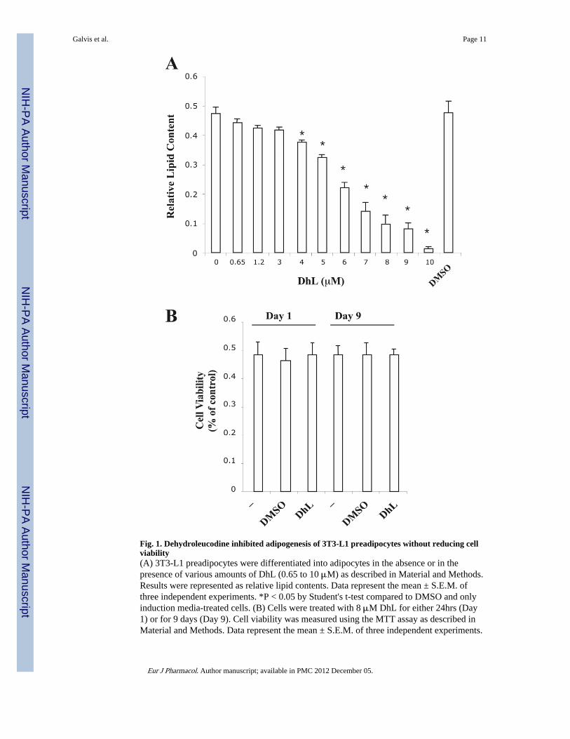
Fig. 1. Dehydroleucodine inhibited adipogenesis of 3T3-L1 preadipocytes without reducing cellviability(A) 3T3-L1 preadipocytes were differentiated into adipocytes in the absence or in thepresence of various amounts of DhL (0.65 to 10 μM) as described in Material and Methods.Results were represented as relative lipid contents. Data represent the mean ± S.E.M. ofthree independent experiments. *P < 0.05 by Student's t-test compared to DMSO and onlyinduction media-treated cells. (B) Cells were treated with 8 μM DhL for either 24hrs (Day1) or for 9 days (Day 9). Cell viability was measured using the MTT assay as described inMaterial and Methods. Data represent the mean ± S.E.M. of three independent experiments.
Galvis et al. Page 11
Eur J Pharmacol. Author manuscript; available in PMC 2012 December 05.
NIH
-PA Author Manuscript
NIH
-PA Author Manuscript
NIH
-PA Author Manuscript

Fig. 2. Dehydroleucodine blocked the formation of lipid droplet by induction media in 3T3-L1cellsAdipocyte differentiation was induced by treating confluent 3T3-L1 preadipocytes withinduction media in the absence or presence of 8 μM DhL. Morphological changes of 3T3-L1 preadipocytes were monitored by microscope and photographed after 9 days from theonset of differentiation. (A) Vehicle only, (B) cells treated induction media in the presenceDMSO, or (C) in the presence of DhL. (D) Nine days after induction of differentiation, cellswere lysed for triglyceride and protein assays as described in Material and Methods. Vehicleonly (line 1), cells treated with induction media alone (line 2), cells treated with inductionmedia in the presence DMSO (line 3), and cells treated with induction media in the presence
Galvis et al. Page 12
Eur J Pharmacol. Author manuscript; available in PMC 2012 December 05.
NIH
-PA Author Manuscript
NIH
-PA Author Manuscript
NIH
-PA Author Manuscript

of 8 μM DhL (line 4). Bars=10μm. Data represent the mean ± S.E.M. of three independentexperiments. *P < 0.05 by Student's t-test compared to DMSO-treated cells and onlyinduction media-treated cells.
Galvis et al. Page 13
Eur J Pharmacol. Author manuscript; available in PMC 2012 December 05.
NIH
-PA Author Manuscript
NIH
-PA Author Manuscript
NIH
-PA Author Manuscript

Fig. 3. Dehydroleucodine attenuated the expression of PPARγ during 3T3-L1 preadipocytedifferentiation3T3-L1 preadipocytes were induced to differentiate by induction media into adipocytes inthe absence (insert: –DhL, control) or in the presence (insert: +DhL) of 8 μM DhL. Totalprotein extracts were prepared at day 9 from each sample. The proteins were subset to 12%SDS-PAGE electrophoresis, blotted to a nitrocellulose membrane, and probed with anti-bodies specific to (A) PPARγ, C-EBPα and tubulin, (B) P-Erk1/2, T-Erk1/2, (C) P-Akt1, T-Akt1, and (D) P-AMPKα and T-AMPKα respectively. Relative levels of proteins weredetermined by densitometry as describe in Material and Methods. Data represent the mean ±
Galvis et al. Page 14
Eur J Pharmacol. Author manuscript; available in PMC 2012 December 05.
NIH
-PA Author Manuscript
NIH
-PA Author Manuscript
NIH
-PA Author Manuscript

S.E.M. of three independent experiments. *P < 0.05 by Student's t-test compared to DMSOinduction media-treated cells (control).
Galvis et al. Page 15
Eur J Pharmacol. Author manuscript; available in PMC 2012 December 05.
NIH
-PA Author Manuscript
NIH
-PA Author Manuscript
NIH
-PA Author Manuscript

Fig. 4. Dehydroleucodine blocked adipocyte differentiation in a time-dependent manner(A) 3T3-L1 preadipocyte cells were treated with 8 μM DhL for the time indicated in theschematic representation of the experiment. (B) In each treatment (1-6), the accumulation oflipid droplets was measured by the incorporation of Oil red O as described in Material andMethods. Data represent the mean ± S.E.M. of three independent experiments. *P < 0.05and **P < 0.01 by Student's t-test compared to DMSO induction media-treated cells(control).
Galvis et al. Page 16
Eur J Pharmacol. Author manuscript; available in PMC 2012 December 05.
NIH
-PA Author Manuscript
NIH
-PA Author Manuscript
NIH
-PA Author Manuscript

Fig. 5. GC analysis of dehydroleucodine and 11,13-dihydro-dehydroleucodine epimersA) DhL isolated from Artemisia douglassiana; (B) Mixture DH-DhL epimers as obtained byreduction of DhL, (#) denotes small amount of DhL after the reduction reaction; (C) DH-DhL epimer S, and, (D) DH-DhL epimer R after separation from the mixture. Chemicalstructures of DhL (compound 1), DH-DhL epimer 11S (compound 2) and DH-DhL epimer11R (compound 3) (numbering according to classical nomenclature).
Galvis et al. Page 17
Eur J Pharmacol. Author manuscript; available in PMC 2012 December 05.
NIH
-PA Author Manuscript
NIH
-PA Author Manuscript
NIH
-PA Author Manuscript

Fig. 6. 11,13-dihydro-dehydroleucodine inhibited 3T3-L1 preadipocyte differentiation(A) 3T3-L1 preadipocytes were differentiated into adipocytes in the absence or in thepresence of either DHL or DH-DhL. Results were represented as relative lipid content. Datarepresent the mean ± S.E.M. of three independent experiments. (B) 3T3-L1 preadipocytecells were incubated with induction media supplemented with either DMSO, 80 μM DH-DhL epimer S or 80 μM DH-DhL epimer R and the incorporation of Oil Red O wasmeasured by as described in Material and Methods. Results were represented as relativelipid content. Data represent the mean ± S.E.M. of three independent experiments. **P <0.01 by Student's t-test compared to DMSO-treated cells.
Galvis et al. Page 18
Eur J Pharmacol. Author manuscript; available in PMC 2012 December 05.
NIH
-PA Author Manuscript
NIH
-PA Author Manuscript
NIH
-PA Author Manuscript