Proximal tubular cells promote fibrogenesis by TGF-β1–mediated induction of peritubular...
12
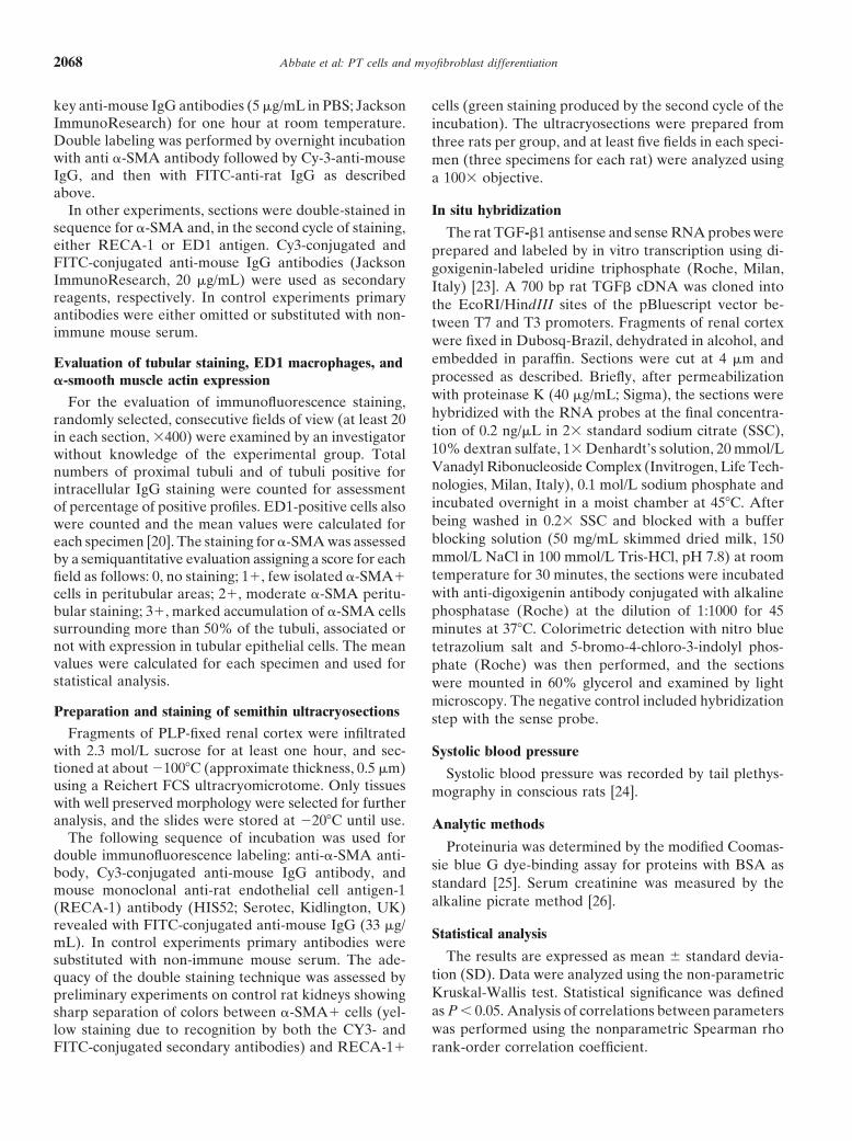
Kidney International, Vol. 61 (2002), pp. 2066–2077 Proximal tubular cells promote fibrogenesis by TGF-1–mediated induction of peritubular myofibroblasts MAURO ABBATE,CARLA ZOJA,DANIELA ROTTOLI,DANIELA CORNA, SUSANNA TOMASONI, and GIUSEPPE REMUZZI Mario Negri Institute for Pharmacological Research, and Unit of Nephrology and Dialysis, Azienda Ospedaliera, Ospedali Riuniti di Bergamo, Bergamo, Italy Proximal tubular cells promote fibrogenesis by TGF-1– In chronic nephropathies with increasingly severe dys- mediated induction of peritubular myofibroblasts. function of the glomerular filtering barrier, interstitial Background. In proteinuric nephropathies with increasingly inflammatory and fibrogenic events are important com- severe defects of the glomerular filtering barrier, interstitial ponents of injury leading to end-stage kidney. Proteins fibrogenesis is a major effector of scarring. An early event in ultrafiltered in excessive amounts may exert renal toxic- this process is the peritubular accumulation of myofibroblasts that express -smooth muscle actin (-SMA) and contribute ity as a consequence of virtually any kind of persistent to abnormal matrix production. Common trigger factors are insult or damage to the barrier [1]. Highlights on the poorly understood. Enhanced protein trafficking may play a mechanisms underlying tubulointerstitial damage have role by up-regulating inflammatory and fibrogenic genes in been obtained using polarized proximal tubular cells as proximal tubular cells. a model to examine cellular responses to the apical chal- Methods. The remnant kidney model in rats was used to (1) analyze interactions between activated proximal tubular cells, lenge to proteins. Exposing the cells to delipidated albu- peritubular cells expressing the myofibroblast marker, and in- min, immunoglobulins (IgG) or transferrin enhanced the flammatory cells at time intervals (days 7, 14, and 30) after rate of synthesis of endothelin-1 in a concentration de- surgery, and (2) evaluate the effects of angiotensin-converting pendent fashion [2]. Similarly, albumin and transferrin enzyme inhibitor (ACEi) on protein trafficking, fibrogenic sig- naling, and -SMA expression. up-regulated the monocyte chemoattractant protein 1 Results. Abnormal uptake of ultrafiltered proteins by proxi- gene (MCP-1), an event abrogated by inhibition of pro- mal tubular cells (IgG staining) occurred at an early stage (day 7) tein uptake with lysine [3]. RANTES (regulated upon and was subsequently associated with macrophage and -SMA activation, normal T cell expressed and secreted) [4] and cell accumulation into the peritubular interstitium. -SMA transforming growth factor-1 (TGF-1) [5] also were cells clustered with macrophages into the interstitium. These changes were associated with appearance of transforming up-regulated in proximal tubular cells on protein chal- growth factor-1 (TGF-1) mRNA in proximal tubular cells lenge. Mediators were released preferentially into the and in the infiltrating cells with time. At day 30, focal -SMA basolateral cell medium in a fashion that in the kidney staining also was found in the tubular cells and in peritubular would incite interstitial inflammatory and fibrogenic re- endothelial cells on semithin ultracryosections. ACEi prevented actions [2–4]. In fact, macrophages are recruited at tubu- both proteinuria and abnormal protein accumulation in tubular cells, as well as the inflammatory and fibrogenic reaction with lar sites of enhanced protein uptake [6] and may addi- peritubular -SMA expression. tionally synthesize fibrogenic stimuli [7]. However, little Conclusions. Profibrogenic signaling from both proximal tu- is known of the interactions at these sites between proxi- bular cells on challenge with filtered protein and inflammatory mal tubular cells, fibroblasts, and other cells relevant to cells is implicated as a key candidate trigger of progressive fibrogenic events. tubulointerstitial injury. One such event in progressive interstitial injury is the accumulation of fibrogenitor cells of heterogeneous origin, the myofibroblasts, induced by local stimuli to express cytoskeletal elements (-smooth muscle actin, -SMA) and to produce extracellular matrix [8, 9]. In anti-glomeru- Key words: proteinuria, -smooth muscle actin, macrophage, fibrosis, lar basement membrane (anti-GBM) disease and mesan- angiotensin converting enzyme inhibitor, progressive renal disease, tubulointerstitial injury. gial glomerulonephritis, selective insults to the glomerular membrane or mesangial components are followed by the Received for publication May 10, 2001 accumulation of -SMA cells into the peritubular in- and in revised form December 19, 2001 Accepted for publication February 5, 2002 terstitium as long as the barrier dysfunction and protein- uria persist [10–13]. The myofibroblast phenotype also 2002 by the International Society of Nephrology 2066
-
Upload
independent -
Category
Documents
-
view
1 -
download
0
Transcript of Proximal tubular cells promote fibrogenesis by TGF-β1–mediated induction of peritubular...
Kidney International, Vol. 61 (2002), pp. 2066–2077
Proximal tubular cells promote fibrogenesis byTGF-�1–mediated induction of peritubular myofibroblasts
MAURO ABBATE, CARLA ZOJA, DANIELA ROTTOLI, DANIELA CORNA,SUSANNA TOMASONI, and GIUSEPPE REMUZZI
Mario Negri Institute for Pharmacological Research, and Unit of Nephrology and Dialysis, Azienda Ospedaliera,Ospedali Riuniti di Bergamo, Bergamo, Italy
Proximal tubular cells promote fibrogenesis by TGF-�1– In chronic nephropathies with increasingly severe dys-mediated induction of peritubular myofibroblasts. function of the glomerular filtering barrier, interstitial
Background. In proteinuric nephropathies with increasingly inflammatory and fibrogenic events are important com-severe defects of the glomerular filtering barrier, interstitialponents of injury leading to end-stage kidney. Proteinsfibrogenesis is a major effector of scarring. An early event inultrafiltered in excessive amounts may exert renal toxic-this process is the peritubular accumulation of myofibroblasts
that express �-smooth muscle actin (�-SMA) and contribute ity as a consequence of virtually any kind of persistentto abnormal matrix production. Common trigger factors are insult or damage to the barrier [1]. Highlights on thepoorly understood. Enhanced protein trafficking may play a mechanisms underlying tubulointerstitial damage haverole by up-regulating inflammatory and fibrogenic genes in
been obtained using polarized proximal tubular cells asproximal tubular cells.a model to examine cellular responses to the apical chal-Methods. The remnant kidney model in rats was used to (1)
analyze interactions between activated proximal tubular cells, lenge to proteins. Exposing the cells to delipidated albu-peritubular cells expressing the myofibroblast marker, and in- min, immunoglobulins (IgG) or transferrin enhanced theflammatory cells at time intervals (days 7, 14, and 30) after
rate of synthesis of endothelin-1 in a concentration de-surgery, and (2) evaluate the effects of angiotensin-convertingpendent fashion [2]. Similarly, albumin and transferrinenzyme inhibitor (ACEi) on protein trafficking, fibrogenic sig-
naling, and �-SMA expression. up-regulated the monocyte chemoattractant protein 1Results. Abnormal uptake of ultrafiltered proteins by proxi- gene (MCP-1), an event abrogated by inhibition of pro-
mal tubular cells (IgG staining) occurred at an early stage (day 7) tein uptake with lysine [3]. RANTES (regulated uponand was subsequently associated with macrophage and �-SMA�
activation, normal T cell expressed and secreted) [4] andcell accumulation into the peritubular interstitium. �-SMA�transforming growth factor-�1 (TGF-�1) [5] also werecells clustered with macrophages into the interstitium. These
changes were associated with appearance of transforming up-regulated in proximal tubular cells on protein chal-growth factor-�1 (TGF-�1) mRNA in proximal tubular cells lenge. Mediators were released preferentially into theand in the infiltrating cells with time. At day 30, focal �-SMA basolateral cell medium in a fashion that in the kidneystaining also was found in the tubular cells and in peritubular
would incite interstitial inflammatory and fibrogenic re-endothelial cells on semithin ultracryosections. ACEi preventedactions [2–4]. In fact, macrophages are recruited at tubu-both proteinuria and abnormal protein accumulation in tubular
cells, as well as the inflammatory and fibrogenic reaction with lar sites of enhanced protein uptake [6] and may addi-peritubular �-SMA expression. tionally synthesize fibrogenic stimuli [7]. However, little
Conclusions. Profibrogenic signaling from both proximal tu- is known of the interactions at these sites between proxi-bular cells on challenge with filtered protein and inflammatorymal tubular cells, fibroblasts, and other cells relevant tocells is implicated as a key candidate trigger of progressivefibrogenic events.tubulointerstitial injury.
One such event in progressive interstitial injury is theaccumulation of fibrogenitor cells of heterogeneous origin,the myofibroblasts, induced by local stimuli to expresscytoskeletal elements (�-smooth muscle actin, �-SMA)and to produce extracellular matrix [8, 9]. In anti-glomeru-
Key words: proteinuria, �-smooth muscle actin, macrophage, fibrosis,lar basement membrane (anti-GBM) disease and mesan-angiotensin converting enzyme inhibitor, progressive renal disease,
tubulointerstitial injury. gial glomerulonephritis, selective insults to the glomerularmembrane or mesangial components are followed by theReceived for publication May 10, 2001accumulation of �-SMA� cells into the peritubular in-and in revised form December 19, 2001
Accepted for publication February 5, 2002 terstitium as long as the barrier dysfunction and protein-uria persist [10–13]. The myofibroblast phenotype also 2002 by the International Society of Nephrology
2066
Abbate et al: PT cells and myofibroblast differentiation 2067
appears in the interstitium in the typical setting of pro- ficed at day 30 (N � 7). To assess the effects of ACEi,rats with renal mass reduction received lisinopril (25teinuria due to nonimmune glomerular injury, the rem-
nant kidney of rats after 5/6 nephrectomy [14]. Remark- mg/L in the drinking water) [19, 20] starting from day 1after surgery and were sacrificed on day 30 (N � 7).ably, the emergence of �-SMA myofibroblasts is not
confined to animal models and, like the related fibrotic In all groups twenty-four hour urines were collected inmetabolic cages both before the time of disease inductionlesion itself, its detection can be used as a morphological
predictor of progressive disease [15–17]. and at sacrifice for the determination of urinary proteinexcretion levels. Systolic blood pressure and serum creat-Our current study tested the hypothesis that the activa-
tion of proximal tubular epithelial cells may contribute inine were assessed on day 30 after surgery.to promote a myofibroblast reaction into peritubular ar-
Tissue preparationeas and that the filtered protein overload may act as alocal stimulatory factor. To this end, remnant kidneys At the end of the study, the animals were anesthetized
with sodium pentobarbital IP (0.1 mL/100 g body wt ofof rats after 5/6 nephrectomy were studied with the fol-lowing specific aims: (1) to provide a comparative immuno- a 65 mg/mL solution in saline) and the kidneys were
fixed by perfusion via abdominal aorta [21]. Kidneyshistochemical analysis of ultrafiltered protein accumulationin proximal tubular cells and of �-SMA in the tubulo- were perfused with Hanks’ solution for five minutes and
then fixed with periodate-lysine paraformaldehyde (PLP)interstitial compartment; (2) to characterize the timecourse and location of abnormal peritubular �-SMA ex- fixative [22] for 10 minutes, followed by overnight fixa-
tion in the same fixative at 4�C. The tissue fragmentspression on semithin ultracryosections; (3) to identifyrelevant macrophage-myofibroblast interactions; and (4) from remnant kidneys were taken from the center of
non-infarcted areas. Fixed tissue specimens were exten-to determine whether the amelioration of the filteringbarrier by angiotensin-converting enzyme inhibitor (ACEi) sively washed with phosphate-buffered saline (PBS; 0.9%
NaCl in 10 mmol/L sodium phosphate buffer, pH 7.4)therapy that limits both the exposure of tubular cells toultrafiltered proteins and subsequent macrophage accu- and stored in the same buffer. Selected specimens were
embedded in Epon and processed for electron micros-mulation, also may cause reductions of abnormal peritu-bular �-SMA� cells and of TGF-�1, a common inducer copy using standard protocol. For in situ hybridization
studies, fragments were fixed in Dubosq-Brazil solution,of myofibroblast phenotype in fibroblasts, endothelialcells, and proximal tubular epithelial cells in the kidney. dehydrated, and embedded in paraffin [23].
ImmunohistochemistryMETHODS
The tissue specimens were immersed in 30% sucrose/Animals PBS for at least one hour at room temperature, embed-
ded in OCT medium, and frozen in liquid nitrogen. Tis-Studies were conducted in male Sprague Dawley, CD-COBS rats (275 to 300 g initial body weight) obtained sue sections were cut at 5 �m thickness using a Mikrom
500 O cryostat (Mikrom, Walldorf, Germany) and eitherfrom Charles River SpA (Calco, Italy). The animals werehoused in a constant temperature room with a 12-hour stained immediately or stored at �20�C until further pro-
cessing. Non-specific binding of antibodies was blockeddark/12-hour light cycle and fed a standard diet. Animalcare and treatment were conducted in conformity with with PBS/1% BSA for 15 minutes at room temperature.
Sections were incubated for direct immunofluorescencethe institutional guidelines that are in compliance withnational (D.L. n.116, G.U., Suppl 40, 18 Febbraio 1992, with fluorescein isothiocyanate (FITC)-conjugated goat
anti-rat IgG (30 �g/mL in PBS; Jackson ImmunoRe-Circolare No 8, G.U., 14 Luglio 1994) and internationallaws and policies (EEC Council Directive 86/609, OJL search Laboratories, West Grove, PA, USA) for one
hour at room temperature. After washes in PBS, the358, Dec 1987; Guide for the Care and Use of LaboratoryAnimals, U.S. National Research Council, 1996). slides were mounted using 100 mmol/L Tris-HCl:glycerol
50:50, 2% N-propyl gallate, pH 8. Sections were exam-Disease model and protocol ined with a Leika DM-R microscope equipped with
epifluorescence and appropriate filters.Five-sixths renal mass reduction (RMR) was accom-plished by surgical removal of right kidney and ligation Mouse monoclonal antibodies were used for the detec-
tion of �-smooth muscle actin (1A4; Sigma Co., St. Louis,of two or three extrarenal branches of the left renalartery [18] in anesthetized rats. Age-matched rats (N � MO, USA) and rat macrophages (ED1; Serotec, Oxford,
UK). Tissue sections were blocked with PBS/1% bovine5) were used as controls after sham operation, consistingof a laparotomy and manipulation of renal pedicles. serum albumin (BSA) and incubated overnight (18
hours) at 4�C with the primary antibody diluted in PBSThree groups of rats with renal mass reduction (N � 7each group) were sacrificed at 7, 14 and 30 days after (1A4, ascites fluid, 1:400; ED1, 10 �g/mL). After washes
in PBS, they were incubated with Cy-3-conjugated don-surgery, respectively; sham-operated controls were sacri-
Abbate et al: PT cells and myofibroblast differentiation2068
key anti-mouse IgG antibodies (5 �g/mL in PBS; Jackson cells (green staining produced by the second cycle of theImmunoResearch) for one hour at room temperature. incubation). The ultracryosections were prepared fromDouble labeling was performed by overnight incubation three rats per group, and at least five fields in each speci-with anti �-SMA antibody followed by Cy-3-anti-mouse men (three specimens for each rat) were analyzed usingIgG, and then with FITC-anti-rat IgG as described a 100� objective.above.
In situ hybridizationIn other experiments, sections were double-stained insequence for �-SMA and, in the second cycle of staining, The rat TGF-�1 antisense and sense RNA probes wereeither RECA-1 or ED1 antigen. Cy3-conjugated and prepared and labeled by in vitro transcription using di-FITC-conjugated anti-mouse IgG antibodies (Jackson goxigenin-labeled uridine triphosphate (Roche, Milan,ImmunoResearch, 20 �g/mL) were used as secondary Italy) [23]. A 700 bp rat TGF� cDNA was cloned intoreagents, respectively. In control experiments primary the EcoRI/HindIII sites of the pBluescript vector be-antibodies were either omitted or substituted with non- tween T7 and T3 promoters. Fragments of renal corteximmune mouse serum. were fixed in Dubosq-Brazil, dehydrated in alcohol, and
embedded in paraffin. Sections were cut at 4 �m andEvaluation of tubular staining, ED1 macrophages, andprocessed as described. Briefly, after permeabilization�-smooth muscle actin expressionwith proteinase K (40 �g/mL; Sigma), the sections wereFor the evaluation of immunofluorescence staining,hybridized with the RNA probes at the final concentra-randomly selected, consecutive fields of view (at least 20tion of 0.2 ng/�L in 2� standard sodium citrate (SSC),in each section, �400) were examined by an investigator10% dextran sulfate, 1� Denhardt’s solution, 20 mmol/Lwithout knowledge of the experimental group. TotalVanadyl Ribonucleoside Complex (Invitrogen, Life Tech-numbers of proximal tubuli and of tubuli positive fornologies, Milan, Italy), 0.1 mol/L sodium phosphate andintracellular IgG staining were counted for assessmentincubated overnight in a moist chamber at 45�C. Afterof percentage of positive profiles. ED1-positive cells alsobeing washed in 0.2� SSC and blocked with a bufferwere counted and the mean values were calculated forblocking solution (50 mg/mL skimmed dried milk, 150each specimen [20]. The staining for �-SMA was assessedmmol/L NaCl in 100 mmol/L Tris-HCl, pH 7.8) at roomby a semiquantitative evaluation assigning a score for eachtemperature for 30 minutes, the sections were incubatedfield as follows: 0, no staining; 1�, few isolated �-SMA�with anti-digoxigenin antibody conjugated with alkalinecells in peritubular areas; 2�, moderate �-SMA peritu-phosphatase (Roche) at the dilution of 1:1000 for 45bular staining; 3�, marked accumulation of �-SMA cells
surrounding more than 50% of the tubuli, associated or minutes at 37�C. Colorimetric detection with nitro bluenot with expression in tubular epithelial cells. The mean tetrazolium salt and 5-bromo-4-chloro-3-indolyl phos-values were calculated for each specimen and used for phate (Roche) was then performed, and the sectionsstatistical analysis. were mounted in 60% glycerol and examined by light
microscopy. The negative control included hybridizationPreparation and staining of semithin ultracryosections step with the sense probe.
Fragments of PLP-fixed renal cortex were infiltratedwith 2.3 mol/L sucrose for at least one hour, and sec- Systolic blood pressuretioned at about �100�C (approximate thickness, 0.5 �m) Systolic blood pressure was recorded by tail plethys-using a Reichert FCS ultracryomicrotome. Only tissues mography in conscious rats [24].with well preserved morphology were selected for furtheranalysis, and the slides were stored at �20�C until use. Analytic methods
The following sequence of incubation was used forProteinuria was determined by the modified Coomas-
double immunofluorescence labeling: anti-�-SMA anti-sie blue G dye-binding assay for proteins with BSA asbody, Cy3-conjugated anti-mouse IgG antibody, andstandard [25]. Serum creatinine was measured by themouse monoclonal anti-rat endothelial cell antigen-1alkaline picrate method [26].(RECA-1) antibody (HIS52; Serotec, Kidlington, UK)
revealed with FITC-conjugated anti-mouse IgG (33 �g/Statistical analysis
mL). In control experiments primary antibodies wereThe results are expressed as mean standard devia-substituted with non-immune mouse serum. The ade-
tion (SD). Data were analyzed using the non-parametricquacy of the double staining technique was assessed byKruskal-Wallis test. Statistical significance was definedpreliminary experiments on control rat kidneys showingas P 0.05. Analysis of correlations between parameterssharp separation of colors between �-SMA� cells (yel-was performed using the nonparametric Spearman rholow staining due to recognition by both the CY3- and
FITC-conjugated secondary antibodies) and RECA-1� rank-order correlation coefficient.
Abbate et al: PT cells and myofibroblast differentiation 2069
RESULTS cells showed less or no IgG staining in respect to otherIgG-positive epithelial cells within individual profiles ofIgG staining in proximal tubuli and expression ofproximal tubule, most presumably reflecting impaired�-smooth muscle actin after 5/6 nephrectomyprotein absorption following the induction of abnormal
In the kidneys of sham-operated rats IgG staining was cell phenotype. Correlation analysis revealed significantundetectable in proximal tubular cells except that for a positive correlations both between proteinuria and prox-weak intracellular granular reactivity with focal distribu- imal tubular IgG staining and between the latter andtion. Intracellular IgG staining was found in increasingly peritubular �-SMA staining (Spearman rho: �0.80 andhigher numbers of proximal tubular profiles at different
�0.88, respectively, P 0.001).time points starting from day 7 (day 7, 21.22 8.66%;day 14, 58.17 9.16%; day 30, 66.71 10.67%; sham TGF-�1 mRNA expression in the tubulointerstitiumcontrol, 3.68 1.51% staining). This change was associ- in remnant kidneysated with increasingly higher levels of urinary protein By in situ hybridization, in kidneys of sham operatedexcretion during time after surgery (day 7, 45.2 18.87; rats signal for TGF-�1 mRNA was found in tubularday 14, 116.18 34.21; day 30, 223.65 94.70; sham epithelial cells in distal segments, and in occasional inter-control, 24.36 4.31 mg/24 h; IgG mean score and pro- stitial cells or peritubular capillary endothelial cells. Stainteinuria, P 0.05 day 7 vs control, P 0.01 day 14 vs. of proximal tubular cells was absent (Fig. 3A). In rem-day 7, and P 0.01 day 30 vs. day 14). nant kidneys at 7 days after surgery, despite a similar
No differences were visualized in the staining for distribution of TGF-�1 mRNA, a focal and weak staining�-SMA in the tubulointerstitial compartment of remnant of epithelial cells was revealed in proximal tubuli (Fig.kidneys at day 7 as compared to sham control kidneys 3B). A focal increase in the extension and intensity of(Fig. 1 A, B; mean score for remnant kidney at day 7, staining was also visualized in proximal tubuli in 14-0.07, and control, 0.03). The staining was confined to day remnant kidneys. In addition, abnormal signal wasvascular smooth muscle cells of arteries, arterioles, and present in peritubular interstitial and capillary areas, parttheir branches splitting into capillaries. In contrast, in of which was associated with infiltrating cells (Fig. 3C). Inremnant kidneys at 14 days after surgery, �-SMA� cells 30-day remnant kidneys the increase in TGF-�1 mRNAbecame detectable in the peritubular areas, either iso- hybridization signal in proximal tubuli also was promi-lated or in clusters. In most fields the abnormal staining nent and diffuse in parallel with marked TGF-�1 mRNAappeared to concentrate in the interstitium (Fig. 1C) in expression in peritubular areas (Fig. 3D).cells with round or elongated fibroblast-like morphology.Abnormal �-SMA expression was more evident and dif- Time course of ED1 macrophage accumulation and
comparison with �-smooth muscle actin infuse in the sections of 30-day remnant kidneys. At thisremnant kidneystime point, it was focally detectable in tubular epithelial
cells in addition to the �-SMA� cells in adjacent peritu- We and others have documented that ED1 monocyte/bular areas (Fig. 1D). The mean scores for �-SMA stain- macrophages and major histocompatibility complex-IIing were 0.71 at day 14 and 1.11 at day 30 (P 0.01 vs. (MHC-II) positive cells initially accumulate into the in-day 7 and vs. day 14, respectively). terstitium of remnant kidneys during the early protein-
uric stages of progressive disease, with significant in-Comparison of the distribution of IgG and �-smooth creases at 14 days after surgery. The time course of ED1muscle actin staining macrophage accumulation was confirmed here, and is
By double staining of the sections for IgG and �-SMA, illustrated in Figure 4 (ED1, green color). Mean countsabnormal IgG staining was visualized in remnant kidneys of ED1� cells (number/high power field) were 6.2 2.2at day 7 in proximal tubular cells, in the absence of at day 7, 16.6 3.5 at day 14, and 28.1 5.7 at day�-SMA staining in surrounding areas (Fig. 2B, compare 30 (P 0.01 days 14 and 30 vs. control, 5.3 1.0).to sham control in Fig. 2A) as predictable from results Additionally, infiltrating macrophages were identified inof single immunofluorescence staining. The initial ex- areas surrounding proximal tubuli in which protein traf-pression of abnormal �-SMA at 14 days was revealed in fic was enhanced [20], thus in a distribution remarkablythe peritubular areas in the vicinity of proximal tubuli similar to that of peritubular �-SMA� cells in remnantthat stained positive for IgG as a consequence of abnor- kidneys as described above in the present study. Consis-mally high cellular uptake of ultrafiltered protein (Fig. tent with such distribution, the results of double staining2C). This association was maintained at 30 days (Fig. experiments revealed a close association of ED1� cells2D). Additionally, �-SMA staining was visualized in and �-SMA� cells in peritubular areas in the sectionsremnant kidneys at 30 days in tubular epithelial cells of 14 day remnant kidneys (Fig. 4C), reflecting the inter-of IgG-positive proximal tubuli, although not confined action between the recruited macrophages and �-SMA�
cells at those sites. ED1 macrophages and �-SMA� cellsexclusively to these segments. Some of the �-SMA�
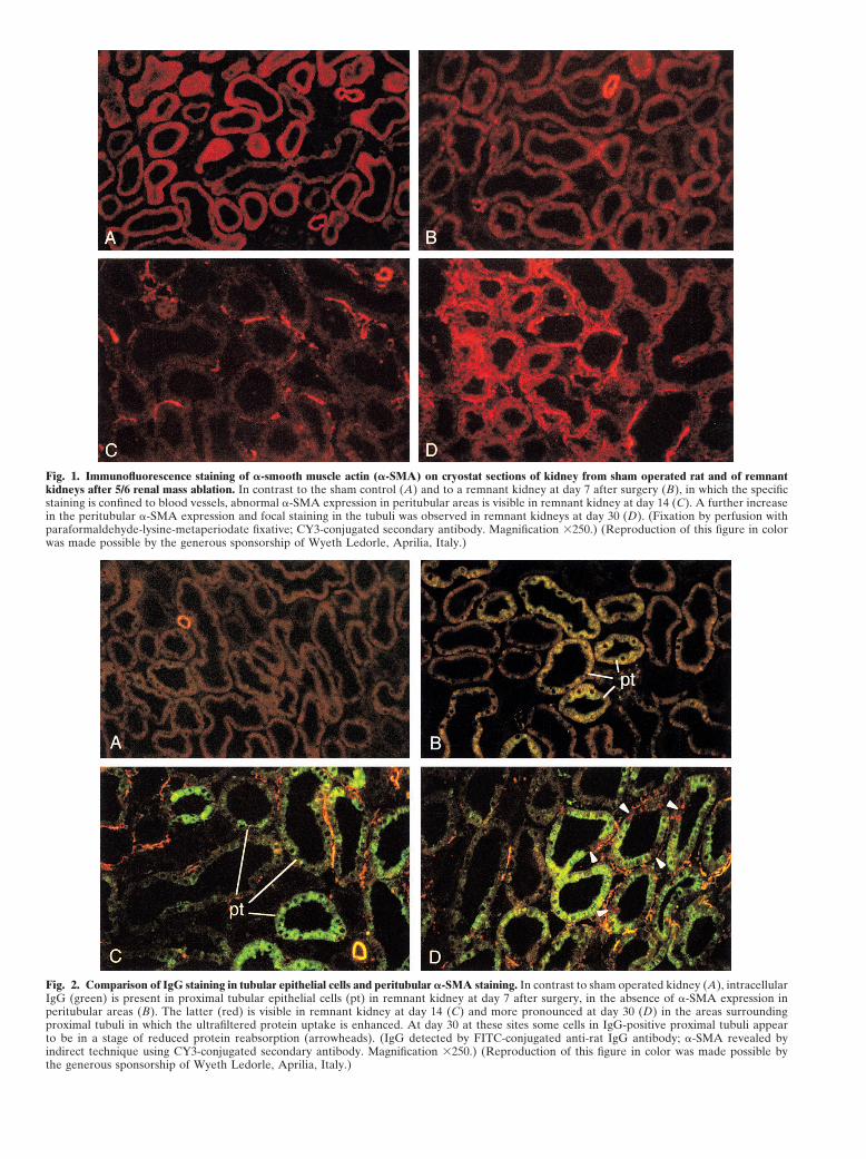
Fig. 1. Immunofluorescence staining of �-smooth muscle actin (�-SMA) on cryostat sections of kidney from sham operated rat and of remnantkidneys after 5/6 renal mass ablation. In contrast to the sham control (A) and to a remnant kidney at day 7 after surgery (B), in which the specificstaining is confined to blood vessels, abnormal �-SMA expression in peritubular areas is visible in remnant kidney at day 14 (C). A further increasein the peritubular �-SMA expression and focal staining in the tubuli was observed in remnant kidneys at day 30 (D). (Fixation by perfusion withparaformaldehyde-lysine-metaperiodate fixative; CY3-conjugated secondary antibody. Magnification �250.) (Reproduction of this figure in colorwas made possible by the generous sponsorship of Wyeth Ledorle, Aprilia, Italy.)
Fig. 2. Comparison of IgG staining in tubular epithelial cells and peritubular �-SMA staining. In contrast to sham operated kidney (A), intracellularIgG (green) is present in proximal tubular epithelial cells (pt) in remnant kidney at day 7 after surgery, in the absence of �-SMA expression inperitubular areas (B). The latter (red) is visible in remnant kidney at day 14 (C) and more pronounced at day 30 (D) in the areas surroundingproximal tubuli in which the ultrafiltered protein uptake is enhanced. At day 30 at these sites some cells in IgG-positive proximal tubuli appearto be in a stage of reduced protein reabsorption (arrowheads). (IgG detected by FITC-conjugated anti-rat IgG antibody; �-SMA revealed byindirect technique using CY3-conjugated secondary antibody. Magnification �250.) (Reproduction of this figure in color was made possible bythe generous sponsorship of Wyeth Ledorle, Aprilia, Italy.)
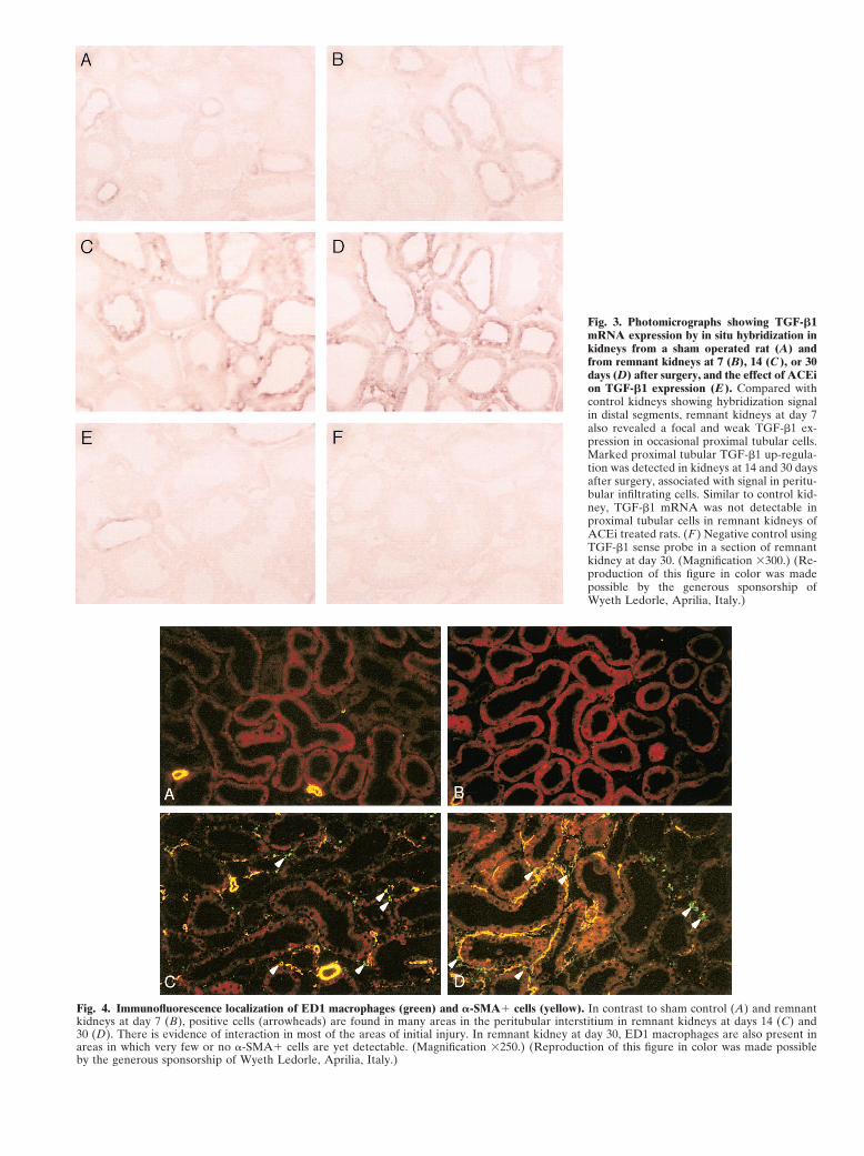
Fig. 3. Photomicrographs showing TGF-�1mRNA expression by in situ hybridization inkidneys from a sham operated rat (A) andfrom remnant kidneys at 7 (B), 14 (C ), or 30days (D) after surgery, and the effect of ACEion TGF-�1 expression (E ). Compared withcontrol kidneys showing hybridization signalin distal segments, remnant kidneys at day 7also revealed a focal and weak TGF-�1 ex-pression in occasional proximal tubular cells.Marked proximal tubular TGF-�1 up-regula-tion was detected in kidneys at 14 and 30 daysafter surgery, associated with signal in peritu-bular infiltrating cells. Similar to control kid-ney, TGF-�1 mRNA was not detectable inproximal tubular cells in remnant kidneys ofACEi treated rats. (F ) Negative control usingTGF-�1 sense probe in a section of remnantkidney at day 30. (Magnification �300.) (Re-production of this figure in color was madepossible by the generous sponsorship ofWyeth Ledorle, Aprilia, Italy.)
Fig. 4. Immunofluorescence localization of ED1 macrophages (green) and �-SMA� cells (yellow). In contrast to sham control (A) and remnantkidneys at day 7 (B), positive cells (arrowheads) are found in many areas in the peritubular interstitium in remnant kidneys at days 14 (C) and30 (D). There is evidence of interaction in most of the areas of initial injury. In remnant kidney at day 30, ED1 macrophages are also present inareas in which very few or no �-SMA� cells are yet detectable. (Magnification �250.) (Reproduction of this figure in color was made possibleby the generous sponsorship of Wyeth Ledorle, Aprilia, Italy.)

Abbate et al: PT cells and myofibroblast differentiation2072
Fig. 5. Identification of �-SMA in tubuloin-terstitial areas on semithin ultracryosectionsof remnant kidneys, and effect of lisinoprilon abnormal �-SMA expression. No �-SMAstaining was localized in peritubular areas inremnant kidney at day 7 (A). Endothelial cells(ec), stained in green for rat endothelial cellantigen -1, are sharply distinguishable from�-SMA� smooth muscle cells (in yellow, in-set). In a remnant kidney at day 14 (B), abnor-mal peritubular �-SMA staining can be seenin myofibroblasts in areas (asterisk) betweenproximal tubuli (pt) and peritubular capillar-ies. In a remnant kidney at day 30 (C), �-SMAis additionally expressed by tubular epithelialcells and appears to be associated with cellsshowing both endothelial location and mor-phology in focal areas (arrowheads). (D)Remnant kidney of lisinopril-treated rat. (Mag-nification �400 in A, C, D, and inset; �1000in B.) (Reproduction of this figure in color wasmade possible by the generous sponsorship ofWyeth Ledorle, Aprilia, Italy.)
also were found to colocalize in peritubular areas in the above analysis, we performed a detailed study usingsemithin ultracryosections, which provide much higherkidneys at 30 days (Fig. 4D), despite more irregular and
diffuse distribution of macrophages during the persistent level of resolution and precise delineation of peritubularstructures. The sections were analyzed for detection ofinflammatory wave.�-SMA and the endothelial cell marker, RECA-1. Re-
Localization of �-smooth muscle actin positive cells sults were unequivocal by inspection and are illustratedon ultracryosections in Figures 5 and 6. Both in sham kidneys and in rem-
nant kidneys at day 7 (Fig. 5A), endothelial cells wereTo gain information on the cell type(s) initially in-duced to express �-SMA, and to extend the results from distinguishable by RECA-1 staining in green color from
Abbate et al: PT cells and myofibroblast differentiation 2073
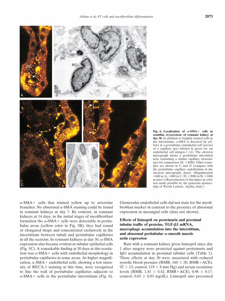
Fig. 6. Localization of �-SMA� cells insemithin cryosections of remnant kidney atday 30. In addition to brightly stained cells inthe interstitium, �-SMA is detected (in yel-low) in a peritubular endothelial cell (arrow)of a capillary (pc) labeled in green for ratendothelial cell antigen-1 (A). The electronmicrograph shows a peritubular interstitialarea containing a similar capillary structure(pc) for comparison (B, �3000). Other exam-ples are shown in C and D (compare withthe peritubular capillary endothelium in theelectron micrograph, inset). (Magnification�600 in A; �800 in C, D; �3000 in B; �1000in inset.) (Reproduction of this figure in colorwas made possible by the generous sponsor-ship of Wyeth Ledorle, Aprilia, Italy.)
�-SMA� cells that stained yellow up to arteriolar Glomerular endothelial cells did not stain for the myofi-broblast marker in contrast to the presence of abnormalbranches. No abnormal �-SMA staining could be found
in remnant kidneys at day 7. By contrast, in remnant expression in mesangial cells (data not shown).kidneys at 14 days, in the initial stages of myofibroblast
Effects of lisinopril on proteinuria and proximalformation the �-SMA� cells were detectable in peritu-tubular traffic of proteins, TGF-�1 mRNA,bular areas (yellow color in Fig. 5B), they had roundmacrophage accumulation into the interstitium,or elongated shape and concentrated exclusively in theand abnormal peritubular �-smooth muscleinterstitium between tubuli and peritubular capillariesactin expressionin all the sections. In remnant kidneys at day 30, �-SMA
expression also became evident in tubular epithelial cells Rats with a remnant kidney given lisinopril since day1 after surgery were protected against proteinuria and(Fig. 5C). A remarkable finding at 30 days at this resolu-
tion was �-SMA� cells with endothelial morphology in IgG accumulation in proximal tubular cells (Table 1).These effects at day 30 were associated with reducedperitubular capillaries in some areas. At higher magnifi-
cation, �-SMA� endothelial cells, showing a low inten- systolic blood pressure (RMR, 160 20; RMR�ACEi,92 13; control, 119 8 mm Hg) and serum creatininesity of RECA-1 staining at this time, were recognized
to line the wall of peritubular capillaries adjacent to levels (RMR, 1.41 0.42; RMR�ACEi, 0.96 0.17;control, 0.63 0.03 mg/dL). Lisinopril also prevented�-SMA� cells in the peritubular interstitium (Fig. 6).
Abbate et al: PT cells and myofibroblast differentiation2074
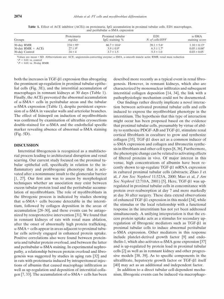
Table 1. Effect of ACE inhibitor (ACEi) on proteinuria, IgG accumulation in proximal tubular cells, ED1 macrophages,and peritubular �-SMA expression
Proteinuria Proximal tubular ED1 �-SMAGroups mg/day IgG staining % N of cells/HPF staining score
30-day RMR 22495a 66.7 10.6a 28.1 5.6a 1.10 0.13a
30-day RMR � ACEi 279b 3.9 0.9b 6.3 1.7b 0.05 0.06b
30-day Control 244 3.7 1.5 5.31.0 0.030.05
Values are mean SD. Abbreviations are: ACE, angiotensin-converting enzyme; �-SMA, �-smooth muscle actin; RMR, renal mass reductiona P 0.01 vs. controlb P 0.01 vs. 30-day RMR
both the increases in TGF-�1 expression thus abrogating described more recently as a typical event in renal fibro-the prominent up-regulation in proximal tubular epithe- genesis. However, in remnant kidneys, which also arelial cells (Fig. 3E), and the interstitial accumulation of characterized by mononuclear infiltrates and subsequentmacrophages in remnant kidneys at 30 days (Table 1). interstitial collagen deposition [14, 34], the link with aFinally, the ACEi prevented the abnormal accumulation pathophysiologic mechanism could not be documented.of �-SMA� cells in peritubular areas and the tubular Our findings rather directly implicate a novel interac-�-SMA expression (Table 1), despite persistent expres- tion between activated proximal tubular cells and cellssion of �-SMA in vascular walls and arteriolar branches. induced to express the myofibroblast phenotype in theThe effect of lisinopril on induction of myofibroblasts interstitium. The hypothesis that this type of interactionwas confirmed by examination of ultrathin cryosections might occur has been proposed based on the evidencedouble-stained for �-SMA and the endothelial specific that proximal tubular cells, presumably by virtue of abil-marker revealing absence of abnormal �-SMA staining ity to synthesize PDGF-AB and TGF-�1, stimulate renal(Fig. 5D). cortical fibroblasts in coculture to grow and synthesize
collagen [35]. TGF-�1 does act as a common inducer of�-SMA expression and collagen and fibronectin synthe-DISCUSSIONsis in fibroblasts and other cell types [8, 36]. Furthermore,
Interstitial fibrogenesis is recognized as a multifacto-the phenotypic change can be related to activating effectsrial process leading to architectural disruption and renalof filtered proteins in vivo. Of major interest in thisscarring. Our current study focused on the proximal tu-venue, high concentrations of albumin have been re-bular epithelial cell, specifically in relation to the in-cently shown to up-regulate TGF-�1 mRNA expressionflammatory and profibrogenic phenotype that is acti-in cultured proximal tubular cells (abstracts; Zhao J etvated after a nonimmune insult to the glomerular barrieral, J Am Soc Nephrol 11:523A, 2000; Mao et al, J Am[1, 27]. Our first aim was to assess by morphologicalSoc Nephrol 12:710A, 2001) [5]. Here, TGF-�1 was up-techniques whether an association may exist betweenregulated in proximal tubular cells in concomitance withexcess tubular protein load and the peritubular accumu-protein over-reabsorption at day 7 and more markedlylation of myofibroblasts. The role of myofibroblasts inat day 30 after surgery. These data extend observationsthe fibrogenic process is indicated by studies showingof enhanced TGF-�1 expression in this model [34], whilethat �-SMA� cells become detectable in the intersti-the stimulus or the local relationship with a functionaltium, followed by collagen deposition in the areas ofresponse in the interstitium has not yet been addressedaccumulation [28–30], and these events can be antago-simultaneously. A unifying interpretation is that the ex-nized by renoprotective intervention [31]. We found thatcess protein uptake acts as a stimulus for secondary up-in remnant kidneys of rats with renal mass ablation,regulation of fibrogenic mediators such as TGF-�1 inafter the onset of abnormally high protein trafficking,proximal tubular cells to induce abnormal peritubular�-SMA� cells appear in areas adjacent to proximal tubu-�-SMA expression. Other mediators in this responselar cells actively engaged in enhanced protein uptake.include platelet-derived growth factor [9] and endo-Positive correlations also were found between protein-thelin-1, which also activates �-SMA gene expression [37]uria and tubular protein overload, and between the latterand is up-regulated by protein load in proximal tubularand peritubular �-SMA staining. In experimental nephro-cells [2] as well as in remnant kidney and other progres-pathy, a relationship between protein overload and fibro-sive models [38, 39]. As to specific components in thegenesis was suggested by studies in aging rats [32] andultrafiltrate, hepatocyte growth factor or TGF-�1 itselfin rats with proteinuria induced by intraperitoneal injec-[40] may act as an additional and independent factor.tions of albumin that caused macrophage infiltration as
In addition to a direct tubular cell-dependent mecha-well as up-regulation and deposition of interstitial colla-gen [7, 33]. The accumulation of �-SMA� cells has been nism, fibrogenic events can be induced via macrophage-
Abbate et al: PT cells and myofibroblast differentiation 2075
mediated pathways. The codistribution of the tubular the nearby epithelium and immune cells and may partici-pate to the fibrogenic reaction. Importantly in thisstaining for IgG with peritubular �-SMA in the present
study replicates the codistribution found with inflamma- perspective, antisense oligodeoxynucleotides comple-mentary to �-SMA inhibited endothelial-mesenchymaltory cells after either immune or nonimmune injury [6].
A cellular interaction is disclosed consistently between transformation and the associated TGF-�1–dependentmigration in vitro [50].macrophages and peritubular �-SMA� cells at day 14
after surgery. No studies have specifically looked at this Angiotensin converting enzyme inhibitors are thetreatment of choice in chronic proteinuric nephropa-aspect in progressive disease models. The macrophage-
myofibroblast interaction, however, has been recently thies. Studies on mechanisms of renoprotection haveidentified, among other targets, glomerular permselec-proposed to have pathogenic significance in chronictive dysfunction. We found previously that lisinopril pre-immune tubulointerstitial injury [16]. In the remnantvents the excess exposure of proximal tubular cells tokidney the initial hit responsible for proteinuria and tu-ultrafiltered proteins [20], which are potentially responsi-bulointerstitial damage is nonimmune. The macrophage-ble for macrophage accumulation into the interstitiummyofibroblast interaction can be at least favored thereby[20, 34]. That ACEi treatment can limit �-SMA expres-by inflammatory mediators, such as MCP-1, RANTES,sion and collagen accumulation also has been reportedosteopontin, and complement, released or activated lo-(abstract; Jiang et al, J Am Soc Nephrol 10:660A, 1999)cally by tubular cells as a consequence of protein over-[31]. In addition, in our current study lisinopril abrogatedreabsorption [3, 4, 7, 41]. That the macrophages furtherTGF-�1 up-regulation and tubular cell- and macro-elaborate fibrogenic molecules is known by results of inphage-myofibroblast interactions at sites of abnormalvitro studies [42, 43] and by evidence here as well as inprotein reabsorption. These effects offer further supportthe protein overload model that peritubular infiltratingto the possibility that proximal tubular cells become acti-cells express TGF-�1 [7]. On the same line, macrophage-vated to promote local myofibroblast accumulation.derived TGF-�1 stimulated rat kidney interstitial fibro-ACEi does ameliorate the permselective function of theblasts to produce collagen types I and III and fibronectinbarrier by now emerging effects on podocyte functional[40].molecules such as nephrin [51]. As a less likely alterna-Regardless of the source of stimulation, the presenttive from such standpoint, ACEi may act on separatereport provides a clue to the origin(s) of peritubularangiotensin II pathways of interstitial injury [52]. How-myofibroblasts. Studies have left unanswered questionsever, given the relevant effects of angiotensin II includingabout whether the initial �-SMA expression in remnantthe ability to induce proteinuria, interstitial �-SMA ex-kidney was associated with interstitial cells [14] or proxi-pression [53], and TGF-�1 expression in tubular cellsmal tubular cells [44]. First, dual-staining experiments[54], its interrelated role as an upstream factor certainlyreveal that cells located in peritubular areas, most pre-must be considered.sumably albeit not exclusively the fibroblast [9, 14], are
In summary, the results of this study indicate thatthe initial targets of fibrogenic signaling. On the otherin an experimental model of progressive nephropathyhand, the finding of �-SMA in tubular cells at day 30proximal tubular cells promote peritubular accumulationis in line with previous results [44], confirming that aof �-SMA� cells, either directly and via macrophagephenotypic transformation of tubular cells occurs in amediated pathways. The initial target cell lies in therelatively late stage, as suggested originally for autoim-interstitium, and presumably the interstitial fibroblastsmune interstitial nephritis [45]. The available data indi-are a major component. In addition to tubular cells atcate that the phenotypic change is mediated in an auto-subsequent stages, peritubular endothelial cells may par-crine fashion and/or by inflammatory cells via TGF-�1,ticipate to the fibrogenic response as a consequence ofepidermal growth factor, or other pathways [36]. Of note,common signaling partly mediated by TGF-�1.the appearance of �-SMA in proximal tubular cells in
some profiles was associated with no or less IgG stainingACKNOWLEDGMENTSas compared with cells showing more abundant reab-
The authors are greatly indebted to Drs. Annalisa Perna and Bo-sorbed protein, a change that could reflect reduced reab-rislav D. Dimitrov for assistance in the statistical analysis.sorption by the transdifferentiating epithelium. Another
Reproduction of Figures 1 to 6 in color was made possible by thenew finding is the detection of �-SMA in peritubular generous sponsorship of Wyeth Ledorle, Aprilia, Italy.endothelial cells in semithin ultracryosections of rem-
Reprint requests to Mauro Abbate, M.D., “Mario Negri” Institutenant kidneys at day 30. Certain types of endothelial cells for Pharmacological Research, Via Gavazzeni 11, 24125 Bergamo, Italy.in culture can be induced to express �-SMA [46–48], for E-mail: [email protected] after heparin depletion, and transdifferentiateinto mesenchymal cells again in response to TGF-�1 REFERENCES[49]. Thus, it seems that the peritubular endothelium, 1. Remuzzi G, Bertani T: Pathophysiology of progressive nephropa-
thies. N Engl J Med 339:1448–1456, 1998less than the fibroblasts, is responsive to signaling from
Abbate et al: PT cells and myofibroblast differentiation2076
2. Zoja C, Morigi M, Figliuzzi H, et al: Proximal tubular cell synthe- 23. Zoja C, Liu XH, Donadelli R, et al: Renal expression of monocytechemoattractant protein-1 in lupus autoimmune mice. J Am Socsis and secretion of endothelin-1 on challenge with albumin andNephrol 8:720–729, 1997other proteins. Am J Kidney Dis 26:934–941, 1995
24. Pfeffer JM, Pfeffer MA, Frohlich ED: Validity of an indirect3. Wang Y, Chen J, Chen L, et al: Induction of monocyte chemoat-tail-cuff method for determining systolic arterial pressure in un-tractant protein-1 in proximal tubule cells by urinary protein. Janesthetized normotensive and spontaneously hypertensive rats.Am Soc Nephrol 8:1537–1545, 1997J Lab Clin Med 78:957–962, 19714. Zoja C, Donadelli R, Colleoni S, et al: Protein overload stimu-
25. Read SM, Northcote DH: Minimization of variation in the re-lates RANTES production by proximal tubular cells dependingsponse to different proteins of the Coomassie blue G dye-bindingon NF-�B activation. Kidney Int 53:1608–1615, 1998assay for protein. Anal Biochem 116:53–64, 19815. Yard BA, Chorianopoulos E, Herr D, van der Woude FJ: Regu-
26. Bonsnes RW, Tausski HA: The colorimetric determination oflation of endothelin-1 and transforming growth factor-beta1 pro-creatinine of the Jaffe reaction. J Biol Chem 158:581, 1945duction in cultured proximal tubular cells by albumin and heparan
27. Abbate M, Remuzzi G: Proteinuria as a mediator of tubulointersti-sulphate glycosaminoglycans. Nephrol Dial Transplant 16:1769–tial injury. Kidney Blood Press Res 22:37–46, 19991775, 2001
28. Tang WW, Van GY, Qi M: Myofibloblast and �1(III) collagen6. Abbate M, Zoja C, Corna D, et al: In progressive nephropathies,expression in experimental tubulointerstitial nephritis. Kidney Intoverload of tubular cells with filtered proteins translates glomerular 51:926–931, 1997
permeability dysfunction into cellular signals of interstitial in- 29. Badid C, Vincent M, McGregor B, et al: Mycophenolate mofetilflammation. J Am Soc Nephrol 9:1213–1224, 1998 reduces myofibroblast infiltration and collagen III deposition in
7. Eddy AA, Giachelli CM: Renal expression of genes that promote rat remnant kidney. Kidney Int 58:51–61, 2000interstitial inflammation and fibrosis in rats with protein-overload 30. Pichler RH, Hugo C, Shankland SJ, et al: SPARC is expressedproteinuria. Kidney Int 47:1546–1557, 1995 in renal interstitial fibrosis and in renal vascular injury. Kidney Int
8. Desmouliere A, Geinoz A, Gabbiani F, Gabbiani G: Trans- 50:1978–1989, 1996forming growth factor-�1 induces �-smooth muscle actin expres- 31. Wu LL, Yang N, Roe CJ, et al: Macrophage and myofibroblastsion in granulation tissue myofibroblasts and in quiescent and grow- proliferation in remnant kidney: Role of angiotensin II. Kidneying cultured fibroblasts. J Cell Biol 122:103–111, 1993 Int 52(Suppl 63):S221–S225, 1997
9. Tang WW, Ulich TR, Lacey DL, et al: Platelet-derived growth 32. Bertani T, Zoja C, Abbate M, et al: Age-related nephropathyfactor-BB induces renal tubulointerstitial myofibroblast formation and proteinuria in rats with intact kidneys exposed to diets withand tubulointerstitial fibrosis. Am J Pathol 148:1169–1180, 1996 different protein contents. Lab Invest 60:196–204, 1989
10. Zhang G, Moorhead PJ, El Nahas AM: Myofibroblasts and the 33. Eddy AA, McCulloch L, Adams J, Liu E: Interstitial nephritisprogression of experimental glomerulonephritis. Exp Nephrol induced by protein-overload proteinuria. Am J Pathol 135:719–733,
19893:308–318, 199534. Taal MW, Zandi-Nejad K, Weening B, et al: Proinflammatory11. El Nahas AM, Muchaneta-Kubara C, Zhang GZ, et al: Pheno-
gene expression and macrophage recruitment in the rat remnanttypic modulation of renal cells during experimental and clinicalkidney. Kidney Int 58:1664–1676, 2000renal scarring. Kidney Int 49(Suppl 54):S23–S27, 1996
35. Johnson DW, Saunders HJ, Baxter RC, et al: Paracrine stimula-12. Suzuki Y, Shirato I, Okumura K, et al: Distinct contribution oftion of human renal fibroblasts by proximal tubule cells. KidneyFc receptors and angiotensin II-dependent pathways in anti-GBMInt 54:747–757, 1998glomerulonephritis. Kidney Int 54:1166–1174, 1998
36. Okada H, Danoff TM, Kalluri R, Neilson EG: Early role of13. Yamamoto T, Noble NA, Miller DE, Border WA: SustainedFsp1 in epithelial-mesenchymal transformation. Am J Physiolexpression of TGF-�1 underlies development of progressive kid-273:F563–F574, 1997ney fibrosis. Kidney Int 45:916–927, 1994
37. Andrawis NS, Wang E, Abernethy DR: Endothelin-1 induces14. Kliem V, Johnson RJ, Alpers CE, et al: Mechanisms involved inan increase in total protein synthesis and expression of the smooththe pathogenesis of tubulointerstitial fibrosis in 5/6-nephrecto-muscle �-actin gene in vascular smooth muscle cells. Life Scimized rats. Kidney Int 49:666–678, 199659:523–528, 199615. Goumenos D, Tsomi K, Iatrou C, et al: Myofibroblasts and the
38. Orisio S, Benigni A, Bruzzi I, et al: Renal endothelin gene expres-progression of crescentic glomerulonephritis. Nephrol Dial Trans-sion is increased in remnant kidney and correlates with diseaseplant 13:1652–1661, 1998progression. Kidney Int 43:354–358, 199316. Pilmore HL, Painter DM, Bishop GA, et al: Early up-regulation
39. Zoja C, Liu X-H, Abbate M, et al: Angiotensin II blockade limitsof macrophages and myofibroblasts. Transplantation 69:2658–2669, tubular protein overreabsorption and the consequent upregulation2000 of endothelin 1 gene in experimental membranous nephropathy.17. Mezzano SA, Droguett MA, Burgos ME, et al: Overexpression Exp Nephrol 6:121–131, 1998
of chemokines, fibrogenic cytokines, and myofibroblast in human 40. Wang SN, LaPage J, Hirschberg R: Role of glomerular ultrafil-membranous nephropathy. Kidney Int 57:147–158, 2000 tration of growth factors in progressive interstitial fibrosis in dia-
18. Olson JL, Hostetter TH, Rennke HG, et al: Altered glomerular betic nephropathy. Kidney Int 57:1002–1214, 2000permselectivity and progressive sclerosis following extreme abla- 41. Donadelli R, Abbate M, Zanchi C, et al: Protein traffic activatestion of renal mass. Kidney Int 22:112–126, 1982 NF-kB gene signaling and promotes MCP-1-dependent interstitial
19. Remuzzi G, Zoja C, Gagliardini E, et al: Combining an antipro- inflammation. Am J Kidney Dis 36:1226–1241, 2000teinuric approach with mycophenolate mofetil fully suppresses 42. Eddy AA: Interstitial macrophages as mediators of renal fibrosis.progressive nephropathy of experimental animals. J Am Soc Exp Nephrol 3:76–79, 1995Nephrol 10:1542–1549, 1999 43. Chong H, Vodovotz Y, Cox GW, Barcellos-Hoff HM: Immuno-
20. Abbate M, Zoja C, Rottoli D, et al: Antiproteinuric therapy cytochemical localization of latent transforming growth factor-betawhile preventing the abnormal protein traffic in proximal tubule 1 activation by stimulated macrophages. J Cell Physiol 178:275–283,abrogates protein- and complement-dependent interstitial in- 1999flammation in experimental renal disease. J Am Soc Nephrol 44. Ng Y-Y, Huang T-P, Yang W-C, et al: Tubular epithelial-myofi-10:804–813, 1999 broblast transdifferentiation in progressive tubulointerstitial fi-
21. Abbate M, Bachinsky D, Zheng G, et al: Location of gp330/�2-m brosis in 5/6 nephrectomized rats. Kidney Int 54:864–876, 1998receptor-associated protein (�2-MRAP) and its binding sites in 45. Strutz F, Okada H, Lo CW, et al: Identification and characteriza-kidney: Distribution of endogenous �2-MRAP is modified by tissue tion of a fibroblast marker: FSP1. J Cell Biol 130:393–405, 1995processing. Eur J Cell Biol 61:139–149, 1993 46. Amberger A, Bauer H, Tontsch U, et al: Reversible expression
22. McLean IW, Nakane PF: Periodate-lysine paraformaldehyde fix- of sm alpha-actin protein and sm alpha-actin mRNA in clonedative: A new fixative for immunoelectron microscopy. J Histochem cerebral endothelial cells. FEBS Lett 287:223–225, 1991
47. DeRuiter MC, Poelmann RE, VanMunsteren JC, et al: Embry-Cytochem 22:1077–1083, 1974
Abbate et al: PT cells and myofibroblast differentiation 2077
onic endothelial cells transdifferentiate into mesenchymal cells hibits endothelial-mesenchymal transformation during chick cardi-ogenesis. Dev Dyn 216:489–498, 1999expressing smooth muscle actins in vivo and in vitro. Circ Res
51. Benigni A, Tomasoni S, Gagliardini E, et al: Blocking angiotensin80:444–451, 1997II synthesis/activity preserves glomerular nephrin in rats with se-48. Hautmann MB, Adam PJ, Owens GK: Similarities and differencesvere nephrosis. J Am Soc Nephrol 12:941–948, 2001in smooth muscle �-actin induction by TGF-� in smooth muscle
52. Border WA, Noble NA: Interactions of transforming growth fac-versus non-smooth muscle cells. Arterioscler Thromb Vasc Bioltor-beta and angiotensin II in renal fibrosis. Hypertens 31:181–188,19:2049–2058, 1999199849. Arciniegas E, Sutton AB, Allen TD, Schor AM: Transforming 53. Johnson RJ, Alpers CE, Yoshimura A, et al: Renal injury from
growth factor beta 1 promotes the differentiation of endothelial angiotensin II-mediated hypertension. Hypertens 19:464–474, 1992cells into smooth muscle-like cells in vitro. J Cell Sci 103:521–529, 54. Wolf G, Mueller E, Stahl RAK, Ziyadeh FN: Angiotensin II-1992 induced hypertrophy of cultured murine proximal tubular cells is
50. Nakajima Y, Yamagishi T, Yoshimura K, et al: Antisense oligo- mediated by endogenous transforming growth factor-�. J Clin In-vest 92:1366–1372, 1993deoxynucleotide complementary to smooth muscle alpha-actin in-