Specific expression of the Fusarium transposon Fot1 and effects on target gene transcription
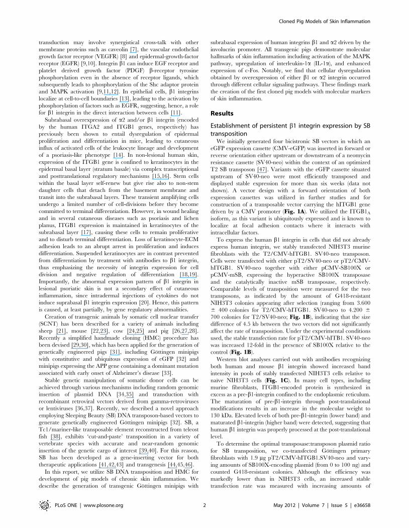
Development of Transgenic Cloned Pig Models of SkinInflammation by DNA Transposon-Directed EctopicExpression of Human b1 and a2 IntegrinNicklas Heine Staunstrup1, Johannes Madsen2, Maria Nascimento Primo1, Juan Li3, Ying Liu3,4,
Peter M. Kragh3, Rong Li3, Mette Schmidt5, Stig Purup3, Frederik Dagnæs-Hansen1, Lars Svensson2,
Thomas K. Petersen2, Henrik Callesen3, Lars Bolund1,6, Jacob Giehm Mikkelsen1*
1 Department of Biomedicine, Aarhus University, Aarhus, Denmark, 2 Department of Disease Pharmacology, LEO Pharma, Ballerup, Denmark, 3 Department of Animal
Science, Aarhus University, Tjele, Denmark, 4 College of Animal Science and Technology, Nanjing Agricultural University, Nanjing, China, 5 Department of Veterinary
Reproduction and Obstetrics, University of Copenhagen, Frederiksberg, Denmark, 6 HuaDa JiYin (BGI), Shenzhen, China
Abstract
Integrins constitute a superfamily of transmembrane signaling receptors that play pivotal roles in cutaneous homeostasis bymodulating cell growth and differentiation as well as inflammatory responses in the skin. Subrabasal expression of integrinsa2 and/or b1 entails hyperproliferation and aberrant differentiation of keratinocytes and leads to dermal and epidermalinflux of activated T-cells. The anatomical and physiological similarities between porcine and human skin make the pig asuitable model for human skin diseases. In efforts to generate a porcine model of cutaneous inflammation, we employedthe Sleeping Beauty DNA transposon system for production of transgenic cloned Gottingen minipigs expressing human b1or a2 integrin under the control of a promoter specific for subrabasal keratinocytes. Using pools of transgenic donorfibroblasts, cloning by somatic cell nuclear transfer was utilized to produce reconstructed embryos that were subsequentlytransferred to surrogate sows. The resulting pigs were all transgenic and harbored from one to six transgene integrants.Molecular analyses on skin biopsies and cultured keratinocytes showed ectopic expression of the human integrins andlocalization within the keratinocyte plasma membrane. Markers of perturbed skin homeostasis, including activation of theMAPK pathway, increased expression of the pro-inflammatory cytokine IL-1a, and enhanced expression of the transcriptionfactor c-Fos, were identified in keratinocytes from b1 and a2 integrin-transgenic minipigs, suggesting the induction of achronic inflammatory phenotype in the skin. Notably, cellular dysregulation obtained by overexpression of either b1 or a2integrin occurred through different cellular signaling pathways. Our findings mark the creation of the first cloned pigmodels with molecular markers of skin inflammation. Despite the absence of an overt psoriatic phenotype, these animalsmay possess increased susceptibility to severe skin damage-induced inflammation and should be of great potential instudies aiming at the development and refinement of topical therapies for cutaneous inflammation including psoriasis.
Citation: Staunstrup NH, Madsen J, Primo MN, Li J, Liu Y, et al. (2012) Development of Transgenic Cloned Pig Models of Skin Inflammation by DNA Transposon-Directed Ectopic Expression of Human b1 and a2 Integrin. PLoS ONE 7(5): e36658. doi:10.1371/journal.pone.0036658
Editor: Xiuhcun (Cindy) Tian, University of Connecticut, United States of America
Received September 26, 2011; Accepted April 4, 2012; Published May 10, 2012
Copyright: � 2012 Staunstrup et al. This is an open-access article distributed under the terms of the Creative Commons Attribution License, which permitsunrestricted use, distribution, and reproduction in any medium, provided the original author and source are credited.
Funding: This work was made possible by support from the Danish National Advanced Technology Foundation (http://hoejteknologifonden.dk/en/), theLundbeck Foundation (http://www.lundbeckfoundation.com/Frontpage.20.aspx), the Novo Nordisk Foundation (http://www.novonordiskfonden.dk/en/), Kgl.Hofbuntmager Aage Bangs Foundation (http://www.danderm-pdv.is.kkh.dk/h11u-5.htm), and Helga and Peter Kornings Foundation (http://www.korningfonden.dk/). The funders had no role in study design, data collection and analysis, decision to publish, or preparation of the manuscript.
Competing Interests: LS and TKP are employed by LEO Pharma A/S, Ballerup, Denmark. JM is a graduate student at LEO Pharma A/S. This does not alter theauthors’ adherence to all the PLoS ONE policies on sharing data and materials.
* E-mail: [email protected]
Introduction
Development and refinement of topically administered medi-
cations directed against cutaneous inflammatory and hyperproli-
ferative conditions, like psoriasis, scleroderma, and cutaneous
lupus erythematosus, demand suitable animal models with a skin
architecture that mimics the human counterpart. The skin of pigs
greatly resembles that of humans with respect to morphology and
physiology [1]. The thickness of the epidermis of pig and human
skin is comparable, and turnover rate of epidermal cells as well as
uptake of topically applied drugs are similar [2].
Integrins play a central role in cell-to-cell and cell-to-extracel-
lular matrix interactions involved in inside-out and outside-in
signaling, contributing to the control of cell function and behavior
[3,4]. Integrins constitute a superfamily of ab-heterodimer
transmembrane receptors that recognize predominately extracel-
lular matrix (ECM) ligands and convey outside-in signaling events
such as cytoplasmic alkalization, potassium channel activation,
and activation of various signaling molecules, including mitogen-
activated protein kinases (MAPKs) and protein kinase C (PKC).
This entails great importance for many cellular aspects such as
proliferation, differentiation, and migration [3,5]. Epidermal
keratinocytes express several extracellular matrix receptors
including a2b1, a3b1, and a5b1 that bind collagen, laminin,
and fibronectin, respectively. In keratinocytes, b1 integrins
regulate stratification and terminal differentiation and are
regarded as a stem cell marker of the epidermis [6]. Besides
signals exclusively mediated by integrins, integrin-directed signal
PLoS ONE | www.plosone.org 1 May 2012 | Volume 7 | Issue 5 | e36658
transduction may involve synergistical cross-talk with other
membrane proteins such as caveolin [7], the vascular endothelial
growth factor receptor (VEGFR) [8] and epidermal-growth-factor
receptor (EGFR) [9,10]. Integrin b1 can induce EGF receptor and
platelet derived growth factor (PDGF) b-receptor tyrosine
phosphorylation even in the absence of receptor ligands, which
subsequently leads to phosphorylation of the Shc adaptor protein
and MAPK activation [9,11,12]. In epithelial cells, b1 integrins
localize at cell-to-cell boundaries [13], leading to the activation by
phosphorylation of factors such as EGFR, suggesting, hence, a role
for b1 integrin in the direct interaction between cells [11].
Subrabasal overexpression of a2 and/or b1 integrin (encoded
by the human ITGA2 and ITGB1 genes, respectively) has
previously been shown to entail dysregulation of epidermal
proliferation and differentiation in mice, leading to cutaneous
influx of activated cells of the leukocyte lineage and development
of a psoriasis-like phenotype [14]. In non-lesional human skin,
expression of the ITGB1 gene is confined to keratinocytes in the
epidermal basal layer (stratum basale) via complex transcriptional
and posttranslational regulatory mechanisms [15,16]. Stem cells
within the basal layer self-renew but give rise also to non-stem
daughter cells that detach from the basement membrane and
transit into the subrabasal layers. These transient amplifying cells
undergo a limited number of cell-divisions before they become
committed to terminal differentiation. However, in wound healing
and in several cutaneous diseases such as psoriasis and lichen
planus, ITGB1 expression is maintained in keratinocytes of the
subrabasal layer [17], causing these cells to remain proliferative
and to disturb terminal differentiation. Loss of keratinocyte-ECM
adhesion leads to an abrupt arrest in proliferation and induces
differentiation. Suspended keratinocytes are in contrast prevented
from differentiation by treatment with antibodies to b1 integrin,
thus emphasizing the necessity of integrin expression for cell
division and negative regulation of differentiation [18,19].
Importantly, the abnormal expression pattern of b1 integrin in
lesional psoriatic skin is not a secondary effect of cutaneous
inflammation, since intradermal injections of cytokines do not
induce suprabasal b1 integrin expression [20]. Hence, this pattern
is caused, at least partially, by gene regulatory abnormalities.
Creation of transgenic animals by somatic cell nuclear transfer
(SCNT) has been described for a variety of animals including
sheep [21], mouse [22,23], cow [24,25] and pig [26,27,28].
Recently a simplified handmade cloning (HMC) procedure has
been devised [29,30], which has been applied for the generation of
genetically engineered pigs [31], including Gottingen minipigs
with constitutive and ubiquitous expression of eGFP [32] and
minipigs expressing the APP gene containing a dominant mutation
associated with early onset of Alzheimer’s disease [33].
Stable genetic manipulation of somatic donor cells can be
achieved through various mechanisms including random genomic
insertion of plasmid DNA [34,35] and transduction with
recombinant retroviral vectors derived from gamma-retroviruses
or lentiviruses [36,37]. Recently, we described a novel approach
employing Sleeping Beauty (SB) DNA transposon-based vectors to
generate genetically engineered Gottingen minipigs [32]. SB, a
Tc1/mariner-like transposable element reconstructed from teleost
fish [38], exhibits ‘cut-and-paste’ transposition in a variety of
vertebrate species with accurate and near-random genomic
insertion of the genetic cargo of interest [39,40]. For this reason,
SB has been developed as a gene-inserting vector for both
therapeutic applications [41,42,43] and transgenesis [44,45,46].
In this report, we utilize SB DNA transposition and HMC for
development of pig models of chronic skin inflammation. We
describe the generation of transgenic Gottingen minipigs with
subrabasal expression of human integrins b1 and a2 driven by the
involucrin promoter. All transgenic pigs demonstrate molecular
hallmarks of skin inflammation including activation of the MAPK
pathway, upregulation of interleukin-1a (IL-1a), and enhanced
expression of c-Fos. Notably, we find that cellular dysregulation
obtained by overexpression of either b1 or a2 integrin occurred
through different cellular signaling pathways. These findings mark
the creation of the first cloned pig models with molecular markers
of skin inflammation.
Results
Establishment of persistent b1 integrin expression by SBtransposition
We initially generated four bicistronic SB vectors in which an
eGFP expression cassette (CMV-eGFP) was inserted in forward or
reverse orientation either upstream or downstream of a neomycin
resistance cassette (SV40-neo) within the context of an optimized
T2 SB transposon [47]. Variants with the eGFP cassette situated
upstream of SV40-neo were most efficiently transposed and
displayed stable expression for more than six weeks (data not
shown). A vector design with a forward orientation of both
expression cassettes was utilized in further studies and for
construction of a transposable vector carrying the hITGB1 gene
driven by a CMV promoter (Fig. 1A). We utilized the ITGB1A
isoform, as this variant is ubiquitously expressed and is known to
localize at focal adhesion contacts where it interacts with
intracellular factors.
To express the human b1 integrin in cells that did not already
express human integrin, we stably transfected NIH3T3 murine
fibroblasts with the T2/CMV-hITGB1. SV40-neo transposon.
Cells were transfected with either pT2/SV40-neo or pT2/CMV-
hITGB1. SV40-neo together with either pCMV-SB100X or
pCMV-mSB, expressing the hyperactive SB100X transposase
and the catalytically inactive mSB transposase, respectively.
Comparable levels of transposition were measured for the two
transposons, as indicated by the amount of G418-resistant
NIH3T3 colonies appearing after selection (ranging from 3.600
6 400 colonies for T2/CMV-hITGB1. SV40-neo to 4.200 6
700 colonies for T2/SV40-neo; Fig. 1B), indicating that the size
difference of 4.5 kb between the two vectors did not significantly
affect the rate of transposition. Under the experimental conditions
used, the stable transfection rate for pT2/CMV-hITB1. SV40-neo
was increased 12-fold in the presence of SB100X relative to the
control (Fig. 1B).
Western blot analyses carried out with antibodies recognizing
both human and mouse b1 integrin showed increased band
intensity in pools of stably transfected NIH3T3 cells relative to
naıve NIH3T3 cells (Fig. 1C). In many cell types, including
murine fibroblasts, ITGB1-encoded protein is synthesized in
excess as a pre-b1-integrin confined to the endoplasmic reticulum.
The maturation of pre-b1-integrin through post-translational
modifications results in an increase in the molecular weight to
130 kDa. Elevated levels of both pre-b1-integrin (lower band) and
maturated b1-integrin (higher band) were detected, suggesting that
human b1 integrin was properly processed at the post-translational
level.
To determine the optimal transposase:transposon plasmid ratio
for SB transposition, we co-transfected Gottingen primary
fibroblasts with 1.9 mg pT2/CMV-hITGB1.SV40-neo and vary-
ing amounts of SB100X-encoding plasmid (from 0 to 100 ng) and
counted G418-resistant colonies. Although the efficiency was
markedly lower than in NIH3T3 cells, an increased stable
transfection rate was measured with increasing amounts of
Cloned Pig Models of Skin Inflammation
PLoS ONE | www.plosone.org 2 May 2012 | Volume 7 | Issue 5 | e36658

Cloned Pig Models of Skin Inflammation
PLoS ONE | www.plosone.org 3 May 2012 | Volume 7 | Issue 5 | e36658
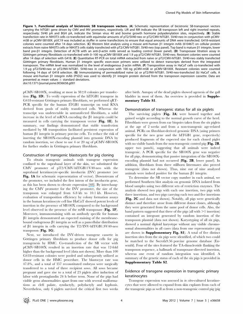
pCMV-SB100X, resulting at most in 3868 colonies per transfec-
tion (Fig. 1D). To verify expression of the hITGB1 transgene in
G418-resistant Gottingen primary fibroblasts, we performed qRT-
PCR specific for the human ITGB1 transcript on total RNA
derived from pools of stably transfected cells. Whereas the
transcript was undetectable in untransfected fibroblasts, a solid
increase in the level of mRNA encoding the b1 integrin could be
measured in cells carrying the transposon vector (Fig. 1E). In
summary, our findings demonstrated that stable transfection
mediated by SB transposition facilitated persistent expression of
human b1 integrin in primary porcine cells. To reduce the risk of
inserting the SB100X-encoding plasmid into genomic DNA by
random insertion, we chose to use 4 or 20 ng of pCMV-SB100X
for further studies in Gottingen primary fibroblasts.
Construction of transgenic blastocysts for pig cloningTo obtain transgenic animals with transgene expression
confined to the suprabasal layer of the skin, we substituted the
CMV promoter of pT2/CMV-hITGB11V40-neo with the
suprabasal keratinocyte-specific involucrin (INV) promoter (see
Fig. 1A for schematic representation of vector). Downstream of
the promoter, we included the first intron of the involucrin gene,
as this has been shown to elevate expression [48]. By interchang-
ing the CMV promoter for the INV promoter, the size of the
transposon was enlarged from 6.8 kb to 10.1 kb. However,
evaluation of transposition efficiency by colony formation assays
in the human keratinocyte cell line HaCaT showed potent levels of
insertion in the presence of SB100X compared to the background
level observed in the presence of the mSB transposase (Fig. 1F).
Moreover, immunostaining with an antibody specific for human
b1 integrin demonstrated an expected staining of the membrane-
bound endogenous b1 integrin as well as a pancellular distribution
of b1 integrin in cells carrying the T2/INV-hITGB1.SV40-neo
transposon (Fig. 1G).
Next, we introduced the INV-driven transgene cassette in
Gottingen primary fibroblasts to produce donor cells for pig
transgenesis by HMC. Co-transfection of the SB vector with
pCMV-SB100X resulted in an insertion rate that was 12-fold
higher than the background level (data not shown). More than 100
G418-resistant colonies were pooled and subsequently utilized as
donor cells in the HMC procedure. The blastocyst rate was
47.8%, and a total of 357 reconstituted embryos were surgically
transferred to a total of three recipient sows. All sows became
pregnant and gave rise to a total of 23 piglets after induction of
labor with prostaglandin 24 h before term. None of the pigs had
visible gross abnormalities apart from one with several malforma-
tions as cleft palate, syndactyly, polydactyly and kyphosis.
Nevertheless, only 6 piglets survived the critical first two weeks
after birth. Autopsy of the dead piglets showed agenesis of the gall
bladder in most of them. An overview is provided in Supple-mentary Table S1.
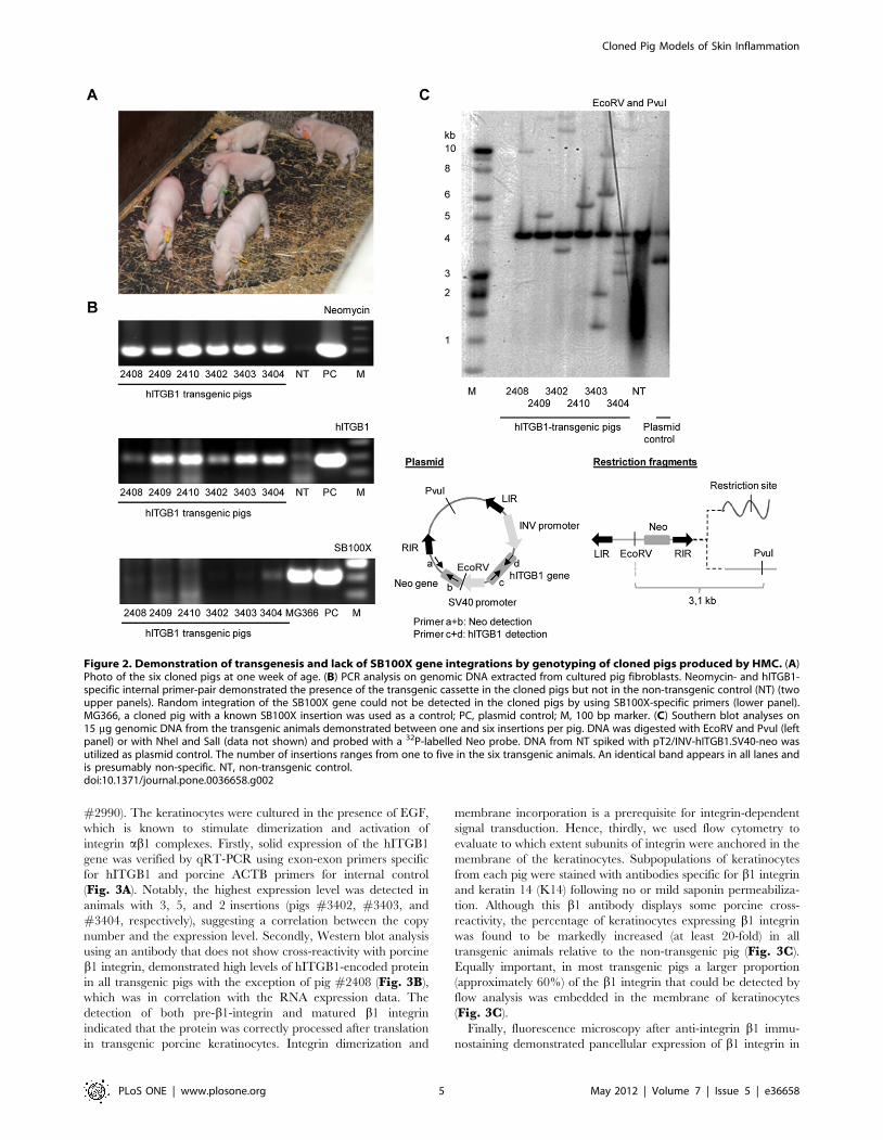
Demonstration of transgenic status for all six pigletsThe surviving piglets (Fig. 2A) were housed together and
gained weight according to the normal growth curve of the herd.
Fibroblasts were grown from ear biopsies taken from the six piglets
at the age of 2 weeks and from a non-transgenic age-related
animal. PCRs on fibroblast-derived genomic DNA (using primers
specific for the neo gene and the hITGB1 gene, respectively)
produced fragments of the expected size for each cloned piglet
with no visible bands from the non-transgenic control pig (Fig. 2B,
upper two panels), suggesting that all animals were indeed
transgenic. A PCR specific for the SB100X gene was negative
for all pigs, demonstrating that passive integration of the SB100X-
encoding plasmid had not occurred (Fig. 2B, lower panel). In
addition, fibroblasts from three stillborn littermates also proved
transgenic (data not shown). Thus, nine out of nine analyzed
animals were indeed positive for the human b1 integrin.
To determine the SB vector copy number in each animal, we
performed Southern blot analysis on genomic DNA isolated from
blood samples using two different sets of restriction enzymes. The
analysis showed two pigs with each one insertion, two pigs with
three insertions, and two pigs carrying each two and five insertions
(Fig. 2C and data not shown). Notably, all pigs were genetically
distinct and therefore arose from different donor clones, although
they were generated from the same pool of donor cells. Also, the
band pattern suggested that three of the pigs (all with .1 insertion)
contained an integrant generated by random insertion of the
transposon plasmid (data not shown). Karyotyping of all six pigs,
showed a normal diploid karyotype without any visible chromo-
somal abnormalities in all cases (data from one representative pig
are shown in Supplementary Fig. S1). A total of five distinct
insertion sites from the six pigs were identified, of which two could
be matched to the Sscrofa9.56 porcine genome database (En-
sembl). Four of the sites featured the TA-dinucleotide flanking the
transposon sequence, a hallmark of transposase-directed insertion,
whereas one event of random integration was identified. A
summary of the genetic status of each of the six pigs is provided in
Supplementary Table S2.
Evidence of transgene expression in transgenic primarykeratinocytes
Transgene expression was assessed in in vitro-cultured keratino-
cytes that were allowed to expand from skin explants from each of
the transgenic pigs as well as from a non-transgenic control pig (pig
Figure 1. Functional analysis of bicistronic SB transposon vectors. (A) Schematic representation of bicistronic SB-transposon vectorscarrying the hITGB1 gene driven by CMV and INV promoters, respectively. LIR and RIR indicate the SB transposon left and right inverted repeats,respectively; SV40 pA and BGH pA, indicate the Simian virus 40 and bovine growth hormone polyadenylation sites, respectively. (B) Stabletransfection rate in NIH3T3 cells co-transfected with equimolar amounts of pT2/SV40-neo or pT2/CMV-hITGB1. SV40-neo in conjunction with pCMV-mSB or pCMV-SB100X. pUC19 was included as stuffer in some transfections to ensure that equal amounts of DNA were transfected. G418-resistantcolonies were counted after 14 days selection. (C) Western blot analysis using a mouse anti-human b1 integrin mAb (610467) on cellular proteinextracts from naıve NIH3T3 cells or NIH3T3 cells stably transfected with pT2/CMV-hITGB1. SV40-neo (top panel). Top band is mature b1 integrin, lowerband pre-b1 integrin. Detection of ACTB with an anti-b-actin mAb served as loading control (lower panel). (D) Transposase titration assay inGottingen primary fibroblasts co-transfected with 0–100 ng pCMV-SB100X and 1.9 mg pT2/CMV-hITGB1. SV40-neo. Resistant colonies were countedafter 14 days of selection with G418. (E) Quantitative RT-PCR on total mRNA extracted from naıve or pT2/CMV-hITGB1. SV40-neo stably transfectedGottingen primary fibroblasts. Human b1 integrin specific exon-exon primers were utilized to detect transcripts derived from the integratedtransposon. The mRNA level was normalized to the level of endogenous b-actin mRNA. (F) Transposition assay in HaCaT cells co-transfected with1.9 mg pT2/SV40-neo or pT2/INV-hITGB1. SV40-neo in conjunction with 100 ng pCMV-mSB or pCMV-SB100X. Resistant colonies were countedfollowing 14 days of G418 selection. (G) Immunostaining of permeabilized naıve (a) or pT2/INV-hITGB1. SV40-neo-transfected (b) HaCaT cells. Amouse anti-human b1 integrin mAb (P5D2) was used to identify b1 integrin protein derived from the transposon expression cassette. Data arepresented as mean values 6 standard deviations.doi:10.1371/journal.pone.0036658.g001
Cloned Pig Models of Skin Inflammation
PLoS ONE | www.plosone.org 4 May 2012 | Volume 7 | Issue 5 | e36658
#2990). The keratinocytes were cultured in the presence of EGF,
which is known to stimulate dimerization and activation of
integrin ab1 complexes. Firstly, solid expression of the hITGB1
gene was verified by qRT-PCR using exon-exon primers specific
for hITGB1 and porcine ACTB primers for internal control
(Fig. 3A). Notably, the highest expression level was detected in
animals with 3, 5, and 2 insertions (pigs #3402, #3403, and
#3404, respectively), suggesting a correlation between the copy
number and the expression level. Secondly, Western blot analysis
using an antibody that does not show cross-reactivity with porcine
b1 integrin, demonstrated high levels of hITGB1-encoded protein
in all transgenic pigs with the exception of pig #2408 (Fig. 3B),
which was in correlation with the RNA expression data. The
detection of both pre-b1-integrin and matured b1 integrin
indicated that the protein was correctly processed after translation
in transgenic porcine keratinocytes. Integrin dimerization and
membrane incorporation is a prerequisite for integrin-dependent
signal transduction. Hence, thirdly, we used flow cytometry to
evaluate to which extent subunits of integrin were anchored in the
membrane of the keratinocytes. Subpopulations of keratinocytes
from each pig were stained with antibodies specific for b1 integrin
and keratin 14 (K14) following no or mild saponin permeabiliza-
tion. Although this b1 antibody displays some porcine cross-
reactivity, the percentage of keratinocytes expressing b1 integrin
was found to be markedly increased (at least 20-fold) in all
transgenic animals relative to the non-transgenic pig (Fig. 3C).
Equally important, in most transgenic pigs a larger proportion
(approximately 60%) of the b1 integrin that could be detected by
flow analysis was embedded in the membrane of keratinocytes
(Fig. 3C).
Finally, fluorescence microscopy after anti-integrin b1 immu-
nostaining demonstrated pancellular expression of b1 integrin in
Figure 2. Demonstration of transgenesis and lack of SB100X gene integrations by genotyping of cloned pigs produced by HMC. (A)Photo of the six cloned pigs at one week of age. (B) PCR analysis on genomic DNA extracted from cultured pig fibroblasts. Neomycin- and hITGB1-specific internal primer-pair demonstrated the presence of the transgenic cassette in the cloned pigs but not in the non-transgenic control (NT) (twoupper panels). Random integration of the SB100X gene could not be detected in the cloned pigs by using SB100X-specific primers (lower panel).MG366, a cloned pig with a known SB100X insertion was used as a control; PC, plasmid control; M, 100 bp marker. (C) Southern blot analyses on15 mg genomic DNA from the transgenic animals demonstrated between one and six insertions per pig. DNA was digested with EcoRV and PvuI (leftpanel) or with NheI and SalI (data not shown) and probed with a 32P-labelled Neo probe. DNA from NT spiked with pT2/INV-hITGB1.SV40-neo wasutilized as plasmid control. The number of insertions ranges from one to five in the six transgenic animals. An identical band appears in all lanes andis presumably non-specific. NT, non-transgenic control.doi:10.1371/journal.pone.0036658.g002
Cloned Pig Models of Skin Inflammation
PLoS ONE | www.plosone.org 5 May 2012 | Volume 7 | Issue 5 | e36658
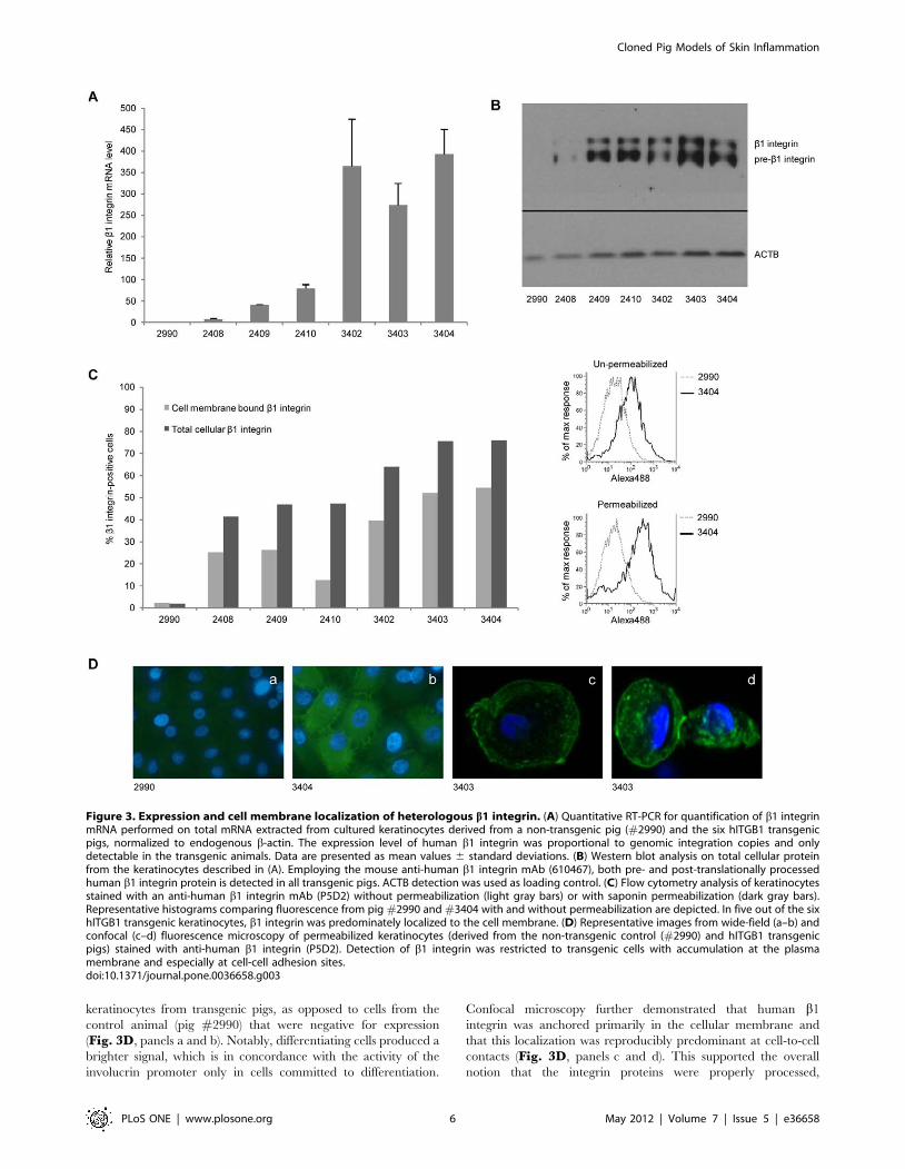
keratinocytes from transgenic pigs, as opposed to cells from the
control animal (pig #2990) that were negative for expression
(Fig. 3D, panels a and b). Notably, differentiating cells produced a
brighter signal, which is in concordance with the activity of the
involucrin promoter only in cells committed to differentiation.
Confocal microscopy further demonstrated that human b1
integrin was anchored primarily in the cellular membrane and
that this localization was reproducibly predominant at cell-to-cell
contacts (Fig. 3D, panels c and d). This supported the overall
notion that the integrin proteins were properly processed,
Figure 3. Expression and cell membrane localization of heterologous b1 integrin. (A) Quantitative RT-PCR for quantification of b1 integrinmRNA performed on total mRNA extracted from cultured keratinocytes derived from a non-transgenic pig (#2990) and the six hITGB1 transgenicpigs, normalized to endogenous b-actin. The expression level of human b1 integrin was proportional to genomic integration copies and onlydetectable in the transgenic animals. Data are presented as mean values 6 standard deviations. (B) Western blot analysis on total cellular proteinfrom the keratinocytes described in (A). Employing the mouse anti-human b1 integrin mAb (610467), both pre- and post-translationally processedhuman b1 integrin protein is detected in all transgenic pigs. ACTB detection was used as loading control. (C) Flow cytometry analysis of keratinocytesstained with an anti-human b1 integrin mAb (P5D2) without permeabilization (light gray bars) or with saponin permeabilization (dark gray bars).Representative histograms comparing fluorescence from pig #2990 and #3404 with and without permeabilization are depicted. In five out of the sixhITGB1 transgenic keratinocytes, b1 integrin was predominately localized to the cell membrane. (D) Representative images from wide-field (a–b) andconfocal (c–d) fluorescence microscopy of permeabilized keratinocytes (derived from the non-transgenic control (#2990) and hITGB1 transgenicpigs) stained with anti-human b1 integrin (P5D2). Detection of b1 integrin was restricted to transgenic cells with accumulation at the plasmamembrane and especially at cell-cell adhesion sites.doi:10.1371/journal.pone.0036658.g003
Cloned Pig Models of Skin Inflammation
PLoS ONE | www.plosone.org 6 May 2012 | Volume 7 | Issue 5 | e36658
dimerized, and distributed in keratinocytes of the transgenic pigs,
rendering them likely to interact with endogenously expressed
membrane receptors.
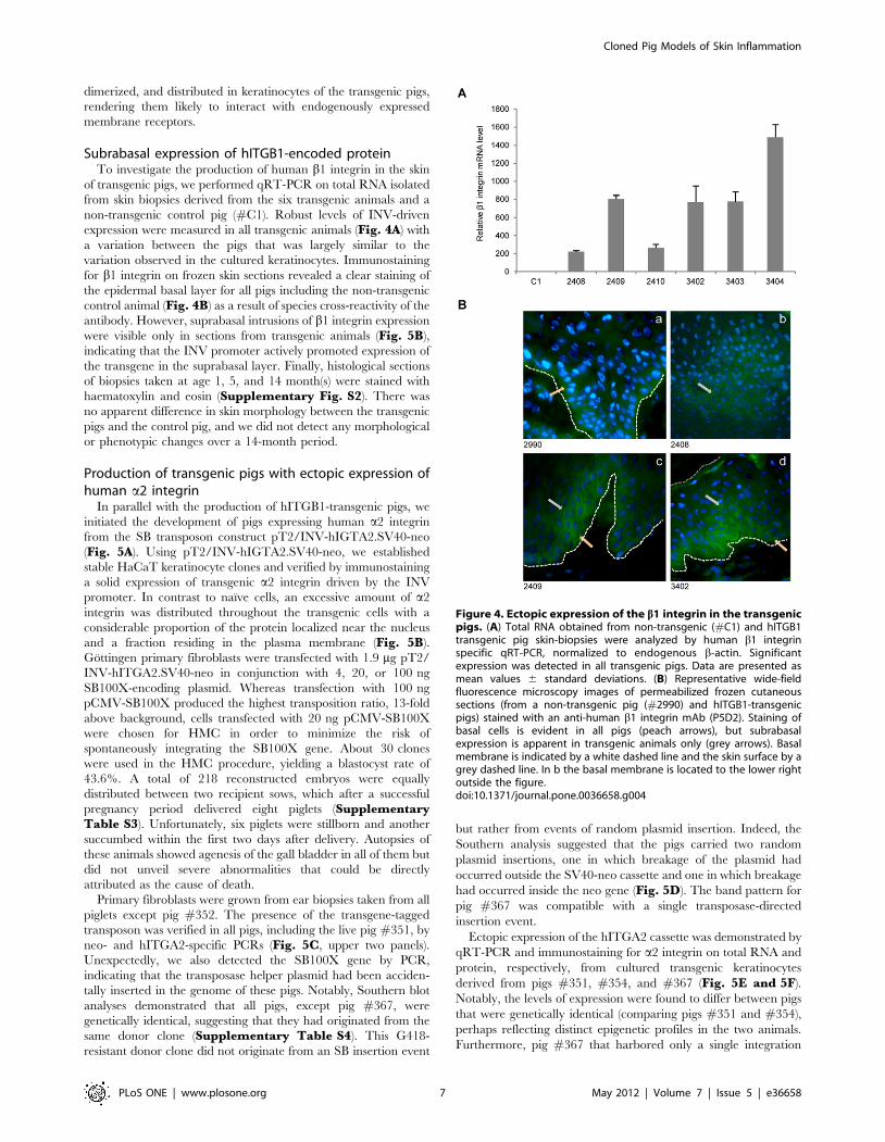
Subrabasal expression of hITGB1-encoded proteinTo investigate the production of human b1 integrin in the skin
of transgenic pigs, we performed qRT-PCR on total RNA isolated
from skin biopsies derived from the six transgenic animals and a
non-transgenic control pig (#C1). Robust levels of INV-driven
expression were measured in all transgenic animals (Fig. 4A) with
a variation between the pigs that was largely similar to the
variation observed in the cultured keratinocytes. Immunostaining
for b1 integrin on frozen skin sections revealed a clear staining of
the epidermal basal layer for all pigs including the non-transgenic
control animal (Fig. 4B) as a result of species cross-reactivity of the
antibody. However, suprabasal intrusions of b1 integrin expression
were visible only in sections from transgenic animals (Fig. 5B),
indicating that the INV promoter actively promoted expression of
the transgene in the suprabasal layer. Finally, histological sections
of biopsies taken at age 1, 5, and 14 month(s) were stained with
haematoxylin and eosin (Supplementary Fig. S2). There was
no apparent difference in skin morphology between the transgenic
pigs and the control pig, and we did not detect any morphological
or phenotypic changes over a 14-month period.
Production of transgenic pigs with ectopic expression ofhuman a2 integrin
In parallel with the production of hITGB1-transgenic pigs, we
initiated the development of pigs expressing human a2 integrin
from the SB transposon construct pT2/INV-hIGTA2.SV40-neo
(Fig. 5A). Using pT2/INV-hIGTA2.SV40-neo, we established
stable HaCaT keratinocyte clones and verified by immunostaining
a solid expression of transgenic a2 integrin driven by the INV
promoter. In contrast to naıve cells, an excessive amount of a2
integrin was distributed throughout the transgenic cells with a
considerable proportion of the protein localized near the nucleus
and a fraction residing in the plasma membrane (Fig. 5B).
Gottingen primary fibroblasts were transfected with 1.9 mg pT2/
INV-hITGA2.SV40-neo in conjunction with 4, 20, or 100 ng
SB100X-encoding plasmid. Whereas transfection with 100 ng
pCMV-SB100X produced the highest transposition ratio, 13-fold
above background, cells transfected with 20 ng pCMV-SB100X
were chosen for HMC in order to minimize the risk of
spontaneously integrating the SB100X gene. About 30 clones
were used in the HMC procedure, yielding a blastocyst rate of
43.6%. A total of 218 reconstructed embryos were equally
distributed between two recipient sows, which after a successful
pregnancy period delivered eight piglets (SupplementaryTable S3). Unfortunately, six piglets were stillborn and another
succumbed within the first two days after delivery. Autopsies of
these animals showed agenesis of the gall bladder in all of them but
did not unveil severe abnormalities that could be directly
attributed as the cause of death.
Primary fibroblasts were grown from ear biopsies taken from all
piglets except pig #352. The presence of the transgene-tagged
transposon was verified in all pigs, including the live pig #351, by
neo- and hITGA2-specific PCRs (Fig. 5C, upper two panels).
Unexpectedly, we also detected the SB100X gene by PCR,
indicating that the transposase helper plasmid had been acciden-
tally inserted in the genome of these pigs. Notably, Southern blot
analyses demonstrated that all pigs, except pig #367, were
genetically identical, suggesting that they had originated from the
same donor clone (Supplementary Table S4). This G418-
resistant donor clone did not originate from an SB insertion event
but rather from events of random plasmid insertion. Indeed, the
Southern analysis suggested that the pigs carried two random
plasmid insertions, one in which breakage of the plasmid had
occurred outside the SV40-neo cassette and one in which breakage
had occurred inside the neo gene (Fig. 5D). The band pattern for
pig #367 was compatible with a single transposase-directed
insertion event.
Ectopic expression of the hITGA2 cassette was demonstrated by
qRT-PCR and immunostaining for a2 integrin on total RNA and
protein, respectively, from cultured transgenic keratinocytes
derived from pigs #351, #354, and #367 (Fig. 5E and 5F).
Notably, the levels of expression were found to differ between pigs
that were genetically identical (comparing pigs #351 and #354),
perhaps reflecting distinct epigenetic profiles in the two animals.
Furthermore, pig #367 that harbored only a single integration
Figure 4. Ectopic expression of the b1 integrin in the transgenicpigs. (A) Total RNA obtained from non-transgenic (#C1) and hITGB1transgenic pig skin-biopsies were analyzed by human b1 integrinspecific qRT-PCR, normalized to endogenous b-actin. Significantexpression was detected in all transgenic pigs. Data are presented asmean values 6 standard deviations. (B) Representative wide-fieldfluorescence microscopy images of permeabilized frozen cutaneoussections (from a non-transgenic pig (#2990) and hITGB1-transgenicpigs) stained with an anti-human b1 integrin mAb (P5D2). Staining ofbasal cells is evident in all pigs (peach arrows), but subrabasalexpression is apparent in transgenic animals only (grey arrows). Basalmembrane is indicated by a white dashed line and the skin surface by agrey dashed line. In b the basal membrane is located to the lower rightoutside the figure.doi:10.1371/journal.pone.0036658.g004
Cloned Pig Models of Skin Inflammation
PLoS ONE | www.plosone.org 7 May 2012 | Volume 7 | Issue 5 | e36658

demonstrated the highest level of expression. To estimate the
extent to which transgenic a2 integrin was localized to the
membrane, keratinocytes from the transgenic animals and a non-
transgenic control (pig #3854) were either permeabilized with
saponin or left untreated prior to staining with an anti-a2 integrin
and an anti-K14 antibody. Flow cytometry analysis for expression
of K14 and a2 integrin demonstrated robust anchoring of a2
integrin in the plasma membrane of transgenic keratinocytes,
although the level of membrane-embedded a2 integrin was
surprisingly heterogeneous (ranging from 16 to 69%) among the
animals (Fig. 5G). To enlarge the herd of hITGA2-positive pigs,
we performed an additional round of cloning using donor
fibroblasts from hITGA2-transgenic animals, resulting in addi-
tional two live, transgenic piglets (#554 and #556) originating
from a mixed pool of fibroblasts derived from pigs #355 and
#366 (Supplementary Table S5).
The involucrin promoter should restrict the expression of
hITGA2 to the subrabasal layers of the skin. To evaluate the
expression level at different locations of the skin and in different
tissues, we sacrificed pig #554, knowing that this pig might not be
representative of the full herd due to its genetic make-up featuring
random integrations. Samples from six distinct areas of the skin
along with samples from eight internal organs including the liver,
heart, and lung were obtained from pig #554. Quantitative RT-
PCR on total RNA revealed comparable levels of human a2
integrin mRNA in all skin samples, whereas a2 integrin mRNA
Figure 5. Generation of hITGA2-transgenic pigs with ectopic expression of human a2 integrin. (A) Schematic figure of bicistronic SBtransposon vectors with expression of human a2 integrin driven by the CMV and INV promoters and the neo selection marker gene driven by theSV40 promoter. LIR and RIR indicate SB transposon left and right inverted repeats, respectively; SV40 pA and BGH pA indicate Simian virus 40 andbovine growth hormone polyadenylation sites, respectively. (B) Immunostaining of permeabilized naıve (a) or pT2/INV-hITGA2.SV40-neo-transfected(b) HaCaT cells with a mouse anti-human a2 integrin mAb (P1E6). Background staining of endogenous a2 integrin is evident in naıve cells, whereastransfected cells show intense pancellular staining. (C) PCR analysis on genomic DNA extracted from cultured hITGA2-transgenic pig fibroblasts. Thepresence of Neo and hITGA2 was confirmed for all transgenic pigs with specific primer-pairs (upper panels). Passive integration of SB100X could beverified in all the cloned pigs by the use of primers specific for the SB100X sequence (lower panel). NT, non-transgenic control; MG366, cloned pigwith known insertion of the SB100X gene; PC, plasmid control; M, 100 bp marker. (D) Southern blot analyses performed with 11 mg genomic DNAextracted from hITGA2-transgenic pig fibroblasts showed that all pigs, except 367, were genetically identical. DNA was digested with EcoRV and PvuI(left panel) or with EcoRV and SalI (right panel) and probed with a 32P-labelled Neo probe. DNA from NT spiked with pT2/INV-hITGA2.SV40-neo wasutilized in copy number controls with 1, 5 and 10 copies, respectively (refer to figure 2C for lengths of restriction fragments). NT, non-transgeniccontrol; M, 1 kb marker. (E) qRT-PCR analysis of a2 integrin mRNA expression levels in hITGA2-transgenic pig keratinocytes, normalized toendogenous b-actin. Expression of human a2 integrin mRNA was evident in all transgenic pig keratinocytes. Representative data are presented asmean values 6 standard deviations. (F) Representative wide-field fluorescence microscopy images of permeabilized and unstained orimmunostained (with anti-human a2 integrin mAb (P1E6)) frozen cutaneous sections from the non-transgenic pig #2990 (a–b) and analyzedhITGA2-transgenic pigs (c–e). A clear staining for human a2 integrin protein was present only in transgenic keratinocytes. (G) Flow cytometry analysison hITGA2-transgenic keratinocytes stained with an anti-human a2 integrin mAb (P1E6) without permeabilization (light gray bars) or with saponinpermeabilization (dark gray bars). Also shown are representative histograms comparing fluorescence intensities from pig 3854 and 352 with andwithout permeabilization.doi:10.1371/journal.pone.0036658.g005
Cloned Pig Models of Skin Inflammation
PLoS ONE | www.plosone.org 8 May 2012 | Volume 7 | Issue 5 | e36658

was not detectable in the control pig. Although considerable
expression of a2 integrin mRNA was evident in all analyzed tissues
of pig #554, expression of the transgene was significantly higher in
all analyzed areas of the skin (p,0.0001) (SupplementaryFig. S3). These findings confirmed the tissue-specificity of the
involucrin promoter, but also indicated that unsolicited expression
from randomly integrated fragments was facilitated by endogenous
promoter elements in pig #554.
Activation of molecular markers of inflammation in theskin of pigs transgenic for either b1 or a2 integrin
A key feature of erroneous or engineered subrabasal expression
of b1 integrin is the elevated expression and secretion of IL-1awhich is not dependent on integrin ligation [49,50]. The
proinflammatory cytokine IL-1a plays pivotal auto- and paracrine
roles in the inflammatory response inflicted in lesional psoriatic
skin. Furthermore, the Erk/MAPK pathway is simultaneously
activated by the autocrine effect of secreted IL-1a and by the
integrin-dependent potentiating effect on EGFR transactivation
[50].
For analysis of IL-1a expression in the pigs, qRT-PCR was first
carried out on total RNA from cultured keratinocytes. In five of
the six hITGB1-transgenic pigs a significantly enhanced IL-1amRNA level was detected ranging from a 2- to 6-fold increase
relative to the control pig #2990 (Fig. 6A). Further enhancement
of IL-1a expression was detected in hITGA2-transgenic animals.
For the three animals that were analyzed, the IL-1a mRNA levels
were increased from 6- to 27-fold relative to three distinct control
pigs (Fig. 6B). Notably, this level of induction was directly
correlated with the a2 integrin expression level (compare with
Fig. 5E), indicating that the induced cytokine response was
triggered by the transgenic expression of a2 integrin. Assessment
of cellular IL-1a release was achieved by an ELISA-based
approach using an antibody specific for porcine IL-1a. Medium
conditioned for 14 hours showed a 2- to 5-fold increment in
secreted IL-1a by cells expressing b1 integrin compared to the
average value obtained with medium conditioned by keratinocytes
from the non-transgenic controls, pigs #301703 and #301706
(Fig. 6C). Together, these findings documented an increased
expression of IL-1a in hITGB1- and hITGA2-transgenic porcine
keratinocytes.
To assess if the general inflammatory profile was altered in the
transgenic animals, a panel of ten crucial markers of inflammation
was analyzed by qRT-PCR on total RNA derived from skin
biopsies taken from the back of the hITGB1-transgenic pigs
#2410 and #3402 and compared to a skin sample from the
wildtype control #301702 (Fig. 7). The chemokines chemokine
(C-C motif) ligand 5 (CCL5), CCL20, chemokine (C-X-C motif)
ligand 10 (CXCL10), and IL-8 are released by several cell types in
the early phase of inflammation and recruit lymphocytes and
granulocytes to the site of inflammation. Compared to the
wildtype control pig, the level of mRNA encoding these factors
was elevated from 2- to 19-fold in the two transgenic animals
(Fig. 7A–D), whereas the level of the skin-specific chemokine
CCL27 was only moderately upregulated in one of the pigs
(Fig. 7E). The level of expression of the cytokines IL-1b, tumor
necrosis factor alpha (TNF-a) and granulocyte-macrophage
colony-stimulating factor (GM-CSF) was increased 2- to 5-fold,
as judged by qRT-PCR (Fig. 7F–H), whereas no changes were
seen in the case of proliferating cell nuclear antigen (PCNA)
(Fig. 7I). The expression of the antimicrobial and chemotatic
protein psoriasin was markedly decreased in pig #2410 but
moderately increased in pig #3402 (Fig. 7J). In summary, these
results indicate that a broad perturbation of cutaneous cytokine
and chemokine expression was inflicted by subrabasal expression
of b1 integrin.
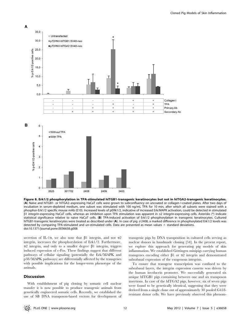
Phosphorylation of Erk1/2 (pErk1/2) is a prerequisite for the
nuclear import and activity of Erk1/2 during induction of the
Erk/MAPK pathway. Elevated levels of nuclear pErk1/2 are
evident in psoriatic lesions and in mouse keratinocytes engineered
to express the hITGB1 gene [49]. To study the phosphorylation of
Erk1/2, we first investigated the effect of stably expressing the
hITGB1 and hITGA2 genes in HaCaT keratinocytes. Naıve and
transgenic cells were cultured in serum-depleted medium in dishes
that were either coated with collagen I or left uncoated. One
subset of cells was subsequently treated with the protein kinase C
(PKC) activator 12-O-tetradecanoyl phorbol-13-acetate (TPA),
after which all cells were trypsinized, immunostained with a
pErk1/2-specific antibody, and analyzed by flow cytometry.
Exposure to TPA increased the number of naıve HaCaT cells
that were positive for pErk1/2 less than 2-fold, whereas the
number of pErk1/2-positive cells carrying the transposon-embed-
ded hITGB1 gene was increased 4-fold by TPA induction, leading
to pErk1/2 labeling of more than 25% of the cells (Fig. 8A). In
contrast to cells stably transfected with the hITGB1 gene, TPA did
not lead to increased phosphorylation of Erk1/2 in hITGA2-
transgenic HaCaT cells. In fact, the pathway seemed to be down-
regulated compared to naıve cells, resulting in approximately half
the proportion of cells that were positive for pErk1/2 relative to
non-transfected cells (Fig. 8A). Together, these findings suggested
that keratinocytes with engineered expression of b1 integrin were
increasingly sensitive to external stimuli and that the response to
extracellular stimuli was differentially affected by engineered
expression of b1 and a2 integrin. We therefore analyzed the level
of phosphorylated Erk1/2 in keratinocytes derived from b1
integrin-transgenic pigs. We succeeded to prepare primary
keratinocytes from three pigs for this analysis. There was no
remarkable change in the level of pErk1/2 in untreated or TPA
treated cells from each control pig, whereas for pig #2408 an 8-
fold increase in the number of positive cells was evident after TPA
stimulation.
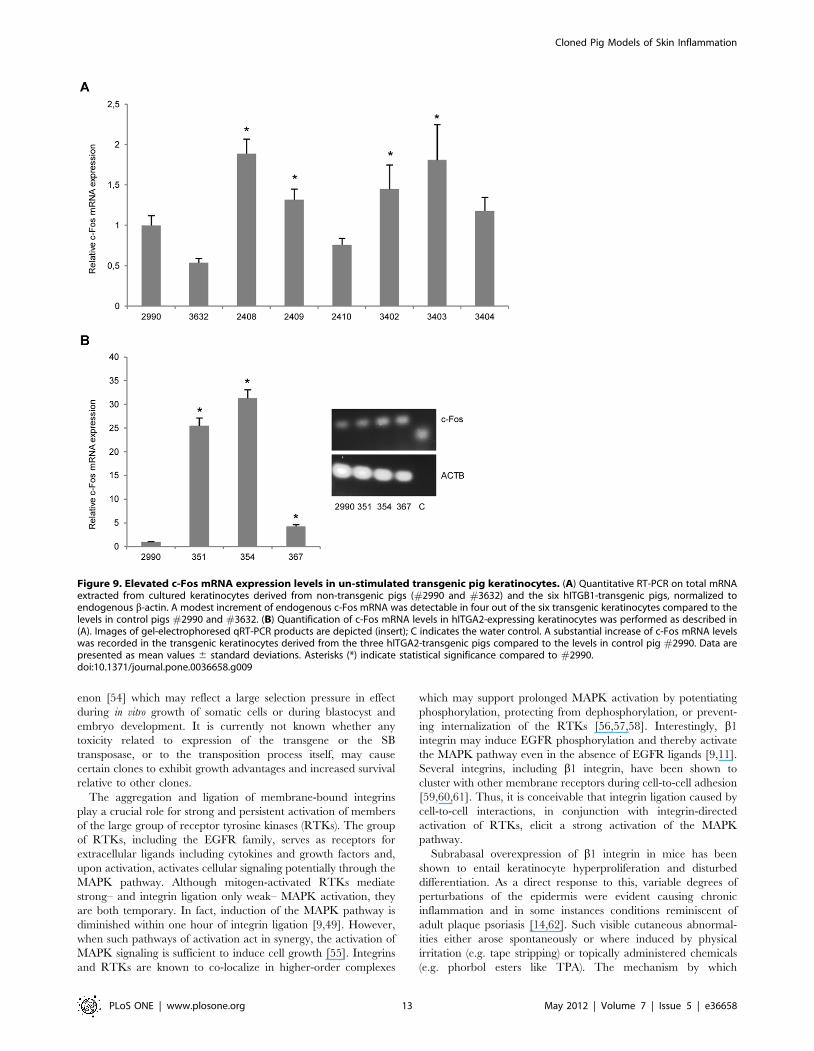
Finally, we inspected the expression of the immediate early gene
c-Fos in transgenic keratinocytes. C-Fos is a downstream effector
of p38/MAPK and Erk/MAPK activation and dimerizes with c-
Jun to form the transcription complex AP-1. AP-1 up-regulates a
variety of genes involved in important biological processes
including cell proliferation, differentiation, survival, and repair
mechanisms as well as embryonic development [51,52]. In the
stratum basale and stratum spinosum, c-Fos is normally not (or
only slightly) expressed [53]. We speculated that transgenic
integrin expression could have an impact on the expression of c-
Fos with possible implications for skin homeostasis. Levels of
keratinocyte c-Fos mRNA was increased in all hITGB1-transgenic
pigs, except one (pig #2410). However, the enhancement was
small (at most 2.5-fold above the average level measured for the
two control pigs) and varied considerably among the animals
(Fig. 9A). In contrast, in case of the keratinocytes derived from the
three a2 integrin-transgenic pigs that were analyzed, we observed
a considerable induction of c-Fos, resulting in at most a 31-fold
enhancement in the level of c-Fos mRNA (Fig. 9B). These
findings suggest that expression of transgenic a2 integrin, and to a
minor degree b1 integrin, triggers cellular signaling pathways that
involve the induction of c-Fos.
In summary, we conclude that molecular markers of induced
skin inflammation could be identified in keratinocytes from pigs
that were transgenic for either human b1 or a2 integrin expressed
from the involucrin promoter exclusively in the suprabasal skin
layer. Although both integrins cause enhanced expression and
Cloned Pig Models of Skin Inflammation
PLoS ONE | www.plosone.org 9 May 2012 | Volume 7 | Issue 5 | e36658
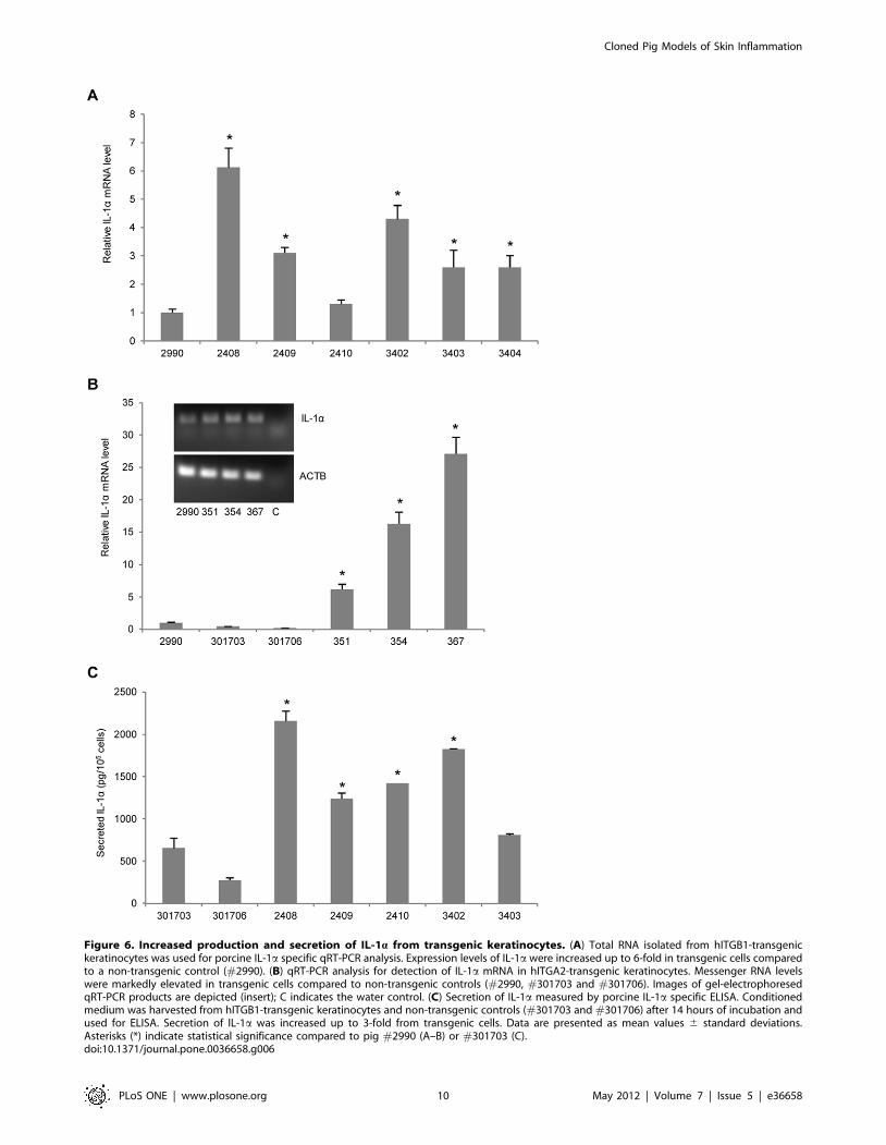
Figure 6. Increased production and secretion of IL-1a from transgenic keratinocytes. (A) Total RNA isolated from hITGB1-transgenickeratinocytes was used for porcine IL-1a specific qRT-PCR analysis. Expression levels of IL-1a were increased up to 6-fold in transgenic cells comparedto a non-transgenic control (#2990). (B) qRT-PCR analysis for detection of IL-1a mRNA in hITGA2-transgenic keratinocytes. Messenger RNA levelswere markedly elevated in transgenic cells compared to non-transgenic controls (#2990, #301703 and #301706). Images of gel-electrophoresedqRT-PCR products are depicted (insert); C indicates the water control. (C) Secretion of IL-1a measured by porcine IL-1a specific ELISA. Conditionedmedium was harvested from hITGB1-transgenic keratinocytes and non-transgenic controls (#301703 and #301706) after 14 hours of incubation andused for ELISA. Secretion of IL-1a was increased up to 3-fold from transgenic cells. Data are presented as mean values 6 standard deviations.Asterisks (*) indicate statistical significance compared to pig #2990 (A–B) or #301703 (C).doi:10.1371/journal.pone.0036658.g006
Cloned Pig Models of Skin Inflammation
PLoS ONE | www.plosone.org 10 May 2012 | Volume 7 | Issue 5 | e36658

Figure 7. Altered cytokine and chemokine profile in hITGB1-transgenic animals. Quantitative RT-PCR directed against ten molecularmarkers of skin-inflammation performed on total RNA extracted from punch biopsies taken from the non-transgenic pig #301702 and the hITGB1-transgenic pigs #2410 and #3402 at age 14 months, normalized to endogenous GAPDH. Data are technical triplicates and presented as mean values6 standard deviations. CCL5, CCL20, and CCL27 (Chemokine (C-C motif) ligand 5, 20, and 27, respectively); CXCL10 (chemokine (C-X-C motif) ligand10); IL-8 and IL-1b (interleukins 8 and 1b); TNF-a (tumor necrosis factor alpha); GM-CSF (granulocyte-macrophage colony-stimulating factor); PCNA(proliferating cell nuclear antigen).doi:10.1371/journal.pone.0036658.g007
Cloned Pig Models of Skin Inflammation
PLoS ONE | www.plosone.org 11 May 2012 | Volume 7 | Issue 5 | e36658
secretion of IL-1a, we also note that b1 integrin, and not a2
integrin, increases the phosphorylation of Erk1/2. Furthermore,
a2 integrin, and only to a smaller degree b1 integrin, triggers
induced expression of c-Fos. These findings suggest that different
pathways of cellular signaling (potentially the Erk/MAPK and
p38/MAPK pathways) are differentially affected by the transgenes
with possible implications for the longer-term phenotype of the
animals.
Discussion
With establishment of pig cloning by somatic cell nuclear
transfer it is now possible to produce transgenic animals from
genetically engineered somatic cells. Recently, we established the
use of SB DNA transposon-based vectors for development of
transgenic pigs by DNA transposition in cultured cells serving as
nuclear donors in handmade cloning [54]. In the present report,
we explore this approach for generating pig models of skin
inflammation. We established Gottingen minipigs carrying human
transgenes encoding either b1 or a2 integrin and demonstrated
subrabasal expression of the exogenous integrins.
To ensure that transgene transcription was confined to the
subrabasal layers, the integrin expression cassette was driven by
the human involucrin promoter. We successfully generated six
unique hITGB1 pigs containing between one and six transposon
insertions. In case of the hITGA2 pigs, however, six of seven pigs
were found to be genetically identical, suggesting that they were
derived from a single clone out of approximately 30 pooled G418-
resistant donor cells. We have previously observed this phenom-
Figure 8. Erk1/2 phosphorylation in TPA-stimulated hITGB1-transgenic keratinocytes but not in hITGA2-transgenic keratinocytes.(A) Naıve and hITGB1- or hITGA2–expressing HaCaT cells were grown to subconfluency on uncoated or collagen I-coated plates. After two days ofincubation in serum-depleted medium, one subset was stimulated with 100 ng/mL TPA for 10 min, after which all subsets were stained with aphosphor-Erk1/2 specific mouse mAb (E10). Increased levels of pERk1/2, indicative of increased Erk/MAPK activation, could be detected in stimulatedb1 integrin-expressing HaCaT cells, whereas an inhibition upon TPA stimulation was apparent in a2 integrin-expressing cells. Asterisks (*) indicatestatistical significance relative to naıve HaCaT cells. (B) TPA-induced activation of Erk1/2 phosphorylation in transgenic keratinocytes. CulturedhITGB1-transgenic keratinocytes were treated as described under (A). In case of pig #2408, a marked difference in phosphorylated Erk1/2 levels wasdetected by comparing TPA-stimulated and un-stimulated cells. Data are presented as mean values 6 standard deviations.doi:10.1371/journal.pone.0036658.g008
Cloned Pig Models of Skin Inflammation
PLoS ONE | www.plosone.org 12 May 2012 | Volume 7 | Issue 5 | e36658
enon [54] which may reflect a large selection pressure in effect
during in vitro growth of somatic cells or during blastocyst and
embryo development. It is currently not known whether any
toxicity related to expression of the transgene or the SB
transposase, or to the transposition process itself, may cause
certain clones to exhibit growth advantages and increased survival
relative to other clones.
The aggregation and ligation of membrane-bound integrins
play a crucial role for strong and persistent activation of members
of the large group of receptor tyrosine kinases (RTKs). The group
of RTKs, including the EGFR family, serves as receptors for
extracellular ligands including cytokines and growth factors and,
upon activation, activates cellular signaling potentially through the
MAPK pathway. Although mitogen-activated RTKs mediate
strong– and integrin ligation only weak– MAPK activation, they
are both temporary. In fact, induction of the MAPK pathway is
diminished within one hour of integrin ligation [9,49]. However,
when such pathways of activation act in synergy, the activation of
MAPK signaling is sufficient to induce cell growth [55]. Integrins
and RTKs are known to co-localize in higher-order complexes
which may support prolonged MAPK activation by potentiating
phosphorylation, protecting from dephosphorylation, or prevent-
ing internalization of the RTKs [56,57,58]. Interestingly, b1
integrin may induce EGFR phosphorylation and thereby activate
the MAPK pathway even in the absence of EGFR ligands [9,11].
Several integrins, including b1 integrin, have been shown to
cluster with other membrane receptors during cell-to-cell adhesion
[59,60,61]. Thus, it is conceivable that integrin ligation caused by
cell-to-cell interactions, in conjunction with integrin-directed
activation of RTKs, elicit a strong activation of the MAPK
pathway.
Subrabasal overexpression of b1 integrin in mice has been
shown to entail keratinocyte hyperproliferation and disturbed
differentiation. As a direct response to this, variable degrees of
perturbations of the epidermis were evident causing chronic
inflammation and in some instances conditions reminiscent of
adult plaque psoriasis [14,62]. Such visible cutaneous abnormal-
ities either arose spontaneously or where induced by physical
irritation (e.g. tape stripping) or topically administered chemicals
(e.g. phorbol esters like TPA). The mechanism by which
Figure 9. Elevated c-Fos mRNA expression levels in un-stimulated transgenic pig keratinocytes. (A) Quantitative RT-PCR on total mRNAextracted from cultured keratinocytes derived from non-transgenic pigs (#2990 and #3632) and the six hITGB1-transgenic pigs, normalized toendogenous b-actin. A modest increment of endogenous c-Fos mRNA was detectable in four out of the six transgenic keratinocytes compared to thelevels in control pigs #2990 and #3632. (B) Quantification of c-Fos mRNA levels in hITGA2-expressing keratinocytes was performed as described in(A). Images of gel-electrophoresed qRT-PCR products are depicted (insert); C indicates the water control. A substantial increase of c-Fos mRNA levelswas recorded in the transgenic keratinocytes derived from the three hITGA2-transgenic pigs compared to the levels in control pig #2990. Data arepresented as mean values 6 standard deviations. Asterisks (*) indicate statistical significance compared to #2990.doi:10.1371/journal.pone.0036658.g009
Cloned Pig Models of Skin Inflammation
PLoS ONE | www.plosone.org 13 May 2012 | Volume 7 | Issue 5 | e36658
subrabasal integrins induce hyperproliferation and altered differ-
entiation is still obscure. However, subrabasal MAPK activation
coincides with b1 integrin expression. Moreover, b1 integrin
ligation on the surface of cultured keratinocytes leads to MAPK
induction, suggesting that the capability of b1 integrins to induce
signal transduction plays a role during the induction of skin
inflammation in the mouse [49].
Confocal microscopy of keratinocytes derived from hITGB1
transgenic pigs demonstrated membrane localization of the
transgenic protein and the accumulation of b1 integrin at cell-
to-cell interaction points. To investigate the potential activation of
the MAPK pathway in transgenic animals, we first studied the
level of Erk1/2 phosphorylation in TPA-stimulated HaCaT
keratinocytes stably expressing b1 or a2 integrin. Notably, only
b1 integrin triggered an elevated level of pErk1/2 in HaCaT cells,
and the effect of b1 integrin was therefore explored in
keratinocytes from b1 integrin-transgenic pigs. At least for one
of three analyzed pigs (#2408) we could monitor a marked
increase in the percentage of cells positive for phosphorylated
Erk1/2 (also referred to as mitogen-activated protein kinase 3 and
1), suggesting that the MAPK pathway was activated upon
stimulation with TPA. Similarly, the Erk/MAPK pathway was
activated upon TPA stimulation of keratinocytes derived from
a2b1 integrin double-transgenic mice [63], suggesting that
constitutive expression of integrins in subrabasal keratinocytes
rendered the keratinocytes hypersensitive to external stimuli. The
fact that the level of pErk1/2 was unaffected by expression of a2
integrin, as based on our findings in TPA-stimulated HaCaT cells,
suggests that b1, but not a2, integrin plays a crucial role for the
activation of the Erk/MAPK pathway in this context. Previous
work has demonstrated that the cytoplasmic tail of a2 integrin
mediates signals via p38/MAPK [64,65]. This effect is probably
stimulated through the small G-proteins Cdc42 and Rac1 [66], a
route of signaling which is not stimulated by TPA. Interestingly,
several reports have shown that p38 has an inhibitory effect on
Erk1/2 which can be mediated either through p38-directed
stimulation of protein phosphatase 2 (PP2A) [67,68] or via a direct
interaction between p38 isoforms and Erk1/2 [69,70].
To further define the inflammatory phenotype of keratinocytes
from transgenic pigs, we analyzed transgenic keratinocytes for
potential induction of IL-1a as a marker for an altered
inflammatory profile. The expression of IL-1a was found to be
elevated in all transgenic pigs, indicating that both b1 and a2
integrin gave rise to transcriptional induction of IL-1a. However,
the enhancement was far more pronounced for a2 integrin pigs
relative to b1-transgenic animals. This suggests that overexpres-
sion of a2 integrin induces cellular signaling conveyed by routes
different from b1 integrin overexpression. IL-1a is a very potent
proinflammatory cytokine which is constitutively produced by
epithelial cells, especially keratinocytes, leading to activation of the
NF-kB pathway and recruitment of activated mononuclear cells–
among a range of effects. Following skin wounding IL-1a is
discharged from intracellular storage vesicles [71]. Secreted IL-1acontributes to keratinocyte hyperproliferation by EGFR-depen-
dent activation of the Erk/MAPK pathway and has been shown to
induce inflammatory conditions in mouse and human epidermis
[72,73]. Under normal conditions the activity of IL-1a is tightly
regulated [74] by mechanisms that do not depend on ECM-
induced integrin ligation [49]. However, there are numerous
examples of signal co-operativity between integrins and IL
receptors, suggesting that integrins may assist in intensifying
cytokine signaling [75,76]. Indeed, integrin-mediated transcrip-
tional activation of IL-1a has previously shown to involve Erk1/2
or NF-kB activation [50] although the exact mechanism by which
the release of IL-1a is stimulated is still uncertain. The
demonstration of increased IL-1a production in b1 and a2
integrin-transgenic pigs supports the notion that increased integrin
signaling correlates with enhanced levels of porcine cytokine
production and an altered inflammation profile.
To clarify if the ectopic b1-integrin expression induced a
general alteration of the cytokine and chemokine profile in the
transgenic animals, we analyzed the mRNA levels of a series of
inflammation markers in biopsies of untreated skin from two
hITGB1-transgenic animals. The inflammatory response instigat-
ed by subrabasal integrin expression has previously shown to
comprise TH1- and TH17-related cytokines like IL-1b, IFN-c,
GM-CSF and TNF-a [62]. Importantly, the TH1 cytokine
signature has been associated with the pathogenesis of psoriasis
[77]. Notably, for all the cytokines investigated, except TNF-a, the
level of mRNA was upregulated in both transgenic animals
compared to an age-matched control. The level of TNF-a mRNA,
in contrast, was only enhanced in one of the two pigs. A general
upregulation of relevant chemokines was also observed. In
addition, an increased level of psoriasin was detected in one of
the two transgenic pigs. Psoriasin has been identified as a marker
for hyperproliferative and inflammatory skin disorders such as
psoriasis and atopic dermatitis [78,79]. Overexpression of
CXCL10 has also been linked to psoriasis [80]. It is predominately
activated by IFN-c, and recruits activated T-lymphocytes and NK
cells. Notably, there was a significant difference in the expression
level of psoriasin and CXCL10 in the two transgenic animals.
Interestingly, it has previously been shown that psoriasin
expression is transcriptionally suppressed by IFN-c [81]. Taken
together, the expression profile of inflammatory markers suggests
that a wide-spread dysregulation of the immune system was
prompted by the ectopic expression of b1-integrin. Peculiarly, the
expression pattern seemed to vary between the two hITGB1-
transgenic pigs, suggesting that the exact expression profile of the
integrin itself played a role on downstream effects.
The expression of c-Fos is induced through both the p38/
MAPK and the Erk/MAPK signaling pathways [82], and this
marker is therefore, like IL-1a, a more general indicator of altered
signaling caused by phosphorylation of transcription factors that
bind c-Fos enhancer elements [83]. We therefore measured the
levels of c-Fos mRNA in transgenic keratinocytes. Whereas the
increase was only moderate in hITGB1-transgenic keratinocytes (a
significant increase in four of six analyzed animals), we observed a
solid enhancement of c-Fos mRNA in all three hITGA2-
transgenic animals that were analyzed. Although the total number
of animals was small, these data support the notion that different
signaling patterns were activated in the two groups of transgenic
animals.
To date we have not registered any visible epidermal
abnormality, or psoriasis-like phenotype, in any of the hITGB1-
or hITGA2-transgenic pigs, neither by direct examination of the
skin nor by analysis of skin morphology by staining of skin section.
However, it is widely accepted that environmental influences, in
conjunction with genetic components, play an important role in
the pathogenesis of inflammatory diseases such as psoriasis. In a2/
b1 integrin double-transgenic mice, skin irritation led to a chronic
condition resembling psoriasis with persistent hyperplasia, cuta-
neous influx of CD4+ and CD8+ T-lymphocytes and secretion of
pro-inflammatory cytokines like TNF-a, IFN-c, IL-1b and IL-6
[62]. Based on our molecular analysis of central indicators of
inflammation findings, we believe that the transgenic pigs may be
pre-disposed for development of an exacerbated and potentially
chronic inflammation by provocation. Ongoing studies have been
designed to address the potential development of chronic plaque
Cloned Pig Models of Skin Inflammation
PLoS ONE | www.plosone.org 14 May 2012 | Volume 7 | Issue 5 | e36658
lesions in the transgenic pigs as a response to mechanically or
chemically induced stress.
In conclusion, we have created Gottingen minipigs transgenic
for the human integrins b1 and a2 and have demonstrated
efficacious transgene expression in skin as well as induced
inflammatory signaling in transgenic keratinocytes. We believe
that such porcine models of skin inflammation will contribute to a
better understanding of the pathogenesis of cutaneous diseases and
will be of great potential in studies aiming at the development and
refinement of topical therapies for cutaneous inflammation
including psoriasis.
Materials and Methods
Ethics StatementAll animal procedures were approved by the Danish Animal
Experiments Inspectorate (license no. 2006/561-1230).
Plasmids and vector constructionSB transposon-based vector constructs were generated based on
the plasmid vector pT2/SV40-neo which has been previously
described [47]. An expression cassette containing the eGFP ORF
driven by the cytomegalovirus (CMV) promoter was PCR-
amplified in two steps from pEGFP.N1 (Clontech), allowing the
insertion of a unique linker between the eGFP gene and the
promoter. Subsequently, the CMV-eGFP fragment was combined
with the bGH polyA signal derived from pTOPO (Invitrogen,
Paisley, UK) in an overlap extension PCR creating a fragment
with an EcoRI-XbaI linker in each terminus. The PCR product
was digested and inserted into pT2/SV40-neo digested with either
EcoRI or XbaI (which cuts upstream and downstream, respec-
tively, of the SV40-neo cassette). This gave rise to four constructs
(pT2/CMV-eGFP-bGHpA.SV40-neo, pT2/bGHpA-eGFP-
CMV.SV40-neo, pT2/SV40-neo.CMV-eGFP-bGHpA and
pT2/SV40-neo.bGHpA-eGFP-CMV) in which individual com-
ponents could be easily exchanged. The eGFP ORF of pT2/
CMV-eGFP-bGHpA.SV40-neo was released with NotI and
substituted with either the hITGB1 cDNA sequence (encoding
b1 integrin) PCR-amplified from pCMV6-XL5 (SC111935,
Origene, Rockville, MD, USA) or the hITGA2 cDNA sequence
(encoding a2 integrin) amplified from pCMV6-XL4 (SC118747,
Origene, Rockville, MD, USA), generating the constructs desig-
nated pT2/CMV-hITGB1-bGHpA.SV40-neo and pT2/CMV-
hITGA2-bGHpA.SV40-neo, respectively. The CMV promoter
was subsequently released by AscI digestion and replaced with the
PCR-amplified involucrin (INV) promoter from pH3700-pL2
(kindly provided by Rikke Christensen, Dept. of Biomedicine,
University of Aarhus, Denmark), leading to pT2/INV-hITGB1-
bGHpA.SV40-neo and pT2/INV-hITGA2-bGHpA.SV40-neo.
pCMV-SB100X has been previously described [84].
CellsThe murine fibroblast cell line NIH3T3 and human keratino-
cyte cell line HaCaT were cultured in Dulbeccos modified Eagles
medium, DMEM (Lonza, Verviers, Belgium) with 10% fetal calf
serum, FCS (Lonza, Verviers, Belgium). Established Gottingen
primary fibroblasts were maintained in DMEM with 15% FCS.
All media were supplemented with 100 U/mL penicillin, 0.1 mg/
mL streptomycin and 265 mg/l L-glutamine and all cell were
maintained at 37uC, 5% CO2. Outgrowth and expansion of
primary fibroblasts and keratinocytes was achieved from explants
derived from pig ear-biopsies. The epidermis was sectionally
isolated and the explants were placed in 25-cm2 culture flasks
(TPP, Trasadingen, Switzerland) and incubated bottom-up O/N
at 37uC, 5% CO2. Fibroblast outgrowth was promoted in
AmnioMAX-C100 (Gibco, Invitrogen, Paisley, UK). Outgrowth
and expansion of keratinocytes was achieved in 15% FCS DMEM
containing the additives as described above plus 10 ng/mL EGF
(Gibco, Invitrogen, Paisley, UK), 50 mM gentamycin and 0,4 mg/
mL hydrocortisone (Sigma-Aldrich, St. Louis, MO, USA) at 37uC,
5% CO2 for 7–10 days, allowing keratinocytes to migrate from the
explants. Hereafter, the medium was substituted with epidermal
growth factor (EGF) and bovine pituitary extract (BPE) containing
serum-free keratinocyte medium, K-SFM (Gibco, Invitrogen,
Paisley, UK).
Handmade cloning (HMC) and transfersHMC was performed as described before [29,31]. Briefly,
cumulus cells were removed from matured cumulus-oocyte
complexes (COCs) by treatment with hyaluronidase. After partial
digestion of zona pellucida, oriented bisection of oocytes was
performed to remove nuclei. Each cytoplast without polar body
attached with a single trangenic fibroblast was fused in fusion
medium (0.3 M mannitol, 0.1 mM MgSO4 and 0.01% [w/v]
PVA) in a fusion chamber (BTX microslide 0.5 mm fusion
chamber, model 450; BTX, San Diego, CA, USA) with a single
direct current (DC) impulse of 2.0 kV/cm for 9 ms. One hour
later, each cytoplast-somatic cell pair was fused with another
cytoplast in activation medium (fusion medium with 0.1 mM
CaCl2) by a single DC pulse of 0.86 kV/cm for 80 ms. After
incubation in porcine zygote medium 3 (PZM-3) supplemented
with 5 mg/ml cytochalasin B, 10 mg/ml cyclohexinmide for 4 h,
the reconstructed embryos were cultured in PZM-3 medium for
another 6 days to develop into transgenic blastocysts. Blastocysts
at day 5 and 6 were surgically transferred into recipient sows [31].
All animal procedures were approved by the Danish Animal
Experiments Inspectorate (license no. 2006/561–1230).
ImmunostainingFor immunocytochemistry, HaCaT cells stably transfected with
pT2/INV-hITGB1.SV40-neo or pT2/INV-hITGA2.SV40-neo
were seeded in slideflasks (Nunc A/S, Roskilde, Denmark) with
a density of 26105 cells/flask. After 24–48 h the cells were fixed in
4% formalin for 5–10 min, washed in PBS and blocked in 0.5%
BSA, 0.3% Triton-X100 PBS for 30 min at ambient temperatures.
Subsequently, the cells were incubated O/N at 4uC with the
mouse anti-human mAb Alexa488-P5D2 (kindly provided by Uffe
Birk Jensen, Dept. of Biomedicine, University of Aarhus, Den-
mark) in 0,5% BSA, 0,3% Triton-X100 PBS for hITGB1-
expressing cells and with the mouse anti-human mAb P1E6
(Santa Cruz Biotechnology Inc, CA, USA) for hITGA2-expressing
cells. The cells were washed in 0.05% Tween20 TBS. and the
mAb P1E6 treated cells were additionally incubated with an
Alexa-488 goat anti-mouse IgG secondary antibody (Invitrogen,
Paisley, UK ) for 1h at room temperature and once again washed
in 0.05% Tween20 TBS. The slides were mounted in mounting
medium (Vectashield, Vector Laboratories Inc, Burlingame, CA)
containing 1.5 mg/mL DAPI and visualized with a Leitz DMRB
microscope (Leica Microsystems CMS GmbH, Wetzlar, Ger-
many). Skin explants from a2 or b2 integrin-transgenic pigs were
placed in slideflasks from which primary keratinocytes were
expanded. After 2 weeks the explants were removed and the slides
were stained as described above. For immunohistochemistry, ear
biopsies from b2 integrin-transgenic pigs were embedded in OCT
(Sakura Finetek Europe, The Netherlands) and snap frozen in
liquid N2 and sections of 6 mm hereof were cut on a cryostat
(Microm HM 500 M, Microm International GmBH, Waldorf,
Germany). Slides were treated as described above.
Cloned Pig Models of Skin Inflammation
PLoS ONE | www.plosone.org 15 May 2012 | Volume 7 | Issue 5 | e36658
Flow CytometryFor flow cytometry assessment of hITGA2 and hITGB1
expression, transgenic and non-transgenic primary porcine kera-
tinocytes were trypsinized and fixed in 4% formalin for 10 min.
The populations were split into two tubes. One part was blocked in
PBS with 0.5% BSA at 4uC, 30 min, the second part was
additionally permeabilized with 0.2% saponin (Sigma-Aldrich, St.
Louis, MO, USA). All samples were washed in TBS +0.5% Tween
and stained with the Alexa 647-conjugated anti-K14 mAb LL002
(kindly provided by Uffe Birk Jensen, Dept. of Biomedicine,
University of Aarhus, Denmark) at 1:100 for 4uC, 60 min.
Additionally, staining for a2 integrin was accomplished with the
monoclonal anti-a2 primary antibody, P1E6 (Santa Cruz
Biotechnology, CA, USA) at 1:50 for 4uC, 60 min., washed and
subsequently incubated with the highly cross-adsorbed Alexa 488
goat anti-mouse IgG, A11029 (Invitrogen, Paisley, UK) at 1:400
for 4uC, 60 min. Staining for b1 integrin was achieved with the
Alexa 488-conjugated monoclonal anti-b1 antibody, P5D2 1:100
for 4uC, 60 min. The cells were washed and resuspended in
300 mL PBS (no MgCl2, no CaCl2). Subsequently, 10,000 events
were analyzed on a BD FACSAria III machine using BD
FACSDiva and FlowJo software. For the evaluation of phosphor-
ylated Erk1/2 in HaCaT cells, 56105 cells were seeded on
uncoated or collagen I-coated P10 plates in 10% FCS DMEM.
After 24h incubation the medium was exchanged with serum-free
DMEM and the cells were incubated for additional 48 hours.
Subsequently, appropriate plates were incubated with 100 ng/mL
TPA (Sigma-Aldrich, St. Louis, MO, USA) for 8 min. All plates
were trypsinized, and the cells were fixed in 90% ice-cold
methanol for 30 min on ice. All samples were washed in TBS
+0,05% Tween and blocked in PBS with 0.5% BSA on ice,
30 min; hereafter, p-Erk1/2 was bound with the mouse mAb E10
(Cell Signaling Technology, MA, USA) at 1:800 for 1 h on ice.
The samples were washed and incubated with the Alexa 488 goat
anti-mouse IgG, A11029 at 1:400 on ice for 1 h. Transgenic and
non-transgenic porcine keratinocytes expanding from skin ex-
plants were induced with 100 ng/mL TPA for 8 min., trypsinized
and processed as described for HaCaT cells.
Confocal microscopyhITGB1-transgenic or control pig keratinocytes were seeded at
a density of 104/chamber in a 8-well poly-L-Lysine coated 1m-slide
plate (Ibidi, Munich, Germany). After 24 h incubation, the cells
were fixed, permeabilized and stained with the Alexa488-P5D2
mAb as described above. The cells were visualized utilizing a
488 nm line of a multiline argon laser (detection of Alexa-488) and
the 405 nm line of a 405–30 nm diode laser (detection of DAPI) in
a confocal laser scanning microscope (LSM 710, Zeiss, Jene,
Germany) using 630 oil-immersion objective with a numerical
aperture of 1.4.
IL-1a ELISAhITGA2- and hITGB1-transgenic keratinocytes as well as
control pig keratinocytes were grown in 3 mL complete K-SFM in
uncoated flasks for 14 h, after which the conditioned medium was
harvested. Secreted porcine IL-1a levels were measured by ELISA
(Cusabio Biotech CO Ltd., Wuhan, Hubei, China) and correlated
to the number of viable cells determined with a NucleoCounter
(ChemoMetec A/S, Allerød, Denmark) and in a Burker-Turk cell
counting chamber.
Additional details on materials and methods are provided in the
‘Supplementary Materials and Methods S1’ file that is available
online from the PLoS One web site.
Supporting Information
Figure S1 Karyotyping of the six hITGB1 transgenicpigs revealed no gross abnormalities. Karyotyping of
mitotic arrested fibroblasts from the six hITGB1 transgenic pigs
showed a normal diploidic karyotype with 36 autosomal and 2 sex
chromosomes for all pigs. Based on the karyogram no gross
chromosomal abnormalities could be detected. A representative
image of the DAPI stained karyogram for pig #3404 is shown.
(TIF)
Figure S2 Histological examination of skin sectionsfrom hITGB1 transgenic pigs by haematoxylin and eosinstaining. Skin biopsies from the six hITGB1-transgenic and
control pigs were taken at the age of 1, 5 and 14 months. The
biopsies were embedded in OCT, snap frozen in liquid nitrogen,
sectioned into 6 mm slices and H&E-stained. No change in skin
morphology could be detected in any of the six hITGB1-
transgenic pigs over the period of 14 months. Representative
pictures are shown for control #2990 (a–b), control #301706 (c)
and hITGB1-transgenic pig #3404 (c–e).
(TIF)
Figure S3 Quantification of hITGA2 mRNA by qRT-PCRin tissues from hITGA2-transgenic pig #554. Tissue
biopsies from the sacrificed pig #554 and an age-related wildtype
pig were grinded after which total RNA was extracted and
employed for hITGA2-directed qRT-PCR, normalized to endog-
enous b-actin mRNA levels. No hITGA2 was detected in any of
the samples from the non-transgenic control pig. Obtained values
in pig #554 are shown relative to the level detected in one of the
skin samples (skin A) which is set to 1. A statistically significant
(p,0.0001) expression was seen in all pig #554 skin samples
relative to internal tissues. Skin samples A–F were taken from
different locations on pig #554. Data are presented as mean
values 6 standard deviations.
(TIF)
Materials and Methods S1 Transposition assays and genera-
tion of donor cells for HMC, H&E staining, PCR and qRT-PCR,
Identification of SB insertion sites by LDI-PCR and sequencing,
Southern blot analysis, Karyotype analysis, and Western blot
analysis.
(DOCX)
Table S1 Summary of cloning efficiencies obtained withhITGB1-transgenic fibroblasts.
(TIF)
Table S2 Summary of genotyping results obtained inhITGB1-transgenic pigs.
(TIF)
Table S3 Summary of cloning efficiencies obtained withhITGA2-transgenic fibroblasts. The only surviving pig is
indicated with an asterisk (*).
(TIF)
Table S4 Summary of genotyping results obtained inhITGA2-transgenic pigs.
(TIF)
Table S5 Summary of re-cloning efficiencies obtainedwith original hITGA2-transgenic fibroblasts.
(TIF)
Cloned Pig Models of Skin Inflammation
PLoS ONE | www.plosone.org 16 May 2012 | Volume 7 | Issue 5 | e36658
Acknowledgments
We thank Lisbeth Dahl Schrøder and Christian Knudsen for excellent
technical assistance throughout the project. We are grateful also to Annette
M. Pedersen, Janne Adamsen, Ruth Kristensen, Klaus Villemoes, and
Anette K. Nielsen for expert technical assistance related to the cloning
work. The authors would like to thank Lajos Mates, Zoltan Ivics, and
Zsuzanna Izsvak for providing the plasmid encoding the SB100X
transposase and Jannik Jakobsen and Arne Lund Jørgensen for providing
non-transgenic cell material from Gottingen minipigs.
Author Contributions
Conceived and designed the experiments: NHS LS TKP LB JGM.
Performed the experiments: NHS JGM MNP FDH. Analyzed the data:
NHS JGM. Contributed reagents/materials/analysis tools: LS TKP HC
JGM. Wrote the paper: NHS JGM. Cloned the animals by hand made
cloning: JL YL RL MS HC PMK SP.
References
1. Meyer W, Schwarz R, Neurand K (1978) The skin of domestic mammals as a
model for the human skin, with special reference to the domestic pig. Curr Probl
Dermatol 7: 39–52.
2. Bartek MJ, LaBudde JA, Maibach HI (1972) Skin permeability in vivo:
comparison in rat, rabbit, pig and man. J Invest Dermatol 58: 114–123.
3. Schwartz MA, Schaller MD, Ginsberg MH (1995) Integrins: emerging
paradigms of signal transduction. Annu Rev Cell Dev Biol 11: 549–599.
4. Takada Y, Ye X, Simon S (2007) The integrins. Genome Biol 8: 215.
5. Clark EA, Brugge JS (1995) Integrins and signal transduction pathways: the road
taken. Science 268: 233–239.
6. Jones PH, Watt FM (1993) Separation of human epidermal stem cells from
transit amplifying cells on the basis of differences in integrin function and
expression. Cell 73: 713–724.
7. Wary KK, Mainiero F, Isakoff SJ, Marcantonio EE, Giancotti FG (1996) The
adaptor protein Shc couples a class of integrins to the control of cell cycle
progression. Cell 87: 733–743.
8. Soldi R, Mitola S, Strasly M, Defilippi P, Tarone G, et al. (1999) Role of
alphavbeta3 integrin in the activation of vascular endothelial growth factor
receptor-2. EMBO J 18: 882–892.
9. Moro L, Venturino M, Bozzo C, Silengo L, Altruda F, et al. (1998) Integrins
induce activation of EGF receptor: role in MAP kinase induction and adhesion-
dependent cell survival. EMBO J 17: 6622–6632.
10. Reginato MJ, Mills KR, Paulus JK, Lynch DK, Sgroi DC, et al. (2003) Integrins
and EGFR coordinately regulate the pro-apoptotic protein Bim to prevent
anoikis. Nat Cell Biol 5: 733–740.
11. Yu X, Miyamoto S, Mekada E (2000) Integrin alpha 2 beta 1-dependent EGF
receptor activation at cell-cell contact sites. J Cell Sci 113 (Pt 12): 2139–2147.
12. Sundberg C, Rubin K (1996) Stimulation of beta1 integrins on fibroblasts
induces PDGF independent tyrosine phosphorylation of PDGF beta-receptors.
J Cell Biol 132: 741–752.
13. Larjava H, Peltonen J, Akiyama SK, Yamada SS, Gralnick HR, et al. (1990)
Novel function for beta 1 integrins in keratinocyte cell-cell interactions. J Cell
Biol 110: 803–815.
14. Carroll JM, Romero MR, Watt FM (1995) Suprabasal integrin expression in the
epidermis of transgenic mice results in developmental defects and a phenotype
resembling psoriasis. Cell 83: 957–968.
15. Hotchin NA, Gandarillas A, Watt FM (1995) Regulation of cell surface beta 1
integrin levels during keratinocyte terminal differentiation. J Cell Biol 128:
1209–1219.
16. Hotchin NA, Kovach NL, Watt FM (1993) Functional down-regulation of alpha
5 beta 1 integrin in keratinocytes is reversible but commitment to terminal
differentiation is not. J Cell Sci 106 (Pt 4): 1131–1138.
17. Ramirez-Amador V, Dekker NP, Lozada-Nur F, Mirowski GW, MacPhail LA,
et al. (1996) Altered interface adhesion molecules in oral lichen planus. Oral Dis
2: 188–192.
18. Romero MR, Carroll JM, Watt FM (1999) Analysis of cultured keratinocytes
from a transgenic mouse model of psoriasis: effects of suprabasal integrin
expression on keratinocyte adhesion, proliferation and terminal differentiation.
Exp Dermatol 8: 53–67.
19. Levy L, Broad S, Diekmann D, Evans RD, Watt FM (2000) beta1 integrins
regulate keratinocyte adhesion and differentiation by distinct mechanisms. Mol
Biol Cell 11: 453–466.
20. Hertle MD, Jones PH, Groves RW, Hudson DL, Watt FM (1995) Integrin
expression by human epidermal keratinocytes can be modulated by interferon-
gamma, transforming growth factor-beta, tumor necrosis factor-alpha, and
culture on a dermal equivalent. J Invest Dermatol 104: 260–265.
21. Wilmut I, Schnieke AE, McWhir J, Kind AJ, Campbell KH (1997) Viable
offspring derived from fetal and adult mammalian cells. Nature 385: 810–813.
22. Wakayama T, Shinkai Y, Tamashiro KL, Niida H, Blanchard DC, et al. (2000)
Cloning of mice to six generations. Nature 407: 318–319.
23. Wakayama T, Yanagimachi R (1999) Cloning of male mice from adult tail-tip
cells. Nat Genet 22: 127–128.
24. Cibelli JB, Stice SL, Golueke PJ, Kane JJ, Jerry J, et al. (1998) Cloned transgenic
calves produced from nonquiescent fetal fibroblasts. Science 280: 1256–1258.
25. Kato Y, Tani T, Sotomaru Y, Kurokawa K, Kato J, et al. (1998) Eight calves
cloned from somatic cells of a single adult. Science 282: 2095–2098.
26. Betthauser J, Forsberg E, Augenstein M, Childs L, Eilertsen K, et al. (2000)
Production of cloned pigs from in vitro systems. Nat Biotechnol 18: 1055–1059.
27. Onishi A, Iwamoto M, Akita T, Mikawa S, Takeda K, et al. (2000) Pig cloningby microinjection of fetal fibroblast nuclei. Science 289: 1188–1190.
28. Polejaeva IA, Chen SH, Vaught TD, Page RL, Mullins J, et al. (2000) Cloned
pigs produced by nuclear transfer from adult somatic cells. Nature 407: 86–90.
29. Du Y, Kragh PM, Zhang Y, Li J, Schmidt M, et al. (2007) Piglets born fromhandmade cloning, an innovative cloning method without micromanipulation.
Theriogenology 68: 1104–1110.
30. Kragh PM, Vajta G, Corydon TJ, Purup S, Bolund L, et al. (2004) Production of
transgenic porcine blastocysts by hand-made cloning. Reprod Fertil Dev 16:315–318.
31. Schmidt M, Kragh PM, Li J, Du Y, Lin L, et al. (2010) Pregnancies and piglets
from large white sow recipients after two transfer methods of cloned and
transgenic embryos of different pig breeds. Theriogenology 74: 1233–1240.
32. Jakobsen JE, Li J, Kragh PM, Moldt B, Lin L, et al. (2010) Pig transgenesis bySleeping Beauty DNA transposition. Transgenic Res.
33. Kragh PM, Nielsen AL, Li J, Du Y, Lin L, et al. (2009) Hemizygous minipigs
produced by random gene insertion and handmade cloning express the
Alzheimer’s disease-causing dominant mutation APPsw. Transgenic Res 18:545–558.
34. Hyun S, Lee G, Kim D, Kim H, Lee S, et al. (2003) Production of nuclear
transfer-derived piglets using porcine fetal fibroblasts transfected with theenhanced green fluorescent protein. Biol Reprod 69: 1060–1068.
35. Watanabe S, Iwamoto M, Suzuki S, Fuchimoto D, Honma D, et al. (2005) A
novel method for the production of transgenic cloned pigs: electroporation-
mediated gene transfer to non-cultured cells and subsequent selection withpuromycin. Biol Reprod 72: 309–315.
36. Park KW, Cheong HT, Lai L, Im GS, Kuhholzer B, et al. (2001) Production of
nuclear transfer-derived swine that express the enhanced green fluorescent
protein. Anim Biotechnol 12: 173–181.
37. Park KW, Lai L, Cheong HT, Cabot R, Sun QY, et al. (2002) Mosaic geneexpression in nuclear transfer-derived embryos and the production of cloned
transgenic pigs from ear-derived fibroblasts. Biol Reprod 66: 1001–1005.
38. Ivics Z, Hackett PB, Plasterk RH, Izsvak Z (1997) Molecular reconstruction ofSleeping Beauty, a Tc1-like transposon from fish, and its transposition in human
cells. Cell 91: 501–510.
39. Izsvak Z, Ivics Z, Plasterk RH (2000) Sleeping Beauty, a wide host-range
transposon vector for genetic transformation in vertebrates. J Mol Biol 302:93–102.
40. Yant SR, Wu X, Huang Y, Garrison B, Burgess SM, et al. (2005) High-
resolution genome-wide mapping of transposon integration in mammals. Mol
Cell Biol 25: 2085–2094.
41. Liu L, Mah C, Fletcher BS (2006) Sustained FVIII expression and phenotypiccorrection of hemophilia A in neonatal mice using an endothelial-targeted
sleeping beauty transposon. Mol Ther 13: 1006–1015.
42. Mikkelsen JG, Yant SR, Meuse L, Huang Z, Xu H, et al. (2003) Helper-
Independent Sleeping Beauty transposon-transposase vectors for efficientnonviral gene delivery and persistent gene expression in vivo. Mol Ther 8:
654–665.
43. Singh H, Manuri PR, Olivares S, Dara N, Dawson MJ, et al. (2008) Redirectingspecificity of T-cell populations for CD19 using the Sleeping Beauty system.
Cancer Res 68: 2961–2971.
44. Davidson AE, Balciunas D, Mohn D, Shaffer J, Hermanson S, et al. (2003)
Efficient gene delivery and gene expression in zebrafish using the SleepingBeauty transposon. Dev Biol 263: 191–202.
45. Dupuy AJ, Clark K, Carlson CM, Fritz S, Davidson AE, et al. (2002)
Mammalian germ-line transgenesis by transposition. Proc Natl Acad Sci U S A99: 4495–4499.
46. Sinzelle L, Vallin J, Coen L, Chesneau A, Du Pasquier D, et al. (2006)Generation of trangenic Xenopus laevis using the Sleeping Beauty transposon
system. Transgenic Res 15: 751–760.
47. Cui Z, Geurts AM, Liu G, Kaufman CD, Hackett PB (2002) Structure-functionanalysis of the inverted terminal repeats of the sleeping beauty transposon. J Mol
Biol 318: 1221–1235.
48. Carroll JM, Taichman LB (1992) Characterization of the human involucrin
promoter using a transient beta-galactosidase assay. J Cell Sci 103 (Pt 4):925–930.
49. Haase I, Hobbs RM, Romero MR, Broad S, Watt FM (2001) A role for
mitogen-activated protein kinase activation by integrins in the pathogenesis of
psoriasis. J Clin Invest 108: 527–536.
Cloned Pig Models of Skin Inflammation
PLoS ONE | www.plosone.org 17 May 2012 | Volume 7 | Issue 5 | e36658
50. Hobbs RM, Watt FM (2003) Regulation of interleukin-1alpha expression by
integrins and epidermal growth factor receptor in keratinocytes from a mousemodel of inflammatory skin disease. J Biol Chem 278: 19798–19807.
51. Shaulian E, Karin M (2002) AP-1 as a regulator of cell life and death. Nat Cell
Biol 4: E131–136.52. Angel P, Szabowski A, Schorpp-Kistner M (2001) Function and regulation of
AP-1 subunits in skin physiology and pathology. Oncogene 20: 2413–2423.53. Eckert RL, Welter JF (1996) Transcription factor regulation of epidermal
keratinocyte gene expression. Mol Biol Rep 23: 59–70.
54. Jakobsen JE, Li J, Kragh PM, Moldt B, Lin L, et al. (2011) Pig transgenesis bySleeping Beauty DNA transposition. Transgenic Res 20: 533–545.
55. Assoian RK, Schwartz MA (2001) Coordinate signaling by integrins andreceptor tyrosine kinases in the regulation of G1 phase cell-cycle progression.
Curr Opin Genet Dev 11: 48–53.56. Miyamoto S, Teramoto H, Gutkind JS, Yamada KM (1996) Integrins can
collaborate with growth factors for phosphorylation of receptor tyrosine kinases
and MAP kinase activation: roles of integrin aggregation and occupancy ofreceptors. J Cell Biol 135: 1633–1642.
57. DeMali KA, Balciunaite E, Kazlauskas A (1999) Integrins enhance platelet-derived growth factor (PDGF)-dependent responses by altering the signal relay
enzymes that are recruited to the PDGF beta receptor. J Biol Chem 274:
19551–19558.58. Schneller M, Vuori K, Ruoslahti E (1997) Alphavbeta3 integrin associates with
activated insulin and PDGFbeta receptors and potentiates the biological activityof PDGF. EMBO J 16: 5600–5607.
59. Xue W, Mizukami I, Todd RF, 3rd, Petty HR (1997) Urokinase-typeplasminogen activator receptors associate with beta1 and beta3 integrins of
fibrosarcoma cells: dependence on extracellular matrix components. Cancer Res
57: 1682–1689.60. Berditchevski F, Tolias KF, Wong K, Carpenter CL, Hemler ME (1997) A novel
link between integrins, transmembrane-4 superfamily proteins (CD63 andCD81), and phosphatidylinositol 4-kinase. J Biol Chem 272: 2595–2598.
61. Alam N, Goel HL, Zarif MJ, Butterfield JE, Perkins HM, et al. (2007) The
integrin-growth factor receptor duet. J Cell Physiol 213: 649–653.62. Teige I, Backlund A, Svensson L, Kvist PH, Petersen TK, et al. (2010) Induced
keratinocyte hyper-proliferation in alpha2beta1 integrin transgenic mice resultsin systemic immune cell activation. Int Immunopharmacol 10: 107–114.
63. Owens DM, Watt FM (2001) Influence of beta1 integrins on epidermalsquamous cell carcinoma formation in a transgenic mouse model: alpha3beta1,
but not alpha2beta1, suppresses malignant conversion. Cancer Res 61:
5248–5254.64. Klekotka PA, Santoro SA, Zutter MM (2001) alpha 2 integrin subunit
cytoplasmic domain-dependent cellular migration requires p38 MAPK. J BiolChem 276: 9503–9511.
65. Klekotka PA, Santoro SA, Wang H, Zutter MM (2001) Specific residues within
the alpha 2 integrin subunit cytoplasmic domain regulate migration and cellcycle progression via distinct MAPK pathways. J Biol Chem 276: 32353–32361.
66. Ivaska J, Reunanen H, Westermarck J, Koivisto L, Kahari VM, et al. (1999)Integrin alpha2beta1 mediates isoform-specific activation of p38 and upregula-
tion of collagen gene transcription by a mechanism involving the alpha2cytoplasmic tail. J Cell Biol 147: 401–416.
67. Li SP, Junttila MR, Han J, Kahari VM, Westermarck J (2003) p38 Mitogen-
activated protein kinase pathway suppresses cell survival by inducingdephosphorylation of mitogen-activated protein/extracellular signal-regulated
kinase kinase1,2. Cancer Res 63: 3473–3477.
68. Westermarck J, Li SP, Kallunki T, Han JH, Kahari VM (2001) p38 mitogen-
activated protein kinase-dependent activation of protein phosphatases 1 and 2Ainhibits MEK1 and MEK2 activity and collagenase 1 (MMP-1) gene expression.
Molecular and Cellular Biology 21: 2373–2383.
69. Efimova T, Broome AM, Eckert RL (2003) A regulatory role for p38 deltaMAPK in keratinocyte differentiation. Evidence for p38 delta-ERK1/2 complex
formation. J Biol Chem 278: 34277–34285.70. Efimova T, Broome AM, Eckert RL (2004) Protein kinase Cdelta regulates
keratinocyte death and survival by regulating activity and subcellular localization
of a p38delta-extracellular signal-regulated kinase 1/2 complex. Mol Cell Biol24: 8167–8183.
71. Lee RT, Briggs WH, Cheng GC, Rossiter HB, Libby P, et al. (1997) Mechanicaldeformation promotes secretion of IL-1 alpha and IL-1 receptor antagonist.
J Immunol 159: 5084–5088.72. Groves RW, Mizutani H, Kieffer JD, Kupper TS (1995) Inflammatory skin
disease in transgenic mice that express high levels of interleukin 1 alpha in basal
epidermis. Proc Natl Acad Sci U S A 92: 11874–11878.73. Groves RW, Ross E, Barker JN, Ross JS, Camp RD, et al. (1992) Effect of in
vivo interleukin-1 on adhesion molecule expression in normal human skin.J Invest Dermatol 98: 384–387.
74. Mee JB, Antonopoulos C, Poole S, Kupper TS, Groves RW (2005) Counter-
regulation of interleukin-1alpha (IL-1alpha) and IL-1 receptor antagonist inmurine keratinocytes. J Invest Dermatol 124: 1267–1274.
75. Ivaska J, Bosca L, Parker PJ (2003) PKCepsilon is a permissive link in integrin-dependent IFN-gamma signalling that facilitates JAK phosphorylation of
STAT1. Nat Cell Biol 5: 363–369.76. Defilippi P, Rosso A, Dentelli P, Calvi C, Garbarino G, et al. (2005) {beta}1
Integrin and IL-3R coordinately regulate STAT5 activation and anchorage-
dependent proliferation. J Cell Biol 168: 1099–1108.77. Schlaak JF, Buslau M, Jochum W, Hermann E, Girndt M, et al. (1994) T cells
involved in psoriasis vulgaris belong to the Th1 subset. J Invest Dermatol 102:145–149.
78. Glaser R, Meyer-Hoffert U, Harder J, Cordes J, Wittersheim M, et al. (2009)
The antimicrobial protein psoriasin (S100A7) is upregulated in atopic dermatitisand after experimental skin barrier disruption. J Invest Dermatol 129: 641–649.
79. Madsen P, Rasmussen HH, Leffers H, Honore B, Dejgaard K, et al. (1991)Molecular cloning, occurrence, and expression of a novel partially secreted
protein "psoriasin" that is highly up-regulated in psoriatic skin. J Invest Dermatol97: 701–712.
80. Ottaviani C, Nasorri F, Bedini C, de Pita O, Girolomoni G, et al. (2006)
CD56brightCD16(-) NK cells accumulate in psoriatic skin in response toCXCL10 and CCL5 and exacerbate skin inflammation. Eur J Immunol 36:
118–128.81. Petersson S, Bylander A, Yhr M, Enerback C (2007) S100A7 (Psoriasin), highly
expressed in ductal carcinoma in situ (DCIS), is regulated by IFN-gamma in
mammary epithelial cells. BMC Cancer 7: 205.82. Schiller M, Bohm M, Dennler S, Ehrchen JM, Mauviel A (2006) Mitogen- and
stress-activated protein kinase 1 is critical for interleukin-1-induced, CREB-mediated, c-fos gene expression in keratinocytes. Oncogene 25: 4449–4457.
83. Wang Y, Prywes R (2000) Activation of the c-fos enhancer by the erk MAPkinase pathway through two sequence elements: the c-fos AP-1 and p62TCF
sites. Oncogene 19: 1379–1385.
84. Mates L, Chuah MK, Belay E, Jerchow B, Manoj N, et al. (2009) Molecularevolution of a novel hyperactive Sleeping Beauty transposase enables robust
stable gene transfer in vertebrates. Nat Genet 41: 753–761.
Cloned Pig Models of Skin Inflammation
PLoS ONE | www.plosone.org 18 May 2012 | Volume 7 | Issue 5 | e36658
Copyright © 2022 FDOKUMEN


![Quantification of [11C]yohimbine binding to α2 adrenoceptors in rat brain in vivo](https://static.fdokumen.com/doc/165x107/633453e376a7ca221d08a4ae/quantification-of-11cyohimbine-binding-to-2-adrenoceptors-in-rat-brain-in-vivo.jpg)