piggyBac Transposon-Mediated Transgenesis in the Apicomplexan Parasite Eimeria tenella
7
piggyBac Transposon-Mediated Transgenesis in the Apicomplexan Parasite Eimeria tenella Huali Su 1 , Xianyong Liu 1 , Wenchao Yan 1 , Tuanyuan Shi 1 , Xinxin Zhao 1 , Damer P. Blake 2,3 , Fiona M. Tomley 2,3 , Xun Suo 1 * 1 National Animal Protozoa Laboratory and College of Veterinary Medicine, China Agricultural University, Beijing, China, 2 Institute for Animal Health, Compton, Berkshire, United Kingdom, 3 Royal Veterinary College, Pathology and Infectious Diseases, North Mymms, Hertfordshire, United Kingdom Abstract piggyBac, a type II transposon that is useful for efficient transgenesis and insertional mutagenesis, has been used for effective and stable transfection in a wide variety of organisms. In this study we investigate the potential use of the piggyBac transposon system for forward genetics studies in the apicomplexan parasite Eimeria tenella. Using the restriction enzyme- mediated integration (REMI) method, E. tenella sporozoites were electroporated with a donor plasmid containing the enhanced yellow fluorescent protein (EYFP) gene flanked by piggyBac inverted terminal repeats (ITRs), an Asc I-linearized helper plasmid containing the transposase gene and the restriction enzyme Asc I. Subsequently, electroporated sporozoites were inoculated into chickens via the cloacal route and transfected progeny oocysts expressing EYFP were sorted by flow cytometry. A transgenic E. tenella population was selected by successive in vivo passage. Southern-blotting analysis showed that exogenous DNA containing the EYFP gene was integrated into the parasite genome at a limited number of integration sites and that the inserted part of the donor plasmid was the fragment located between the 59 and 39 ITRs as indicated by primer-specific PCR screening. Genome walking revealed that the insertion sites were TTAA-specific, which is consistent with the transposition characteristics of piggyBac. Citation: Su H, Liu X, Yan W, Shi T, Zhao X, et al. (2012) piggyBac Transposon-Mediated Transgenesis in the Apicomplexan Parasite Eimeria tenella. PLoS ONE 7(6): e40075. doi:10.1371/journal.pone.0040075 Editor: Xiaolei Xu, Mayo Clinic, United States of America Received December 10, 2011; Accepted June 5, 2012; Published June 29, 2012 Copyright: ß 2012 Su et al. This is an open-access article distributed under the terms of the Creative Commons Attribution License, which permits unrestricted use, distribution, and reproduction in any medium, provided the original author and source are credited. Funding: This study was supported by the Ministry of Education Academic Awards for Excellent PhD Students in China and the National Key Programs of China (Project Nos. 2009ZX08009-152B). Interactions between the authors’ laboratories have been facilitated by BBSRC China Partnering Award 1340. The funders had no role in study design, data collection and analysis, decision to publish, or preparation of the manuscript. Competing Interests: The authors have declared that no competing interests exist. * E-mail: [email protected] Introduction Avian coccidiosis caused by infection with one or more Eimeria species parasite incurs global economic losses of ,£1,500 million annually [1]. Traditional measures of control including prophy- lactic chemotherapy are becoming increasingly ineffective because of the emergence of resistance to anticoccidial drugs [2]. While several live attenuated Eimeria vaccines are commercially available [3], there is an urgent need for more cost-effective vaccines. A better understanding of the biology of Eimeria parasites is essential for the development of new strategies for effective control of avian coccidia. Recent progress in genomic and proteomic studies focused on Eimeria are now providing valuable insights into eimerian biology [4,5]. Additionally, the development of protocols supporting genetic manipulation including transient and stable transfection of Eimeria tenella is beginning to provide complementary tools for functional genomic and transcriptomic analyses [6,7,8,9,10]. Nonetheless, despite these efforts functional genomic studies remain limited for Eimeria because of the lack of powerful and user-friendly molecular tools. piggyBac transposable elements have been used for effective and stable transformation with a wide variety of organisms ranging from lower eukaryotes including Plasmodium [11,12] to higher invertebrates like Drosophila [13] and mammalian vertebrates [14]. For random mutagenesis piggyBac has a better ability to non- preferentially integrate into the genome of Drosophila melanogaster, compared to the popular P-element [13]. However, piggyBac shows a high preference for integration into predicted transcriptional units in the apicomplexan parasite Plasmodium berghei [12], which suggests its potential for disrupting normal gene expression in the related apicomplexan parasite, Eimeria tenella. Importantly, high levels of transformation efficiency have been reported in mouse and human cells using piggyBac [14]. Combined, these character- istics suggest that piggyBac has the potential to allow functional genomic studies to be performed in E. tenella by generation of random gene mutations. The piggyBac transposon was originally derived from the insect cell line TN-368 [15,16]. Structurally, the 594 amino acid piggyBac transposase is flanked by 13 bp inverted terminal repeats (ITRs) [17,18]. As a class II transposable element, piggyBac is found to cut and paste into TTAA target-site sequences [18]. Thus, the transposon was considered to be the typical member of the TTAA- specific piggyBac transposon family. The transposition function of piggyBac has been developed into a binary system which includes a donor plasmid that can carry a gene of interest or a selectable marker flanked by piggyBac 59 and 39 ITRs and a helper plasmid that encodes the piggyBac transposase [17]. Following consideration of the efficient and extensive use of the piggyBac binary system for transformation we decided to test its potential with the protozoan parasite E. tenella in this study. PLoS ONE | www.plosone.org 1 June 2012 | Volume 7 | Issue 6 | e40075
Transcript of piggyBac Transposon-Mediated Transgenesis in the Apicomplexan Parasite Eimeria tenella

piggyBac Transposon-Mediated Transgenesis in theApicomplexan Parasite Eimeria tenellaHuali Su1, Xianyong Liu1, Wenchao Yan1, Tuanyuan Shi1, Xinxin Zhao1, Damer P. Blake2,3,
Fiona M. Tomley2,3, Xun Suo1*
1 National Animal Protozoa Laboratory and College of Veterinary Medicine, China Agricultural University, Beijing, China, 2 Institute for Animal Health, Compton, Berkshire,
United Kingdom, 3 Royal Veterinary College, Pathology and Infectious Diseases, North Mymms, Hertfordshire, United Kingdom
Abstract
piggyBac, a type II transposon that is useful for efficient transgenesis and insertional mutagenesis, has been used foreffective and stable transfection in a wide variety of organisms. In this study we investigate the potential use of the piggyBactransposon system for forward genetics studies in the apicomplexan parasite Eimeria tenella. Using the restriction enzyme-mediated integration (REMI) method, E. tenella sporozoites were electroporated with a donor plasmid containing theenhanced yellow fluorescent protein (EYFP) gene flanked by piggyBac inverted terminal repeats (ITRs), an Asc I-linearizedhelper plasmid containing the transposase gene and the restriction enzyme Asc I. Subsequently, electroporated sporozoiteswere inoculated into chickens via the cloacal route and transfected progeny oocysts expressing EYFP were sorted by flowcytometry. A transgenic E. tenella population was selected by successive in vivo passage. Southern-blotting analysis showedthat exogenous DNA containing the EYFP gene was integrated into the parasite genome at a limited number of integrationsites and that the inserted part of the donor plasmid was the fragment located between the 59 and 39 ITRs as indicated byprimer-specific PCR screening. Genome walking revealed that the insertion sites were TTAA-specific, which is consistentwith the transposition characteristics of piggyBac.
Citation: Su H, Liu X, Yan W, Shi T, Zhao X, et al. (2012) piggyBac Transposon-Mediated Transgenesis in the Apicomplexan Parasite Eimeria tenella. PLoS ONE 7(6):e40075. doi:10.1371/journal.pone.0040075
Editor: Xiaolei Xu, Mayo Clinic, United States of America
Received December 10, 2011; Accepted June 5, 2012; Published June 29, 2012
Copyright: � 2012 Su et al. This is an open-access article distributed under the terms of the Creative Commons Attribution License, which permits unrestricteduse, distribution, and reproduction in any medium, provided the original author and source are credited.
Funding: This study was supported by the Ministry of Education Academic Awards for Excellent PhD Students in China and the National Key Programs of China(Project Nos. 2009ZX08009-152B). Interactions between the authors’ laboratories have been facilitated by BBSRC China Partnering Award 1340. The funders hadno role in study design, data collection and analysis, decision to publish, or preparation of the manuscript.
Competing Interests: The authors have declared that no competing interests exist.
* E-mail: [email protected]
Introduction
Avian coccidiosis caused by infection with one or more Eimeria
species parasite incurs global economic losses of ,£1,500 million
annually [1]. Traditional measures of control including prophy-
lactic chemotherapy are becoming increasingly ineffective because
of the emergence of resistance to anticoccidial drugs [2]. While
several live attenuated Eimeria vaccines are commercially available
[3], there is an urgent need for more cost-effective vaccines. A
better understanding of the biology of Eimeria parasites is essential
for the development of new strategies for effective control of avian
coccidia.
Recent progress in genomic and proteomic studies focused on
Eimeria are now providing valuable insights into eimerian biology
[4,5]. Additionally, the development of protocols supporting
genetic manipulation including transient and stable transfection
of Eimeria tenella is beginning to provide complementary tools for
functional genomic and transcriptomic analyses [6,7,8,9,10].
Nonetheless, despite these efforts functional genomic studies
remain limited for Eimeria because of the lack of powerful and
user-friendly molecular tools.
piggyBac transposable elements have been used for effective and
stable transformation with a wide variety of organisms ranging
from lower eukaryotes including Plasmodium [11,12] to higher
invertebrates like Drosophila [13] and mammalian vertebrates [14].
For random mutagenesis piggyBac has a better ability to non-
preferentially integrate into the genome of Drosophila melanogaster,
compared to the popular P-element [13]. However, piggyBac shows
a high preference for integration into predicted transcriptional
units in the apicomplexan parasite Plasmodium berghei [12], which
suggests its potential for disrupting normal gene expression in the
related apicomplexan parasite, Eimeria tenella. Importantly, high
levels of transformation efficiency have been reported in mouse
and human cells using piggyBac [14]. Combined, these character-
istics suggest that piggyBac has the potential to allow functional
genomic studies to be performed in E. tenella by generation of
random gene mutations.
The piggyBac transposon was originally derived from the insect
cell line TN-368 [15,16]. Structurally, the 594 amino acid piggyBac
transposase is flanked by 13 bp inverted terminal repeats (ITRs)
[17,18]. As a class II transposable element, piggyBac is found to cut
and paste into TTAA target-site sequences [18]. Thus, the
transposon was considered to be the typical member of the TTAA-
specific piggyBac transposon family. The transposition function of
piggyBac has been developed into a binary system which includes a
donor plasmid that can carry a gene of interest or a selectable
marker flanked by piggyBac 59 and 39 ITRs and a helper plasmid
that encodes the piggyBac transposase [17]. Following consideration
of the efficient and extensive use of the piggyBac binary system for
transformation we decided to test its potential with the protozoan
parasite E. tenella in this study.
PLoS ONE | www.plosone.org 1 June 2012 | Volume 7 | Issue 6 | e40075
Results
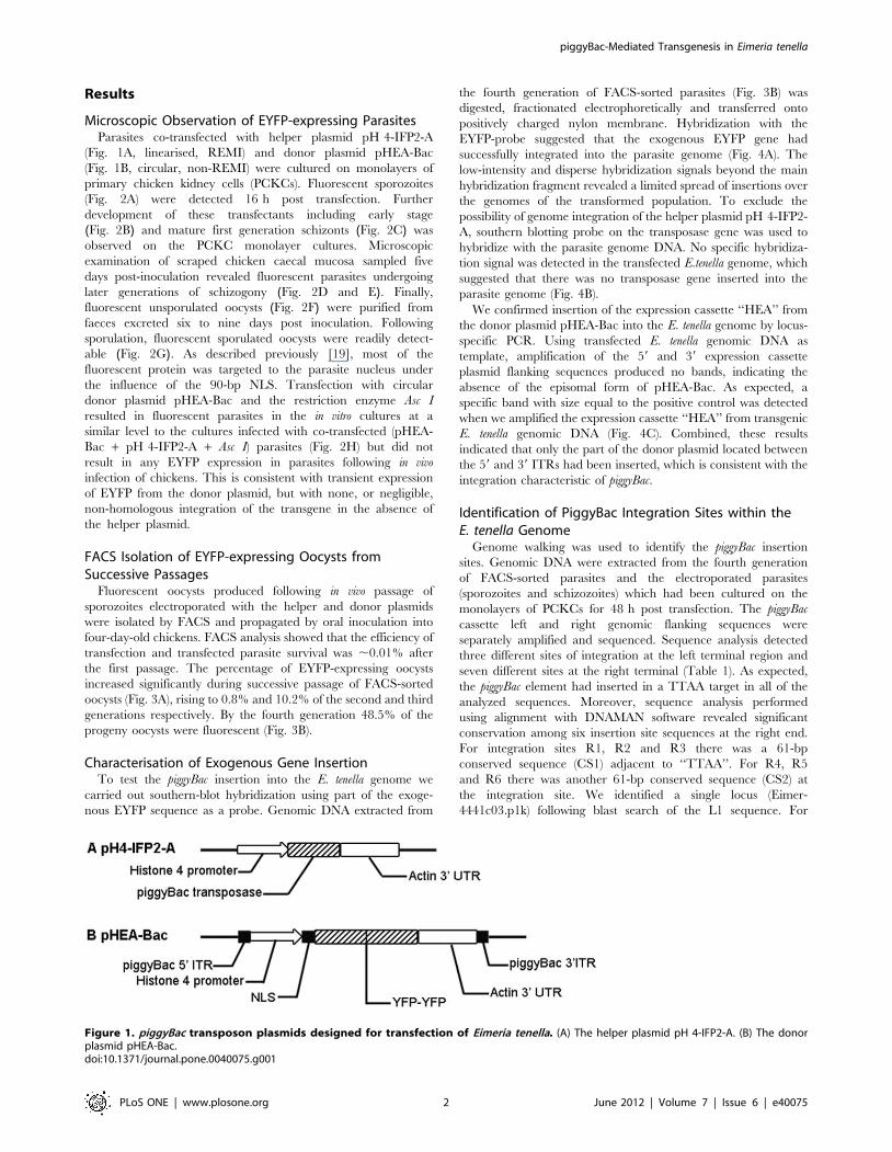
Microscopic Observation of EYFP-expressing ParasitesParasites co-transfected with helper plasmid pH 4-IFP2-A
(Fig. 1A, linearised, REMI) and donor plasmid pHEA-Bac
(Fig. 1B, circular, non-REMI) were cultured on monolayers of
primary chicken kidney cells (PCKCs). Fluorescent sporozoites
(Fig. 2A) were detected 16 h post transfection. Further
development of these transfectants including early stage
(Fig. 2B) and mature first generation schizonts (Fig. 2C) was
observed on the PCKC monolayer cultures. Microscopic
examination of scraped chicken caecal mucosa sampled five
days post-inoculation revealed fluorescent parasites undergoing
later generations of schizogony (Fig. 2D and E). Finally,
fluorescent unsporulated oocysts (Fig. 2F) were purified from
faeces excreted six to nine days post inoculation. Following
sporulation, fluorescent sporulated oocysts were readily detect-
able (Fig. 2G). As described previously [19], most of the
fluorescent protein was targeted to the parasite nucleus under
the influence of the 90-bp NLS. Transfection with circular
donor plasmid pHEA-Bac and the restriction enzyme Asc I
resulted in fluorescent parasites in the in vitro cultures at a
similar level to the cultures infected with co-transfected (pHEA-
Bac + pH 4-IFP2-A + Asc I) parasites (Fig. 2H) but did not
result in any EYFP expression in parasites following in vivo
infection of chickens. This is consistent with transient expression
of EYFP from the donor plasmid, but with none, or negligible,
non-homologous integration of the transgene in the absence of
the helper plasmid.
FACS Isolation of EYFP-expressing Oocysts fromSuccessive Passages
Fluorescent oocysts produced following in vivo passage of
sporozoites electroporated with the helper and donor plasmids
were isolated by FACS and propagated by oral inoculation into
four-day-old chickens. FACS analysis showed that the efficiency of
transfection and transfected parasite survival was ,0.01% after
the first passage. The percentage of EYFP-expressing oocysts
increased significantly during successive passage of FACS-sorted
oocysts (Fig. 3A), rising to 0.8% and 10.2% of the second and third
generations respectively. By the fourth generation 48.5% of the
progeny oocysts were fluorescent (Fig. 3B).
Characterisation of Exogenous Gene InsertionTo test the piggyBac insertion into the E. tenella genome we
carried out southern-blot hybridization using part of the exoge-
nous EYFP sequence as a probe. Genomic DNA extracted from
the fourth generation of FACS-sorted parasites (Fig. 3B) was
digested, fractionated electrophoretically and transferred onto
positively charged nylon membrane. Hybridization with the
EYFP-probe suggested that the exogenous EYFP gene had
successfully integrated into the parasite genome (Fig. 4A). The
low-intensity and disperse hybridization signals beyond the main
hybridization fragment revealed a limited spread of insertions over
the genomes of the transformed population. To exclude the
possibility of genome integration of the helper plasmid pH 4-IFP2-
A, southern blotting probe on the transposase gene was used to
hybridize with the parasite genome DNA. No specific hybridiza-
tion signal was detected in the transfected E.tenella genome, which
suggested that there was no transposase gene inserted into the
parasite genome (Fig. 4B).
We confirmed insertion of the expression cassette ‘‘HEA’’ from
the donor plasmid pHEA-Bac into the E. tenella genome by locus-
specific PCR. Using transfected E. tenella genomic DNA as
template, amplification of the 59 and 39 expression cassette
plasmid flanking sequences produced no bands, indicating the
absence of the episomal form of pHEA-Bac. As expected, a
specific band with size equal to the positive control was detected
when we amplified the expression cassette ‘‘HEA’’ from transgenic
E. tenella genomic DNA (Fig. 4C). Combined, these results
indicated that only the part of the donor plasmid located between
the 59 and 39 ITRs had been inserted, which is consistent with the
integration characteristic of piggyBac.
Identification of PiggyBac Integration Sites within theE. tenella Genome
Genome walking was used to identify the piggyBac insertion
sites. Genomic DNA were extracted from the fourth generation
of FACS-sorted parasites and the electroporated parasites
(sporozoites and schizozoites) which had been cultured on the
monolayers of PCKCs for 48 h post transfection. The piggyBac
cassette left and right genomic flanking sequences were
separately amplified and sequenced. Sequence analysis detected
three different sites of integration at the left terminal region and
seven different sites at the right terminal (Table 1). As expected,
the piggyBac element had inserted in a TTAA target in all of the
analyzed sequences. Moreover, sequence analysis performed
using alignment with DNAMAN software revealed significant
conservation among six insertion site sequences at the right end.
For integration sites R1, R2 and R3 there was a 61-bp
conserved sequence (CS1) adjacent to ‘‘TTAA’’. For R4, R5
and R6 there was another 61-bp conserved sequence (CS2) at
the integration site. We identified a single locus (Eimer-
4441c03.p1k) following blast search of the L1 sequence. For
Figure 1. piggyBac transposon plasmids designed for transfection of Eimeria tenella. (A) The helper plasmid pH 4-IFP2-A. (B) The donorplasmid pHEA-Bac.doi:10.1371/journal.pone.0040075.g001
piggyBac-Mediated Transgenesis in Eimeria tenella
PLoS ONE | www.plosone.org 2 June 2012 | Volume 7 | Issue 6 | e40075

L2 and L3, we could not find a highly similar sequence through
blast search using the E. tenella omniblast server http://www.
sanger.ac.uk/cgi-bin/blast/submitblast/e_tenella/omni, possibly
due to the incomplete nature of the E. tenella genome sequence.
At the right terminal we identified two loci as integration sites
for R1 and R4 respectively (Eimer-4441c03.p1k and Eimer-
642h02.p1k). Homologous sequences were not found for sites of
R2, R3, R5, R6 and R7 when we performed blast searches as
described previously, although a large number of repetitive
sequences were identified with a low level of significance. Using
sequences associated with the two genomic insertion points
suggested that each mapped to a predicted non-coding region
within ,400 bp of a putative coding sequence. The conserved
CS1 and CS2 sequences were both found at several loci without
annotation in E. tenella databases of the Wellcome Trust Sanger
Institute.
Figure 2. Transgenic fluorescent Eimeria tenella parasites. Sporozoite (A), early stage (B) and mature (C) first generation schizonts expressedEYFP predominantly in the parasite nuclei following transfection with the helper plasmid pH 4-IFP2-A and the donor plasmid pHEA-Bac. Lategeneration merozoite (D) and schizont (E), unsporulated (F) and sporulated (G) oocysts also presented fluorescent nuclei following transfection withpH 4-IFP2-A and pHEA-Bac. In the transfection efficient assays (H), fluorescent parasites in each well (3 wells per group) were counted at 36 h posttransfection. The mean and standard deviations were analysed by the Student t test using the SPSS 13.0 software. The significance level was set at0.05.doi:10.1371/journal.pone.0040075.g002
Figure 3. Isolation of fluorescent oocysts by FACS. (A) The percentage of fluorescent oocysts increased from 0.01% in the first generation to48.5% in the fourth generation. (B) EYFP-expressing oocysts (48.5%) from the fourth generation.doi:10.1371/journal.pone.0040075.g003
piggyBac-Mediated Transgenesis in Eimeria tenella
PLoS ONE | www.plosone.org 3 June 2012 | Volume 7 | Issue 6 | e40075

Calculation of Average EYFP Insertion RateWe extracted genomic DNA from transgenic E. tenella harvested
from the fourth round of propagation to calculate the average
EYFP cassette insertion rate using Real-time PCR with the
fluorescence dye SYBR Green I. By comparing the number of
EYFP copies with the total number of transgenic parasite genomes
(real-time PCR for total parasite genomes corrected for the
percentage that were found to be fluorescent by microscopy) there
were on average ,14 EYFP copies per expressing genome,
indicating ,7 insertions since the EYFP was presented as a
tandem copy reporter.
Discussion
Genomic resources for Eimeria species parasites are rare.
Although great progress has been achieved in a whole-genome
sequencing project for E. tenella through a combination of Sanger
and next-generation sequencing [20], the genome assembly
remains fragmented and many genes are yet to be identified or
characterized. Strategies supporting genetic manipulation of the
E. tenella genome are developing rapidly but direct methods for
verification of gene function by targeted gene knockout remain
elusive, complicated by low insertion efficiency and a requirement
for serial in vivo passage to select and stabilize transgenic parasite
lines. Another way to determine gene function is to employ
forward genetic approaches to generate random gene mutations
and characterize the resulting phenotypes. Thus, the piggyBac
transposon system, which has shown high levels of efficiency in
other organisms, may provide a powerful approach to study gene
function in E. tenella.
We report here that the piggyBac transposon system functions
in E. tenella by transposing exogenous sequences into TTAA sites.
Previous studies have reported that piggyBac insertions showed a
high preference of predicted transcriptional units in mice [14] and
59 UTRs of genes in Plasmodium falciparum [11]. In our study only
three of the ten genomic insertion flanking sequences could be
mapped to the genome assembly, identifying two as yet
unannotated loci predicted to be located in non-coding regions
proximal to putative coding sequences. Several genomic insertion
flanking sequences were found to be repetitive, featuring tandemly
repeated sequences or a high AT content which may be associated
with transposon-like elements and telomere-like repeats. In this
study, we got limited insertions sites in E.tenella. This may be due to
the much higher overall GC content of the Eimeria genome
(http://www.sanger.ac.uk/Projects/E_tenella/). Another reason
for the small number of cloned insertion sites may be the
amplification of parasite through successive passage, in which
EYFP-transgenic parasites with some selective or growth advan-
tage were selected for outgrowth. For this reason, we performed
three independent transfections in which the electroporated
sporozoites were cultured on the monolayer of PCKC cells other
than passaged through the chicken. As the plasmid contamination
was difficult to be avoid in the freshly electroporated samples, we
got only one insertion site at each terminal region (L3 and R7). So
there are still much more work to be done to obtain new
integration sequences to understand integration preferences in E.
tenella. Although it is far from making any assumption, we
identified the conserved sequences CS1 and CS2 at most of the
minimal insertion sites except for L1, L2, L3 and R7. As expected,
all the identified insertion sites do support the existence of a short
TTAA piggyBac target insertion sequence.
Black et al. reported that REMI could dramatically increase the
transfection efficiency of Toxoplasma gondii and enhance stable
transfection [21]. In our study, REMI was used to increase the
transfection efficiency of E.tenella. But in piggyBac system, trans-
posase gene should be discharged from parasite or else the
piggyBac transposon could keep on jumping around the genome.
In order to reduce the possibility of insertion of the helper gene
into the parasite genome, we didn’t do any selection for
transposase gene through the successive passages. Fortunately,
genome hybridization with the transposase-probe produced no
specific signal, which successfully excluded the possibility of
genome integration of the piggyBac transposase gene.
Plasmids with relatively large sizes (.7 kb) often produce low
transfection efficiencies in vitro and therefore reduce the potential
for obtaining fluorescent oocysts from the first in vivo passage of
electroporated sporozoites [22]. The current study also failed to
obtain transfected oocysts post in vivo passage of sporozoites
electroporated with the circular donor plasmid pHEA-Bac and the
restriction enzyme Asc I (non-REMI enzyme for pHEA-Bac),
indicating a very low level of non-homologous integration in the
absence of the helper plasmid. However, when the piggyBac
Figure 4. Confirmation of exogenous gene insertion. (A) Southern-blot hybridization using the EYFP gene as the probe. Lanes 1 and 2:transgenic and wild type Eimeria tenella genomic DNA, lane 3: donor plasmid pHEA-Bac (presenting EYFP as a positive control). (B) Southern-blothybridization using the transposase gene as the probe. Lane 1: transgenic E.tenella genomic DNA, lane2: wild type E.tenella genomic DNA, lane 3:helper plasmid pH 4-IFP2-A (presenting transposase gene as a positive control). (C) PCR for confirmation of insertion of part of pHEA-Bac. Theexpression cassette ‘‘HEA’’ was only amplified from transgenic parasite genomic DNA and the 39 and 59 plasmid flanking sequences lying outside ofthe ITR sequences in pHEA-Bac were not detected. Lanes including ‘‘39P’’, ‘‘59P’’ and ‘‘HEAP’’ represented positive controls (Amplicons from theplasmid pHEA-Bac).doi:10.1371/journal.pone.0040075.g004
piggyBac-Mediated Transgenesis in Eimeria tenella
PLoS ONE | www.plosone.org 4 June 2012 | Volume 7 | Issue 6 | e40075

transposase were applied together with the circular donor plasmid,
fluorescent oocysts were successfully obtained, indicating that the
efficient transfection of the transposase plasmid could help increase
integration of exogenous genes. While, the action of the AscI may
or may not have helped with genome access. Increased integration
facilitates in vivo selection of transfected oocysts, suggesting that
piggyBac may be a practical molecular genetics tool for use in E.
tenella transgenesis. Experiments performed in this report provide a
first step toward development of an effective mutagenesis platform
in the apicomplexan parasite E. tenella using the piggyBac system.
Materials and Methods
Parasites and AnimalsAll experimental procedures were approved by the China
Agricultural University Animal Ethics Committee. (The certificate
of Beijing Laboratory Animal employee, ID: 18086). The E. tenella
Beijing (BJ) strain [7] was maintained, purified, and sporulated as
described elsewhere [23]. Before transfection, purified oocysts were
ground with a 7-ml glass tissue grinder to release sporocysts which
were then treated with trypsin-bile solution (0.75% [w/v] trypsin
and 10% [v/v] bile in PBS) to release sporozoites. Purification of
sporozoites was carried out by DE-52 anion-exchange chromatog-
raphy [24]. Freshly purified sporozoites were washed once in
incomplete cytomix (10 mM K2HPO4:KH2PO4, pH 7.6;
120 mM KCl; 0.15 mM CaCl2; 25 mM HEPES; 2 mM EGTA;
and 5 mM MgCl2) [6] and then suspended in the same buffer,
supplemented with 2 mM ATP and 5 mM glutathione. In vitro cell
culture of transfectants was carried out using primary chicken
kidney cells (PCKCs) prepared from a ten-day-old chicken as
described previously [19]. Arbor Acres (AA) broiler chickens were
used for preparation of PCKCs and parasite propagation in vivo.
Plasmid ConstructsThe helper plasmid pH 4-IFP2-A (Fig. 1A) was constructed by
cloning the piggyBac transposase coding sequence from pBSII-
IFP2orf [25] using primers 59-ACTAGTATGGGTAGTTCTT-
TAG-39 that created a Spe I site and 59-GCGGCCGCTCA-
GAAACAACTTTGGCACA -39 that added a Not I site, under the
control of the E. tenella Histone 4 promoter and the E. tenella actin
39 untranslated region (UTR). The Spe I and Not I digested PCR
product was then inserted into the Spe I and Not I digested vector
pEtHEA [8].
The donor plasmid pHEA-Bac (Fig. 1B) was constructed by
cutting the fragment containing the EYFP sequence under the
control of the E. tenella Histone 4 promoter and incorporating a 59
90-bp nuclear location signal sequence (NLS) and a 39 E. tenella
actin 39 UTR from pH-2E-A39 [26] using Eco RV and Bgl II. The
digestion product was cloned between the ITRs of the piggyBac
element in the vector pXL-BAC II [25].
TransfectionEimeria tenella sporozoites were electroporated with the donor
and helper plasmids using a restriction enzyme-mediated integra-
tion (REMI) method [22] where only the helper plasmid was
subject to REMI. Briefly, 16107 freshly excysted sporozoites
suspended in complete Cytomix [6] were mixed with 60 mg
circular donor plasmid pHEA-Bac and 30 mg Asc I linearized
helper plasmid pH 4-IFP2-A. Next, 200 U restriction enzyme Asc
I was added to the sporozoite/plasmid mixture. Prior to
transfection the total volume of the mixture was made up to
800 ml in a 0.4 cm cuvette (BioRad). Transfection was conducted
by electroporating at 2.0 kV, 25 mF, resulting in a pulse time of
0.3–0.4 ms, with a Gene Pulser XcellTM Electroporation System
(BioRad). Transfection with the donor plasmid pHEA-Bac alone
was set as control. The transfected sporozoites were allowed to rest
for 20 min at room temperature. For in vitro cultivation,
electroporated sporozoites at 16105 cells/ml were added to
monolayers of PCKCs and incubated at 41uC in a 5% CO2
incubator. PCKCs were grown in a 12-well multiwell plate (BD
FalconTM) in 2 ml/well of DMEM (Gibco) containing fetal bovine
serum (10% v/v), penicillin (200 U ml21) and streptomycin
(20 mg ml21). For in vivo parasite propagation transfected
sporozoites electroporated with the helper and donor plasmids
were inoculated into four-day-old chickens via the cloacal route
(26106 parasites per bird).
Microscopic Observation of EYFP-expressing ParasitesE. tenella parasites electroporated with both donor and helper
plasmids, or with donor plasmid alone, were cultured in vitro within
PCKC and observed at 16 h and 36 h post-electroporation using
an inverted fluorescent microscope (IX 71, Olympus, Japan) with
488-nm excitation and 508-nm emission filters, under which the
expressed yellow fluorescence appeared green. At 36 h, the
numbers of green intracellular sporozoites, or developing first-
generation schizonts, were counted (Liu et al, 2008). Transfected
E. tenella parasites within in vivo mucosal scrapings prepared from
the caeca five days post-inoculation, and progeny oocysts purified
from faeces excreted six to nine days post-inoculation, were also
examined by fluorescence microscopy.
Selection of Stable Transformed ParasitesPositive selections of transfectants were performed by Fluores-
cence Activated Cell Sorting (FACS) using a MoFlo Cell Sorter
(Dako Cytomation, Fort Collins, CO, USA) as described
previously [8]. The sorted oocysts were sporulated and inoculated
into four-day-old chickens for another round of selection. After
four successive passages with selection, the transgenic E. tenella
population was collected for genomic DNA analysis.
Table 1. Identification of genomic sequences flanking thepiggyBac insertion sites in Eimeria tenella.
Integration site Left end Integration locus
L1 Aggacacact-TTAA-piggyBac Eimer-4441c03.p1k
L2 Aacgcgggac-TTAA-piggyBac Unknown locus 1
L3 Gtcgtattac-TTAA-piggyBac Unknown locus 2
Right end
R1 piggyBac-TTAA-CS1a Eimer-4441c03.p1k
R2 piggyBac-TTAA-CS1a Unknown locus 3c
R3 piggyBac-TTAA-CS1a Unknown locus 4
R4 piggyBac-TTAA-CS2b Eimer-642h02.p1k
R5 piggyBac-TTAA-CS2b Unknown locus 5d
R6 piggyBac-TTAA-CS2b Unknown locus 6
R7 piggyBac-TTAA-cagggcgcaa Unknown locus 7
aConservative sequence 1 (CS1):GTAATACGACTCACTATAGGGCGAATTGAAGCTGCCCTTTGGTGCAGATGAACTTC-AGGGT.bConservative sequence 2 (CS2):GAGGCGGAGTGTCCGCTGTTGCTGTGGGCAGAAAGAGGGCGGCGTAGAGAGGCA-TTTAGTG.cR2 flanking region was revealed as an AT-rich DNA sequence.dR5 flanking region presented in the form of tandem repeat sequences.doi:10.1371/journal.pone.0040075.t001
piggyBac-Mediated Transgenesis in Eimeria tenella
PLoS ONE | www.plosone.org 5 June 2012 | Volume 7 | Issue 6 | e40075

Southern-blot HybridizationWild type or transgenic E. tenella genomic DNA was prepared from
freshly purified sporozoites according to the Instruction Manual of
TIANamp Genomic DNA Kit (TIANGEN, Beijing, China). Bam HI
linearized donor plasmid pHEA-Bac was used as the positive control.
For Southern blotting, 3 mg of genomic DNA was digested with Nde I
and electrophoresed on a 0.8% (w/v) agarose gel (in 16TAE, 60 V,
4 h). Size-fractionated DNA was blotted onto positively charged
nylon membrane (Amersham Biosciences, NJ, USA) by capillary
transfer and fixed to the membranes by u.v.-crosslinking for 10 min.
The hybridization probe corresponding to the EYFP gene sequence
was amplified with the primers 59-ATGGTGAGCAAGGGCGAG-
GAGC-39 and 59-CTAAAGCTTCTTGTACAGCTC-39 from the
plasmidpHEA-Bac.The transposasegeneprobewascloned fromthe
plasmid pH 4-IFP2-A with the primers 59-AATGGAATGCCT-
TATTTGGG-39 and 59-GTGTCCACTCCGCCTTTAGT-39.
Probe labeling, hybridization, washing and detection were carried
out according to the manufacturer’s guide using a DIG High Prime
DNA Labeling and Detection Starter Kit II as described previously
[8].
PCR Confirmation of Genomic Fragment InsertionPrimers specific for the donor plasmid expression cassette
(containing the E. tenella Histone 4 promoter, EYFP gene and
E. tenella actin 39 UTR; P1 and P2, Table 2) located between the
piggyBac 59 and 39 ITRs were used to confirm insertion of the
donor plasmid pHEA-Bac vectored marker cassette into the E.
tenella genome. Two further primer pairs were used to confirm the
absence of plasmid DNA flanking the ITR sequences from the
genome (P3 and P4, P5 and P6, Table 2). Briefly, the PCR
conditions were 35 cycles of 94uC for 30 s, 55uC for 30 s, and
72uC for 3 min 30 s. PCR products were electrophoresed using a
0.8% (w/v) agarose gel (in 16TAE, 80 V, 20 min).
Genome Walking Through 59 and 39 ITR Flanking RegionsIn order to identify the genomic piggyBac insertion sites, piggyBac
59 and 39 ITR flanking sequences were amplified and sequenced
using a genome walking method (Genome Walking Kit; TaKaRa).
For the fourth generation of FACS-sorted parasites, genomic DNA
was prepared from freshly purified sporozoites as described
previously. For the electroporated parasites, sporozoites or
schizozoites were harvested 48 h post transfection by digestion
of the PCKCs with 0.25% trypsin-EDTA for 5 min at 37uC. The
harvested parasites and PCKCs were treated with DNase I (5 U/
mg plasmid DNA) for 10 min at 37uC and washed five times with
PBS (pH 7.2) [6] before genomic DNA was extracted with
TIANamp Genomic DNA Kit (TIANGEN, Beijing, China). Two
sets of three primers (P6, P7, P8 and P9, P10, P11) specific for the
known pHEA-Bac vectored gene sequence were used successively
in combination with the Arbitrary Primers (AP) provided by the
kit. PCR products were gel purified, ligated into the cloning vector
pEasy-T1 (TransGen Biotech, Beijing, China) and sequenced
using the universal primers M13F & M13R. Analysis of the
sequencing results was performed using DNAMAN software
(Lynnon Biosoft, USA). Insertion sites were identified by similarity
using BLAST searches with the E. tenella omniblast server http://
www.sanger.ac.uk/cgi-bin/blast/submitblast/e_tenella/omni
against the September 2010 genome assembly. Associated putative
coding sequences were identified by BLASTX comparison of
5 Kb genomic sequence flanking the identified insertion site with
the September 2010 E. tenella predicted proteins dataset.
Genomic Insertion Quantification Using Real-time PCRThe average number of donor cassette insertions per parasite
genome was determined using EYFP- and Eimeria genus-specific
Real-Time PCR. Briefly, the total number of E. tenella genomes
was determined for each test sample in triplicate using a genus
specific Real-Time PCR assay targeting the 5 S rRNA coding
sequence with primers P13 and P14 (Table 2) [27]. EYFP coding
sequence copy number was evaluated in triplicate using primers of
P15 and P16. Plasmids containing the EYFP or E. tenella 5 s rDNA
sequence were constructed for use as DNA standards. Plasmid
concentration was initially calculated using a spectrophotometer
reading at 260 nm and 280 nm (NanoDrop 2000c, USA) prior to
Table 2. Primers used during the application and confirmation of piggyBac mediated transfection of Eimeria tenella.
Primer ID Sequence (59–39) Purpose
P1 GATATCTGGTTAGGGCCTCAAGGGAA Confirm donor insertion
P2 AGATCTACACTCCGCCTCTTAATCTTT Confirm donor insertion
P3 CGGGTTTCGCCACCTCTGACT Absence of cassette flanking sequence insertion
P4 CGCCGTTTAGGCTGCTTCTGC Absence of cassette flanking sequence insertion
P5 GTAGGTATGGGAGGAGTGTAGTTGG Absence of cassette flanking sequence insertion
P6 TAAATCGGAACCCTAAAGGGAGCCC Absence of cassette flanking sequence insertion
P7 CGCCGATACCATCACGAACAACAACA Genome walking nested primer
P8 GCTCCTCGCCCTTGCTCACCAT Genome walking nested primer
P9 TGGTGCAGATGAACTTCAGGGT Genome walking nested primer
P10 CGACCACTACCAGCAGAACACC Genome walking nested primer
P11 AGTCCGCCCTGAGCAAAGACCC Genome walking nested primer
P12 CGTTTGCAGCAGAGTAGTTCAT Genome walking nested primer
P13 GTTGCAGCTAGGTGCGAGACA Eimeria 5 S rDNA qPCR
P14 CAGCGCGTCCTCTCTACCAA Eimeria 5 S rDNA qPCR
P15 TCCAGGAGCGCACCATCTT EYFP qPCR
P16 ATGCCCTTCAGCTCGATGC EYFP qPCR
doi:10.1371/journal.pone.0040075.t002
piggyBac-Mediated Transgenesis in Eimeria tenella
PLoS ONE | www.plosone.org 6 June 2012 | Volume 7 | Issue 6 | e40075

the preparation of ten-fold serial dilutions representing 106–100
copies of each plasmid. 2 ml of plasmid or transgenic E. tenella
genomic DNA were used in each amplification reaction using the
real-time PCR buffer Power SYBRH Green PCR Master Mix
(Ambion, USA). Milli-Q water served as negative no template
control. Amplifications were performed using Applied Biosystems
7500 Real-Time PCR System equipment and subjected to 50uCfor 2 min, then 95uC for 10 min, followed by 40 cycles at 95uC for
15 sec and 60uC for 1 min. Figures produced by absolute
quantification of target EYFP insert and 5 S rDNA host template
copy number were used to calculate the average number of donor
plasmid insertions per transgenic parasite genome based upon the
total number of parasite genomes detected and the percentage
found to be transgenic (i.e. fluorescent) by microscopy.
Acknowledgments
We acknowledge Dr. Malcolm J. Fraser (University of Notre Dame) for
kindly granting us the piggyBac plasmids pBSII-IFP2-orf and pXL-BacII.
Author Contributions
Conceived and designed the experiments: HS DPB FMT XS. Performed
the experiments: HS XL XZ DPB. Analyzed the data: HS DPB FMT XS.
Contributed reagents/materials/analysis tools: WY TS. Wrote the paper:
HS DPB FMT XS.
References
1. Shirley MW, Smith AL, Tomley FM (2005) The biology of avian Eimeria withan emphasis on their control by vaccination. Adv Parasitol 60: 285–330.
2. Williams RB (1998) Epidemiological aspects of the use of live anticoccidialvaccines for chickens. Int J Parasitol 28: 1089–1098.
3. Suo X, Zhang JX, Li ZG, Yang CT, Min QR, et al. (2006) The efficacy and
economic benefits of Supercox, a live anticoccidial vaccine in a commercial trialin broiler chickens in China. Vet Parasitol 142: 63–70.
4. Bromley E, Leeds N, Clark J, McGregor E, Ward M, et al. (2003) Defining theprotein repertoire of microneme secretory organelles in the apicomplexan
parasite Eimeria tenella. Proteomics 3: 1553–1561.
5. Shirley MW, Ivens A, Gruber A, Madeira AM, Wan KL, et al. (2004) TheEimeria genome projects: a sequence of events. Trends Parasitol 20: 199–201.
6. Kelleher M, Tomley FM (1998) Transient expression of beta-galactosidase indifferentiating sporozoites of Eimeria tenella. Mol Biochem Parasitol 97: 21–31.
7. Hao L, Liu X, Zhou X, Li J, Suo X (2007) Transient transfection of Eimeriatenella using yellow or red fluorescent protein as a marker. Mol Biochem
Parasitol 153: 213–215.
8. Yan W, Liu X, Shi T, Hao L, Tomley FM, et al. (2009) Stable transfection ofEimeria tenella: constitutive expression of the YFP-YFP molecule throughout
the life cycle. Int J Parasitol 39: 109–117.9. Clark JD, Billington K, Bumstead JM, Oakes RD, Soon PE, et al. (2008) A
toolbox facilitating stable transfection of Eimeria species. Mol Biochem Parasitol
162: 77–86.10. Kurth M, Entzeroth R (2009) Reporter gene expression in cell culture stages and
oocysts ofEimeria nieschulzi (Coccidia, Apicomplexa). Parasitol Res 104: 303–310.11. Balu B, Shoue DA, Fraser MJ Jr, Adams JH (2005) High-efficiency
transformation of Plasmodium falciparum by the lepidopteran transposable
element piggyBac. Proc Natl Acad Sci U S A 102: 16391–16396.12. Fonager J, Franke-Fayard BM, Adams JH, Ramesar J, Klop O, et al. (2011)
Development of the piggyBac transposable system for Plasmodium berghei and itsapplication for random mutagenesis in malaria parasites. BMC Genomics 12: 155.
13. Thibault ST, Singer MA, Miyazaki WY, Milash B, Dompe NA, et al. (2004) Acomplementary transposon tool kit for Drosophila melanogaster using P and
piggyBac. Nat Genet 36: 283–287.
14. Ding S, Wu X, Li G, Han M, Zhuang Y, et al. (2005) Efficient transposition ofthe piggyBac (PB) transposon in mammalian cells and mice. Cell 122: 473–483.
15. Fraser MJ, Smith GE, Summers MD (1983) Acquisition of Host Cell DNASequences by Baculoviruses: Relationship Between Host DNA Insertions and FP
Mutants of Autographa californica and Galleria mellonella Nuclear PolyhedrosisViruses. J Virol 47: 287–300.
16. Cary LC, Goebel M, Corsaro BG, Wang HG, Rosen E, et al. (1989) Transposonmutagenesis ofbaculoviruses: analysis ofTrichoplusia ni transposonIFP2 insertions
within the FP-locus of nuclear polyhedrosis viruses. Virology 172: 156–169.
17. Fraser MJ, Cary L, Boonvisudhi K, Wang HG (1995) Assay for movement ofLepidopteran transposon IFP2 in insect cells using a baculovirus genome as a
target DNA. Virology 211: 397–407.18. Fraser MJ, Ciszczon T, Elick T, Bauser C (1996) Precise excision of TTAA-
specific lepidopteran transposons piggyBac (IFP2) and tagalong (TFP3) from the
baculovirus genome in cell lines from two species of Lepidoptera. Insect Mol Biol5: 141–151.
19. Shi TY, Liu XY, Hao LL, Li JD, Gh AN, et al. (2008) Transfected Eimeriatenella could complete its endogenous development in vitro. J Parasitol 94: 978–
980.20. Ling KH, Rajandream MA, Rivailler P, Ivens A, Yap SJ, et al. (2007)
Sequencing and analysis of chromosome 1 of Eimeria tenella reveals a unique
segmental organization. Genome Res 17: 311–319.21. Black M, Seeber F, Soldati D, Kim K, Boothroyd JC (1995) Restriction enzyme-
mediated integration elevates transformation frequency and enables co-transfection of Toxoplasma gondii. Mol Biochem Parasitol 74: 55–63.
22. Liu X, Shi T, Ren H, Su H, Yan W, et al. (2008) Restriction enzyme-mediated
transfection improved transfection efficiency in vitro in Apicomplexan parasiteEimeria tenella. Mol Biochem Parasitol 161: 72–75.
23. LongPL,MillardBJ, JoynerLP,NortonCC(1976)Aguide to laboratory techniquesused in the study and diagnosis of avian coccidiosis. Folia Vet Lat 6: 201–217.
24. Schmatz DM, Crane MS, Murray PK (1984) Purification of Eimeria sporozoites
by DE-52 anion exchange chromatography. J Protozool 31: 181–183.25. Li X, Harrell RA, Handler AM, Beam T, Hennessy K, et al. (2005) piggyBac
internal sequences are necessary for efficient transformation of target genomes.Insect Mol Biol 14: 17–30.
26. Shi T, Yan W, Ren H, Liu X, Suo X (2009) Dynamic development ofparasitophorous vacuole of Eimeria tenella transfected with the yellow
fluorescent protein gene fused to different signal sequences from apicomplexan
parasites. Parasitol Res 104: 315–320.27. Blake DP, Hesketh P, Archer A, Shirley MW, Smith AL (2006) Eimeria maxima:
the influence of host genotype on parasite reproduction as revealed byquantitative real-time PCR. Int J Parasitol 36: 97–105.
piggyBac-Mediated Transgenesis in Eimeria tenella
PLoS ONE | www.plosone.org 7 June 2012 | Volume 7 | Issue 6 | e40075