
A Novel Family of Apicomplexan Glideosome-associated Proteins with an Inner Membrane-anchoring Role
11
A Novel Family of Apicomplexan Glideosome-associated Proteins with an Inner Membrane-anchoring Role * □ S Received for publication, June 22, 2009 Published, JBC Papers in Press, June 26, 2009, DOI 10.1074/jbc.M109.036772 Hayley E. Bullen ‡§¶1 , Christopher J. Tonkin ¶ , Rebecca A. O’Donnell ¶ , Wai-Hong Tham ¶ , Anthony T. Papenfuss , Sven Gould**, Alan F. Cowman ¶2 , Brendan S. Crabb ‡2 , and Paul R. Gilson ‡3 From the ‡ Macfarlane Burnet Institute for Medical Research & Public Health, 85 Commercial Road, Melbourne, Victoria 3004, the § Department of Medical Biology, The University of Melbourne, Parkville, Victoria 3010, the ¶ Infection and Immunity Division, and the Bioinformatics Division, The Walter & Eliza Hall Institute of Medical Research, 1G Royal Parade, Parkville, Victoria 3050, and the **School of Botany, The University of Melbourne, Parkville, Victoria 3050, Australia The phylum Apicomplexa are a group of obligate intracellular parasites responsible for a wide range of important diseases. Cen- tral to the lifecycle of these unicellular parasites is their ability to migrate through animal tissue and invade target host cells. Api- complexan movement is generated by a unique system of gliding motility in which substrate adhesins and invasion-related pro- teins are pulled across the plasma membrane by an underlying actin-myosin motor. The myosins of this motor are inserted into a dual membrane layer called the inner membrane complex (IMC) that is sandwiched between the plasma membrane and an underlying cytoskeletal basket. Central to our understanding of gliding motility is the characterization of proteins residing within the IMC, but to date only a few proteins are known. We report here a novel family of six-pass transmembrane proteins, termed the GAPM family, which are highly conserved and spe- cific to Apicomplexa. In Plasmodium falciparum and Toxo- plasma gondii the GAPMs localize to the IMC where they form highly SDS-resistant oligomeric complexes. The GAPMs co-pu- rify with the cytoskeletal alveolin proteins and also to some degree with the actin-myosin motor itself. Hence, these proteins are strong candidates for an IMC-anchoring role, either directly or indirectly tethering the motor to the cytoskeleton. Apicomplexan parasites cause a multitude of illnesses through infection of both human and livestock hosts. Members of this phy- lum include the opportunistic human parasites Toxoplasma gon- dii and Cryptosporidium parvum, pathogens of livestock, includ- ing Theileria annulata and Eimeria tenalla, and most notably the Plasmodium species, the causative agents of malaria in humans. Infection with P. falciparum results in 1–3 million deaths and a further 500 million infections annually (1). During various stages of the Apicomplexan lifecycle the par- asites require motility to migrate through their insect and ver- tebrate hosts and to invade and internalize themselves within targeted host cells (2– 4). The parasite’s unique mechanism of gliding motility is powered by an Apicomplexan-specific motor complex termed the actin-myosin motor (5), which resides between the outer plasma membrane and inner membrane complex (IMC) 4 (6). The IMC is a continuous patchwork of flattened vesicular cisternae located directly beneath the plasma membrane and overlying the cytoskeletal network (7, 8). The IMC appears to arise from Golgi-associated vesicles flattened during parasite maturation to form large membra- nous sheets, which envelope the parasite and leave only a small gap at the extreme parasite apex (9). The myosin component of the actin-myosin motor has pre- viously been defined as a tetrameric complex consisting of a class XIV myosin termed Myo-A (10), a myosin tail interacting protein (also called myosin light chain) (7) and the two glideo- some-associated proteins GAP45 and GAP50 (11). These motor components are linked to the outer IMC membrane via the membrane proteins GAP45/50 (11). Between the plasma membrane and the IMC are actin filaments held in place through aldolase-mediated contact with the C-terminal tails of plasma membrane-spanning adhesive proteins whose ectodo- mains bind substrate and host cells (2). To power the forward movement of apicomplexan zoite stages, myosin pulls the actin filaments and their attached adhesins rearward. For this to suc- ceed the GAP-myosin complex must presumably be fixed to the IMC, possibly via interactions with unidentified proteins link- ing the motor to the underlying cytoskeleton. Studies of fluo- rescently tagged GAP50 confirm it is relatively immobile within the IMC, however attempts to identify potential anchoring pro- teins have not been successful and have instead indicated that GAP50 may be immobilized by the lipid-raft like properties of the IMC membranes (12). The actin-myosin complex is confined to the outer IMC mem- brane while the opposing innermost IMC membrane is studded with 9 nm intramembranous particles, revealed by electron mi- croscopy of freeze fractured Toxoplasma tachyzoites and Plasmo- * This work was supported, in part, by the National Health and Medical Research Council of Australia and Wellcome Trust, UK. Author’s Choice—Final version full access. □ S The on-line version of this article (available at http://www.jbc.org) contains supplemental Figs. S1–S7 and Tables S1 and S2. 1 Recipient of an Australian Postgraduate Award. 2 International Scholars of the Howard Hughes Medical Institute. 3 To whom correspondence should be addressed: Macfarlane Burnet Institute for Medical Research & Public Health, 85 Commercial Road, Melbourne, Victoria 3004, Australia. Tel.: 61-03-8506-2481; Fax: 61-03-9282-2100; E-mail: [email protected]. 4 The abbreviations used are: IMC, inner membrane complex; DRM, deter- gent-resistant membrane; HA, hemagglutinin; GFP, green fluorescent protein; RIPA, radioimmune precipitation assay; bis-tris, 2-[bis(2-hy- droxyethyl)amino]-2-(hydroxymethyl)propane-1,3-diol; mAb, mono- clonal antibody; ChFP, cherry fluorescent protein; DSP, dithiobis succin- imidyl propionate; IMP, intramembranous particle; ALV, alveolin; GPI, glycosylphosphatidylinositol. THE JOURNAL OF BIOLOGICAL CHEMISTRY VOL. 284, NO. 37, pp. 25353–25363, September 11, 2009 Author’s Choice © 2009 by The American Society for Biochemistry and Molecular Biology, Inc. Printed in the U.S.A. SEPTEMBER 11, 2009 • VOLUME 284 • NUMBER 37 JOURNAL OF BIOLOGICAL CHEMISTRY 25353 at Universitaets- und Landesbibliothek Duesseldorf, on February 10, 2012 www.jbc.org Downloaded from http://www.jbc.org/content/suppl/2009/06/25/M109.036772.DC1.html Supplemental Material can be found at:
-
Upload
independent -
Category
Documents
-
view
0 -
download
0
Transcript of A Novel Family of Apicomplexan Glideosome-associated Proteins with an Inner Membrane-anchoring Role

A Novel Family of Apicomplexan Glideosome-associatedProteins with an Inner Membrane-anchoring Role*□S
Received for publication, June 22, 2009 Published, JBC Papers in Press, June 26, 2009, DOI 10.1074/jbc.M109.036772
Hayley E. Bullen‡§¶1, Christopher J. Tonkin¶, Rebecca A. O’Donnell¶, Wai-Hong Tham¶, Anthony T. Papenfuss�,Sven Gould**, Alan F. Cowman¶2, Brendan S. Crabb‡2, and Paul R. Gilson‡3
From the ‡Macfarlane Burnet Institute for Medical Research & Public Health, 85 Commercial Road, Melbourne, Victoria 3004, the§Department of Medical Biology, The University of Melbourne, Parkville, Victoria 3010, the ¶Infection and Immunity Division, andthe �Bioinformatics Division, The Walter & Eliza Hall Institute of Medical Research, 1G Royal Parade, Parkville, Victoria 3050, and the**School of Botany, The University of Melbourne, Parkville, Victoria 3050, Australia
The phylum Apicomplexa are a group of obligate intracellularparasites responsible for a wide range of important diseases. Cen-tral to the lifecycle of these unicellular parasites is their ability tomigrate through animal tissue and invade target host cells. Api-complexanmovement is generated by a unique systemof glidingmotility in which substrate adhesins and invasion-related pro-teins are pulled across the plasma membrane by an underlyingactin-myosinmotor. Themyosins of thismotor are inserted intoa dual membrane layer called the inner membrane complex(IMC) that is sandwiched between the plasmamembrane and anunderlying cytoskeletal basket. Central to our understanding ofgliding motility is the characterization of proteins residingwithin the IMC, but to date only a few proteins are known. Wereport here a novel family of six-pass transmembrane proteins,termed the GAPM family, which are highly conserved and spe-cific to Apicomplexa. In Plasmodium falciparum and Toxo-plasma gondii the GAPMs localize to the IMC where they formhighly SDS-resistant oligomeric complexes. TheGAPMs co-pu-rify with the cytoskeletal alveolin proteins and also to somedegreewith the actin-myosinmotor itself. Hence, these proteinsare strong candidates for an IMC-anchoring role, either directlyor indirectly tethering the motor to the cytoskeleton.
Apicomplexan parasites cause a multitude of illnesses throughinfectionof bothhumanand livestockhosts.Members of this phy-lum include the opportunistic human parasites Toxoplasma gon-dii and Cryptosporidium parvum, pathogens of livestock, includ-ingTheileria annulata and Eimeria tenalla, andmost notably thePlasmodium species, the causative agents of malaria in humans.Infection with P. falciparum results in �1–3million deaths and afurther 500million infections annually (1).During various stages of the Apicomplexan lifecycle the par-
asites require motility to migrate through their insect and ver-
tebrate hosts and to invade and internalize themselves withintargeted host cells (2–4). The parasite’s unique mechanism ofglidingmotility is powered by anApicomplexan-specific motorcomplex termed the actin-myosin motor (5), which residesbetween the outer plasma membrane and inner membranecomplex (IMC)4 (6). The IMC is a continuous patchwork offlattened vesicular cisternae located directly beneath theplasma membrane and overlying the cytoskeletal network (7,8). The IMC appears to arise from Golgi-associated vesiclesflattened during parasite maturation to form large membra-nous sheets, which envelope the parasite and leave only a smallgap at the extreme parasite apex (9).The myosin component of the actin-myosin motor has pre-
viously been defined as a tetrameric complex consisting of aclass XIV myosin termedMyo-A (10), a myosin tail interactingprotein (also called myosin light chain) (7) and the two glideo-some-associated proteins GAP45 and GAP50 (11). Thesemotor components are linked to the outer IMC membrane viathe membrane proteins GAP45/50 (11). Between the plasmamembrane and the IMC are actin filaments held in placethrough aldolase-mediated contact with the C-terminal tails ofplasma membrane-spanning adhesive proteins whose ectodo-mains bind substrate and host cells (2). To power the forwardmovement of apicomplexan zoite stages, myosin pulls the actinfilaments and their attached adhesins rearward. For this to suc-ceed theGAP-myosin complexmust presumably be fixed to theIMC, possibly via interactions with unidentified proteins link-ing the motor to the underlying cytoskeleton. Studies of fluo-rescently taggedGAP50 confirm it is relatively immobilewithinthe IMC, however attempts to identify potential anchoring pro-teins have not been successful and have instead indicated thatGAP50 may be immobilized by the lipid-raft like properties ofthe IMC membranes (12).The actin-myosin complex is confined to the outer IMCmem-
brane while the opposing innermost IMC membrane is studdedwith 9 nm intramembranous particles, revealed by electron mi-croscopy of freeze fracturedToxoplasma tachyzoites andPlasmo-* This work was supported, in part, by the National Health and Medical
Research Council of Australia and Wellcome Trust, UK.Author’s Choice—Final version full access.
□S The on-line version of this article (available at http://www.jbc.org) containssupplemental Figs. S1–S7 and Tables S1 and S2.
1 Recipient of an Australian Postgraduate Award.2 International Scholars of the Howard Hughes Medical Institute.3 To whom correspondence should be addressed: Macfarlane Burnet Institute
for Medical Research & Public Health, 85 Commercial Road, Melbourne,Victoria 3004, Australia. Tel.: 61-03-8506-2481; Fax: 61-03-9282-2100;E-mail: [email protected].
4 The abbreviations used are: IMC, inner membrane complex; DRM, deter-gent-resistant membrane; HA, hemagglutinin; GFP, green fluorescentprotein; RIPA, radioimmune precipitation assay; bis-tris, 2-[bis(2-hy-droxyethyl)amino]-2-(hydroxymethyl)propane-1,3-diol; mAb, mono-clonal antibody; ChFP, cherry fluorescent protein; DSP, dithiobis succin-imidyl propionate; IMP, intramembranous particle; ALV, alveolin; GPI,glycosylphosphatidylinositol.
THE JOURNAL OF BIOLOGICAL CHEMISTRY VOL. 284, NO. 37, pp. 25353–25363, September 11, 2009Author’s Choice © 2009 by The American Society for Biochemistry and Molecular Biology, Inc. Printed in the U.S.A.
SEPTEMBER 11, 2009 • VOLUME 284 • NUMBER 37 JOURNAL OF BIOLOGICAL CHEMISTRY 25353
at Universitaets- und Landesbibliothek D
uesseldorf, on February 10, 2012
ww
w.jbc.org
Dow
nloaded from
http://www.jbc.org/content/suppl/2009/06/25/M109.036772.DC1.html Supplemental Material can be found at:

dium ookinetes (13, 14). The size of these particles suggests thatthe proteins involved are likely to form high molecular weightcomplexes that overlay the parasite’s cytoskeletal network andpossibly anchor the IMC to the cytoskeleton (12–15). Due to theclose appositionof the inner andouter IMCmembranes (14, 16), itis possible that the intramembranous particles could bridge theIMC lumen and interact with the GAP-myosin complex contrib-uting to its stabilization within the IMC.To identify putative proteins that might be components of
the intramembranous particles, we examined data from thedetergent-resistant membrane (DRM) proteome of schizont-stage P. falciparum parasites containing developingmerozoites(17, 18). DRMs, or lipid-rafts, were of considerable interest,because they appeared to harbor proteins involved in host cellinvasion such as glycosylphosphatidylinositol (GPI)-anchoredmerozoite surface proteins. Our data also indicated that P. fal-ciparum schizont-stage DRMs contained the IMC proteinsPfGAP45/50 (17), and recent studies in T. gondii have also sug-gested that the IMC is enriched in DRMs (12). Another studyindicated that when P. falciparum DRM protein complexeswere separated by blue native gel electrophoresis, a band wasproduced containing PfGAP45/50 and PfMyo-A as well as anovel six-pass transmembrane protein (PlasmoDB: PFD1110w,GenBankTM: CAD49269) (18). This protein was related toanother six-pass transmembrane DRM protein (PlasmoDB:MAL13P1.130, GenBankTM: CAD52385) we had previouslyidentified in P. falciparum schizont-stage DRMs (17).
We show here that MAL13P1.130 and PFD1110w, termedPfGAPM1 and PfGAPM2 (glideosome-associated protein withmultiple-membrane spans), respectively, belong to a family ofproteins specific to the Apicomplexa and demonstrate that P.falciparumGAPM proteins, and their orthologues in T. gondii,localize to the parasite IMC. The GAPMs form high molecularweight complexes that are resistant to dissociation and solubi-lization by a variety of common detergents and could thereforebe components of the intramembranous particles seen in elec-tron microscopy. When isolated by immunoprecipitation, theGAPMcomplexesco-purifywithcomponentsof theactin-myosinmotor and particularly the parasite cytoskeletal network suggest-ing GAPMs could anchor the IMC to the cytoskeleton and per-haps even play a role in tethering themotor to cytoskeleton.
EXPERIMENTAL PROCEDURES
Sequence and Phylogenetic Analysis—Orthologues ofPfGAPM1, -2, and -3 were identified by BLASTP searches(E-value �0.01) of the following databases; NCBI Non-Redun-dant data base, PlasmoDB, GeneDB, and OrthoMclDB and arelisted in supplemental Table S1. The GAPM proteins werealigned with MUSCLE (19), evolutionary distances were esti-mated under the JTT model, and neighbor joining trees wereconstructed with Phylip (J. Felsenstein, Phylogeny InferencePackage version 3.6, Dept. of Genome Sciences, University ofWashington, Seattle). Bootstrap support was also calculated.GAPM Plasmid Construction—gapm genes were PCR-am-
plified using cDNA extracted from either strain 3D7 P. falcip-arum parasites or strain RH T. gondii parasites (primers listedin supplemental Table S2). Products were ligated into thepGEM T-easy vector (Promega) and were verified by sequenc-
ing. The P. falciparum gapm genes were then excised with PstIand ligated into the PstI site of a modified version of pTGFP-GPI (20, 21), called pTM2HA and pTM2GFP to create genefusions with a double HA epitope and GFP, respectively, calledpPfGAPM1/2/3-HA and pPfGAPM1/2/3-GFP (supplementalFig. S2). Tggapm1a, Tggapm2b, and Tggapm3 were excisedfrom pGEM T-easy with BglII and AvrII and ligated intopCTCh3H to create a gene fusion with mCherry and the tripleHA epitope called pTggapm1/2/3-ChFP-HA (supplementalFig. S2). pCTCh3H is a derivative of pCTG in which GFP hasbeen replaced with the mCherry/HA fusion (22).Parasite Culture—P. falciparum strain 3D7 parasites were
transfected with either 100 �g of pPfGAPM1/2/3-HA or pPf-GAPM1/2/3-GFP and cultured continuously (23). Transfec-tants were selected with 10 nM WR99210 (24). T. gondiistrain-RH parasites were transfected with pTgGAPM1/2/3-ChFP-HA plasmids using standard conditions (25) and cul-tured in human foreskin fibroblasts and vero cells. Addition of20 �M chloramphenicol to cultures permitted selection of sta-ble transgenic parasites.Western Blot Analyses—Schizont stage P. falciparum para-
sites were treated with 0.15% saponin in RPMImedia to releasehemoglobin from the red blood cells. The hemoglobin wasremoved by washing the parasite pellet three times in coldphosphate-buffered saline. Parasites were solubilized at roomtemperature for 10 min in either 1% Triton X-100, RIPA (1%Triton X-100, 1% sodium deoxycholate, 0.1% SDS, 150 mM
NaCl, 25mMTris-HCl, pH 8, in phosphate-buffered saline), 2%SDS, or two-dimensional sample buffer (7 M urea, 2 M thiourea,2% ASB-14). Soluble proteins were separated from the insolu-ble by centrifugation in a microcentrifuge at 13,000 rpm for 10min. A portion of the soluble fractions as well as the insolublepellet fractions (washed in phosphate-buffered saline) wereincubated in two-dimensional sample buffer overnight at roomtemperature to further extract the GAPMs. Prior to electro-phoresis protein samplesweremixedwith reducing SDS-PAGEsample buffer at 1� concentration (0.05 M Tris-HCl, pH 6.8,10% glycerol, 2 mM EDTA, 2% SDS, 0.05% bromphenol blue,100 mM dithiothreitol). Samples not containing urea wereheated at 80 °C for 10 min prior to SDS-PAGE. T. gondiitachyzoites were solubilized by sonication in RIPA buffer andsoluble fractions were subsequently treated with either reduc-ing SDS-PAGE sample buffer or reducing two-dimensionalsample buffer. Samples were electrophoresed in pre-cast4–12% acrylamide gradient bis-tris gels (Invitrogen) and blot-ted onto nitrocellulose or polyvinylidene difluoride prior toprobingwith specific primary antibodies;mouse anti-HA (mAb12CA5) (1:500), rabbit anti-ALV repeat (ARILKPLIQEKIVEI-MKPEIEEKIIEVPQVQYIEKLVEVPHVILQEKLIHIPKPVIH-ERIKKCSKTIFQEKIVEVPQIKVVDKIVEVPQYVYQEKIIEV-PKIMVQERIIPVPKKIVKEKIVEIPQIELKNIDIEKVQEIPEY-IPE, 1:100), rabbit anti-GFP (a gift from Emanuella Handman,1:500), rabbit anti-PfGAP45 (1:200) (26), rabbit anti-PfGAP50(1:100) (26), rabbit anti-aldolase (1:500) (26), rabbit anti-SAG1(1:1000) (a gift from David Sibley), or rabbit anti-TgGAP45(1:2000) (a gift from Con Beckers). 700 and 800 nm secondaryantibodies were all from Rockland Immunochemicals. Bound
Glideosome-associated Proteins with an IMC-anchoring Role
25354 JOURNAL OF BIOLOGICAL CHEMISTRY VOLUME 284 • NUMBER 37 • SEPTEMBER 11, 2009
at Universitaets- und Landesbibliothek D
uesseldorf, on February 10, 2012
ww
w.jbc.org
Dow
nloaded from

antibody probes were detected with LiCor Odyssey infraredimager.Microscopy—Live cell microscopy was performed on P.
falciparum transfectants expressing PfGAPM1/2-GFP and T.gondii RH-strain transfectants expressing TgGAPM1/2/3-ChFP-HA stained with 1 mM 4,6-diamidino-2-phenylindole.For immunofluorescence microscopy, PfGAPM1/3-HA para-sites were fixed in either ice-cold acetone/methanol or 4%paraformaldehyde and probed with the following primary anti-body combinations; mouse anti-HA (mAb 12CA5, 1:50) andeither rabbit anti-GAP45 (1:50) (26) or rabbit anti-MSP119(1:50). Secondary antibodies were Alexa Fluor 568 nm goatanti-mouse IgG (1:2000, Molecular Probes) and Alexa Fluor488 nm goat anti-rabbit IgG (1:2000, Molecular Probes).TgGAPM-ChFP-HA transfectants were treated with Clostrid-ium septicum�-toxin as previously described (27). Treated par-asites were fixed in 4% paraformaldehyde and probed withmouse anti-HA mAb 12CA5 (1:200) and rabbit anti-SAG1(1:200) (a gift from David Sibley).Affinity Purification Assays—For co-immunoprecipitation
assays,saponin-lysedPfGAPM1-HA,PfGAPM2-GFP,PfGAPM3-HA, and control 3D7 parasites as well as TgGAPM1/2/3-HA-ChFP and control RH strain parasites (100 �l pellets), weresolubilized by sonication in RIPA buffer (2 ml) with Completeprotease inhibitor mixture (Roche Applied Science). Insolublematerial was pelleted, and either goat-polyclonal anti-HA aga-rose beads (AbCam, 50–100 �l) or rat anti-GFP-agarose beads(Medical and Biological Laboratories, 50–100 �l) were addedto the supernatant, and the mixture was incubated at 4 °C for3–4 h. For immunoprecipitation assays with parasite-specificantibodies, soluble 1% Triton X-100 lysates of the PfGAPM1-HA, PfGAPM2-GFP, and PfGAPM3-HA parasites were pre-pared as described above. Rabbit anti-PfGAP45 (10�l) or rabbitanti-ALV repeat (10 �l) was added to the lysate for 16 h at 4 °C.Sheep anti-rabbit Dynal beads (Invitrogen) were added to thelysates, and the mixture was incubated at 4 °C for 3 h. Boundproteins were eluted, reduced in two-dimensional samplebuffer or 2% SDS, and analyzed by Western blotting asdescribed as above.Cross-linking Analyses—Saponin-lysed P. falciparum
PfGAPM1-HA, PfGAPM2-GFP, or PfGAPM3-HA schizontstage parasites were prepared as described above (�20-�l pel-lets) and resuspended in 0.4 ml of phosphate-buffered salinecontaining 0.5 mM dithiobis succinimidyl propionate for 30min. Cross-linkers were later quenched in 100mMNaCl, 25mM
Tris, pH 7.5. Cross-linked pellets were resuspended in two-dimensional sample buffer, and soluble fractions were subse-quently either reduced (addition of 100 mM dithiothreitol) orleft non-reduced.
RESULTS
The GAPM Family Is Restricted to the Apicomplexa andComprises Three Phylogenetically Distinct Groups—Two hypo-thetical proteins PfGAPM1 (MAL13P1.130) and PfGAPM2(PFD1110w),with six transmembrane domainswere previouslyidentified in DRMs extracted from developing P. falciparumschizont/merozoite lysates (17, 18). Alignment of the aminoacid sequences of these proteins revealed a low to moderate
degree of similarity (29.9% similar and 18.1% identical) (Fig.1A). Sequence similarity searches of the Plasmodium genomedata base (28) indicated that P. falciparum encoded an addi-tional member of the family termed PfGAPM3 (PlasmoDB:PF14_0065, GenBankTM: AAN36677). All three proteins werepredicted by TMHMM (29) to have six transmembranedomains with the N- and C-terminal tails and loops 2 and 4facing the cytoplasmic side of the membrane for the GAPM1and -2 and the opposite orientation for GAPM3 (Fig. 1B).Subsequent searches of the Apicomplexan genome data base
(30) revealed that most other Apicomplexan genomes alsoencode three of the GAPM proteins. Phylogenetic analysis oftheir sequences indicated that each of these proteins falls intoone of three distinct orthologous groups, which we have calledGAPM1, GAPM2, and GAPM3 (Fig. 1B). Interestingly, T. gon-dii encodes five GAPMs, two each in groups GAPM1(TgGAPM1a and TgGAPM1b; GenBankTM: EEB00395 andEEB00396) and GAPM2 (TgGAPM2a and TgGAPM2b; Gen-BankTM: EEB03551 and EEB00710) and one GAPM3 protein(TgGAPM3; GenBankTM: EEA98700) (Fig. 1). Protein similaritysearches of GenBankTM revealed no other obvious homologs ofthe GAPM family outside the Apicomplexan phylum.GAPM sequence alignment revealed interesting patterns of
amino acid conservation across the entire GAPM family as wellas within the three groups (Fig. 1 and supplemental Fig. S1).Inspection of the residues forming the five short loops separat-ing the six transmembrane domains reveals that loops two andfour are highly conserved across all Apicomplexa. Compara-tively, loops 1, 3, and 5 are less conserved. Such a high level ofconservation in loops 2 and 4 suggests that these residues mayassist in maintaining overall GAPM integrity or facilitatinginteractions with other proteins. Interestingly, the N and C ter-mini appear to be highly conservedwithin but not between eachof the three GAPM groups and may potentially determinegroup specific functions.GAPMs Localize to the IMC—To facilitate fluorescent and
immunodetection ofGAPMs in the absence of antibodies to theendogenous proteins, members from each of the three groupsin both P. falciparum and T. gondii were tagged at their C ter-minus with either green fluorescent protein (GFP), cherry flu-orescent protein (ChFP) (31) and/or a hemagglutinin epitopetag (HA). For tagging the GAPMs in P. falciparum, cDNAsequences were ligated in front and in-frame with thesequences of GFP or a double HA tag. The gene fusions wereinserted into amodified version of the pTGFP-GPI plasmid (21)(supplemental Fig. S2). In this plasmid system the expression ofgene fusions can be suppressed by the addition of the tetracy-cline analogue anhydrotetracycline and induced by removingthe drug. Once induced, the gene fusions come under tran-scriptional control of a transactivator protein (TATi2) whoseexpression is in turn regulated by a schizont blood stage mero-zoite surface protein 2 promoter (20, 21). Examination of themicroarray gene transcription profile of thePfgapms confirmedthe endogenous genes were also expressed during schizogony(32, 33). Once parasite lines transfected with the Pfgapm genefusion plasmids were established, anhydrotetracycline wasremoved and those expressing the GFP fusion proteins wereexamined bymicroscopy.GFP fluorescencewasonlyobserved for
Glideosome-associated Proteins with an IMC-anchoring Role
SEPTEMBER 11, 2009 • VOLUME 284 • NUMBER 37 JOURNAL OF BIOLOGICAL CHEMISTRY 25355
at Universitaets- und Landesbibliothek D
uesseldorf, on February 10, 2012
ww
w.jbc.org
Dow
nloaded from

the PfGAPM1-GFP and PfGAPM2-GFP fusions and initially only in a lowpercentage of schizont andmerozoitestage parasites. Over several weekshowever, this percentage increasedgreatly even in the presence of anhy-drotetracycline, which should havesilenced expression. This indicatedthat the plasmids containing thePfGAPM-GFP fusion may haverecombined into the Pfgapm chro-mosomal locus tagging the endoge-nous gene with GFP. Southern blotanalysis of PfGAMP2-GFP parasitesconfirmed this (supplemental Fig.S2). The PfGAPM1-GFP parasitesgrew poorly and no further workwas done with this line. Our atten-tion instead turned to theHA-tagged PfGAPM transfectants.These were examined by immuno-fluorescence microscopy with a HAmonoclonal antibody (12CA5mAb;used for all subsequent experi-ments) after several weeks in cul-ture. Most schizont/merozoites ex-pressed the PfGAPM1-HA fusionprotein and Southern blot analysisalso confirmed integration into theendogenous locus (supplementalFig. S2). PfGAPM3-HA-expressingparasites were similarly produced,but integration was not confirmedby Southern blot analysis. Althoughtransfected lines containing thePfGAPM2-HA and PfGAPM3-GFPplasmids were generated, the result-ant lines did not express detectablelevels of fusion proteins possiblydue to some unforeseen deleteriouseffects.To determine in which cellular
membrane the GAPMs resided, P.falciparum schizonts were exam-ined by immunofluorescence mi-croscopy. In Plasmodium asexualblood stages, cell replication occursby schizogony where several roundsof nuclear division and associatedorganelle development occurwithina common cytoplasm bound by a sin-gle plasma membrane. Daughtermerozoites are formed late inschizogony when cytokinesis pinchesoff the individual merozoites. Wemicroscopically examined immaturePfGAPM1-HA and PfGAPM3-HAtransgenic schizonts, because the
Glideosome-associated Proteins with an IMC-anchoring Role
25356 JOURNAL OF BIOLOGICAL CHEMISTRY VOLUME 284 • NUMBER 37 • SEPTEMBER 11, 2009
at Universitaets- und Landesbibliothek D
uesseldorf, on February 10, 2012
ww
w.jbc.org
Dow
nloaded from

multiple forming IMCs are distinct from the single boundingplasmamembrane. Parasiteswere labeledwith anti-HAandeitherthe IMC-specific PfGAP45 rabbit antibody or parasite plasmamembrane-specific rabbit antibody for the C-terminal domain ofmerozoite surface protein 1 (MSP119). The localization pattern ofPfGAPM1-HA and PfGAPM3-HA was indistinguishable fromthatofPfGAP45andmarkedlydistinct fromthatof knownplasmamembrane protein MSP1 (Fig. 2A and supplemental Fig. S4).Additionally, live fluorescencemicroscopy of PfGAPM1-GFP andPfGAPM2-GFP transgenic schizonts demonstrated an identicalIMC expression pattern to that of PfGAPM1-HA andPfGAPM3-HA throughout schizogony and a developmentalseries of images is shown in supplemental Fig. S3.Toascertain if theGAPMs localize to the IMCinotherApicom-
plexa, the GAPMs of T. gondii were investigated by live fluo-rescence microscopy. The cDNA sequences of Tggapm1a,Tggapm2b, and Tggapm3 were N-terminally joined to a fusion ofboth cherry fluorescent protein and theHA epitopes (ChFP-HA).The gene fusionswere ligated into the pCTCh3HAplasmid underthe control of the �-tubulin promoter expressed during thetachyzoite stage (supplemental Fig. S2).After transfection, ectopicexpression of the fusion proteins was observed near the surface oftachyzoites developing withinmammalian host cells (supplemen-tal Fig. S5). The GAPMs were particularly concentrated in mem-branous organelles of the developing daughter cells suggestive ofIMC localization (supplemental Fig. S5).To confirm this,T. gondiiGAPM-ChFP-HAtransgenicparasiteswere treatedwithClostrid-ium septicum �-toxin, which swells the parasite membrane awayfrom the underlying IMC (27). Treated transgenic parasites werefixed and labeled with antibodies specific to the parasite plasmamembrane (SAG1) and the TgGAPM-ChFP-HA fusion protein(anti-HA). Upon fluorescence microscopy, treated parasites
exhibited markedly swollen outermembranes, whereas TgGAPM-ChFP-HA labeling was only appar-ent in the normal crescent shape ofthe parasite confirming IMC local-ization (Fig. 2B).GAPM Proteins Are Present in
Large and Extremely Stable Com-plexes—To ensure that the GAPMfusion proteins were full-length andhad been tagged as predicted, the P.falciparum schizonts expressingthese proteins were examined byWestern blot analysis. The parasit-ized red blood cells were firstlytreated with saponin to remove hostcell proteins, and the remainingschizonts were then solubilized instandard SDS-PAGE protein sam-ple buffer containing 2% SDS and
the reducing agent dithiothreitol.Western blots of the proteinsrevealed monomeric bands slightly smaller than the sizesexpected (PfGAPM1-HA: 36.6 kDa, PfGAPM2-GFP: 65.2 kDa,and PfGAPM3-HA: 34 kDa) as well as high molecular massspecies particularly for PfGAPM1-HA (data not shown). Theresistance of these proteins to SDS dissociation prompted fur-ther investigation. PfGAPM1-HA-, PfGAPM2-GFP-, andPfGAPM3-HA-expressing schizonts were saponin treated asdescribed above and were then solubilized in four detergent/denaturant mixes of differing strengths. From weakest tostrongest the mixes were; 1) 1% of the non-ionic detergent Tri-ton X-100; 2) RIPA buffer containing 1% Triton X-100, 1% ofthe weak ionic detergent deoxycholate, and 0.1% of the strongionic detergent SDS; 3) 2%SDS; and 4) two-dimensional samplebuffer containing the denaturant urea. Soluble and insolublematerial were separated by centrifugation and prior to fraction-ation by SDS-PAGE, the soluble fractionwasmixedwith stand-ard 2% SDS sample buffer or two-dimensional sample buffer(Fig. 3A). The insoluble pellet material was treated with two-dimensional sample buffer only. Immunoblots of the proteinsprobedwith their corresponding antibodies indicated the sol-ubility of PfGAPM1-HA was poor in Triton X-100, better inRIPA buffer, and substantially increased with the stronger 2%SDS and two-dimensional buffers (Fig. 3A). PfGAPM2-GFPwas the most easily extracted and was mostly solubilized in allbuffers (Fig. 3A). PfGAPM3-HA was also solubilized by allthe buffers; however, the stronger 2% SDS and two-dimen-sional buffers were required for best extraction (Fig. 3A). Thesolubility of other IMC-associated proteins was also investi-gated. Actin-myosin motor components PfGAP45 andPfGAP50 were easily solubilized by all detergent treatments
FIGURE 1. GAPMs form an Apicomplexan-specific protein family characterized by six transmembrane domains and short cytoplasmic loops/tails. A, amultiple alignment of the protein sequences of the P. falciparum and T. gondii GAPM sequences with their amino acids shaded according to their side chaincharacteristics. Transmembrane domains were identified using TMHMM (version 2.0) (29). TM, transmembrane domain; L, loop. B, neighbor-joining treeconstructed using all available amino acids sequences of GAPM family members. Evolutionary distances are estimated under the JTT model. Calculatedbootstrap values are indicated on significant branches. Each GAPM protein falls into one of three distinct orthologous groups; GAPM1, -2 or -3. Diagramsindicating predicted GAPM membrane orientation predicted by TMHMM are shown beside each GAPM group.
FIGURE 2. GAPM proteins localize to the IMC. A, immunofluorescence images of PfGAPM1-HA andPfGAPM3-HA expressing P. falciparum schizonts probed with an anti-HA antibody and antibodies to either theIMC-resident protein PfGAP45 or the MSP119 subunit of the plasma membrane protein MSP1. PfGAPM1-HAand PfGAPM3-HA co-localize with PfGAP45 but not with MSP119. B, TgGAPM-ChFP-HA-expressing parasiteswere treated with C. septicum �-toxin to swell the parasite membrane away from the IMC. Probing fixedparasites with anti-HA and the parasite membrane marker SAG1 validates that TgGAPM proteins reside in theIMC. Scale bar, 1 �m.
Glideosome-associated Proteins with an IMC-anchoring Role
SEPTEMBER 11, 2009 • VOLUME 284 • NUMBER 37 JOURNAL OF BIOLOGICAL CHEMISTRY 25357
at Universitaets- und Landesbibliothek D
uesseldorf, on February 10, 2012
ww
w.jbc.org
Dow
nloaded from

and only a small amount of these proteins remained in theinsoluble pellet fractions in Triton X-100 and RIPA (Fig. 3A).To investigate the solubility of the IMC-associated cytoskel-
eton, antisera were raised to a repeat region common to thealveolin family of cytoskeletal proteins characteristic of thealveolates, a vast group of organisms that include apicomplexa,dinoflagellates, and ciliates (34). ThePlasmodium alveolin anti-body (PfALV) recognized three major bands that migrated at75, 42, and 37 kDa, which probably correspond to the alveolinsPfALV2, PfALV4, and PfALV5, respectively (34), that microar-
ray data predict are expressed in blood stages (35, 32). Thepredicted masses of these blood-stage alveolins are 62, 34, and30 kDa, respectively, so the proteins migrate more slowly thanexpected, which has been reported previously for the alveolinsof other species (34). Interestingly, the solubility of the blood-stage alveolinswas similar to PfGAPM1 and -3 in that theywereonly partially solubilized with the weaker Triton X-100 andRIPA detergent mixes and required the harsher 2% SDS andtwo-dimensional sample buffers for better extraction (Fig. 3A).The resistance of some of the PfGAPM proteins to solubili-
zation in mild detergent mixes and to complete dissociation inSDS-containing buffers suggested that the proteins could becomponents of large, stable complexes within the IMC. Toinvestigate this further, live PfGAPM schizont stage parasiteswere cross-linked with the reduction sensitive, cell-permeablecross-linker dithiobis succinimidyl propionate (DSP) after hostcell proteins had been removed by saponin lysis. The parasiteswere then solubilized in two-dimensional urea sample bufferwithout a reducing agent, and the insoluble material was pel-leted by centrifugation. Dithiothreitol was then added to par-tially reduce the DSP cross-links in the insoluble pellet and alsoto half of the soluble fraction. Western blots of the materialprobed with anti-HA and anti-GFP indicated that DSP cross-linking had caused most PfGAPM proteins that had previouslybeen soluble to become insoluble (Fig. 3B). PfGAPM1-HA andPfGAPM3-HA, which previously had been efficiently reducedtomonomer and dimer in two-dimensional urea sample buffer,now contained many larger bands that appeared to increase inincrements of 28 kDa suggesting that the proteins were derivedfrom partially reduced homo-oligomeric complexes of up to150 kDa (pentameric) (Fig. 3B). PfGAPM2-GFP was also onlypresent in DSP-treated pellet samples as a high molecularweight smear suggesting that it too forms poorly soluble mac-romolecular complexes when cross-linked (Fig. 3B). The onlyPfGAPM proteins that were present in the soluble fractionswere mostly low molecular weight monomers and dimers.These could have been reduced and then dissociated from largecomplexes or might have been derived from newly synthesizedproteins that had not yet assembled into the IMC complexes.Similar experiments were completed to investigate the solu-
bility of GAPM proteins in T. gondii (supplemental Fig. S6).These data revealed that the Toxoplasma proteins follow simi-lar trends to those of Plasmodium in not only migrating morequickly than expected but also in their respective resistance tobreakdown into monomers. TgGAPM1, like PfGAPM1, is themost resistant, TgGAPM2 and PfGAPM2 are the least resist-ant, and TgGAPM3 and PfGAPM3 are intermediate (Fig. 3Aand supplemental Fig. S6). Overall, however, the ToxoplasmaGAPM aggregates appeared more resistant to breakdown thantheir Plasmodium counterparts (supplemental Fig. S6).GAPM Proteins Interact with Components of the Actin-myo-
sin Motor and Cytoskeletal Network—The stability of GAPMprotein complexes, in addition to their large size, suggested thattheymay play a structural rolewithin the IMC such as pinning themembranes together, anchoring the actin-myosin motor or link-ing the IMC to the cytoskeleton. To explore potential interactionsbetween the GAPM proteins and other parasite proteins,PfGAPM1-HAwas immunoprecipitated fromschizont-stagepar-
FIGURE 3. P. falciparum GAPM proteins are present in oligomeric com-plexes resistant to disassociation. A, Western blots of schizont stage trans-genic parasites solubilized with detergent mixes of different strengths (1%TX-100 � RIPA � 2% SDS � 2D) indicate that PfGAPM1-HA is the most resist-ant to solubilisation, PfGAPM3-HA is intermediate and PfGAPM2-GFP themost easily solubilized. Other known IMC proteins, PfGAP45 and PfGAP50, areeasily solubilized by all detergent treatments. Cytoskeletal ALVs are mostreadily solubilized by the harsher detergent treatments. B, cross-linking par-asite proteins with DSP indicated that the PfGAPMs form insoluble highmolecular weight oligomers. Mono- and dimeric forms of the PfGAPMs aremore soluble, and the majority of the GAPM protein remained in the insolublepellet fraction. NR, non-reduced; R, reduced.
Glideosome-associated Proteins with an IMC-anchoring Role
25358 JOURNAL OF BIOLOGICAL CHEMISTRY VOLUME 284 • NUMBER 37 • SEPTEMBER 11, 2009
at Universitaets- und Landesbibliothek D
uesseldorf, on February 10, 2012
ww
w.jbc.org
Dow
nloaded from
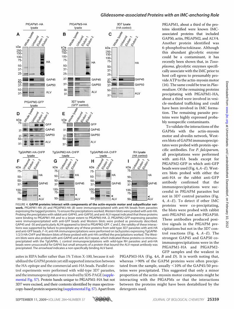
asites in RIPA buffer rather than 1% Triton X-100, because it sol-ubilized theGAPMproteinyet still supported interactionbetweenthe HA-epitope and the commercial anti-HA beads. Parallel con-trol experiments were performed with wild-type 3D7 parasites,andthe immunoprecipitateswereresolvedbySDS-PAGE(supple-mental Fig. S7). Protein bands unique to PfGAPM1-HA but not3D7 were excised, and their contents identified bymass spectros-copy-basedprotein sequencing (supplementalFig. S7).Apart from
PfGAPM1, about a third of the pro-teins identified were known IMC-associated proteins that includedGAP50, actin, PfGAPM2, and ALV4.Another protein identified was6-phosphofructokinase. Althoughthis abundant glycolytic enzymecould be a contaminant, it hasrecently been shown that, in Toxo-plasma, glycolytic enzymes specifi-cally associate with the IMCprior tohost cell egress to presumably pro-videATP to the actin-myosinmotor(16). The same could be true inPlas-modium. Of the remaining proteinsprecipitating with PfGAPM1-HA,about a third were involved in vesi-cle-mediated trafficking and couldhave been involved in IMC forma-tion. The remaining parasite pro-teins were highly expressed proba-bly nonspecific contaminants.To validate the interactions of the
GAPMs with the actin-myosinmotor and alveolin network, West-ern blots of GAPM immunoprecipi-tates were probed with protein-spe-cific antibodies. For P. falciparum,all precipitations were performedwith anti-HA beads except forPfGAPM2-GFP in which anti-GFPbeadswere used (Fig. 4,A–E).West-ern blots probed with either theanti-HA or the rabbit anti-GFPantibody confirmed that theimmunoprecipitations were suc-cessful in PfGAPM parasites butnot in 3D7 control parasites (Fig.4, A–E). To detect if other IMCproteins were co-precipitating,the blots were probed with rabbitanti-PfGAP45 and anti-PfGAP50.These antibodies produced posi-tive signals in the PfGAPM pre-cipitations but not in the 3D7 con-trol reactions (Fig. 4, A–E). Thestrongest GAP45 and GAP50 co-immunoprecipitations were in thePfGAPM1-HA and PfGAPM2-GFP samples and the weakest in
PfGAPM3-HA (Fig. 4A, B and D). It is worth noting that,whereas �90% of the GAPM proteins were often precipi-tated from the sample, usually �10% of the GAP45/50 pro-teins were precipitated. This suggested that only a minorproportion of the actin-myosin motor components might beinteracting with the PfGAPMs or that the interactionsbetween the proteins might have been destabilized by thedetergents used.
FIGURE 4. GAPM proteins interact with components of the actin-myosin motor and subpellicular net-work. PfGAPM1-HA (A) and PfGAPM3-HA (B) were immunoprecipitated with anti-HA beads from parasitesexpressing the tagged proteins. To ensure the precipitations worked, Western blots were probed with anti-HA.Probing the precipitates with rabbit anti-GAP45, anti-GAP50, and anti-ALV repeat indicated that these proteinswere binding to PfGAPM1-HA and to a lesser extent to PfGAPM3-HA. D, PfGAPM2-GFP-expressing parasiteswere immunoprecipitated with anti-GFP beads and Western blots were probed as previously described.GAP45 and -50 and particularly ALV4 appeared to bind to PfGAPM2-GFP. C and E, the validity of these interac-tions was supported by failure to precipitate any of these proteins from wild type 3D7 parasites with anti-HAand anti-GFP beads. F–H, anti-HA immunoprecipitations were performed on tachyzoites expressing TgGAPM-1/2/3-HA-ChFP and Western blots of these probed with anti-HA certified the precipitations worked. The West-ern blots were also probed with anti-GAP45 and anti-ALV repeat, which indicated these proteins co-immuno-precipitated with the TgGAPMs. I, control immunoprecipitations with wild-type RH parasites and anti-HAbeads were unsuccessful for GAP45 but small amounts of a protein that bound the ALV-repeat antibody wasprecipitated. The arrowhead indicates a non-specifically binding ALV band.
Glideosome-associated Proteins with an IMC-anchoring Role
SEPTEMBER 11, 2009 • VOLUME 284 • NUMBER 37 JOURNAL OF BIOLOGICAL CHEMISTRY 25359
at Universitaets- und Landesbibliothek D
uesseldorf, on February 10, 2012
ww
w.jbc.org
Dow
nloaded from

Interactions of PfGAPMwith the alveolins were also investi-gated by probing with anti-ALV. This antibody detected a42-kDa band in precipitates probably corresponding to PfALV4in all three GAPMs (Fig. 4, A–E). An additional �35-kDa bandwas observed in the PfGAPM2-GFP precipitation that could beALV5 (Fig. 4D). PfGAPM1, PfGAPM2, and PfGAPM3, respec-tively, precipitated �17%, 26%, and 16% of the total ALVdetected in the sample. A degree of specificity was thereforeobserved between the PfGAPMs with PfGAPM2-GFP precipi-tating the most ALV4, as well as the additional alveolin, ALV5(Fig. 4D).When probed with anti-HA, a ladder of proteins ranging
from 28 kDa to 150 kDa was seen in the second elution for bothPfGAPM1-HA and PfGAPM3-HA immunoprecipitations (Fig.4,A andB). The origins of these ladders are unclear because thetwo-dimensional sample buffer used to solubilize the elutedproteins should have dissociated the complexes. We speculatethat the high concentration of PfGAPM proteins collecting onthe anti-HA agarose beads during the immunoprecipitationmay have caused the hydrophobic PfGAPM proteins to aggre-gate, further increasing their resistance to complete denatur-ation into monomers and dimers.To investigate whether T. gondii GAPMs also co-immuno-
precipitatedmembers of the actin-myosinmotor, transgenicT.gondii parasites expressing C-terminally ChFP/HA-taggedGAPM proteins were used in immunoprecipitation assays asabove. These samples were subsequently analyzed by Westernblotting that revealed each TgGAPM investigated also co-im-munoprecipitated TgGAP45 while RH-strain control parasitesdid not. Additionally, ALV-specific bands were detected at 62,74, 82, and 92 kDa for each TgGAPM (Fig. 4, F–H); however,because minor amounts of the 62-kDa band were also detectedin RH-strain control parasites, this might not represent a gen-uine TgGAPM-specific interaction. Toxoplasma encodesnearly twice as many alveolins as P. falciparum (12 versus 7) soit is not possible to speculate which specific alveolins wereinteracting with the TgGAPM1/2/3-ChFP-HA proteins. Wenote however that similar sized alveolins were previouslydetected with a TgALV3 antibody (34).Aswas seen for PfGAPM immunoprecipitations, only a small
fraction of the total TgGAP45 and ALV proteins were detectedin the immunoprecipitated material. We were unable to inves-tigate proposed TgGAPM interactions with TgGAP50 due to alack of antibody to this protein.To validate the proposed interactions between the GAPM
proteins and the actin-myosin motor, reciprocal immunopre-cipitation experiments were performed. Anti-ALV and anti-GAP45 antibodieswere found to interact poorlywith their anti-gens in RIPA buffer (data not shown), therefore reciprocalimmunoprecipitations were performed in 1% Triton X-100.Tagged P. falciparum GAPM schizonts were solubilized andincubated with either rabbit anti-ALV or rabbit anti-PfGAP45.Immune complexes were precipitated with anti-rabbit conju-gated Dynal beads. Antibodies specific to PfGAP45 co-precip-itated small amounts of PfGAPM2 and PfGAPM3 (Fig. 5, B andC). Slightly larger amounts of these proteins were co-precipi-tated with anti-ALV suggestive of a more extensive interactionbetween the GAPMs and the ALVs than between the GAPMs
and PfGAP45 (Fig. 5, B and C). PfGAPM1 was co-immunopre-cipitated poorly with ALV and GAP45 probably due to its poorsolubility in Triton X-100 (Fig. 5A).
DISCUSSION
The GAPMs identified in this study constitute a three-member family of six-pass transmembrane proteins presentin all sequenced Apicomplexa. In the groups tested here,namely Plasmodium blood-stage merozoites and Toxo-plasma tachyzoites, the GAPMs localize to the IMC imply-ing that the IMCs of all motile Apicomplexan zoite stages willcontain these proteins. No related proteins are present outsideof this phylum suggesting that GAPMs perform vital, Apicom-plexan-specific functions.The tagged GAPMs interact with the cytoskeletal alveolins
and to a slightly lesser degree the actin-myosin motor compo-
FIGURE 5. Reciprocal immunoprecipitation assays indicate that thealveolins and GAP45 interact with GAPM proteins. Western blots of alveo-lin and GAP45 immunoprecipitation assays on lysates from P. falciparum schi-zonts expressing either PfGAPM1-HA (A), PfGAPM2-GFP (B), or PfGAPM3-HA(C) were probed with antibodies listed in individual panels. Probing with anti-ALV or anti-GAP45 confirmed that immunoprecipitations were successful.Probing with either anti-HA (A and C) or anti-GFP (B) indicated that eachGAPM co-purified with the alveolins and GAP45. The arrow represents theanti-ALV IgG band.
Glideosome-associated Proteins with an IMC-anchoring Role
25360 JOURNAL OF BIOLOGICAL CHEMISTRY VOLUME 284 • NUMBER 37 • SEPTEMBER 11, 2009
at Universitaets- und Landesbibliothek D
uesseldorf, on February 10, 2012
ww
w.jbc.org
Dow
nloaded from

nentsGAP45/50. This suggests that theGAPMsmight functionas protein “rivets” that attach the IMC to the cytoskeleton oreven as protein tethers that span the IMC to link and immobi-lize the myosin complex onto the cytoskeleton (Fig. 6). If true,then why have previous immunoprecipitation-based attemptsto identify anchors of the GAP45/50/myosin complex failed toidentify the GAPMs (11, 12)? We believe these attempts mayhave failed because the interactions of the GAPMs withGAP45/50 do not survive the immunoprecipitation processwell and the GAPMs are difficult to resolve inmonomeric formby SDS-PAGE and to detect without specific reagents.With sixtransmembrane-spanning domains and the size of the com-plexes they form (�200 kDa) the GAPMs are probably firmlyembedded in the IMCmembrane. For immunoprecipitation towork the GAPMs have to be solubilized, which in their caserequires extensive sonication in the stringent RIPA-detergentmix. Both of these treatments can disrupt protein-proteininteractions, and it is no surprise that little (�10%) of theGAP45/50 andGAPM interaction survives co-immunoprecipi-tation. The sonication is probably necessary to shear thecytoskeleton and detach the IMC, which is also why only�20%of the available alveolins co-precipitate with the GAPMs.To validate interaction of the GAPMs with the alveolins and
GAP45/50 we attempted reciprocal immunoprecipitationswith anti-PfGAP45 in P. falciparum, anti-TgGAP45 in Toxo-plasma, and anti-ALV in both species. We found these immu-noprecipitations worked poorly in RIPA, because the stringentbuffer reduced interaction of the antibodies with their ligands.These precipitations were therefore repeated in 1% TritonX-100. In P. falciparum this detergent easily extractedGAP45/50 but only poorly the PfGAPMs, especially PfGAPM1.This probably accounts for why the GAPMs were only poorlyco-precipitated with GAP45 and alveolin, with much of theGAPMs remaining in the pellet after detergent extraction. Pre-vious attempts to identify GAP45/50/myosin-tethering pro-teins in Toxoplasma were probably unsuccessful because they
were performed with Triton X-100,and the GAPMs in this parasite areeven more insoluble than in P. fal-ciparum (11). Chemical cross-link-ers were also used to identify pro-teins attaching to the GAP45/50/myosin complex (12) but as we haveshown they cause the GAPMs tobecome extremely insoluble.Even if the GAPM proteins could
be co-immunoprecipitated in ap-preciable amounts with GAP45/50,theymayhave avoideddetection afterSDS-PAGE, because they migratedas indistinct high molecular weightaggregates.We found it necessary totreat the immunoprecipitates withtwo-dimensional sample buffer todissociate the GAPMs into mono-meric form. We note that, althoughPfGAPM2-GFP was the most easilyextracted protein, this may have
been assisted by the highly soluble GFPmoiety that is almost aslarge as PfGAPM2 itself.All of our P. falciparum immunoprecipitations with the
GAPs, GAPMs, and alveolins successfully co-precipitated eachother suggesting they all physically bind to each other thoughnot necessarily in equimolar ratios. Another interpretation ofour data is that small fragments of IMC containing all of theseproteins are being nonspecifically precipitated, because theyare resistant to mechanical and detergent disruption. Thesefragments would, however, have to be small, because the para-site lysates were cleared by centrifugation prior to immunopre-cipitation.We also observed a certain degree of specificity withour immunoprecipitations that argues against theGAPMspull-ing down IMC fragments with attached cytoskeleton. Forexample, PfGAPM2 specifically precipitates two alveolins,whereas the others only precipitate a single cytoskeletal pro-tein. To absolutely resolve the IMC fragment issue wouldrequire the use of a resident IMC protein known not to bind tothe proteins mentioned above as a negative control in theimmunoprecipitation reactions, but unfortunately no suchpro-tein is known. In conclusion, based on the numerous times wehave repeated these precipitations and the fact that some workin the stringent RIPA buffer, we are confident that GAPMs docontact both the alveolin cytoskeleton on one side of the IMCand the GAP-myosin complex on the other side (Fig. 6).In each of the immunoprecipitation assays shown here, the
strongest interactions were seen between the GAPMs and thealveolins of the subpellicular network. This network resides onthe cytoplasmic face of the IMC suggesting that the GAPMsmay also localize to this face. In addition, the organization ofGAPMs into highly stable oligomers makes these proteinsprime candidates for being constituents of the uncharacterizedfamily of 9 nm intramembranous particles (IMPs) seenthroughout the IMC in freeze-fracture studies (6, 13–15). It hasbeen observed that IMPs localize to different parts of the IMC.A single line of IMPs overlay the cytoskeletal filaments (proba-
FIGURE 6. Proposed model of GAPM interaction. GAPM proteins reside within the IMC and have been shownto interact with components of the actin-myosin motor and subpellicular network. The plus signs represent thesolubility of each of the proteins in each buffer as judged by Western blotting: �, proteins readily soluble inlisted buffer; ����, proteins poorly soluble in listed buffer.
Glideosome-associated Proteins with an IMC-anchoring Role
SEPTEMBER 11, 2009 • VOLUME 284 • NUMBER 37 JOURNAL OF BIOLOGICAL CHEMISTRY 25361
at Universitaets- und Landesbibliothek D
uesseldorf, on February 10, 2012
ww
w.jbc.org
Dow
nloaded from
bly 10 nm alveolin containing filaments) (15) and a double rowlays over microtubules (13). Furthermore, other types of IMPsmight form pores in the IMC or may suture the edges of theflattened vesicles that comprise the IMC (14). This degree ofspecialization hints that if the GAPMs are indeed true IMPproteins, then each type of IMP might contain a differentGAPM.To establishwhether theGAPMs bind to each otherweattempted tomake specific antibodies to theN- andC-terminaltails of PfGAPM1 to determine if the protein co-immunopre-cipitates with the GFP- and HA-tagged PfGAPM2 andPfGAPM3 proteins. Unfortunately, the PfGAPM1 antibodiesperformed very poorly (data not shown), so specific antibodieswill have to be raised to the other GAPMs. It is worth noting,though, that when partially cross-linked, the PfGAPM1-HAand -3HA proteinsmigrated in precise increments of their ownmolecular weights hinting that they might be homo-oli-gomers. Sizes up to the pentamer (�150 kDa) have beendetected by immunoblot analysis of partially cross-linkedmaterial, although to form a 9 nm particle the protein com-plexes would probably have to be �500 kDa.We have attempted immunoelectron microscopy to deter-
mine if the GAPMs are components of these 9 nm IMPs. Ifpresent, immunogold particles specific for the HA-taggedGAPMs might label the IMC with a periodicity correspondingto IMPs observed by freeze fracture microscopy. Unfortu-nately, the immunoelectron microscopy experiments were notsuccessful because we could not achieve specific labeling of thedeveloping IMC in gluteraldehyde-fixed schizont stage P. fal-ciparum parasites (data not shown). It is possible that the glu-teraldehyde used for electron microscopy may have cross-linked the GAPMs to such an extent that access to the HAepitope was greatly reduced, resulting in poor labeling. Sup-porting this is our finding that GAPM labeling with anti-HA oranti-GFP is greatly reduced in immunofluorescence micros-copy of cells fixed in paraformaldehyde when compared withmethanol/acetone (data not shown). Successful immunoelec-tron microscopy of the GAPMs may therefore require alterna-tive cryo-preservation methods.The GAPM proteins appear to form complexes, possibly
homo-oligomers, though how large they are remains to beascertained. Unless reduced to mono- or dimeric forms theGAPM proteins when in complexes appear very insoluble.The presence of GAPMs in IMPs would explain why theGAPMs poorly precipitate GAPs and alveolins. The largemembrane particles may only bind to a small number ofneighboring actin-myosin complexes and alveolin subunitsparticularly if the latter were forming a filament. Data pre-sented here suggest that the GAPMproteins form complexeson the cytoplasmic face of the IMC where they interact withcomponents of the subpellicular network. This localizationwould permit interactions with the GAP45/50 complex,because it is believed that the majority of GAP50 resideswithin the IMC (11) and would explain why only a smallproportion of the GAP45/50 proteins can be co-immunopre-cipitated with the GAPM proteins (Fig. 6). Establishingwhere the GAPMs reside within the IMC and what each trulyinteracts with will do much to determine the structural
organization of this important apicomplexan organelle ofmotility.
Acknowledgments—We thank Jake Baum for the GAP45 and -50antibodies, Con Beckers for the TgGAP45 antibody, and Paul Sandersand Thomas Nebl for technical advice. We also thank Giel vanDooren of the University of Georgia, Athens, for his gift of the T. gondiitransfection plasmid pCTGand J. I. Rood,D. Lyras, andC. L. KennedyofMonashUniversity, Victoria, Australia for the kind provision of theC. septicum �-toxin. We also thank the Walter and Eliza Hall Insti-tute’s antibody production facility and the Australian Red Cross forsupplying blood.
REFERENCES1. Snow, R.W., Guerra, C. A., Noor, A.M.,Myint, H. Y., andHay, S. I. (2005)
Nature 434, 214–2172. Baum, J., Gilberger, T. W., Frischknecht, F., and Meissner, M. (2008)
Trends Parasitol. 24, 557–5633. Cowman, A. F., and Crabb, B. S. (2006) Cell 124, 755–7664. Tardieux, I., and Menard, R. (2008) Traffic 9, 627–6355. Keeley, A., and Soldati, D. (2004) Trends Cell Biol. 14, 528–5326. Soldati, D., and Meissner, M. (2004) Curr. Opin Cell Biol. 16, 32–407. Bergman, L. W., Kaiser, K., Fujioka, H., Coppens, I., Daly, T. M., Fox, S.,
Matuschewski, K., Nussenzweig, V., and Kappe, S. H. (2003) J. Cell Sci.116, 39–49
8. Pinder, J. C., Fowler, R. E., Dluzewski, A. R., Bannister, L. H., Lavin, F. M.,Mitchell, G. H., Wilson, R. J., and Gratzer, W. B. (1998) J. Cell Sci. 111,1831–1839
9. Bannister, L. H., Hopkins, J. M., Fowler, R. E., Krishna, S., and Mitchell,G. H. (2000) Parasitology 121, 273–287
10. Pinder, J., Fowler, R., Bannister, L., Dluzewski, A., and Mitchell, G. H.(2000) Parasitol Today 16, 240–245
11. Gaskins, E., Gilk, S., DeVore, N., Mann, T., Ward, G., and Beckers, C.(2004) J. Cell Biol. 165, 383–393
12. Johnson, T.M., Rajfur, Z., Jacobson, K., and Beckers, C. J. (2007)Mol. Biol.Cell 18, 3039–3046
13. Morrissette, N. S., Murray, J. M., and Roos, D. S. (1997) J. Cell Sci. 110,35–42
14. Raibaud, A., Lupetti, P., Paul, R. E., Mercati, D., Brey, P. T., Sinden, R. E.,Heuser, J. E., and Dallai, R. (2001) J. Struct. Biol. 135, 47–57
15. Mann, T., and Beckers, C. (2001)Mol. Biochem. Parasitol. 115, 257–26816. Pomel, S., Luk, F. C., and Beckers, C. J. (2008) PLoS Pathog. 4, e100018817. Sanders, P. R., Gilson, P. R., Cantin, G. T., Greenbaum, D. C., Nebl, T.,
Carucci, D. J., McConville, M. J., Schofield, L., Hodder, A. N., Yates, J. R.,3rd, and Crabb, B. S. (2005) J. Biol. Chem. 280, 40169–40176
18. Sanders, P. R., Cantin, G. T., Greenbaum, D. C., Gilson, P. R., Nebl, T.,Moritz, R. L., Yates, J. R., 3rd, Hodder, A. N., and Crabb, B. S. (2007)Mol.Biochem. Parasitol. 154, 148–157
19. Edgar, R. C. (2004) BMC Bioinformatics 5, 11320. Gilson, P. R., O’Donnell, R. A., Nebl, T., Sanders, P. R., Wickham, M. E.,
McElwain, T. F., de Koning-Ward, T. F., and Crabb, B. S. (2008) Mol.Microbiol. 68, 124–138
21. Meissner, M., Krejany, E., Gilson, P. R., de Koning-Ward, T. F., Soldati,D., and Crabb, B. S. (2005) Proc. Natl. Acad. Sci. U.S.A. 102, 2980–2985
22. van Dooren, G. G., Tomova, C., Agrawal, S., Humbel, B. M., and Striepen,B. (2008) Proc. Natl. Acad. Sci. U. S. A. 105, 13574–13579
23. Trager, W., and Jensen, J. B. (1976) Science 193, 673–67524. Crabb, B. S., Rug,M., Gilberger, T.W., Thompson, J. K., Triglia, T., Maier,
A. G., and Cowman, A. F. (2004)Methods Mol. Biol. 270, 263–27625. Striepen, B., He, C. Y., Matrajt, M., Soldati, D., and Roos, D. S. (1998)Mol.
Biochem. Parasitol. 92, 325–33826. Baum, J., Richard, D., Healer, J., Rug, M., Krnajski, Z., Gilberger, T. W.,
Green, J. L., Holder, A. A., and Cowman, A. F. (2006) J. Biol. Chem. 281,5197–5208
27. Wichroski, M. J., Melton, J. A., Donahue, C. G., Tweten, R. K., andWard,
Glideosome-associated Proteins with an IMC-anchoring Role
25362 JOURNAL OF BIOLOGICAL CHEMISTRY VOLUME 284 • NUMBER 37 • SEPTEMBER 11, 2009
at Universitaets- und Landesbibliothek D
uesseldorf, on February 10, 2012
ww
w.jbc.org
Dow
nloaded from
G. E. (2002) Infect. Immun. 70, 4353–436128. Aurrecoechea, C., Brestelli, J., Brunk, B. P., Dommer, J., Fischer, S.,
Gajria, B., Gao, X., Gingle, A., Grant, G., Harb, O. S., Heiges, M.,Innamorato, F., Iodice, J., Kissinger, J. C., Kraemer, E., Li, W., Miller,J. A., Nayak, V., Pennington, C., Pinney, D. F., Roos, D. S., Ross, C.,Stoeckert, C. J., Jr., Treatman, C., and Wang, H. (2009) Nucleic AcidsRes. 37, D539–D543
29. Krogh, A., Larsson, B., von Heijne, G., and Sonnhammer, E. L. (2001) J.Mol. Biol. 305, 567–580
30. Aurrecoechea, C., Heiges, M., Wang, H., Wang, Z., Fischer, S., Rhodes, P.,Miller, J., Kraemer, E., Stoeckert, C. J., Jr., Roos, D. S., and Kissinger, J. C.(2007) Nucleic Acids Res. 35, D427–D430
31. Giepmans, B. N., Adams, S. R., Ellisman, M. H., and Tsien, R. Y. (2006)Science 312, 217–224
32. Bozdech, Z., Llinas,M., Pulliam, B. L.,Wong, E. D., Zhu, J., andDeRisi, J. L.(2003) PLoS Biol. 1, E5
33. Le Roch, K. G., Johnson, J. R., Florens, L., Zhou, Y., Santrosyan, A.,Grainger, M., Yan, S. F., Williamson, K. C., Holder, A. A., Carucci, D. J.,Yates, J. R., 3rd, andWinzeler, E. A. (2004)Genome Res. 14, 2308–2318
34. Gould, S. B., Tham, W. H., Cowman, A. F., McFadden, G. I., and Waller,R. F. (2008)Mol. Biol. Evol. 25, 1219–1230
35. Le Roch, K. G., Zhou, Y., Blair, P. L., Grainger, M., Moch, J. K., Haynes,J. D., De LaVega, P., Holder, A. A., Batalov, S., Carucci, D. J., andWinzeler,E. A. (2003) Science 301, 1503–1508
Glideosome-associated Proteins with an IMC-anchoring Role
SEPTEMBER 11, 2009 • VOLUME 284 • NUMBER 37 JOURNAL OF BIOLOGICAL CHEMISTRY 25363
at Universitaets- und Landesbibliothek D
uesseldorf, on February 10, 2012
ww
w.jbc.org
Dow
nloaded from


![Clad Inner Surface Temperature l°C]](https://static.fdokumen.com/doc/165x107/633831c324ea072f160c74b1/clad-inner-surface-temperature-lc.jpg)


