Enzymes of type II fatty acid synthesis and apicoplast differentiation and division in Eimeria...
19
Enzymes of type II fatty acid synthesis and apicoplast differentiation and division in Eimeria tenella q D.J.P. Ferguson a, * , S.A. Campbell b,1 , F.L. Henriquez b,2 , L. Phan c , E. Mui c , T.A. Richards d , S.P. Muench e , M. Allary f , J.Z. Lu f , S.T. Prigge f , F. Tomley g , M.W. Shirley g , D.W. Rice e , R. McLeod c , C.W. Roberts b a Nuffield Department of Pathology, University of Oxford, John Radcliffe Hospital, Oxford OX3 9DU, UK b Department of Immunology, University of Strathclyde, Glasgow G4 0NR, UK c Department of Ophthalmology and Visual Sciences, Pediatrics (Infectious Diseases), Pathology and Committees on Genetic, Molecular Medicine, and Immunology, University of Chicago, Chicago, IL 60637, USA d School of Biosciences, University of Exeter, Geoffrey Pope Building, Stocker Road, Exeter EX4 4QG, UK e Department of Molecular Biology and Biotechnology, The University of Sheffield, Sheffield S10 2TN, UK f School of Public Health, Johns Hopkins University, Baltimore, MD 21205, USA g Division of Molecular Biology, Institute of Animal Health, Compton, Newbury, Berkshire RG20 7NN, UK Received 24 July 2006; received in revised form 26 September 2006; accepted 3 October 2006 Abstract Apicomplexan parasites, Eimeria tenella, Plasmodium spp. and Toxoplasma gondii, possess a homologous plastid-like organelle termed the apicoplast, derived from the endosymbiotic enslavement of a photosynthetic alga. However, currently no eimerian nuclear encoded apicoplast targeted proteins have been identified, unlike in Plasmodium spp. and T. gondii. In this study, we demonstrate that nuclear encoded enoyl reductase of E. tenella (EtENR) has a predicted N-terminal bipartite transit sequence, typical of apicoplast-tar- geted proteins. Using a combination of immunocytochemistry and EM we demonstrate that this fatty acid biosynthesis protein is located in the apicoplast of E. tenella. Using the EtENR as a tool to mark apicoplast development during the Eimeria lifecycle, we demonstrate that nuclear and apicoplast division appear to be independent events, both organelles dividing prior to daughter cell formation, with each daughter cell possessing one to four apicoplasts. We believe this is the first report of multiple apicoplasts present in the infectious stage of an apicomplexan parasite. Furthermore, the microgametes lacked an identifiable apicoplast consistent with maternal inheritance via the macrogamete. It was found that the size of the organelle and the abundance of EtENR varied with developmental stage of the E. tenella lifecycle. The high levels of EtENR protein observed during asexual development and macrogametogony is potentially associated with the increased synthesis of fatty acids required for the rapid formation of numerous merozoites and for the extracellular development and survival of the oocyst. Taken together the data demonstrate that the E. tenella apicoplast participates in type II fatty acid biosynthesis with increased expression of ENR during parasite growth. Apicoplast division results in the simultaneous formation of multiple frag- ments. The division mechanism is unknown, but is independent of nuclear division and occurs prior to daughter formation. Ó 2006 Australian Society for Parasitology Inc. Published by Elsevier Ltd. All rights reserved. Keywords: Eimeria tenella; Apicoplast; Enoyl reductase; Immunocytochemistry; Stage specific expression 1. Introduction The group of parasites forming the Apicomplexa con- tains numerous and diverse parasitic forms of medical and veterinary importance (Fast et al., 2002). The signifi- cance of Plasmodium spp. (malaria causes two million 0020-7519/$30.00 Ó 2006 Australian Society for Parasitology Inc. Published by Elsevier Ltd. All rights reserved. doi:10.1016/j.ijpara.2006.10.003 q Nucleotide sequences reported in this paper are available in the GenBank database under the accession number AY566297. * Corresponding author. Tel.: +44 1865 220 514; fax: +44 1865 228 980. E-mail address: [email protected] (D.J.P. Ferguson). 1 Present address: School of Life Sciences, Faculty of Health and Life Sciences, Napier University, Edinburgh, UK. 2 Present address: School of Engineering and Science, University of Paisley, Paisley, Scotland, UK. www.elsevier.com/locate/ijpara International Journal for Parasitology 37 (2007) 33–51
-
Upload
independent -
Category
Documents
-
view
3 -
download
0
Transcript of Enzymes of type II fatty acid synthesis and apicoplast differentiation and division in Eimeria...
www.elsevier.com/locate/ijpara
International Journal for Parasitology 37 (2007) 33–51
Enzymes of type II fatty acid synthesis and apicoplastdifferentiation and division in Eimeria tenella q
D.J.P. Ferguson a,*, S.A. Campbell b,1, F.L. Henriquez b,2, L. Phan c, E. Mui c,T.A. Richards d, S.P. Muench e, M. Allary f, J.Z. Lu f, S.T. Prigge f, F. Tomley g,
M.W. Shirley g, D.W. Rice e, R. McLeod c, C.W. Roberts b
a Nuffield Department of Pathology, University of Oxford, John Radcliffe Hospital, Oxford OX3 9DU, UKb Department of Immunology, University of Strathclyde, Glasgow G4 0NR, UK
c Department of Ophthalmology and Visual Sciences, Pediatrics (Infectious Diseases), Pathology and Committees on Genetic,
Molecular Medicine, and Immunology, University of Chicago, Chicago, IL 60637, USAd School of Biosciences, University of Exeter, Geoffrey Pope Building, Stocker Road, Exeter EX4 4QG, UKe Department of Molecular Biology and Biotechnology, The University of Sheffield, Sheffield S10 2TN, UK
f School of Public Health, Johns Hopkins University, Baltimore, MD 21205, USAg Division of Molecular Biology, Institute of Animal Health, Compton, Newbury, Berkshire RG20 7NN, UK
Received 24 July 2006; received in revised form 26 September 2006; accepted 3 October 2006
Abstract
Apicomplexan parasites, Eimeria tenella, Plasmodium spp. and Toxoplasma gondii, possess a homologous plastid-like organelletermed the apicoplast, derived from the endosymbiotic enslavement of a photosynthetic alga. However, currently no eimerian nuclearencoded apicoplast targeted proteins have been identified, unlike in Plasmodium spp. and T. gondii. In this study, we demonstrate thatnuclear encoded enoyl reductase of E. tenella (EtENR) has a predicted N-terminal bipartite transit sequence, typical of apicoplast-tar-geted proteins. Using a combination of immunocytochemistry and EM we demonstrate that this fatty acid biosynthesis protein is locatedin the apicoplast of E. tenella. Using the EtENR as a tool to mark apicoplast development during the Eimeria lifecycle, we demonstratethat nuclear and apicoplast division appear to be independent events, both organelles dividing prior to daughter cell formation, with eachdaughter cell possessing one to four apicoplasts. We believe this is the first report of multiple apicoplasts present in the infectious stage ofan apicomplexan parasite. Furthermore, the microgametes lacked an identifiable apicoplast consistent with maternal inheritance via themacrogamete. It was found that the size of the organelle and the abundance of EtENR varied with developmental stage of the E. tenella
lifecycle. The high levels of EtENR protein observed during asexual development and macrogametogony is potentially associated withthe increased synthesis of fatty acids required for the rapid formation of numerous merozoites and for the extracellular development andsurvival of the oocyst. Taken together the data demonstrate that the E. tenella apicoplast participates in type II fatty acid biosynthesiswith increased expression of ENR during parasite growth. Apicoplast division results in the simultaneous formation of multiple frag-ments. The division mechanism is unknown, but is independent of nuclear division and occurs prior to daughter formation.� 2006 Australian Society for Parasitology Inc. Published by Elsevier Ltd. All rights reserved.
Keywords: Eimeria tenella; Apicoplast; Enoyl reductase; Immunocytochemistry; Stage specific expression
0020-7519/$30.00 � 2006 Australian Society for Parasitology Inc. Published b
doi:10.1016/j.ijpara.2006.10.003
q Nucleotide sequences reported in this paper are available in theGenBank database under the accession number AY566297.
* Corresponding author. Tel.: +44 1865 220 514; fax: +44 1865 228 980.E-mail address: [email protected] (D.J.P. Ferguson).
1 Present address: School of Life Sciences, Faculty of Health and LifeSciences, Napier University, Edinburgh, UK.
2 Present address: School of Engineering and Science, University ofPaisley, Paisley, Scotland, UK.
1. Introduction
The group of parasites forming the Apicomplexa con-tains numerous and diverse parasitic forms of medicaland veterinary importance (Fast et al., 2002). The signifi-cance of Plasmodium spp. (malaria causes two million
y Elsevier Ltd. All rights reserved.

34 D.J.P. Ferguson et al. / International Journal for Parasitology 37 (2007) 33–51
childhood deaths per year) and Toxoplasma gondii (two bil-lion chronically infected individuals) as human pathogensare well known (Ferguson, 2002; Snow et al., 2005). Eime-
ria spp. are important pathogens in domestic animals beingassociated with losses of approximately two billion dollarsper year in the poultry industry of the USA alone (Wallach,2002). It is such a serious and intractable problem thatcommercial poultry food contains drugs to control theinfection. Apicomplexan parasites are characterised by aset of organelles (rhoptries, micronemes and dense gran-ules) located in the apical cytoplasm (Levine et al., 1980).However, an additional organelle, found in many membersof this phylum, is the apicoplast. The consensus is that thisorganelle is the plastid remnant of an endosymbiontalthough the exact origin is still the subject of debate(reviewed Wilson, 2002; Waller and McFadden, 2005).Studies using in situ hybridisation, have demonstrated thepresence of a multi-membraned (four unit membranes)organelle present in the peri-nuclear cytoplasm of T. gondii
with a 35 kb circular genome (Kohler et al., 1997; McFad-den and Waller, 1997). Thus the apicoplast is different fromthe other DNA-containing organelle, the mitochondrion,which is encapsulated by two membranes. The apicoplasthad previously been identified as a distinct organelle byEM and termed the Golgi adjunct in T. gondii (Sheffieldand Melton, 1968) or the multi-membranous organelle inEimeria species (Ferguson et al., 1976). The presence ofmultiple (four) membranes is believed to be the conse-quence of the secondary endosymbiotic event (Kohleret al., 1997; Cavalier-Smith, 2000). Although the apicoplasthas its own small genome, many of the apicoplast-specificproteins are coded by genes located in the nucleus, whichare subsequently transferred into the apicoplast (Walleret al., 1998). The functions of the apicoplast are still incom-pletely understood, but it is known to be involved in type IIfatty acid synthesis and isoprenoid biosynthesis in T. gondii
plus heme biosynthesis in Plasmodium spp. (Waller et al.,1998; Zuther et al., 1999; McLeod et al., 2001; Suroliaand Surolia, 2001; Wilson, 2002; Roberts et al., 2003;Ralph et al., 2004; Waller and McFadden, 2005).
To date, studies of the apicoplast have been limited tothe in vitro proliferative form (tachyzoite) of T. gondii,the erythrocytic stages of P. falciparum and the asexualmultiplication of Sarcocystis neurona (reviewed Vaishnavaand Striepen, 2006). In addition, stage specific variationsduring in vivo development of T. gondii in both the inter-mediate and final hosts have been described (Fergusonet al., 2005). Recently, the E. tenella apicoplast genomehas been sequenced (Cai et al., 2003). However, to dateno nuclear encoded, apicoplast localised proteins have beenidentified and little is known about the development ordivision of the apicoplast in Eimeria spp. during a lifecyclethat has many different developmental stages.
In the present study, we have examined the E. tenellagenome database (www.sanger.ac.uk/Projects/E_tenella/)to identify the genes involved in type II fatty acid synthesis.Enoyl reductase was identified, sequenced and expressed as
a recombinant protein. Antibodies to the recombinant eno-yl reductase of E. tenella (EtENR), raised in mice, wereemployed using light and EM immunocytochemical tech-niques to study the location of the apicoplast and alsothe relative level of expression of EtENR during in vivodevelopment. This was correlated with detailed ultrastruc-tural studies of the apicoplast and its division duringasexual and sexual development.
2. Materials and methods
2.1. Parasites
The Houghton strain of E. tenella was used in this study.
2.2. Identification of genes involved in type 2 fatty acid
synthesis
The E. tenella genome project was searched for enzymesinvolved in type II fatty acid synthesis. Preliminary datawas obtained from the E. tenella genome sequencing pro-ject (Wellcome Trust Sanger Institute http://www.sanger.ac.uk/cgi-bin/blast/submitblast/e_tenella/omni). Using thetBLASTn algorithm, the genome was interrogated withknown enzyme amino acid sequences to identify DNAsequences containing potential coding regions homologousto the enzymes (acetyl-coA carboxylase, acetyl-CoA-ACPtransacylase, malonyl-CoA-ACP transacylase [ACAT],b-ketoacyl-ACP synthase [b-KAS], b-ketoacyl-ACP reduc-tase [b-KAR], b-hydroxyacyl-ACP dehydrase [b-HAD]and enoyl-ACP reductase [ENR]).
2.3. Cloning, expression and purification of recombinant
EtENR
RNA was extracted from sporozoite stage E. tenella
W15 parasites using RNAeasy Maxi kit (Qiagen, Crawley,UK) as per the manufacturer’s instructions and used togenerate cDNA using Superscript III reverse transcriptase(Invitrogen, Paisley, UK) as per the manufacturer’sinstructions.
The coding sequence of E. tenella ENR was amplifiedfrom cDNA using the following primers EtENR for 5 0
GTGTCGGCTGTCACCTCC3 0 and EtENRrev 5 0CTCAAGGGGTGAGGGACTTG3 0. PCR reactions were per-formed in a volume of 25 ll and contained a final concentra-tion of 1· ReddyMix�. PCR Mastermix with 1.5 mMMgCl2 (ABgene) and 25 pM of each primer. PCR productswere separated on a 1% agarose gel and visualised by ethi-dium bromide staining. Following excision from the gel,PCR products were cleaned up using the QIAquick Gel Puri-fication Kit (Qiagen) according to the manufacturer’sinstructions and ligated into pDRIVE using the QiagenPCR Cloning Kit (Qiagen). Plasmid DNA was isolated fromindividual clones, analysed by restriction enzyme digest anda number of positive clones were sequenced commercially(MWG Biotech, Milton Keynes, UK) and assembled using

D.J.P. Ferguson et al. / International Journal for Parasitology 37 (2007) 33–51 35
Sequencher (Genecodes, USA). EtENR was expressed as amaltose binding protein (MBP) fusion protein usingpMALLcHT vector (Muench et al., 2003) in BL21-Star(DE3) in the presence of pRIL, which encodes the tobaccoetch virus (TEV) protease (Kapust and Waugh, 2000).
2.4. Antibodies
To generate antibodies to EtENR, 50 lg of purifiedrecombinant EtENR protein was emulsified in FCA (Sigma)(1:1) and injected s.c. into BALB/c mice. On day 14, a sec-ondary immunisation of 50 lg of recombinant EtENRemulsified in Freund’s incomplete adjuvant (Sigma) (1:1)was administered s.c. Blood was collected 14 days after thesecondary immunisation. Serum samples were checked byWestern blot analysis against purified recombinant EtENR.
Additional antibodies used in the study were: anti-EtMIC2, which is a rabbit antibody to the microneme 2protein of E. tenella (Tomley et al., 1996) and anti-EtSAG4which stained the surface of the merozoites of E. tenella
(Tabares et al., 2004).
2.5. Western blot analysis
Samples for analysis were mixed 1:1 with SDS–PAGEsample buffer (125 mM Tris, 4% SDS, 20% glycerol, 10%b-mercaptoethanol, 0.0025% bromophenol blue) and wereboiled for 5 min before resolving on a 4% stacking/12%resolving SDS gel at 200 V constant voltage (Laemmli,1970). The separated proteins were transferred toHybond-ECL nitrocellulose membrane (Amersham Biosci-ences) using the Xcell Surelock system (Invitrogen) in TrisGlycine transfer buffer (50 mM Tris, 192 mM glycine, 20%methanol) for 1 h at 100 V constant voltage. After blotting,the membrane was blocked with 1% milk powder in PBS.Membranes were incubated for 1 h at room temperaturewith each mouse EtENR antibody (1:10,000) and washedthree times with PBS for 5 min. Membranes were thenincubated with a horse anti-mouse IgG (Cell SignallingTechnology) (1:3000) for 1 h, washed three times withPBS for 5 min, and developed using SuperSignal West PicoChemiluminescent Substrate (Pierce).
2.6. Infection
Six week old Light Sussex chickens were infected by feed-ing sporulated oocysts of E. tenella. The infective dose wasvaried depending on the time to autopsy. Ten million oocystswere fed to chickens autopsied at 48 (first generation schizo-nts), 72 and 94 h, while 250,000 and 100,000 oocysts wereused for chickens autopsied at 112 h (second generationschizonts) and 136 h (sexual stages) p.i., respectively.
2.7. Autopsy
The caecum of the chickens was removed and dividedinto two portions, one of which was fixed in 2% parafor-
maldehyde in 0.1 M phosphate buffer. Parts of this samplewere dehydrated and embedded in wax for routine andimmuno light microscopy. In addition, small portions weredehydrated and embedded in LR White resin for immuno-electron microscopy. The other portion was fixed in 4%glutaraldehyde in 0.1 M phosphate buffer and processedfor routine EM as described previously (Ferguson et al.,1999). One micron sections of the plastic embedded mate-rial were also stained with azure A and examined by lightmicroscopy to provide improved morphological detailand to identify areas of interest for EM.
2.8. Immunocytochemistry
Sections were pre-treated by pressure-cooking prior toimmunostaining. Non-specific staining was blocked with1% BSA in PBS. For immuno-peroxidase staining, serialsections from each time point were exposed to mouseanti-EtENR, rabbit anti-EtMIC2 or chicken anti-EtSAG4.After washing, the sections were exposed to antibodies tothe primary species immunoglobulins conjugated to perox-idase. DAB/H2O2 was used as the chromogen and the sec-tions were counter-stained with haematoxylin prior toexamination. For immunofluorescence staining, after pres-sure cooking and blocking, sections were single-labelledwith anti-EtENR or double-labelled with mouse anti-EtENR and rabbit anti-EtMIC2, or chicken anti-EtSAG4followed by anti-mouse Ig conjugated to fluorescein isothi-ocyanate (FITC) and anti-rabbit Ig or anti-chicken Igconjugated to Texas red. Sections were counter stainedwith 4 0,6-diamidino-2-phenylindole (DAPI) prior toexamination.
In addition, smears of sporozoites obtained by excystingoocysts were fixed in paraformaldehyde and treated withacetone. Smears were stained with anti-EtENR or for dou-ble labelling, mouse anti-EtENR and rabbit anti-EtMIC2followed by anti-mouse Ig conjugated to FITC or in com-bination with anti-rabbit Ig conjugated to Texas red.Smears were finally counter-stained with DAPI.
2.9. Electron microscopy and immunoelectron microscopy
For routine EM, thin sections were stained with uranylacetate and lead citrate prior to examination in a Jeol1200EX electron microscope. For immunoelectron micros-copy, thin sections were place on formvar-coated nickelgrids. The grids were floated on drops of 1% BSA in PBSto block non-specific staining and then on the primary anti-body (EtENR or EtMIC2) in PBS. After washing, the gridswere exposed to secondary anti-primary Ig conjugated to10 nm gold particles. Sections were counter-stained withuranyl acetate prior to examination.
2.10. Phylogenetic analyses of ENR gene
BLASTp and tBLASTn were used to sample similarhomologues to EtENR from the following genome project

36 D.J.P. Ferguson et al. / International Journal for Parasitology 37 (2007) 33–51
databases: GenBank nr database and the Entamoeba
histolytica, Dictyostelium discoideum, Giardia intestinalis,Trichomonas vaginalis, Leishmania major, Trypanosoma
brucei, Trypanosoma cruzi, Plasmodium yoelii, P. falcipa-
rum, Tetrahymena thermophila, Phytophthora ramorum,Thalassiosira pseudonana, Ustilago maydis, Cryptococcusneoformans, Rhizopus oryzae, Neurospora crassa, Chla-
mydomonas reinhardtii, Arabidopsis thaliana, Homo sapiens,Drosophila melanogaster, Caenorhabditis elegans andCyanidioschyzon merolae genome databases. Sequence datawas obtained from The Institute for Genomic Researchwebsite (http://www.tigr.org), Department of Energy JointGenome Institute (http://www.jgi.doe.gov) and the C. mer-
olae genome website (http://merolae.biol.s.u-tokyo.ac.jp/).In addition, a range of prokaryote putative homologueswere sampled, attention was paid to include best scoringcyanobacteria and the a-proteobacterial sequences.Sequences were sampled when BLAST e-scores exceeded4e-22 to focus the analyses on a putative set of orthologuesor closely related paralogues. Sequences were aligned usingT-COFFEE (Notredame et al., 2000) and manually refinedusing SE-AL (Rambaut, A., 1996. Se-Al:Sequence Align-ment Editor. Available at http://evolve.zoo.ox.ac.uk/.).Ambiguous or highly variant alignment positions wereexcluded (masked) in preparation for phylogenetic analy-ses. The edited and masked alignment was then evaluatedusing the program MODELGENERATOR (Keane,T.M., 2004. MODELGENERATOR download [http://bioinf.nuim.ie/software/modelgenerator/].) to ascertainthe best model for phylogeny (which compares the likeli-hood ratio score of a neighbour-joining tree) given thealignment using different models of amino acid substitutionwith different combinations of C and Pinvar corrections forsite rate heterogeneity (WAG + C8 + I). Bayesian analysiswas performed using MRBAYES 3.1.2 (Ronquist andHuelsenbeck, 2003) incorporating a rate variation acrosssites model with eight category gamma distribution andproportion of invariant sites. The MCMCMC was runtwice independently, each with four chains (one cold, threeheated) for 500,000 generations with a sampling frequencyof every 100 generations. Parameters and likelihood valueswere plotted to confirm that the values had convergedbetween the two analyses and reached a plateau by50,000 generations (500 samples) allowing us to conserva-tively discard the first 500 samples as ‘‘burnin’’. MaximumLikelihood bootstrap values from 1,000 replicates weregenerated using PHYML (Guindon and Gascuel, 2003;Guindon et al., 2005) using the same model as predictedby MODELGENERATOR.
2.11. Bioinformatics for division proteins
Sequences of proteins known to be involved in plastiddivision of A. thaliana were identified in the NCBI nr data-base (www.ncbi.nlm.nih.gov) and used as queries to searchthe E. tenella genome project database (http://www.sanger.ac.uk) using the tBLASTn algorithm. Those searched
for were: MinD, Dynamin, Centrin, FtsZ and FtsK,MSCL proteins, ZipA and ArcII. The Eimeria homologoussequences found were translated in MacVector� 7.0. Thepredicted open reading frames (ORFs) were aligned usingCLUSTALW (Thompson et al., 1994).
3. Results
3.1. Identification of Type II fatty acid synthase enzymes
DNA sequences were identified that contained potentialcoding regions homologous to several enzymes of theFASII pathway (Type II b-ketoacyl-ACP synthase[b-KAS]. b-ketoacyl-ACP reductase [b-KAR], b-hydroxya-cyl-ACP dehydrase [b-HAD] and enoyl-ACP reductase[ENR]) which show approximately, 30%, 40%, 30% and40% sequence identity to their homologues in P. falcipa-
rum, respectively. Moreover, analysis of these sequenceswhen aligned with their homologues from other speciesreveals that they all retain the residues which have beenshown to be important for catalysis (SupplementaryFig. 1; Rafferty et al., 1995; Heath et al., 2001; Priceet al., 2001, 2004; Swarnamukhi et al., 2006). Of theremaining two enzymes of the FASII pathway only partialsequence could be obtained containing 150 residues thataligned with the C-terminal section of the P. falciparum
Type III b-KAS protein with approximately 50% sequenceidentity, whereas the sequence for malonyl-CoA-ACPtransacylase [ACAT] is made up of several contigs. Theseresults indicate that E. tenella like its apicomplexan coun-terparts T. gondii and P. falciparum can utilize a FASIIpathway.
Further analysis focussed on the genomic contig2257242.c009402138, as it contained potential codingsequences similar to those already described in knownENR proteins that are the target of the antimicrobial, tri-closan. As the genomic sequence appeared to include multi-ple introns, a region flanking the predicted ENR ORF wasamplified by PCR from E. tenella cDNA, cloned andsequenced. This revealed an ORF of 1233 bp encoding aprotein of 411 amino acids with a predicted molecularweight of 42.8 kDa. Comparison of the cDNA with thegDNA reveals that the coding region is composed of fiveintrons and six exons.
3.2. Analysis of EtENR
The amino acid sequence of EtENR is highly conservedwhen compared with other ENR homologues with signifi-cant sequence similarity of 65%, 59% and 42% betweenthe ENR enzyme from T. gondii, Brassica napus and P. fal-
ciparum, respectively, which suggests that it is a homolo-gous protein with the potential to adopt a fold similar tothat seen for other members of the ENR family. In com-mon with previously described apicomplexan ENRs,EtENR has a predicted bi-partite transit sequence consist-ing of a cleavable von Heijne secretory signal (amino acids

D.J.P. Ferguson et al. / International Journal for Parasitology 37 (2007) 33–51 37
1–25) and a chloroplast-like transit sequence (amino acids26–91) facilitating targeting and entry to the apicoplast(Fig. 1). A common feature of both plant and apicomplex-an ENR sequences is the presence of two significant inser-tions, both before and after b3 [Arg58 to Ile71 and Leu79to Asp103 (TgENR numbering) which form a groove onthe top of the ENR tetramer, a feature absent in the bacte-rial ENR homologues (Muench et al., unpublished data).Furthermore, EtENR also contains a hydrophilic insertafter a7 common to the apicomplexan ENR family whichflanks the substrate binding domain and can vary in lengthfrom 37 amino acids (P. falciparum) to six amino acids(T. gondii), with close sequence similarity for the later inEtENR (Fig. 1). This sequence character, although highlyvariable, is present only in the chromalveolate sequencesand so represents a character (synapomorphy) consistentwith chromalveolate holophyly (Harper and Keeling,2003) confirmed with 83% bootstrap support for monophy-ly of these taxa (Fig. 2). Phylogenetic analyses also demon-strates that the EtENR gene is an orthologue of thepreviously studied Toxoplasma gene (Ferguson et al.,2005) and that the chromalveolate ENR genes branch withthe Viridaeplantae and the Chlamydia genes consistentwith an origin from the plastid progenitor genome (Brink-man et al., 2002). Several branching relationships wereweakly supported, suggesting that these relationships areundefined. In addition, the relationships within the Api-complexa did not conform to expected species relation-ships, with the Plasmodium species branching separatelyfrom the other apicomplexans. This unexpected branchingrelationship suggests the possibility of hidden paralogywithin the chromalveolates or, alternatively, the presenceof a tree reconstruction artifact in the analyses. As suchthe results of the phylogenetic analyses are unlikely to rep-resent a good model for chromalveolate evolutionary rela-tionships. All the residues known to be involved in bindingthe antimicrobial triclosan, a potent inhibitor of ENR, areconserved between EtENR and other apicomplexan homo-logues (Fig. 1a and b). Interestingly, of those residues thatappeared to be conserved across all species, one exceptionis found within EtENR in which Ala119 is replaced by acysteine. This is not a PCR error as several different PCRproducts were sequenced from a number of independentreactions and this sequence also corresponds with the geno-mic sequence obtained from the database. A feature that isunique to EtENR is that Cys 295 lies in close proximity toCys312 with their separation being consistent with disul-phide bond formation (Fig. 1c). Other than the potentialof these amino acid substitutions to facilitate a disulphidebond formation, the full implications of this observationare yet to be realised. However, their positions on a6 andb6, (based on known structures), would place this changeapproximately 11 A from the active site. Furthermore, thisresidue is buried within the core of the protein and makesinteractions with less conserved residues and as such areplacement from alanine to cysteine would appear to havelittle effect if any on the EtENR structure (Fig. 1b and c).
The antibodies generated against the recombinantEtENR protein were tested against both recombinant pro-tein and also an extract from second generation merozoitesand were found to recognise a band of approximately42.8 kDa in both cases (Supplementary Fig. 2).
3.3. Genome-to-genome comparisons of candidate apicoplast
division machinery
BLAST searches identified contigs in E. tenella genomicdatabase that contain ORFs with similarity to the plastiddivision machinery of A. thaliana, C. merolae and C. rein-hardtii. Partial Dynamin and MinD sequences were identi-fied as well as two Centrin sequences. FtsZ, FtsK, MSCL,ArcII and ZipA proteins were not found. The highestBLAST hits for E. tenella Dynamin, MinD and Centrinare other apicomplexans, ciliates, bacteria and metazoans(Table 1). These proteins are involved in the division ofother organelles in various taxa and therefore cannot bedefinitely associated with apicoplast division withoutfurther experimental work.
3.4. Microscopy
3.4.1. Sporozoite
When sporozoites were examined by immunocytochem-istry, a small EtENR positive apicoplast was observedadjacent to the nucleus (Fig. 3a and b). Occasionally sporo-zoites (<5%) were observed with two or three apicoplasts,often on opposite sides of the nucleus (Fig. 3a). TheEtENR signal appeared to colocalise with a small DAPIpositive structure consistent with the structure of theapicoplast (Fig. 3b–d).
3.4.2. Merozoite
All the mature first and second generation merozoites inthe gut appeared to contain at least one small EtENR posi-tive apicoplast (Fig. 3f). However, based on counting 100organisms, approximately 20% of merozoites had multipleapicoplasts with between two and four located around thenucleus, but predominantly posterior to the nucleus(Fig. 3e and f). These appearances correlated with thespherical multi-membrane apicoplasts identified by EM(Fig. 4a and b). In addition, immunoelectron microscopyconfirmed that the EtENR protein was located within thesestructures (Fig. 4c). The apicoplast was much smaller(approximately 400–450 nm in diameter) than the nucleus(approximately 1.3–1.5 lm diameter) and contained finegranular material (Fig. 4b).
3.4.3. Asexual development
Eimeria tenella undergoes a number of asexual cyclesthat have been identified as two generations based on dif-ferences of the timing, location and appearances of theschizonts (Fig. 3g and h). The second-generation schizontsof E. tenella were very large, produced a few hundred

38 D.J.P. Ferguson et al. / International Journal for Parasitology 37 (2007) 33–51
daughters and were located in epithelial cells within themucosa of the caecum (Fig. 3h).
The structure and location of the apicoplast during asex-ual development were examined by immunocytochemistry
using the peroxidase technique which allowed the host tis-sue architecture to be evaluated (Fig. 3g) or in double-la-belled immunofluorescence in which antibodies tomicroneme proteins (EtMIC2) and surface proteins
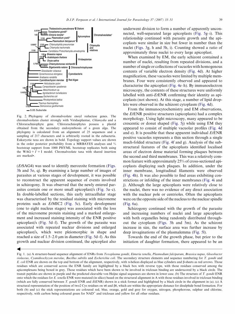
Fig. 2. Phylogeny of chromalveolate enoyl reductase genes. Thechromalveolates cluster strongly with Viridaeplantae, Chlamydia and aChlorarachniophyte gene. Chlorarachniophytes possess a plastidobtained from the secondary endosymbiosis of a green alga. Thephylogeny is calculated from an alignment of 25 sequences and asampling of 217 characters and is arbitrarily rooted in the eubacteria.Eukaryotic taxa are shown in bold. Topology support values are shownin the order posterior probability from a MRBAYES analyses and %bootstrap support from 1000 PHYML bootstrap replicates both usingthe WAG + C + I model. Chromalveolate taxa with shared insertionare marked*.
D.J.P. Ferguson et al. / International Journal for Parasitology 37 (2007) 33–51 39
(EtSAG4) was used to identify merozoite formation (Figs.3h and 5o, q). By examining a large number of images ofparasites at various stages of development, it was possibleto reconstruct the apparent sequence of events involvedin schizogony. It was observed that the newly entered par-asites contain one or more small apicoplast/s (Fig. 5a–c).In double-labelled sections, this early intracellular stagewas characterised by the residual staining with micronemeproteins such as EtMIC2 (Fig. 5c). Early development(one to eight nucleus stages) was associated with the lossof the microneme protein staining and a marked enlarge-ment and increased staining intensity of the ENR positiveapicoplast/s (Fig. 5d–f). The growth of the parasite wasassociated with repeated nuclear divisions and enlargedapicoplast/s, which were pleiomorphic in shape andreached a size of 1.5–2.0 lm in diameter (Fig. 5d–f). As thisgrowth and nuclear division continued, the apicoplast also
Fig. 1. (a) A structure-based sequence alignment of ENRs from Toxoplasma g
violaceus, Cyanidioschyzon merolae, Bacillus subtilis and Escherichia coli. TheE. coli ENR are shown on the top and bottom of the alignment, respectively, wresidues which are conserved across the ENR family are highlighted by aapicomplexans being boxed in grey. Those residues which have been shown ttransit peptides are shown in purple and the predicted cleavable von Heijne sigonto which the residues for E. tenella ENR were mutated (in silico) based on the(which are fully conserved between T. gondii ENR and EtENR) shown in a ststructural representation of the position of two2 Cys residues on a6 and b6, whboth (b) and (c) the stick representations are coloured red, blue, orange, grespectively, with carbon being coloured green for NAD+ and triclosan and y
b
underwent division to form a number of apparently uncon-nected, well-separated large apicoplasts (Fig. 5g–i). Thisrelationship continued with parasite growth and the api-coplasts were similar in size but fewer in number than thenuclei (Figs. 3g, h and 5h, i). Counting showed a ratio ofapproximately three nuclei to every large apicoplast.
When examined by EM, the early schizont contained anumber of nuclei, resulting from repeated divisions, and anumber of single or collections of vacuoles with homogenouscontents of variable electron density (Fig. 4d). At highermagnification, these vacuoles were limited by multiple mem-branes. Four were consistently observed and appeared tocharacterise the apicoplast (Fig. 4e–h). By immunoelectronmicroscopy, the contents of these structures were uniformlylabelled with anti-EtENR confirming their identity as api-coplasts (not shown). At this stage, a number of lipid drop-lets were observed in the schizont cytoplasm (Fig. 4d).
From the immunocytochemistry and EM observations,the EtENR positive structures (apicoplasts) had a complexmorphology. Using light microscopy, many appeared to becrescentic or donut shaped (Fig. 5j) while using EM theyappeared to consist of multiple vacuolar profiles (Fig. 4dand e). It is possible that these apparent individual EtENRpositive vacuoles represents a thin section through a singlemuch-folded structure (Fig. 4f and g). Analysis of the sub-structural features of the apicoplasts identified localisedareas of electron dense material forming plaques betweenthe second and third membranes. This was a relatively com-mon feature with approximately 25% of cross-sectioned api-coplasts displaying such plaques. In addition, under theinner membrane, longitudinal filaments were observed(Fig. 4h). It was also possible to find areas exhibiting con-strictions or infolding of the inner membranes (Fig. 4i andj). Although the large apicoplasts were relatively close tothe nuclei, there was no evidence of any direct associationwith the nuclear pole or centrioles. Often the apicoplastswere on the opposite side of the nucleus to the nuclear spindle(Fig. 6a).
Schizogony continued with the growth of the parasiteand increasing numbers of nuclei and large apicoplastswith both organelles being randomly distributed through-out the cytoplasm (Figs. 3h and 5m). As the schizontincrease in size, the surface area was further increase bydeep invaginations of the plasmalemma (Fig. 5l).
Towards the end of the growth phase, but prior to theinitiation of daughter formation, there appeared to be an
ondii, Eimeria tenella, Plasmodium falciparum, Brassica napus, Gloeobacter
secondary structure elements and sequence numbering for T. gondii andith a-helices displayed as blue cylinders and b-sheets as red arrows. Thoseblack box with reverse type, with those residues conserved among theo be involved in triclosan binding are underscored by a black circle. Thenal sequences are shown in lower case. (b) The structure of T. gondii ENRstructural alignment in A with those residues involved in triclosan binding
ick format and highlighted by a black circle in the alignment in (a). (c) Aich are within the appropriate distance for disulphide bond formation. Forold and grey for oxygen, nitrogen, phosphorous, sulphur and chlorine,ellow for all other residues.

Table 1Predicted dynamin, MinD and Centrin proteins found in the Eimeria tenella genome project share similarity to a wide variety of organisms
E. tenella predicted protein Organism with closest sequence similarity(gene description)
Accession number of publishedsequence
Probability
Dynamin Eimer_2900c04.plk Dictyostelium discoideum (Dynamin like protein) XP_642112 3e-54Homo sapiens (Dynamin 3) NP_056384 2e-53Arabidopsis thaliana (Dynamin like protein) AAM61220 2e-49
MinD Eimer_2796f01.qlk Cryptosporidium parvum (MRP like MinD familyATPase)
EAK88253 3e-11
Clostridium acetobutylicum (MinD family ATPase) AEO07795 3e-09
Centrin1 Eimer bac47a4Ef12.qlk
Plasmodium falciparum (Centrin putative) NP_702332 8e-51Paramecium tetraurelia (Centrin putative) CAH03655 1e-41Tetrahymena thermophilia (Centrin) AAF66602 3e-35A. thaliana (Centrin) CAB62315 2e-28
Centrin2 Eimer bac47a4Ef12.qlk
Ochromonas danica (Centrin) BAD20712 6e-43Giardia intestinalis (Centrin) AAC47395 3e-39
The organisms with closest sequence similarities for each of the organellar division proteins and the accession numbers of the proteins or predicted proteinsare provided. The role of these proteins in organellar (plastid, mitochondrial or nuclear) division remains to be determined.
40 D.J.P. Ferguson et al. / International Journal for Parasitology 37 (2007) 33–51
unwinding of the large apicoplast into a thin elongatedstructure (Fig. 5j and m). These structures developed abead-like appearance with numerous regular constrictionsalong its length and appeared to result in the formationof numerous small EtENR positive apicoplasts (Fig. 5kand n). The small spherical EtENR positive structures weresimilar in size (approximately 0.45 lm diameter) to theapicoplasts of the sporozoites and merozoite. The smallapicoplasts were present in greater numbers (approximate-ly 3:1) than the nuclei and were randomly distributedthroughout the cytoplasm (Fig. 3g and h). This final api-coplast division occurred before daughter formation wasinitiated as confirmed by the limited EtSAG4 stainingand the absence of EtMIC2 staining (Figs. 3h and 5n).
The differentiation phase, resulting in merozoite forma-tion, was associated with the nuclei and apicoplasts movingclose to the schizont plasmalemma or its deep invagin-ations (Fig. 3h). The earliest stage of daughter formationwas identified by increased expression of the anti-EtSAG4at the schizont surface (Fig. 5o). At this stage, daughterformation was initiated by the formation of the conoidand inner membrane complex beneath the plasmalemmaas observed by EM (Fig. 6b and c). The small EtENRpositive apicoplasts were predominately located on thecytoplasmic side of the nuclei (Figs. 3h and 5o). The mer-ozoites were formed by budding into the parasitophorousvacuole while the inner membrane complex remaininganchored to the junction with the plasmalemma (Fig. 6c–f). The nuclear poles were directed into the forming daugh-ters as the nucleus entered. As this process occurred, one ormore apicoplasts also entered the developing merozoite. Atthis time, the micronemes formed in the anterior of thedaughters as shown by positive staining for EtMIC2 (Figs.3h and 5o). In the fully formed merozoite, between one andfour small apicoplasts were located around the nucleus ineach daughter (Fig. 5p and q). The segregation processduring the differentiation phase resulted in a single nucleus
and one to four apicoplasts entering every daughter withnone left in the residual body.
3.4.4. Sexual stages
Microgametocyte development. The microgametocytesdeveloped in the superficial epithelial cells of the crypts.In the heavily infected caecum, micro- and macrogameto-cytes developed in adjacent or even the same cell(Fig. 7a–c). The early stages were characterised by thepresence of a numbers of open nuclei. At this time, oneor more small apicoplasts with a relatively low EtENRsignal were located in the cytoplasm (Fig. 7b, c, e, fand i). During maturation the number of nuclei increasedand became peripherally located with increasingly con-densed chromatin (Fig. 7b and c). During this time, littlechange was seen in the apicoplast (Fig. 7b and c). Inmature microgametocytes, the residual cytoplasm exhibit-ed little evidence of apicoplasts and apicoplasts were notobserved in the crescentic shaped microgametes formed(Fig. 7b and c).
Macrogametocyte development. In contrast to themicrogametocytes, there was strong anti-EtENR stainingof enlarged lobated apicoplast/s located adjacent to thelarge single nucleus during the early stages of develop-ment (Fig. 7b–i). Due to the absence of suitable antibod-ies for staining other organelles of the macrogametocytes,the appearance of the apicoplast within the developingmacrogamete is best seen using the peroxidase technique(Fig. 7b, e, g and h). Consistent with the lobed appear-ance of the apicoplast by immunofluorescence, multipleprofiles of apicoplast-like structures adjacent to thenucleus were observed by EM (Fig. 8a and b) and con-firmed by immunoelectron microscopy (Fig. 8c). Thestrongly staining apicoplast was maintained during thegrowth and maturation of the macrogametocyte withthe development of the various types of wall formingbodies, polysaccharide granules and lipid droplets

Fig. 3. Immuno-fluorescent (a–f, h) and immuno-peroxidase (g) images of various asexual stages of Eimeria tenella labelled with anti-EtENR visualisedwith fluorescein isothiocyanate (green) or DAB/H2O2 (brown). The nuclei have been counter stained with 4 0,6-diamidino-2-phenylindole (DAPI). Barsrepresent 1 lm in a–e and 10 lm in g and h. (a) Sporozoite showing two EtENR positive apicoplasts (arrows) either side of the nucleus (N). (b) Sporozoitewith a single apicoplast (green) anterior to the nucleus. The micronemes in the anterior are labelled with anti-EtMIC2 (red), which outlines the anteriorrefractile body (RB). (c) Detail of the green channel for the enclosed area in b showing the strong labelling of the apicoplast with anti-EtENR (arrow). (d)Grey level detail from the blue channel showing DAPI staining of the apicoplast (arrow) adjacent to the strongly staining nucleus. (e) Merozoite for the gutlumen showing the surface labelling with anti-EtSAG4 (red) and three EtENR positive (green) apicoplasts (arrows) around the nucleus (blue). (f)Merozoite showing the apical labelling of the micronemes with anti-EtMIC2 (red) and the EtENR positive apicoplast (arrow) adjacent to the nucleus (N).(f) Section of the caecum at 48 h p.i. showing mid stage (MS) first generation schizonts with a number of large irregular shaped EtENR positive (brown)structures and late stage (LS) schizonts with numerous small spherical EtENR positive apicoplasts. In addition, a number of small newly entered parasites(T) with one or two small EtENR positive apicoplasts were also located within the epithelial cells. (h) Section of caecum at 112 h p.i. illustrating variousstages of schizogony from the early stages (ES) with a few large EtENR positive structures through the mid stages (MS) with a number of large EtENRpositive structures to the late stage (LS) with numerous small EtENR positive apicoplasts and finally the mature schizonts with fully formed merozoites(Me) characterised by the labelling with anti-EtMIC2 (red), which contain small apicoplasts (green).
D.J.P. Ferguson et al. / International Journal for Parasitology 37 (2007) 33–51 41
(Fig. 7f and g). Large or multiple EtENR positiveapicoplasts were still present within developing oocystsidentified by the formation of the oocyst wall (Fig. 7h).Therefore, in the absence of apicoplasts associated withthe microgamete, oocysts only inherited this organellefrom the macrogametes, which is consistent with mater-nal inheritance by the sporozoites.
4. Discussion
Previous studies have identified enzymes of type II fattyacid synthesis in a number of apicomplexans includingT. gondii and Plasmodium spp., but these are notablyabsent from Cryptosporidium parvum and Cryptosporidium
hominis that lack apicoplasts (Abrahamsen et al., 2004; Xu
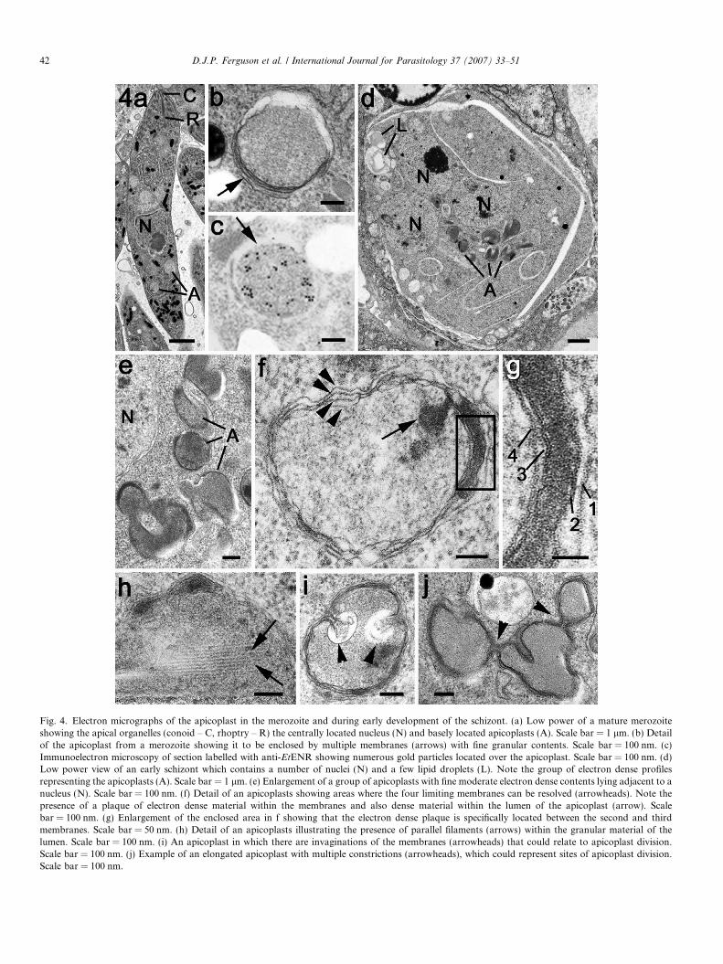
Fig. 4. Electron micrographs of the apicoplast in the merozoite and during early development of the schizont. (a) Low power of a mature merozoiteshowing the apical organelles (conoid – C, rhoptry – R) the centrally located nucleus (N) and basely located apicoplasts (A). Scale bar = 1 lm. (b) Detailof the apicoplast from a merozoite showing it to be enclosed by multiple membranes (arrows) with fine granular contents. Scale bar = 100 nm. (c)Immunoelectron microscopy of section labelled with anti-EtENR showing numerous gold particles located over the apicoplast. Scale bar = 100 nm. (d)Low power view of an early schizont which contains a number of nuclei (N) and a few lipid droplets (L). Note the group of electron dense profilesrepresenting the apicoplasts (A). Scale bar = 1 lm. (e) Enlargement of a group of apicoplasts with fine moderate electron dense contents lying adjacent to anucleus (N). Scale bar = 100 nm. (f) Detail of an apicoplasts showing areas where the four limiting membranes can be resolved (arrowheads). Note thepresence of a plaque of electron dense material within the membranes and also dense material within the lumen of the apicoplast (arrow). Scalebar = 100 nm. (g) Enlargement of the enclosed area in f showing that the electron dense plaque is specifically located between the second and thirdmembranes. Scale bar = 50 nm. (h) Detail of an apicoplasts illustrating the presence of parallel filaments (arrows) within the granular material of thelumen. Scale bar = 100 nm. (i) An apicoplast in which there are invaginations of the membranes (arrowheads) that could relate to apicoplast division.Scale bar = 100 nm. (j) Example of an elongated apicoplast with multiple constrictions (arrowheads), which could represent sites of apicoplast division.Scale bar = 100 nm.
42 D.J.P. Ferguson et al. / International Journal for Parasitology 37 (2007) 33–51

Fig. 5. Details of the asexual multiplication of Eimeria tenella in the chicken caecum observed in plastic sections after staining with azure A (a, d, g, l andp), after immunoperoxidase staining with anti-EtENR (b, e and h) and double labelling with anti-EtENR (green) and anti-EtMIC2 (red) (c and f) or anti-EtSAG4 (red) (i, m, n, o and q). Scale bars = 1 lm (a–f, j and k), 5 lm (g–i) and 10 lm (l–q). (a–c) Early stage of newly entered merozoites showing twosmall apicoplasts (arrowheads) with some residual anti-EtMIC2 staining (red). (d–f) Early growth stage in which there is marked expansion of the size andsignal from the apicoplasts (A). (g–i) Mid-stage with repeated nuclear division resulting in numerous nuclei. Note the increased number of largepleiomorphic-shaped EtENR positive apicoplasts (A). (j) Deconvoluted enlargement of the enclosed area in m showing the donut and crescentic shape ofthe large apicoplasts. (k) Deconvoluted enlargement of the enclosed area in n showing repeated constrictions (arrowheads) along the enlarged apicoplastsand the formation of small spherical apicoplasts. (l) Late schizont with numerous nuclei (N) and deep invaginations of the plasmalemma (arrows)increasing the surface area of the schizont. (m) Similar stage to that in l showing a number of randomly distributed large apicoplasts (green). N – nucleus.(n) Slightly later stage than in m showing the apicoplasts as elongated structures with a bead-like appearance and numerous small apicoplasts. The surfaceof the schizont is still negative for EtSAG4 at this stage. N – nucleus. (o) Late stage in schizogony showing the initiation of daughter formation identifiedby EtSAG4 positivity (red) at the schizont surface. Note the nuclei (N) and small apicoplasts (A) have become peripherally located. (p) Mature schizontconsisting of numerous fully formed merozoites (Me). (q) Mature schizont showing the fully formed merozoites (Me) outline by the EtSAG4 staining (red)and containing one to three small apicoplasts (A).
D.J.P. Ferguson et al. / International Journal for Parasitology 37 (2007) 33–51 43
et al., 2004). Moreover, the type II fatty acid biosynthesishas been suggested as an antimicrobial agent target inT. gondii and Plasmodium spp. and as such the identifica-
tion of the enzymes of type II fatty acid biosynthesis inE. tenella may suggest that this pathway holds promise asan antimicrobial target. Analysis of these genes reveals that

Fig. 6. Electron micrographs illustrating the later stages in schizogony with formation of the merozoites. (a) Section through a nucleus (N) in a lateschizont. Note the absence of apicoplast-like structures associated with the poles (NP) of intranuclear spindle but an apicoplast (A) is located on theopposite side of the nucleus. Scale bar = 500 nm. (b) Low power through part of the periphery of a late schizont showing the peripherally located nucleiand the conical restructures at the plasmalemma representing the initiation of daughter formation (arrows). Scale bar = 1 lm. (c) Detail of the very earlystage of merozoites formation showing the conoid (C) and electron dense inner membrane complex of the daughter (arrowhead) budding into the lumen ofthe parasitophorous vacuole. Note the nucleus associated with the forming daughter and the basally located apicoplast (A). G – Golgi body. Scalebar = 200 nm. (d) A more advanced stage of daughter formation with continued growth into the parasitophorous vacuole. There is the initiation ofrhoptry formation (R) and the nucleus (N) and apicoplast (A) are partially within the developing daughter. Scale bar = 200 nm. (e) Later stage with thenucleus (N) within the developing daughter and the rhoptry (R) has a partially developed duct. C – conoid. Scale bar = 200 nm. (f) Fully formed daughterbudding from the surface of the schizont. N – nucleus; R – rhoptry. Scale bar = 200 nm.
44 D.J.P. Ferguson et al. / International Journal for Parasitology 37 (2007) 33–51
they all show significant sequence similarity to their bacte-rial and apicomplexan homologues, in particular those res-idues which have been implicated in catalysis are all fullyconserved in the E. tenella enzymes. This analysis may sug-gest that E. tenella contains a fully functioning type II fattyacid biosynthesis pathway. Of the seven enzymes involvedin Type II fatty acid synthesis evident in the genome ofE. tenella, we elected to further characterise ENR. This isbecause the structure of a number of ENRs has been solvedboth independently and in complex with several families ofantimicrobial compounds including the diazaborines,aminopyridine-based inhibitors and triclosan (Baldocket al., 1996; Levy et al., 1999; Payne et al., 2002; Seefeldet al., 2003). Modelling of EtENR based on the closehomologue T. gondii ENR has revealed that those residueswhich form close interactions to the biocide triclosan arefully conserved in EtENR and that features unique to the
plastid but absent in the bacterial ENR family are con-served, including two significant sequence inserts beforeand after b3, common to the chromalveolate ENR ortho-logue (Fig. 1). Moreover, Cys295 is positioned in closeproximity to Cys312 with their separation being consistentwith disulphide bond formation a possibility that willrequire further investigation (Fig. 1c).
Expression of the predicted mature protein also facilitat-ed the production of polyclonal antisera. These sera notonly allowed confirmation of the apicoplast location ofthe enzyme, but also made it possible to visualise detailedchanges in the morphology of the apicoplast throughoutthe life cycle of E. tenella. The present study involved theuse of tissue samples from infected chickens. This is theonly method available for examining many of the naturaldevelopmental processes involved in the coccidian lifecyclebecause these cannot be reproduced in tissue culture. This

Fig. 7. Details of the changes in the apicoplast during sexual development using plastic sections stained with azure A (a and d), immunoperoxidase usinganti-EtENR visualised with DAB/H2O2 (b, e, g and h) and double-labelled immunofluorescence using anti-EtENR visualised with fluoresceinisothiocyanate (green) and anti-EtSAG4 visualised with Texas red (red) (c, f and i). Scale bar = 10 lm in a–c and 1 lm in d–i. (a–c) Lower power imagesthrough the caecum of a chicken at 142 h p.i. in which numerous stages of microgametogony (Mi) and macrogametogony (Ma) can be seen. Note the largeEtENR-positive structures adjacent to the nucleus at all stages of macrogametogenesis and the few small EtENR-positive apicoplasts associated with thedeveloping microgametocytes. (d) Detail showing a late macrogametocyte (Ma) with numerous cytoplasmic granules and wall forming bodies and a midstage microgametocyte (Mi) with numerous small dense nuclei located towards the periphery. (e) Similar stage to that in d showing macrogametocytes(Ma) with large strongly EtENR positive apicoplasts compared to the small apicoplasts present in the microgametocyte (Mi). (f) Similar stage to that in dshowing the strongly staining apicoplast with the macrogametocyte (Ma) and the weakly staining structure (arrowhead) within the microgametocyte (Mi).Neither sexual stage is stained with anti-EtSAG4. (g) Mature macrogametocytes (Ma) showing the EtENR positive apicoplasts as lobated or multiplestructure. (h) Early oocyst identified by oocyst wall formation (O) showing a large EtENR positive structure centrally located within the oocyst. (i) Similarsection to that in f showing the large apicoplast within the macrogametocyte (Ma) and a small positive structure (arrowhead) within the microgametocyte(Mi). Note the adjacent merozoites (Me), which are positively stained for EtSAG4 (red) and contain small apicoplasts.
D.J.P. Ferguson et al. / International Journal for Parasitology 37 (2007) 33–51 45
is particularly true for the coccidian stages of development(asexual and sexual) present in the definitive hosts. Unfor-tunately, there are a number of disadvantages of having toexamine sections of fixed and embedded tissue. It is not
possible to follow dynamic developmental processes withina given parasite. Second, the parasites are randomly orien-tated within the tissue increasing the difficulty of obtainingclear images, in contrast to the situation in cultured cells

Fig. 8. Electron micrographs of apicoplasts within the macrogametocyte. (a) Mature macrogametocyte showing the centrally located nucleus (N) with thecytoplasm containing peripherally located wall forming bodies type 1 (W1), wall forming bodies type 2 (W2), numerous lipid droplets (L) andpolysaccharide granules (PG). Note the membrane-bound structures representing the apicoplasts around the nucleus (arrows). Scale bar = 2 lm. (b)Detail from the periphery of a nucleus (N) showing a number of profiles of the apicoplast. PG – polysaccharide granules. Scale bar = 100 nm. (c)Immunoelectron microscopy of a section through a macrogametocyte labelled with anti-EtENR showing numerous gold particles over the apicoplast(arrow) located between the nucleus (N) and the polysaccharide granules (PG). Scale bar = 100 nm.
46 D.J.P. Ferguson et al. / International Journal for Parasitology 37 (2007) 33–51
where the parasites are constrained to a flattened two-di-mensional orientation. Third, many of the antibodies andfluorescent stains, which have added greatly to our knowl-edge of in vitro development, will not work with formalinfixed/wax embedded tissue. For example, in tissue sections,DAPI stain is insensitive and cannot be used to identify theapicoplast within the gut stages, probably due to extractionof material or a change in architecture during processing.The absence of DAPI staining of the macrogametocyte/macrogamete nucleus is unexpected, but similar to thatreported for the macrogamete of T. gondii and is thoughtto relate the dispersed nature of the chromatin (Fergusonet al., 2005). However, careful examination of large num-bers of immunostained parasites, correlated with detailedroutine and immunoelectron microscopy can amelioratethese limitations.
The changes observed during sexual development aresimilar to those reported previously for T. gondii (Fergu-son et al., 2005). The development of the microgametocyteresults in little change to the apicoplast size, shape orstaining intensity and the microgametes formed lackedan identifiable apicoplast. In the case of macrogametocytedevelopment, there is a marked increase in size and stain-ing intensity from the early stages up to and including themature macrogamete and early oocyst. Unlike the
schizont, there was no evident apicoplast division orreduced ENR signal in the mature macrogamete. Thiswould point to a continued physiological function of theapicoplasts throughout macrogamete maturation. Thismay be reflected in the build up of storage lipid and poly-saccharide granules within the cytoplasm. The apparentabsence of the apicoplast in microgametes suggests thatthe apicoplast is inherited from the macrogamete and isthus maternally derived as previously described for T. gon-
dii and Plasmodium spp. (Ferguson et al., 2005; Vaidyaet al., 1993; Creasey et al., 1994). Maternal inheritanceof the plastid thus might be a common feature of theApicomplexa.
In the infectious stages, the apicoplast is a small spheri-cal structure with a relatively low EtENR signal afterimmunostaining. While the physiological processes under-taken by the apicoplast are still incompletely understood,it is likely that the size of the apicoplast and level ofEtENR signal reflect changes in the activity of the apicop-last and type II fatty acid synthesis. Using the immunoflu-orescence technique, it is impossible to absolutely quantifyEtENR levels. However, differences in the size and signalintensity between identically stained parasites will representquantitative changes in the relative concentrations of theprotein. Using this criterion, it is obvious that one of the

D.J.P. Ferguson et al. / International Journal for Parasitology 37 (2007) 33–51 47
early changes during schizogony and macrogametogony isa marked increase in the size of the apicoplast and the levelof EtENR signal, which is not observed during microgame-togony. Therefore, it is interesting to note that the stagesshowing an apparent increase in the EtENR protein arealso the stages (early schizont and macrogametocyte) whenlipid droplets have been identified by EM. This increase insize and EtENR signal is similar to that reported duringendopolygeny in T. gondii (Ferguson et al., 2005).
The apicoplast within the infectious stage of E. tenella
appears as single or multiple peri-nuclear spherical struc-tures. Careful examination of merozoites by immunocy-tochemistry and electron microscopy confirm that thesewere individual apicoplasts and not profiles of an elon-gated structure. The presence of multiple apicoplasts inthe merozoites has not been reported for the infectiousstages of other members of the Apicomplexa. This wasrelatively uncommon in sporozoites (less than 5%), butmore common in second generation merozoites (approx-imately 20%). Although the organelle is limited by fourmembranes there are areas where the membranes are dif-ficult to resolve. This is similar to that described for T.gondii, P. falciparum and Sarcocystis sp. (Sheffield andMelton, 1968; Vivier and Petitprez, 1972; McFaddenand Roos, 1999; Tomova et al., 2006) although threemembranes have also been reported for P. falciparum
(Hopkins et al., 1999). In the present study, no evidencewas found to support the proposal that there are twomembranes with the multi-membraned appearance result-ing from extensive folding of the inner membrane (Koh-ler, 2005). A unique observation in E. tenella was theappearance of electron dense plaques of material betweenthe second and third membranes (Fig. 4f and g) but therole of this material is unknown.
In the present study, of the large schizonts of E. tenella,there was evidence for some division of the large apicop-lasts during the proliferative phase. This has not beenobserved previously in the smaller schizonts of T. gondiiand P. falciparum (Waller et al., 2000; Ferguson et al.,2005; van Dooren et al., 2005) and probably relates tothe very large size of the second generation schizonts.The finding of merozoites with multiple apicoplasts hasnot been observed before. In P. falciparum, it has been pro-posed that apicoplast division was delayed until afternuclear division by an unknown feedback mechanism todetermine how many portions the apicoplast would berequired to divide into (Waller et al., 2000). It is possible,that in the large complex schizonts of E. tenella with hun-dreds of nuclei, such a feedback mechanism may not func-tion and therefore the production of an excess number ofapicoplasts may represent insurance against a shortage ofapicoplasts. It is known that merozoites lacking an apicop-last are non-viable (He et al., 2001). Therefore, if there is nodetrimental effect of having more than one apicoplast, thentoo many is better than too few.
The division mechanism of the apicoplast during asexualdevelopment within the Apicomplexa has been the subject
of much controversy. Plastid division had been extensivelystudied in other systems and a range of proteins identifiedthat play vital roles during bacterial cell division and thedivision of plastids (chloroplasts) in algae and plants(reviewed by Hashimoto, 2003). However, searches of thegenomic data bases of apicomplexan parasites, in particu-lar P. falciparum, T. gondii and E. tenella (Vaishnava andStriepen, 2006; present study) have failed to identify a com-plete set of homologous genes for the plastid division pro-teins. Therefore a new mechanism had to be consideredand two hypotheses are proposed from studies of endody-ogeny in T. gondii. One involved a physical pull/pushmechanism in which the centrioles/nuclear poles of thenuclear spindle are connected to and pull the apicoplastinto an elongated structure as they move apart duringnuclear division with the subsequent posterior growth ofthe inner membrane complex of the developing daughterspushing on the apicoplast to cause division (Striepenet al., 2000). The second hypothesis proposed that, evenin the apparent absence of plastid division proteins, anunknown mechanism involving division/constriction ringsmay be involved in apicoplast division (Matsuzaki et al.,2001).
However, when comparing the results for apicoplastdivision and segregation within the Apicomplexa, it isimportant to realise that asexual multiplication is not asingle process, but that there are subtle differences inthe timing and location of both nuclear division anddaughter formation undergone by different species andeven between lifecycle stages of the same species. Thesedifferences may be relevant to understanding apicoplastdivision. We would propose that there are four distinctprocesses rather than the three suggested in a recentreview (Vaishnava and Striepen, 2006). These we havetermed ‘‘classical’’ schizogony, endodyogeny, ‘‘Toxoplas-
ma’’ endopolygeny and ‘‘Sarcocystis’’ endopolygeny, withthe distinctive and characteristic features being summa-rised in Fig. 9. The term endopolygeny has been usedto describe asexual division involving internal formationof multiple daughters in both T. gondii and Sarcocystis
sp. (Piekarski et al., 1971; Speer and Dubey, 2001). How-ever, there are significant differences in relation to thepresence or absence of repeated nuclear divisions in theseprocesses (Fig. 9). Therefore we distinguish between theprocesses by using the terms ‘‘Toxoplasma’’ endopolyge-ny where there are repeated nuclear divisions and ‘‘Sar-cocystis’’ endopolygeny where there are repeated DNAreplications but no nuclear division (Fig. 9).
There is support for the pull/push hypothesis from stud-ies of endodyogeny and ‘‘Sarcocystis’’ endopolygeny (Strie-pen et al., 2000; Vaishnava et al., 2005; Vaishnava andStriepen, 2006). However, it is difficult to reconcile apicop-last division directly to nuclear division and daughter for-mation in ‘‘Toxoplasma’’ endopolygeny and ‘‘classical’’schizogony (Ferguson et al., 2005; Waller et al., 2002; pres-ent study) where repeated nuclear divisions occur in theabsence of apicoplast division and apicoplast division

Fig. 9. Diagram illustrating the differences in the timing and location of nuclear division and daughter formation associated with asexual developmentwithin the Apicomplexa. There are four distinct processes: (a) ‘‘Classical’’ schizogony undergone by the majority of apicomplexans, including the generaPlasmodium and Eimeria, where there is a proliferative phase with repeated nuclear divisions followed by a differentiation phase with daughter formationinitiated at and budding from the parasite plasmalemma. (b) Endodyogeny in which there is no proliferative phase, but one cycle of DNA replication withthe initiation of the formation of two daughters within the mother cell cytoplasm prior to the completion of nuclear division. (c) ‘‘Toxoplasma’’endopolygeny, used to describe the asexual division of Toxoplasma gondii in the definitive host, involves a proliferative phase with repeated nucleardivision, similar to ‘‘classical’’ schizogony, but differs in that daughter formation is initiated within the cytoplasm in a similar manner to endodyogeny. (d)‘‘Sarcocystis’’ endopolygeny has been reported for certain Sarcocystis species and involves an initial phase with repeated cycles of DNA replication andformation of numerous nuclear spindles, but no division of the nucleus. Formation of multiple daughters is initiated within the cytoplasm and the largepolyploidy nucleus fragments and one haploid nucleus enters each developing daughter. In the diagram, centrioles are represented by green circles,apicoplasts by red structures and nuclear poles by black triangles.
48 D.J.P. Ferguson et al. / International Journal for Parasitology 37 (2007) 33–51
occurs prior to daughter formation. In endodyogeny and‘‘Sarcocystis’’ endopolygeny, although the processes ofnuclear division, apicoplast division and initiation ofdaughter formation appear to be closely associated, theycould still represent independent events.
In support of a constriction ring hypothesis is the obser-vation of repeated constrictions along the large apicoplastin the absence of any association with nuclear division ordaughter formation in ‘‘classical’’ schizogony and‘‘Toxoplasma’’ endopolygeny. Indeed our search of theE. tenella genome has identified a number of genes forproteins that are believed to be associated with organellardivision such as dynamin and MinD. However, there isno evidence that these are directly involved in apicoplastdivision and the very high similarity of the putative E. tenella
MinD with a homologous protein in C. parvum, whichlacks an identifiable apicoplast, suggests an alternativefunction for this molecule. Therefore, at the present time,the mechanism of apicoplast division remains an openquestion.
It should be noted that it is unclear how any of the gen-ome containing organelles of the Apicomplexa (nucleus,mitochondrion and apicoplast) divide while maintainingintact membranes. It has recently been reported that divi-sion of the Golgi body during endodyogeny in T. gondii
involved elongation of the stack of flattened vacuoles form-ing the organelle followed by division in the central regionto form two organelles (Pelletier et al., 2002). Unfortunate-ly, no mechanism was provided for how this elongationand central division of the membranes occurs but it bearscertain similarities to apicoplast elongation seen duringendodyogeny.
However, irrespective of the involvement of the centri-oles/nuclear pole in apicoplast division, they appear toplay a central role in coordinating and organising daugh-ter formation and organelle segregation. In the presentstudy, just prior to the initiation of daughter formation,there is a redistribution of the organelles with the ran-domly distributed nuclei and apicoplasts moving to theperiphery of the schizont close to the plasmalemma. In

D.J.P. Ferguson et al. / International Journal for Parasitology 37 (2007) 33–51 49
this situation, the centrioles/nuclear pole complex ispointed towards the plasmalemma and may direct theinitiation of daughter formation thus ensuring the num-ber of daughters matches the number of nuclei. The re-establishment of the nucleus and apicoplast relationshipand the movement of the centrioles/nuclear pole intothe developing daughter will also ensure correct organellesegregation. The involvement of the centriolar complexduring segregation, but not division, of the plastid wouldbe consistent with that reported in certain brown algathat also acquired their plastid apparatus from a second-ary endosymbiotic event (Nagasato and Motomura,2002). Therefore the centrioles/nuclear pole may act asa coordinating centre ensuring efficient formation of via-ble daughters in all variations of the asexual proliferationprocess undergone by the Apicomplexa (Waller et al.,2000; Striepen et al., 2000; Ferguson et al., 2005; Vaish-nava et al., 2005).
In conclusion, we propose a unifying theory for thenature of apicoplast development, which would be con-sistent with the observations across the Apicomplexa(Striepen et al., 2000; Waller et al., 2000; Fergusonet al., 2005; Vaishnava et al., 2005): (i) Nuclear and api-coplast division are independent events. (ii) Apicoplastdivision is independent of daughter formation and couldinvolve constriction rings of unknown composition. (iii)The centriolar/nuclear complex appears to play a centralrole in coordinating daughter formation and organellesegregation. It is possible that, an as yet to be discov-ered, unifying mechanism may explain how the Golgibody, nucleus, apicoplast and mitochondrion dividewhile retaining intact membranes. A better understandingof how these processes are controlled and coordinatedwould be a major advance in our understanding of theApicomplexa.
Acknowledgements
This work was supported by NIH grants R01 AI43228(R.M. and C.W.R.) and AI27530 (R.M.), the Research toPrevent Blindness Foundation (R.M.) and a WellcomeTrust equipment grant (D.J.P.F). Ms. H. Beard is thankedfor assistance in preparing the diagram in Fig. 9.
Appendix A. Supplementary data
Supplementary data associated with this article can befound, in the online version, at doi:10.1016/j.ijpara.2006.10.003.
References
Abrahamsen, M.S., Templeton, T.J., Enomoto, S., Abrahante, J.E., Zhu,G., Lancto, C.A., Deng, M., Liu, C., Widmer, G., Tzipori, S., Buck,G.A., Xu, P., Bankier, A.T., Dear, P.H., Konfortov, B.A., Spriggs,H.F., Iyer, L., Anantharaman, V., Aravind, L., Kapur, V., 2004.Complete genome sequence of the apicomplexan, Cryptosporidium
parvum. Science 304, 441–445.
Baldock, C., Rafferty, J.B., Sedelnikova, S.E., Baker, P.J., Stuitje, A.R.,Slabas, A.R., Hawkes, T.R., Rice, D.W., 1996. A mechanism of drugaction revealed by structural studies of enoyl reductase. Science 274,2107–2110.
Brinkman, F.S., Blanchard, J.L., Cherkasov, A., Av-Gay, Y., Brunham,R.C., Fernandez, R.C., Finlay, B.B., Otto, S.P., Ouellette, B.F.,Keeling, P.J., Rose, A.M., Hancock, R.E., Jones, S.J., Greberg, H.,2002. Evidence that plant-like genes in Chlamydia species reflect anancestral relationship between Chlamydiaceae, cyanobacteria, and thechloroplast. Genome Res. 12, 1159–1167.
Cai, X., Fuller, A.L., McDougald, L.R., Zhu, G., 2003. Apicoplastgenome of the coccidian Eimeria tenella. Gene 321, 39–46.
Cavalier-Smith, T., 2000. Membrane heredity and early chloroplastevolution. Trends Plant Sci. 5, 174–182.
Creasey, A., Mendis, K., Carlton, J., Williamson, D., Wilson, I., Carter,R., 1994. Maternal inheritance of extrachromosomal DNA in malariaparasites. Mol. Biochem. Parasitol. 65, 95–98.
Fast, N.M., Xue, L., Bingham, S., Keeling, P.J., 2002. Re-examiningalveolate evolution using multiple protein molecular phylogenies. J.Eukaryot. Microbiol. 49, 30–37.
Ferguson, D.J., 2002. Toxoplasma gondii and sex: essential or optionalextra? Trends Parasitol. 18, 355–359.
Ferguson, D.J., Birch-Andersen, A., Hutchison, W.M., Siim, J.C.,1976. Ultrastructural studies on the endogenous development ofEimeria brunetti, I. Schizogony. Acta Pathol. Microbiol. Scand. B84, 401–413.
Ferguson, D.J., Cesbron-Delauw, M.F., Dubremetz, J.F., Sibley, L.D.,Joiner, K.A., Wright, S., 1999. The expression and distribution ofdense granule proteins in the enteric (Coccidian) forms of Toxoplasma
gondii in the small intestine of the cat. Exp. Parasitol. 91, 203–211.Ferguson, D.J., Henriquez, F.L., Kirisits, M.J., Muench, S.P., Prigge,
S.T., Rice, D.W., Roberts, C.W., McLeod, R.L., 2005. Maternalinheritance and stage-specific variation of the apicoplast in Toxoplas-
ma gondii during development in the intermediate and definitive host.Eukaryot. Cell 4, 814–826.
Guindon, S., Gascuel, O., 2003. A simple, fast, and accurate algorithm toestimate large phylogenies by maximum likelihood. Syst. Biol. 52,696–704.
Guindon, S., Lethiec, F., Duroux, P., Gascuel, O., 2005. PHYML Online– a web server for fast maximum likelihood-based phylogeneticinference. Nucl. Acids Res. 33, W557–W559.
Harper, J.T., Keeling, P.J., 2003. Nucleus-encoded, plastid-targetedglyceraldehyde-3-phosphate dehydrogenase (GAPDH) indicates asingle origin of chromalveolate plastids. Mol. Biol. Evol. 20, 1730–1735.
Hashimoto, H., 2003. Plastid division: its origins and evolution. Int. Rev.Cytol. 222, 63–98.
He, C.Y., Shaw, M.K., Pletcher, C.H., Striepen, B., Tilney, L.G., Roos,D.S., 2001. A plastid segregation defect in the protozoan parasiteToxoplasma gondii. EMBO J. 20, 330–339.
Heath, R.J., White, S.W., Rock, C.O., 2001. Lipid biosynthesis as a targetfor antibacterial agents. Prog. Lipid Res. 40, 467–497.
Hopkins, J., Fowler, R., Krishna, S., Wilson, I., Mitchell, G., Bannister,L., 1999. The plastid in Plasmodium falciparum asexual blood stages: athree-dimensional ultrastructural analysis. Protist 150, 283–295.
Kapust, R.B., Waugh, D.S., 2000. Controlled intracellular processing offusion proteins by TEV protease. Protein Expr. Purif. 19, 312–318.
Kohler, S., 2005. Multi-membrane-bound structures of Apicomplexa: I.the architecture of the Toxoplasma gondii apicoplast. Parasitol. Res.96, 258–272.
Kohler, S., Delwiche, C.F., Denny, P.W., Tilney, L.G., Webster, P.,Wilson, R.J., Palmer, J.D., Roos, D.S., 1997. A plastid of probablegreen algal origin in Apicomplexan parasites. Science 275, 1485–1489.
Laemmli, U.K., 1970. Cleavage of structural proteins during the assemblyof the head of bacteriophage T4. Nature 227, 680–685.
Levine, N.D., Corliss, J.O., Cox, F.E., Deroux, G., Grain, J., Honigberg,B.M., Leedale, G.F., Loeblich Jr., A.R., Lom, J., Lynn, D., Merinfeld,E.G., Page, F.C., Poljansky, G., Sprague, V., Vavra, J., Wallace, F.G.,

50 D.J.P. Ferguson et al. / International Journal for Parasitology 37 (2007) 33–51
1980. A newly revised classification of the protozoa. J. Protozool. 27,37–58.
Levy, C.W., Roujeinikova, A., Sedelnikova, S., Baker, P.J., Stuitje, A.R.,Slabas, A.R., Rice, D.W., Rafferty, J.B., 1999. Molecular basis oftriclosan activity. Nature 398, 383–384.
Matsuzaki, M., Kikuchi, T., Kita, K., Kojima, S., Kuroiwa, T., 2001.Large amounts of apicoplast nucleoid DNA and its segregation inToxoplasma gondii. Protoplasma 218, 180–191.
McFadden, G.I., Roos, D.S., 1999. Apicomplexan plastids as drug targets.Trends Microbiol. 7, 328–333.
McFadden, G.I., Waller, R.F., 1997. Plastids in parasites of humans.Bioessays 19, 1033–1040.
McLeod, R., Muench, S.P., Rafferty, J.B., Kyle, D.E., Mui, E.J., Kirisits,M.J., Mack, D.G., Roberts, C.W., Samuel, B.U., Lyons, R.E., Dorris,M., Milhous, W.K., Rice, D.W., 2001. Triclosan inhibits the growth ofPlasmodium falciparum and Toxoplasma gondii by inhibition ofapicomplexan Fab I. Int. J. Parasitol. 31, 109–113.
Muench, S.P., Rafferty, J.B., McLeod, R.L., Rice, D.W., Prigge, S.T.,2003. Expression, purification and crystallization of the Plasmodium
falciparum enoyl reductase. Acta Crystallogr. D Biol. Crystallogr. 59,1246–1248.
Nagasato, C., Motomura, T., 2002. Influence of the centrosome incytokinesis of brown algae: polyspermic zygotes of Scytosiphon
lomentaria (Scytosiphonales, Phaeophyceae). J. Cell Sci. 115, 2541–2548.
Notredame, C., Higgins, D.G., Heringa, J., 2000. T-Coffee: A novelmethod for fast and accurate multiple sequence alignment. J. Mol.Biol. 302, 205–217.
Payne, D.J., Miller, W.H., Berry, V., Brosky, J., Burgess, W.J., Chen,E., DeWolf Jr., W.E., Fosberry, A.P., Greenwood, R., Head,M.S., Heerding, D.A., Janson, C.A., Jaworski, D.D., Keller, P.M.,Manley, P.J., Moore, T.D., Newlander, K.A., Pearson, S., Polizzi,B.J., Qiu, X., Rittenhouse, S.F., Slater-Radosti, C., Salyers, K.L.,Seefeld, M.A., Smyth, M.G., Takata, D.T., Uzinskas, I.N.,Vaidya, K., Wallis, N.G., Winram, S.B., Yuan, C.C., Huffman,W.F., 2002. Discovery of a novel and potent class of FabI-directed antibacterial agents. Antimicrob. Agents. Chemother. 46,3118–3124.
Pelletier, L., Stern, C.A., Pypaert, M., Sheff, D., Ngo, H.M., Roper, N.,He, C.Y., Hu, K., Toomre, D., Coppens, I., Roos, D.S., Joiner, K.A.,Warren, G., 2002. Golgi biogenesis in Toxoplasma gondii. Nature 418,548–552.
Piekarski, G., Pelster, B., Witte, H.M., 1971. Endopolygenie bei inToxoplasma gondii. Z. Parasitenkd. 36, 122–130.
Price, A.C., Choi, K.H., Heath, R.J., Li, Z., White, S.W., Rock, C.O.,2001. Inhibition of beta-ketoacyl-acyl carrier protein synthases bythiolactomycin and cerulenin. Structure and mechanism. J. Biol.Chem. 276, 6551–6559.
Price, A.C., Zhang, Y.M., Rock, C.O., White, S.W., 2004. Cofactor-induced conformational rearrangements establish a catalytically com-petent active site and a proton relay conduit in FabG. Structure 12,417–428.
Rafferty, J.B., Simon, J.W., Baldock, C., Artymiuk, P.J., Baker, P.J.,Stuitje, A.R., Slabas, A.R., Rice, D.W., 1995. Common themes inredox chemistry emerge from the X-ray structure of oilseed rape(Brassica napus) enoyl acyl carrier protein reductase. Structure 3,927–938.
Ralph, S.A., van Dooren, G.G., Waller, R.F., Crawford, M.J., Martin,J.M., Foth, B.J., Tonkin, C.J., Roos, D.S., McFadden, G.I., 2004.Metabolic maps and functions of the Plasmodium falciparum apicop-last. Nat. Rev. Microbiol. 2, 203–216.
Roberts, C.W., McLeod, R., Rice, D.W., Ginger, M., Chance, M.L.,Goad, L.J., 2003. Fatty acid and sterol metabolism: potentialantimicrobial targets in apicomplexan and trypanosomatid parasiticprotozoa. Mol. Biochem. Parasitol. 126, 129–142.
Ronquist, F., Huelsenbeck, J.P., 2003. MrBayes 3: Bayesian phylo-genetic inference under mixed models. Bioinformatics 19, 1572–1574.
Seefeld, M.A., Miller, W.H., Newlander, K.A., Burgess, W.J., DeWolf Jr.,W.E., Elkins, P.A., Head, M.S., Jakas, D.R., Janson, C.A., Keller,P.M., Manley, P.J., Moore, T.D., Payne, D.J., Pearson, S., Polizzi,B.J., Qiu, X., Rittenhouse, S.F., Uzinskas, I.N., Wallis, N.G.,Huffman, W.F., 2003. Indole naphthyridinones as inhibitors ofbacterial enoyl-ACP reductases FabI and FabK. J. Med. Chem. 46,1627–1635.
Sheffield, H.G., Melton, M.L., 1968. The fine structure and reproductionof Toxoplasma gondii. J. Parasitol. 54, 209–226.
Snow, R.W., Guerra, C.A., Noor, A.M., Myint, H.Y., Hay, S.I., 2005.The global distribution of clinical episodes of Plasmodium falciparum
malaria. Nature 434, 214–217.Speer, C.A., Dubey, J.P., 2001. Ultrastructure of schizonts and merozoites
of Sarcocystis neurona. Vet. Parasitol. 95, 263–271.Striepen, B., Crawford, M.J., Shaw, M.K., Tilney, L.G., Seeber, F., Roos,
D.S., 2000. The plastid of Toxoplasma gondii is divided by associationwith the centrosomes. J. Cell. Biol. 151, 1423–1434.
Surolia, N., Surolia, A., 2001. Triclosan offers protection against bloodstages of malaria by inhibiting enoyl-ACP reductase of Plasmodium
falciparum. Nat. Med. 7, 167–173.Swarnamukhi, P.L., Sharma, S.K., Bajaj, P., Surolia, N., Surolia, A.,
Suguna, K., 2006. Crystal structure of dimeric FabZ of Plasmodium
falciparum reveals conformational switching to active hexamers bypeptide flips. FEBS Lett. 580, 2653–2660.
Tabares, E., Ferguson, D., Clark, J., Soon, P.E., Wan, K.L., Tomley, F.,2004. Eimeria tenella sporozoites and merozoites differentially expressglycosylphosphatidylinositol-anchored variant surface proteins. Mol.Biochem. Parasitol. 135, 123–132.
Tomova, C., Geerts, W.J., Muller-Reichert, T., Entzeroth, R.,Humbel, B.M., 2006. New comprehension of the apicoplast ofSarcocystis by transmission electron tomography. Biol. Cell. 98,535–545.
Thompson, J.D., Higgins, D.G., Gibson, T.J., 1994. CLUSTAL W:improving the sensitivity of progressive multiple sequence alignmentthrough sequence weighting, position-specific gap penalties and weightmatrix choice. Nucl. Acids Res. 22, 4673–4680.
Tomley, F.M., Bumstead, J.M., Billington, K.J., Dunn, P.P., 1996.Molecular cloning and characterization of a novel acidic micronemeprotein (Etmic-2) from the apicomplexan protozoan parasite, Eimeria
tenella. Mol. Biochem. Parasitol. 79, 195–206.Vaidya, AB., Morrisey, J., Plowe, CV., Kaslow, DC., Wellems, TE.,
1993. Unidirectional dominance of cytoplasmic inheritance in twogenetic crosses of Plasmodium falciparum. Mol. Cell Biol. 13, 7349–7357.
Vaishnava, S., Striepen, B., 2006. The cell biology of secondaryendosymbiosis – how parasites build, divide and segregate theapicoplast. Mol. Microbiol. 61, 1380–1387.
Vaishnava, S., Morrison, D.P., Gaji, R.Y., Murray, J.M., Entzeroth, R.,Howe, D.K., Striepen, B., 2005. Plastid segregation and cell division inthe apicomplexan parasite Sarcocystis neurona. J. Cell. Sci. 118, 3397–3407.
van Dooren, G.G., Marti, M., Tonkin, C.J., Stimmler, L.M., Cowman,A.F., McFadden, G.I., 2005. Development of the endoplasmicreticulum, mitochondrion and apicoplast during the asexual life cycleof Plasmodium falciparum. Mol. Microbiol. 57, 405–419.
Vivier, E., Petitprez, A., 1972. Donnees ultrastructurales complementairesmorphologiques et cytochemiques, sur Toxoplasma gondii. Protisto-logica 8, 199–221.
Wallach, M., 2002. The development of CoxAbic� a novel vaccine againstcoccidiosis. World Poultry 18, 2–4.
Waller, R.F., McFadden, G.I., 2005. The apicoplast: a review of thederived plastid of apicomplexan parasites. Curr. Issues Mol. Biol. 7,57–79.
Waller, R.F., Keeling, P.J., Donald, R.G., Striepen, B., Handman, E.,Lang-Unnasch, N., Cowman, A.F., Besra, G.S., Roos, D.S., McFad-den, G.I., 1998. Nuclear-encoded proteins target to the plastid inToxoplasma gondii and Plasmodium falciparum. Proc. Natl. Acad. Sci.USA 95, 12352–12357.
D.J.P. Ferguson et al. / International Journal for Parasitology 37 (2007) 33–51 51
Waller, R.F., Reed, M.B., Cowman, A.F., McFadden, G.I., 2000. Proteintrafficking to the plastid of Plasmodium falciparum is via the secretorypathway. EMBO J. 19, 1794–1802.
Wilson, R.J., 2002. Progress with parasite plastids. J. Mol. Biol. 319,257–274.
Xu, P., Widmer, G., Wang, Y., Ozaki, L.S., Alves, J.M., Serrano, M.G.,Puiu, D., Manque, P., Akiyoshi, D., Mackey, A.J., Pearson, W.R.,
Dear, P.H., Bankier, A.T., Peterson, D.L., Abrahamsen, M.S., Kapur,V., Tzipori, S., Buck, G.A., 2004. The genome of Cryptosporidium
hominis. Nature 431, 1107–1112.Zuther, E., Johnson, J.J., Haselkorn, R., McLeod, R., Gornicki, P., 1999.
Growth of Toxoplasma gondii is inhibited by aryloxyphenoxypropio-nate herbicides targeting acetyl-CoA carboxylase. Proc. Natl. Acad.Sci. USA 96, 13387–13392.