Youth work, Volunteering, Recognition and Employability: Defining and recognizing competences

EUKARYOTIC CELL, Feb. 2008, p. 202–211 Vol. 7, No. 21535-9778/08/$08.00�0 doi:10.1128/EC.00292-07Copyright © 2008, American Society for Microbiology. All Rights Reserved.
Excystation of Eimeria tenella Sporozoites Impaired by AntibodyRecognizing Gametocyte/Oocyst Antigens GAM22 and GAM56�
Jurgen Krucken,1* Ralf J. Hosse,1 Aimdip N. Mouafo,2 Rolf Entzeroth,2 Stefan Bierbaum,1Predrag Marinovski,1 Karolina Hain,1 Gisela Greif,3 and Frank Wunderlich1
Division of Molecular Parasitology and Biological and Medical Research Centre, Heinrich-Heine-University, Dusseldorf,1 Institute ofZoology, TU-Dresden, Dresden,2 and Animal Health Business Group, Research & Development, Bayer AG, Monheim,3 Germany
Received 10 August 2007/Accepted 14 November 2007
Eimeria tenella is the causative agent of coccidiosis in poultry. Infection of the chicken intestine begins withingestion of sporulated oocysts releasing sporocysts, which in turn release invasive sporozoites. The monoclo-nal antibody E2E5 recognizes wall-forming body type II (WFBII) in gametocytes and the WFBII-derived innerwall of oocysts. Here we describe that this antibody also binds to the stieda body of sporocysts and significantlyimpairs in vitro excystation of sporozoites. Using affinity chromatography and protein sequence analysis, E2E5is shown to recognize EtGAM56, the E. tenella ortholog of the Eimeria maxima gametocyte-specific GAM56protein. In addition, this antibody was used to screen a genomic phage display library presenting E. tenellaantigens as fusion proteins with the gene VIII product on the surfaces of phagemid particles and identified thenovel 22-kDa histidine- and proline-rich protein EtGAM22. The Etgam22 mRNA is expressed predominantlyat the gametocyte stage, as detected by Northern blotting. Southern blot analysis in combination with data fromthe E. tenella genome project revealed that Etgam22 is an intronless multicopy gene, with approximately 12 to22 copies in head-to-tail arrangement. Conspicuously, Etgam56 is also intronless and is localized adjacent toanother gam56-like gene, Etgam59. Our data suggest that amplification is common for genes encoding oocystwall proteins.
Coccidiosis in poultry is caused by protozoan parasites of thegenus Eimeria. Worldwide economic losses due to these para-sites have been estimated to exceed 1.2 billion U.S. dollars perannum (41). The most virulent species is Eimeria tenella, caus-ing severe hemorrhagic enteritis by infection of the epitheliumand submucosa of the ceca and, eventually, death of infectedchickens (24).
Eimeria infections occur by ingestion of oocysts (24). In theintestine, oocysts release four sporocysts, each containing twosporozoites. After excystation, motile infective sporozoites ac-tively enter cells in the epithelium of the cecum. Three roundsof asexual multiplication in the epithelium and submucosa arethen followed by differentiation to sexual stages of micro- andmacrogametocytes (23). After fertilization of macrogametes, acomplex, two-layered wall is secreted around the young oocystby exocytosis of wall-forming body type I and type II (WFBIand WFBII) (35). While the 10-nm-thick outer oocyst wall isbuilt up by the contents of WFBI, the 90-nm inner oocyst wallis composed mainly of glycoproteins that were stored in WFBII(31, 37). The oocyst displays a remarkable rigidity and protectsthe parasite from several physically and chemically adverseinfluences, such as commonly used disinfectants (34). A poten-tial use of gametocyte antigens involved in formation of theoocyst wall as protective transmission-blocking vaccines hasbeen described for Eimeria maxima (2, 4, 25, 38–40, 46).
The formation of oocyst and sporocyst walls and sporozoite
excystation are rather complex processes that we are just be-ginning to understand. Only a few WFBII-localized glycopro-teins have been characterized for E. tenella (10) and for E.maxima. They have been shown to undergo site-specific pro-teolysis before incorporation into the mature oocyst wall (1, 3,4, 5). Moreover, there is compelling evidence for the occur-rence of cross-linking of these tyrosine-rich proteins. This pro-cess of sclerotization involves the formation of dityrosinebonds as well as the emergence of covalent bonds betweenproteins by peroxidase-mediated mechanisms involving L-3,4-dihydroxyphenylalanine (DOPA). Incidentally, sclerotizationof the oocyst wall appears to be comparable to other hardeningprocesses in extracellular matrices, such as insect and nema-tode cuticles, yeast cell walls, mussel byssal threads, and seaurchin fertilization membranes (3).
In E. tenella, the WFBII of macrogametocytes and the inneroocyst wall can be labeled specifically by the monoclonal an-tibody E2E5 (26). This antibody recognizes a complex, devel-opmentally regulated pattern of protein bands in Western blotanalysis (26). Here we show that E2E5 recognizes proteinsencoded by at least two different genes and impairs sporozoiteexcystation.
MATERIALS AND METHODS
Animals, parasites, and infections. The strain E. tenella VT-2 was usedthroughout all experiments. Male chickens of Leghorn type strain LSL (JosefBrinkschulte GmbH, Senden, Germany) were infected with 15,000 oocysts. Forpreparation of oocysts, infected chickens were killed, and the contents of thececum were flushed out with 2% potassium dichromate solution. Sporulation ofoocysts was completed after they were stirred in 2% potassium dichromate at28°C for 48 h.
Cell culture. The hybridoma cell lines E1D8 and E2E5 were previously de-scribed to specifically recognize antigens in WFBI and WFBII, respectively (26).
* Corresponding author. Mailing address: Division of MolecularParasitology, Heinrich-Heine-University, Universitatsstr. 1, 40225Dusseldorf, Germany. Phone: 49-211-8114733. Fax: 49-211-8114734.E-mail: [email protected].
� Published ahead of print on 14 December 2007.
202
on February 11, 2015 by guest
http://ec.asm.org/
Dow
nloaded from

Hybridomas and the human T-cell lymphoma cell line Jurkat were cultivated inRPMI 1640 supplemented with 10% fetal calf serum at 37°C, 5% CO2, and 100%humidity. For most experiments, supernatants were concentrated 50-fold usingVivaspin concentrators (Sartorius AG, Gottingen, Germany) with a 100-kDacutoff.
Immunofluorescence. Reactivity of E2E5 to intracellular E. tenella stages wasanalyzed as described previously (26). Briefly, semithin sections of LR-White-embedded ceca from E. tenella-infected chickens were probed with undilutedhybridoma culture supernatants before detection with fluorescein isothiocyanate(FITC)-coupled anti-mouse immunoglobulin G (IgG) (for E2E5) or rhodamine-coupled anti-mouse immunoglobulin M (IgM) (for E1D8) antibody (Sigma,Germany). Sections were examined using a Zeiss Axioscop 2 microscope.
Oocysts were vortexed in the presence of glass beads until rupturing of mostwalls of oocysts and sporocysts. After centrifugation, the pellet was resuspendedin methanol (�20°C), incubated for 10 min at �20°C, washed in phosphate-buffered saline (PBS), and subjected to immunofluorescence microscopy as de-scribed recently (18). Oocysts and sporocysts were treated with 0.1% TritonX-100 in PBS for 10 min, blocked for 1 h in PBS-1% bovine serum albumin(BSA), incubated with a 1:40 dilution of concentrated E2E5 supernatant for 2 h,and finally visualized with a 1:100 dilution of a secondary goat anti-mouseantibody coupled to Alexa Fluor 488 (Molecular Probes, Karlsruhe, Germany).
Excystation assay. Freshly sporulated oocysts were ruptured in a glass Teflonpotter. Free sporocysts were incubated overnight at 4°C in PBS containing 10�g/ml enrofloxacin (Baytril; Bayer, Leverkusen, Germany). Sporocysts were thencollected by centrifugation and resuspended in 20 ml PBS containing 2.5 �g/mltrypsin before adding 1 ml of chicken bile. Aliquots of 250 �l were incubated inthe presence of either 12.5 �l E2E5 concentrated 50-fold or control supernatantsat 41.5°C and 5% CO2 for 5 h. Numbers of sporocysts before excystation and offree sporozoites after excystation were counted in a Neubauer chamber. Statis-tical comparisons between groups were done using paired Student’s t test.
Affinity chromatography and Edman degradation. E. tenella gametocytes werepurified as described recently (26). Proteins were solubilized with 0.5% TritonX-100–PBS containing 1 mM phenylmethylsulfonyl fluoride. Columns containing4 ml protein A–Sepharose CL-4B covalently cross-linked to E2E5 (AmershamBiosciences, Freiburg, Germany) were loaded with detergent-solubilized game-tocytes at a flow rate of 0.5 ml/min at 4°C. Unbound material was washed off with100 ml PBS supplemented with 1 M NaCl, 0.5% Triton X-100, and 1 mM EDTA.Elution of antigen was performed with 0.1 M diethylamine (pH 11.5) and 0.1%Triton X-100. Fractions of 1 ml were immediately neutralized with 200 �l Tris-Cl(pH 7.5) and then dialyzed against 0.1 mM Tris-Cl (pH 6.8) before beinglyophilized. The purified 51-kDa protein was separated by sodium dodecyl sul-fate-polyacrylamide gel electrophoresis (SDS-PAGE). Gels were either silverstained or transferred to polyvinylidene difluoride membranes (Millipore,Schwalbach, Germany). The sequence of the NH2 terminus of the protein wasdetermined using Edman degradation at the University of Gent (Belgium).
Construction of phage display library. Genomic DNA was isolated fromoocysts according to the method of Blin and Stafford (7). Sheared DNA (150 bpto 800 bp) was ligated into SnaBI-digested pG8SAET (45) and transformed intoelectrocompetent Escherichia coli TG1 cells (Stratagene, Heidelberg, Germany).Clones from several transformations were pooled to obtain a final library with 4.7 �106 independent clones (�95% recombinant clones). Phagemids were preparedusing the helper phage R408 (Promega, Heidelberg, Germany).
Screening of phage display library. In order to identify phage clones express-ing fusion proteins reacting with E2E5, the latter was immobilized on magneticpan-mouse IgG Dynabeads (Invitrogen) according to the manufacturer’s instruc-tions. All incubations containing Dynabeads were carried out at 4°C with rota-tion. After being blocked with PBS-0.1% BSA, beads were collected using aDynal MPC-S magnet (Deutsche Dynal GmbH). For every screening round, twoparallel binding reaction mixtures containing 2 � 107 control or E2E5-coupledbeads and 200 �l phagemids in a final volume of 400 �l PBS-0.1% BSA were setup. After overnight binding, beads were washed 10 times for 5 min each with 2ml PBS-0.1% BSA and once for 15 min with 150 mM NaCl–50 mM sodiumcitrate (pH 4.5) before phagemids were eluted with 400 �l 150 mM NaCl–50 mMsodium citrate (pH 1.8). After neutralization with 40 �l 2 M Tris-Cl (pH 8.6), thephagemids were used to reinfect E. coli TG1 cells, titrated, and amplified beforeuse in consecutive screening rounds.
RNA and protein isolation. For reverse transcription-PCR (RT-PCR), DNA-free total RNAs from ruptured oocysts and chicken ceca were isolated using anInvisorb RNA kit II (Invitek, Berlin, Germany). For Northern blot analysis,RNAs were isolated from ceca and oocysts by using Trizol (Invitrogen). Subse-quently, proteins were extracted from the same samples for Western blotting.
Western blotting. Protein extracts from E. coli TG1 overnight cultures wereseparated by SDS-PAGE, according to the method of Laemmli (20), and then
transferred to membranes. A 1:200 dilution of concentrated E2E5 hybridomasupernatant in 1� RotiBlock (Roth, Karlsruhe, Germany) was used as theprimary antibody. For detection, a 1:4,000 dilution of goat anti-mouse IgGcoupled to horseradish peroxidase (Jackson ImmunoResearch Laboratories,Cambridge, United Kingdom) was combined with an ECL detection system(Amersham Biosciences, Freiburg, Germany). For analysis of protein expressionin E. tenella, proteins were isolated from infected ceca or oocysts by using Trizol(Invitrogen). Sixty-microgram samples from ceca or 20-�g samples from oocystswere separated by 9 to 15% gradient SDS-PAGE, transferred to a nitrocellulosemembrane, and developed as described above.
Sequence analyses. DNA sequencing was performed as described previously(19), using a LI-COR 4000 automatic DNA sequencer. All nucleic acid anddeduced protein sequences were compared to the GenBank and E. tenella ge-nome project databases by using BLAST algorithms. Protein sequences wereanalyzed using the InterProScan server (44). NH2-terminal signal peptides wereidentified using SignalP 3.0 (6), and glycosylation site prediction was performedwith NetOGlyc 3.1 (13). The genomic sequence data for Eimeria tenella wereproduced by the Eimeria Sequencing Group at the Sanger Institute and can beobtainedonline(ftp://ftp.sanger.ac.uk/pub/pathogens/Eimeria/tenella/).ForToxo-plasma gondii, preliminary cDNA sequence data were accessed via http://ToxoDB.org. Genomic data were provided by The Institute for Genomic Re-search and by the Sanger Center (Wellcome Trust). Expressed sequence tagsequences were generated by Washington University.
Cloning of full-length Etgam22 cDNA. Rapid amplification of cDNA ends(RACE) was carried out with RNA from sporulated oocysts as a template, usinga 5�-3�-RACE kit (Roche, Mannheim, Germany). For 3�-RACE, cDNA wassynthesized from 2 �g total RNA, using an oligo(dT) anchor primer. PCRmixtures contained 1 �l of cDNA, 10 �l Q solution (Qiagen, Hilden, Germany),a 0.4 mM concentration of each deoxynucleoside triphosphate (dNTP), 0.25 �Mof the PCR anchor primer, the primer 5�-TGAGGACTATCCTAGCCACCCTAGTCGGTTTC-3�, and 5 U Expand high-fidelity DNA polymerase (Roche,Mannheim, Germany) in 50 �l high-fidelity buffer. The enzyme was added duringthe first annealing phase to achieve a hot start.
Synthesis of cDNA for 5�-RACE was started with the Etgam22-specific primer5�-AGGATGCGCAAAATGGTAGTCATGGTGATAAT-3�. The cDNA waspurified and tailed with dATP, using terminal nucleotide transferase. PCR wascarried out with 5 �l purified cDNA as the template, as described for 3�-RACE,using the PCR anchor primer and the primer 5�-TATGTTCATGATGATGATGGTGAGGATGATGG-3� for the first round, with the primer 5�-GTGGGGATGATGGTCGGG-3� used for nested PCR. All PCRs were carried out with thesame cycling parameters. After 2 min of denaturation at 94°C, 35 cycles of 15 sat 94°C, 30 s at 63°C, and 4 min at 72°C were executed, with a final extension for10 min at 72°C. PCR products were cloned into the pCR2.1 TOPO vector(Invitrogen, Karlsruhe, Germany).
Southern blotting. Genomic DNAs were isolated from sporozoites by using anInvisorb genomic DNA kit II. Southern blots were carried out as detailed pre-viously (18). Briefly, 10 �g genomic DNA was digested with the indicated re-striction enzymes, separated in 0.7% agarose gels, and transferred to Hybond-Nnylon membranes (Amersham). Hybridization was performed using the full-length Etgam22 cDNA as a probe. For estimation of the Etgam22 copy number,the Etgam22 cDNA in plasmid pcDNA3.1 was linearized with XhoI, and definedamounts of linearized plasmid and DraI-digested genomic DNA were mixedbefore analysis of twofold serial dilutions of the mixture by Southern blotting.Blots were exposed to Kodak Biomax MS film with an intensifying screen at�80°C or to a Fuji BAS-MS imaging plate and were read with a Fuji BAS-1500phosphorimager. Densitometric analysis was performed with QuantiScan 1.5(Biosoft, Cambridge, United Kingdom). Signal intensities from the plasmidDNA were plotted against the copy number, and the copy number of genomicDNA was calculated by assuming a haploid genome size of 60 Mb.
RT-PCR. RNA (3.5 �g) was reverse transcribed in 50 �l 1� avian myeloblas-tosis virus (AMV) buffer containing a 0.4 mM concentration of each dNTP, 5mM dithiothreitol, 2.5 �M random hexamer primers, 50 U AMV reverse tran-scriptase (all from Roche, Mannheim, Germany), and 80 U RNasin (Promega,Heidelberg, Germany). Reaction mixtures were incubated at 22°C for 10 min, at42°C for 30 min, and at 55°C for 30 min before inactivation of the enzyme byheating at 95°C for 5 min. As negative controls, parallel reaction mixtures wereincubated without AMV reverse transcriptase or without RNA. PCR mixturescontained 5 �l of the RT products, a 0.4 mM concentration of each dNTP, 0.4�M of each primer, 10 �l Q solution (Qiagen, Hilden, Germany), and 5 UExpand high-fidelity DNA polymerase (Roche, Mannheim, Germany) in 50 �l1� high-fidelity buffer. Primers for amplification of Etgam22 were 5�-TCCTCATCCTTATCATCCTCATCCT-3� and 5�-GTGGGGATGATGGTCGGG-3�.Control PCRs amplifying an actin fragment used the primers 5�-CTGTGAGA
VOL. 7, 2008 IMPAIRED EXCYSTATION OF EIMERIA TENELLA 203
on February 11, 2015 by guest
http://ec.asm.org/
Dow
nloaded from
AGAACCGGGTGCTCTTC-3� and 5�-CGTGCGAAAATGCCGGACGAAGAG-3�. The reaction mixtures were denatured for 2 min at 94°C, followed by 35cycles of 15 s at 94°C, 30 s at 63°C, and 1 min at 72°C. A final extension for 10min at 72°C completed the reactions.
Amplification of full-length Etgam22 cDNA was carried out with 5 �l cDNAfrom infected ceca at 137 h postinfection (p.i.). Reactions were performed asdescribed above, using the primers 5�-CAGGACCCCAAAATAAAATCAAAGGCTATCACA-3� and 5�-TGACCGGTGGTGTGTACTTCGTAAC-3�.
Northern blotting. RNAs (20 �g) were glyoxylated, separated in agarose gels,transferred to Hybond-N membranes (Amersham), and hybridized as describedrecently (42).
In vitro translation. Coupled in vitro transcription and translation were per-formed using the TnT quick coupled transcription/translation system (Promega).In vitro translation products were separated by SDS-PAGE and subjected toWestern blotting using the E2E5 antibody.
Nucleotide sequence accession number. The nucleotide sequence data re-ported in this paper appear in the DDBJ, EMBL, and GenBank nucleotidesequence databases under accession number CS000361.
RESULTS
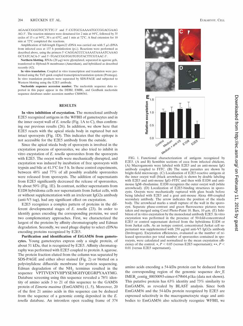
In vitro inhibition of excystation. The monoclonal antibodyE2E5 recognized antigens in the WFBII of gametocytes and inthe inner oocyst wall of E. tenella (Fig. 1A to C), thus confirm-ing our previous results (26). In addition, we show here thatE2E5 reacts with the apical stieda body in ruptured but notintact sporocysts (Fig. 1D). This indicates that the epitope isnot accessible for the E2E5 antibody from the outside.
Since the apical stieda body of sporocysts is involved in theexcystation process of sporozoites, we also tried to inhibit invitro excystation of E. tenella sporozoites from the sporocystswith E2E5. The oocyst walls were mechanically disrupted, andexcystation was induced by incubation of free sporocysts withtrypsin and bile at 41.5°C for 5 h. In these control experiments,between 48% and 77% of all possibly available sporozoiteswere released from sporocysts. The addition of supernatantsfrom E2E5 significantly decreased the release of sporozoites,by about 50% (Fig. 1E). In contrast, neither supernatants fromE1D8 hybridoma cells nor supernatants from Jurkat cells, withor without supplementation with an irrelevant IgG2a antibody(anti-V5 tag), had any significant effect on excystation.
E2E5 recognizes a complex pattern of proteins in the dif-ferent developmental stages of E. tenella (23). In order toidentify genes encoding the corresponding proteins, we usedtwo complementary approaches. First, we characterized thelargest of the proteins by affinity chromatography and Edmandegradation. Secondly, we used phage display to select cDNAsencoding proteins recognized by E2E5.
Purification and identification of EtGAM56 from gameto-cytes. Young gametocytes express only a single protein, ofabout 51 kDa, that is recognized by E2E5. Affinity chromatog-raphy was performed with E2E5 coupled to protein A agarose.The protein fraction eluted from the column was separated bySDS-PAGE and either silver stained (Fig. 2) or blotted on apolyvinylidene difluoride membrane for protein sequencing.Edman degradation of the NH2 terminus resulted in thesequence VPTTVENTVHPYSEMGHYQEGRPYAAYMG.Database screening using this sequence revealed a 78% iden-tity of amino acids 3 to 21 of this sequence to the GAM56protein of Eimeria maxima (EmGAM56) (1, 5). Moreover, 20of the first 21 amino acids in this sequence can be deducedfrom the sequence of a genomic contig deposited in the E.tenella database. An intronless open reading frame of 374
amino acids encoding a 54-kDa protein can be deduced fromthe corresponding region of the genomic sequence dev_EIMER_contig_00030093-eimer-679b04.p1ka (data not shown).This putative protein has 63% identity and 73% similarity toEmGAM56, as revealed by BLAST analysis. Since bothEmGAM56 and the 54-kDa protein recognized by E2E5 areexpressed selectively in the macrogametocyte stage and anti-bodies to EmGAM56 also selectively recognize WFBII, we
FIG. 1. Functional characterization of antigens recognized byE2E5. (A and B) Semithin sections of ceca from infected chickens.(A) Macrogamonts were labeled with E2E5 and an anti-mouse IgGantibody coupled to FITC. (B) The same parasites are shown bybright-field microscopy. (C) Localization of E2E5-reactive antigens atthe inner oocyst wall (black arrowhead) is shown by double labelingwith E2E5 and anti-mouse IgG–FITC and then with E1D8 and anti-mouse IgM–rhodamine. E1D8 recognizes the outer oocyst wall (whitearrowhead). (D) Localization of E2E5-binding structures in sporo-cysts. Oocysts were mechanically ruptured with glass beads beforebeing labeled with E2E5 and a goat anti-mouse Alexa 488-coupledsecondary antibody. The arrow indicates the position of the stiedabody. The arrowhead marks a small rupture of the wall in the sporo-cyst. Separate phase-contrast and green fluorescence pictures weretaken and merged using Corel Photo-Paint 10. Bars, 10 �m. (E) Inhi-bition of in vitro excystation by the monoclonal antibody E2E5. In vitroexcystation was performed in the presence of 50-fold-concentratedE2E5 or control supernatant derived from the hybridoma E1D8 orfrom Jurkat cells. As an isotype control, concentrated Jurkat cell su-pernatant was supplemented with 250 �g/ml anti-V5 IgG2a antibody(Invitrogen). Excystation efficiencies, evaluated as the number of re-leased sporozoites per total number of sporozoites contained in spo-rocysts, were calculated and normalized to the mean excystation effi-ciency of the control. *, P � 0.05 (versus E2E5 supernatant); **, P �0.01 (versus E2E5 supernatant).
204 KRUCKEN ET AL. EUKARYOT. CELL
on February 11, 2015 by guest
http://ec.asm.org/
Dow
nloaded from

propose that the 54-kDa protein is the E. tenella ortholog toGAM56 of E. maxima and should be designated EtGAM56.
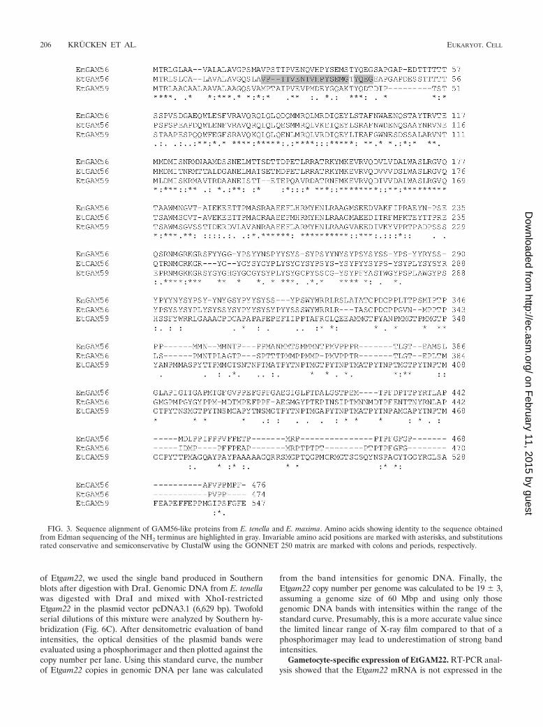
Interestingly, a second GAM56-like protein, of 59 kDa, isencoded on the same contig and in the same orientation, about2.3 kb upstream of Etgam56. We designate this intronless geneEtgam59, although we have not shown gametocyte-specific ex-pression of Etgam59 yet. Figure 3 shows a multiple sequencealignment of both deduced E. tenella proteins with the GAM56sequence of E. maxima. The sequences of all three GAM56-like proteins are rich in the amino acids Ala, Pro, Thr, and Ser.Moreover, all of these proteins contain a Tyr-rich domain.EmGAM56 is known to be present in the WFBII, the inneroocyst wall, and the apical part of the sporocyst, and this fitsperfectly well to our data from immunofluorescence and im-munoelectron microscopy presented in Fig. 1 and to the dataof Mouafo et al. (26). Remarkably, EmGAM56 has beenshown to be proteolytically cleaved during oocyst formationinto mature polypeptides of 33 kDa and 12 kDa (3). Indeed,the protein band corresponding to EtGAM56 disappears inunsporulated oocysts, and a strong band of slightly larger than30 kDa appears (26).
Cloning of EtGAM22 by phage display. In addition toEtGAM56 and its 33-kDa proteolytic fragment, E2E5 recognizestwo smaller proteins, of approximately 23 kDa and 25 kDa, inunsporulated oocysts and an approximately 80-kDa protein insporulated oocysts (26). In order to identify additional proteinsrecognized by E2E5, a genomic phage display library present-ing recombinant E. tenella proteins as fusions with the geneVIII product of filamentous phage on the surfaces of phagemidparticles was constructed. After four rounds of library panningagainst E2E5 coupled to Dynabeads, enrichment of a clonecontaining an insert of 145 bp was observed (data not shown).Western blot analysis clearly revealed that this phage clone,clone A17, expresses a recombinant gene VIII product fusionprotein that is recognized by E2E5, while other clones wereconsistently negative (Fig. 4).
Using 5�- and 3�-RACE PCR, we cloned the complete openreading frame of the corresponding mRNA. Figure 5 shows thecDNA sequence of Etgam22 and the deduced protein se-quence. The EtGAM22 protein has a length of 198 aminoacids, a molecular mass of 22.8 kDa, and a predicted pI of 6.8.At the NH2 terminus, a signal peptide for cotranslationaltransport into the endoplasmic reticulum (ER) is predicted bythe SignalP 3.0 program (6). The amino acid composition ofthe mature EtGAM22 protein without a signal peptide is veryunusual, since only four amino acids, His (25.7%), Pro (19%),Gln (8.4%), and Ala (6.1%), account for nearly 60% of allresidues. In particular, an extremely His- and Pro-rich domainbetween Pro73 and His188 can be identified. This partially re-sembles the case for EmGAM56, which contains 12.8% Proand 8% Ala residues. In contrast to EtGAM22, however,EmGAM56 contains only 0.6% His and 2.9% Gln residues,whereas EtGAM22 contains no Tyr-rich domain characteristicof other gametocyte-specific proteins, such as EmGAM56 andEmGAM82 (3). The sequence of the original phage clone A17containing the E2E5 epitope is completely localized within thisHis/Pro-rich domain. Moreover, there are putative O glycosyl-ation sites at Thr74, Thr80, and Thr81, as predicted byNetOGlyc 3.1.
Genomic organization of EtGAM22. BLAST analysis of thesequences deposited by the E. tenella genome project identifiedseveral perfect matches showing that the Etgam22 gene—likeEmgam56, Etgam56, and Etgam59–-does not contain any in-trons. Conspicuously, there are two contigs (dev_EIMER_contig_00030727-eimerbac28g8Fb07.q1k [data not shown] anddev_EIMER_contig_00016586-eimer-437a12.q1k) with twoidentical head-to-tail repeats of the Etgam22 gene. One ofthese contigs is drawn schematically in Fig. 6A. In addition, theintergenic regions between the copies are also nearly perfectlyconserved (99% identity), indicating that the promoter regionsare virtually identical. In order to demonstrate that Etgam22 isindeed a multicopy gene, we performed genomic Southern blotanalysis using six different restriction enzymes (Fig. 6B). Withthe exception of DraI, all enzymes produced one intensiveband and one or two faint bands, which is in accordance withseveral virtually identical head-to-tail copies of Etgam22. Inparticular, it should be mentioned that the restriction sites forall the enzymes used here are located outside the Etgam22open reading frame, indicating a high degree of conservationeven in the noncoding sequences of the cluster. The faintbands represent unique fragments from the end of theEtgam22 cluster. We used two complementary approaches toestimate the Etgam22 copy number in the E. tenella genome.The genomic DNA digested with BglI and MvaI (Fig. 6B)yielded two bands, i.e., a faint band representing a single copylocated at the end of the Etgam22 cluster and a strong banddue to hybridization to the other members of the cluster. Aftercorrection for different overlaps of internal and flankingEtgam22 copies with the hybridization probe, it should bepossible to calculate the copy number from the ratio of bandintensities. Our results indicate that there are 14 times morehybridization targets in the strong than in the faint BglI bandand 11 times more targets in the strong than in the faint MvaIband. Therefore, we assume that there are between 12 and 15copies of Etgam22 in the genome.
For an independent approach to estimate the copy number
FIG. 2. Purification of 51-kDa antigen by affinity chromatography.Protein extracts from purified gametocytes (lane 1) and proteins elutedfrom the E2E5 affinity column (lane 2) were separated by SDS-PAGEand silver stained. Positions of molecular size marker bands are indi-cated.
VOL. 7, 2008 IMPAIRED EXCYSTATION OF EIMERIA TENELLA 205
on February 11, 2015 by guest
http://ec.asm.org/
Dow
nloaded from
of Etgam22, we used the single band produced in Southernblots after digestion with DraI. Genomic DNA from E. tenellawas digested with DraI and mixed with XhoI-restrictedEtgam22 in the plasmid vector pcDNA3.1 (6,629 bp). Twofoldserial dilutions of this mixture were analyzed by Southern hy-bridization (Fig. 6C). After densitometric evaluation of bandintensities, the optical densities of the plasmid bands wereevaluated using a phosphorimager and then plotted against thecopy number per lane. Using this standard curve, the numberof Etgam22 copies in genomic DNA per lane was calculated
from the band intensities for genomic DNA. Finally, theEtgam22 copy number per genome was calculated to be 19 � 3,assuming a genome size of 60 Mbp and using only thosegenomic DNA bands with intensities within the range of thestandard curve. Presumably, this is a more accurate value sincethe limited linear range of X-ray film compared to that of aphosphorimager may lead to underestimation of strong bandintensities.
Gametocyte-specific expression of EtGAM22. RT-PCR anal-ysis showed that the Etgam22 mRNA is not expressed in the
FIG. 3. Sequence alignment of GAM56-like proteins from E. tenella and E. maxima. Amino acids showing identity to the sequence obtainedfrom Edman sequencing of the NH2 terminus are highlighted in gray. Invariable amino acid positions are marked with asterisks, and substitutionsrated conservative and semiconservative by ClustalW using the GONNET 250 matrix are marked with colons and periods, respectively.
206 KRUCKEN ET AL. EUKARYOT. CELL
on February 11, 2015 by guest
http://ec.asm.org/
Dow
nloaded from

early stage of infection, at 72 h p.i., whereas it is readily de-tectable at 137 h p.i. and 148 h p.i. and in sporulated oocysts(Fig. 7A). However, expression levels in these stages differwidely, as revealed by Northern blot analysis (Fig. 7B). Thus,Etgam22 was not detectable in sporulated oocysts, while a fainthybridization signal was obtained from RNAs of gametocytesat 138 h p.i. Expression of Etgam22 mRNA then increasesdramatically at 148 h p.i. and 168 h p.i., when mature game-tocytes and a mixture of gametocytes and unsporulated oo-
cysts, respectively, are present in the ceca. Expression ofEtgam56 mRNA was already very weakly detectable at 132 hp.i. and showed maximum expression between 144 h p.i. and168 h p.i.
We also extracted proteins from the same tissue samples andanalyzed the expression of proteins by using the antibody E2E5(Fig. 7B). Weak expression of the approximately 60-kDa bandcould be detected as early as 132 h p.i. Bands of about 33 kDaand 25 kDa did not appear before 168 h p.i. and were stilldetectable in sporulated oocysts. In this context, it is notewor-thy that in E. maxima, the ortholog EmGAM56 is cleaved intoa 33-kDa polypeptide during oocyst formation (3). The 33-kDaprotein in Fig. 7B is therefore assumed to represent the pro-teolytic fragment of EtGAM56. In accordance with our previ-ous results, a 25-kDa band was also observed at 168 h p.i.However, we could not reproduce the weak 23-kDa band de-tected previously in purified unsporulated oocysts (26). Sincein vitro translation of Etgam22 cDNA also produced apolypeptide of about 25 kDa (Fig. 7C), the 25-kDa band at168 h p.i. most likely corresponds to EtGAM22. This view isalso consistent with the fact that processing of EmGAM56does not give rise to polypeptide fragments in the size range of20 to 25 kDa (3).
DISCUSSION
The WFBII in gametocytes provides essential componentsfor the rigid and impermeable walls of oocysts, whose forma-tion is a critical step in the life cycle of Eimeria parasites, which
FIG. 4. Identification of E2E5-binding phagemid clones by West-ern blotting. Protein lysates of bacterial cultures were separated bySDS-PAGE. As a positive control (Pos), a protein extract from an E.tenella-infected cecum at 137 h p.i. was used. The negative control(Neg) consisted of a bacterial culture containing the empty pG8SAETphagemid vector.
FIG. 5. cDNA and deduced protein sequences of EtGAM22. The NH2-terminal signal peptide is shaded in light gray, and the sequencecorresponding to the original phage display clone is shaded in dark gray.
VOL. 7, 2008 IMPAIRED EXCYSTATION OF EIMERIA TENELLA 207
on February 11, 2015 by guest
http://ec.asm.org/
Dow
nloaded from

208 KRUCKEN ET AL. EUKARYOT. CELL
on February 11, 2015 by guest
http://ec.asm.org/
Dow
nloaded from

rely exclusively on the fecal/oral route for infection of newhosts. Using the monoclonal antibody E2E5, we describe theE. tenella GAM56 ortholog and confirm its proteolytic process-ing during oocyst wall formation. Moreover, we identify anovel type of small gametocyte-specific protein, EtGAM22,which also appears to be a structural component of the oocystwall.
The genes encoding EtGAM22 and EtGAM56, identifiedhere for E. tenella, and the previously characterized genes forthe WFBII-localized proteins EmGAM56 and EmGAM82 inE. maxima have several characteristics in common. All of themare expressed specifically in gametocytes. Moreover, there is a
remarkable absence of introns in these genes. AlthoughEmgam56 was described to be a single-copy gene (5), the datapresented here indicate the presence of two closely relatedgenes, Etgam56 and Etgam59, in the genome of E. tenella.Etgam22 is the first multicopy gene described for Eimeria spe-cies, and its extraordinarily high copy number and the ex-tremely conserved sequence between the copies suggest thatEtgam22 has a particularly important role in oocyst wall for-mation. The high degree of conservation indicates that ampli-fication of the Etgam22 gene was a very recent evolutionaryevent or that the sequences of the copies are frequently equal-ized by unequal crossover or gene conversion, which prevents
FIG. 6. Genomic organization of Etgam22. (A) Schematic drawing showing one end of the sequence contig 2257242.c007101021.contig1,containing two copies of the Etgam22 open reading frame (Etgam22). Some of the predicted restriction fragments rely on the assumption that atleast one additional copy of Etgam22 can be found further upstream (indicated in light gray on the left). (B) Genomic Southern blot using theenzymes BglI (lane 1), ClaI (lane 2), KpnI (lane 3), AccI (lane 4), DraI (lane 5), and MvaI (lane 6). The blot was hybridized with the Etgam22cDNA probe indicated in panel A. (C) Southern blot containing serial dilutions of genomic DNA from E. tenella digested with DraI and Etgam22cDNA in pcDNA3.1 linearized with XhoI. The lanes (from left to right) contained 1.7 �g (25,856,313 copies, assuming a genome size of 60 Mbp),0.85 �g, 0.425 �g, 0.213 �g, 0.106 �g, 0.053 �g, and 0.027 �g of genomic DNA and 726 pg (100,000 copies), 363 pg, 181.5 pg, 90.7 pg, 45.4 pg,22.7 pg, and 11.3 pg of plasmid DNA. The 6,629-bp band corresponds to the plasmid DNA, while the 995-bp band was shown in panel B to resultfrom binding of the probe to DraI-digested genomic DNA.
FIG. 7. Expression analysis of Etgam22. (A) RT-PCR with primers specific for Etgam22 and Etactin cDNAs. RNAs were isolated fromsporulated oocysts, from the ceca of noninfected chickens (n.i.), or from the ceca of chickens infected with E. tenella for the indicated times. Thereaction mixtures contained reverse transcriptase and RNA (lanes 1) or were performed in the absence of either the reverse transcriptase (lanes2) or the RNA template (lanes 3). (B) RNA and protein were isolated from the same samples of oocysts and infected ceca at the indicated times,using Trizol. With these matched RNA and protein samples, Northern blot analyses of Etgam22 and Etgam56 mRNA expression and Western blotanalysis with E2E5 were performed. (C) In vitro translation of EtGAM22. Products of coupled in vitro transcription and translation were separatedby SDS-PAGE and transferred to a nitrocellulose membrane, and EtGAM22 was detected using the E2E5 antibody in Western blot analysis. Thenegative control reaction mix contained no plasmid DNA.
VOL. 7, 2008 IMPAIRED EXCYSTATION OF EIMERIA TENELLA 209
on February 11, 2015 by guest
http://ec.asm.org/
Dow
nloaded from
sequence diversification, as previously described for multigenefamilies, including rRNA (33) and histone (30, 32) genes. Ap-parently, such mechanisms do not act on gam56-like genes.
Among the apicomplexa, low-copy-number head-to-tail clus-ters of homologous intronless genes have been described forsome of the T. gondii SAG genes that encode glycosylphos-phatidylinositol-coupled surface antigens expressed by the in-vasive parasite stages (11, 21, 36). Sequence similarity andexpression levels in different developmental stages vary widelybetween family members in the same cluster, and intergenicregions are usually not conserved, with the exception of thoseof SAG5B and SAG5C (36). In contrast, the intergenic regionsin the Etgam22 gene cluster appear to be highly conserved, asfar as can be deduced from the few sequenced repeats and theconservation of restriction sites, although we do not yet knowwhether all genes in the cluster are transcriptionally active.Expression of both Etgam56 and Etgam22 mRNAs is veryhigh, since exposure times of Northern blots of as short as 2.5 hand 4 h, respectively, were sufficient to produce clearly detect-able bands, despite the fact that the RNAs were not isolatedfrom pure parasites but from infected ceca and would there-fore contain large amounts of chicken RNA. It is thereforetempting to speculate that amplification of the Etgam22 genecopy number might be a mechanism to allow the production oflarge amounts of RNA within a very short period, as previouslydescribed for histone genes, which are highly expressed onlyduring a short phase of the cell cycle (22). Despite the highlevel of Etgam22 mRNA, however, the protein band detectedin Western blots is surprisingly quite faint. There are severalpossible explanations for this observation. First, the reactivityof the E2E5 antibody might be better for EtGAM56 than forEtGAM22, or extraction of EtGAM56 from the tissue mightbe easier than that of EtGAM22. Second, translation ofEtgam22 mRNA might be inefficient, with only low levels ofprotein being produced. Third, translation of Etgam22 mRNAmay be developmentally regulated and may not occur in mac-rogametocytes but in oocysts after fusion of gametes or duringsporulation. This view is also supported by the fact that theEtGAM22 protein is not detectable at 144 h p.i., when tran-script levels are maximal, although transcription starts at least6 h earlier. However, the protein becomes detectable at 168 hp.i., when the first unsporulated oocysts appear in the cecum,as revealed by the beginning of proteolytic processing ofEtGAM56.
At the protein level, similarity between EtGAM22 andEtGAM56 manifests itself as cross-reactivity with E2E5, as thepresence of putative N-glycosylation sites, and as NH2-termi-nal signal peptides. The presence of a signal peptide and thefact that, in macrogametocytes, E2E5 exclusively recognizesWFBII, i.e., specialized regions within the rough ER (9), sug-gest that EtGAM22—like EmGAM56—is transported to theWFBII and participates in formation of the inner oocyst walland/or the stieda body. For E. maxima, a GAM56-specificmonoclonal antibody has been reported to recognize the WFBIIin macrogametocytes, the oocyst wall, the outer sporocyst wall,and the stieda body (5). Since this pattern largely overlaps withthe pattern of E2E5 labeling in E. tenella, a monoclonal anti-body against EtGAM22 that does not cross-react withEtGAM56 will be necessary to evaluate the exact localizationof EtGAM22.
Other His-rich proteins are known for parasites belonging tothe apicomplexa, with the histidine-rich protein of Plasmodiumlophurae (78% His residues) (12) displaying the most excep-tional composition. However, the low sequence complexities ofthese proteins and EtGAM22 do not permit a phylogeneticanalysis. Indeed, those His-rich proteins for which a function isknown are surely not homologous to EtGAM22. For instance,the knob-associated histidine-rich protein of Plasmodium fal-ciparum is involved in the interaction of parasite proteins inknobs on the erythrocyte surface with the host cell cytoskeleton(27, 29), whereas the histidine-rich protein 2 has been impli-cated in hemozoin formation of P. falciparum (28). In contrast,BLAST comparison of EtGAM22 with proteins predictedfrom the Toxoplasma gondii genome reveals the presence of atleast five proteins with NH2-terminal signal peptides which arerelatively rich in proline and histidine and have lengths in therange of 117 to 269 amino acids. Although there is not yetanything known about the function or developmental expres-sion pattern of these proteins, it is tempting to speculate thatsome of them represent extracellular structural proteins, suchas many proline- or hydroxyproline-rich glycoproteins in plantcell walls (15, 16) or histidine-rich proteins in Hydra nemato-cysts (17). Cross-linking of EmGAM56- and EmGAM82-de-rived peptides via dopamine has been shown in the oocyst wall(3), and His-rich proteins such as EtGAM22 might also beinvolved in stabilizing extracellular structures via cross-linksbetween His and catechols, as described for insect cuticles (8,14, 43).
The in vitro excystation inhibition assay presented hereshows that the antibody E2E5 can significantly interfere withparasite development. Remarkably, vaccination with a prepa-ration of native gametocyte antigens enriched for GAM56,GAM82, and GAM230, which are all involved in wall formingin E. maxima, has been shown to convey at least partial pro-tection against homologous challenge (25, 39, 46). SinceEtGAM22 represents a new family of oocyst wall proteins, itmight be a useful supplement to a protective gametocyte-spe-cific cocktail vaccine.
ACKNOWLEDGMENTS
We thank Carsten Angenendt for valuable technical assistance.Genomic data were provided by The Institute for Genomic Re-
search (supported by NIH grant AI05093) and by the Sanger Center(Wellcome Trust). Expressed sequence tag sequences were generatedby Washington University (NIH grant 1R01AI045806-01A1).
REFERENCES
1. Belli, S. I., M. Lee, P. Thebo, M. G. Wallach, B. Schwartsburd, and N. C.Smith. 2002. Biochemical characterisation of the 56 and 82 kDa immuno-dominant gametocyte antigens from Eimeria maxima. Int. J. Parasitol. 32:805–816.
2. Belli, S. I., K. Mai, C. D. Skene, M. T. Gleeson, D. M. Witcombe, M. Katrib,A. Finger, M. G. Wallach, and N. C. Smith. 2004. Characterisation of theantigenic and immunogenic properties of bacterially expressed, sexual stageantigens of the coccidian parasite Eimeria maxima. Vaccine 22:4316–4325.
3. Belli, S. I., M. G. Wallach, C. Luxford, M. J. Davies, and N. C. Smith. 2003.Roles of tyrosine-rich precursor glycoproteins and dityrosine- and 3,4-dihy-droxyphenylalanine-mediated protein cross-linking in development of theoocyst wall in the coccidian parasite Eimeria maxima. Eukaryot. Cell 2:456–464.
4. Belli, S. I., M. G. Wallach, and N. C. Smith. 2003. Cloning and character-ization of the 82 kDa tyrosine-rich sexual stage glycoprotein, GAM82, and itsrole in oocyst wall formation in the apicomplexan parasite Eimeria maxima.Gene 307:201–212.
5. Belli, S. I., D. Witcombe, M. G. Wallach, and N. C. Smith. 2002. Functional
210 KRUCKEN ET AL. EUKARYOT. CELL
on February 11, 2015 by guest
http://ec.asm.org/
Dow
nloaded from
genomics of gam56: characterisation of the role of a 56 kilodalton sexualstage antigen in oocyst wall formation in Eimeria maxima. Int. J. Parasitol.32:1727–1737.
6. Bendtsen, J. D., H. Nielsen, G. von Heijne, and S. Brunak. 2004. Improvedprediction of signal peptides: SignalP 3.0. J. Mol. Biol. 340:783–795.
7. Blin, N., and D. W. Stafford. 1976. A general method for isolation of highmolecular weight DNA from eukaryotes. Nucleic Acids Res. 3:2303–2308.
8. Christensen, A. M., J. Schaefer, K. J. Kramer, T. D. Morgan, and T. L.Hopkins. 1991. Detection of cross-links in insect cuticle by redor NMR-spectroscopy. J. Am. Chem. Soc. 113:6799–6802.
9. Ferguson, D. J., S. I. Belli, N. C. Smith, and M. G. Wallach. 2003. Thedevelopment of the macrogamete and oocyst wall in Eimeria maxima: im-muno-light and electron microscopy. Int. J. Parasitol. 33:1329–1340.
10. Fetterer, R. H., and R. C. Barfield. 2003. Characterization of a developmen-tally regulated oocyst protein from Eimeria tenella. J. Parasitol. 89:553–564.
11. Hehl, A., T. Krieger, and J. C. Boothroyd. 1997. Identification and charac-terization of SRS1, a Toxoplasma gondii surface antigen upstream of andrelated to SAG1. Mol. Biochem. Parasitol. 89:271–282.
12. Irving, D. O., and G. A. Cross. 1986. Primary structure of the histidine-richprotein of Plasmodium lophurae. Nature 324:492.
13. Julenius, K., A. Molgaard, R. Gupta, and S. Brunak. 2005. Prediction,conservation analysis, and structural characterization of mammalian mucin-type O-glycosylation sites. Glycobiology 15:153–164.
14. Kerwin, J. L., F. Turecek, R. D. Xu, K. J. Kramer, T. L. Hopkins, C. L.Gatlin, and J. R. Yates. 1999. Mass spectrometric analysis of catechol-histidine adducts from insect cuticle. Anal. Biochem. 268:229–237.
15. Kieliszewski, M. J., and D. T. Lamport. 1994. Extensin: repetitive motifs,functional sites, post-translational codes, and phylogeny. Plant J. 5:157–172.
16. Kieliszewski, M. J., and E. Shpak. 2001. Synthetic genes for the elucidationof glycosylation codes for arabinogalactan-proteins and other hydroxypro-line-rich glycoproteins. Cell Mol. Life Sci. 58:1386–1398.
17. Koch, A. W., T. W. Holstein, C. Mala, E. Kurz, J. Engel, and C. N. David.1998. Spinalin, a new glycine- and histidine-rich protein in spines of Hydranematocysts. J. Cell Sci. 111:1545–1554.
18. Krucken, J., R. M. Schroetel, I. U. Muller, N. Saıdani, P. Marinovski, W. P.Benten, O. Stamm, and F. Wunderlich. 2004. Comparative analysis of thehuman gimap gene cluster encoding a novel GTPase family. Gene 341:291–304.
19. Krucken, J., O. Stamm, H.-P. Schmitt-Wrede, A. Mincheva, P. Lichter, andF. Wunderlich. 1999. Spleen-specific expression of the malaria-inducibleintronless mouse gene imap38. J. Biol. Chem. 274:24383–24391.
20. Laemmli, U. K. 1970. Cleavage of structural proteins during the assembly ofthe head of bacteriophage T4. Nature 227:680–685.
21. Lekutis, C., D. J. Ferguson, M. E. Grigg, M. Camps, and J. C. Boothroyd.2001. Surface antigens of Toxoplasma gondii: variations on a theme. Int. J.Parasitol. 31:1285–1292.
22. Marzluff, W. F., P. Gongidi, K. R. Woods, J. Jin, and L. J. Maltais. 2002. Thehuman and mouse replication-dependent histone genes. Genomics 80:487–498.
23. McDonald, V., and M. E. Rose. 1987. Eimeria tenella and E. necatrix: a thirdgeneration of schizogony is an obligatory part of the developmental cycle. J.Parasitol. 73:617–622.
24. Mehlhorn, H. 2005. Eimeria, p. 198–199. In H. Mehlhorn (ed.), Encyclopedicreference of parasitology. Biology, structure, function. Springer, Berlin, Ger-many.
25. Michael, A. 2002. The practical use of a maternal vaccine against coccidiosis.World Poultry 19:24–26.
26. Mouafo, A. N., A. Weck-Heimann, J. F. Dubremetz, and R. Entzeroth. 2002.Monoclonal antibodies specific for the two types of wall-forming bodies ofEimeria tenella macrogametes (Coccidia, Apicomplexa). Parasitol. Res. 88:217–224.
27. Oh, S. S., S. Voigt, D. Fisher, S. J. Yi, P. J. LeRoy, L. H. Derick, S. Liu, andA. H. Chishti. 2000. Plasmodium falciparum erythrocyte membrane protein 1
is anchored to the actin-spectrin junction and knob-associated histidine-richprotein in the erythrocyte skeleton. Mol. Biochem. Parasitol. 108:237–247.
28. Pandey, A. V., V. K. Babbarwal, J. N. Okoyeh, R. M. Joshi, S. K. Puri, R. L.Singh, and V. S. Chauhan. 2003. Hemozoin formation in malaria: a two-stepprocess involving histidine-rich proteins and lipids. Biochem. Biophys. Res.Commun. 308:736–743.
29. Pei, X., X. An, X. Guo, M. Tarnawski, R. Coppel, and N. Mohandas. 2005.Structural and functional studies of interaction between Plasmodium falcip-arum knob-associated histidine-rich protein (KAHRP) and erythrocyte spec-trin. J. Biol. Chem. 280:31166–31171.
30. Piontkivska, H., A. P. Rooney, and M. Nei. 2002. Purifying selection andbirth-and-death evolution in the histone H4 gene family. Mol. Biol. Evol.19:689–697.
31. Pittilo, R. M., and S. J. Ball. 1980. The ultrastructural development of theoocyst wall of Eimeria maxima. Parasitology 81:115–122.
32. Rooney, A. P., H. Piontkivska, and M. Nei. 2002. Molecular evolution of thenontandemly repeated genes of the histone 3 multigene family. Mol. Biol.Evol. 19:68–75.
33. Rooney, A. P., and T. J. Ward. 2005. Evolution of a large ribosomal RNAmultigene family in filamentous fungi: birth and death of a concerted evo-lution paradigm. Proc. Natl. Acad. Sci. USA 102:5084–5089.
34. Ryley, J. F. 1973. Cytochemistry, physiology and biochemistry, p. 145–181. InD. M. Hammond and P. L. Long (ed.), The coccidia. University Park Press,London, United Kingdom.
35. Scholtyseck, E., H. Mehlhorn, and D. M. Hammond. 1971. Fine structure ofmacrogametes and oocysts of Coccidia and related organisms. Z. Para-sitenkd. 37:1–43.
36. Spano, F., I. Ricci, M. Di Cristina, A. Possenti, M. Tinti, N. Dendouga, S.Tomavo, and A. Crisanti. 2002. The SAG5 locus of Toxoplasma gondiiencodes three novel proteins belonging to the SAG1 family of surface anti-gens. Int. J. Parasitol. 32:121–131.
37. Stotish, R. L., C. C. Wang, and M. Meyenhofer. 1978. Structure and com-position of the oocyst wall of Eimeria tenella. J. Parasitol. 64:1074–1081.
38. Wallach, M. 1997. The importance of transmission-blocking immunity in thecontrol of infections by apicomplexan parasites. Int. J. Parasitol. 27:1159–1167.
39. Wallach, M. 2002. The development of a novel vaccine against coccidiosis.World Poultry 19:24–26.
40. Wallach, M. G., D. Mencher, S. Yarus, G. Pillemer, A. Halabi, and T.Pugatsch. 1989. Eimeria maxima: identification of gametocyte protein anti-gens. Exp. Parasitol. 68:49–56.
41. Williams, R. B., W. W. Carlyle, D. R. Bond, and I. A. Brown. 1999. Theefficacy and economic benefits of Paracox, a live attenuated anticoccidialvaccine, in commercial trials with standard broiler chickens in the UnitedKingdom. Int. J. Parasitol. 29:341–355.
42. Wunderlich, F., M. A. Dkhil, L. I. Mehnert, J. V. Braun, M. El Khadragy, E.Borsch, D. Hermsen, W. P. Benten, K. Pfeffer, H. Mossmann, and J.Krucken. 2005. Testosterone responsiveness of spleen and liver in femalelymphotoxin beta receptor-deficient mice resistant to blood-stage malaria.Microbes Infect. 7:399–409.
43. Xu, R. D., X. Huang, T. L. Hopkins, and K. J. Kramer. 1997. Catecholamineand histidyl protein cross-linked structures in sclerotized insect cuticle. In-sect Biochem. Mol. Biol. 27:101–108.
44. Zdobnov, E. M., and R. Apweiler. 2001. InterProScan—an integration plat-form for the signature-recognition methods in InterPro. Bioinformatics 17:847–848.
45. Zhang, L., K. Jacobsson, K. Strom, M. Lindberg, and L. Frykberg. 1999.Staphylococcus aureus expresses a cell surface protein that binds both IgGand beta2-glycoprotein I. Microbiology 145:177–183.
46. Ziomko, R., J. Karamon, T. Cencek, E. Gornowicz, A. Skoracki, and U.Ashash. 2005. Prevention of broiler chick coccidiosis using the inactivatedsubunit vaccine CoxAbic. Bull. Vet. Inst. Puawy 49:299–302.
VOL. 7, 2008 IMPAIRED EXCYSTATION OF EIMERIA TENELLA 211
on February 11, 2015 by guest
http://ec.asm.org/
Dow
nloaded from
Copyright © 2022 FDOKUMEN