Melusin, a muscle-specific integrin β1–interacting protein, is required to prevent cardiac...
8
68 NATURE MEDICINE • VOLUME 9 • NUMBER 1 • JANUARY 2003 ARTICLES Arterial hypertension imposes a biomechanical stress on the left ventricle, requiring the heart to work harder than under normal circumstances. Under these conditions, the heart undergoes hy- pertrophy resulting from increased synthesis and assembly of the contractile proteins of the actomyosin fibrils. Cardiac hy- pertrophy is considered beneficial because it allows the genera- tion of greater contractile force, but it may also result in activation of pathways that reduce the efficiency of structural adaptation, promoting the transition toward cardiac dilation and failure 1 . Identifying the molecular mechanisms involved in cardiac hypertrophy and the transition toward dilation and dys- function is thus an important challenge of cardiovascular biol- ogy and medicine 2 . Cardiac hypertrophic remodeling is triggered by a combined action of mechanical stretching of cardiac walls and activation of neurohumoral growth factors 1,3,4 . Mechanical stress is consid- ered the primary trigger to induce the growth response in the overloaded myocardium. Indeed, cardiomyocytes possess an in- trinsic mechanosensing mechanism, as indicated by the ability of cultured cardiomyocytes to undergo hypertrophy when sub- jected to mechanical stretching in vitro 5 . Stretching is thought to activate the release of a cascade of autocrine and paracrine hu- moral factors, such as angiotensin II, endothelin 1, insulin like growth factor (IGF-1), transforming growth factor-β (TGF-β), fi- broblast growth factor (FGF) and cardiotrophin-1 (CT-1) (refs. 3,6–8), that in turn promote cardiomyocyte growth and heart tissue remodeling. The intracellular signaling pathways triggered by mechanical stress and by humoral trophic factors are proba- bly temporally and mechanistically distinct; however, whether these pathways can act independently is still under debate. In vitro and in vivo studies have identified several signaling mole- cules and pathways involved in cardiac hypertrophy, including the αGq subunit of the heterotrimeric G protein coupled to the β-adrenergic receptors 9–11 ; the phospholipase Cβ and protein ki- nase C, acting downstream of the G proteins 12 ; the phospho- inositide 3-kinase; the calcineurin/NF-AT3 pathway; the Ras cascade, including Raf-1 and ERK1/2 MAP kinases; the stress ki- nases Jnk and p38; and the Jak-STAT pathway 13,14 . Although most of these molecules are activated by both biomechanical and hu- moral factors, some are specifically involved in cardiac hypertro- phy induced by humoral factors. In fact, inactivation of the genes encoding angiotensin II type 1 (AT-1) and the α 1b adrener- gic receptor (α 1b -AR) abrogates cardiac hypertrophy induced by sub-pressor doses of angiotensin II or phenylephrine in the ab- sence of mechanical stress, but does not affect the hypertrophic response to mechanical overload imposed by aortic coarcta- Melusin, a muscle-specific integrin β 1 –interacting protein, is required to prevent cardiac failure in response to chronic pressure overload MARA BRANCACCIO 1 , LUIGI FRATTA 2 , ANTONELLA NOTTE 2 , EMILIO HIRSCH 1,3 , ROBERTA POULET 2 , SIMONA GUAZZONE 1 , MARIKA DE ACETIS 1 , CARMINE VECCHIONE 2 , GENNARO MARINO 2 , FIORELLA ALTRUDA 1,3 , LORENZO SILENGO 1,3 , GUIDO TARONE 1,3 & GIUSEPPE LEMBO 2,4 1 Department of Genetics, Biology, and Biochemistry, Turin University, 10126 Turin, Italy 2 Department of Angiocardioneurology, IRCCS ‘Neuromed’, 86077 Pozzilli (IS), Italy 3 Experimental Medicine Research Center, San Giovanni Battista Hospital, 10126 Turin, Italy 4 Department of Experimental Medicine and Pathology, University of Rome ‘La Sapienza’ Rome, Italy M.B. and L.F. contributed equally to this work. Correspondence should be addressed to G.T. (e-mail: [email protected]) or G.L. (email: [email protected]) Published online 23 December 2002, doi:10.1038/nm805 Cardiac hypertrophy is an adaptive response to a variety of mechanical and hormonal stimuli, and represents an early event in the clinical course leading to heart failure. By gene inactivation, we demonstrate here a crucial role of melusin, a muscle-specific protein that interacts with the integrin β 1 cytoplasmic domain, in the hypertrophic response to mechanical overload. Melusin- null mice showed normal cardiac structure and function in physiological conditions, but when subjected to pressure overload—a condition that induces a hypertrophic response in wild-type controls—they developed an abnormal cardiac remodeling that evolved into dilated cardiomy- opathy and contractile dysfunction. In contrast, the hypertrophic response was identical in wild- type and melusin-null mice after chronic administration of angiotensin II or phenylephrine at doses that do not increase blood pressure—that is, in the absence of cardiac biomechanical stress. Analysis of intracellular signaling events induced by pressure overload indicated that phosphorylation of glycogen synthase kinase-3β (GSK-3β) was specifically blunted in melusin- null hearts. Thus, melusin prevents cardiac dilation during chronic pressure overload by specifi- cally sensing mechanical stress. © 2003 Nature Publishing Group http://www.nature.com/naturemedicine
Transcript of Melusin, a muscle-specific integrin β1–interacting protein, is required to prevent cardiac...
68 NATURE MEDICINE • VOLUME 9 • NUMBER 1 • JANUARY 2003
ARTICLES
Arterial hypertension imposes a biomechanical stress on the leftventricle, requiring the heart to work harder than under normalcircumstances. Under these conditions, the heart undergoes hy-pertrophy resulting from increased synthesis and assembly ofthe contractile proteins of the actomyosin fibrils. Cardiac hy-pertrophy is considered beneficial because it allows the genera-tion of greater contractile force, but it may also result inactivation of pathways that reduce the efficiency of structuraladaptation, promoting the transition toward cardiac dilationand failure1. Identifying the molecular mechanisms involved incardiac hypertrophy and the transition toward dilation and dys-function is thus an important challenge of cardiovascular biol-ogy and medicine2.
Cardiac hypertrophic remodeling is triggered by a combinedaction of mechanical stretching of cardiac walls and activationof neurohumoral growth factors1,3,4. Mechanical stress is consid-ered the primary trigger to induce the growth response in theoverloaded myocardium. Indeed, cardiomyocytes possess an in-trinsic mechanosensing mechanism, as indicated by the abilityof cultured cardiomyocytes to undergo hypertrophy when sub-jected to mechanical stretching in vitro5. Stretching is thought toactivate the release of a cascade of autocrine and paracrine hu-moral factors, such as angiotensin II, endothelin 1, insulin like
growth factor (IGF-1), transforming growth factor-β (TGF-β), fi-broblast growth factor (FGF) and cardiotrophin-1 (CT-1) (refs.3,6–8), that in turn promote cardiomyocyte growth and hearttissue remodeling. The intracellular signaling pathways triggeredby mechanical stress and by humoral trophic factors are proba-bly temporally and mechanistically distinct; however, whetherthese pathways can act independently is still under debate. Invitro and in vivo studies have identified several signaling mole-cules and pathways involved in cardiac hypertrophy, includingthe αGq subunit of the heterotrimeric G protein coupled to theβ-adrenergic receptors9–11; the phospholipase Cβ and protein ki-nase C, acting downstream of the G proteins12; the phospho-inositide 3-kinase; the calcineurin/NF-AT3 pathway; the Rascascade, including Raf-1 and ERK1/2 MAP kinases; the stress ki-nases Jnk and p38; and the Jak-STAT pathway13,14. Although mostof these molecules are activated by both biomechanical and hu-moral factors, some are specifically involved in cardiac hypertro-phy induced by humoral factors. In fact, inactivation of thegenes encoding angiotensin II type 1 (AT-1) and the α1b adrener-gic receptor (α1b-AR) abrogates cardiac hypertrophy induced bysub-pressor doses of angiotensin II or phenylephrine in the ab-sence of mechanical stress, but does not affect the hypertrophicresponse to mechanical overload imposed by aortic coarcta-
Melusin, a muscle-specific integrin β1–interacting protein, is required to prevent cardiac failure in response
to chronic pressure overload
MARA BRANCACCIO1, LUIGI FRATTA2, ANTONELLA NOTTE2, EMILIO HIRSCH1,3, ROBERTA POULET2,SIMONA GUAZZONE1, MARIKA DE ACETIS1, CARMINE VECCHIONE2, GENNARO MARINO2,
FIORELLA ALTRUDA1,3, LORENZO SILENGO1,3, GUIDO TARONE1,3 & GIUSEPPE LEMBO2,4
1Department of Genetics, Biology, and Biochemistry, Turin University, 10126 Turin, Italy2Department of Angiocardioneurology, IRCCS ‘Neuromed’, 86077 Pozzilli (IS), Italy
3Experimental Medicine Research Center, San Giovanni Battista Hospital, 10126 Turin, Italy4Department of Experimental Medicine and Pathology, University of Rome ‘La Sapienza’ Rome, Italy
M.B. and L.F. contributed equally to this work.Correspondence should be addressed to G.T. (e-mail: [email protected]) or G.L. (email: [email protected])
Published online 23 December 2002, doi:10.1038/nm805
Cardiac hypertrophy is an adaptive response to a variety of mechanical and hormonal stimuli,and represents an early event in the clinical course leading to heart failure. By gene inactivation,we demonstrate here a crucial role of melusin, a muscle-specific protein that interacts with theintegrin β1 cytoplasmic domain, in the hypertrophic response to mechanical overload. Melusin-null mice showed normal cardiac structure and function in physiological conditions, but whensubjected to pressure overload—a condition that induces a hypertrophic response in wild-typecontrols—they developed an abnormal cardiac remodeling that evolved into dilated cardiomy-opathy and contractile dysfunction. In contrast, the hypertrophic response was identical in wild-type and melusin-null mice after chronic administration of angiotensin II or phenylephrine atdoses that do not increase blood pressure—that is, in the absence of cardiac biomechanicalstress. Analysis of intracellular signaling events induced by pressure overload indicated thatphosphorylation of glycogen synthase kinase-3β (GSK-3β) was specifically blunted in melusin-null hearts. Thus, melusin prevents cardiac dilation during chronic pressure overload by specifi-cally sensing mechanical stress.©
2003
Nat
ure
Pu
blis
hin
g G
rou
p
htt
p:/
/ww
w.n
atu
re.c
om
/nat
ure
med
icin
e
NATURE MEDICINE • VOLUME 9 • NUMBER 1 • JANUARY 2003 69
ARTICLES
tion15,16. This indicates the existence of a pathway triggered byangiotensin II or norepinephrine via AT-1 and an α1b-AR recep-tors, independently of mechanical stress.
In contrast, it is not known which molecules are required tocouple mechanical stress to the induction of intracellular sig-nals responsible for the hypertrophic response. Integrins andthe associated signaling machinery are likely candidates, be-cause of their role in linking the force-generating actin cy-toskeleton inside the cell to the extracellular matrix17.
Integrins are crucial to heart developmentand function, as indicated both by in vitroand in vivo studies. Both chimeric mice andembryoid bodies constructed from integrinβ1–null cells show delayed development anddifferentiation of β1-deficient cells along thecardiac lineage, as well as abnormal sarcom-erogenesis18. In addition, dominant-negativedisruption of integrin β1 function19 as well asselective cardiac excision of the integrin β1
gene20 result in myocardial fibrosis and car-diac failure.
We recently identified melusin, a proteinbinding to the integrin β1 cytoplasmic domainand specifically expressed in striated muscletissue, where it localizes at costameres inproximity of the Z line along with integrinsand vinculin21. By inactivating the gene en-coding melusin, we show here that the ab-sence of melusin does not affect cardiacdevelopment or basal function, but leads to areduced left ventricular hypertrophy and fa-vors the transition toward dilated cardiomy-opathy and contractile dysfunction inresponse to chronic pressure overload. Yet theabsence of melusin does not influence the de-velopment of cardiac hypertrophy to humoralfactors such as angiotensin II or phenyle-phrine in the absence of biomechanical stress.Thus, melusin is most likely a specificmechanosensor transducing signal that actsin preventing the transition to heart failure inconditions of biomechanical stress.
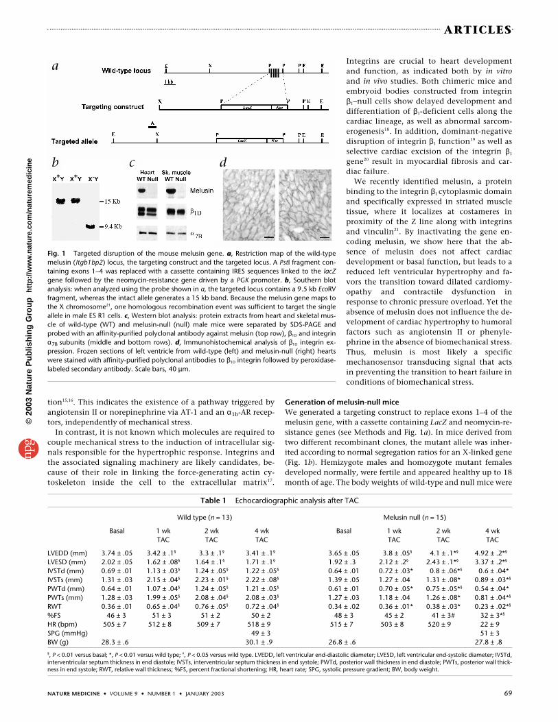
Generation of melusin-null miceWe generated a targeting construct to replace exons 1–4 of themelusin gene, with a cassette containing LacZ and neomycin-re-sistance genes (see Methods and Fig. 1a). In mice derived fromtwo different recombinant clones, the mutant allele was inher-ited according to normal segregation ratios for an X-linked gene(Fig. 1b). Hemizygote males and homozygote mutant femalesdeveloped normally, were fertile and appeared healthy up to 18month of age. The body weights of wild-type and null mice were
a
b c d
Fig. 1 Targeted disruption of the mouse melusin gene. a, Restriction map of the wild-typemelusin (Itgb1bp2) locus, the targeting construct and the targeted locus. A PstI fragment con-taining exons 1–4 was replaced with a cassette containing IRES sequences linked to the lacZgene followed by the neomycin-resistance gene driven by a PGK promoter. b, Southern blotanalysis: when analyzed using the probe shown in a, the targeted locus contains a 9.5 kb EcoRVfragment, whereas the intact allele generates a 15 kb band. Because the melusin gene maps tothe X chromosome21, one homologous recombination event was sufficient to target the singleallele in male ES R1 cells. c, Western blot analysis: protein extracts from heart and skeletal mus-cle of wild-type (WT) and melusin-null (null) male mice were separated by SDS-PAGE andprobed with an affinity-purified polyclonal antibody against melusin (top row), β1D and integrinα7B subunits (middle and bottom rows). d, Immunohistochemical analysis of β1D integrin ex-pression. Frozen sections of left ventricle from wild-type (left) and melusin-null (right) heartswere stained with affinity-purified polyclonal antibodies to β1D integrin followed by peroxidase-labeled secondary antibody. Scale bars, 40 µm.
Table 1 Echocardiographic analysis after TAC
Wild type (n = 13) Melusin null (n = 15)
Basal 1 wk 2 wk 4 wk Basal 1 wk 2 wk 4 wkTAC TAC TAC TAC TAC TAC
LVEDD (mm) 3.74 ± .05 3.42 ± .1§ 3.3 ± .1§ 3.41 ± .1§ 3.65 ± .05 3.8 ± .05§ 4.1 ± .1*§ 4.92 ± .2*§
LVESD (mm) 2.02 ± .05 1.62 ± .08§ 1.64 ± .1§ 1.71 ± .1§ 1.92 ± .3 2.12 ± .2§ 2.43 ± .1*§ 3.37 ± .2*§
IVSTd (mm) 0.69 ± .01 1.13 ± .03§ 1.24 ± .05§ 1.22 ± .05§ 0.64 ± .01 0.72 ± .03* 0.8 ± .06*§ 0.6 ± .04*IVSTs (mm) 1.31 ± .03 2.15 ± .04§ 2.23 ± .01§ 2.22 ± .08§ 1.39 ± .05 1.27 ± .04 1.31 ± .08* 0.89 ± .03*§
PWTd (mm) 0.64 ± .01 1.07 ± .04§ 1.24 ± .05§ 1.21 ± .05§ 0.61 ± .01 0.70 ± .05* 0.75 ± .05*§ 0.54 ± .04*PWTs (mm) 1.28 ± .03 1.99 ± .05§ 2.08 ± .04§ 2.08 ± .03§ 1.27 ± .03 1.18 ± .04 1.26 ± .08* 0.81 ± .04*§
RWT 0.36 ± .01 0.65 ± .04§ 0.76 ± .05§ 0.72 ± .04§ 0.34 ± .02 0.36 ± .01* 0.38 ± .03* 0.23 ± .02*§
%FS 46 ± 3 51 ± 3 51 ± 2 50 ± 2 48 ± 3 45 ± 2 41 ± 3# 32 ± 3*§
HR (bpm) 505 ± 7 512 ± 8 509 ± 7 518 ± 9 515 ± 7 503 ± 8 520 ± 9 22 ± 9SPG (mmHg) 49 ± 3 51 ± 3BW (g) 28.3 ± .6 30.1 ± .9 26.8 ± .6 27.8 ± .8§, P < 0.01 versus basal; *, P < 0.01 versus wild type; #, P < 0.05 versus wild type. LVEDD, left ventricular end-diastolic diameter; LVESD, left ventricular end-systolic diameter; IVSTd,interventricular septum thickness in end diastole; IVSTs, interventricular septum thickness in end systole; PWTd, posterior wall thickness in end diastole; PWTs, posterior wall thick-ness in end systole; RWT, relative wall thickness; %FS, percent fractional shortening; HR, heart rate; SPG, systolic pressure gradient; BW, body weight.
©20
03 N
atu
re P
ub
lish
ing
Gro
up
h
ttp
://w
ww
.nat
ure
.co
m/n
atu
rem
edic
ine
70 NATURE MEDICINE • VOLUME 9 • NUMBER 1 • JANUARY 2003
ARTICLES
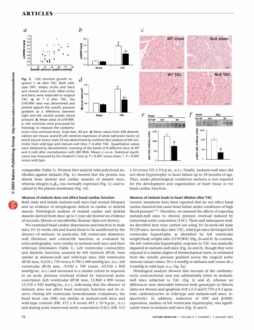
Fig. 2. Left ventricle growth re-sponse 1 wk after TAC. Both wild-type (WT, empty circles and bars)and mutant mice (null, filled circlesand bars) were subjected to surgicalTAC. a, At 7 d after TAC, theLVW/BW ratio was determined andplotted against the systolic pressuregradient as a difference betweenright and left carotid systolic bloodpressure. b, Mean value of LVW/BW.c, Left ventricles were processed forhistology to measure the cardiomy-ocyte cross-sectional areas. Scale bars, 40 µm. d, Mean values from 200 determi-nations per mouse. e and f, Left ventricle expression of atrial natriuretic factor (e)and β-myosin heavy chain (f) was determined by northern blot analysis of left ven-tricles from wild-type and melusin-null mice 7 d after TAC. Quantitative valueswere obtained by densitometric scanning of the bands of 8 different mice (4 WTand 4 null) after normalization with 28S RNA. Means ± s.e.m. Statistical signifi-cance was measured by the Student’s t test; §, P < 0.001 versus sham; *, P < 0.001versus wild type.
comparable (Table 1). Western blot analysis with polyclonal an-tibodies against melusin (Fig. 1c) showed that the protein wasabsent from skeletal and cardiac muscles of mutant mice,whereas integrin α7Bβ1D was normally expressed (Fig. 1c) and lo-calized to the plasma membrane (Fig. 1d).
Absence of melusin does not affect basal cardiac functionBoth male and female melusin-null mice had normal lifespansand no evidence of morphogenic defects in cardiac or skeletalmuscle. Histological analysis of mutant cardiac and skeletalmuscles derived from mice up to 1 year old showed no evidenceof necrosis, fibrosis or myofibrillar disarray (data not shown).
We examined basal cardiac dimensions and function in malemice 10–16 weeks old and found them to be unaffected by theabsence of melusin. In particular, left ventricular diameters,wall thickness and contractile function, as evaluated byechocardiography, were similar in melusin-null mice and theirwild-type littermates (Table 1). Left ventricular contractilityand diastolic function, assessed by left ventricle dP/dt, weresimilar in melusin-null and wild-type mice (left ventriculardP/dt max, 9,610 ± 750 versus 9,700 ± 600 mmHg/sec, n.s.; leftventricular dP/dt min, –9,930 ± 700 versus –10,530 ± 850mmHg/sec, n.s.) and increased to a similar extent in responseto an acute pressure overload evoked by transversal aorticcoarctation (left ventricular dP/dt max, 15,860 ± 890 versus15,535 ± 920 mmHg/sec, n.s.), indicating that the absence ofmelusin does not affect basal inotropic function and its re-serve. During left ventricular hemodynamic evaluations, thebasal heart rate (HR) was similar in melusin-null mice andwild-type controls (HR, 475 ± 8 versus 491 ± 10 b.p.m., n.s.)and during acute transversal aortic coarctation (TAC) (HR, 513
± 10 versus 521 ± 9 b.p.m., n.s.). Finally, melusin-null mice didnot show hypertrophy or heart failure up to 18 months of age.Thus, under physiological conditions melusin is not requiredfor the development and organization of heart tissue or forbasal cardiac function.
Absence of melusin leads to heart dilation after TACGenetic mutations have been reported that do not affect basalcardiac function but cause heart failure under conditions of highblood pressure22,23. Therefore, we assessed the effects of exposingmelusin-null mice to chronic pressure overload induced bytransversal aortic coarctation (TAC). These and subsequent stud-ies described here were carried out using 10–16-week-old maleSV129 mice. Seven days after TAC, wild-type mice developed leftventricular hypertrophy as identified by left ventricularweight/body weight ratio (LVW/BW) (Fig. 2a and b). In contrast,the left ventricular hypertrophic response to TAC was markedlyimpaired in melusin-null mice (Fig. 2a and b), though they wereexposed to a similar degree of biomechanical stress, as estimatedfrom the systolic pressure gradient across the surgical aorticstenosis (mean values, 50 ± 4 mmHg in melusin-null versus 46 ±4 mmHg in wild-type, n.s.; Fig. 2a).
Histological analysis showed that increase of the cardiomy-ocyte cross-sectional area was substantially lower in melusin-null mice subjected to TAC (Fig. 2c and d), whereas nodifferences were detectable between both genotypes in fibrosis(data not shown) and apoptosis (0.8 ± 0.2 and 0.75% ± 0.3 apop-totic cardiomyocytes in wild-type and melusin-null mice, re-spectively). In addition, induction of ANF and β-MHCexpression, markers of left ventricular hypertrophy, was signifi-cantly lower in melusin-null mice (Fig. 2e and f).
a b c
d
e f
©20
03 N
atu
re P
ub
lish
ing
Gro
up
h
ttp
://w
ww
.nat
ure
.co
m/n
atu
rem
edic
ine
NATURE MEDICINE • VOLUME 9 • NUMBER 1 • JANUARY 2003 71
ARTICLES
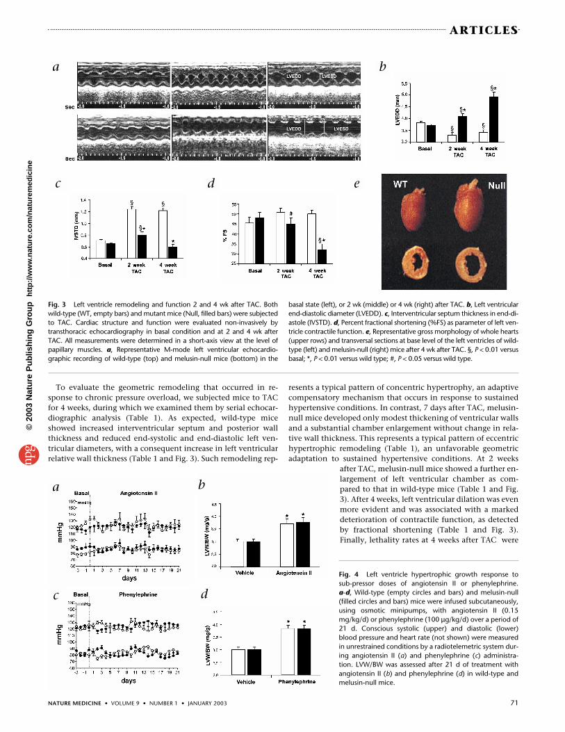
To evaluate the geometric remodeling that occurred in re-sponse to chronic pressure overload, we subjected mice to TACfor 4 weeks, during which we examined them by serial echocar-diographic analysis (Table 1). As expected, wild-type miceshowed increased interventricular septum and posterior wallthickness and reduced end-systolic and end-diastolic left ven-tricular diameters, with a consequent increase in left ventricularrelative wall thickness (Table 1 and Fig. 3). Such remodeling rep-
resents a typical pattern of concentric hypertrophy, an adaptivecompensatory mechanism that occurs in response to sustainedhypertensive conditions. In contrast, 7 days after TAC, melusin-null mice developed only modest thickening of ventricular wallsand a substantial chamber enlargement without change in rela-tive wall thickness. This represents a typical pattern of eccentrichypertrophic remodeling (Table 1), an unfavorable geometricadaptation to sustained hypertensive conditions. At 2 weeks
after TAC, melusin-null mice showed a further en-largement of left ventricular chamber as com-pared to that in wild-type mice (Table 1 and Fig.3). After 4 weeks, left ventricular dilation was evenmore evident and was associated with a markeddeterioration of contractile function, as detectedby fractional shortening (Table 1 and Fig. 3).Finally, lethality rates at 4 weeks after TAC were
Fig. 3 Left ventricle remodeling and function 2 and 4 wk after TAC. Bothwild-type (WT, empty bars) and mutant mice (Null, filled bars) were subjectedto TAC. Cardiac structure and function were evaluated non-invasively bytransthoracic echocardiography in basal condition and at 2 and 4 wk afterTAC. All measurements were determined in a short-axis view at the level ofpapillary muscles. a, Representative M-mode left ventricular echocardio-graphic recording of wild-type (top) and melusin-null mice (bottom) in the
basal state (left), or 2 wk (middle) or 4 wk (right) after TAC. b, Left ventricularend-diastolic diameter (LVEDD). c, Interventricular septum thickness in end-di-astole (IVSTD). d, Percent fractional shortening (%FS) as parameter of left ven-tricle contractile function. e, Representative gross morphology of whole hearts(upper rows) and transversal sections at base level of the left ventricles of wild-type (left) and melusin-null (right) mice after 4 wk after TAC. §, P < 0.01 versusbasal; *, P < 0.01 versus wild type; #, P < 0.05 versus wild type.
a b
c d e
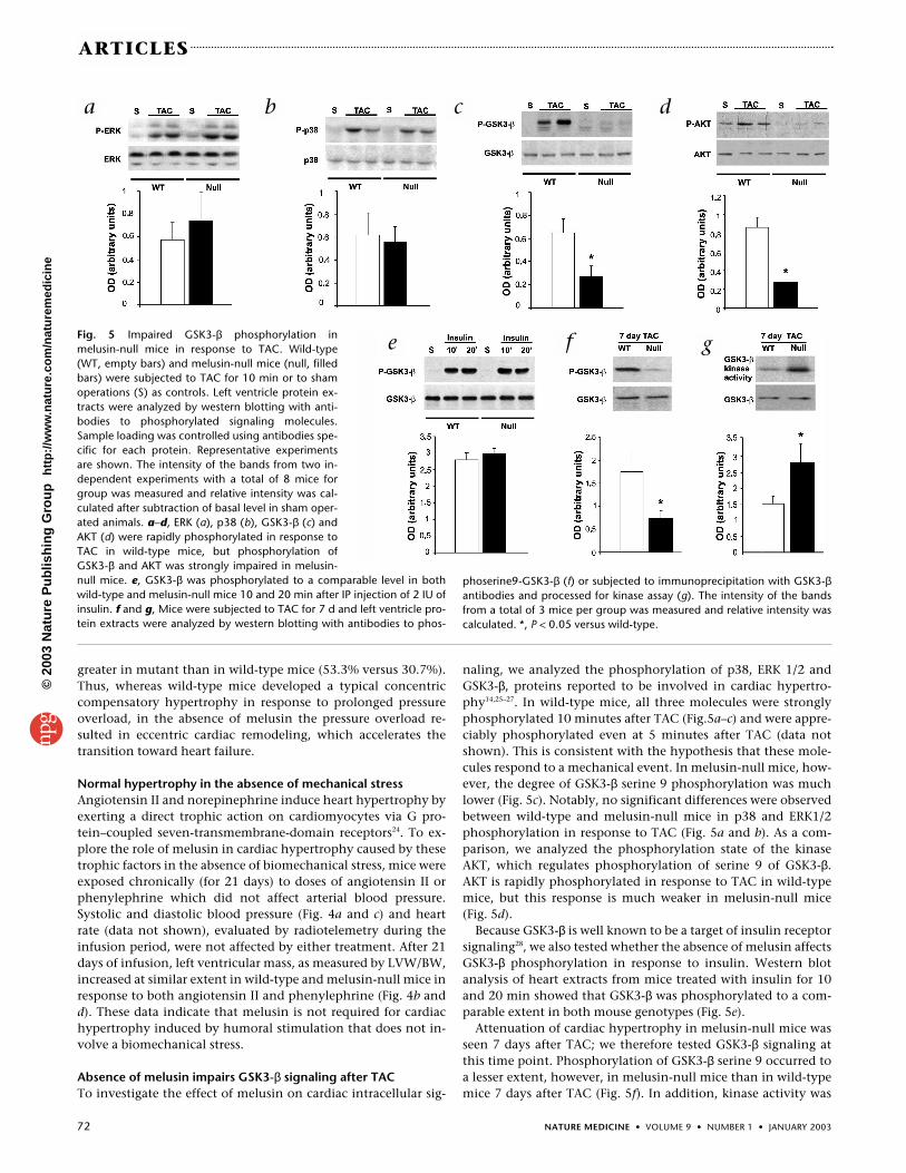
Fig. 4 Left ventricle hypertrophic growth response tosub-pressor doses of angiotensin II or phenylephrine. a-d, Wild-type (empty circles and bars) and melusin-null(filled circles and bars) mice were infused subcutaneously,using osmotic minipumps, with angiotensin II (0.15mg/kg/d) or phenylephrine (100 µg/kg/d) over a period of21 d. Conscious systolic (upper) and diastolic (lower)blood pressure and heart rate (not shown) were measuredin unrestrained conditions by a radiotelemetric system dur-ing angiotensin II (a) and phenylephrine (c) administra-tion. LVW/BW was assessed after 21 d of treatment withangiotensin II (b) and phenylephrine (d) in wild-type andmelusin-null mice.
a b
c d
©20
03 N
atu
re P
ub
lish
ing
Gro
up
h
ttp
://w
ww
.nat
ure
.co
m/n
atu
rem
edic
ine
72 NATURE MEDICINE • VOLUME 9 • NUMBER 1 • JANUARY 2003
ARTICLES
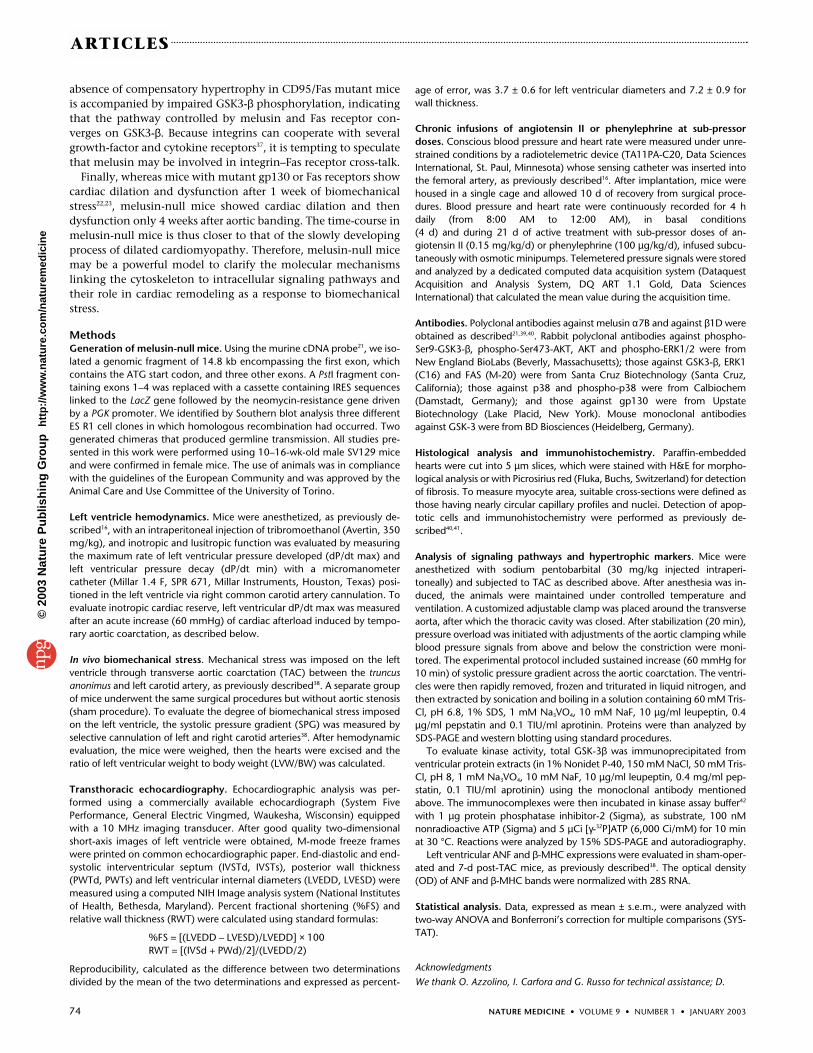
Fig. 5 Impaired GSK3-β phosphorylation inmelusin-null mice in response to TAC. Wild-type(WT, empty bars) and melusin-null mice (null, filledbars) were subjected to TAC for 10 min or to shamoperations (S) as controls. Left ventricle protein ex-tracts were analyzed by western blotting with anti-bodies to phosphorylated signaling molecules.Sample loading was controlled using antibodies spe-cific for each protein. Representative experimentsare shown. The intensity of the bands from two in-dependent experiments with a total of 8 mice forgroup was measured and relative intensity was cal-culated after subtraction of basal level in sham oper-ated animals. a–d, ERK (a), p38 (b), GSK3-β (c) andAKT (d) were rapidly phosphorylated in response toTAC in wild-type mice, but phosphorylation ofGSK3-β and AKT was strongly impaired in melusin-null mice. e, GSK3-β was phosphorylated to a comparable level in bothwild-type and melusin-null mice 10 and 20 min after IP injection of 2 IU ofinsulin. f and g, Mice were subjected to TAC for 7 d and left ventricle pro-tein extracts were analyzed by western blotting with antibodies to phos-
greater in mutant than in wild-type mice (53.3% versus 30.7%).Thus, whereas wild-type mice developed a typical concentriccompensatory hypertrophy in response to prolonged pressureoverload, in the absence of melusin the pressure overload re-sulted in eccentric cardiac remodeling, which accelerates thetransition toward heart failure.
Normal hypertrophy in the absence of mechanical stressAngiotensin II and norepinephrine induce heart hypertrophy byexerting a direct trophic action on cardiomyocytes via G pro-tein–coupled seven-transmembrane-domain receptors24. To ex-plore the role of melusin in cardiac hypertrophy caused by thesetrophic factors in the absence of biomechanical stress, mice wereexposed chronically (for 21 days) to doses of angiotensin II orphenylephrine which did not affect arterial blood pressure.Systolic and diastolic blood pressure (Fig. 4a and c) and heartrate (data not shown), evaluated by radiotelemetry during theinfusion period, were not affected by either treatment. After 21days of infusion, left ventricular mass, as measured by LVW/BW,increased at similar extent in wild-type and melusin-null mice inresponse to both angiotensin II and phenylephrine (Fig. 4b andd). These data indicate that melusin is not required for cardiachypertrophy induced by humoral stimulation that does not in-volve a biomechanical stress.
Absence of melusin impairs GSK3-β signaling after TACTo investigate the effect of melusin on cardiac intracellular sig-
naling, we analyzed the phosphorylation of p38, ERK 1/2 andGSK3-β, proteins reported to be involved in cardiac hypertro-phy14,25–27. In wild-type mice, all three molecules were stronglyphosphorylated 10 minutes after TAC (Fig.5a–c) and were appre-ciably phosphorylated even at 5 minutes after TAC (data notshown). This is consistent with the hypothesis that these mole-cules respond to a mechanical event. In melusin-null mice, how-ever, the degree of GSK3-β serine 9 phosphorylation was muchlower (Fig. 5c). Notably, no significant differences were observedbetween wild-type and melusin-null mice in p38 and ERK1/2phosphorylation in response to TAC (Fig. 5a and b). As a com-parison, we analyzed the phosphorylation state of the kinaseAKT, which regulates phosphorylation of serine 9 of GSK3-β.AKT is rapidly phosphorylated in response to TAC in wild-typemice, but this response is much weaker in melusin-null mice(Fig. 5d).
Because GSK3-β is well known to be a target of insulin receptorsignaling28, we also tested whether the absence of melusin affectsGSK3-β phosphorylation in response to insulin. Western blotanalysis of heart extracts from mice treated with insulin for 10and 20 min showed that GSK3-β was phosphorylated to a com-parable extent in both mouse genotypes (Fig. 5e).
Attenuation of cardiac hypertrophy in melusin-null mice wasseen 7 days after TAC; we therefore tested GSK3-β signaling atthis time point. Phosphorylation of GSK3-β serine 9 occurred toa lesser extent, however, in melusin-null mice than in wild-typemice 7 days after TAC (Fig. 5f). In addition, kinase activity was
a b c d
e f g
phoserine9-GSK3-β (f) or subjected to immunoprecipitation with GSK3-βantibodies and processed for kinase assay (g). The intensity of the bandsfrom a total of 3 mice per group was measured and relative intensity wascalculated. *, P < 0.05 versus wild-type.
©20
03 N
atu
re P
ub
lish
ing
Gro
up
h
ttp
://w
ww
.nat
ure
.co
m/n
atu
rem
edic
ine

NATURE MEDICINE • VOLUME 9 • NUMBER 1 • JANUARY 2003 73
ARTICLES
greater in melusin-null mice, as would be predicted given the in-hibitory effect of serine 9 phosphorylation (Fig. 5g). Thus alteredGSK3-β signaling persists in melusin-null mice exposed to the ef-fects of TAC for 7 days. Together, these data indicate that the ab-sence of melusin selectively impairs left ventricular GSK3-βphosphorylation in response to biomechanical stress.
DiscussionWe previously identified melusin as a protein that binds to theintegrin β1 cytoplasmic domain and specifically expressed in stri-ated muscle21. The phenotype of melusin-null mice reportedhere demonstrates that melusin, though dispensable for cardiacmuscle development and for cardiac function under physiologi-cal conditions, is required to sustain compensatory left ventriclehypertrophy in response to pressure overload. Notably, the ab-sence of melusin does not influence the cardiac hypertrophyevoked by trophic stimuli such as angiotensin II or phenyl-ephrine at sub-pressor doses that do not impose a mechanicaloverload. As melusin interacts with integrin β1 cytoplasmic do-main and localizes at costameres21, where contractile filamentsare anchored laterally to the sarcolemma, we suggest thatmelusin senses high threshold levels of mechanical stress andconsequently activates signaling pathways required to supportcardiomyocyte hypertrophy.
Our analysis of the signaling events occurring in response tomechanical stress indicated that phosphorylation of p38 andERK1/2 MAP kinases, as well as GSK3-β, is strongly stimulatedwithin 5 minutes of induction of the stress in wild-type mice.These molecules are phosphorylated in response to integrin stim-ulation by matrix ligands in vitro17,29,30, supporting the hypothesisthat integrin stimulation could be responsible for their phospho-rylation in heart after mechanical stress. The absence of melusinexpression specifically reduced phosphorylation of GSK3-β serine9, but did not substantially affect phosphorylation of p38 and ERK1/2. In addition, phosphorylation of AKT was greatly impaired inaortic-banded heart of melusin-null mice. As AKT is the principalkinase that phosphorylates the inhibitory site of GSK3-β, this re-sult points to an important role of melusin in regulating this path-way. GSK3-β is also phosphorylated in response to hormonalstimuli not involving mechanical stress, such as insulin28; thispathway, however, was unaffected in the melusin-null back-ground. This observation, together with the very rapid kinetics ofGSK3-β phosphorylation in response to aortic coarctaction, sup-ports the hypothesis that melusin selectively senses the biome-chanical stimuli. In contrast to other kinases, GSK3-β is highlyactive in unstimulated cells and becomes inactivated by phospho-rylation of serine 9 in response to several stimuli28. Recent studieshave shown that this inactivation is required for cardiac hypertro-phy. In cardiomyocytes, active GSK3-β phosphorylates the tran-scription factors NF-AT and GATA4, inducing their translocationfrom the nucleus to the cytoplasm and thus inhibiting their tran-scriptional activity25–27. In addition, active GSK3-β also induces in-hibition of elF2B, a principal initiation factor regulating proteinsynthesis. Thus, the phosphorylation of serine 9 seen during aor-tic coarctation may allow the activation of both protein synthesisand transcriptional events required for the hypertrophic response.Indeed, expression of a constitutively active form of GSK3-β intransgenic mice causes an impaired cardiac hypertrophy in re-sponse to pressure overload27. It is thus likely that the reducedGSK3-β phosphorylation in response to pressure overload that weobserved in melusin-null hearts can account, at least partially, forthe defective hypertrophic response in these mice.
The impaired left ventricle remodeling of melusin-null mice inresponse to mechanical overload shows the typical pattern of ec-centric left ventricular hypertrophy, characterized by an enlarge-ment of the left ventricular chamber and an impaired wallthickening. This is an unfavorable geometric adaptation to hyper-tensive conditions that markedly accelerates the evolution towardheart failure31. Indeed, in contrast to the compensatory hypertro-phy developed by control animals, melusin-null mice undergo aprogressive left ventricle dilation followed by the onset of cardiacfailure over a period of 4 weeks after TAC, and this is associatedwith an increased mortality rate. We conclude that the absence ofmelusin causes a deleterious left ventricular remodeling in condi-tions of sustained biomechanical stress, such as eccentric hyper-trophy, thus accelerating the transition toward heart failure. Incontrast, other studies32 have reported that a reduced left ventricu-lar hypertrophy is protective from the onset of cardiac dilationand dysfunction in two strains of genetically modified mice defec-tive either for Gq signaling (Gq-transgenic) or for norepinephrinesynthesis (Dbh null). A possible explanation for this conflictingevidence could be the difference in genetic background betweenthe mice used in the two laboratories: we used inbred SV129,whereas the other researchers probably used different strains.Another possible explanation is that in both Gq-transgenic andDbh-null mice, the genetic mutations affect G protein–coupled re-ceptor (GPCR) signaling that is unaltered in melusin-null mice, asjudged from the normal hypertrophy response to angiotensin II orphenyl-ephrine. It is therefore likely that the differences in cardiac remod-eling in response to long-standing pressure overload betweenmelusin-null and Gq-transgenic or Dbh-null mice are attributableto the perturbation of two independent pathways that have op-posing effects on heart remodeling. Whereas the GPCR pathway,when active, favors left ventricle dilation, the integrin-linkedmelusin pathway supports concentric compensatory hypertro-phy.
Several genetically modified mouse models of dilated cardiomy-opathy have been described, and have enabled different structuraland signaling proteins involved in this process to be identi-fied1,33–35. In most cases, however, altered expression of these mol-ecules causes basal alteration in cardiac structure and function,with spontaneous development of dilated cardiomyopathy. Fewerexamples have been reported in which abrogation of protein func-tion by gene inactivation does not affect basal cardiac physiology,but causes dilated cardiomyopathy and failure in response to me-chanical overload. The proteins involved in these cases includethe gp130 cytokine receptor22 and CD95/Fas receptor23. Mice inwhich gp130 is specifically inactivated in the heart have normalcardiac structure and function, but develop rapid-onset dilatedcardiomyopathy, resulting from massive myocyte apoptosis,within 7 days after aortic pressure overload. The mechanism un-derlying cardiac dilation in these mice is distinct from that seen inmelusin-null mice, as the latter do not develop cardiomyocyteapoptosis after pressure overload. The CD95/Fas receptor is a well-studied molecule that controls apoptosis in several cell types. Incardiomyocytes, however, Fas induces the pro-hypertrophic tran-scription factor AP-1 rather than inducing apoptosis36. Mice with-out a functional Fas receptor develop rapid-onset left ventriculardilation and failure in response to aortic coarctation, but no apop-totic mechanism is involved23. The possibility that the absence ofmelusin expression prevents normal of gp130 and/or CD95/Fasexpression was excluded by western blot analysis of mutant andwild-type hearts (M.B., unpublished observations). Notably, the
©20
03 N
atu
re P
ub
lish
ing
Gro
up
h
ttp
://w
ww
.nat
ure
.co
m/n
atu
rem
edic
ine
74 NATURE MEDICINE • VOLUME 9 • NUMBER 1 • JANUARY 2003
ARTICLES
absence of compensatory hypertrophy in CD95/Fas mutant miceis accompanied by impaired GSK3-β phosphorylation, indicatingthat the pathway controlled by melusin and Fas receptor con-verges on GSK3-β. Because integrins can cooperate with severalgrowth-factor and cytokine receptors37, it is tempting to speculatethat melusin may be involved in integrin–Fas receptor cross-talk.
Finally, whereas mice with mutant gp130 or Fas receptors showcardiac dilation and dysfunction after 1 week of biomechanicalstress22,23, melusin-null mice showed cardiac dilation and thendysfunction only 4 weeks after aortic banding. The time-course inmelusin-null mice is thus closer to that of the slowly developingprocess of dilated cardiomyopathy. Therefore, melusin-null micemay be a powerful model to clarify the molecular mechanismslinking the cytoskeleton to intracellular signaling pathways andtheir role in cardiac remodeling as a response to biomechanicalstress.
MethodsGeneration of melusin-null mice. Using the murine cDNA probe21, we iso-lated a genomic fragment of 14.8 kb encompassing the first exon, whichcontains the ATG start codon, and three other exons. A PstI fragment con-taining exons 1–4 was replaced with a cassette containing IRES sequenceslinked to the LacZ gene followed by the neomycin-resistance gene drivenby a PGK promoter. We identified by Southern blot analysis three differentES R1 cell clones in which homologous recombination had occurred. Twogenerated chimeras that produced germline transmission. All studies pre-sented in this work were performed using 10–16-wk-old male SV129 miceand were confirmed in female mice. The use of animals was in compliancewith the guidelines of the European Community and was approved by theAnimal Care and Use Committee of the University of Torino.
Left ventricle hemodynamics. Mice were anesthetized, as previously de-scribed16, with an intraperitoneal injection of tribromoethanol (Avertin, 350mg/kg), and inotropic and lusitropic function was evaluated by measuringthe maximum rate of left ventricular pressure developed (dP/dt max) andleft ventricular pressure decay (dP/dt min) with a micromanometercatheter (Millar 1.4 F, SPR 671, Millar Instruments, Houston, Texas) posi-tioned in the left ventricle via right common carotid artery cannulation. Toevaluate inotropic cardiac reserve, left ventricular dP/dt max was measuredafter an acute increase (60 mmHg) of cardiac afterload induced by tempo-rary aortic coarctation, as described below.
In vivo biomechanical stress. Mechanical stress was imposed on the leftventricle through transverse aortic coarctation (TAC) between the truncusanonimus and left carotid artery, as previously described38. A separate groupof mice underwent the same surgical procedures but without aortic stenosis(sham procedure). To evaluate the degree of biomechanical stress imposedon the left ventricle, the systolic pressure gradient (SPG) was measured byselective cannulation of left and right carotid arteries38. After hemodynamicevaluation, the mice were weighed, then the hearts were excised and theratio of left ventricular weight to body weight (LVW/BW) was calculated.
Transthoracic echocardiography. Echocardiographic analysis was per-formed using a commercially available echocardiograph (System FivePerformance, General Electric Vingmed, Waukesha, Wisconsin) equippedwith a 10 MHz imaging transducer. After good quality two-dimensionalshort-axis images of left ventricle were obtained, M-mode freeze frameswere printed on common echocardiographic paper. End-diastolic and end-systolic interventricular septum (IVSTd, IVSTs), posterior wall thickness(PWTd, PWTs) and left ventricular internal diameters (LVEDD, LVESD) weremeasured using a computed NIH Image analysis system (National Institutesof Health, Bethesda, Maryland). Percent fractional shortening (%FS) andrelative wall thickness (RWT) were calculated using standard formulas:
%FS = [(LVEDD – LVESD)/LVEDD] × 100RWT = [(IVSd + PWd)/2]/(LVEDD/2)
Reproducibility, calculated as the difference between two determinationsdivided by the mean of the two determinations and expressed as percent-
age of error, was 3.7 ± 0.6 for left ventricular diameters and 7.2 ± 0.9 forwall thickness.
Chronic infusions of angiotensin II or phenylephrine at sub-pressordoses. Conscious blood pressure and heart rate were measured under unre-strained conditions by a radiotelemetric device (TA11PA-C20, Data SciencesInternational, St. Paul, Minnesota) whose sensing catheter was inserted intothe femoral artery, as previously described16. After implantation, mice werehoused in a single cage and allowed 10 d of recovery from surgical proce-dures. Blood pressure and heart rate were continuously recorded for 4 h daily (from 8:00 AM to 12:00 AM), in basal conditions (4 d) and during 21 d of active treatment with sub-pressor doses of an-giotensin II (0.15 mg/kg/d) or phenylephrine (100 µg/kg/d), infused subcu-taneously with osmotic minipumps. Telemetered pressure signals were storedand analyzed by a dedicated computed data acquisition system (DataquestAcquisition and Analysis System, DQ ART 1.1 Gold, Data SciencesInternational) that calculated the mean value during the acquisition time.
Antibodies. Polyclonal antibodies against melusin α7B and against β1D wereobtained as described21,39,40. Rabbit polyclonal antibodies against phospho-Ser9-GSK3-β, phospho-Ser473-AKT, AKT and phospho-ERK1/2 were fromNew England BioLabs (Beverly, Massachusetts); those against GSK3-β, ERK1(C16) and FAS (M-20) were from Santa Cruz Biotechnology (Santa Cruz,California); those against p38 and phospho-p38 were from Calbiochem(Damstadt, Germany); and those against gp130 were from UpstateBiotechnology (Lake Placid, New York). Mouse monoclonal antibodiesagainst GSK-3 were from BD Biosciences (Heidelberg, Germany).
Histological analysis and immunohistochemistry. Paraffin-embeddedhearts were cut into 5 µm slices, which were stained with H&E for morpho-logical analysis or with Picrosirius red (Fluka, Buchs, Switzerland) for detectionof fibrosis. To measure myocyte area, suitable cross-sections were defined asthose having nearly circular capillary profiles and nuclei. Detection of apop-totic cells and immunohistochemistry were performed as previously de-scribed40,41.
Analysis of signaling pathways and hypertrophic markers. Mice wereanesthetized with sodium pentobarbital (30 mg/kg injected intraperi-toneally) and subjected to TAC as described above. After anesthesia was in-duced, the animals were maintained under controlled temperature andventilation. A customized adjustable clamp was placed around the transverseaorta, after which the thoracic cavity was closed. After stabilization (20 min),pressure overload was initiated with adjustments of the aortic clamping whileblood pressure signals from above and below the constriction were moni-tored. The experimental protocol included sustained increase (60 mmHg for10 min) of systolic pressure gradient across the aortic coarctation. The ventri-cles were then rapidly removed, frozen and triturated in liquid nitrogen, andthen extracted by sonication and boiling in a solution containing 60 mM Tris-Cl, pH 6.8, 1% SDS, 1 mM Na3VO4, 10 mM NaF, 10 µg/ml leupeptin, 0.4µg/ml pepstatin and 0.1 TIU/ml aprotinin. Proteins were than analyzed bySDS-PAGE and western blotting using standard procedures.
To evaluate kinase activity, total GSK-3β was immunoprecipitated fromventricular protein extracts (in 1% Nonidet P-40, 150 mM NaCl, 50 mM Tris-Cl, pH 8, 1 mM Na3VO4, 10 mM NaF, 10 µg/ml leupeptin, 0.4 mg/ml pep-statin, 0.1 TIU/ml aprotinin) using the monoclonal antibody mentionedabove. The immunocomplexes were then incubated in kinase assay buffer42
with 1 µg protein phosphatase inhibitor-2 (Sigma), as substrate, 100 nMnonradioactive ATP (Sigma) and 5 µCi [γ-32P]ATP (6,000 Ci/mM) for 10 minat 30 °C. Reactions were analyzed by 15% SDS-PAGE and autoradiography.
Left ventricular ANF and β-MHC expressions were evaluated in sham-oper-ated and 7-d post-TAC mice, as previously described38. The optical density(OD) of ANF and β-MHC bands were normalized with 28S RNA.
Statistical analysis. Data, expressed as mean ± s.e.m., were analyzed withtwo-way ANOVA and Bonferroni’s correction for multiple comparisons (SYS-TAT).
AcknowledgmentsWe thank O. Azzolino, I. Carfora and G. Russo for technical assistance; D.
©20
03 N
atu
re P
ub
lish
ing
Gro
up
h
ttp
://w
ww
.nat
ure
.co
m/n
atu
rem
edic
ine
NATURE MEDICINE • VOLUME 9 • NUMBER 1 • JANUARY 2003 75
ARTICLES
Bongioanni, B. Canepa, R. Ferretti and M. Sbroggiò for help with several exper-iments; V. Poli and S. Cabodi for suggestions and critical reading of the manu-script; and A. Fubini for great enthusiasm and support in the initial analysis ofthe mouse phenotype. This work was supported by grants from Telethon toG.T., the Ministry of University and Research to G.T. and G.L., the ItalianNational Research Council to F.A. and E.H., and the Italian Ministry of Healthto G.L.
Competing interests statementThe authors declare that they have no competing financial interests.
RECEIVED 19 SEPTEMBER; ACCEPTED 22 NOVEMBER 2002
1. Chien, K.R. Stress pathways and heart failure. Cell 98, 555–558 (1999).2. Chien, K.R. & Olson, E.N. Converging pathways and principles in heart develop-
ment and disease: CV@CSH. Cell 110, 153–162 (2002).3. Sadoshima, J. & Izumo, S. The cellular and molecular response of cardiac my-
ocytes to mechanical stress. Annu. Rev. Physiol. 59, 551–571 (1997).4. Ruwhof, C. & van der Laarse, A. Mechanical stress-induced cardiac hypertrophy:
mechanisms and signal transduction pathways. Cardiovasc. Res. 47, 23–37(2000).
5. Sadoshima, J. & Izumo, S. Mechanical stretch rapidly activates multiple signaltransduction pathways in cardiac myocytes: potential involvement of an au-tocrine/paracrine mechanism. EMBO J. 12, 1681–1692 (1993).
6. Schultz Jel, J. et al. TGF-β1 mediates the hypertrophic cardiomyocyte growth in-duced by angiotensin II. J. Clin. Invest. 109, 787–796 (2002).
7. MacLellan, W.R. & Schneider, M.D. Genetic dissection of cardiac growth controlpathways. Annu. Rev. Physiol. 62, 289–319 (2000).
8. Yamazaki, T. et al. Endothelin-1 is involved in mechanical stress-induced car-diomyocyte hypertrophy. J. Biol. Chem. 271, 3221–3228 (1996).
9. Wettschureck, N. et al. Absence of pressure overload induced myocardial hyper-trophy after conditional inactivation of Gαq/Gα11 in cardiomyocytes. Nat. Med.7, 1236–1240 (2001).
10. Akhter, S.A. et al. Targeting the receptor-Gq interface to inhibit in vivo pressureoverload myocardial hypertrophy. Science 280, 574–577 (1998).
11. Dorn, G.W. 2nd & Brown, J H. Gq signaling in cardiac adaptation and maladap-tation. Trends Cardiovasc. Med. 9, 26–34 (1999).
12. Wakasaki, H. et al. Targeted overexpression of protein kinase C β2 isoform inmyocardium causes cardiomyopathy. Proc. Natl. Acad. Sci. USA 94, 9320–9325(1997).
13. Sugden, P.H. & Clerk, A. Cellular mechanisms of cardiac hypertrophy. J. Mol.Med. 76, 725–746 (1998).
14. Hunter, J.J. & Chien, K.R. Signaling pathways for cardiac hypertrophy and fail-ure. N. Engl. J. Med. 341, 1276–1283 (1999).
15. Harada, K. et al. Pressure overload induces cardiac hypertrophy in angiotensin IItype 1A receptor knockout mice. Circulation 97, 1952–1959 (1998).
16. Vecchione, C. et al. Cardiovascular influences of α1b-adrenergic receptor defectin mice. Circulation 105, 1700–1707 (2002).
17. Schwartz, M.A., Schaller, M.D. & Ginsberg, M.H. Integrins: emerging para-digms of signal transduction. Annu. Rev. Cell Dev. Biol. 11, 549–599 (1995).
18. Fassler, R. et al. Differentiation and integrity of cardiac muscle cells are impairedin the absence of β1 integrin. J. Cell Sci. 109, 2989–2899 (1996).
19. Keller, R.S. et al. Disruption of integrin function in the murine myocardium leadsto perinatal lethality, fibrosis, and abnormal cardiac performance. Am. J. Pathol.158, 1079–1090 (2001).
20. Shai, S.Y. et al. Cardiac myocyte-specific excision of the β1 integrin gene resultsin myocardial fibrosis and cardiac failure. Circ. Res. 90, 458–464 (2002).
21. Brancaccio, M. et al. Melusin is a new muscle-specific interactor for β1 integrincytoplasmic domain. J. Biol. Chem. 274, 29282–29288 (1999).
22. Hirota, H. et al. Loss of a gp130 cardiac muscle cell survival pathway is a criticalevent in the onset of heart failure during biomechanical stress. Cell 97, 189–198(1999).
23. Badorff, C. et al. Fas receptor signaling inhibits glycogen synthase kinase 3β andinduces cardiac hypertrophy following pressure overload. J. Clin. Invest. 109,373–381 (2002).
24. Rockman, H.A., Koch, W.J. & Lefkowitz, R.J. Seven-transmembrane-spanning re-ceptors and heart function. Nature 415, 206–212 (2002).
25. Haq, S. et al. Glycogen synthase kinase-3β is a negative regulator of cardiomy-ocyte hypertrophy. J. Cell Biol. 151, 117–130 (2000).
26. Morisco, C. et al. Glycogen synthase kinase 3β regulates GATA4 in cardiac my-ocytes. J. Biol. Chem. 276, 28586–28597 (2001).
27. Antos, C.L. et al. Activated glycogen synthase-3β suppresses cardiac hypertro-phy in vivo. Proc. Natl. Acad. Sci. USA 99, 907–912 (2002).
28. Cohen, P. & Frame, S. The renaissance of GSK3. Nat. Rev. Mol. Cell Biol. 2,769–776 (2001).
29. Persad, S. et al. Regulation of protein kinase B/Akt-serine 473 phosphorylationby integrin-linked kinase: critical roles for kinase activity and amino acids argi-nine 211 and serine 343. J. Biol. Chem. 276, 27462–27469 (2001).
30. Ivaska, J. et al. Integrin α2β1 mediates isoform-specific activation of p38 and up-regulation of collagen gene transcription by a mechanism involving the α2 cy-toplasmic tail. J. Cell Biol. 147, 401–416 (1999).
31. Norton, G.R. et al. Heart failure in pressure overload hypertrophy. The relativeroles of ventricular remodeling and myocardial dysfunction. J. Am. Coll. Cardiol.39, 664–671 (2002).
32. Esposito, G. et al. Genetic alterations that inhibit in vivo pressure-overload hy-pertrophy prevent cardiac dysfunction despite increased wall stress. Circulation105, 85–92 (2002).
33. Wang, Y. Signal transduction in cardiac hypertrophy—dissecting compensatoryversus pathological pathways utilizing a transgenic approach. Curr. Opin.Pharmacol. 1, 134–140 (2001).
34. MacLellan, W.R. Advances in the molecular mechanisms of heart failure. Curr.Opin. Cardiol. 15, 128–135 (2000).
35. Nicol, R.L. et al. Activated MEK5 induces serial assembly of sarcomeres and ec-centric cardiac hypertrophy. EMBO J. 20, 2757–2767 (2001).
36. Wollert, K.C. et al. The cardiac Fas (APO-1/CD95) Receptor/Fas ligand systemrelation to diastolic wall stress in volume-overload hypertrophy in vivo and acti-vation of the transcription factor AP-1 in cardiac myocytes. Circulation 101,1172–1178 (2000).
37. Schwartz, M.A. & Ginsberg, M.H. Networks and crosstalk: integrin signallingspreads. Nat. Cell Biol. 4, E65–E658 (2002).
38. Lembo, G. et al. Elevated blood pressure and enhanced myocardial contractilityin mice with severe IGF-1 deficiency. J. Clin. Invest. 98, 2648–2655 (1996).
39. Belkin, A.M. et al. Muscle β1D integrin reinforces the cytoskeleton-matrix link:modulation of integrin adhesive function by alternative splicing. J. Cell Biol. 139,1583–1595 (1997).
40. Brancaccio, M. et al. Differential onset of expression of α7 and β1D integrinsduring mouse heart and skeletal muscle development. Cell Adhes. Commun. 5,193–205 (1998).
41. Condorelli, G. et al. Increased cardiomyocyte apoptosis and changes inproapoptotic and antiapoptotic genes bax and bcl-2 during left ventricularadaptations to chronic pressure overload in the rat. Circulation 99, 3071–3078(1999).
42. Cabodi, S. et al. A PKC-β/Fyn-dependent pathway leading to keratinocytegrowth arrest and differentiation. Mol. Cell 6, 1121–1129 (2000).
©20
03 N
atu
re P
ub
lish
ing
Gro
up
h
ttp
://w
ww
.nat
ure
.co
m/n
atu
rem
edic
ine