Predictive and prognostic molecular markers for cancer medicine
Upload
independentCategory
view
0download
0
BioMed CentralJournal of Biomedical Science
ss
Open AcceResearchIncreased expression of Matrix Metalloproteinase 9 in liver from NZB/W F1 mice received antibody against human parvovirus B19 VP1 unique region proteinChun-Chou Tsai†1, Bor-Show Tzang†2, Szu-Yi Chiang3, Gwo-Jong Hsu4 and Tsai-Ching Hsu*1Address: 1Institute of Immunology, Chung Shan Medical University, Taichung, Taiwan, 2Institute of Biochemistry and Biotechnology, Chung Shan Medical University, Taichung, Taiwan, 3Department of Health, Executive Yuan, Hua-Lien Hospital, Hua-Lien, Taiwan and 4Division of Infectious disease and Department of Internal Medicine, Chia-Yi Christian Hospital, Chia-Yi, Taiwan
Email: Chun-Chou Tsai - [email protected]; Bor-Show Tzang - [email protected]; Szu-Yi Chiang - [email protected]; Gwo-Jong Hsu - [email protected]; Tsai-Ching Hsu* - [email protected]
* Corresponding author †Equal contributors
AbstractBackground: Human parvovirus B19 infection has been postulated to the anti-phospholipidsyndrome (APS) in autoimmunity. However, the influence of anti-B19-VP1u antibody inautoimmune diseases is still obscure.
Methods: To elucidate the effect of anti-B19-VP1u antibodies in systemic lupus erythematosus(SLE), passive transfer of rabbit anti-B19-VP1u IgG was injected intravenously into NZB/W F1 mice.
Results: Significant reduction of platelet count and prolonged thrombocytopenia time weredetected in anti-B19-VP1u IgG group as compared to other groups, whereas significant increasesof anti-B19-VP1u, anti-phospholipid (APhL), and anti-double strand DNA (dsDNA) antibodybinding activity were detected in anti-B19-VP1u group. Additionally, significant increases of matrixmetalloproteinase-9 (MMP9) activity and protein expression were detected in B19-VP1u IgGgroup. Notably, phosphatidylinositol 3-phosphate kinase (PI3K) and phosphorylated extracellularsignal-regulated kinase (ERK) proteins were involved in the induction of MMP9.
Conclusion: These experimental results firstly demonstrated the aggravated effects of anti-B19-VP1u antibody in disease activity of SLE.
BackgroundHuman parvovirus B19 (B19) is known as a parvovirus ofhuman pathogen [1] that consists two structural proteinsincluding VP1 and VP2, which are identical except for the227 amino acids at the amino-terminal end of the VP1-protein, the so-called VP1-unique region (VP1u) [2].Recently, B19-VP1u has been reported to have the phos-pholipase A2 (PLA2) motif and secreted phospholipases
A2 (sPLA2) activity [3-6], and is associated with variousautoimmune diseases [7].
The infection of B19 has been postulated to the genera-tion of various autoantibodies including anti-nuclearantibody (ANA), anticardiolipin antibody (aCL), andanti-phospholipid antibody (APhL) [7-11], as well as theanti-phospholipid syndrome (APS) [8]. Notably, a signif-
Published: 26 January 2009
Journal of Biomedical Science 2009, 16:14 doi:10.1186/1423-0127-16-14
Received: 18 November 2008Accepted: 26 January 2009
This article is available from: http://www.jbiomedsci.com/content/16/1/14
© 2009 Tsai et al; licensee BioMed Central Ltd. This is an Open Access article distributed under the terms of the Creative Commons Attribution License (http://creativecommons.org/licenses/by/2.0), which permits unrestricted use, distribution, and reproduction in any medium, provided the original work is properly cited.
Page 1 of 9(page number not for citation purposes)

Journal of Biomedical Science 2009, 16:14 http://www.jbiomedsci.com/content/16/1/14
icant similarity existed in the specificity of APhL betweenpatients with B19 infection or systemic lupus erythemato-sus (SLE) was reported [9,10]. Recent studies have sug-gested that B19 may exacerbate or even induce SLE [7,8].Our recent findings indicated that serum from patientswith acute B19 infection have a high frequency in recog-nition of cardiolipin (CL) and 2GPI, and the phospholi-pase domain observed in the B19-VP1u may contribute tothe generation of APhL [12]. Additionally, the BALB/cmice immunized with anti-B19-VP1u IgG developedthrombocytopenia, prolongation of aPTT, and autoanti-body against 2GPI and PhL and suggested the associa-tion among anti-B19-VP1u IgG and production of anti-2GPI antibodies, APhL, and APS-like autoimmunity[13].
However, no further study was performed in elucidatingthe effect of anti-B19-VP1u antibody on disease activity inSLE. In current study, we treated NZB/W F1 mice with pas-sive transfer of rabbit anti-B19-VP1u antibody to investi-gate the effect of elicited anti-B19-VP1u antibody ondiseases activity in SLE.
MethodsPreparation of recombinant human B19-VP1 unique region protein and rabbit anti-B19-VP1 unique region antibodyConstruction of B19-VP1u cDNA into pET-32a expressionvector (Novagene, Cambridge, MA) was performed andthe recombinant B19-VP1u protein was purified asdescribed in our recent publication [12,13]. For genera-tion of antisera against the B19-VP1u, four female NewZealand White rabbits were immunized subcutaneous inthe neck region with 0.5 mg of purified recombinant B19-VP1u protein in Freund's complete adjuvant (Sigma, SaintLouis Mo, USA) followed by injection at two-week inter-vals with 0.25 mg of B19-VP1uproteininFreund'sincom-pleteadjuvant (Sigma, Saint Louis Mo, USA). A controlgroup were immunized with Freund's complete adjuvantand followed by injection with Freund's incomplete adju-vant. All sera reacted specially with the B19-VP1u byimmunoblotting analysis.
Animals and induction of experimental APS by passive transferTwenty-four female NZB/W F1 mice at age of 8 weekswere purchased from National Taiwan University, Labora-tory Animal Center, Taiwan and housed under supervi-sion of the Institutional Animal Care and Use Committeeat Chung Shan Medical University, Taichung, Taiwan.Induction of experimental APS by passive transfer wasperformed according to the method of Blank [14]. Diseaseactivity of mice was determined by monitoring the pro-teinuria biweekly with Albustix test strips from the age of14 weeks for ten weeks as described previously [15]. All
rabbit IgG were isolated using Protein A beads asdescribed in our recent report [13,16]. The dosage of anti-B19-VP1u IgG is based on a previous study of inducingAPS in mice with anti-cardiolopin antibodies [14]. Pro-portionally, the common used concentration of immu-noglobulins-preparations in mice is 20 ug/dosage and thetiter of 20 ug rabbit anti-B19-VP1u IgG is similar to 27 Uafter determination and mathematics conversion (IBL-America, MN, USA) while the values of other control IgGpreparations are less than 7 U. The titer greater than 12 Uis considered as positive. The mice at age of 20 weeks weredivided into four groups and were intravenously receivedrabbit anti-B19-VP1u IgG (20 ug), normal rabbit IgG (20ug), rabbit anti-B19-NS1 IgG (20 ug), and PBS throughthe tail vein, respectively. The mice were then sacrificed onday 30 by CO2 asphyxiation and the heart blood sampleswere collected. APS clinical parameters, including throm-bocytomenia and prolonged activated partial thrombo-plastin time [aPTT], were evaluated and the plateletcounts were detected using Systemex (KX-21, KOBE,Japan). The presence of lupus anticoagulants were evalu-ated by the prolongation of aPTT in a mixing tests by add-ing 1 volume of plasma from whole blood mixed withsodium citrate 0.123 mol/l in a 9:1 ratio to 1 volume ofeach cephalin and incubating for 2 minutes at 37°C.Another volume of 0.025 M CaCl2 (Sigma, Saint LouisMo, USA) was added, and the clotting time was recordedin seconds using Coatron M1 (TECO GmbH, NeufahrnNB, Germany).
ELISADirect antigen-specific ELISA kits were used to detect APhLIgG (Louisville APL Diagnostics, Inc. GA, USA) and anti-dsDNA IgG (INOVA Diagnostics, Inc. CA, USA) was per-formed as described in our recent publication [12,13]. Thecolor reaction was performed as described above. Fordetecting the binding activity of anti-B19-VP1u antibody,recombinant B19-VP1u was coated in a 96 well plate andELISA was performed as described in our recent report[13]. The cutoff value for each ELISA experiment wasobtained (mean+3SD) and the absorbance above thevalue is regarded as positive.
Preparation of tissue extract and determination of proteinAll procedures were performed at 4°C. Liver samplesobtained from NZB/W F1 mice were homogenized in 600ul PRO-PREP™ solution (iNtRON Biotech, Korea) by 30strokes using a Dounce Homogenizer (Knotes Glass, Vine-land, NJ). The homogenates were centrifuged at 13,000rpm for 10 minutes at 4°C and the supernatant was storedat -80°C until use. Protein concentration of tissue extractswas determined according to the method described byBradford [17] using bovine serum albumin as standards.
Page 2 of 9(page number not for citation purposes)

Journal of Biomedical Science 2009, 16:14 http://www.jbiomedsci.com/content/16/1/14
Gel zymographyMMP-2 and MMP-9 activities were analyzed by gelatin-zymography assays as previously described [18]. Tenmicroliters of ten-fold diluted serum or 25 g proteinlysates of liver tissue from NZB/W F1 mice with passivetransfer anti-idiotype antibodies were separated by an 8%Sodium dodecyl sulfate-polyacrylamide gel electrophore-sis (SDS-PAGE) gels containing 0.1% gelatin. Gels werewashed for 30 min in 2.5% Triton X-100 to remove theSDS and then soaked in the reaction buffer containing 40mM Tris-HCl, pH8.0, 10 mM CaCl2 and 0.02% NaN3 for30 min. The reaction buffer was changed to a fresh one,and the gels were incubated at 37°C for 24 h. Gelatino-lytic activity was visualized by staining the gels with 0.5%Coomassie brillant blue R-250, destained with methanol-acetic acid water, and relative MMP levels were quanti-tated by a gel documentation and analysis system(Appraise, Beckman-Coulter, Brea, California, USA).
Western blotThe loading sample for each lane of Western blot was apool of four random selected mice of the same group. Pro-tein samples were separated in 12.5 or 10% of SDS-PAGEand electrophoretically transferred to nitrocellulose mem-brane (Amersham Biosciences, Piscataway, NJ, USA)according to the method of Towbin [19]. After blockingwith 5% non-fat dry milk in (PBS), antibodies againstMMP2, MMP9, phosphatidylinositol 3-phosphate kinase(PI3K) and phosphorylated extracellular signal-regulatedkinase 1/2 (p-ERK1/2), and actin (Upstates, Charlottes-ville, Virginia, USA) were diluted in PBS with 2.5% BSAand incubated for 1.5 hr with gentle agitation at roomtemperature. The membranes were then incubated withhorseradish peroxidase (HRP) conjugated secondary anti-
body. Pierce's Supersignal West Dura HRP Detection Kit(Pierce Biotechnology Inc., Rockford, IL) was used todetect the antigen-antibody complexes. The blots werescanned and quantified by densitometry (Appraise, Beck-man-Coulter, Brea, California, USA).
Statistical analysesThe paired t test and one-way ANOVA were used to ana-lyze for statistical significance. A P value < 0.05 was con-sidered significant.
ResultsEnhanced APS-like syndrome in NZB/W F1 by passive transfer of purified rabbit anti-B19-VP1u antibodyTo clarify the influence of anti-B19-VP1u antibody in dis-ease activity and development of SLE, we employed andmodified the experimental model as described in our pre-viously report by immunizing NZB/W F1 mice intrave-nously with various kinds of purified rabbit IgG. The bodyweight and various clinical parameters including, WBC,RBC, HGB, HCT, MCV, MCH and MCHC revealed no sig-nificant variation in all groups of mice (Table 1). Notably,significant decreases of platelet counts and aPTT wereobserved in sera from NZB/W F1 mice that were receivedpurified rabbit anti-B19-VP1u IgG, compared to thosemice that were received normal rabbit IgG, rabbit anti-B19-NS1 IgG, or PBS, respectively (Table 1). Additionally,ELISA experiments were performed to elucidate the effectof anti-B19-VP1u IgG by analyzing the binding activitiesof APhL antibodies in NZB/W F1 mice that were receivedpurified rabbit anti-B19-VP1u IgG. Elevated titers of anti-B19-VP1u and APhL and anti-dsDNA antibodies weredetected in serum from NZB/W F1 mice that were receivedpurified rabbit anti-B19-VP1u IgG as compared to those
Table 1: Mice infused with various rabbit antibody or reagent
Antibodies or reagent infused into mice
PBS[n = 6]
Control IgG[n = 6]
B19-NS1[n = 6]
B19-VP1u[n = 6]
Body Weight (gram) 33.9 ± 1.7 33.9 ± 0.2 32.6 ± 2.5 33.6 ± 2.7WBC (103 cells/ul) 6.3 ± 0.7 5.5 ± 1.1 6.2 ± 1.6 4.3 ± 1.4RBC (106 cells/ul) 7.7 ± 0.6 8.7 ± 0.2 8.1 ± 0.2 7.8 ± 0.2HGB (g/dL) 12.5 ± 1.1 15 ± 0.3 13.6 ± 0.5 13.3 ± 0.6HCT (%) 44.2 ± 1.3 53.3 ± 2.2 50.4 ± 1.9 47.2 ± 1.4MCV (fL) 57.3 ± 2.5 61.1 ± 1.1 62.3 ± 0.8 60.4 ± 0.9MCH (pg) 16.2 ± 0.3 17.2 ± 0.2 16.9 ± 0.3 17.4 ± 0.6MCHC (g/dL) 28.3 ± 1.7 28.2 ± 0.8 27.1 ± 0.2 28.3 ± 1.3aPTT (second) 69.5 ± 8.1 76.4 ± 11.6 87 ± 6.0 156 ± 27.0* ,#,¥
Platelet count (103 cells/mm3) 1186 ± 219 1198 ± 124 1374 ± 179 808 ± 90.0* ,#,¥
The unit of the parameter is presented in bracket.NS1: non-structure protein 1; VP1u: VP1 unique region protein; WBC: white blood cell; RBC: red blood cell; HGB: Hemoglobin; HCT: Hematocrit; MCV: Mean corpuscular volume; MCH: mean corpuscular hemoglobin; MCHC: hemoglobin concentration; aPTT: activated partial thromboplastin time.*, #, and ¥ indicate P < 0.05, as compared to the PBS, control IgG, or B19-NS1 group, respectively.
Page 3 of 9(page number not for citation purposes)

Journal of Biomedical Science 2009, 16:14 http://www.jbiomedsci.com/content/16/1/14
mice that were received normal rabbit IgG, anti-B19-NS1IgG, or PBS, respectively (Fig. 1).
Enhanced MMP-9 activity and expression in liver from NZB/W F1 by passive transfer of purified rabbit anti-B19-VP1u antibodyTo further examine the effect of anti-B19-VP1u antibodyon pathogenesis of liver in NZB/W F1 mice, MMPs activityand protein expression were examined. Significantincrease of MMP9 activity was observed in liver of NZB/WF1 mice that were received rabbit anti-B19 VP1u IgG ascompared to PBS, Control IgG, or B19-NS1 IgG group,respectively (Fig. 2A). However, no significant variationwas detected in MMP-2 activity among all experimentalgroups (Fig. 2A). The quantified results of MMP-9/MMP-2 ratio were shown in lower panel of Fig 2A. Moreover,Western blots were performed to examine the expressionof MMP9 and MMP2. Significant increase of MMP-9/MMP-2 ratio was detected in B19-VP1u group as com-pared to PBS, Control IgG, or B19-NS1 IgG group, respec-tively (Fig 2B). Quantified results were shown in the lowerpanel of Fig 2B.
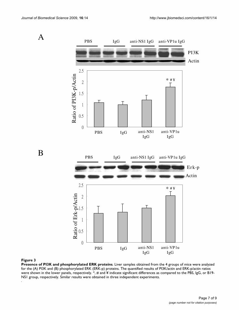
Increased phosphorylation of ERK 1/2 and PI3K proteins in NZB/W F1 by passive transfer of purified rabbitanti-B19-VP1u antibodyTo clarify the possible signaling pathway involved in theactivation of MMP9 by B19-VP1u, various signaling mol-ecules including, PI3K, Erk1/2-p, p38-p, and JNK-p wereexamined. Notably, the PI3K and phosphorylation of ERK1/2 proteins were observed in liver from NZB/W F1 micethat were treated with rabbit anti-B19 VP1u IgG, as com-pared to PBS, Control IgG, or B19-NS1 IgG group, respec-tively (Fig 3). However, no significant variations of p38-pand JNK-p were observed in all experimental groups (datanot shown). Quantified results were shown in lower pan-els of Fig. 3A and 3B.
DiscussionB19 infection has been implicated as a trigger of variousautoimmune diseases including the induction of autoan-tibodies in patients with SLE [7-11]. However, the roles ofB19-VP1u and anti-B19-VP1u antibody in pathogenesisof autoimmune diseases remain unclear. In current study,we demonstrated the aggravated effect of anti-B19-VP1uantibody on disease activity in SLE by analyzing the APS-like syndrome. Significant aggravated disease activities,including reduced platelet count, prolonged thrombocy-topenia time, increased binding activity of autoantibody,elevated MMP9 activity and protein expression, wereobserved in NZB/W F1 mice that were received anti-B19-VP1u IgG as compared to those mice that were receivedrabbit control IgG, rabbit anti-B19-NS1 IgG or PBS,respectively.
APS-like syndrome is recognized as a striking analogybetween the clinical features and hematologic findings inpatients with SLE or B19 infection [7-11]. APS is charac-terized by raised titers of circulating APhL that bind targetmolecules primarily via 2GPI, and/or lupus anticoagu-lant in association with recurrent fetal loss, thromboem-bolic phenomena, thrombocytopenia, CNS, heart, andother organ involvement [20,21]. 2GPI shares a com-mon amino acid sequence with various microbial patho-gens, and may be the cause of APS or production of thecross reacting autoantibodies [22-24]. Our previous studyidentified four cases of B19 infection associated with theproduction of aCL and anti-2GPI antibodies [12]. Con-sistently, our recent study has demonstrated a spectrum ofexperimental APS-like autoimmunity induced by passivetransfer of purified rabbit anti-B19-VP1u IgG antibodiesand provided a connection between anti-B19-VP1u IgGand pathogenesis of SLE [13]. In current study, we indi-cated the significant increases of APS-like syndromesincluding increased platelet count, prolonged PTT, andincreased binding activity of APhL and dsDNA in NZB/WF1 mice that were received purified anti-B19-VP1u IgG.These findings strikingly suggested the aggravated effect ofanti-B19-VP1u IgG in SLE.
Numerous studies have suggested that B19-VP1u plays acrucial role in induction of anti-2GPI and APhL antibod-ies [8,9,13]. Adsorption experiment revealed the partiallyreduced reactivity of anti-B19-VP1u antibody to CL and2GPI [12], suggesting similar epitopes or conformationmay exist between B19-VP1u, CL and 2GPI [8,9,12,13].These experimental results may also account for the cross-reactivity of anti-B19-VP1u antibody against CL, 2GPI,and ph ospholipid and suggest underlying mechanisms indevelopment of APS-like syndrome such as APhL anti-body. In current study, significant increase of APhL anti-bodies was observed in NZB/W F1 mice that were receivedpurified rabbit anti-B19-VP1u IgG, as compared to thosemice that were received control rabbit IgG, rabbit anti-B19-NS1 IgG or PBS, respectively. This finding may be dueto the similar epitopes and anti-idiotype networks amonganti-B19-VP1u IgG, and APhL antibody. However, theunderlying mechanism is still unclear and deed meritedfurther investigations.
Previous studies have postulated MMPs to the pathogene-sis of SLE [25-28]. Cleavage of myelin basic protein ortype II gelatins by MMP-9 will produce remnant epitopesand contribute to the development of autoimmunity[27,28]. In recent studies, elevated MMP-9 activity wasfounded in both human and mice model with SLE [28-31] and recognized to play crucial roles in development ofSLE. Additionally, various studies have indicated theinvolvement of ERK and PI3K in activation of MMP-9. Inmonocytes from patients with rheumatoid arthritis, inhi-
Page 4 of 9(page number not for citation purposes)

Journal of Biomedical Science 2009, 16:14 http://www.jbiomedsci.com/content/16/1/14
Page 5 of 9(page number not for citation purposes)
Binding activity of various autoantibodiesFigure 1Binding activity of various autoantibodies. Anti-sera from the 4 groups of mice were analyzed for the binding activities of (A) B19-VP1u and (B) PhL, and (C) dsDNA. Bars represent the values of optical density. *, # and ¥ indicate significant differ-ences as compared to the PBS, IgG, or B19-NS1 group, respectively. Similar results were obtained in three independent exper-iments.
A
B
B1 9
-VP
1u b
ind i
n g a
c ti v
it y
(Va l
u e o
f op t
ica l
den
s ity
)P
h Lb i
n di n
g a c
tivity
(V
a lu e
of o
p tic
a l d
ens i
ty)
B19 VP1u Control IgG B19 NS1PBS
* # ¥
* # ¥
C
B19 VP1u Control IgG B19 NS1PBS
0
0.5
1
1.5
2
2.5
3
3.5
B19 VP1u Control IgG B19 NS1PBS
* # ¥
d sD
NA
b in d
i ng
a ctiv
ity
(Va l
u e o
f op t
ica l
den
s ity
)

Journal of Biomedical Science 2009, 16:14 http://www.jbiomedsci.com/content/16/1/14
Page 6 of 9(page number not for citation purposes)
Activities and protein expression of MMP-9 andMMP-2Figure 2Activities and protein expression of MMP-9 andMMP-2. Liver samples obtained from the 4 groups of mice were ana-lyzed for the (A) MMP-9 and MMP-2 activity and (B) MMP-9 and MMP-2 protein expression. The quantified results of MMP-9/MMP-2, MMP-9/actin, and MMP-2/actin ratio were shown in the lower panels, respectively. *, # and ¥ indicate significant differ-ences as compared to the PBS, IgG, or B19-NS1 group, respectively. Similar results were obtained in three independent exper-iments.
MMP-9
MMP-2
Diluted serum
anti-VP1u IgGanti-NS1 IgG
MMP-9
MMP-2
A
B IgGPBS
anti-VP1u IgG
anti-NS1 IgGIgGPBS
anti-VP1u IgG
anti-NS1 IgG
IgGPBS
Rat
io o
f M
MP9
/MM
P2
Actin
Rat
io o
f M
MP2
/Act
in
Rat
io o
f M
MP9
/Act
in
anti-VP1u IgG
anti-NS1 IgG
IgGPBS anti-VP1u IgG
anti-NS1 IgG
IgGPBS
* # ¥
* # ¥
Journal of Biomedical Science 2009, 16:14 http://www.jbiomedsci.com/content/16/1/14
Page 7 of 9(page number not for citation purposes)
Presence of PI3K and phosphorylated ERK proteinsFigure 3Presence of PI3K and phosphorylated ERK proteins. Liver samples obtained from the 4 groups of mice were analyzed for the (A) PI3K and (B) phosphorylated ERK (ERK-p) proteins. The quantified results of PI3K/actin and ERK-p/actin ratios were shown in the lower panels, respectively. *, # and ¥ indicate significant differences as compared to the PBS, IgG, or B19-NS1 group, respectively. Similar results were obtained in three independent experiments.
PI3K
Erk-p
anti-VP1u IgGanti-NS1 IgGIgGPBS
anti-VP1u IgGanti-NS1 IgGIgGPBS
A
B
Actin
Rat
io o
f PI
3K-p
/Act
in
anti-VP1u IgG
anti-NS1 IgG
IgGPBS
Actin
anti-VP1u IgG
anti-NS1 IgG
IgGPBS
Rat
io o
f E
rk-p
/Act
in* # ¥
* # ¥

Journal of Biomedical Science 2009, 16:14 http://www.jbiomedsci.com/content/16/1/14
bition of extracellular signal-regulated kinase (ERK) abol-ished Cyclosporine A-induced MMP-9 expression [32].Another study reported that PI3K/Akt activation promotestranscriptional co-factor p300 recruitment and activationand led to increased proMMP-9 expression in rat astrocyte[33]. In current study, significant increases of MMP-9activity and protein expression were detected in NZB/WF1 mice that were received anti-B19-VP1u IgG, as well asthe increased PI3K and phosphorylated ERK proteins.These data suggest the aggravated effect of anti-B19-VP1uIgG in pathogenesis of SLE and the involvement of activa-tion of MMP-9 via PI3K and ERK signaling pathway. Itcould provide clues in treatment of SLE by inhibiting PI3Kand EKR signaling pathway.
ConclusionTaken together, our experimental results firstly demon-strated the aggravated APS-like syndromes in NZB/W F1mice that were received anti-B19-VP1u IgG. Additionally,it could provide clues in understanding the roles of anti-B19-VP1u IgG in SLE and suggest possible therapeuticpotential by inhibiting PI3K or AKT signaling pathway.
Competing interestsThe authors declare that they have no competing interests.
Authors' contributionsCCT performed the animal study, ELISA, zymography,and Western blotting. BST conceived this study, draftedthe manuscript, and performed the performed statisticalanalyses. SYC and GJH provided material support andencouragement for this work. TCH provided material sup-port and direction, and drafted significant portions of themanuscript.
AcknowledgementsAll experiments were performed in accordance with the guidelines of the Ethics Committee of Institutional Animal Care, Chung Shan Medical Uni-versity, Taichung, Taiwan. This study was supported by the grant NSC 97-2314-B-040-009 from the National Science Council, Taiwan, Republic of China.
References1. Young NS, Brown KE: Parvovirus B19. N Engl J Med 2004,
350:586-597.2. Rayment FB, Crosdale E, Morris DJ, Pattison JR, Talbot P, Clare JJ:
The production of human parvovirus capsid proteins inEscherichia coli and their potential as diagnostic antigens. JGen Virol 1990, 71:2665-2672.
3. Dorsch S, Liebisch G, Kaufmann B, von Landenberg P, Hoffmann JH,Drobnik W, Modrow S: The VP1 unique region of parvovirusB19 and its constituent phospholipase A2-like activity. J Virol2002, 76:2014-2018.
4. Canaan S, Zádori Z, Ghomashchi F, Bollinger J, Sadilek M, Moreau ME,Tijssen P, Gelb MH: Interfacial enzymology of parvovirus phos-pholipases A2. J Biol Chem 2004, 279:14502-14508.
5. Lu J, Zhi N, Wong S, Brown KF: Activation of Synoviocytes bythe Secreted Phospholipase A2 Motif in the VP1-UniqueRegion of Parvovirus B19 Minor Capsid Protein. J Infect Dis2006, 193:582-590.
6. Filippone C, Zhi N, Wong S, Lu J, Kajigaya S, Gallinella G, Kakkola L,Söderlund-Venermo M, Young NS, Brown KE: VP1u phospholi-pase activity is critical for infectivity of full-length parvovirusB19 genomic clones. Virology 2008, 374:444-452.
7. Lehmann HW, Modrow S: Parvovirus B19 infection and autoim-mune disease. Autoimmun Rev 2003, 2:218-223.
8. von Landenberg P, Modrow S: Human parvovirus B19 infectionand antiphospholipid-syndrome: the two sides of one medal.J Vet Med B Infect Dis Vet Public Health 2005, 52:353-355.
9. von Landenberg P, Lehmann HW, Modrow S: Human parvovirusB19 infection and antiphospholipid antibodies. Autoimmun Rev2007, 6:278-285.
10. Loizou S, Cazabon JK, Walport MJ, Tait D, So AK: Similarities ofspecificity and cofactor dependence in serum antiphospholi-pid antibodies from patients with human parvovirus B19infection and from those with systemic lupus erythematosus.Arthritis Rheum 1997, 40:103-108.
11. von Landenberg P, Lehmann HW, Knoll A, Dorsch S, Modrow S:Antiphospholipid antibodies in pediatric and adult patientswith rheumatic disease are associated with parvovirus B19infection. Arthritis Rheum 2003, 48:1939-1947.
12. Tzang BS, Tsay GJ, Lee YJ, Li C, Sun YS, Hsu TC: The associationof VP1 unique region protein in acute parvovirus B19 infec-tion and antiphospholipid antibody production. Clin Chim Acta2007, 378:59-65.
13. Tzang BS, Lee YJ, Yang TP, Tsay GJ, Shi JY, Tsai CC, Hsu TC: Induc-tion of antiphospholipid antibodies and antiphospholipid syn-drome-like autoimmunity in naive mice with antibodyagainst human Parvovirus B19 VP1 unique region protein.Clin Chim Acta 2007, 382:31-36.
14. Blank M, Cohen J, Toder V, Shoenfeld Y: Induction of anti-phos-pholipid syndrome in naive mice with mouse lupus mono-clonal and human polyclonal anti-cardiolipin antibodies. ProcNatl Acad Sci USA 1991, 88:3069-3073.
15. Hsu TC, Chiang SY, Huang CY, Tsay GJ, Yang CW, Huang CN, TzangBS: Beneficial effects of treatment with transglutaminasesinhibitor cystamine on macrophage response in NZB/W F1mice. Exp Biol Med 2007, 232:195-203.
16. Tzang BS, Tsai CC, Chiu CC, Shi JY, Hsu TC: Up-regulation ofadhesion molecule expression and induction of TNF- on vas-cular endothelial cells by antibody against human parvovirusB19 VP1 unique region protein. Clin Chim Acta 2008, 395:77-83.
17. Bradford MM: A rapid and sensitive method for the quantita-tion of microgram quantities of protein utilizing the princi-ple of protein-dye binding. Anal Biochem 1976, 72:248-254.
18. Kuo WH, Yang SF, Chu SC, Lu SO, Chou FP, Hsieh YS: Differentialinductions of matrix metalloproteinase-2 and -9 in host tis-sues during the growth of ascitic sarcoma 180 cells in mice.Cancer Lett 2003, 189:103-112.
19. Towbin H, Staehelin T, Gordon J: Electrophoretic transfer ofproteins from polyacrylamide gels to nitrocellulose sheet:procedure and some applications. Proc Natl Acad Sci USA 1979,76:4350-4354.
20. Blank M, Shoenfeld Y: Beta-2-glycoprotein-I, infections,antiphospholipid syndrome and therapeutic considerations.Clin Immunol 2004, 112:190-199.
21. Lim W, Crowther MA, Eikelboom JW: Management of antiphos-pholipid antibody syndrome: a systematic review. JAMA 2006,295:1050-1057.
22. Shoenfeld Y, Blank M, Cervera R, Font J, Raschi E, Meroni PL: Infec-tious origin of the antiphospholipid syndrome. Ann Rheum Dis2006, 65:2-6.
23. Harel M, Aron-Maor A, Sherer Y, Blank M, Shoenfeld Y: The infec-tious etiology of the antiphospholipid syndrome: linksbetween infection and autoimmunity. Immunobiology 2005,210:743-747.
24. Gharavi EE, Chaimovich H, Cucurull E, Celli CM, Tang H, Wilson WA,Gharavi AE: Induction of antiphospholipid antibodies byimmunization with synthetic viral and bacterial peptides.Lupus 1999, 8:449-455.
25. Goetzl EJ, Banda MJ, Leppert D: Matrix metalloproteinases andimmunity. J Immunol 1996, 156:1-4.
26. Zucker S, Hymowitz M, Conner C, Zarrabi HM, Hurewitz AN, Mat-risian L, Boyd D, Nicolson G, Montana S: Measurement of matrixmetalloproteinases and tissue inhibitors of metalloprotein-
Page 8 of 9(page number not for citation purposes)

Journal of Biomedical Science 2009, 16:14 http://www.jbiomedsci.com/content/16/1/14
Publish with BioMed Central and every scientist can read your work free of charge
"BioMed Central will be the most significant development for disseminating the results of biomedical research in our lifetime."
Sir Paul Nurse, Cancer Research UK
Your research papers will be:
available free of charge to the entire biomedical community
peer reviewed and published immediately upon acceptance
cited in PubMed and archived on PubMed Central
yours — you keep the copyright
Submit your manuscript here:http://www.biomedcentral.com/info/publishing_adv.asp
BioMedcentral
ases in blood and tissues. Clinical and experimental applica-tions. Ann N Y Acad Sci 1999, 878:212-227.
27. Opdenaker G, Steen PE Van der, Van Damme J, Gelatinase B: Aturner and amplifier of immune functions. Trends Immunol2001, 22:571-579.
28. Ram M, Sherer Y, Shoenfeld Y: Matrix Metalloproteinase-9 andautoimmune diseases. J Clin Immunol 2006, 26:299-307.
29. Faber-Elmann A, Sthoeger Z, Tcherniack A, Dayan M, Mozes E:Activity of matrix metalloproteinase-9 is elevated in sera ofpatients with systemic lupus erythematosus. Clin Exp Immunol2002, 127:393-398.
30. Ainiala H, Hietaharju A, Dastidar P, Loukkola J, Lehtimäki T, Peltola J,Korpela M, Heinonen T, Nikkari ST: Increased serum matrixmetalloproteinase 9 levels in systemic lupus erythematosuspatients with neuropsychiatric manifestations and brainmagnetic resonance imaging abnormalities. Arthritis Rheum2004, 50:858-865.
31. Hsu TC, Chen YC, Lai WX, Chiang SY, Huang CY, Tzang BS: Bene-ficial effects of treatment with cystamine on brain in NZB/WF1 mice. Eur J Pharmacol 2008, 591:307-314.
32. Yang Y, Lu N, Zhou J, Chen ZN, Zhu P: Cyclophilin A up-regu-lates MMP-9 expression and adhesion of monocytes/macro-phages via CD147 signalling pathway in rheumatoid arthritis.Rheumatology 2008, 47:1299-310.
33. Wu CY, Hsieh HL, Sun CC, Tseng CP, Yang CM: IL-1 beta inducesproMMP-9 expression via c-Src-dependent PDGFR/PI3K/Akt/p300 cascade in rat brain astrocytes. J Neurochem 2008,105:1499-1512.
Page 9 of 9(page number not for citation purposes)
Copyright © 2022 FDOKUMEN