Twitching Motility Is Essential for Virulence in Dichelobacter nodosus
Upload
morehouse-medCategory
view
0download
0
BioMed CentralBMC Cancer
ss
Open AcceResearch articleInhibition of cyclooxygenase-2 decreases breast cancer cell motility, invasion and matrix metalloproteinase expressionTeri L Larkins1, Marchele Nowell2, Shailesh Singh1,3 and Gary L Sanford*1Address: 1Department of Microbiology, Immunology and Biochemistry Morehouse School of Medicine Atlanta, GA 30310-1495, USA, 2Georgia State University Atlanta, GA 30302-3965, USA and 3James Graham Brown Cancer Center University of Louisville, School of Medicine Louisville, KY 40202-1756, USA
Email: Teri L Larkins - [email protected]; Marchele Nowell - [email protected]; Shailesh Singh - [email protected]; Gary L Sanford* - [email protected]
* Corresponding author
AbstractBackground: Cyclooxygenase (COX) is the rate-limiting enzyme that catalyzes the formation ofprostaglandins. The inducible isoform of COX (COX-2) is highly expressed in aggressive metastaticbreast cancers and may play a critical role in cancer progression (i.e. growth and metastasis).However, the exact mechanism(s) for COX-2-enhanced metastasis has yet to be clearly defined.It is well established that one of the direct results of COX-2 action is increased prostaglandinproduction, especially prostaglandin E2 (PGE2). Here, we correlate the inhibition of COX-2 activitywith decreased breast cancer cell proliferation, migration, invasion and matrix metalloproteinase(MMP) expression.
Methods: Breast cancer cells (Hs578T, MDA-MB-231 and MCF-7) were treated with selectiveCOX-2 inhibitors (NS-398 and Niflumic acid, NA). Cell proliferation was measured by staining witherythrosin B and counting the viable cells using a hemacytometer. Cell migration and invasion weremeasured using migration and invasion chamber systems. MMP expression was determined byenzyme immunoassay (secreted protein) and real-time quantitative polymerase chain reaction(mRNA).
Results: Our results show that there is a decline in proliferation, migration and invasion by theHs578T and MDA-MB-231 breast cancer cell lines in the presence of either low concentrations (1μM or lower) NA or NS-398. We also report that MMP mRNA and protein expression by Hs578Tcells is inhibited by NS-398; there was a 50% decrease by 100 μM NS-398. PGE2 completelyreversed the inhibitory effect of NS-398 on MMP mRNA expression.
Conclusion: Our data suggests that COX-2-dependent activity is a necessary component forcellular and molecular mechanisms of breast cancer cell motility and invasion. COX-2 activity alsomodulates the expression of MMPs, which may be a part of the molecular mechanism by whichCOX-2 promotes cell invasion and migration. The studies suggest that COX-2 assists indetermining and defining the metastatic signaling pathways that promote the breast cancerprogression to metastasis.
Published: 10 July 2006
BMC Cancer 2006, 6:181 doi:10.1186/1471-2407-6-181
Received: 14 January 2006Accepted: 10 July 2006
This article is available from: http://www.biomedcentral.com/1471-2407/6/181
© 2006 Larkins et al; licensee BioMed Central Ltd.This is an Open Access article distributed under the terms of the Creative Commons Attribution License (http://creativecommons.org/licenses/by/2.0), which permits unrestricted use, distribution, and reproduction in any medium, provided the original work is properly cited.
Page 1 of 12(page number not for citation purposes)
BMC Cancer 2006, 6:181 http://www.biomedcentral.com/1471-2407/6/181
BackgroundNumerous studies indicate that cyclooxygenase-2 (COX-2) is highly expressed in a variety of human cancers,including colorectal, breast and prostate. In breast cancer,the expression of the COX-2 gene is associated with hightumor grade [1], which suggests it may serve as a prognos-tic biomarker for the presence of breast cancer. Research-ers also found high expression of COX-2 in highlyinvasive estrogen independent breast cancer cell lines,(MDA-MB-231 (MDA-231) and Hs578T) as well as 12, 0-tetradecanoylphorbol-13-acetate (TPA)-induced COX-2expression, while a poorly invasive and estrogen depend-ent cell line (MCF-7) did not express COX-2 [2,2,3]. Rista-maki et al. [4] also confirmed that the elevated COX-2expression seen in 37.4% of the 1567 invasive breast can-cers were associated with a large tumor size, high tumorgrade, negative estrogen receptor status, high p53 expres-sion and unfavorable prognosis. Transgenic mice thatoverexpressed COX-2 in mammary epithelial cells pro-moted mammary gland tumorigenesis and decreasedapoptosis by reducing the expression levels of proapop-totic genes [4,5]. When transfecting the breast cancer cellline, MDA-MB-435 with COX-2, the cells migrated signif-icantly better than the untransfected control cells [6]. Theexpression of COX-2 in breast tumors can be correlatedwith high metastatic potential.
Many of the critical steps of malignant tumorigenesis,such as cell proliferation, evading apoptosis, stimulatingangiogenesis, enhancing cell motility, cell invasivenessand mediating immune suppression, have been associ-ated with cyclooxygenase-2 expression. The end-productsof COX-2 activity are prostaglandins and thromboxaneswhich may mediate these changes in cancer cell progres-sion.
Elevated levels of prostaglandins, notably PGE2, havebeen detected in breast cancer cell lines, as well as invasivebreast cancer [3,7,8]. Gilhooly et al. [2] induced COX-2expression and activity in breast cancer cell lines with TPAwhich increased the production of PGE2. PGE2 was shownto stimulate cell proliferation indirectly by increasingestrogen levels via the induction of the aromatase geneexpression [9]. Other researchers have shown that PGE2,prostacyclin and thromboxanes A2 contribute to tumorangiogenesis by mediating endothelial cell migrationthrough integrin αVβ3 and by aiding in the production ofangiogenic growth factors [10,11].
Recent data suggest a correlation between COX-2 expres-sion and cell invasiveness. In order for cancer cells tometastasize, the cells must digest and dissolve the extracel-lular matrix (ECM) and the basement membrane, whichrequires the secretion and activation of MMPs. The expres-sion and activation of MMPs may be directly proportional
to the overexpression of COX-2 in tumor cells. One grouphas shown that Hs578T breast cancer cells transfectedwith COX-2 resulted in the activation of MMP-2 [12].Sivula et al. [13] found increased COX-2 expression inbreast cancer specimens, which also exhibited elevatedMMP-2 expression and decreased disease specific survival.MMP-2 was elevated in 56 out of 59 invasive breast carci-nomas in which expression of COX-2 was moderate tohigh. Studies also suggest that COX-2 may mediate uroki-nase plasminogen activator (uPA) production in meta-static breast cancer cell lines that overexpress COX-2. TheuPA activates proteases and MMPs that degrade the base-ment membrane and mediate cytoskeleton reorganiza-tion. [6,12,14].
To our knowledge, we are the first to report evidence thatCOX-2 activity and expression may modulate the expres-sion and activity of several MMPs in COX-2 expressingbreast cancer cells. In this study, we screened for eightMMPs in breast cancer cells that were treated with andwithout of a COX-2 inhibitor. To date, only three groupshave reported on studies focused only on the effect ofCOX-2 activity on the secretion of the gelatinases (MMP-2 and -9); all were done on cancers other than breast.Attiga et al. [15] have reported the inhibition of MMP-2and MMP-9 by COX-2 inhibitors in prostate cancer. Tsujiet al. [16] observed an increase in MMP-2 activation andincrease in MMP-14 mRNA expression by Caco-2 coloncancer cells, which showed high levels of COX-2. MMP-2levels were decreased in the non-small cell lung cancer celllines, A549 and H157 when treated with a COX-2 specificinhibitor [17].
The results of a number of epidemiological, clinical andlaboratory studies suggest that the administration of COXinhibitors (NSAIDs, aspirin, indomethacin) reduces theincidence of breast, colon and prostate cancers [15,18-23]. Although several researchers have reported on theassociation of COX-2 overexpression with tumorigenesisin various cancers, many do not address the mechanismby which COX-2 promotes tumorigenesis. Using COX-2inhibitors to prevent tumorigenesis will allow us to studythe properties of COX-2 that influence breast cancermetastasis. The aim of the study reported here was to fur-ther elucidate the mechanism in which COX-2 promotesmetastasis by using COX-2 selective inhibitors to studythe role of COX-2 breast cancer motility and invasion. Wewere able to demonstrate that low and achievable concen-trations of specific COX-2 inhibitors were sufficient toreduce the proliferation, migration and invasion of COX-2 expressing breast cancer cells. This study also suggeststhat COX-2 modulates the expression and activity of mul-tiple MMPs involved in breast cancer metastasis.
Page 2 of 12(page number not for citation purposes)
BMC Cancer 2006, 6:181 http://www.biomedcentral.com/1471-2407/6/181
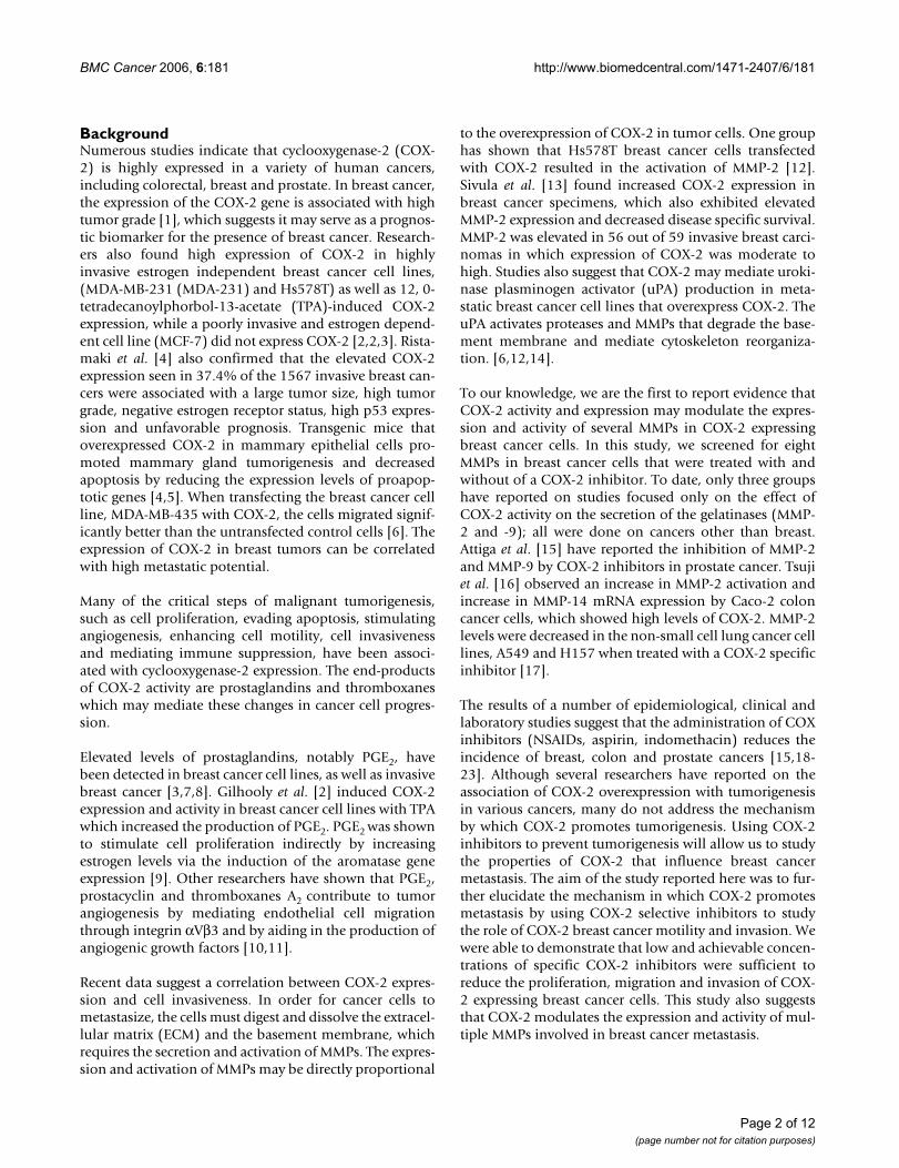
MethodsCell lines and cell cultureThe Hs578T, MDA-231 and MCF-7 breast cancer cell lineswere all obtained from the American Type Culture Collec-tion (Manassas, VA). The Hs578T and MDA-231 are estro-gen-independent and highly invasive breast cancer cells.Although there have been studies that have looked at theexpression of COX-2 in these breast cancer cell lines[2,3,24], we wanted to be certain that under our experi-mental conditions we saw similar results. We found highexpression of COX-2 by both cell lines (Fig. 1). The MCF-7 cell line is an estrogen-dependent and poorly invasive
breast cancer cell line that did not express COX-2. All celllines were adapted for growth in Dulbecco's modifiedEagle's medium (DMEM, Cambrex-Biowhittaker Walkers-ville, MD) supplemented with 0.1 Unit/ml bovine insulin(Sigma Chemical Co. St. Louis, MO), 100 I. U. penicillin,100 μg/ml streptomyocin, 0.25 μg/ml amphotericin B(Cellgro Herndon, VA) and 10% fetal bovine serum (FBSHyclone, Logan UT). When cells reached confluency, thecells were harvested by trypsinizing with 0.25% trypsin,2.21 mM EDTA-Na in Hank's Balanced Salt solution w/Ca2+, Mg2+ and NaHCO3 (Cellgro Herndon, VA), pelleted,resuspended in fresh medium and seeded in multiple 75-cm2 flasks (Corning Corning, NY).
Cell proliferation and viabilityThe Hs578T (1.5 × 10⁵), MDA-231 (1 × 10⁴) and MCF-7cells (1.5 × 10⁵) were seeded on Falcon Multiwell™ 6-or12-well plates (Becton Dickinson-BD Biosciences Discov-ery Labware Franklin Lakes, NJ) and grown for 48–72 h.The plates were washed with serum-free DMEM once andthen incubated for 1 h in DMEM with 2% FBS. Controlcells were treated with the vehicle only. Experimental cellswere incubated with 0.1, 1.0 and 10 μM NS-398 (N-(2-cyclohexyloxy-4-nitrophenyl)-methanesulfonamide) or10 μM niflumic acid (NA) for 24 h at 37°C in 5% CO2,harvested and homogeneous cell suspension preparedand counted. The number of viable cells was determinedby the dye exclusion method using erythrosin B and meas-ured by hemacytometer counting.
Migration assayCell migration assays were performed using a modifica-tion of the protocol described by Attiga et al [15]. The BDFalcon Cell Culture Insert System containing PET (poly-ethylene terephthalate) membranes with 8 μm pores (BDBiosciences Discovery Labware Franklin Lakes, NJ) wasutilized in the assay. The Hs578T and MDA-231 cells wereharvested and resuspended into serum-free medium con-taining NS-398 (0.1, 1.0 and 10 μM) or the vehicle. Theupper chamber of the insert was filled with 500 μl of thecell and drug suspension (1 × 10⁵ cells) and 1.5 ml of(NIH/3T3) fibroblast-conditioned medium (FCM) wasadded to the lower chamber. FCM served as the chemoat-tractant. The conditioned medium was collected fromNIH 3T3 cells grown in serum-free DMEM after 24 h. Theplate was incubated in a humidified environment at 37°Cwith 5% CO2 for 24 h. After incubation, the cells wereremoved from upper surface of the membrane by wipingwith a moist cotton swab. The lower surface of the mem-brane (cells that migrated) was stained for 10 min with0.5% crystal violet in 25% methanol, rinsed with distilledwater to remove excess stain not absorbed by cells and air-dried overnight. Digital images of the stained cells wereobtained prior to the extraction of dye. The crystal violetwas then extracted with 900 μl of 0.1 M sodium citrate in
Expression of COX-2 protein in different breast cancer cell linesFigure 1Expression of COX-2 protein in different breast cancer cell lines. (Hs578T, MCF-7 and MDA-231). Protein was extracted with RIPA buffer and the expression of COX-2 protein detected by immunoblotting and then quantitated by densit-ometry. Quantitated data were normalized to GAPDH. The relative units (RU) and ratio for the COX-2 and GAPDH bands for the different breast cancer cell lines were as fol-lows: Hs578T (COX-2-57878/GAPDH-80182) = 0.7218, MCF-7 (COX-2-5871/GAPDH-74734) = 0.05, MDA-231 (COX-2-43861/GAPDH-61470) = 0.7135. Data for invasive cells lines were compared to poorly invasive cell line by one-way ANOVA followed by Student-Newman-Keuls test (,**p < 0.01).
Page 3 of 12(page number not for citation purposes)
BMC Cancer 2006, 6:181 http://www.biomedcentral.com/1471-2407/6/181
50% ethanol. The absorbance was measured at 585 nm(using Genova Life Science Analyser (Jenway Felsted, Eng-land) spectrophotometer).
Wound migration assayThe Hs578T and MDA-231 cells (2.0 × 10⁵) were seededinto six-well plates and grown to 100% confluency. Theconfluent cells were carefully wounded with sterile pol-ished pasteur pipet tips and any cellular debris wasremoved by washing with PBS. The wounded monolayerswere then incubated in the presence of NA (1.0 and 100μM) for 0, 5 and 24 h time periods and digitally photo-graphed. The distance between the wound edges wasmeasured using Adobe Photoshop 6.0.
Invasion assayCell invasion of the breast cancer cells were assessed byusing the BD BioCoat™ FluoroBlok™ Invasion System (BDBiosciences Franklin, NJ) and procedures were followedaccording to the manufacturer. Monolayer cells grown to80% confluency and labeled in situ with 10 μg/ml of 1,1'-didodecyl-3,3,3',3'-tetramethylindocarbocyanine per-chlorate (DiIC12(3)) DiI fluorophore lipophilic tracer(Molecular Probe, Invitrogen Carlsbad, CA) in mediumfor 1 h at 37°C. The FluoroBlok Invasion insert plate wasrehydrated with warm PBS for 2 h at 37°C. Cells were har-vested and resuspended in serum-free DMEM containingNS-398 (0.1, 1.0 and 10 μM) or the vehicle. NIH/3T3fibroblast-conditioned medium (FCM) 750 μl was addedto the lower chamber and the cell suspension (500 μl) wasadded to the upper chamber. The system was incubated22–24 h at 37°C and the fluorescence of the cells thatinvaded was read directly with a GENios Pro fluorescenceplate reader (Tecan, San Jose, CA) at excitation/emissionwavelengths of 535/590 nm.
RNA isolation and real-time PCRTotal RNA was extracted from the Hs578T cells treatedwith NS-398 and/or PGE2 (control cells received the vehi-cle only), using the RNeasy mini Kit (Qiagen Valencia,CA) according to the manufacturer's instructions. TheRNA was eluted and resuspended in RNA secure (AmbionSan Diego, CA). The concentration and purity of the RNAwas determined by measuring the absorbance at 260 nm(A260) and determining the ratio of the readings at 260 nmand 280 nm (A260/A280). Complimentary DNA (cDNA)was prepared from 2 μg of RNA using Omniscript ReverseTranscriptase (Qiagen Valencia, CA), 2.5 μM randomprimers (Invitrogen Carlsbad, CA) and 0.5 U/μl RNaseInhibitor (Ambion Austin, TX) and incubated at 37°C for1 h.
The cDNA generated from the reverse transcriptase reac-tion was amplified by real-time PCR using specific MMP-(1, 2, 3, 9, 10, 11, 13, 14) and 18S primers and the SYBR
Green polymerase chain reaction master mix reagents. The18S rRNA was used a as a standard. We performed thePCR reaction according to the previously published meth-ods in Singh et al [25]. The master mix for each cDNAsample was composed of 5 μl of the diluted cDNA (20 μlcDNA + 80 μl of nuclease-free water Ambion Austin, TX),15 μl H2O, 5 μl 10 ng/ml reverse and forward primers,and the 25 μl Q™SyBr Green Supermix (Bio-Rad Hercules,CA). The reaction was performed according to the follow-ing program: 10 min at 95°C for activating the polymer-ase, the 40 cycles of 15 s at denaturation temperature of95°C, 1 min of annealing at 60°C and the reaction washeld at 4°C. The relative MMP mRNA expression to the18S rRNA copies was quantified by real-time PCR analysisusing the Bio-Rad Icycler and software (Bio-Rad Hercules,CA).
Pro and active metalloproteinase protein detectionCells (1 × 10⁵) were seeded in 12-well plates and grown to70–80% confluency. The plates were washed with serum-free DMEM once and then incubated for 1 h in 1 ml ofDMEM with 2% FBS and either NS-398 or the vehicle. Thepro and active gelatinases (MMP-2 and MMP-9), colla-genases (MMP-1 and MMP-13), and stromelysins (MMP-3 and MMP-10) levels released in the media were meas-ured using a Quantikine colorimetric and Fluorokinefluorometric enzyme immunoassay (EIA) kit (R&D Sys-tems Minneapolis, MN) using the manufacturer's instruc-tions. After 24 h, the media were collected inmicrocentrifuge tubes and centrifuged for 10 min at10,000 g in order to remove particulates. The sampleswere stored frozen at -80°C prior to the assay. The sam-ples and MMP standards were incubated on a pre-coatedpolyclonal or monoclonal MMP antibody 96 well-platefor 2 h on a shaker at room temperature. The intensity ofcolor on the Quantikine EIA plates was determined bySpectra Max 190 and SOFTmax-Pro 4.3 Life Sciences Ed.(Molecular Devices Sunnyvale, CA) at a wavelength of450 nm with a correction of 540 nm. The fluorescent sig-nal in the sample on the Fluorokine EIA wells was deter-mined by the GENios Pro fluorescence plate reader(Tecan, San Jose, CA) at excitation/emission wavelengthsof 340 nm/465 nm.
Statistical analysisStatistical significance was determined by one-wayANOVA with Student-Newman-Keuls post test was per-formed using GraphPad InStat v3.00. The data wasexpressed as the mean ± S.E.; significance was achieved atp values < 0.05.
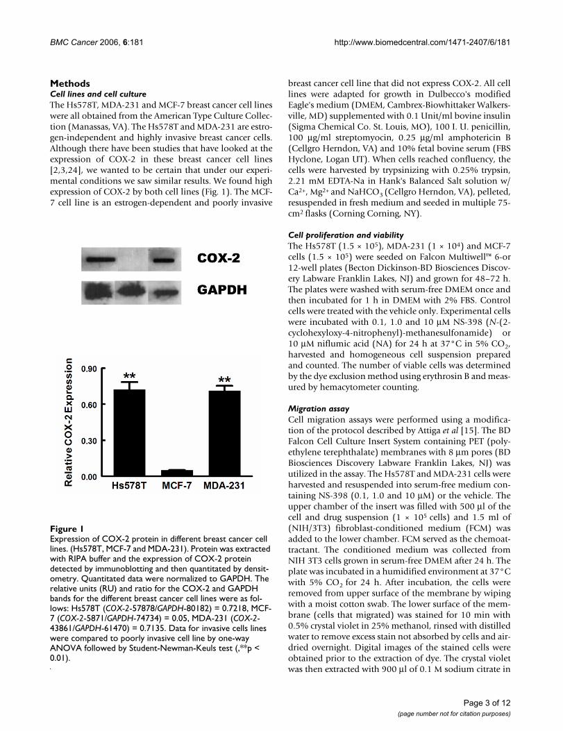
ResultsGrowth inhibition of COX-2 expressing breast cancer cellsWe treated the Hs578T, MDA-231 and MCF-7 breast can-cer cells with NS-398 and NA for 24 h and examined the
Page 4 of 12(page number not for citation purposes)
BMC Cancer 2006, 6:181 http://www.biomedcentral.com/1471-2407/6/181
effects of the cell proliferation. Treatment of the two COX-2 expressing cell lines, Hs578T and MDA-231 with 0.1,1.0, 10 μM NS-398 and NA impeded the growth of thecells. NS-398 resulted in a 30% growth inhibition ofHs578T cells (p < 0.01) and 40% growth inhibition ofMDA-23l cells (p < 0.05) at 0.1 μM (Fig. 2). We also founda 37% inhibition (p < 0.05) of MDA-231 growth by 10μM NA compared to the control (Fig. 2b). The MCF-7 cellsdid not show a difference in growth when treated with NS-398 (data not shown). The COX-2 inhibitors did not sig-nificantly affect the cell viability. There was 85%, orhigher, cell viability seen after treatment with COX-2inhibitors, up to 10 μM indicated no cytotoxicity.
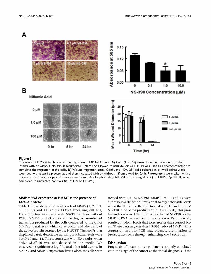
Inhibition of cell migration and invasion with a COX-2 inhibitorWe evaluated the effect of the COX-2 inhibitors on the cellmotility and invasiveness of MDA-231 and Hs578T breastcancer cells. Cell motility and invasion are a measure ofmetastatic potential cancer cells. Treatment with 1.0 μMNS-398 inhibited MDA-231 cell motility by 35% (p <0.05) (Fig. 3a). Confluent MDA-231 cells subjected to thescratch wound assay in the presence of 1.0 and 100 μMNA showed a significant delay in cells moving into theinjury area. The control cells migrated into the woundarea by 5 h to such an extent that the wound edges wereindistinguishable, whereas the experimental group of cellsdid not migrate into and completely close the wound areauntil 24 h (Fig. 3b).
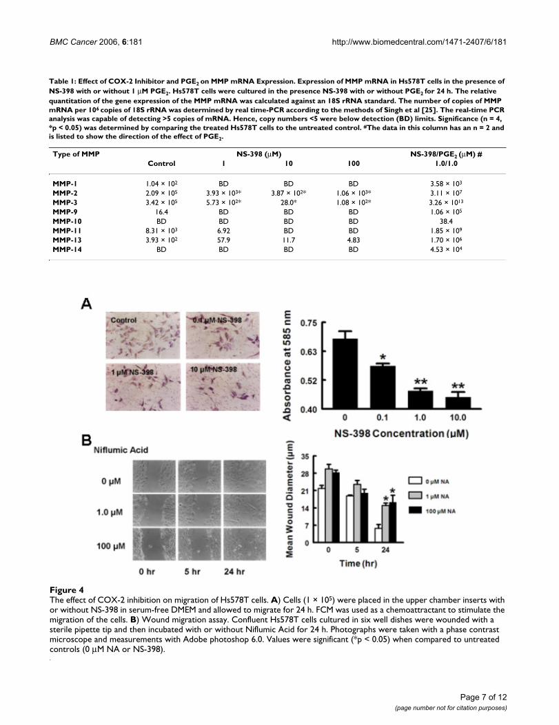
Figure 4a shows that the motility of Hs578T cells was sig-nificantly decreased by 21% (p < 0.05) by 10 μM NS-398when compared to the control. Inhibition of Hs578T cellmotility by COX-2 inhibitors was also confirmed by thescratch wound assay. Figure 4b shows that Hs578T woundincubation with 1 and 100 μM NA resulted in a significantdelay in cell migration compared to the control (Fig. 4b).
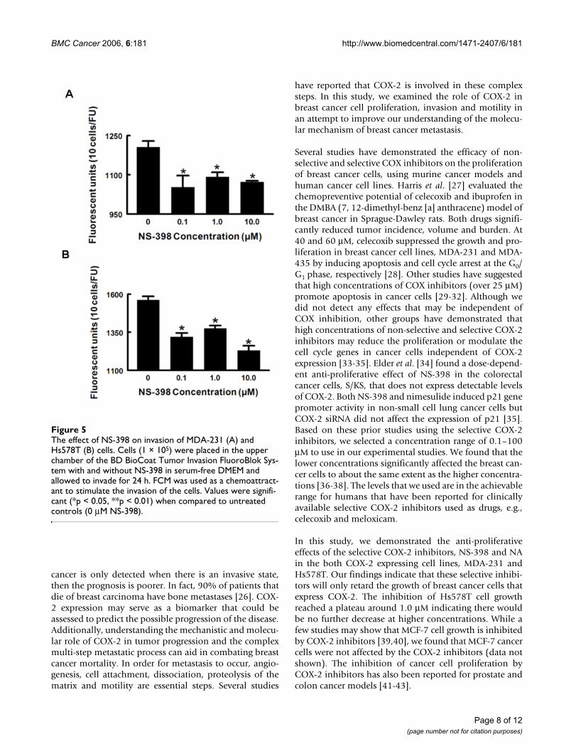
Prior to the migration phase of metastasis, breast cancercells must invade the basement membrane and the extra-cellular matrix. We measured the invasive ability of breastcancer cells on Matrigel coated membranes. Invasion ofMDA-231 cells was inhibited by 11% in response to 10μM NS-398 (p < 0.01) (Fig. 5a). We found a 21% decreaseof the invasion of Hs578T cells by 10 μM NS-398 (Fig.5b).
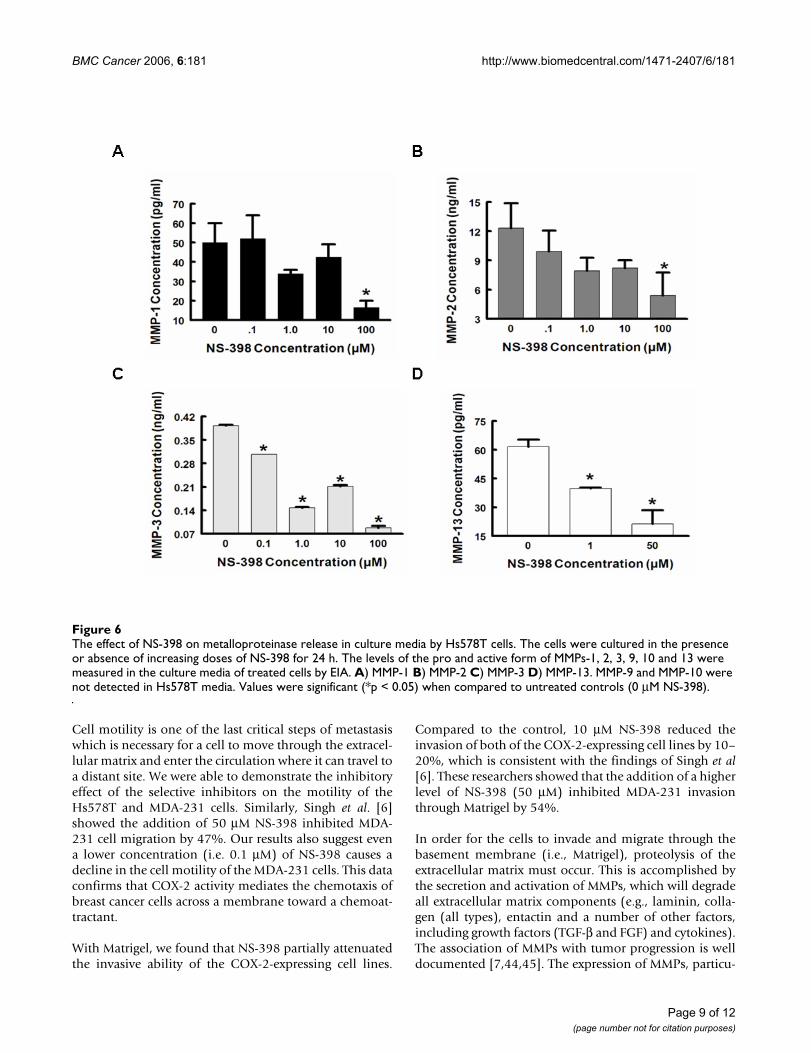
Effect of COX-2 inhibition on the secretion of MMPsWe assessed the dose-dependent effects of NS-398 onMMP secretion by Hs578T and MDA-231 cells. After 24 h,there was a 67% decrease in MMP-1, 45% in MMP-2 and65% in MMP-3 secretion by Hs578T cells treated with 100μM NS-398 (Fig. 6). We also observed a significant reduc-tion (66%) of MMP-13 when the cells were treated with50 μM NS-398 (Fig. 6d). In contrast, MMP-9 and MMP-10were undetectable in the media of Hs578T cells, suggest-ing that the cells did not secrete these MMPs.
Out of the six MMPs assessed by EIA, we were only able todetect the presence of MMP-1 and MMP-9 in the media ofMDA-231 cells. There was about a 20% decrease for bothMMP-1 and MMP-9 secreted by these cells treated with 50μM NS-398 (Fig. 7). We found a 15% reduction in activeMMP-2 and 20–30% reduction in active MMP-9 byimmunoblotting analysis for MDA-231 cells treated withthe NA or NS-398, respectively (data not shown).
Effect of COX-2 inhibitors on breast cancer cell proliferationFigure 2Effect of COX-2 inhibitors on breast cancer cell prolifera-tion. A) 1 × 10⁴ MDA-231 cells and B) 1.5 × 10⁵ Hs578T were seeded, grown for 48 h and then treated with COX-2 inhibitors (NS-398 and Niflumic Acid) or the vehicle for 24 h. Cells were harvested and counted as described in the Meth-ods. Data for treated cells were compared to untreated con-trols by one-way ANOVA followed by Student-Newman-Keuls test (*p < 0.05, **p < 0.01).
Page 5 of 12(page number not for citation purposes)
BMC Cancer 2006, 6:181 http://www.biomedcentral.com/1471-2407/6/181
MMP mRNA expression in Hs578T in the presence of COX-2 inhibitorTable 1 shows detectable basal levels of MMPs (1, 2, 3, 9,10, 11, 13 and 14) in the COX-2 expressing cell line,Hs578T before treatment with NS-398 with or withoutPGE2. MMP-2 and -3 exhibited the highest number oftranscripts produced by the cells compared to the otherMMPs at basal levels which corresponds with the trend ofthe active protein secreted by the Hs578T. The MMPs thatdisplayed barely detectable transcripts at basal levels wereMMP-10 and -14. This is consistent with EIA results, whenactive MMP-10 was not detected in the media. Weobserved a significant 2 log-fold and 4 log-fold decline inMMP-2 and MMP-3 expression levels when the cells were
treated with 10 μM NS-398. MMP 1, 9, 11 and 14 wereeither below detection limits or at barely detectable levelswhen the Hs578T cells were treated with 10 and 100 μMNS-398. One of the products of COX-2 is PGE2; this pros-taglandin reversed the inhibitory effect of NS-398 on theMMP mRNA expression. In some cases PGE2 actuallyresulted in MMP levels that were greater than control lev-els. These data suggests that NS-398 reduced MMP mRNAexpression and that PGE2 may promote the invasion ofbreast cancer cells through enhancing MMP secretion.
DiscussionPrognosis of breast cancer patients is strongly correlatedwith the stage of the cancer at the initial diagnosis. If the
The effect of COX-2 inhibition on the migration of MDA-231 cellsFigure 3The effect of COX-2 inhibition on the migration of MDA-231 cells. A) Cells (1 × 10⁵) were placed in the upper chamber inserts with or without NS-398 in serum-free DMEM and allowed to migrate for 24 h. FCM was used as a chemoattractant to stimulate the migration of the cells. B) Wound migration assay. Confluent MDA-231 cells cultured in six well dishes were wounded with a sterile pipette tip and then incubated with or without Niflumic Acid for 24 h. Photographs were taken with a phase contrast microscope and measurements with Adobe photoshop 6.0. Values were significant (*p < 0.05, **p < 0.01) when compared to untreated controls (0 μM NA or NS-398).
Page 6 of 12(page number not for citation purposes)
BMC Cancer 2006, 6:181 http://www.biomedcentral.com/1471-2407/6/181
Page 7 of 12(page number not for citation purposes)
The effect of COX-2 inhibition on migration of Hs578T cellsFigure 4The effect of COX-2 inhibition on migration of Hs578T cells. A) Cells (1 × 10⁵) were placed in the upper chamber inserts with or without NS-398 in serum-free DMEM and allowed to migrate for 24 h. FCM was used as a chemoattractant to stimulate the migration of the cells. B) Wound migration assay. Confluent Hs578T cells cultured in six well dishes were wounded with a sterile pipette tip and then incubated with or without Niflumic Acid for 24 h. Photographs were taken with a phase contrast microscope and measurements with Adobe photoshop 6.0. Values were significant (*p < 0.05) when compared to untreated controls (0 μM NA or NS-398).
Table 1: Effect of COX-2 Inhibitor and PGE2 on MMP mRNA Expression. Expression of MMP mRNA in Hs578T cells in the presence of NS-398 with or without 1 μM PGE2. Hs578T cells were cultured in the presence NS-398 with or without PGE2 for 24 h. The relative quantitation of the gene expression of the MMP mRNA was calculated against an 18S rRNA standard. The number of copies of MMP mRNA per 106 copies of 18S rRNA was determined by real time-PCR according to the methods of Singh et al [25]. The real-time PCR analysis was capable of detecting >5 copies of mRNA. Hence, copy numbers <5 were below detection (BD) limits. Significance (n = 4, *p < 0.05) was determined by comparing the treated Hs578T cells to the untreated control. #The data in this column has an n = 2 and is listed to show the direction of the effect of PGE2.
Type of MMP NS-398 (μM) NS-398/PGE2 (μM) #Control 1 10 100 1.0/1.0
MMP-1 1.04 × 10² BD BD BD 3.58 × 10³
MMP-2 2.09 × 10⁵ 3.93 × 103* 3.87 × 102* 1.06 × 103* 3.11 × 107
MMP-3 3.42 × 10⁵ 5.73 × 102* 28.0* 1.08 × 102* 3.26 × 1013
MMP-9 16.4 BD BD BD 1.06 × 10⁵
MMP-10 BD BD BD BD 38.4MMP-11 8.31 × 10³ 6.92 BD BD 1.85 × 10⁹
MMP-13 3.93 × 10² 57.9 11.7 4.83 1.70 × 10⁶
MMP-14 BD BD BD BD 4.53 × 10⁴
BMC Cancer 2006, 6:181 http://www.biomedcentral.com/1471-2407/6/181
cancer is only detected when there is an invasive state,then the prognosis is poorer. In fact, 90% of patients thatdie of breast carcinoma have bone metastases [26]. COX-2 expression may serve as a biomarker that could beassessed to predict the possible progression of the disease.Additionally, understanding the mechanistic and molecu-lar role of COX-2 in tumor progression and the complexmulti-step metastatic process can aid in combating breastcancer mortality. In order for metastasis to occur, angio-genesis, cell attachment, dissociation, proteolysis of thematrix and motility are essential steps. Several studies
have reported that COX-2 is involved in these complexsteps. In this study, we examined the role of COX-2 inbreast cancer cell proliferation, invasion and motility inan attempt to improve our understanding of the molecu-lar mechanism of breast cancer metastasis.
Several studies have demonstrated the efficacy of non-selective and selective COX inhibitors on the proliferationof breast cancer cells, using murine cancer models andhuman cancer cell lines. Harris et al. [27] evaluated thechemopreventive potential of celecoxib and ibuprofen inthe DMBA (7, 12-dimethyl-benz [a] anthracene) model ofbreast cancer in Sprague-Dawley rats. Both drugs signifi-cantly reduced tumor incidence, volume and burden. At40 and 60 μM, celecoxib suppressed the growth and pro-liferation in breast cancer cell lines, MDA-231 and MDA-435 by inducing apoptosis and cell cycle arrest at the G0/G1 phase, respectively [28]. Other studies have suggestedthat high concentrations of COX inhibitors (over 25 μM)promote apoptosis in cancer cells [29-32]. Although wedid not detect any effects that may be independent ofCOX inhibition, other groups have demonstrated thathigh concentrations of non-selective and selective COX-2inhibitors may reduce the proliferation or modulate thecell cycle genes in cancer cells independent of COX-2expression [33-35]. Elder et al. [34] found a dose-depend-ent anti-proliferative effect of NS-398 in the colorectalcancer cells, S/KS, that does not express detectable levelsof COX-2. Both NS-398 and nimesulide induced p21 genepromoter activity in non-small cell lung cancer cells butCOX-2 siRNA did not affect the expression of p21 [35].Based on these prior studies using the selective COX-2inhibitors, we selected a concentration range of 0.1–100μM to use in our experimental studies. We found that thelower concentrations significantly affected the breast can-cer cells to about the same extent as the higher concentra-tions [36-38]. The levels that we used are in the achievablerange for humans that have been reported for clinicallyavailable selective COX-2 inhibitors used as drugs, e.g.,celecoxib and meloxicam.
In this study, we demonstrated the anti-proliferativeeffects of the selective COX-2 inhibitors, NS-398 and NAin the both COX-2 expressing cell lines, MDA-231 andHs578T. Our findings indicate that these selective inhibi-tors will only retard the growth of breast cancer cells thatexpress COX-2. The inhibition of Hs578T cell growthreached a plateau around 1.0 μM indicating there wouldbe no further decrease at higher concentrations. While afew studies may show that MCF-7 cell growth is inhibitedby COX-2 inhibitors [39,40], we found that MCF-7 cancercells were not affected by the COX-2 inhibitors (data notshown). The inhibition of cancer cell proliferation byCOX-2 inhibitors has also been reported for prostate andcolon cancer models [41-43].
The effect of NS-398 on invasion of MDA-231 (A) and Hs578T (B) cellsFigure 5The effect of NS-398 on invasion of MDA-231 (A) and Hs578T (B) cells. Cells (1 × 10⁵) were placed in the upper chamber of the BD BioCoat Tumor Invasion FluoroBlok Sys-tem with and without NS-398 in serum-free DMEM and allowed to invade for 24 h. FCM was used as a chemoattract-ant to stimulate the invasion of the cells. Values were signifi-cant (*p < 0.05, **p < 0.01) when compared to untreated controls (0 μM NS-398).
Page 8 of 12(page number not for citation purposes)
BMC Cancer 2006, 6:181 http://www.biomedcentral.com/1471-2407/6/181
Cell motility is one of the last critical steps of metastasiswhich is necessary for a cell to move through the extracel-lular matrix and enter the circulation where it can travel toa distant site. We were able to demonstrate the inhibitoryeffect of the selective inhibitors on the motility of theHs578T and MDA-231 cells. Similarly, Singh et al. [6]showed the addition of 50 μM NS-398 inhibited MDA-231 cell migration by 47%. Our results also suggest evena lower concentration (i.e. 0.1 μM) of NS-398 causes adecline in the cell motility of the MDA-231 cells. This dataconfirms that COX-2 activity mediates the chemotaxis ofbreast cancer cells across a membrane toward a chemoat-tractant.
With Matrigel, we found that NS-398 partially attenuatedthe invasive ability of the COX-2-expressing cell lines.
Compared to the control, 10 μM NS-398 reduced theinvasion of both of the COX-2-expressing cell lines by 10–20%, which is consistent with the findings of Singh et al[6]. These researchers showed that the addition of a higherlevel of NS-398 (50 μM) inhibited MDA-231 invasionthrough Matrigel by 54%.
In order for the cells to invade and migrate through thebasement membrane (i.e., Matrigel), proteolysis of theextracellular matrix must occur. This is accomplished bythe secretion and activation of MMPs, which will degradeall extracellular matrix components (e.g., laminin, colla-gen (all types), entactin and a number of other factors,including growth factors (TGF-β and FGF) and cytokines).The association of MMPs with tumor progression is welldocumented [7,44,45]. The expression of MMPs, particu-
The effect of NS-398 on metalloproteinase release in culture media by Hs578T cellsFigure 6The effect of NS-398 on metalloproteinase release in culture media by Hs578T cells. The cells were cultured in the presence or absence of increasing doses of NS-398 for 24 h. The levels of the pro and active form of MMPs-1, 2, 3, 9, 10 and 13 were measured in the culture media of treated cells by EIA. A) MMP-1 B) MMP-2 C) MMP-3 D) MMP-13. MMP-9 and MMP-10 were not detected in Hs578T media. Values were significant (*p < 0.05) when compared to untreated controls (0 μM NS-398).
Page 9 of 12(page number not for citation purposes)
BMC Cancer 2006, 6:181 http://www.biomedcentral.com/1471-2407/6/181
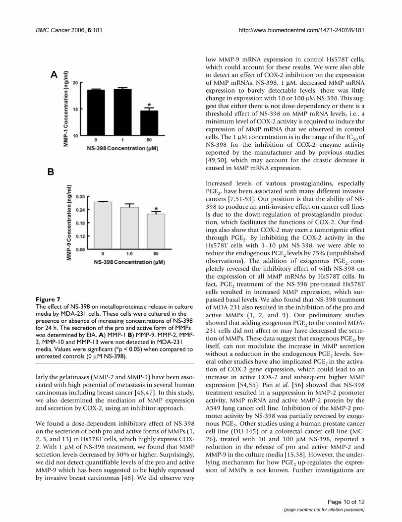
larly the gelatinases (MMP-2 and MMP-9) have been asso-ciated with high potential of metastasis in several humancarcinomas including breast cancer [46,47]. In this study,we also determined the mediation of MMP expressionand secretion by COX-2, using an inhibitor approach.
We found a dose-dependent inhibitory effect of NS-398on the secretion of both pro and active forms of MMPs (1,2, 3, and 13) in Hs578T cells, which highly express COX-2. With 1 μM of NS-398 treatment, we found that MMPsecretion levels decreased by 50% or higher. Surprisingly,we did not detect quantifiable levels of the pro and activeMMP-9 which has been suggested to be highly expressedby invasive breast carcinomas [48]. We did observe very
low MMP-9 mRNA expression in control Hs578T cells,which could account for these results. We were also ableto detect an effect of COX-2 inhibition on the expressionof MMP mRNAs. NS-398, 1 μM, decreased MMP mRNAexpression to barely detectable levels; there was littlechange in expression with 10 or 100 μM NS-398. This sug-gest that either there is not dose-dependency or there is athreshold effect of NS-398 on MMP mRNA levels, i.e., aminimum level of COX-2 activity is required to induce theexpression of MMP mRNA that we observed in controlcells. The 1 μM concentration is in the range of the IC50 ofNS-398 for the inhibition of COX-2 enzyme activityreported by the manufacturer and by previous studies[49,50], which may account for the drastic decrease itcaused in MMP mRNA expression.
Increased levels of various prostaglandins, especiallyPGE2, have been associated with many different invasivecancers [7,51-53]. Our position is that the ability of NS-398 to produce an anti-invasive effect on cancer cell linesis due to the down-regulation of prostaglandin produc-tion, which facilitates the functions of COX-2. Our find-ings also show that COX-2 may exert a tumorigenic effectthrough PGE2. By inhibiting the COX-2 activity in theHs578T cells with 1–10 μM NS-398, we were able toreduce the endogenous PGE2 levels by 75% (unpublishedobservations). The addition of exogenous PGE2 com-pletely reversed the inhibitory effect of with NS-398 onthe expression of all MMP mRNAs by Hs578T cells. Infact, PGE2 treatment of the NS-398 pre-treated Hs578Tcells resulted in increased MMP expression, which sur-passed basal levels. We also found that NS-398 treatmentof MDA-231 also resulted in the inhibition of the pro andactive MMPs (1, 2, and 9). Our preliminary studiesshowed that adding exogenous PGE2 to the control MDA-231 cells did not affect or may have decreased the secre-tion of MMPs. These data suggest that exogenous PGE2, byitself, can not modulate the increase in MMP secretionwithout a reduction in the endogenous PGE2 levels. Sev-eral other studies have also implicated PGE2 in the activa-tion of COX-2 gene expression, which could lead to anincrease in active COX-2 and subsequent higher MMPexpression [54,55]. Pan et al. [56] showed that NS-398treatment resulted in a suppression in MMP-2 promoteractivity, MMP mRNA and active MMP-2 protein by theA549 lung cancer cell line. Inhibition of the MMP-2 pro-moter activity by NS-398 was partially reversed by exoge-nous PGE2. Other studies using a human prostate cancercell line (DU-145) or a colorectal cancer cell line (MC-26), treated with 10 and 100 μM NS-398, reported areduction in the release of pro and active MMP-2 andMMP-9 in the culture media [15,38]. However, the under-lying mechanism for how PGE2 up-regulates the expres-sion of MMPs is not known. Further investigations are
The effect of NS-398 on metalloproteinase release in culture media by MDA-231 cellsFigure 7The effect of NS-398 on metalloproteinase release in culture media by MDA-231 cells. These cells were cultured in the presence or absence of increasing concentrations of NS-398 for 24 h. The secretion of the pro and active form of MMPs was determined by EIA. A) MMP-1 B) MMP-9. MMP-2, MMP-3, MMP-10 and MMP-13 were not detected in MDA-231 media. Values were significant (*p < 0.05) when compared to untreated controls (0 μM NS-398).
Page 10 of 12(page number not for citation purposes)

BMC Cancer 2006, 6:181 http://www.biomedcentral.com/1471-2407/6/181
needed to properly explain the phenomenon that weobserved in our study.
Contrary to our expectation, we did not see exactly thesame MMPs secreted by both of the COX-2-expressing celllines. We suspect that the difference could be a result ofthe heterogeneity of the Hs578T cell line or the cell'sresponse to its extracellular environment. The time periodused for transcription of the various MMPs in these cellsmay also explain some of the variability observed. In an invivo system, the stroma of breast carcinomas may alsosecrete MMPs in response to high COX-2 expression bythe breast tumor, which would also aid in tumor cell inva-sion. To our knowledge, we are the first to report that theinhibition of COX-2 reduces both MMPs mRNA expres-sion and secretion of pro and active MMPs in breast can-cer cell lines where COX-2 is highly expressed. Our studysuggests that the MMPs may promote some of the delete-rious effects of COX-2, and could possibly be studied as auseful target for combination chemotherapy for breastcancer patients that overexpress COX-2.
ConclusionUsing an inhibitory approach, we examined the involve-ment of COX-2 activity promoting breast cancer meta-static behavior. In this report, we confirm that theexpression and activity of COX-2 may be a required com-ponent for breast cancer cell proliferation, motility andinvasion. In our experiments, we showed that treatingbreast cancer cell lines that express COX-2 with a COX-2inhibitor decreased proliferation, migration, invasion,and MMP production. Furthermore, we report that PGE2may mediate the effects COX-2 activity by activating sign-aling pathways via PGE2 (EP) receptors. However, exoge-nous PGE2 alone was not able to induce a number ofchanges seen by exogenous PGE2 when COX-2 was firstinhibited. Since currently there is no clear understandingof the mechanism by which COX-2 facilitates the progres-sion of cancer, our studies suggest a strategy for assessingthe COX-2 pathway through elucidating possible down-stream signaling mediators that have effects on the migra-tion, invasion and expression of MMPs by highly invasivebreast cancer cell lines.
Competing interestsThe author(s) declare that they have no competing inter-ests.
Authors' contributionsTLL implemented the experimental design, carried out theassays and drafted the manuscript. MN participated inperforming the cell proliferation studies and the westernblotting analysis for MMP protein expression. SS designedand provided the MMP primers, 18S primers, 18S stand-ard and the set-up for the real-time PCR. GLS aided in the
experimental design for the studies, provided the majorityof the laboratory materials/equipment and in drafting themanuscript.
AcknowledgementsThis research was supported in part by funds from NASA NCC9-112, Georgia Cancer Coalition and NIH grants: RR 03034, GM 08248 and MD 00525. We would like to thank Drs. James Lillard, Ward Kirlin, Gale New-man, Veena Rao and Sandra Harris-Hooker for the use of their resources and scientific advice. Special thanks to other members of the Sanford labo-ratory- Debra Ellerson, Brandi Brandon and Tammy Wallace for their sup-port and help in this research effort.
References1. Parrett ML, Harris RE, Joarder FS, Ross MS, Clausen KP, Robertson
FM: Cyclooxygenase-2 gene expression in human breast can-cer. Int J Oncol 1997, 10:503-507.
2. Gilhooly EM, Rose DP: The association between a mutated rasgene and cyclooxygenase-2 expression in human breast can-cer cell lines. Int J Oncol 1999, 15:267-270.
3. Liu XH, Rose DP: Differential expression and regulation ofcyclooxygenase-1 and -2 in two human breast cancer celllines. Cancer Res 1996, 56:5125-5127.
4. Ristimaki A, Sivula A, Lundin J, Lundin M, Salminen T, Haglund C, Joen-suu H, Isola J: Prognostic significance of elevated cyclooxygen-ase-2 expression in breast cancer. Cancer Res 2002, 62:632-635.
5. Liu CH, Chang SH, Narko K, Trifan OC, Wu MT, Smith E, Hauden-schild C, Lane TF, Hla T: Overexpression of cyclooxygenase-2 issufficient to induce tumorigenesis in transgenic mice. J BiolChem 2001, 276:18563-18569.
6. Singh B, Berry JA, Shoher A, Ramakrishnan V, Lucci A: COX-2 over-expression increases motility and invasion of breast cancercells. Int J Oncol 2005, 26:1393-1399.
7. Rolland PH, Martin PM, Jacquemier J, Rolland AM, Toga M: Prostag-landin in human breast cancer: Evidence suggesting that anelevated prostaglandin production is a marker of high meta-static potential for neoplastic cells. J Natl Cancer Inst 1980,64:1061-1070.
8. Karmali RA, Welt S, Thaler HT, Lefevre F: Prostaglandins inbreast cancer: relationship to disease stage and hormonestatus. Br J Cancer 1983, 48:689-696.
9. Zhao Y, Agarwal VR, Mendelson CR, Simpson ER: Estrogen biosyn-thesis proximal to a breast tumor is stimulated by PGE2 viacyclic AMP, leading to activation of promoter II of theCYP19 (aromatase) gene. Endocrinology 1996, 137:5739-5742.
10. Dormond O, Foletti A, Paroz C, Ruegg C: NSAIDs inhibit alpha Vbeta 3 integrin-mediated and Cdc42/Rac-dependentendothelial-cell spreading, migration and angiogenesis. NatMed 2001, 7:1041-1047.
11. Gately S: The contributions of cyclooxygenase-2 to tumorangiogenesis. Cancer Metastasis Rev 2000, 19:19-27.
12. Takahashi Y, Kawahara F, Noguchi M, Miwa K, Sato H, Seiki M, InoueH, Tanabe T, Yoshimoto T: Activation of matrix metalloprotei-nase-2 in human breast cancer cells overexpressing cycloox-ygenase-1 or -2. FEBS Lett 1999, 460:145-148.
13. Sivula A, Talvensaari-Mattila A, Lundin J, Joensuu H, Haglund C, Risti-maki A, Turpeenniemi-Hujanen T: Association of cyclooxygen-ase-2 and matrix metalloproteinase-2 expression in humanbreast cancer. Breast Cancer Res Treat 2005, 89:215-220.
14. Guyton DP, Evans DM, Sloan-Stakleff KD: Urokinase PlasminogenActivator Receptor (uPAR): A Potential Indicator of Inva-sion for In Situ Breast Cancer. Breast J 2000, 6:130-136.
15. Attiga FA, Fernandez PM, Weeraratna AT, Manyak MJ, Patierno SR:Inhibitors of prostaglandin synthesis inhibit human prostatetumor cell invasiveness and reduce the release of matrixmetalloproteinases. Cancer Res 2000, 60:4629-4637.
16. Tsujii M, Kawano S, DuBois RN: Cyclooxygenase-2 expression inhuman colon cancer cells increases metastatic potential.Proc Natl Acad Sci U S A 1997, 94:3336-3340.
17. Dohadwala M, Batra RK, Luo J, Lin Y, Krysan K, Pold M, Sharma S,Dubinett SM: Autocrine/paracrine prostaglandin E2 produc-tion by non-small cell lung cancer cells regulates matrix met-
Page 11 of 12(page number not for citation purposes)
BMC Cancer 2006, 6:181 http://www.biomedcentral.com/1471-2407/6/181
alloproteinase-2 and CD44 in cyclooxygenase-2-dependentinvasion. J Biol Chem 2002, 277:50828-50833.
18. Schreinemachers DM, Everson RB: Aspirin use and lung, colon,and breast cancer incidence in a prospective study. Epidemiol-ogy 1994, 5:138-146.
19. Harris RE, Namboodiri KK, Farrar WB: Nonsteroidal antiinflam-matory drugs and breast cancer. Epidemiology 1996, 7:203-205.
20. Kawamori T, Rao CV, Seibert K, Reddy BS: Chemopreventiveactivity of celecoxib, a specific cyclooxygenase-2 inhibitor,against colon carcinogenesis. Cancer Res 1998, 58:409-412.
21. Kundu N, Fulton AM: Selective cyclooxygenase (COX)-1 orCOX-2 inhibitors control metastatic disease in a murinemodel of breast cancer. Cancer Res 2002, 62:2343-2346.
22. Subbaramaiah K, Zakim D, Weksler BB, Dannenberg AJ: Inhibitionof cyclooxygenase: a novel approach to cancer prevention.Proc Soc Exp Biol Med 1997, 216:201-210.
23. Norrish AE, Jackson RT, McRae CU: Non-steroidal anti-inflam-matory drugs and prostate cancer progression. Int J Cancer1998, 77:511-515.
24. Half E, Tang XM, Gwyn K, Sahin A, Wathen K, Sinicrope FA:Cyclooxygenase-2 expression in human breast cancers andadjacent ductal carcinoma in situ. Cancer Res 2002,62:1676-1681.
25. Singh S, Singh UP, Stiles JK, Grizzle WE, Lillard JWJ: Expression andfunctional role of CCR9 in prostate cancer cell migration andinvasion. Clin Cancer Res 2004, 10:8743-8750.
26. Mundy GR, Yoneda T: Facilitation and suppression of bonemetastasis. Clin Orthop Relat Res 1995:34-44.
27. Harris RE, Alshafie GA, bou-Issa H, Seibert K: Chemopreventionof breast cancer in rats by celecoxib, a cyclooxygenase 2inhibitor. Cancer Res 2000, 60:2101-2103.
28. Basu GD, Pathangey LB, Tinder TL, Gendler SJ, Mukherjee P: Mech-anisms underlying the growth inhibitory effects of the cyclo-oxygenase-2 inhibitor celecoxib in human breast cancercells. Breast Cancer Res 2005, 7:R422-R435.
29. Gee J, Lee IL, Jendiroba D, Fischer SM, Grossman HB, Sabichi AL:Selective cyclooxygenase-2 inhibitors inhibit growth andinduce apoptosis of bladder cancer. Oncol Rep 2006,15:471-477.
30. Hiraga T, Myoui A, Choi ME, Yoshikawa H, Yoneda T: Stimulationof cyclooxygenase-2 expression by bone-derived transform-ing growth factor-beta enhances bone metastases in breastcancer. Cancer Res 2006, 66:2067-2073.
31. Liu XH, Yao S, Kirschenbaum A, Levine AC: NS398, a selectivecyclooxygenase-2 inhibitor, induces apoptosis and down-reg-ulates bcl-2 expression in LNCaP cells. Cancer Res 1998,58:4245-4249.
32. Mohseni H, Zaslau S, McFadden D, Riggs DR, Jackson BJ, Kandzari S:COX-2 inhibition demonstrates potent anti-proliferativeeffects on bladder cancer in vitro. J Surg Res 2004, 119:138-142.
33. Chan CM, Ma BB, Wong SC, Chan AT: Celecoxib induces dosedependent growth inhibition in nasopharyngeal carcinomacell lines independent of cyclooxygenase-2 expression.Biomed Pharmacother 2005, 59 Suppl 2:S268-S271.
34. Elder DJ, Halton DE, Hague A, Paraskeva C: Induction of apoptoticcell death in human colorectal carcinoma cell lines by acyclooxygenase-2 (COX-2)-selective nonsteroidal anti-inflammatory drug: independence from COX-2 proteinexpression. Clin Cancer Res 1997, 3:1679-1683.
35. Han S, Roman J: COX-2 inhibitors suppress lung cancer cellgrowth by inducing p21 via COX-2 independent signals. LungCancer 2006, 51:283-296.
36. Gao XQ, Han JX, Huang HY, Song B, Zhu B, Song CZ: Effect ofNS398 on metastasis-associated gene expression in a humancolon cancer cell line. World J Gastroenterol 2005, 11:4337-4343.
37. Rozic JG, Chakraborty C, Lala PK: Cyclooxygenase inhibitorsretard murine mammary tumor progression by reducingtumor cell migration, invasiveness and angiogenesis. Int J Can-cer 2001, 93:497-506.
38. Yao M, Lam EC, Kelly CR, Zhou W, Wolfe MM: Cyclooxygenase-2 selective inhibition with NS-398 suppresses proliferationand invasiveness and delays liver metastasis in colorectalcancer. Br J Cancer 2004, 90:712-719.
39. Matsumoto G, Rahman MA, Muta M, Nakamura T, Bando H, Saji S,Tsuruta K, Okamoto A, Toi M: DFU, a selective COX-2 inhibi-
tor, suppresses MCF-7 xenograft tumor growth in mice.Oncol Rep 2004, 12:281-285.
40. Srinath P, Rao PN, Knaus EE, Suresh MR: Effect of cyclooxygen-ase-2 (COX-2) inhibitors on prostate cancer cell prolifera-tion. Anticancer Res 2003, 23:3923-3928.
41. Sheng H, Shao J, Kirkland SC, Isakson P, Coffey RJ, Morrow J, Beau-champ RD, DuBois RN: Inhibition of human colon cancer cellgrowth by selective inhibition of cyclooxygenase-2. J Clin Invest1997, 99:2254-2259.
42. Yao M, Kargman S, Lam EC, Kelly CR, Zheng Y, Luk P, Kwong E, EvansJF, Wolfe MM: Inhibition of cyclooxygenase-2 by rofecoxibattenuates the growth and metastatic potential of colorectalcarcinoma in mice. Cancer Res 2003, 63:586-592.
43. Liu XH, Kirschenbaum A, Yao S, Lee R, Holland JF, Levine AC: Inhi-bition of cyclooxygenase-2 suppresses angiogenesis and thegrowth of prostate cancer in vivo. J Urol 2000, 164:820-825.
44. Chambers AF, Matrisian LM: Changing views of the role ofmatrix metalloproteinases in metastasis. J Natl Cancer Inst1997, 89:1260-1270.
45. Duffy MJ, Maguire TM, Hill A, McDermott E, O'Higgins N: Metallo-proteinases: role in breast carcinogenesis, invasion andmetastasis. Breast Cancer Res 2000, 2:252-257.
46. Barsky SH, Togo S, Garbisa S, Liotta LA: Type IV collagenaseimmunoreactivity in invasive breast carcinoma. Lancet 1983,1:296-297.
47. Pacheco MM, Mourao M, Mantovani EB, Nishimoto IN, Brentani MM:Expression of gelatinases A and B, stromelysin-3 and matri-lysin genes in breast carcinomas: clinico-pathological corre-lations. Clin Exp Metastasis 1998, 16:577-585.
48. Iwata H, Kobayashi S, Iwase H, Masaoka A, Fujimoto N, Okada Y:Production of matrix metalloproteinases and tissue inhibi-tors of metalloproteinases in human breast carcinomas. JpnJ Cancer Res 1996, 87:602-611.
49. Futaki N, Takahashi S, Kitagawa T, Yamakawa Y, Tanaka M, Higuchi S:Selective inhibition of cyclooxygenase-2 by NS-398 in endo-toxin shock rats in vivo. Inflamm Res 1997, 46:496-502.
50. Ogino K, Hatanaka K, Kawamura M, Katori M, Harada Y: Evaluationof pharmacological profile of meloxicam as an anti-inflam-matory agent, with particular reference to its relative selec-tivity for cyclooxygenase-2 over cyclooxygenase-1.Pharmacology 1997, 55:44-53.
51. Rigas B, Goldman IS, Levine L: Altered eicosanoid levels inhuman colon cancer. J Lab Clin Med 1993, 122:518-523.
52. Uefuji K, Ichikura T, Mochizuki H: Cyclooxygenase-2 expressionis related to prostaglandin biosynthesis and angiogenesis inhuman gastric cancer. Clin Cancer Res 2000, 6:135-138.
53. Wang D, DuBois RN: Prostaglandins and cancer. Gut 2005.54. Tjandrawinata RR, Hughes-Fulford M: Up-regulation of cyclooxy-
genase-2 by product-prostaglandin E2. Adv Exp Med Biol 1997,407:163-170.
55. Tjandrawinata RR, Dahiya R, Hughes-Fulford M: Induction of cyclo-oxygenase-2 mRNA by prostaglandin E2 in human prostaticcarcinoma cells. Br J Cancer 1997, 75:1111-1118.
56. Pan MR, Chuang LY, Hung WC: Non-steroidal anti-inflamma-tory drugs inhibit matrix metalloproteinase-2 expression viarepression of transcription in lung cancer cells. FEBS Lett 2001,508:365-368.
Pre-publication historyThe pre-publication history for this paper can be accessedhere:
http://www.biomedcentral.com/1471-2407/6/181/prepub
Page 12 of 12(page number not for citation purposes)
Copyright © 2022 FDOKUMEN