Motility, Nervous Control, and Blood Circulation - Cornell
36
UNIT XII 753 General Principles of Gastrointestinal Function— Motility, Nervous Control, and Blood Circulation CHAPTER 62 The alimentary tract pro- vides the body with a con- tinual supply of water, electrolytes, vitamins, and nutrients. To achieve this requires (1) movement of food through the alimen- tary tract; (2) secretion of digestive juices and digestion of the food; (3) absorption of water, various electrolytes, vitamins, and digestive products; (4) circulation of blood through the gastrointestinal organs to carry away the absorbed substances; and (5) control of all these functions by local, nervous, and hormonal systems. Figure 62-1 shows the entire alimentary tract. Each part is adapted to its specific functions: some to simple passage of food, such as the esophagus; others to tempo- rary storage of food, such as the stomach; and others to digestion and absorption, such as the small intestine. In this chapter, we discuss the basic principles of function in the entire alimentary tract; in the following chapters, we discuss the specific functions of different segments of the tract. General Principles of Gastrointestinal Motility Physiologic Anatomy of the Gastrointestinal Wall Figure 62-2 shows a typical cross section of the intesti- nal wall, including the following layers from outer sur- face inward: (1) the serosa, (2) a longitudinal smooth muscle layer, (3) a circular smooth muscle layer, (4) the submucosa, and (5) the mucosa. In addition, sparse bun- dles of smooth muscle fibers, the mucosal muscle, lie in the deeper layers of the mucosa. The motor functions of the gut are performed by the different layers of smooth muscle. The general characteristics of smooth muscle and its function are discussed in Chapter 8, which should be reviewed as a background for the following sections of this chapter. The specific characteristics of smooth muscle in the gut are the following. Gastrointestinal Smooth Muscle Functions as a Syncytium. The individual smooth muscle fibers in the gastrointestinal tract are 200 to 500 micrometers in length and 2 to 10 micrometers in diameter, and they are arranged in bundles of as many as 1000 parallel fibers. In the longitudinal muscle layer, the bundles extend longi- tudinally down the intestinal tract; in the circular muscle layer, they extend around the gut. Within each bundle, the muscle fibers are electrically connected with one another through large numbers of gap junctions that allow low-resistance movement of ions from one muscle cell to the next. Therefore, electrical signals that initiate muscle contractions can travel readily from one fiber to the next within each bun- dle but more rapidly along the length of the bundle than sideways. Parotid gland Mouth Salivary glands Esophagus Liver Gallbladder Duodenum Ascending colon Transverse colon Stomach Pancreas Jejunum Descending colon Ileum Anus Figure 62-1 Alimentary tract.
-
Upload
khangminh22 -
Category
Documents
-
view
1 -
download
0
Transcript of Motility, Nervous Control, and Blood Circulation - Cornell

Un
it X
ii
753
General Principles of Gastrointestinal Function—Motility, Nervous Control, and Blood Circulation
chapter 62
The alimentary tract pro-vides the body with a con-tinual supply of water, electrolytes, vitamins, and nutrients. To achieve this requires (1) movement of food through the alimen-
tary tract; (2) secretion of digestive juices and digestion of the food; (3) absorption of water, various electrolytes, vitamins, and digestive products; (4) circulation of blood through the gastrointestinal organs to carry away the absorbed substances; and (5) control of all these functions by local, nervous, and hormonal systems.
Figure 62-1 shows the entire alimentary tract. Each part is adapted to its specific functions: some to simple passage of food, such as the esophagus; others to tempo-rary storage of food, such as the stomach; and others to digestion and absorption, such as the small intestine. In this chapter, we discuss the basic principles of function in the entire alimentary tract; in the following chapters, we discuss the specific functions of different segments of the tract.
General Principles of Gastrointestinal Motility
Physiologic Anatomy of the Gastrointestinal Wall
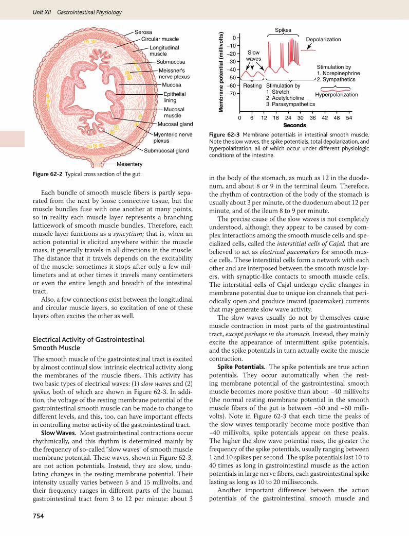
Figure 62-2 shows a typical cross section of the intesti-nal wall, including the following layers from outer sur-face inward: (1) the serosa, (2) a longitudinal smooth muscle layer, (3) a circular smooth muscle layer, (4) the submucosa, and (5) the mucosa. In addition, sparse bun-dles of smooth muscle fibers, the mucosal muscle, lie in the deeper layers of the mucosa. The motor functions of the gut are performed by the different layers of smooth muscle.
The general characteristics of smooth muscle and its function are discussed in Chapter 8, which should be reviewed as a background for the following sections of this chapter. The specific characteristics of smooth muscle in the gut are the following.
Gastrointestinal Smooth Muscle Functions as a Syncytium. The individual smooth muscle fibers in the gastrointestinal tract are 200 to 500 micrometers in length and 2 to 10 micrometers in diameter, and they are arranged in bundles of as many as 1000 parallel fibers. In the longitudinal muscle layer, the bundles extend longi-tudinally down the intestinal tract; in the circular muscle layer, they extend around the gut.
Within each bundle, the muscle fibers are electrically connected with one another through large numbers of gap junctions that allow low-resistance movement of ions from one muscle cell to the next. Therefore, electrical signals that initiate muscle contractions can travel readily from one fiber to the next within each bun-dle but more rapidly along the length of the bundle than sideways.
Parotid glandMouth
Salivary glands
Esophagus
Liver
Gallbladder
Duodenum
Ascendingcolon
Transversecolon
Stomach
Pancreas
Jejunum
Descendingcolon
Ileum
Anus
Figure 62-1 Alimentary tract.
Unit XII Gastrointestinal Physiology
754
Each bundle of smooth muscle fibers is partly sepa-rated from the next by loose connective tissue, but the muscle bundles fuse with one another at many points, so in reality each muscle layer represents a branching latticework of smooth muscle bundles. Therefore, each muscle layer functions as a syncytium; that is, when an action potential is elicited anywhere within the muscle mass, it generally travels in all directions in the muscle. The distance that it travels depends on the excitability of the muscle; sometimes it stops after only a few mil-limeters and at other times it travels many centimeters or even the entire length and breadth of the intestinal tract.
Also, a few connections exist between the longitudinal and circular muscle layers, so excitation of one of these layers often excites the other as well.
Electrical Activity of Gastrointestinal Smooth Muscle
The smooth muscle of the gastrointestinal tract is excited by almost continual slow, intrinsic electrical activity along the membranes of the muscle fibers. This activity has two basic types of electrical waves: (1) slow waves and (2) spikes, both of which are shown in Figure 62-3. In addi-tion, the voltage of the resting membrane potential of the gastrointestinal smooth muscle can be made to change to different levels, and this, too, can have important effects in controlling motor activity of the gastrointestinal tract.
Slow Waves. Most gastrointestinal contractions occur rhythmically, and this rhythm is determined mainly by the frequency of so-called “slow waves” of smooth muscle membrane potential. These waves, shown in Figure 62-3, are not action potentials. Instead, they are slow, undu-lating changes in the resting membrane potential. Their intensity usually varies between 5 and 15 millivolts, and their frequency ranges in different parts of the human gastrointestinal tract from 3 to 12 per minute: about 3
in the body of the stomach, as much as 12 in the duode-num, and about 8 or 9 in the terminal ileum. Therefore, the rhythm of contraction of the body of the stomach is usually about 3 per minute, of the duodenum about 12 per minute, and of the ileum 8 to 9 per minute.
The precise cause of the slow waves is not completely understood, although they appear to be caused by com-plex interactions among the smooth muscle cells and spe-cialized cells, called the interstitial cells of Cajal, that are believed to act as electrical pacemakers for smooth mus-cle cells. These interstitial cells form a network with each other and are interposed between the smooth muscle lay-ers, with synaptic-like contacts to smooth muscle cells. The interstitial cells of Cajal undergo cyclic changes in membrane potential due to unique ion channels that peri-odically open and produce inward (pacemaker) currents that may generate slow wave activity.
The slow waves usually do not by themselves cause muscle contraction in most parts of the gastrointestinal tract, except perhaps in the stomach. Instead, they mainly excite the appearance of intermittent spike potentials, and the spike potentials in turn actually excite the muscle contraction.
Spike Potentials. The spike potentials are true action potentials. They occur automatically when the rest-ing membrane potential of the gastrointestinal smooth muscle becomes more positive than about −40 millivolts (the normal resting membrane potential in the smooth muscle fibers of the gut is between −50 and −60 milli-volts). Note in Figure 62-3 that each time the peaks of the slow waves temporarily become more positive than −40 millivolts, spike potentials appear on these peaks. The higher the slow wave potential rises, the greater the frequency of the spike potentials, usually ranging between 1 and 10 spikes per second. The spike potentials last 10 to 40 times as long in gastrointestinal muscle as the action potentials in large nerve fibers, each gastrointestinal spike lasting as long as 10 to 20 milliseconds.
Another important difference between the action potentials of the gastrointestinal smooth muscle and
SerosaCircular muscle
Longitudinalmuscle
Submucosa
Mucosa
Meissner'snerve plexus
Epitheliallining
Mucosalmuscle
Mucosal gland
Submucosal gland
Mesentery
Myenteric nerveplexus
Figure 62-2 Typical cross section of the gut.
Mem
bra
ne
po
ten
tial
(m
illiv
olt
s)
-70-60
-50
-40
-30-20
-10
0
0 6 12 18
Spikes
Depolarization
Stimulation by1. Norepinephrine2. Sympathetics
Stimulation by1. Stretch2. Acetylcholine3. Parasympathetics
Resting
Hyperpolarization
Slowwaves
24 30 36 42 48 54SecondsSeconds
Figure 62-3 Membrane potentials in intestinal smooth muscle. Note the slow waves, the spike potentials, total depolarization, and hyperpolarization, all of which occur under different physiologic conditions of the intestine.
Chapter 62 General Principles of Gastrointestinal Function—Motility, Nervous Control, and Blood Circulation
755
Un
it X
iithose of nerve fibers is the manner in which they are gen-erated. In nerve fibers, the action potentials are caused almost entirely by rapid entry of sodium ions through sodium channels to the interior of the fibers. In gastro-intestinal smooth muscle fibers, the channels responsi-ble for the action potentials are somewhat different; they allow especially large numbers of calcium ions to enter along with smaller numbers of sodium ions and therefore are called calcium-sodium channels. These channels are much slower to open and close than are the rapid sodium channels of large nerve fibers. The slowness of opening and closing of the calcium-sodium channels accounts for the long duration of the action potentials. Also, the move-ment of large amounts of calcium ions to the interior of the muscle fiber during the action potential plays a special role in causing the intestinal muscle fibers to contract, as we discuss shortly.
Changes in Voltage of the Resting Membrane Potential. In addition to the slow waves and spike poten-tials, the baseline voltage level of the smooth muscle rest-ing membrane potential can also change. Under normal conditions, the resting membrane potential averages about −56 millivolts, but multiple factors can change this level. When the potential becomes less negative, which is called depolarization of the membrane, the muscle fibers become more excitable. When the potential becomes more negative, which is called hyperpolarization, the fibers become less excitable.
Factors that depolarize the membrane—that is, make it more excitable—are (1) stretching of the muscle, (2) stimulation by acetylcholine released from the endings of parasympathetic nerves, and (3) stimulation by several specific gastrointestinal hormones.
Important factors that make the membrane potential more negative—that is, hyperpolarize the membrane and make the muscle fibers less excitable—are (1) the effect of norepinephrine or epinephrine on the fiber membrane and (2) stimulation of the sympathetic nerves that secrete mainly norepinephrine at their endings.
Calcium Ions and Muscle Contraction. Smooth mus-cle contraction occurs in response to entry of calcium ions into the muscle fiber. As explained in Chapter 8, cal-cium ions, acting through a calmodulin control mecha-nism, activate the myosin filaments in the fiber, causing attractive forces to develop between the myosin filaments and the actin filaments, thereby causing the muscle to contract.
The slow waves do not cause calcium ions to enter the smooth muscle fiber (only sodium ions). Therefore, the slow waves by themselves usually cause no muscle con-traction. Instead, it is during the spike potentials, gen-erated at the peaks of the slow waves, that significant quantities of calcium ions do enter the fibers and cause most of the contraction.
Tonic Contraction of Some Gastrointestinal Smooth Muscle. Some smooth muscle of the gastrointestinal tract exhibits tonic contraction, as well as, or instead of, rhythmical contractions. Tonic contraction is continu-
ous, not associated with the basic electrical rhythm of the slow waves but often lasting several minutes or even hours. The tonic contraction often increases or decreases in intensity but continues.
Tonic contraction is sometimes caused by contin-uous repetitive spike potentials—the greater the fre-quency, the greater the degree of contraction. At other times, tonic contraction is caused by hormones or other factors that bring about continuous partial depolariza-tion of the smooth muscle membrane without causing action potentials. A third cause of tonic contraction is continuous entry of calcium ions into the interior of the cell brought about in ways not associated with changes in membrane potential. The details of these mechanisms are still unclear.
Neural Control of Gastrointestinal Function—Enteric Nervous System
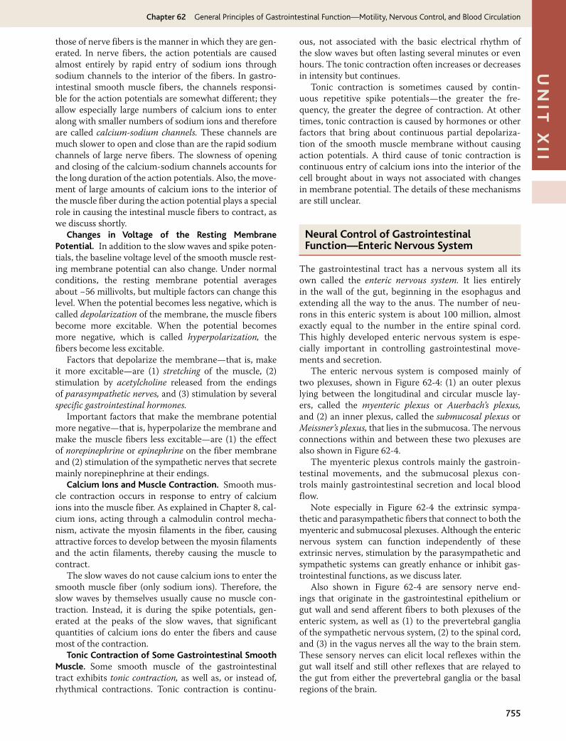
The gastrointestinal tract has a nervous system all its own called the enteric nervous system. It lies entirely in the wall of the gut, beginning in the esophagus and extending all the way to the anus. The number of neu-rons in this enteric system is about 100 million, almost exactly equal to the number in the entire spinal cord. This highly developed enteric nervous system is espe-cially important in controlling gastrointestinal move-ments and secretion.
The enteric nervous system is composed mainly of two plexuses, shown in Figure 62-4: (1) an outer plexus lying between the longitudinal and circular muscle lay-ers, called the myenteric plexus or Auerbach’s plexus, and (2) an inner plexus, called the submucosal plexus or Meissner’s plexus, that lies in the submucosa. The nervous connections within and between these two plexuses are also shown in Figure 62-4.
The myenteric plexus controls mainly the gastroin-testinal movements, and the submucosal plexus con-trols mainly gastrointestinal secretion and local blood flow.
Note especially in Figure 62-4 the extrinsic sympa-thetic and parasympathetic fibers that connect to both the myenteric and submucosal plexuses. Although the enteric nervous system can function independently of these extrinsic nerves, stimulation by the parasympathetic and sympathetic systems can greatly enhance or inhibit gas-trointestinal functions, as we discuss later.
Also shown in Figure 62-4 are sensory nerve end-ings that originate in the gastrointestinal epithelium or gut wall and send afferent fibers to both plexuses of the enteric system, as well as (1) to the prevertebral ganglia of the sympathetic nervous system, (2) to the spinal cord, and (3) in the vagus nerves all the way to the brain stem. These sensory nerves can elicit local reflexes within the gut wall itself and still other reflexes that are relayed to the gut from either the prevertebral ganglia or the basal regions of the brain.
Unit XII Gastrointestinal Physiology
756
Differences Between the Myenteric and Submucosal Plexuses
The myenteric plexus consists mostly of a linear chain of many interconnecting neurons that extends the entire length of the gastrointestinal tract. A section of this chain is shown in Figure 62-4.
Because the myenteric plexus extends all the way along the intestinal wall and because it lies between the longi-tudinal and circular layers of intestinal smooth muscle, it is concerned mainly with controlling muscle activity along the length of the gut. When this plexus is stimu-lated, its principal effects are (1) increased tonic contrac-tion, or “tone,” of the gut wall; (2) increased intensity of the rhythmical contractions; (3) slightly increased rate of the rhythm of contraction; and (4) increased velocity of conduction of excitatory waves along the gut wall, causing more rapid movement of the gut peristaltic waves.
The myenteric plexus should not be considered entirely excitatory because some of its neurons are inhibitory; their fiber endings secrete an inhibitory transmitter, possibly vasoactive intestinal polypeptide or some other inhibi-tory peptide. The resulting inhibitory signals are espe-cially useful for inhibiting some of the intestinal sphincter muscles that impede movement of food along successive segments of the gastrointestinal tract, such as the pyloric sphincter, which controls emptying of the stomach into the duodenum, and the sphincter of the ileocecal valve, which controls emptying from the small intestine into the cecum.
The submucosal plexus, in contrast to the myenteric plexus, is mainly concerned with controlling function within the inner wall of each minute segment of the intes-tine. For instance, many sensory signals originate from the gastrointestinal epithelium and are then integrated in the submucosal plexus to help control local intestinal secretion, local absorption, and local contraction of the
submucosal muscle that causes various degrees of infold-ing of the gastrointestinal mucosa.
Types of Neurotransmitters Secreted by Enteric Neurons
In an attempt to understand better the multiple functions of the gastrointestinal enteric nervous system, research workers the world over have identified a dozen or more different neurotransmitter substances that are released by the nerve endings of different types of enteric neu-rons. Two of them with which we are already familiar are (1) acetylcholine and (2) norepinephrine. Others are (3) adenosine triphosphate, (4) serotonin, (5) dopamine, (6) cholecystokinin, (7) substance P, (8) vasoactive intes-tinal polypeptide, (9) somatostatin, (10) leu-enkephalin, (11) met-enkephalin, and (12) bombesin. The specific functions of many of these are not known well enough to justify discussion here, other than to point out the following.
Acetylcholine most often excites gastrointestinal activ-ity. Norepinephrine almost always inhibits gastrointestinal activity. This is also true of epinephrine, which reaches the gastrointestinal tract mainly by way of the blood after it is secreted by the adrenal medullae into the circulation. The other aforementioned transmitter substances are a mix-ture of excitatory and inhibitory agents, some of which we discuss in the following chapter.
Autonomic Control of the Gastrointestinal Tract
Parasympathetic Stimulation Increases Activity of the Enteric Nervous System. The parasympathetic sup-ply to the gut is divided into cranial and sacral divisions, which were discussed in Chapter 60.
Except for a few parasympathetic fibers to the mouth and pharyngeal regions of the alimentary tract, the cranial parasympathetic nerve fibers are almost entirely in the
To prevertebralganglia, spinalcord, and brainstem
Sensoryneurons
Submucosalplexus
Myentericplexus
Epithelium
Sympathetic
(mainly postganglionic)
Parasympathetic
(preganglionic)
Figure 62-4 Neural control of the gut wall, showing (1) the myenteric and submucosal plexuses (black fibers); (2) extrinsic control of these plexuses by the sympathetic and parasympathetic ner-vous systems (red fibers); and (3) sensory fibers passing from the luminal epithelium and gut wall to the enteric plexuses, then to the prevertebral ganglia of the spinal cord and directly to the spinal cord and brain stem (dashed fibers).

Chapter 62 General Principles of Gastrointestinal Function—Motility, Nervous Control, and Blood Circulation
757
Un
it X
iivagus nerves. These fibers provide extensive innervation to the esophagus, stomach, and pancreas and somewhat less to the intestines down through the first half of the large intestine.
The sacral parasympathetics originate in the second, third, and fourth sacral segments of the spinal cord and pass through the pelvic nerves to the distal half of the large intestine and all the way to the anus. The sigmoidal, rec-tal, and anal regions are considerably better supplied with parasympathetic fibers than are the other intestinal areas. These fibers function especially to execute the defecation reflexes, discussed in Chapter 63.
The postganglionic neurons of the gastrointestinal parasympathetic system are located mainly in the myen-teric and submucosal plexuses. Stimulation of these para-sympathetic nerves causes general increase in activity of the entire enteric nervous system. This in turn enhances activity of most gastrointestinal functions.
Sympathetic Stimulation Usually Inhibits Gastro-intestinal Tract Activity. The sympathetic fibers to the gastrointestinal tract originate in the spinal cord between segments T5 and L2. Most of the preganglionic fibers that innervate the gut, after leaving the cord, enter the sympa-thetic chains that lie lateral to the spinal column, and many of these fibers then pass on through the chains to outlying ganglia such as to the celiac ganglion and various mesen-teric ganglia. Most of the postganglionic sympathetic neu-ron bodies are in these ganglia, and postganglionic fibers then spread through postganglionic sympathetic nerves to all parts of the gut. The sympathetics innervate essen-tially all of the gastrointestinal tract, rather than being more extensive nearest the oral cavity and anus, as is true of the parasympathetics. The sympathetic nerve endings secrete mainly norepinephrine but also small amounts of epinephrine.
In general, stimulation of the sympathetic nervous sys-tem inhibits activity of the gastrointestinal tract, causing many effects opposite to those of the parasympathetic sys-tem. It exerts its effects in two ways: (1) to a slight extent by direct effect of secreted norepinephrine to inhibit intestinal tract smooth muscle (except the mucosal mus-cle, which it excites) and (2) to a major extent by an inhib-itory effect of norepinephrine on the neurons of the entire enteric nervous system.
Strong stimulation of the sympathetic system can inhibit motor movements of the gut so greatly that this can literally block movement of food through the gastro-intestinal tract.
Afferent Sensory Nerve Fibers from the Gut
Many afferent sensory nerve fibers innervate the gut. Some of them have their cell bodies in the enteric ner-vous system itself and some in the dorsal root ganglia of the spinal cord. These sensory nerves can be stimu-lated by (1) irritation of the gut mucosa, (2) excessive distention of the gut, or (3) presence of specific chemi-cal substances in the gut. Signals transmitted through the fibers can then cause excitation or, under other
conditions, inhibition of intestinal movements or intes-tinal secretion.
In addition, other sensory signals from the gut go all the way to multiple areas of the spinal cord and even the brain stem. For example, 80 percent of the nerve fibers in the vagus nerves are afferent rather than efferent. These afferent fibers transmit sensory signals from the gastroin-testinal tract into the brain medulla, which in turn initi-ates vagal reflex signals that return to the gastrointestinal tract to control many of its functions.
Gastrointestinal Reflexes
The anatomical arrangement of the enteric nervous system and its connections with the sympathetic and parasympathetic systems support three types of gastroin-testinal reflexes that are essential to gastrointestinal con-trol. They are the following:
1. Reflexes that are integrated entirely within the gut wall enteric nervous system. These include reflexes that con-trol much gastrointestinal secretion, peristalsis, mixing contractions, local inhibitory effects, and so forth.
2. Reflexes from the gut to the prevertebral sympathetic ganglia and then back to the gastrointestinal tract. These reflexes transmit signals long distances to other areas of the gastrointestinal tract, such as signals from the stomach to cause evacuation of the colon (the gas-trocolic reflex), signals from the colon and small intes-tine to inhibit stomach motility and stomach secretion (the enterogastric reflexes), and reflexes from the colon to inhibit emptying of ileal contents into the colon (the colonoileal reflex).
3. Reflexes from the gut to the spinal cord or brain stem and then back to the gastrointestinal tract. These include especially (1) reflexes from the stomach and duode-num to the brain stem and back to the stomach—by way of the vagus nerves—to control gastric motor and secretory activity; (2) pain reflexes that cause general inhibition of the entire gastrointestinal tract; and (3) defecation reflexes that travel from the colon and rec-tum to the spinal cord and back again to produce the powerful colonic, rectal, and abdominal contractions required for defecation (the defecation reflexes).
Hormonal Control of Gastrointestinal Motility
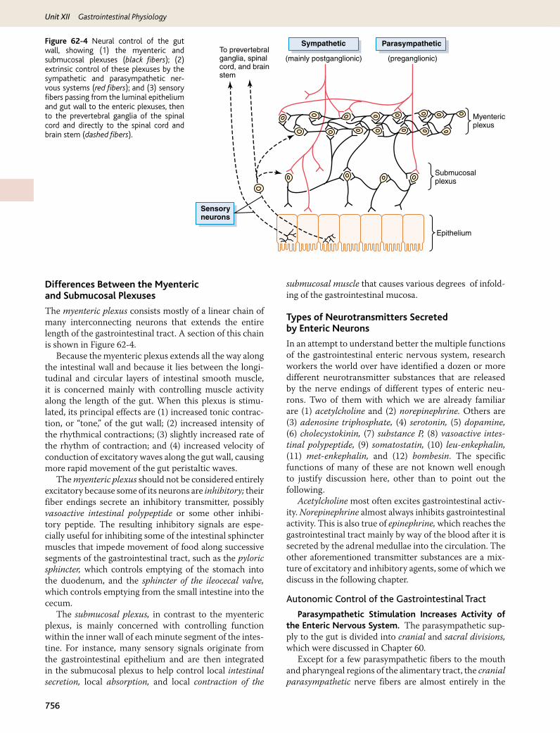
The gastrointestinal hormones are released into the portal circulation and exert physiological actions on target cells with specific receptors for the hormone. The effects of the hormones persist even after all nervous connections between the site of release and the site of action have been severed. Table 62-1 outlines the actions of each gastroin-testinal hormone, as well as the stimuli for secretion and sites at which secretion takes place.
In Chapter 64, we discuss the extreme importance of several hormones for controlling gastrointestinal secre-tion. Most of these same hormones also affect motility in some parts of the gastrointestinal tract. Although the
Unit XII Gastrointestinal Physiology
758
motility effects are usually less important than the secre-tory effects of the hormones, some of the more important of them are the following.
Gastrin is secreted by the “G” cells of the antrum of the stomach in response to stimuli associated with ingestion of a meal, such as distention of the stomach, the products of proteins, and gastrin releasing peptide, which is released by the nerves of the gastric mucosa during vagal stimula-tion. The primary actions of gastrin are (1) stimulation of gastric acid secretion and (2) stimulation of growth of the gastric mucosa.
Cholecystokinin (CCK) is secreted by “I” cells in the mucosa of the duodenum and jejunum mainly in response to digestive products of fat, fatty acids, and monoglycerides in the intestinal contents. This hor-mone strongly contracts the gallbladder, expelling bile into the small intestine, where the bile in turn plays important roles in emulsifying fatty substances, and allowing them to be digested and absorbed. CCK also inhibits stomach contraction moderately. Therefore, at the same time that this hormone causes emptying of the gallbladder, it also slows the emptying of food from the stomach to give adequate time for digestion of the fats in the upper intestinal tract. CCK also inhibits appe-tite to prevent overeating during meals by stimulating sensory afferent nerve fibers in the duodenum; these fibers, in turn, send signals by way of the vagus nerve
to inhibit feeding centers in the brain as discussed in Chapter 71.
Secretin was the first gastrointestinal hormone dis-covered and is secreted by the “S” cells in the mucosa of the duodenum in response to acidic gastric juice empty-ing into the duodenum from the pylorus of the stomach. Secretin has a mild effect on motility of the gastrointes-tinal tract and acts to promote pancreatic secretion of bicarbonate, which in turn helps to neutralize the acid in the small intestine.
Gastric inhibitory peptide (GIP) is secreted by the mucosa of the upper small intestine, mainly in response to fatty acids and amino acids but to a lesser extent in response to carbohydrate. It has a mild effect in decreas-ing motor activity of the stomach and therefore slows emptying of gastric contents into the duodenum when the upper small intestine is already overloaded with food products. GIP, at blood levels even lower than those needed to inhibit gastric motility, also stimulates insulin secretion and for this reason is also known as glucose-dependent insulinotropic peptide.
Motilin is secreted by the stomach and upper duode-num during fasting, and the only known function of this hormone is to increase gastrointestinal motility. Motilin is released cyclically and stimulates waves of gastrointes-tinal motility called interdigestive myoelectric complexes that move through the stomach and small intestine every
Hormone Stimuli for Secretion Site of Secretion Actions
Gastrin Protein G cells of the antrum, duodenum, and jejunum
StimulatesDistention Gastric acid secretionNerve(Acid inhibits release)
Mucosal growth
Cholecystokinin Protein I cells of the duodenum, jejunum, and ileum
StimulatesFat Pancreatic enzyme secretionAcid Pancreatic bicarbonate secretion
Gallbladder contraction Growth of exocrine pancreasInhibits
Gastric emptying
Secretin
AcidFat
S cells of the duodenum, jejunum, and ileum
Stimulates Pepsin secretion Pancreatic bicarbonate secretion Biliary bicarbonate secretion Growth of exocrine pancreasInhibits Gastric acid secretion
Gastric inhibitory peptide Protein K cells of the duodenum and jejunum
StimulatesFat Insulin releaseCarbohydrate Inhibits
Gastric acid secretion
Motilin Fat M cells of the duodenum and jejunum
StimulatesAcid Gastric motilityNerve Intestinal motility
Table 62-1 Gastrointestinal Hormone Actions, Stimuli for Secretion, and Site of Secretion

Chapter 62 General Principles of Gastrointestinal Function—Motility, Nervous Control, and Blood Circulation
759
Un
it X
ii90 minutes in a fasted person. Motilin secretion is inhib-ited after ingestion by mechanisms that are not fully understood.
Functional Types of Movements in the Gastrointestinal Tract
Two types of movements occur in the gastrointesti-nal tract: (1) propulsive movements, which cause food to move forward along the tract at an appropriate rate to accommodate digestion and absorption, and (2) mix-ing movements, which keep the intestinal contents thor-oughly mixed at all times.
Propulsive Movements—Peristalsis
The basic propulsive movement of the gastrointesti-nal tract is peristalsis, which is illustrated in Figure 62-5. A contractile ring appears around the gut and then moves forward; this is analogous to putting one’s fingers around a thin distended tube, then constricting the fingers and sliding them forward along the tube. Any material in front of the contractile ring is moved forward.
Peristalsis is an inherent property of many syncytial smooth muscle tubes; stimulation at any point in the gut can cause a contractile ring to appear in the circu-lar muscle, and this ring then spreads along the gut tube. (Peristalsis also occurs in the bile ducts, glandular ducts, ureters, and many other smooth muscle tubes of the body.)
The usual stimulus for intestinal peristalsis is disten-tion of the gut. That is, if a large amount of food collects at any point in the gut, the stretching of the gut wall stimu-lates the enteric nervous system to contract the gut wall 2 to 3 centimeters behind this point, and a contractile ring appears that initiates a peristaltic movement. Other stim-uli that can initiate peristalsis include chemical or physi-cal irritation of the epithelial lining in the gut. Also, strong parasympathetic nervous signals to the gut will elicit strong peristalsis.
Function of the Myenteric Plexus in Peristalsis. Peristalsis occurs only weakly or not at all in any portion of the gastrointestinal tract that has congenital absence of the myenteric plexus. Also, it is greatly depressed or
completely blocked in the entire gut when a person is treated with atropine to paralyze the cholinergic nerve endings of the myenteric plexus. Therefore, effectual peri-stalsis requires an active myenteric plexus.
Directional Movement of Peristaltic Waves Toward the Anus. Peristalsis, theoretically, can occur in either direction from a stimulated point, but it normally dies out rapidly in the orad (toward the mouth) direction while continuing for a considerable distance toward the anus. The exact cause of this directional transmission of peri-stalsis has never been ascertained, although it probably results mainly from the fact that the myenteric plexus itself is “polarized” in the anal direction, which can be explained as follows.
Peristaltic Reflex and the “Law of the Gut”. When a segment of the intestinal tract is excited by distention and thereby initiates peristalsis, the contractile ring caus-ing the peristalsis normally begins on the orad side of the distended segment and moves toward the distended seg-ment, pushing the intestinal contents in the anal direction for 5 to 10 centimeters before dying out. At the same time, the gut sometimes relaxes several centimeters down-stream toward the anus, which is called “receptive relax-ation,” thus allowing the food to be propelled more easily toward the anus than toward the mouth.
This complex pattern does not occur in the absence of the myenteric plexus. Therefore, the complex is called the myenteric reflex or the peristaltic reflex. The peristaltic reflex plus the anal direction of movement of the peristal-sis is called the “law of the gut.”
Mixing Movements
Mixing movements differ in different parts of the ali-mentary tract. In some areas, the peristaltic contrac-tions themselves cause most of the mixing. This is especially true when forward progression of the intes-tinal contents is blocked by a sphincter so that a peri-staltic wave can then only churn the intestinal contents, rather than propelling them forward. At other times, local intermittent constrictive contractions occur every few centimeters in the gut wall. These constrictions usually last only 5 to 30 seconds; then new constrictions occur at other points in the gut, thus “chopping” and “shearing” the contents first here and then there. These peristaltic and constrictive movements are modified in different parts of the gastrointestinal tract for proper propulsion and mixing, as discussed for each portion of the tract in Chapter 63.
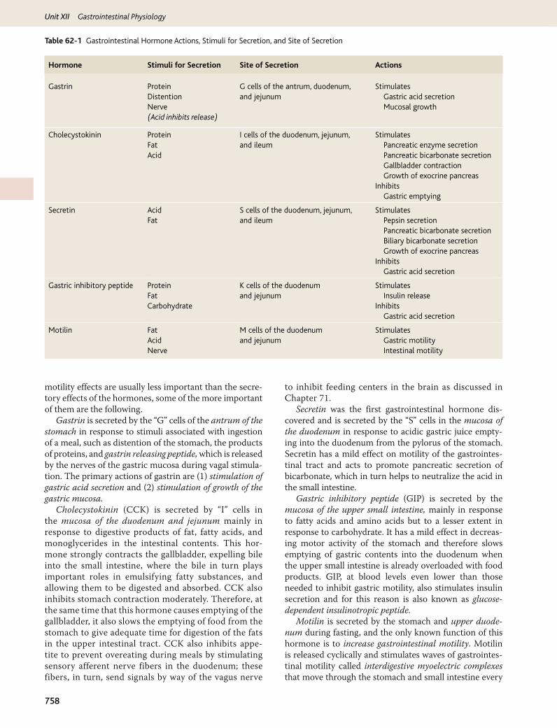
Gastrointestinal Blood Flow—“Splanchnic Circulation”
The blood vessels of the gastrointestinal system are part of a more extensive system called the splanchnic circu-lation, shown in Figure 62-6. It includes the blood flow
Leading wave of distention
Zero time
5 seconds later
Peristaltic contraction
Figure 62-5 Peristalsis.
Unit XII Gastrointestinal Physiology
760
through the gut itself plus blood flows through the spleen, pancreas, and liver. The design of this system is such that all the blood that courses through the gut, spleen, and pancreas then flows immediately into the liver by way of the portal vein. In the liver, the blood passes through
millions of minute liver sinusoids and finally leaves the liver by way of hepatic veins that empty into the vena cava of the general circulation. This flow of blood through the liver, before it empties into the vena cava, allows the retic-uloendothelial cells that line the liver sinusoids to remove bacteria and other particulate matter that might enter the blood from the gastrointestinal tract, thus prevent-ing direct transport of potentially harmful agents into the remainder of the body.
The nonfat, water-soluble nutrients absorbed from the gut (such as carbohydrates and proteins) are transported in the portal venous blood to the same liver sinusoids. Here, both the reticuloendothelial cells and the principal parenchymal cells of the liver, the hepatic cells, absorb and store temporarily from one half to three quarters of the nutrients. Also, much chemical intermediary pro-cessing of these nutrients occurs in the liver cells. We dis-cuss these nutritional functions of the liver in Chapters 67 through 71. Almost all of the fats absorbed from the intestinal tract are not carried in the portal blood but instead are absorbed into the intestinal lymphatics and then conducted to the systemic circulating blood by way of the thoracic duct, bypassing the liver.
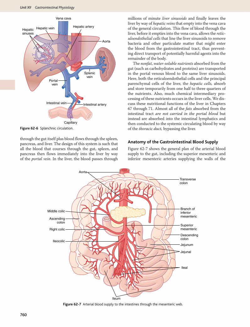
Anatomy of the Gastrointestinal Blood Supply
Figure 62-7 shows the general plan of the arterial blood supply to the gut, including the superior mesenteric and inferior mesenteric arteries supplying the walls of the
Vena cava
Hepatic artery
Aorta
Splenicvein
Intestinal arteryIntestinal vein
Capillary
Portalvein
Hepatic veinHepaticsinuses
Figure 62-6 Splanchnic circulation.
Transversecolon
Descendingcolon
Jejunum
Jejunal
Ileal
Ileum
Branch ofinferiormesenteric
SuperiormesentericRight colic
Ascendingcolon
Middle colic
Aorta
Ileocolic
Figure 62-7 Arterial blood supply to the intestines through the mesenteric web.
Chapter 62 General Principles of Gastrointestinal Function—Motility, Nervous Control, and Blood Circulation
761
Un
it X
iismall and large intestines by way of an arching arterial system. Not shown in the figure is the celiac artery, which provides a similar blood supply to the stomach.
On entering the wall of the gut, the arteries branch and send smaller arteries circling in both directions around the gut, with the tips of these arteries meeting on the side of the gut wall opposite the mesenteric attachment. From the circling arteries, still much smaller arteries penetrate into the intestinal wall and spread (1) along the muscle bundles, (2) into the intestinal villi, and (3) into submu-cosal vessels beneath the epithelium to serve the secre-tory and absorptive functions of the gut.
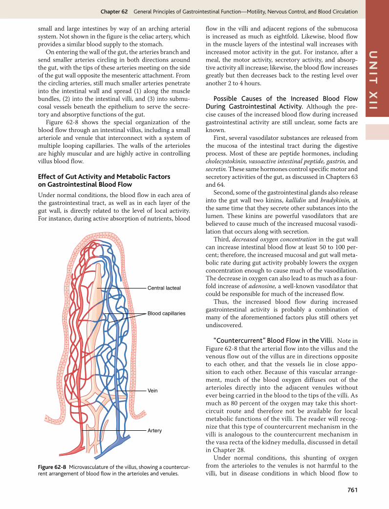
Figure 62-8 shows the special organization of the blood flow through an intestinal villus, including a small arteriole and venule that interconnect with a system of multiple looping capillaries. The walls of the arterioles are highly muscular and are highly active in controlling villus blood flow.
Effect of Gut Activity and Metabolic Factors on Gastrointestinal Blood Flow
Under normal conditions, the blood flow in each area of the gastrointestinal tract, as well as in each layer of the gut wall, is directly related to the level of local activity. For instance, during active absorption of nutrients, blood
flow in the villi and adjacent regions of the submucosa is increased as much as eightfold. Likewise, blood flow in the muscle layers of the intestinal wall increases with increased motor activity in the gut. For instance, after a meal, the motor activity, secretory activity, and absorp-tive activity all increase; likewise, the blood flow increases greatly but then decreases back to the resting level over another 2 to 4 hours.
Possible Causes of the Increased Blood Flow During Gastrointestinal Activity. Although the pre-cise causes of the increased blood flow during increased gastrointestinal activity are still unclear, some facts are known.
First, several vasodilator substances are released from the mucosa of the intestinal tract during the digestive process. Most of these are peptide hormones, including cholecystokinin, vasoactive intestinal peptide, gastrin, and secretin. These same hormones control specific motor and secretory activities of the gut, as discussed in Chapters 63 and 64.
Second, some of the gastrointestinal glands also release into the gut wall two kinins, kallidin and bradykinin, at the same time that they secrete other substances into the lumen. These kinins are powerful vasodilators that are believed to cause much of the increased mucosal vasodi-lation that occurs along with secretion.
Third, decreased oxygen concentration in the gut wall can increase intestinal blood flow at least 50 to 100 per-cent; therefore, the increased mucosal and gut wall meta-bolic rate during gut activity probably lowers the oxygen concentration enough to cause much of the vasodilation. The decrease in oxygen can also lead to as much as a four-fold increase of adenosine, a well-known vasodilator that could be responsible for much of the increased flow.
Thus, the increased blood flow during increased gastrointestinal activity is probably a combination of many of the aforementioned factors plus still others yet undiscovered.
“Countercurrent” Blood Flow in the Villi. Note in Figure 62-8 that the arterial flow into the villus and the venous flow out of the villus are in directions opposite to each other, and that the vessels lie in close appo-sition to each other. Because of this vascular arrange-ment, much of the blood oxygen diffuses out of the arterioles directly into the adjacent venules without ever being carried in the blood to the tips of the villi. As much as 80 percent of the oxygen may take this short-circuit route and therefore not be available for local metabolic functions of the villi. The reader will recog-nize that this type of countercurrent mechanism in the villi is analogous to the countercurrent mechanism in the vasa recta of the kidney medulla, discussed in detail in Chapter 28.
Under normal conditions, this shunting of oxygen from the arterioles to the venules is not harmful to the villi, but in disease conditions in which blood flow to
Central lacteal
Vein
Artery
Blood capillaries
Figure 62-8 Microvasculature of the villus, showing a countercur-rent arrangement of blood flow in the arterioles and venules.
Unit XII Gastrointestinal Physiology
762
the gut becomes greatly curtailed, such as in circulatory shock, the oxygen deficit in the tips of the villi can become so great that the villus tip or even the whole villus suffers ischemic death and can disintegrate. Therefore, for this reason and others, in many gastrointestinal diseases the villi become seriously blunted, leading to greatly dimin-ished intestinal absorptive capacity.
Nervous Control of Gastrointestinal Blood Flow
Stimulation of the parasympathetic nerves going to the stomach and lower colon increases local blood flow at the same time that it increases glandular secretion. This increased flow probably results secondarily from the increased glandular activity and not as a direct effect of the nervous stimulation.
Sympathetic stimulation, by contrast, has a direct effect on essentially all the gastrointestinal tract to cause intense vasoconstriction of the arterioles with greatly decreased blood flow. After a few minutes of this vasoconstric-tion, the flow often returns to near normal by means of a mechanism called “autoregulatory escape.” That is, the local metabolic vasodilator mechanisms that are elicited by ischemia override the sympathetic vasoconstriction, returning toward normal the necessary nutrient blood flow to the gastrointestinal glands and muscle.
Importance of Nervous Depression of Gastrointestinal Blood Flow When Other Parts of the Body Need Extra Blood Flow. A major value of sympathetic vasoconstriction in the gut is that it allows shutoff of gas-trointestinal and other splanchnic blood flow for short periods of time during heavy exercise, when the skeletal muscle and heart need increased flow. Also, in circulatory shock, when all the body’s vital tissues are in danger of cellular death for lack of blood flow—especially the brain and the heart—sympathetic stimulation can decrease splanchnic blood flow to very little for many hours.
Sympathetic stimulation also causes strong vasocon-striction of the large-volume intestinal and mesenteric veins. This decreases the volume of these veins, thereby dis-placing large amounts of blood into other parts of the cir-culation. In hemorrhagic shock or other states of low blood volume, this mechanism can provide as much as 200 to 400 milliliters of extra blood to sustain the general circulation.
Bibliography
Adelson DW, Million M: Tracking the moveable feast: sonomicrometry and gastrointestinal motility, News Physiol Sci 19:27, 2004.
Daniel EE: Physiology and pathophysiology of the interstitial cell of Cajal: from bench to bedside. III. Interaction of interstitial cells of Cajal with neuromediators: an interim assessment, Am J Physiol Gastrointest Liver Physiol 281:G1329, 2001.
Grundy D, Al-Chaer ED, Aziz Q, et al: Fundamentals of neurogastroenterol-ogy: basic science, Gastroenterology 130:1391, 2006.
Hobson AR, Aziz Q: Central nervous system processing of human visceral pain in health and disease, News Physiol Sci 18:109, 2003.
Holst JJ: The physiology of glucagon-like peptide 1, Physiol Rev 87:1409, 2009.
Huizinga JD: Physiology and pathophysiology of the interstitial cell of Cajal: from bench to bedside. II. Gastric motility: lessons from mutant mice on slow waves and innervation, Am J Physiol Gastrointest Liver Physiol 281:G1129, 2001.
Huizinga JD, Lammers WJ: Gut peristalsis is governed by a multitude of cooperating mechanisms, Am J Physiol Gastrointest Liver Physiol 296:G1, 2009.
Jeays AD, Lawford PV, Gillott R, et al: A framework for the modeling of gut blood flow regulation and postprandial hyperaemia, World J Gastroenterol 13:1393, 2007.
Johnson LR: Gastrointestinal Physiology, ed 3, St. Louis, 2001, Mosby.Kim W, Egan JM: The role of incretins in glucose homeostasis and diabetes
treatment, Pharmacol Rev 60:470, 2009.Kolkman JJ, Bargeman M, Huisman AB, Geelkerken RH: Diagnosis and
management of splanchnic ischemia, World J Gastroenterol 14:7309, 2008.
Lammers WJ, Slack JR: Of slow waves and spike patches, News Physiol Sci 16:138, 2001.
Moran TH, Dailey MJ: Minireview: Gut peptides: targets for antiobesity drug development? Endocrinology 150:2526, 2009.
Nauck MA: Unraveling the science of incretin biology, Am J Med 122(Suppl 6):S3, 2009.
Powley TL, Phillips RJ: Musings on the wanderer: what’s new in our under-standing of vago-vagal reflexes? I. Morphology and topography of vagal afferents innervating the GI tract, Am J Physiol Gastrointest Liver Physiol 283:G1217, 2002.
Phillips RJ, Powley TL: Innervation of the gastrointestinal tract: patterns of aging, Auton Neurosci 136:1, 2007.
Sanders KM, Ordog T, Ward SM: Physiology and pathophysiology of the interstitial cells of Cajal: from bench to bedside. IV. Genetic and animal models of GI motility disorders caused by loss of intersti-tial cells of Cajal, Am J Physiol Gastrointest Liver Physiol 282:G747, 2002.
Schubert ML, Peura DA: Control of gastric acid secretion in health and dis-ease, Gastroenterology 134:1842, 2008.
Vanden Berghe P, Tack J, Boesmans W: Highlighting synaptic commu-nication in the enteric nervous system, Gastroenterology 135:20, 2008.
773
Un
it X
iiSecretory Functions of the Alimentary Tract
chapter 64
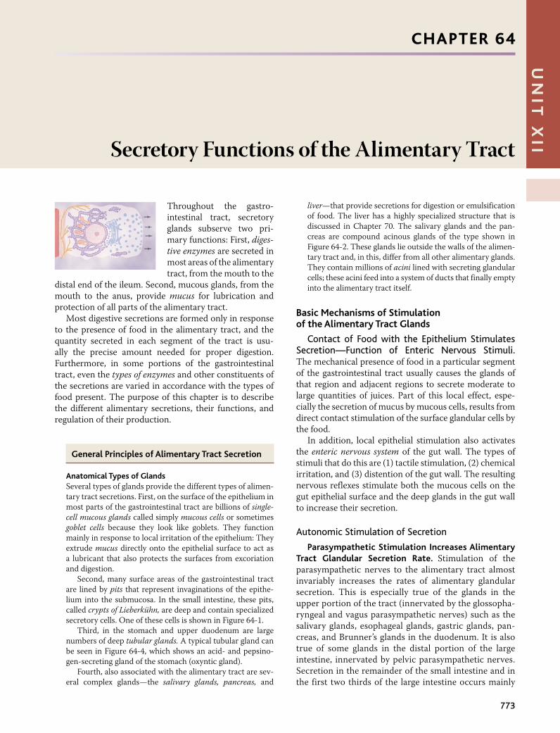
Throughout the gastro-intestinal tract, secretory glands subserve two pri-mary functions: First, diges-tive enzymes are secreted in most areas of the alimentary tract, from the mouth to the
distal end of the ileum. Second, mucous glands, from the mouth to the anus, provide mucus for lubrication and protection of all parts of the alimentary tract.
Most digestive secretions are formed only in response to the presence of food in the alimentary tract, and the quantity secreted in each segment of the tract is usu-ally the precise amount needed for proper digestion. Furthermore, in some portions of the gastrointestinal tract, even the types of enzymes and other constituents of the secretions are varied in accordance with the types of food present. The purpose of this chapter is to describe the different alimentary secretions, their functions, and regulation of their production.
General Principles of Alimentary Tract Secretion
Anatomical Types of GlandsSeveral types of glands provide the different types of alimen-tary tract secretions. First, on the surface of the epithelium in most parts of the gastrointestinal tract are billions of single-cell mucous glands called simply mucous cells or sometimes goblet cells because they look like goblets. They function mainly in response to local irritation of the epithelium: They extrude mucus directly onto the epithelial surface to act as a lubricant that also protects the surfaces from excoriation and digestion.
Second, many surface areas of the gastrointestinal tract are lined by pits that represent invaginations of the epithe-lium into the submucosa. In the small intestine, these pits, called crypts of Lieberkühn, are deep and contain specialized secretory cells. One of these cells is shown in Figure 64-1.
Third, in the stomach and upper duodenum are large numbers of deep tubular glands. A typical tubular gland can be seen in Figure 64-4, which shows an acid- and pepsino-gen-secreting gland of the stomach (oxyntic gland).
Fourth, also associated with the alimentary tract are sev-eral complex glands—the salivary glands, pancreas, and
liver—that provide secretions for digestion or emulsification of food. The liver has a highly specialized structure that is discussed in Chapter 70. The salivary glands and the pan-creas are compound acinous glands of the type shown in Figure 64-2. These glands lie outside the walls of the alimen-tary tract and, in this, differ from all other alimentary glands. They contain millions of acini lined with secreting glandular cells; these acini feed into a system of ducts that finally empty into the alimentary tract itself.
Basic Mechanisms of Stimulation of the Alimentary Tract Glands
Contact of Food with the Epithelium Stimulates Secretion—Function of Enteric Nervous Stimuli. The mechanical presence of food in a particular segment of the gastrointestinal tract usually causes the glands of that region and adjacent regions to secrete moderate to large quantities of juices. Part of this local effect, espe-cially the secretion of mucus by mucous cells, results from direct contact stimulation of the surface glandular cells by the food.
In addition, local epithelial stimulation also activates the enteric nervous system of the gut wall. The types of stimuli that do this are (1) tactile stimulation, (2) chemical irritation, and (3) distention of the gut wall. The resulting nervous reflexes stimulate both the mucous cells on the gut epithelial surface and the deep glands in the gut wall to increase their secretion.
Autonomic Stimulation of Secretion
Parasympathetic Stimulation Increases Alimentary Tract Glandular Secretion Rate. Stimulation of the parasympathetic nerves to the alimentary tract almost invariably increases the rates of alimentary glandular secretion. This is especially true of the glands in the upper portion of the tract (innervated by the glossopha-ryngeal and vagus parasympathetic nerves) such as the salivary glands, esophageal glands, gastric glands, pan-creas, and Brunner’s glands in the duodenum. It is also true of some glands in the distal portion of the large intestine, innervated by pelvic parasympathetic nerves. Secretion in the remainder of the small intestine and in the first two thirds of the large intestine occurs mainly
Unit XII Gastrointestinal Physiology
774
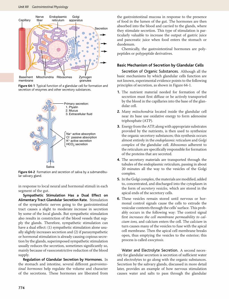
Primary secretion: 1. Ptyalin 2. Mucus 3. Extracellular fluid
Saliva
Na+ active absorptionCl− passive absorptionK+ active secretionHCO3
− secretion
Figure 64-2 Formation and secretion of saliva by a submandibu-lar salivary gland.
in response to local neural and hormonal stimuli in each segment of the gut.
Sympathetic Stimulation Has a Dual Effect on Alimentary Tract Glandular Secretion Rate. Stimulation of the sympathetic nerves going to the gastrointestinal tract causes a slight to moderate increase in secretion by some of the local glands. But sympathetic stimulation also results in constriction of the blood vessels that sup-ply the glands. Therefore, sympathetic stimulation can have a dual effect: (1) sympathetic stimulation alone usu-ally slightly increases secretion and (2) if parasympathetic or hormonal stimulation is already causing copious secre-tion by the glands, superimposed sympathetic stimulation usually reduces the secretion, sometimes significantly so, mainly because of vasoconstrictive reduction of the blood supply.
Regulation of Glandular Secretion by Hormones. In the stomach and intestine, several different gastrointes-tinal hormones help regulate the volume and character of the secretions. These hormones are liberated from
the gastrointestinal mucosa in response to the presence of food in the lumen of the gut. The hormones are then absorbed into the blood and carried to the glands, where they stimulate secretion. This type of stimulation is par-ticularly valuable to increase the output of gastric juice and pancreatic juice when food enters the stomach or duodenum.
Chemically, the gastrointestinal hormones are poly-peptides or polypeptide derivatives.
Basic Mechanism of Secretion by Glandular Cells
Secretion of Organic Substances. Although all the basic mechanisms by which glandular cells function are not known, experimental evidence points to the following principles of secretion, as shown in Figure 64-1.
1. The nutrient material needed for formation of the secretion must first diffuse or be actively transported by the blood in the capillaries into the base of the glan-dular cell.
2. Many mitochondria located inside the glandular cell near its base use oxidative energy to form adenosine triphosphate (ATP).
3. Energy from the ATP, along with appropriate substrates provided by the nutrients, is then used to synthesize the organic secretory substances; this synthesis occurs almost entirely in the endoplasmic reticulum and Golgi complex of the glandular cell. Ribosomes adherent to the reticulum are specifically responsible for formation of the proteins that are secreted.
4. The secretory materials are transported through the tubules of the endoplasmic reticulum, passing in about 20 minutes all the way to the vesicles of the Golgi complex.
5. In the Golgi complex, the materials are modified, added to, concentrated, and discharged into the cytoplasm in the form of secretory vesicles, which are stored in the apical ends of the secretory cells.
6. These vesicles remain stored until nervous or hor-monal control signals cause the cells to extrude the vesicular contents through the cells’ surface. This prob-ably occurs in the following way: The control signal first increases the cell membrane permeability to cal-cium ions, and calcium enters the cell. The calcium in turn causes many of the vesicles to fuse with the apical cell membrane. Then the apical cell membrane breaks open, thus emptying the vesicles to the exterior; this process is called exocytosis.
Water and Electrolyte Secretion. A second neces-sity for glandular secretion is secretion of sufficient water and electrolytes to go along with the organic substances. Secretion by the salivary glands, discussed in more detail later, provides an example of how nervous stimulation causes water and salts to pass through the glandular
Zymogengranules
RibosomesMitochondria
Nervefiber
Basementmembrane
Endoplasmicreticulum
Golgiapparatus
Secretion
Capillary
Figure 64-1 Typical function of a glandular cell for formation and secretion of enzymes and other secretory substances.
Chapter 64 Secretory Functions of the Alimentary Tract
775
Un
it X
iicells in great profusion, washing the organic substances through the secretory border of the cells at the same time. Hormones acting on the cell membrane of some glandu-lar cells are believed also to cause secretory effects similar to those caused by nervous stimulation.
Lubricating and Protective Properties of Mucus, and Importance of Mucus in the Gastrointestinal TractMucus is a thick secretion composed mainly of water, elec-trolytes, and a mixture of several glycoproteins, which them-selves are composed of large polysaccharides bound with much smaller quantities of protein. Mucus is slightly differ-ent in different parts of the gastrointestinal tract, but every-where it has several important characteristics that make it both an excellent lubricant and a protectant for the wall of the gut. First, mucus has adherent qualities that make it adhere tightly to the food or other particles and to spread as a thin film over the surfaces. Second, it has sufficient body that it coats the wall of the gut and prevents actual contact of most food particles with the mucosa. Third, mucus has a low resistance for slippage, so the particles can slide along the epithelium with great ease. Fourth, mucus causes fecal particles to adhere to one another to form the feces that are expelled during a bowel movement. Fifth, mucus is strongly resistant to digestion by the gastrointestinal enzymes. And sixth, the glycoproteins of mucus have amphoteric proper-ties, which means that they are capable of buffering small amounts of either acids or alkalies; also, mucus often con-tains moderate quantities of bicarbonate ions, which specifi-cally neutralize acids.
In summary, mucus has the ability to allow easy slip-page of food along the gastrointestinal tract and to prevent excoriative or chemical damage to the epithelium. A person becomes acutely aware of the lubricating qualities of mucus when the salivary glands fail to secrete saliva, because then it is difficult to swallow solid food even when it is eaten along with large amounts of water.
Secretion of Saliva
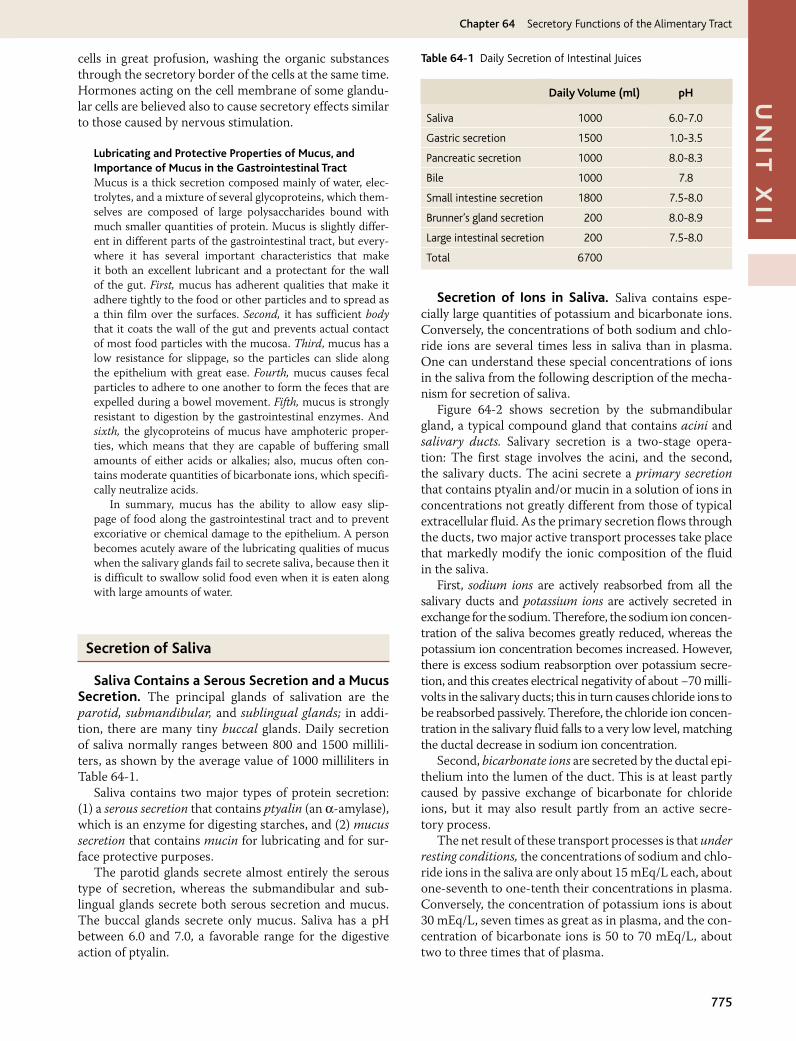
Saliva Contains a Serous Secretion and a Mucus Secretion. The principal glands of salivation are the parotid, submandibular, and sublingual glands; in addi-tion, there are many tiny buccal glands. Daily secretion of saliva normally ranges between 800 and 1500 millili-ters, as shown by the average value of 1000 milliliters in Table 64-1.
Saliva contains two major types of protein secretion: (1) a serous secretion that contains ptyalin (an α-amylase), which is an enzyme for digesting starches, and (2) mucus secretion that contains mucin for lubricating and for sur-face protective purposes.
The parotid glands secrete almost entirely the serous type of secretion, whereas the submandibular and sub-lingual glands secrete both serous secretion and mucus. The buccal glands secrete only mucus. Saliva has a pH between 6.0 and 7.0, a favorable range for the digestive action of ptyalin.
Secretion of Ions in Saliva. Saliva contains espe-cially large quantities of potassium and bicarbonate ions. Conversely, the concentrations of both sodium and chlo-ride ions are several times less in saliva than in plasma. One can understand these special concentrations of ions in the saliva from the following description of the mecha-nism for secretion of saliva.
Figure 64-2 shows secretion by the submandibular gland, a typical compound gland that contains acini and salivary ducts. Salivary secretion is a two-stage opera-tion: The first stage involves the acini, and the second, the salivary ducts. The acini secrete a primary secretion that contains ptyalin and/or mucin in a solution of ions in concentrations not greatly different from those of typical extracellular fluid. As the primary secretion flows through the ducts, two major active transport processes take place that markedly modify the ionic composition of the fluid in the saliva.
First, sodium ions are actively reabsorbed from all the salivary ducts and potassium ions are actively secreted in exchange for the sodium. Therefore, the sodium ion concen-tration of the saliva becomes greatly reduced, whereas the potassium ion concentration becomes increased. However, there is excess sodium reabsorption over potassium secre-tion, and this creates electrical negativity of about −70 milli-volts in the salivary ducts; this in turn causes chloride ions to be reabsorbed passively. Therefore, the chloride ion concen-tration in the salivary fluid falls to a very low level, matching the ductal decrease in sodium ion concentration.
Second, bicarbonate ions are secreted by the ductal epi-thelium into the lumen of the duct. This is at least partly caused by passive exchange of bicarbonate for chloride ions, but it may also result partly from an active secre-tory process.
The net result of these transport processes is that under resting conditions, the concentrations of sodium and chlo-ride ions in the saliva are only about 15 mEq/L each, about one-seventh to one-tenth their concentrations in plasma. Conversely, the concentration of potassium ions is about 30 mEq/L, seven times as great as in plasma, and the con-centration of bicarbonate ions is 50 to 70 mEq/L, about two to three times that of plasma.
Daily Volume (ml) pH
Saliva 1000 6.0-7.0
Gastric secretion 1500 1.0-3.5
Pancreatic secretion 1000 8.0-8.3
Bile 1000 7.8
Small intestine secretion 1800 7.5-8.0
Brunner’s gland secretion 200 8.0-8.9
Large intestinal secretion 200 7.5-8.0
Total 6700
Table 64-1 Daily Secretion of Intestinal Juices
Unit XII Gastrointestinal Physiology
776
Superior and inferiorsalivatory nuclei
Submandibular gland
Submandibularganglion
Sublingual glandChordatympani
Otic ganglion Taste andtactile stimuli
Tongue
Glossopharyngealnerve
Parotidgland
Tractussolitarius
Facialnerve
Figure 64-3 Parasympathetic nervous regulation of salivary secretion.
During maximal salivation, the salivary ionic con-centrations change considerably because the rate of for-mation of primary secretion by the acini can increase as much as 20-fold. This acinar secretion then flows through the ducts so rapidly that the ductal reconditioning of the secretion is considerably reduced. Therefore, when copi-ous quantities of saliva are being secreted, the sodium chloride concentration is about one-half or two-thirds that of plasma, and the potassium concentration rises to only four times that of plasma.
Function of Saliva for Oral Hygiene. Under basal awake conditions, about 0.5 milliliter of saliva, almost entirely of the mucous type, is secreted each minute; but during sleep, little secretion occurs. This secretion plays an exceedingly important role for maintaining healthy oral tissues. The mouth is loaded with pathogenic bacteria that can easily destroy tissues and cause dental caries. Saliva helps prevent the deteriorative processes in several ways.
First, the flow of saliva itself helps wash away pathogenic bacteria, as well as food particles that provide their metabolic support.
Second, saliva contains several factors that destroy bac-teria. One of these is thiocyanate ions and another is several proteolytic enzymes—most important, lysozyme—that (a) attack the bacteria, (b) aid the thiocyanate ions in entering the bacteria where these ions in turn become bactericidal, and (c) digest food particles, thus helping further to remove the bacterial metabolic support.
Third, saliva often contains significant amounts of protein antibodies that can destroy oral bacteria, including some that cause dental caries. In the absence of salivation, oral tissues often become ulcerated and otherwise infected, and caries of the teeth can become rampant.
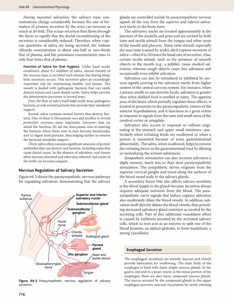
Nervous Regulation of Salivary Secretion
Figure 64-3 shows the parasympathetic nervous pathways for regulating salivation, demonstrating that the salivary
glands are controlled mainly by parasympathetic nervous signals all the way from the superior and inferior saliva-tory nuclei in the brain stem.
The salivatory nuclei are located approximately at the juncture of the medulla and pons and are excited by both taste and tactile stimuli from the tongue and other areas of the mouth and pharynx. Many taste stimuli, especially the sour taste (caused by acids), elicit copious secretion of saliva—often 8 to 20 times the basal rate of secretion. Also, certain tactile stimuli, such as the presence of smooth objects in the mouth (e.g., a pebble), cause marked sal-ivation, whereas rough objects cause less salivation and occasionally even inhibit salivation.
Salivation can also be stimulated or inhibited by ner-vous signals arriving in the salivatory nuclei from higher centers of the central nervous system. For instance, when a person smells or eats favorite foods, salivation is greater than when disliked food is smelled or eaten. The appetite area of the brain, which partially regulates these effects, is located in proximity to the parasympathetic centers of the anterior hypothalamus, and it functions to a great extent in response to signals from the taste and smell areas of the cerebral cortex or amygdala.
Salivation also occurs in response to reflexes origi-nating in the stomach and upper small intestines—par-ticularly when irritating foods are swallowed or when a person is nauseated because of some gastrointestinal abnormality. The saliva, when swallowed, helps to remove the irritating factor in the gastrointestinal tract by diluting or neutralizing the irritant substances.
Sympathetic stimulation can also increase salivation a slight amount, much less so than does parasympathetic stimulation. The sympathetic nerves originate from the superior cervical ganglia and travel along the surfaces of the blood vessel walls to the salivary glands.
A secondary factor that also affects salivary secretion is the blood supply to the glands because secretion always requires adequate nutrients from the blood. The para-sympathetic nerve signals that induce copious salivation also moderately dilate the blood vessels. In addition, sali-vation itself directly dilates the blood vessels, thus provid-ing increased salivatory gland nutrition as needed by the secreting cells. Part of this additional vasodilator effect is caused by kallikrein secreted by the activated salivary cells, which in turn acts as an enzyme to split one of the blood proteins, an alpha2-globulin, to form bradykinin, a strong vasodilator.
Esophageal Secretion
The esophageal secretions are entirely mucous and mainly provide lubrication for swallowing. The main body of the esophagus is lined with many simple mucous glands. At the gastric end and to a lesser extent in the initial portion of the esophagus, there are also many compound mucous glands. The mucus secreted by the compound glands in the upper esophagus prevents mucosal excoriation by newly entering
Chapter 64 Secretory Functions of the Alimentary Tract
777
Un
it X
iifood, whereas the compound glands located near the esophagogastric junction protect the esophageal wall from digestion by acidic gastric juices that often reflux from the stomach back into the lower esophagus. Despite this protec-tion, a peptic ulcer at times can still occur at the gastric end of the esophagus.
Gastric Secretion
Characteristics of the Gastric Secretions
In addition to mucus-secreting cells that line the entire surface of the stomach, the stomach mucosa has two important types of tubular glands: oxyntic glands (also called gastric glands) and pyloric glands. The oxyntic (acid-forming) glands secrete hydrochloric acid, pepsino-gen, intrinsic factor, and mucus. The pyloric glands secrete mainly mucus for protection of the pyloric mucosa from the stomach acid. They also secrete the hormone gastrin.
The oxyntic glands are located on the inside surfaces of the body and fundus of the stomach, constituting the proximal 80 percent of the stomach. The pyloric glands are located in the antral portion of the stomach, the distal 20 percent of the stomach.
Secretions from the Oxyntic (Gastric) Glands
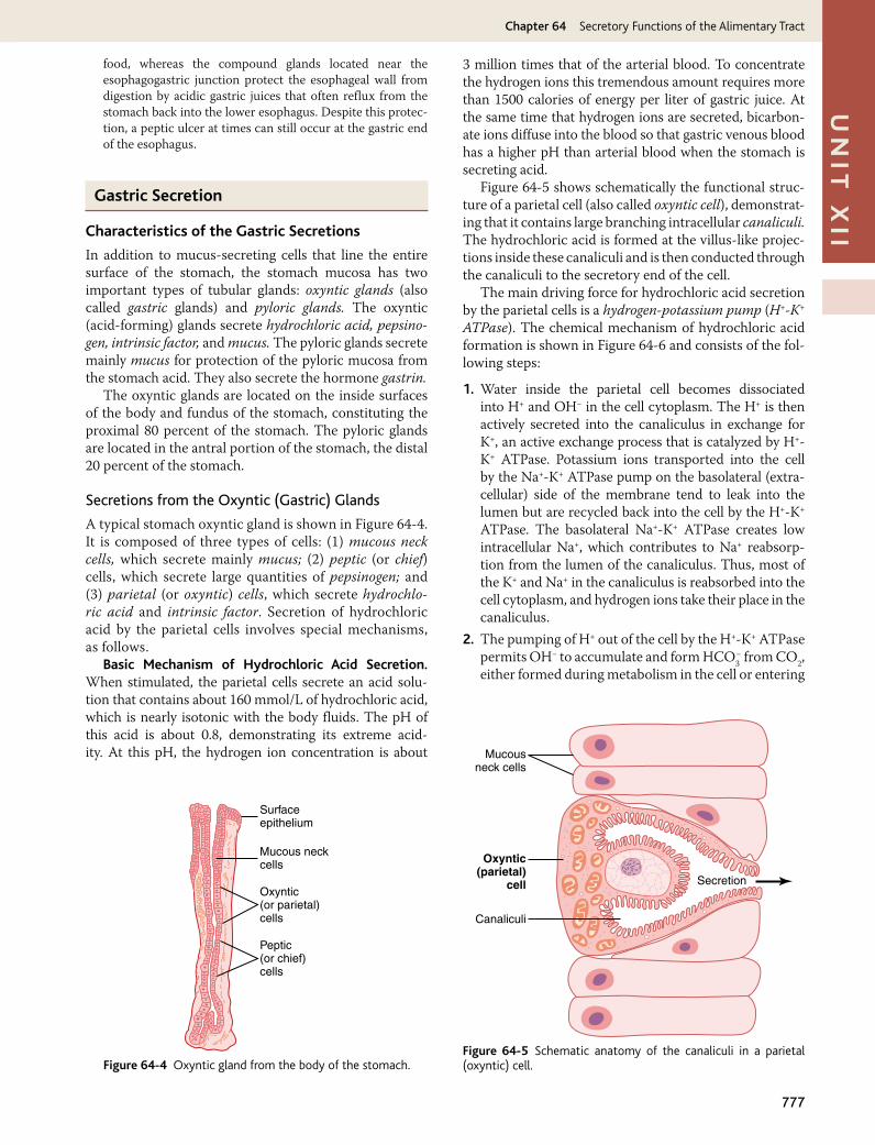
A typical stomach oxyntic gland is shown in Figure 64-4. It is composed of three types of cells: (1) mucous neck cells, which secrete mainly mucus; (2) peptic (or chief) cells, which secrete large quantities of pepsinogen; and (3) parietal (or oxyntic) cells, which secrete hydrochlo-ric acid and intrinsic factor. Secretion of hydrochloric acid by the parietal cells involves special mechanisms, as follows.
Basic Mechanism of Hydrochloric Acid Secretion. When stimulated, the parietal cells secrete an acid solu-tion that contains about 160 mmol/L of hydrochloric acid, which is nearly isotonic with the body fluids. The pH of this acid is about 0.8, demonstrating its extreme acid-ity. At this pH, the hydrogen ion concentration is about
3 million times that of the arterial blood. To concentrate the hydrogen ions this tremendous amount requires more than 1500 calories of energy per liter of gastric juice. At the same time that hydrogen ions are secreted, bicarbon-ate ions diffuse into the blood so that gastric venous blood has a higher pH than arterial blood when the stomach is secreting acid.
Figure 64-5 shows schematically the functional struc-ture of a parietal cell (also called oxyntic cell), demonstrat-ing that it contains large branching intracellular canaliculi. The hydrochloric acid is formed at the villus-like projec-tions inside these canaliculi and is then conducted through the canaliculi to the secretory end of the cell.
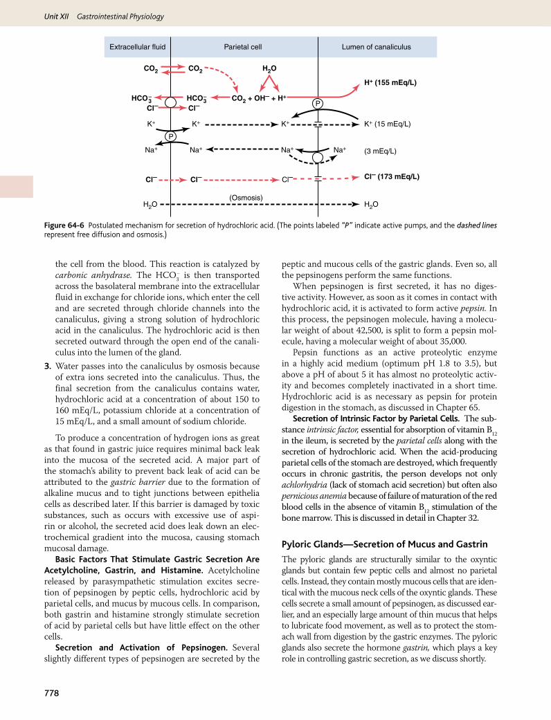
The main driving force for hydrochloric acid secretion by the parietal cells is a hydrogen-potassium pump (H+-K+ ATPase). The chemical mechanism of hydrochloric acid formation is shown in Figure 64-6 and consists of the fol-lowing steps:
1. Water inside the parietal cell becomes dissociated into H+ and OH− in the cell cytoplasm. The H+ is then actively secreted into the canaliculus in exchange for K+, an active exchange process that is catalyzed by H+-K+ ATPase. Potassium ions transported into the cell by the Na+-K+ ATPase pump on the basolateral (extra-cellular) side of the membrane tend to leak into the lumen but are recycled back into the cell by the H+-K+ ATPase. The basolateral Na+-K+ ATPase creates low intracellular Na+, which contributes to Na+ reabsorp-tion from the lumen of the canaliculus. Thus, most of the K+ and Na+ in the canaliculus is reabsorbed into the cell cytoplasm, and hydrogen ions take their place in the canaliculus.
2. The pumping of H+ out of the cell by the H+-K+ ATPase permits OH− to accumulate and form HCO3
− from CO2, either formed during metabolism in the cell or entering
Surfaceepithelium
Mucous neckcells
Oxyntic(or parietal)cells
Peptic(or chief)cells
Figure 64-4 Oxyntic gland from the body of the stomach.
Mucousneck cells
Oxyntic(parietal)
cell
Canaliculi
Secretion
Figure 64-5 Schematic anatomy of the canaliculi in a parietal (oxyntic) cell.
Unit XII Gastrointestinal Physiology
778
Extracellular fluid
CO2 CO2 H2O
H2OH2O(Osmosis)
P
P
HCO3-HCO3
- CO2 + OH- + H+
K+K+K+
Na+ Na+ Na+Na+
Cl- Cl-
Cl- Cl-
(3 mEq/L)
Cl-
H+ (155 mEq/L)
K+ (15 mEq/L)
Cl- (173 mEq/L)
Parietal cell Lumen of canaliculus
Figure 64-6 Postulated mechanism for secretion of hydrochloric acid. (The points labeled “P” indicate active pumps, and the dashed lines represent free diffusion and osmosis.)
the cell from the blood. This reaction is catalyzed by carbonic anhydrase. The HCO3
− is then transported across the basolateral membrane into the extracellular fluid in exchange for chloride ions, which enter the cell and are secreted through chloride channels into the canaliculus, giving a strong solution of hydrochloric acid in the canaliculus. The hydrochloric acid is then secreted outward through the open end of the canali-culus into the lumen of the gland.
3. Water passes into the canaliculus by osmosis because of extra ions secreted into the canaliculus. Thus, the final secretion from the canaliculus contains water, hydrochloric acid at a concentration of about 150 to 160 mEq/L, potassium chloride at a concentration of 15 mEq/L, and a small amount of sodium chloride.
To produce a concentration of hydrogen ions as great as that found in gastric juice requires minimal back leak into the mucosa of the secreted acid. A major part of the stomach’s ability to prevent back leak of acid can be attributed to the gastric barrier due to the formation of alkaline mucus and to tight junctions between epithelia cells as described later. If this barrier is damaged by toxic substances, such as occurs with excessive use of aspi-rin or alcohol, the secreted acid does leak down an elec-trochemical gradient into the mucosa, causing stomach mucosal damage.
Basic Factors That Stimulate Gastric Secretion Are Acetylcholine, Gastrin, and Histamine. Acetylcholine released by parasympathetic stimulation excites secre-tion of pepsinogen by peptic cells, hydrochloric acid by parietal cells, and mucus by mucous cells. In comparison, both gastrin and histamine strongly stimulate secretion of acid by parietal cells but have little effect on the other cells.
Secretion and Activation of Pepsinogen. Several slightly different types of pepsinogen are secreted by the
peptic and mucous cells of the gastric glands. Even so, all the pepsinogens perform the same functions.
When pepsinogen is first secreted, it has no diges-tive activity. However, as soon as it comes in contact with hydrochloric acid, it is activated to form active pepsin. In this process, the pepsinogen molecule, having a molecu-lar weight of about 42,500, is split to form a pepsin mol-ecule, having a molecular weight of about 35,000.
Pepsin functions as an active proteolytic enzyme in a highly acid medium (optimum pH 1.8 to 3.5), but above a pH of about 5 it has almost no proteolytic activ-ity and becomes completely inactivated in a short time. Hydrochloric acid is as necessary as pepsin for protein digestion in the stomach, as discussed in Chapter 65.
Secretion of Intrinsic Factor by Parietal Cells. The sub-stance intrinsic factor, essential for absorption of vitamin B12 in the ileum, is secreted by the parietal cells along with the secretion of hydrochloric acid. When the acid-producing parietal cells of the stomach are destroyed, which frequently occurs in chronic gastritis, the person develops not only achlorhydria (lack of stomach acid secretion) but often also pernicious anemia because of failure of maturation of the red blood cells in the absence of vitamin B12 stimulation of the bone marrow. This is discussed in detail in Chapter 32.
Pyloric Glands—Secretion of Mucus and Gastrin
The pyloric glands are structurally similar to the oxyntic glands but contain few peptic cells and almost no parietal cells. Instead, they contain mostly mucous cells that are iden-tical with the mucous neck cells of the oxyntic glands. These cells secrete a small amount of pepsinogen, as discussed ear-lier, and an especially large amount of thin mucus that helps to lubricate food movement, as well as to protect the stom-ach wall from digestion by the gastric enzymes. The pyloric glands also secrete the hormone gastrin, which plays a key role in controlling gastric secretion, as we discuss shortly.
Chapter 64 Secretory Functions of the Alimentary Tract
779
Un
it X
iiSurface Mucous Cells
The entire surface of the stomach mucosa between glands has a continuous layer of a special type of mucous cells called simply “surface mucous cells.” They secrete large quantities of viscid mucus that coats the stomach mucosa with a gel layer of mucus often more than 1 millimeter thick, thus providing a major shell of protection for the stomach wall, as well as contributing to lubrication of food transport.
Another characteristic of this mucus is that it is alka-line. Therefore, the normal underlying stomach wall is not directly exposed to the highly acidic, proteolytic stomach secretion. Even the slightest contact with food or any irritation of the mucosa directly stimulates the surface mucous cells to secrete additional quantities of this thick, alkaline, viscid mucus.
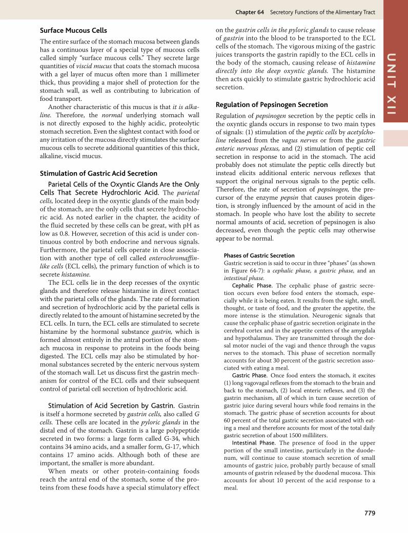
Stimulation of Gastric Acid Secretion
Parietal Cells of the Oxyntic Glands Are the Only Cells That Secrete Hydrochloric Acid. The parietal cells, located deep in the oxyntic glands of the main body of the stomach, are the only cells that secrete hydrochlo-ric acid. As noted earlier in the chapter, the acidity of the fluid secreted by these cells can be great, with pH as low as 0.8. However, secretion of this acid is under con-tinuous control by both endocrine and nervous signals. Furthermore, the parietal cells operate in close associa-tion with another type of cell called enterochromaffin-like cells (ECL cells), the primary function of which is to secrete histamine.
The ECL cells lie in the deep recesses of the oxyntic glands and therefore release histamine in direct contact with the parietal cells of the glands. The rate of formation and secretion of hydrochloric acid by the parietal cells is directly related to the amount of histamine secreted by the ECL cells. In turn, the ECL cells are stimulated to secrete histamine by the hormonal substance gastrin, which is formed almost entirely in the antral portion of the stom-ach mucosa in response to proteins in the foods being digested. The ECL cells may also be stimulated by hor-monal substances secreted by the enteric nervous system of the stomach wall. Let us discuss first the gastrin mech-anism for control of the ECL cells and their subsequent control of parietal cell secretion of hydrochloric acid.
Stimulation of Acid Secretion by Gastrin. Gastrin is itself a hormone secreted by gastrin cells, also called G cells. These cells are located in the pyloric glands in the distal end of the stomach. Gastrin is a large polypeptide secreted in two forms: a large form called G-34, which contains 34 amino acids, and a smaller form, G-17, which contains 17 amino acids. Although both of these are important, the smaller is more abundant.
When meats or other protein-containing foods reach the antral end of the stomach, some of the pro-teins from these foods have a special stimulatory effect
on the gastrin cells in the pyloric glands to cause release of gastrin into the blood to be transported to the ECL cells of the stomach. The vigorous mixing of the gastric juices transports the gastrin rapidly to the ECL cells in the body of the stomach, causing release of histamine directly into the deep oxyntic glands. The histamine then acts quickly to stimulate gastric hydrochloric acid secretion.
Regulation of Pepsinogen Secretion
Regulation of pepsinogen secretion by the peptic cells in the oxyntic glands occurs in response to two main types of signals: (1) stimulation of the peptic cells by acetylcho-line released from the vagus nerves or from the gastric enteric nervous plexus, and (2) stimulation of peptic cell secretion in response to acid in the stomach. The acid probably does not stimulate the peptic cells directly but instead elicits additional enteric nervous reflexes that support the original nervous signals to the peptic cells. Therefore, the rate of secretion of pepsinogen, the pre-cursor of the enzyme pepsin that causes protein diges-tion, is strongly influenced by the amount of acid in the stomach. In people who have lost the ability to secrete normal amounts of acid, secretion of pepsinogen is also decreased, even though the peptic cells may otherwise appear to be normal.
Phases of Gastric SecretionGastric secretion is said to occur in three “phases” (as shown in Figure 64-7): a cephalic phase, a gastric phase, and an intestinal phase.
Cephalic Phase. The cephalic phase of gastric secre-tion occurs even before food enters the stomach, espe-cially while it is being eaten. It results from the sight, smell, thought, or taste of food, and the greater the appetite, the more intense is the stimulation. Neurogenic signals that cause the cephalic phase of gastric secretion originate in the cerebral cortex and in the appetite centers of the amygdala and hypothalamus. They are transmitted through the dor-sal motor nuclei of the vagi and thence through the vagus nerves to the stomach. This phase of secretion normally accounts for about 30 percent of the gastric secretion asso-ciated with eating a meal.
Gastric Phase. Once food enters the stomach, it excites (1) long vagovagal reflexes from the stomach to the brain and back to the stomach, (2) local enteric reflexes, and (3) the gastrin mechanism, all of which in turn cause secretion of gastric juice during several hours while food remains in the stomach. The gastric phase of secretion accounts for about 60 percent of the total gastric secretion associated with eat-ing a meal and therefore accounts for most of the total daily gastric secretion of about 1500 milliliters.
Intestinal Phase. The presence of food in the upper portion of the small intestine, particularly in the duode-num, will continue to cause stomach secretion of small amounts of gastric juice, probably partly because of small amounts of gastrin released by the duodenal mucosa. This accounts for about 10 percent of the acid response to a meal.
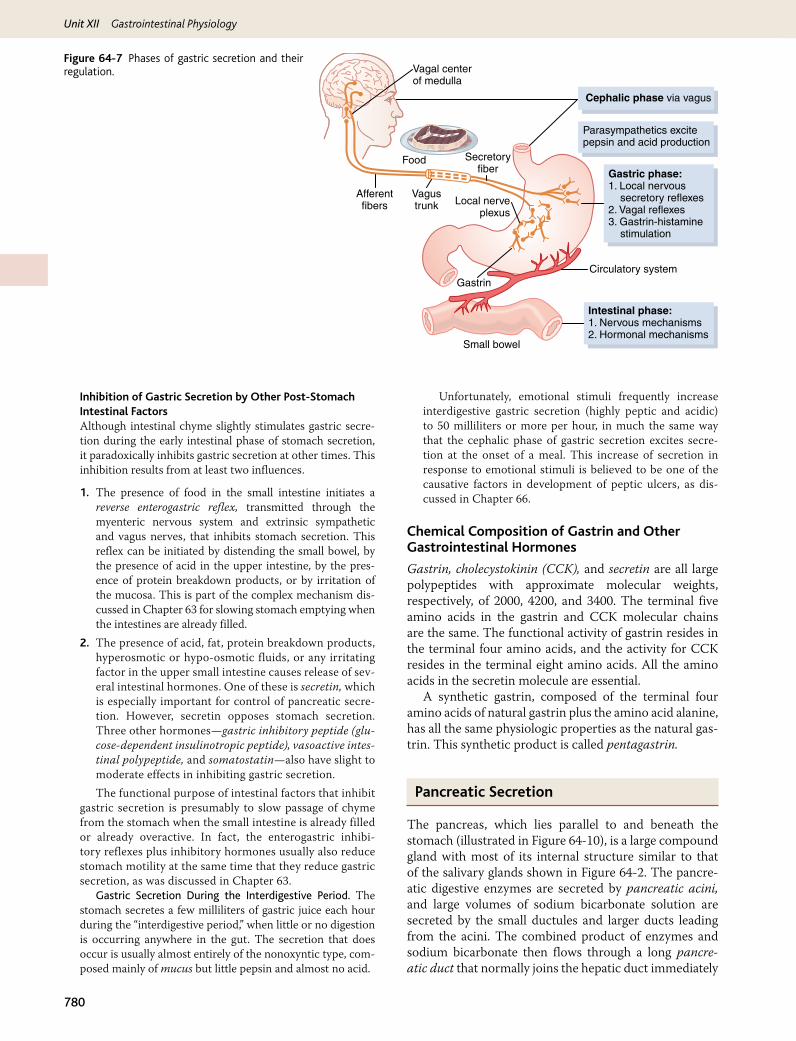
Unit XII Gastrointestinal Physiology
780
Inhibition of Gastric Secretion by Other Post-Stomach Intestinal FactorsAlthough intestinal chyme slightly stimulates gastric secre-tion during the early intestinal phase of stomach secretion, it paradoxically inhibits gastric secretion at other times. This inhibition results from at least two influences.
1. The presence of food in the small intestine initiates a reverse enterogastric reflex, transmitted through the myenteric nervous system and extrinsic sympathetic and vagus nerves, that inhibits stomach secretion. This reflex can be initiated by distending the small bowel, by the presence of acid in the upper intestine, by the pres-ence of protein breakdown products, or by irritation of the mucosa. This is part of the complex mechanism dis-cussed in Chapter 63 for slowing stomach emptying when the intestines are already filled.
2. The presence of acid, fat, protein breakdown products, hyperosmotic or hypo-osmotic fluids, or any irritating factor in the upper small intestine causes release of sev-eral intestinal hormones. One of these is secretin, which is especially important for control of pancreatic secre-tion. However, secretin opposes stomach secretion. Three other hormones—gastric inhibitory peptide (glu-cose-dependent insulinotropic peptide), vasoactive intes-tinal polypeptide, and somatostatin—also have slight to moderate effects in inhibiting gastric secretion.The functional purpose of intestinal factors that inhibit
gastric secretion is presumably to slow passage of chyme from the stomach when the small intestine is already filled or already overactive. In fact, the enterogastric inhibi-tory reflexes plus inhibitory hormones usually also reduce stomach motility at the same time that they reduce gastric secretion, as was discussed in Chapter 63.
Gastric Secretion During the Interdigestive Period. The stomach secretes a few milliliters of gastric juice each hour during the “interdigestive period,” when little or no digestion is occurring anywhere in the gut. The secretion that does occur is usually almost entirely of the nonoxyntic type, com-posed mainly of mucus but little pepsin and almost no acid.
Unfortunately, emotional stimuli frequently increase interdigestive gastric secretion (highly peptic and acidic) to 50 milliliters or more per hour, in much the same way that the cephalic phase of gastric secretion excites secre-tion at the onset of a meal. This increase of secretion in response to emotional stimuli is believed to be one of the causative factors in development of peptic ulcers, as dis-cussed in Chapter 66.
Chemical Composition of Gastrin and Other Gastrointestinal Hormones
Gastrin, cholecystokinin (CCK), and secretin are all large polypeptides with approximate molecular weights, respectively, of 2000, 4200, and 3400. The terminal five amino acids in the gastrin and CCK molecular chains are the same. The functional activity of gastrin resides in the terminal four amino acids, and the activity for CCK resides in the terminal eight amino acids. All the amino acids in the secretin molecule are essential.
A synthetic gastrin, composed of the terminal four amino acids of natural gastrin plus the amino acid alanine, has all the same physiologic properties as the natural gas-trin. This synthetic product is called pentagastrin.
Pancreatic Secretion
The pancreas, which lies parallel to and beneath the stomach (illustrated in Figure 64-10), is a large compound gland with most of its internal structure similar to that of the salivary glands shown in Figure 64-2. The pancre-atic digestive enzymes are secreted by pancreatic acini, and large volumes of sodium bicarbonate solution are secreted by the small ductules and larger ducts leading from the acini. The combined product of enzymes and sodium bicarbonate then flows through a long pancre-atic duct that normally joins the hepatic duct immediately
Vagal centerof medulla
Cephalic phase via vagus
Parasympathetics excitepepsin and acid production
Intestinal phase:1. Nervous mechanisms2. Hormonal mechanisms
Circulatory system
Afferentfibers
Vagustrunk
Secretoryfiber
Small bowel
Gastrin
Food
Local nerveplexus
Gastric phase:1. Local nervous secretory reflexes2. Vagal reflexes3. Gastrin-histamine stimulation
Figure 64-7 Phases of gastric secretion and their regulation.
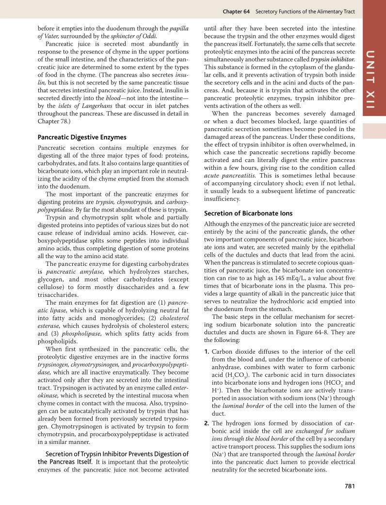
Chapter 64 Secretory Functions of the Alimentary Tract
781
Un
it X
iibefore it empties into the duodenum through the papilla of Vater, surrounded by the sphincter of Oddi.
Pancreatic juice is secreted most abundantly in response to the presence of chyme in the upper portions of the small intestine, and the characteristics of the pan-creatic juice are determined to some extent by the types of food in the chyme. (The pancreas also secretes insu-lin, but this is not secreted by the same pancreatic tissue that secretes intestinal pancreatic juice. Instead, insulin is secreted directly into the blood—not into the intestine—by the islets of Langerhans that occur in islet patches throughout the pancreas. These are discussed in detail in Chapter 78.)
Pancreatic Digestive Enzymes
Pancreatic secretion contains multiple enzymes for digesting all of the three major types of food: proteins, carbohydrates, and fats. It also contains large quantities of bicarbonate ions, which play an important role in neutral-izing the acidity of the chyme emptied from the stomach into the duodenum.
The most important of the pancreatic enzymes for digesting proteins are trypsin, chymotrypsin, and carboxy-polypeptidase. By far the most abundant of these is trypsin.
Trypsin and chymotrypsin split whole and partially digested proteins into peptides of various sizes but do not cause release of individual amino acids. However, car-boxypolypeptidase splits some peptides into individual amino acids, thus completing digestion of some proteins all the way to the amino acid state.
The pancreatic enzyme for digesting carbohydrates is pancreatic amylase, which hydrolyzes starches, glycogen, and most other carbohydrates (except cellulose) to form mostly disaccharides and a few trisaccharides.
The main enzymes for fat digestion are (1) pancre-atic lipase, which is capable of hydrolyzing neutral fat into fatty acids and monoglycerides; (2) cholesterol esterase, which causes hydrolysis of cholesterol esters; and (3) phospholipase, which splits fatty acids from phospholipids.
When first synthesized in the pancreatic cells, the proteolytic digestive enzymes are in the inactive forms trypsinogen, chymotrypsinogen, and procarboxypolypepti-dase, which are all inactive enzymatically. They become activated only after they are secreted into the intestinal tract. Trypsinogen is activated by an enzyme called enter-okinase, which is secreted by the intestinal mucosa when chyme comes in contact with the mucosa. Also, trypsino-gen can be autocatalytically activated by trypsin that has already been formed from previously secreted trypsino-gen. Chymotrypsinogen is activated by trypsin to form chymotrypsin, and procarboxypolypeptidase is activated in a similar manner.
Secretion of Trypsin Inhibitor Prevents Digestion of the Pancreas Itself. It is important that the proteolytic enzymes of the pancreatic juice not become activated
until after they have been secreted into the intestine because the trypsin and the other enzymes would digest the pancreas itself. Fortunately, the same cells that secrete proteolytic enzymes into the acini of the pancreas secrete simultaneously another substance called trypsin inhibitor. This substance is formed in the cytoplasm of the glandu-lar cells, and it prevents activation of trypsin both inside the secretory cells and in the acini and ducts of the pan-creas. And, because it is trypsin that activates the other pancreatic proteolytic enzymes, trypsin inhibitor pre-vents activation of the others as well.
When the pancreas becomes severely damaged or when a duct becomes blocked, large quantities of pancreatic secretion sometimes become pooled in the damaged areas of the pancreas. Under these conditions, the effect of trypsin inhibitor is often overwhelmed, in which case the pancreatic secretions rapidly become activated and can literally digest the entire pancreas within a few hours, giving rise to the condition called acute pancreatitis. This is sometimes lethal because of accompanying circulatory shock; even if not lethal, it usually leads to a subsequent lifetime of pancreatic insufficiency.
Secretion of Bicarbonate Ions
Although the enzymes of the pancreatic juice are secreted entirely by the acini of the pancreatic glands, the other two important components of pancreatic juice, bicarbon-ate ions and water, are secreted mainly by the epithelial cells of the ductules and ducts that lead from the acini. When the pancreas is stimulated to secrete copious quan-tities of pancreatic juice, the bicarbonate ion concentra-tion can rise to as high as 145 mEq/L, a value about five times that of bicarbonate ions in the plasma. This pro-vides a large quantity of alkali in the pancreatic juice that serves to neutralize the hydrochloric acid emptied into the duodenum from the stomach.
The basic steps in the cellular mechanism for secret-ing sodium bicarbonate solution into the pancreatic ductules and ducts are shown in Figure 64-8. They are the following:
1. Carbon dioxide diffuses to the interior of the cell from the blood and, under the influence of carbonic anhydrase, combines with water to form carbonic acid (H2CO3). The carbonic acid in turn dissociates into bicarbonate ions and hydrogen ions (HCO3
− and H+). Then the bicarbonate ions are actively trans-ported in association with sodium ions (Na+) through the luminal border of the cell into the lumen of the duct.
2. The hydrogen ions formed by dissociation of car-bonic acid inside the cell are exchanged for sodium ions through the blood border of the cell by a secondary active transport process. This supplies the sodium ions (Na+) that are transported through the luminal border into the pancreatic duct lumen to provide electrical neutrality for the secreted bicarbonate ions.
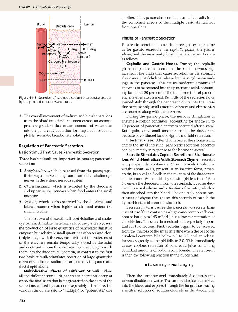
Unit XII Gastrointestinal Physiology
782
3. The overall movement of sodium and bicarbonate ions from the blood into the duct lumen creates an osmotic pressure gradient that causes osmosis of water also into the pancreatic duct, thus forming an almost com-pletely isosmotic bicarbonate solution.
Regulation of Pancreatic Secretion
Basic Stimuli That Cause Pancreatic Secretion
Three basic stimuli are important in causing pancreatic secretion:
1. Acetylcholine, which is released from the parasympa-thetic vagus nerve endings and from other cholinergic nerves in the enteric nervous system
2. Cholecystokinin, which is secreted by the duodenal and upper jejunal mucosa when food enters the small intestine
3. Secretin, which is also secreted by the duodenal and jejunal mucosa when highly acidic food enters the small intestine
The first two of these stimuli, acetylcholine and chole-cystokinin, stimulate the acinar cells of the pancreas, caus-ing production of large quantities of pancreatic digestive enzymes but relatively small quantities of water and elec-trolytes to go with the enzymes. Without the water, most of the enzymes remain temporarily stored in the acini and ducts until more fluid secretion comes along to wash them into the duodenum. Secretin, in contrast to the first two basic stimuli, stimulates secretion of large quantities of water solution of sodium bicarbonate by the pancreatic ductal epithelium.
Multiplicative Effects of Different Stimuli. When all the different stimuli of pancreatic secretion occur at once, the total secretion is far greater than the sum of the secretions caused by each one separately. Therefore, the various stimuli are said to “multiply,” or “potentiate,” one
another. Thus, pancreatic secretion normally results from the combined effects of the multiple basic stimuli, not from one alone.
Phases of Pancreatic Secretion
Pancreatic secretion occurs in three phases, the same as for gastric secretion: the cephalic phase, the gastric phase, and the intestinal phase. Their characteristics are as follows.
Cephalic and Gastric Phases. During the cephalic phase of pancreatic secretion, the same nervous sig-nals from the brain that cause secretion in the stomach also cause acetylcholine release by the vagal nerve end-ings in the pancreas. This causes moderate amounts of enzymes to be secreted into the pancreatic acini, account-ing for about 20 percent of the total secretion of pancre-atic enzymes after a meal. But little of the secretion flows immediately through the pancreatic ducts into the intes-tine because only small amounts of water and electrolytes are secreted along with the enzymes.
During the gastric phase, the nervous stimulation of enzyme secretion continues, accounting for another 5 to 10 percent of pancreatic enzymes secreted after a meal. But, again, only small amounts reach the duodenum because of continued lack of significant fluid secretion.
Intestinal Phase. After chyme leaves the stomach and enters the small intestine, pancreatic secretion becomes copious, mainly in response to the hormone secretin.
Secretin Stimulates Copious Secretion of Bicarbonate Ions, Which Neutralizes Acidic Stomach Chyme. Secretin is a polypeptide, containing 27 amino acids (molecular weight about 3400), present in an inactive form, prose-cretin, in so-called S cells in the mucosa of the duodenum and jejunum. When acid chyme with pH less than 4.5 to 5.0 enters the duodenum from the stomach, it causes duo-denal mucosal release and activation of secretin, which is then absorbed into the blood. The one truly potent con-stituent of chyme that causes this secretin release is the hydrochloric acid from the stomach.
Secretin in turn causes the pancreas to secrete large quantities of fluid containing a high concentration of bicar-bonate ion (up to 145 mEq/L) but a low concentration of chloride ion. The secretin mechanism is especially impor-tant for two reasons: First, secretin begins to be released from the mucosa of the small intestine when the pH of the duodenal contents falls below 4.5 to 5.0, and its release increases greatly as the pH falls to 3.0. This immediately causes copious secretion of pancreatic juice containing abundant amounts of sodium bicarbonate. The net result is then the following reaction in the duodenum:
HCl + NaHCO3 NaCl + H2CO3Æ
Then the carbonic acid immediately dissociates into carbon dioxide and water. The carbon dioxide is absorbed into the blood and expired through the lungs, thus leaving a neutral solution of sodium chloride in the duodenum.
Ductule cells
(Activetransport) (Active
transport)
(Carbonic anhydrase)
Blood Lumen
Na+Na+
H+Na+
H+HCO3
−HCO3−
H2CO3
H2O+
CO2 CO2
H2O H2O
Figure 64-8 Secretion of isosmotic sodium bicarbonate solution by the pancreatic ductules and ducts.
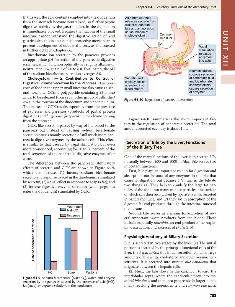
Chapter 64 Secretory Functions of the Alimentary Tract
783
Un
it X
iiIn this way, the acid contents emptied into the duodenum from the stomach become neutralized, so further peptic digestive activity by the gastric juices in the duodenum is immediately blocked. Because the mucosa of the small intestine cannot withstand the digestive action of acid gastric juice, this is an essential protective mechanism to prevent development of duodenal ulcers, as is discussed in further detail in Chapter 66.
Bicarbonate ion secretion by the pancreas provides an appropriate pH for action of the pancreatic digestive enzymes, which function optimally in a slightly alkaline or neutral medium, at a pH of 7.0 to 8.0. Fortunately, the pH of the sodium bicarbonate secretion averages 8.0.
Cholecystokinin—Its Contribution to Control of Digestive Enzyme Secretion by the Pancreas. The pres-ence of food in the upper small intestine also causes a sec-ond hormone, CCK, a polypeptide containing 33 amino acids, to be released from yet another group of cells, the I cells, in the mucosa of the duodenum and upper jejunum. This release of CCK results especially from the presence of proteoses and peptones (products of partial protein digestion) and long-chain fatty acids in the chyme coming from the stomach.
CCK, like secretin, passes by way of the blood to the pancreas but instead of causing sodium bicarbonate secretion causes mainly secretion of still much more pan-creatic digestive enzymes by the acinar cells. This effect is similar to that caused by vagal stimulation but even more pronounced, accounting for 70 to 80 percent of the total secretion of the pancreatic digestive enzymes after a meal.
The differences between the pancreatic stimulatory effects of secretin and CCK are shown in Figure 64-9, which demonstrates (1) intense sodium bicarbonate secretion in response to acid in the duodenum, stimulated by secretin; (2) a dual effect in response to soap (a fat); and (3) intense digestive enzyme secretion (when peptones enter the duodenum) stimulated by CCK.
Figure 64-10 summarizes the more important fac-tors in the regulation of pancreatic secretion. The total amount secreted each day is about 1 liter.
Secretion of Bile by the Liver; Functions of the Biliary Tree
One of the many functions of the liver is to secrete bile, normally between 600 and 1000 ml/day. Bile serves two important functions.
First, bile plays an important role in fat digestion and absorption, not because of any enzymes in the bile that cause fat digestion, but because bile acids in the bile do two things: (1) They help to emulsify the large fat par-ticles of the food into many minute particles, the surface of which can then be attacked by lipase enzymes secreted in pancreatic juice, and (2) they aid in absorption of the digested fat end products through the intestinal mucosal membrane.
Second, bile serves as a means for excretion of sev-eral important waste products from the blood. These include especially bilirubin, an end product of hemoglo-bin destruction, and excesses of cholesterol.
Physiologic Anatomy of Biliary Secretion
Bile is secreted in two stages by the liver: (1) The initial portion is secreted by the principal functional cells of the liver, the hepatocytes; this initial secretion contains large amounts of bile acids, cholesterol, and other organic con-stituents. It is secreted into minute bile canaliculi that originate between the hepatic cells.
(2) Next, the bile flows in the canaliculi toward the interlobular septa, where the canaliculi empty into ter-minal bile ducts and then into progressively larger ducts, finally reaching the hepatic duct and common bile duct.
Vagalstimulationreleasesenzymesinto acini
Secretin causescopious secretionof pancreatic fluidand bicarbonate;cholecystokinincauses secretionof enzymes
Acid from stomachreleases secretin fromwall of duodenum;fats and amino acidscause release ofcholecystokinin Common
bile duct
Secretin andcholecystokininabsorbed intoblood stream
Figure 64-10 Regulation of pancreatic secretion.
Water andNaHCO3
Enzymes
HCI Soap Peptone
Rat
e o
f p
ancr
eati
c se
cret
ion
Figure 64-9 Sodium bicarbonate (NaHCO3), water, and enzyme
secretion by the pancreas, caused by the presence of acid (HCl), fat (soap), or peptone solutions in the duodenum.
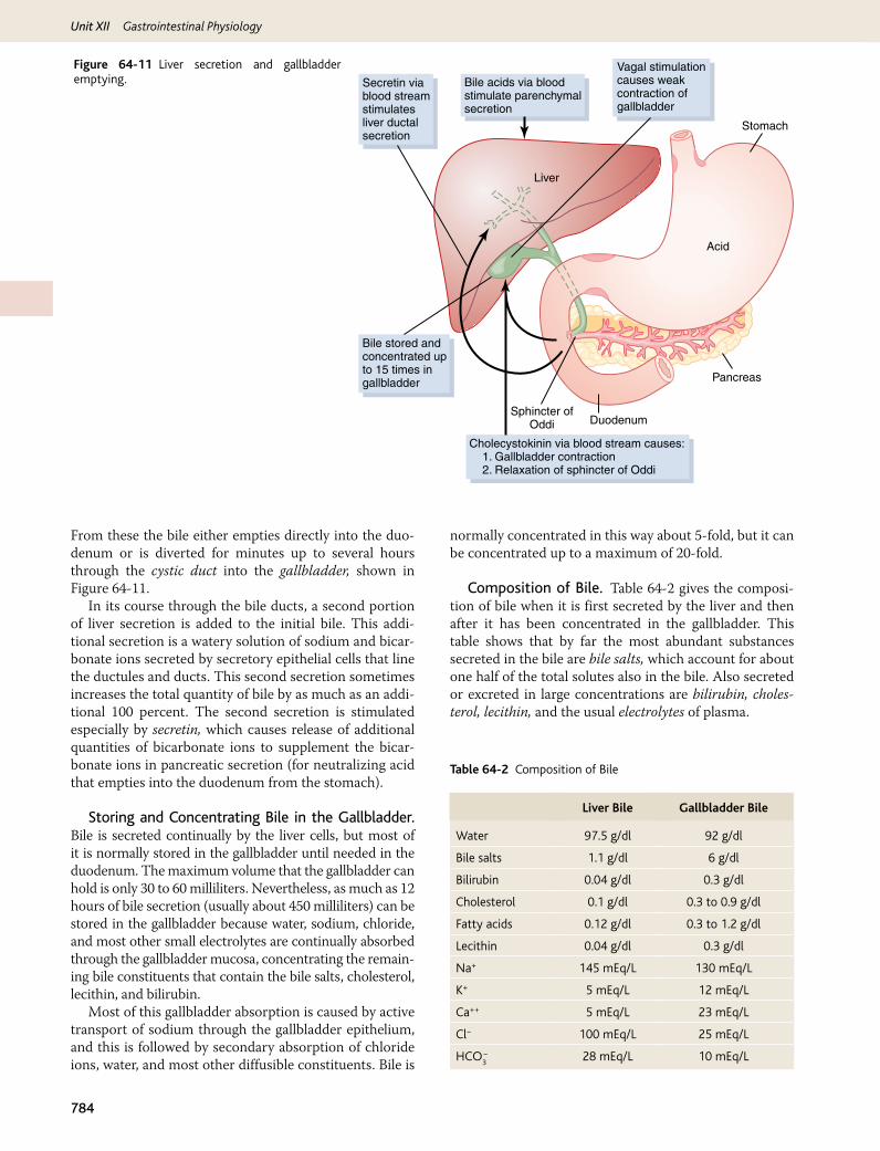
Unit XII Gastrointestinal Physiology
784
From these the bile either empties directly into the duo-denum or is diverted for minutes up to several hours through the cystic duct into the gallbladder, shown in Figure 64-11.
In its course through the bile ducts, a second portion of liver secretion is added to the initial bile. This addi-tional secretion is a watery solution of sodium and bicar-bonate ions secreted by secretory epithelial cells that line the ductules and ducts. This second secretion sometimes increases the total quantity of bile by as much as an addi-tional 100 percent. The second secretion is stimulated especially by secretin, which causes release of additional quantities of bicarbonate ions to supplement the bicar-bonate ions in pancreatic secretion (for neutralizing acid that empties into the duodenum from the stomach).
Storing and Concentrating Bile in the Gallbladder. Bile is secreted continually by the liver cells, but most of it is normally stored in the gallbladder until needed in the duodenum. The maximum volume that the gallbladder can hold is only 30 to 60 milliliters. Nevertheless, as much as 12 hours of bile secretion (usually about 450 milliliters) can be stored in the gallbladder because water, sodium, chloride, and most other small electrolytes are continually absorbed through the gallbladder mucosa, concentrating the remain-ing bile constituents that contain the bile salts, cholesterol, lecithin, and bilirubin.
Most of this gallbladder absorption is caused by active transport of sodium through the gallbladder epithelium, and this is followed by secondary absorption of chloride ions, water, and most other diffusible constituents. Bile is
normally concentrated in this way about 5-fold, but it can be concentrated up to a maximum of 20-fold.
Composition of Bile. Table 64-2 gives the composi-tion of bile when it is first secreted by the liver and then after it has been concentrated in the gallbladder. This table shows that by far the most abundant substances secreted in the bile are bile salts, which account for about one half of the total solutes also in the bile. Also secreted or excreted in large concentrations are bilirubin, choles-terol, lecithin, and the usual electrolytes of plasma.
Stomach
Acid
Liver
Bile acids via bloodstimulate parenchymalsecretion
Secretin viablood streamstimulatesliver ductalsecretion
Bile stored andconcentrated upto 15 times ingallbladder
Cholecystokinin via blood stream causes: 1. Gallbladder contraction 2. Relaxation of sphincter of Oddi
Vagal stimulationcauses weakcontraction ofgallbladder
Pancreas
Sphincter ofOddi Duodenum
Figure 64-11 Liver secretion and gallbladder emptying.
Liver Bile Gallbladder Bile
Water 97.5 g/dl 92 g/dl
Bile salts 1.1 g/dl 6 g/dl
Bilirubin 0.04 g/dl 0.3 g/dl
Cholesterol 0.1 g/dl 0.3 to 0.9 g/dl
Fatty acids 0.12 g/dl 0.3 to 1.2 g/dl
Lecithin 0.04 g/dl 0.3 g/dl
Na+ 145 mEq/L 130 mEq/L
K+ 5 mEq/L 12 mEq/L
Ca++ 5 mEq/L 23 mEq/L
Cl− 100 mEq/L 25 mEq/L
HCO3− 28 mEq/L 10 mEq/L
Table 64-2 Composition of Bile

Chapter 64 Secretory Functions of the Alimentary Tract
785
Un
it X
iiIn the concentrating process in the gallbladder, water
and large portions of the electrolytes (except calcium ions) are reabsorbed by the gallbladder mucosa; essen-tially all other constituents, especially the bile salts and the lipid substances cholesterol and lecithin, are not reab-sorbed and, therefore, become highly concentrated in the gallbladder bile.
Emptying of the Gallbladder—Stimulatory Role of Cholecystokinin. When food begins to be digested in the upper gastrointestinal tract, the gallbladder begins to empty, especially when fatty foods reach the duodenum about 30 minutes after a meal. The mechanism of gall-bladder emptying is rhythmical contractions of the wall of the gallbladder, but effective emptying also requires simul-taneous relaxation of the sphincter of Oddi, which guards the exit of the common bile duct into the duodenum.
By far the most potent stimulus for causing the gall-bladder contractions is the hormone CCK. This is the same CCK discussed earlier that causes increased secre-tion of digestive enzymes by the acinar cells of the pan-creas. The stimulus for CCK entry into the blood from the duodenal mucosa is mainly the presence of fatty foods in the duodenum.
The gallbladder is also stimulated less strongly by ace-tylcholine-secreting nerve fibers from both the vagi and the intestinal enteric nervous system. They are the same nerves that promote motility and secretion in other parts of the upper gastrointestinal tract.
In summary, the gallbladder empties its store of con-centrated bile into the duodenum mainly in response to the CCK stimulus that itself is initiated mainly by fatty foods. When fat is not in the food, the gallbladder emp-ties poorly, but when significant quantities of fat are pres-ent, the gallbladder normally empties completely in about 1 hour. Figure 64-11 summarizes the secretion of bile, its storage in the gallbladder, and its ultimate release from the bladder to the duodenum.
Function of Bile Salts in Fat Digestion and Absorption
The liver cells synthesize about 6 grams of bile salts daily. The precursor of the bile salts is cholesterol, which is either present in the diet or synthesized in the liver cells during the course of fat metabolism. The cholesterol is first con-verted to cholic acid or chenodeoxycholic acid in about equal quantities. These acids in turn combine principally with glycine and to a lesser extent with taurine to form glyco- and tauro-conjugated bile acids. The salts of these acids, mainly sodium salts, are then secreted in the bile.
The bile salts have two important actions in the intes-tinal tract:
First, they have a detergent action on the fat particles in the food. This decreases the surface tension of the par-ticles and allows agitation in the intestinal tract to break the fat globules into minute sizes. This is called the emul-sifying or detergent function of bile salts.
Second, and even more important than the emulsify-ing function, bile salts help in the absorption of (1) fatty acids, (2) monoglycerides, (3) cholesterol, and (4) other lipids from the intestinal tract. They do this by forming small physical complexes with these lipids; the complexes are called micelles, and they are semisoluble in the chyme because of the electrical charges of the bile salts. The intestinal lipids are “ferried” in this form to the intesti-nal mucosa, where they are then absorbed into the blood, as will be described in detail in Chapter 65. Without the presence of bile salts in the intestinal tract, up to 40 per-cent of the ingested fats are lost into the feces and the person often develops a metabolic deficit because of this nutrient loss.
Enterohepatic Circulation of Bile Salts. About 94 percent of the bile salts are reabsorbed into the blood from the small intestine, about one half of this by diffusion through the mucosa in the early portions of the small intestine and the remainder by an active transport process through the intes-tinal mucosa in the distal ileum. They then enter the por-tal blood and pass back to the liver. On reaching the liver, on first passage through the venous sinusoids these salts are absorbed almost entirely back into the hepatic cells and then resecreted into the bile.
In this way, about 94 percent of all the bile salts are recir-culated into the bile, so on the average these salts make the entire circuit some 17 times before being carried out in the feces. The small quantities of bile salts lost into the feces are replaced by new amounts formed continually by the liver cells. This recirculation of the bile salts is called the enterohe-patic circulation of bile salts.
The quantity of bile secreted by the liver each day is highly dependent on the availability of bile salts—the greater the quantity of bile salts in the enterohepatic circu-lation (usually a total of only about 2.5 grams), the greater the rate of bile secretion. Indeed, ingestion of supplemen-tal bile salts can increase bile secretion by several hundred milliliters per day.
If a bile fistula empties the bile salts to the exterior for sev-eral days to several weeks so that they cannot be reabsorbed from the ileum, the liver increases its production of bile salts 6- to 10-fold, which increases the rate of bile secretion most of the way back to normal. This demonstrates that the daily rate of liver bile salt secretion is actively controlled by the availability (or lack of availability) of bile salts in the entero-hepatic circulation.
Role of Secretin in Controlling Bile Secretion. In addi-tion to the strong stimulating effect of bile acids to cause bile secretion, the hormone secretin that also stimulates pancre-atic secretion increases bile secretion, sometimes more than doubling its secretion for several hours after a meal. This increase in secretion is almost entirely secretion of a sodium bicarbonate–rich watery solution by the epithelial cells of the bile ductules and ducts, and not increased secretion by the liver parenchymal cells themselves. The bicarbonate in turn passes into the small intestine and joins the bicarbonate from the pancreas in neutralizing the hydrochloric acid from the stomach. Thus, the secretin feedback mechanism for neu-tralizing duodenal acid operates not only through its effects on pancreatic secretion but also to a lesser extent through its effect on secretion by the liver ductules and ducts.
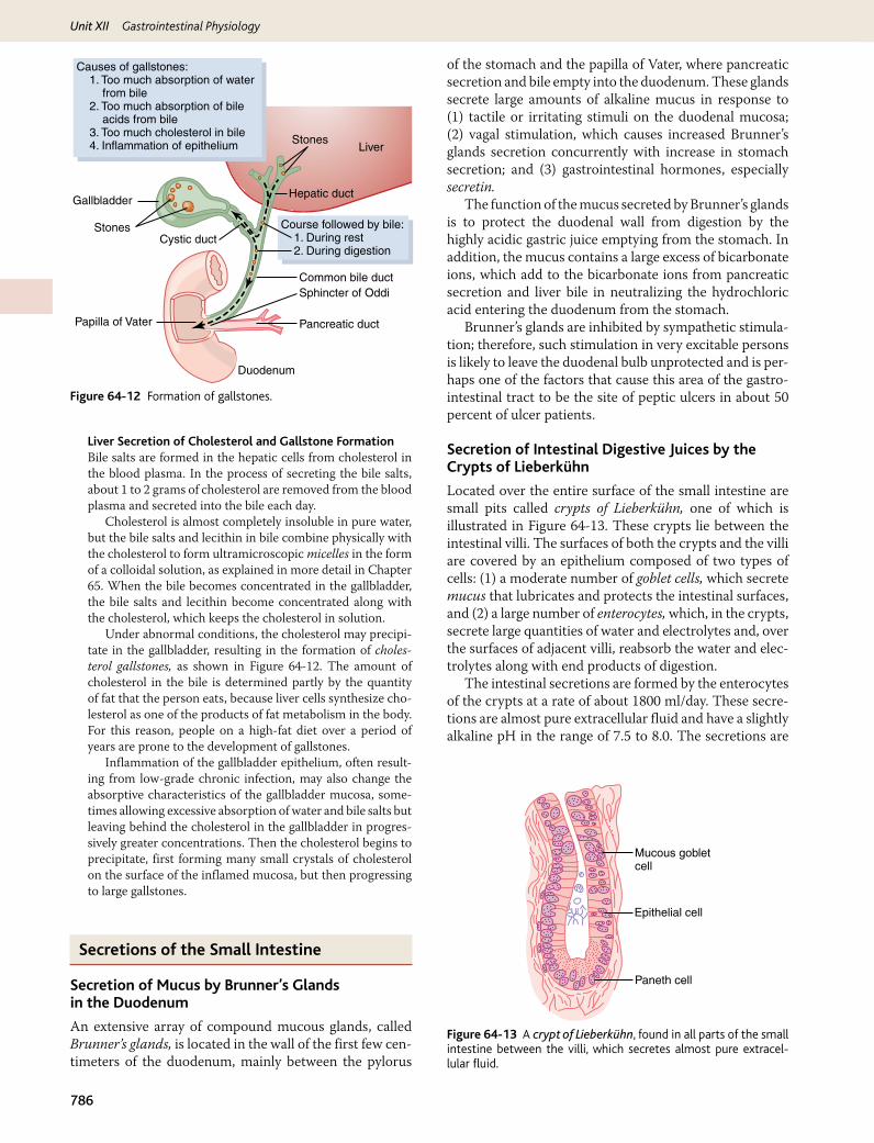
Unit XII Gastrointestinal Physiology
786
Liver Secretion of Cholesterol and Gallstone FormationBile salts are formed in the hepatic cells from cholesterol in the blood plasma. In the process of secreting the bile salts, about 1 to 2 grams of cholesterol are removed from the blood plasma and secreted into the bile each day.
Cholesterol is almost completely insoluble in pure water, but the bile salts and lecithin in bile combine physically with the cholesterol to form ultramicroscopic micelles in the form of a colloidal solution, as explained in more detail in Chapter 65. When the bile becomes concentrated in the gallbladder, the bile salts and lecithin become concentrated along with the cholesterol, which keeps the cholesterol in solution.
Under abnormal conditions, the cholesterol may precipi-tate in the gallbladder, resulting in the formation of choles-terol gallstones, as shown in Figure 64-12. The amount of cholesterol in the bile is determined partly by the quantity of fat that the person eats, because liver cells synthesize cho-lesterol as one of the products of fat metabolism in the body. For this reason, people on a high-fat diet over a period of years are prone to the development of gallstones.
Inflammation of the gallbladder epithelium, often result-ing from low-grade chronic infection, may also change the absorptive characteristics of the gallbladder mucosa, some-times allowing excessive absorption of water and bile salts but leaving behind the cholesterol in the gallbladder in progres-sively greater concentrations. Then the cholesterol begins to precipitate, first forming many small crystals of cholesterol on the surface of the inflamed mucosa, but then progressing to large gallstones.
Secretions of the Small Intestine
Secretion of Mucus by Brunner’s Glands in the Duodenum
An extensive array of compound mucous glands, called Brunner’s glands, is located in the wall of the first few cen-timeters of the duodenum, mainly between the pylorus
of the stomach and the papilla of Vater, where pancreatic secretion and bile empty into the duodenum. These glands secrete large amounts of alkaline mucus in response to (1) tactile or irritating stimuli on the duodenal mucosa; (2) vagal stimulation, which causes increased Brunner’s glands secretion concurrently with increase in stomach secretion; and (3) gastrointestinal hormones, especially secretin.
The function of the mucus secreted by Brunner’s glands is to protect the duodenal wall from digestion by the highly acidic gastric juice emptying from the stomach. In addition, the mucus contains a large excess of bicarbonate ions, which add to the bicarbonate ions from pancreatic secretion and liver bile in neutralizing the hydrochloric acid entering the duodenum from the stomach.
Brunner’s glands are inhibited by sympathetic stimula-tion; therefore, such stimulation in very excitable persons is likely to leave the duodenal bulb unprotected and is per-haps one of the factors that cause this area of the gastro-intestinal tract to be the site of peptic ulcers in about 50 percent of ulcer patients.
Secretion of Intestinal Digestive Juices by the Crypts of Lieberkühn
Located over the entire surface of the small intestine are small pits called crypts of Lieberkühn, one of which is illustrated in Figure 64-13. These crypts lie between the intestinal villi. The surfaces of both the crypts and the villi are covered by an epithelium composed of two types of cells: (1) a moderate number of goblet cells, which secrete mucus that lubricates and protects the intestinal surfaces, and (2) a large number of enterocytes, which, in the crypts, secrete large quantities of water and electrolytes and, over the surfaces of adjacent villi, reabsorb the water and elec-trolytes along with end products of digestion.
The intestinal secretions are formed by the enterocytes of the crypts at a rate of about 1800 ml/day. These secre-tions are almost pure extracellular fluid and have a slightly alkaline pH in the range of 7.5 to 8.0. The secretions are
Paneth cell
Epithelial cell
Mucous gobletcell
Figure 64-13 A crypt of Lieberkühn, found in all parts of the small intestine between the villi, which secretes almost pure extracel-lular fluid.
Course followed by bile: 1. During rest 2. During digestion
LiverStones
Hepatic duct
Common bile ductSphincter of Oddi
Pancreatic duct
Duodenum
Papilla of Vater
Cystic ductStones
Gallbladder
Causes of gallstones: 1. Too much absorption of water from bile 2. Too much absorption of bile acids from bile 3. Too much cholesterol in bile 4. Inflammation of epithelium
Figure 64-12 Formation of gallstones.
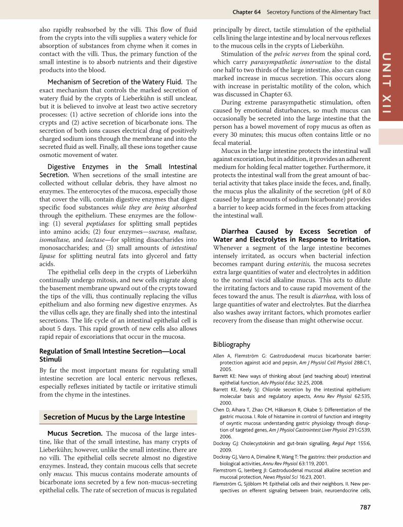
Chapter 64 Secretory Functions of the Alimentary Tract
787
Un
it X
iialso rapidly reabsorbed by the villi. This flow of fluid from the crypts into the villi supplies a watery vehicle for absorption of substances from chyme when it comes in contact with the villi. Thus, the primary function of the small intestine is to absorb nutrients and their digestive products into the blood.
Mechanism of Secretion of the Watery Fluid. The exact mechanism that controls the marked secretion of watery fluid by the crypts of Lieberkühn is still unclear, but it is believed to involve at least two active secretory processes: (1) active secretion of chloride ions into the crypts and (2) active secretion of bicarbonate ions. The secretion of both ions causes electrical drag of positively charged sodium ions through the membrane and into the secreted fluid as well. Finally, all these ions together cause osmotic movement of water.
Digestive Enzymes in the Small Intestinal Secretion. When secretions of the small intestine are collected without cellular debris, they have almost no enzymes. The enterocytes of the mucosa, especially those that cover the villi, contain digestive enzymes that digest specific food substances while they are being absorbed through the epithelium. These enzymes are the follow-ing: (1) several peptidases for splitting small peptides into amino acids; (2) four enzymes—sucrase, maltase, isomaltase, and lactase—for splitting disaccharides into monosaccharides; and (3) small amounts of intestinal lipase for splitting neutral fats into glycerol and fatty acids.
The epithelial cells deep in the crypts of Lieberkühn continually undergo mitosis, and new cells migrate along the basement membrane upward out of the crypts toward the tips of the villi, thus continually replacing the villus epithelium and also forming new digestive enzymes. As the villus cells age, they are finally shed into the intestinal secretions. The life cycle of an intestinal epithelial cell is about 5 days. This rapid growth of new cells also allows rapid repair of excoriations that occur in the mucosa.
Regulation of Small Intestine Secretion—Local Stimuli
By far the most important means for regulating small intestine secretion are local enteric nervous reflexes, especially reflexes initiated by tactile or irritative stimuli from the chyme in the intestines.
Secretion of Mucus by the Large Intestine
Mucus Secretion. The mucosa of the large intes-tine, like that of the small intestine, has many crypts of Lieberkühn; however, unlike the small intestine, there are no villi. The epithelial cells secrete almost no digestive enzymes. Instead, they contain mucous cells that secrete only mucus. This mucus contains moderate amounts of bicarbonate ions secreted by a few non-mucus-secreting epithelial cells. The rate of secretion of mucus is regulated
principally by direct, tactile stimulation of the epithelial cells lining the large intestine and by local nervous reflexes to the mucous cells in the crypts of Lieberkühn.
Stimulation of the pelvic nerves from the spinal cord, which carry parasympathetic innervation to the distal one half to two thirds of the large intestine, also can cause marked increase in mucus secretion. This occurs along with increase in peristaltic motility of the colon, which was discussed in Chapter 63.
During extreme parasympathetic stimulation, often caused by emotional disturbances, so much mucus can occasionally be secreted into the large intestine that the person has a bowel movement of ropy mucus as often as every 30 minutes; this mucus often contains little or no fecal material.
Mucus in the large intestine protects the intestinal wall against excoriation, but in addition, it provides an adherent medium for holding fecal matter together. Furthermore, it protects the intestinal wall from the great amount of bac-terial activity that takes place inside the feces, and, finally, the mucus plus the alkalinity of the secretion (pH of 8.0 caused by large amounts of sodium bicarbonate) provides a barrier to keep acids formed in the feces from attacking the intestinal wall.
Diarrhea Caused by Excess Secretion of Water and Electrolytes in Response to Irritation. Whenever a segment of the large intestine becomes intensely irritated, as occurs when bacterial infection becomes rampant during enteritis, the mucosa secretes extra large quantities of water and electrolytes in addition to the normal viscid alkaline mucus. This acts to dilute the irritating factors and to cause rapid movement of the feces toward the anus. The result is diarrhea, with loss of large quantities of water and electrolytes. But the diarrhea also washes away irritant factors, which promotes earlier recovery from the disease than might otherwise occur.
Bibliography
Allen A, Flemström G: Gastroduodenal mucus bicarbonate barrier: protection against acid and pepsin, Am J Physiol Cell Physiol 288:C1, 2005.
Barrett KE: New ways of thinking about (and teaching about) intestinal epithelial function, Adv Physiol Educ 32:25, 2008.
Barrett KE, Keely SJ: Chloride secretion by the intestinal epithelium: molecular basis and regulatory aspects, Annu Rev Physiol 62:535, 2000.
Chen D, Aihara T, Zhao CM, Håkanson R, Okabe S: Differentiation of the gastric mucosa. I. Role of histamine in control of function and integrity of oxyntic mucosa: understanding gastric physiology through disrup-tion of targeted genes, Am J Physiol Gastrointest Liver Physiol 291:G539, 2006.
Dockray GJ: Cholecystokinin and gut-brain signalling, Regul Pept 155:6, 2009.
Dockray GJ, Varro A, Dimaline R, Wang T: The gastrins: their production and biological activities, Annu Rev Physiol 63:119, 2001.
Flemstrom G, Isenberg JI: Gastroduodenal mucosal alkaline secretion and mucosal protection, News Physiol Sci 16:23, 2001.
Flemström G, Sjöblom M: Epithelial cells and their neighbors. II. New per-spectives on efferent signaling between brain, neuroendocrine cells,
Unit XII Gastrointestinal Physiology
788
and gut epithelial cells, Am J Physiol Gastrointest Liver Physiol 289:G377, 2005.
Heitzmann D, Warth R: Physiology and pathophysiology of potassium channels in gastrointestinal epithelia, Physiol Rev 88:1119, 2008.
Hocker M: Molecular mechanisms of gastrin-dependent gene regulation, Ann N Y Acad Sci 1014:97, 2004.
Hylemon PB, Zhou H, Pandak WM, Ren S, Gil G, Dent P: Bile acids as regula-tory molecules, J Lipid Res 50:1509, 2009.
Jain RN, Samuelson LC: Differentiation of the gastric mucosa. II. Role of gas-trin in gastric epithelial cell proliferation and maturation, Am J Physiol Gastrointest Liver Physiol 291:G762, 2006.
Laine L, Takeuchi K, Tarnawski A: Gastric mucosal defense and cytoprotec-tion: bench to bedside, Gastroenterology 135:41, 2008.
Lefebvre P, Cariou B, Lien F, et al: Role of bile acids and bile acid recep-tors in metabolic regulation, Physiol Rev 89:147, 2009.
Portincasa P, Di Ciaula A, Wang HH, et al: Coordinate regulation of gallbladder motor function in the gut-liver axis, Hepatology 47:2112, 2008.
Portincasa P, Moschetta A, Palasciano G: Cholesterol gallstone disease, Lancet 368:230, 2006.
Russell DW: Fifty years of advances in bile acid synthesis and metabolism, J Lipid Res 50(Suppl):S120, 2009.
Trauner M, Boyer JL: Bile salt transporters: molecular characterization, func-tion, and regulation, Physiol Rev 83:633, 2003.
Wallace JL: Prostaglandins, NSAIDs, and gastric mucosal protection: why doesn’t the stomach digest itself? Physiol Rev 88:1547, 2008.
Williams JA, Chen X, Sabbatini ME: Small G proteins as key regulators of pancreatic digestive enzyme secretion, Am J Physiol Endocrinol Metab 296:E405, 2009.
Zanner R, Gratzl M, Prinz C: Circle of life of secretory vesicles in gastric enterochromaffin-like cells, Ann N Y Acad Sci 971:389, 2002.
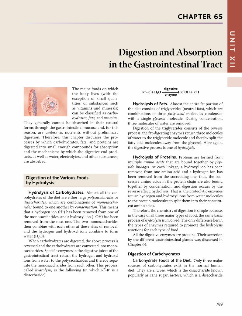
789
chapter 65U
nit
Xii
Digestion and Absorption in the Gastrointestinal Tract
The major foods on which the body lives (with the exception of small quan-tities of substances such as vitamins and minerals) can be classified as carbo-hydrates, fats, and proteins.
They generally cannot be absorbed in their natural forms through the gastrointestinal mucosa and, for this reason, are useless as nutrients without preliminary digestion. Therefore, this chapter discusses the pro-cesses by which carbohydrates, fats, and proteins are digested into small enough compounds for absorption and the mechanisms by which the digestive end prod-ucts, as well as water, electrolytes, and other substances, are absorbed.
Digestion of the Various Foods by Hydrolysis
Hydrolysis of Carbohydrates. Almost all the car-bohydrates of the diet are either large polysaccharides or disaccharides, which are combinations of monosaccha-rides bound to one another by condensation. This means that a hydrogen ion (H+) has been removed from one of the monosaccharides, and a hydroxyl ion (−OH) has been removed from the next one. The two monosaccharides then combine with each other at these sites of removal, and the hydrogen and hydroxyl ions combine to form water (H2O).
When carbohydrates are digested, the above process is reversed and the carbohydrates are converted into mono-saccharides. Specific enzymes in the digestive juices of the gastrointestinal tract return the hydrogen and hydroxyl ions from water to the polysaccharides and thereby sepa-rate the monosaccharides from each other. This process, called hydrolysis, is the following (in which R″-R′ is a disaccharide):
Hydrolysis of Fats. Almost the entire fat portion of the diet consists of triglycerides (neutral fats), which are combinations of three fatty acid molecules condensed with a single glycerol molecule. During condensation, three molecules of water are removed.
Digestion of the triglycerides consists of the reverse process: the fat-digesting enzymes return three molecules of water to the triglyceride molecule and thereby split the fatty acid molecules away from the glycerol. Here again, the digestive process is one of hydrolysis.
Hydrolysis of Proteins. Proteins are formed from multiple amino acids that are bound together by pep-tide linkages. At each linkage, a hydroxyl ion has been removed from one amino acid and a hydrogen ion has been removed from the succeeding one; thus, the suc-cessive amino acids in the protein chain are also bound together by condensation, and digestion occurs by the reverse effect: hydrolysis. That is, the proteolytic enzymes return hydrogen and hydroxyl ions from water molecules to the protein molecules to split them into their constitu-ent amino acids.
Therefore, the chemistry of digestion is simple because, in the case of all three major types of food, the same basic process of hydrolysis is involved. The only difference lies in the types of enzymes required to promote the hydrolysis reactions for each type of food.
All the digestive enzymes are proteins. Their secretion by the different gastrointestinal glands was discussed in Chapter 64.
Digestion of Carbohydrates
Carbohydrate Foods of the Diet. Only three major sources of carbohydrates exist in the normal human diet. They are sucrose, which is the disaccharide known popularly as cane sugar; lactose, which is a disaccharide
æ ÆææR¢¢-R¢ + H2Odigestive
enzymeR¢¢OH + R¢H
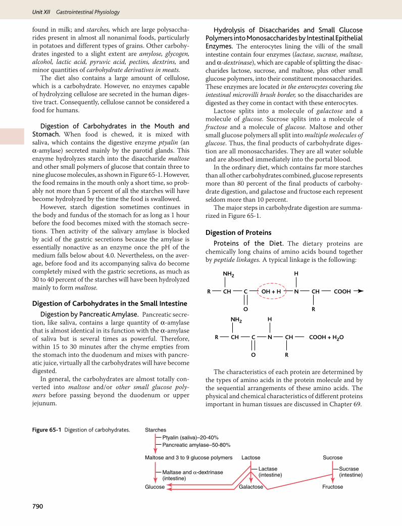
Unit XII Gastrointestinal Physiology
790
found in milk; and starches, which are large polysaccha-rides present in almost all nonanimal foods, particularly in potatoes and different types of grains. Other carbohy-drates ingested to a slight extent are amylose, glycogen, alcohol, lactic acid, pyruvic acid, pectins, dextrins, and minor quantities of carbohydrate derivatives in meats.
The diet also contains a large amount of cellulose, which is a carbohydrate. However, no enzymes capable of hydrolyzing cellulose are secreted in the human diges-tive tract. Consequently, cellulose cannot be considered a food for humans.
Digestion of Carbohydrates in the Mouth and Stomach. When food is chewed, it is mixed with saliva, which contains the digestive enzyme ptyalin (an α-amylase) secreted mainly by the parotid glands. This enzyme hydrolyzes starch into the disaccharide maltose and other small polymers of glucose that contain three to nine glucose molecules, as shown in Figure 65-1. However, the food remains in the mouth only a short time, so prob-ably not more than 5 percent of all the starches will have become hydrolyzed by the time the food is swallowed.
However, starch digestion sometimes continues in the body and fundus of the stomach for as long as 1 hour before the food becomes mixed with the stomach secre-tions. Then activity of the salivary amylase is blocked by acid of the gastric secretions because the amylase is essentially nonactive as an enzyme once the pH of the medium falls below about 4.0. Nevertheless, on the aver-age, before food and its accompanying saliva do become completely mixed with the gastric secretions, as much as 30 to 40 percent of the starches will have been hydrolyzed mainly to form maltose.
Digestion of Carbohydrates in the Small Intestine
Digestion by Pancreatic Amylase. Pancreatic secre-tion, like saliva, contains a large quantity of α-amylase that is almost identical in its function with the α-amylase of saliva but is several times as powerful. Therefore, within 15 to 30 minutes after the chyme empties from the stomach into the duodenum and mixes with pancre-atic juice, virtually all the carbohydrates will have become digested.
In general, the carbohydrates are almost totally con-verted into maltose and/or other small glucose poly-mers before passing beyond the duodenum or upper jejunum.
Hydrolysis of Disaccharides and Small Glucose Polymers into Monosaccharides by Intestinal Epithelial Enzymes. The enterocytes lining the villi of the small intestine contain four enzymes (lactase, sucrase, maltase, and α-dextrinase), which are capable of splitting the disac-charides lactose, sucrose, and maltose, plus other small glucose polymers, into their constituent monosaccharides. These enzymes are located in the enterocytes covering the intestinal microvilli brush border, so the disaccharides are digested as they come in contact with these enterocytes.
Lactose splits into a molecule of galactose and a molecule of glucose. Sucrose splits into a molecule of fructose and a molecule of glucose. Maltose and other small glucose polymers all split into multiple molecules of glucose. Thus, the final products of carbohydrate diges-tion are all monosaccharides. They are all water soluble and are absorbed immediately into the portal blood.
In the ordinary diet, which contains far more starches than all other carbohydrates combined, glucose represents more than 80 percent of the final products of carbohy-drate digestion, and galactose and fructose each represent seldom more than 10 percent.
The major steps in carbohydrate digestion are summa-rized in Figure 65-1.
Digestion of Proteins
Proteins of the Diet. The dietary proteins are chemically long chains of amino acids bound together by peptide linkages. A typical linkage is the following:
The characteristics of each protein are determined by the types of amino acids in the protein molecule and by the sequential arrangements of these amino acids. The physical and chemical characteristics of different proteins important in human tissues are discussed in Chapter 69.
C N
O
CH COOHOH + H
NH2
CHR
R
H
C N
O
CH COOH + H2O
NH2
CHR
R
H
Ptyalin (saliva)–20-40%Starches
Glucose
Maltose and 3 to 9 glucose polymers Lactose Sucrose
FructoseGalactose
Sucrase(intestine)
Lactase(intestine)
Pancreatic amylase–50-80%
Maltase and a-dextrinase(intestine)
Figure 65-1 Digestion of carbohydrates.

791
Un
it X
iiChapter 65 Digestion and Absorption in the Gastrointestinal Tract
Digestion of Proteins in the Stomach. Pepsin, the important peptic enzyme of the stomach, is most active at a pH of 2.0 to 3.0 and is inactive at a pH above about 5.0. Consequently, for this enzyme to cause digestion of protein, the stomach juices must be acidic. As explained in Chapter 64, the gastric glands secrete a large quantity of hydrochloric acid. This hydrochloric acid is secreted by the parietal (oxyntic) cells in the glands at a pH of about 0.8, but by the time it is mixed with the stomach contents and with secretions from the nonoxyntic glandular cells of the stomach, the pH then averages around 2.0 to 3.0, a highly favorable range of acidity for pepsin activity.
One of the important features of pepsin digestion is its ability to digest the protein collagen, an albuminoid type of protein that is affected little by other digestive enzymes. Collagen is a major constituent of the intercel-lular connective tissue of meats; therefore, for the diges-tive enzymes of the digestive tract to penetrate meats and digest the other meat proteins, it is necessary that the collagen fibers be digested. Consequently, in persons who lack pepsin in the stomach juices, the ingested meats are less well penetrated by the other digestive enzymes and, therefore, may be poorly digested.
As shown in Figure 65-2, pepsin only initiates the pro-cess of protein digestion, usually providing only 10 to 20 percent of the total protein digestion to convert the pro-tein to proteoses, peptones, and a few polypeptides. This splitting of proteins occurs as a result of hydrolysis at the peptide linkages between amino acids.
Most Protein Digestion Results from Actions of Pancreatic Proteolytic Enzymes. Most protein diges-tion occurs in the upper small intestine, in the duode-num and jejunum, under the influence of proteolytic enzymes from pancreatic secretion. Immediately on entering the small intestine from the stomach, the partial breakdown products of the protein foods are attacked by major proteolytic pancreatic enzymes: trypsin, chymotrypsin, carboxypolypeptidase, and proelastase, as shown in Figure 65-2.
Both trypsin and chymotrypsin split protein mole-cules into small polypeptides; carboxypolypeptidase then cleaves individual amino acids from the carboxyl ends of the polypeptides. Proelastase, in turn, is converted into elastase, which then digests elastin fibers that partially hold meats together.
Only a small percentage of the proteins are digested all the way to their constituent amino acids by the pancreatic juices. Most remain as dipeptides and tripeptides.
Digestion of Peptides by Peptidases in the Enterocytes That Line the Small Intestinal Villi. The last digestive stage of the proteins in the intestinal lumen is achieved by the enterocytes that line the villi of the small intestine, mainly in the duodenum and jejunum. These cells have a brush border that consists of hundreds of microvilli projecting from the surface of each cell. In the membrane of each of these microvilli are multiple peptidases that pro-trude through the membranes to the exterior, where they come in contact with the intestinal fluids.
Two types of peptidase enzymes are especially impor-tant, aminopolypeptidase and several dipeptidases. They succeed in splitting the remaining larger polypeptides into tripeptides and dipeptides and a few into amino acids. Both the amino acids plus the dipeptides and tripeptides are easily transported through the microvillar membrane to the interior of the enterocyte.
Finally, inside the cytosol of the enterocyte are multiple other peptidases that are specific for the remaining types of linkages between amino acids. Within minutes, virtu-ally all the last dipeptides and tripeptides are digested to the final stage to form single amino acids; these then pass on through to the other side of the enterocyte and thence into the blood.
More than 99 percent of the final protein digestive products that are absorbed are individual amino acids, with only rare absorption of peptides and very, very rare absorption of whole protein molecules. Even these few absorbed molecules of whole protein can sometimes cause serious allergic or immunologic disturbances, as discussed in Chapter 34.
Digestion of Fats
Fats of the Diet. By far the most abundant fats of the diet are the neutral fats, also known as triglycerides, each molecule of which is composed of a glycerol nucleus and three fatty acid side chains, as shown in Figure 65-3.
Pepsin
PeptidasesAmino acids
Polypeptides+
Amino acids
ProteosesPeptonesPolypeptides
Proteins
Trypsin, chymotrypsin, carboxypolypeptidase,proelastase
Figure 65-2 Digestion of proteins.
CH3 (CH2)16 C
O
O CH2
CH3 (CH2)16 C
O
O CH2
CH3 (CH2)16 C
OLipase
(Tristearin)
CH3 (CH2)16 (CH2)16C
O
O C
O
OH
HO CH2
HO CH2(2-Monoglyceride) (Stearic acid)
O CH + 2H2O
CH + 2CH3
Figure 65-3 Hydrolysis of neutral fat catalyzed by lipase.

Unit XII Gastrointestinal Physiology
792
Neutral fat is a major constituent in food of animal origin but much, much less so in food of plant origin.
In the usual diet are also small quantities of phospho-lipids, cholesterol, and cholesterol esters. The phospholip-ids and cholesterol esters contain fatty acid and therefore can be considered fats. Cholesterol, however, is a sterol compound that contains no fatty acid, but it does exhibit some of the physical and chemical characteristics of fats; plus, it is derived from fats and is metabolized similarly to fats. Therefore, cholesterol is considered, from a dietary point of view, a fat.
Digestion of Fats in the Intestine. A small amount of triglycerides is digested in the stomach by lingual lipase that is secreted by lingual glands in the mouth and swal-lowed with the saliva. This amount of digestion is less than 10 percent and generally unimportant. Instead, essentially all fat digestion occurs in the small intestine as follows.
The First Step in Fat Digestion Is Emulsification by Bile Acids and Lecithin. The first step in fat digestion is physically to break the fat globules into small sizes so that the water-soluble digestive enzymes can act on the glob-ule surfaces. This process is called emulsification of the fat, and it begins by agitation in the stomach to mix the fat with the products of stomach digestion.
Then, most of the emulsification occurs in the duo-denum under the influence of bile, the secretion from the liver that does not contain any digestive enzymes. However, bile does contain a large quantity of bile salts, as well as the phospholipid lecithin. Both of these, but especially the lecithin, are extremely important for emul-sification of the fat. The polar parts (the points where ionization occurs in water) of the bile salts and lecithin molecules are highly soluble in water, whereas most of the remaining portions of their molecules are highly soluble in fat. Therefore, the fat-soluble portions of these liver secretions dissolve in the surface layer of the fat glob-ules, with the polar portions projecting. The polar pro-jections, in turn, are soluble in the surrounding watery fluids, which greatly decreases the interfacial tension of the fat and makes it soluble as well.
When the interfacial tension of a globule of nonmisci-ble fluid is low, this nonmiscible fluid, on agitation, can be broken up into many tiny particles far more easily than it can when the interfacial tension is great. Consequently, a major function of the bile salts and lecithin, especially the lecithin, in the bile is to make the fat globules readily frag-mentable by agitation with the water in the small bowel. This action is the same as that of many detergents that are widely used in household cleaners for removing grease.
Each time the diameters of the fat globules are signifi-cantly decreased as a result of agitation in the small intestine, the total surface area of the fat increases manyfold. Because the average diameter of the fat particles in the intestine after emulsification has occurred is less than 1 micrometer, this represents an increase of as much as 1000-fold in total sur-face areas of the fats caused by the emulsification process.
The lipase enzymes are water-soluble compounds and can attack the fat globules only on their surfaces. Consequently, this detergent function of bile salts and lecithin is very important for digestion of fats.
Triglycerides Are Digested by Pancreatic Lipase. By far the most important enzyme for digestion of the triglyc-erides is pancreatic lipase, present in enormous quantities in pancreatic juice, enough to digest within 1 minute all triglycerides that it can reach. In addition, the enterocytes of the small intestine contain additional lipase, known as enteric lipase, but this is usually not needed.
End Products of Fat Digestion Are Free Fatty Acids. Most of the triglycerides of the diet are split by pan-creatic lipase into free fatty acids and 2-monoglycerides, as shown in Figure 65-4.
Bile Salts Form Micelles That Accelerate Fat Digestion. The hydrolysis of triglycerides is a highly reversible process; therefore, accumulation of monoglyc-erides and free fatty acids in the vicinity of digesting fats quickly blocks further digestion. But the bile salts play the additional important role of removing the monoglyc-erides and free fatty acids from the vicinity of the digest-ing fat globules almost as rapidly as these end products of digestion are formed. This occurs in the following way.
Bile salts, when in high enough concentration in water, have the propensity to form micelles, which are small spher-ical, cylindrical globules 3 to 6 nanometers in diameter composed of 20 to 40 molecules of bile salt. These develop because each bile salt molecule is composed of a sterol nucleus that is highly fat-soluble and a polar group that is highly water-soluble. The sterol nucleus encompasses the fat digestate, forming a small fat globule in the middle of a resulting micelle, with polar groups of bile salts project-ing outward to cover the surface of the micelle. Because these polar groups are negatively charged, they allow the entire micelle globule to dissolve in the water of the diges-tive fluids and to remain in stable solution until the fat is absorbed into the blood.
The bile salt micelles also act as a transport medium to carry the monoglycerides and free fatty acids, both of which would otherwise be relatively insoluble, to the brush borders of the intestinal epithelial cells. There the monoglycerides and free fatty acids are absorbed into the blood, as discussed later, but the bile salts themselves are released back into the chyme to be used again and again for this “ferrying” process.
Digestion of Cholesterol Esters and Phospholipids. Most cholesterol in the diet is in the form of
(Bile + Agitation)
Pancreatic lipase
Emulsified fat
Fatty acids and2-monoglycerides
Fat
Emulsified fat
Figure 65-4 Digestion of fats.
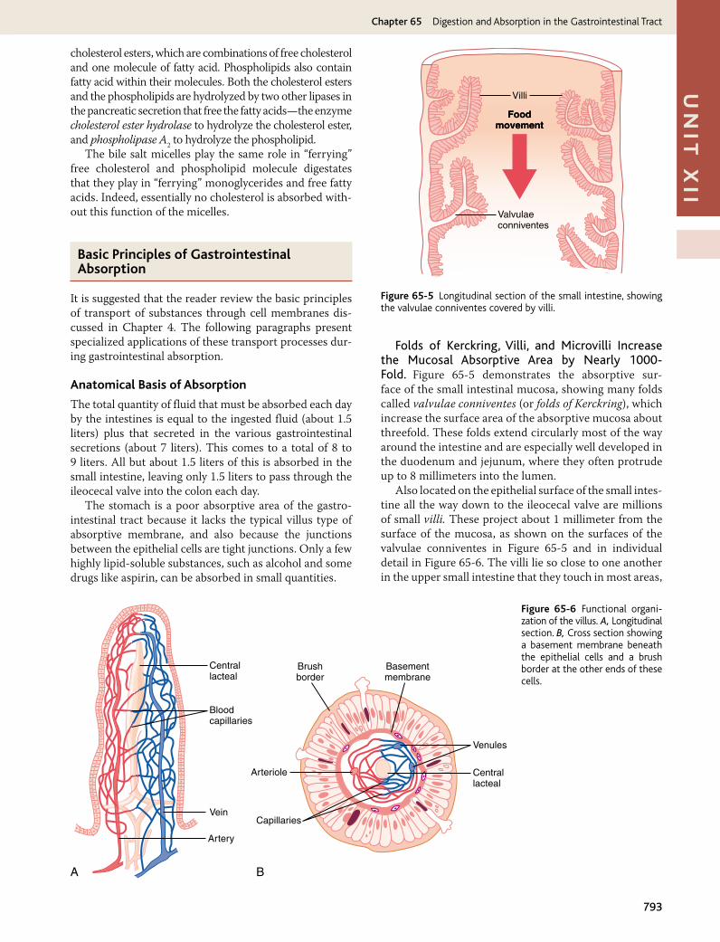
793
Un
it X
iiChapter 65 Digestion and Absorption in the Gastrointestinal Tract
cholesterol esters, which are combinations of free cholesterol and one molecule of fatty acid. Phospholipids also contain fatty acid within their molecules. Both the cholesterol esters and the phospholipids are hydrolyzed by two other lipases in the pancreatic secretion that free the fatty acids—the enzyme cholesterol ester hydrolase to hydrolyze the cholesterol ester, and phospholipase A2 to hydrolyze the phospholipid.
The bile salt micelles play the same role in “ferrying” free cholesterol and phospholipid molecule digestates that they play in “ferrying” monoglycerides and free fatty acids. Indeed, essentially no cholesterol is absorbed with-out this function of the micelles.
Basic Principles of Gastrointestinal Absorption
It is suggested that the reader review the basic principles of transport of substances through cell membranes dis-cussed in Chapter 4. The following paragraphs present specialized applications of these transport processes dur-ing gastrointestinal absorption.
Anatomical Basis of Absorption
The total quantity of fluid that must be absorbed each day by the intestines is equal to the ingested fluid (about 1.5 liters) plus that secreted in the various gastrointestinal secretions (about 7 liters). This comes to a total of 8 to 9 liters. All but about 1.5 liters of this is absorbed in the small intestine, leaving only 1.5 liters to pass through the ileocecal valve into the colon each day.
The stomach is a poor absorptive area of the gastro-intestinal tract because it lacks the typical villus type of absorptive membrane, and also because the junctions between the epithelial cells are tight junctions. Only a few highly lipid-soluble substances, such as alcohol and some drugs like aspirin, can be absorbed in small quantities.
Folds of Kerckring, Villi, and Microvilli Increase the Mucosal Absorptive Area by Nearly 1000Fold. Figure 65-5 demonstrates the absorptive sur-face of the small intestinal mucosa, showing many folds called valvulae conniventes (or folds of Kerckring), which increase the surface area of the absorptive mucosa about threefold. These folds extend circularly most of the way around the intestine and are especially well developed in the duodenum and jejunum, where they often protrude up to 8 millimeters into the lumen.
Also located on the epithelial surface of the small intes-tine all the way down to the ileocecal valve are millions of small villi. These project about 1 millimeter from the surface of the mucosa, as shown on the surfaces of the valvulae conniventes in Figure 65-5 and in individual detail in Figure 65-6. The villi lie so close to one another in the upper small intestine that they touch in most areas,
Villi
Foodmovement
Foodmovement
Valvulaeconniventes
Figure 65-5 Longitudinal section of the small intestine, showing the valvulae conniventes covered by villi.
Centrallacteal
Centrallacteal
Venules
Basementmembrane
A B
Brushborder
Arteriole
CapillariesVein
Artery
Bloodcapillaries
Figure 65-6 Functional organi-zation of the villus. A, Longitudinal section. B, Cross section showing a basement membrane beneath the epithelial cells and a brush border at the other ends of these cells.
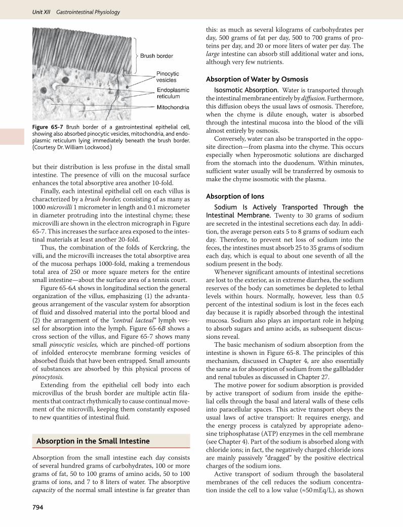
Unit XII Gastrointestinal Physiology
794
but their distribution is less profuse in the distal small intestine. The presence of villi on the mucosal surface enhances the total absorptive area another 10-fold.
Finally, each intestinal epithelial cell on each villus is characterized by a brush border, consisting of as many as 1000 microvilli 1 micrometer in length and 0.1 micrometer in diameter protruding into the intestinal chyme; these microvilli are shown in the electron micrograph in Figure 65-7. This increases the surface area exposed to the intes-tinal materials at least another 20-fold.
Thus, the combination of the folds of Kerckring, the villi, and the microvilli increases the total absorptive area of the mucosa perhaps 1000-fold, making a tremendous total area of 250 or more square meters for the entire small intestine—about the surface area of a tennis court.
Figure 65-6A shows in longitudinal section the general organization of the villus, emphasizing (1) the advanta-geous arrangement of the vascular system for absorption of fluid and dissolved material into the portal blood and (2) the arrangement of the “central lacteal” lymph ves-sel for absorption into the lymph. Figure 65-6B shows a cross section of the villus, and Figure 65-7 shows many small pinocytic vesicles, which are pinched-off portions of infolded enterocyte membrane forming vesicles of absorbed fluids that have been entrapped. Small amounts of substances are absorbed by this physical process of pinocytosis.
Extending from the epithelial cell body into each microvillus of the brush border are multiple actin fila-ments that contract rhythmically to cause continual move-ment of the microvilli, keeping them constantly exposed to new quantities of intestinal fluid.
Absorption in the Small Intestine
Absorption from the small intestine each day consists of several hundred grams of carbohydrates, 100 or more grams of fat, 50 to 100 grams of amino acids, 50 to 100 grams of ions, and 7 to 8 liters of water. The absorptive capacity of the normal small intestine is far greater than
this: as much as several kilograms of carbohydrates per day, 500 grams of fat per day, 500 to 700 grams of pro-teins per day, and 20 or more liters of water per day. The large intestine can absorb still additional water and ions, although very few nutrients.
Absorption of Water by Osmosis
Isosmotic Absorption. Water is transported through the intestinal membrane entirely by diffusion. Furthermore, this diffusion obeys the usual laws of osmosis. Therefore, when the chyme is dilute enough, water is absorbed through the intestinal mucosa into the blood of the villi almost entirely by osmosis.
Conversely, water can also be transported in the oppo-site direction—from plasma into the chyme. This occurs especially when hyperosmotic solutions are discharged from the stomach into the duodenum. Within minutes, sufficient water usually will be transferred by osmosis to make the chyme isosmotic with the plasma.
Absorption of Ions
Sodium Is Actively Transported Through the Intestinal Membrane. Twenty to 30 grams of sodium are secreted in the intestinal secretions each day. In addi-tion, the average person eats 5 to 8 grams of sodium each day. Therefore, to prevent net loss of sodium into the feces, the intestines must absorb 25 to 35 grams of sodium each day, which is equal to about one seventh of all the sodium present in the body.
Whenever significant amounts of intestinal secretions are lost to the exterior, as in extreme diarrhea, the sodium reserves of the body can sometimes be depleted to lethal levels within hours. Normally, however, less than 0.5 percent of the intestinal sodium is lost in the feces each day because it is rapidly absorbed through the intestinal mucosa. Sodium also plays an important role in helping to absorb sugars and amino acids, as subsequent discus-sions reveal.
The basic mechanism of sodium absorption from the intestine is shown in Figure 65-8. The principles of this mechanism, discussed in Chapter 4, are also essentially the same as for absorption of sodium from the gallbladder and renal tubules as discussed in Chapter 27.
The motive power for sodium absorption is provided by active transport of sodium from inside the epithe-lial cells through the basal and lateral walls of these cells into paracellular spaces. This active transport obeys the usual laws of active transport: It requires energy, and the energy process is catalyzed by appropriate adeno-sine triphosphatase (ATP) enzymes in the cell membrane (see Chapter 4). Part of the sodium is absorbed along with chloride ions; in fact, the negatively charged chloride ions are mainly passively “dragged” by the positive electrical charges of the sodium ions.
Active transport of sodium through the basolateral membranes of the cell reduces the sodium concentra-tion inside the cell to a low value (≈50 mEq/L), as shown
Figure 65-7 Brush border of a gastrointestinal epithelial cell, showing also absorbed pinocytic vesicles, mitochondria, and endo-plasmic reticulum lying immediately beneath the brush border. (Courtesy Dr. William Lockwood.)
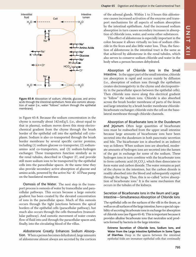
795
Un
it X
iiChapter 65 Digestion and Absorption in the Gastrointestinal Tract
in Figure 65-8. Because the sodium concentration in the chyme is normally about 142 mEq/L (i.e., about equal to that in plasma), sodium moves down this steep electro-chemical gradient from the chyme through the brush border of the epithelial cell into the epithelial cell cyto-plasm. Sodium is also co-transported through the brush border membrane by several specific carrier proteins, including (1) sodium-glucose co-transporter, (2) sodium-amino acid co-transporters, and (3) sodium-hydrogen exchanger. These transporters function similarly as in the renal tubules, described in Chapter 27, and provide still more sodium ions to be transported by the epithelial cells into the paracellular spaces. At the same time they also provide secondary active absorption of glucose and amino acids, powered by the active Na+-K+ ATPase pump on the basolateral membrane.
Osmosis of the Water. The next step in the trans-port process is osmosis of water by transcellular and para-cellular pathways. This occurs because a large osmotic gradient has been created by the elevated concentration of ions in the paracellular space. Much of this osmosis occurs through the tight junctions between the apical borders of the epithelial cells (paracellular pathway), but much also occurs through the cells themselves (transcel-lular pathway). And osmotic movement of water creates flow of fluid into and through the paracellular spaces and, finally, into the circulating blood of the villus.
Aldosterone Greatly Enhances Sodium Absorption. When a person becomes dehydrated, large amounts of aldosterone almost always are secreted by the cortices
of the adrenal glands. Within 1 to 3 hours this aldoster-one causes increased activation of the enzyme and trans-port mechanisms for all aspects of sodium absorption by the intestinal epithelium. And the increased sodium absorption in turn causes secondary increases in absorp-tion of chloride ions, water, and some other substances.
This effect of aldosterone is especially important in the colon because it allows virtually no loss of sodium chlo-ride in the feces and also little water loss. Thus, the func-tion of aldosterone in the intestinal tract is the same as that achieved by aldosterone in the renal tubules, which also serves to conserve sodium chloride and water in the body when a person becomes dehydrated.
Absorption of Chloride Ions in the Small Intestine. In the upper part of the small intestine, chloride ion absorption is rapid and occurs mainly by diffusion (i.e., absorption of sodium ions through the epithelium creates electro negativity in the chyme and electropositiv-ity in the paracellular spaces between the epithelial cells). Then chloride ions move along this electrical gradient to “follow” the sodium ions. Chloride is also absorbed across the brush border membrane of parts of the ileum and large intestine by a brush border membrane chloride-bicarbonate exchanger; chloride exits the cell on the baso-lateral membrane through chloride channels.
Absorption of Bicarbonate Ions in the Duodenum and Jejunum. Often large quantities of bicarbonate ions must be reabsorbed from the upper small intestine because large amounts of bicarbonate ions have been secreted into the duodenum in both pancreatic secretion and bile. The bicarbonate ion is absorbed in an indirect way as follows: When sodium ions are absorbed, moder-ate amounts of hydrogen ions are secreted into the lumen of the gut in exchange for some of the sodium. These hydrogen ions in turn combine with the bicarbonate ions to form carbonic acid (H2CO3), which then dissociates to form water and carbon dioxide. The water remains as part of the chyme in the intestines, but the carbon dioxide is readily absorbed into the blood and subsequently expired through the lungs. Thus, this is so-called “active absorp-tion of bicarbonate ions.” It is the same mechanism that occurs in the tubules of the kidneys.
Secretion of Bicarbonate Ions in the Ileum and Large Intestine—Simultaneous Absorption of Chloride Ions
The epithelial cells on the surfaces of the villi in the ileum, as well as on all surfaces of the large intestine, have a special capa-bility of secreting bicarbonate ions in exchange for absorption of chloride ions (see Figure 65-8). This is important because it provides alkaline bicarbonate ions that neutralize acid prod-ucts formed by bacteria in the large intestine.
Extreme Secretion of Chloride Ions, Sodium Ions, and Water from the Large Intestine Epithelium in Some Types of Diarrhea. Deep in the spaces between the intestinal epithelial folds are immature epithelial cells that continually
Na+
K+
H2OH2O
H+
Interstitialfluid
IntestinelumenCell
H2OH2O
Na+Na+
Amino AcidsAmino Acids
Na+Na+
GlucoseGlucose
Na+Na+
Cl-
Cl- HCO3-
Na+
K+
Na+
K+
Figure 65-8 Absorption of sodium, chloride, glucose, and amino acids through the intestinal epithelium. Note also osmotic absorp-tion of water (i.e., water “follows” sodium through the epithelial membrane).
Unit XII Gastrointestinal Physiology
796
divide to form new epithelial cells. These in turn spread out-ward over the luminal surfaces of the intestines. While still in the deep folds, the epithelial cells secrete sodium chloride and water into the intestinal lumen. This secretion in turn is reabsorbed by the older epithelial cells outside the folds, thus providing flow of water for absorbing intestinal digestates.
The toxins of cholera and of some other types of diarrheal bacteria can stimulate the epithelial fold secretion so greatly that this secretion often becomes much greater than can be reab-sorbed, thus sometimes causing loss of 5 to 10 liters of water and sodium chloride as diarrhea each day. Within 1 to 5 days, many severely affected patients die from this loss of fluid alone.
Extreme diarrheal secretion is initiated by entry of a sub-unit of cholera toxin into the epithelial cells. This stimulates formation of excess cyclic adenosine monophosphate, which opens tremendous numbers of chloride channels, allowing chloride ions to flow rapidly from inside the cell into the intestinal crypts. In turn, this is believed to activate a sodium pump that pumps sodium ions into the crypts to go along with the chloride ions. Finally, all this extra sodium chloride causes extreme osmosis of water from the blood, thus pro-viding rapid flow of fluid along with the salt. All this excess fluid washes away most of the bacteria and is of value in com-bating the disease, but too much of a good thing can be lethal because of serious dehydration of the whole body that might ensue. In most instances, the life of a cholera victim can be saved by administration of tremendous amounts of sodium chloride solution to make up for the loss.
Active Absorption of Calcium, Iron, Potassium, Magnesium, and Phosphate. Calcium ions are actively absorbed into the blood, especially from the duodenum, and the amount of calcium ion absorption is exactly con-trolled to supply the daily need of the body for calcium. One important factor controlling calcium absorption is parathyroid hormone secreted by the parathyroid glands, and another is vitamin D. Parathyroid hormone activates vitamin D, and the activated vitamin D in turn greatly enhances calcium absorption. These effects are dis-cussed in Chapter 79.
Iron ions are also actively absorbed from the small intestine. The principles of iron absorption and regula-tion of its absorption in proportion to the body’s need for iron, especially for the formation of hemoglobin, are dis-cussed in Chapter 32.
Potassium, magnesium, phosphate, and probably still other ions can also be actively absorbed through the intes-tinal mucosa. In general, the monovalent ions are absorbed with ease and in great quantities. Conversely, bivalent ions are normally absorbed in only small amounts; for example, maximum absorption of calcium ions is only 1/50 as great as the normal absorption of sodium ions. Fortunately, only small quantities of the bivalent ions are normally required daily by the body.
Absorption of Nutrients
Carbohydrates Are Mainly Absorbed as Monosaccharides
Essentially all the carbohydrates in the food are absorbed in the form of monosaccharides; only a small fraction
is absorbed as disaccharides and almost none as larger carbohydrate compounds. By far the most abundant of the absorbed monosaccharides is glucose, usually accounting for more than 80 percent of carbohydrate calories absorbed. The reason for this is that glucose is the final digestion product of our most abundant carbo-hydrate food, the starches. The remaining 20 percent of absorbed monosaccharides is composed almost entirely of galactose and fructose, the galactose derived from milk and the fructose as one of the monosaccharides digested from cane sugar.
Virtually all the monosaccharides are absorbed by an active transport process. Let us first discuss the absorp-tion of glucose.
Glucose Is Transported by a Sodium Co-Transport Mechanism. In the absence of sodium transport through the intestinal membrane, virtually no glucose can be absorbed. The reason is that glucose absorption occurs in a co-transport mode with active transport of sodium (see Figure 65-8).
There are two stages in the transport of sodium through the intestinal membrane. First is active transport of sodium ions through the basolateral membranes of the intestinal epithelial cells into the blood, thereby deplet-ing sodium inside the epithelial cells. Second, decrease of sodium inside the cells causes sodium from the intestinal lumen to move through the brush border of the epithelial cells to the cell interiors by a process of secondary active transport. That is, a sodium ion combines with a trans-port protein, but the transport protein will not transport the sodium to the interior of the cell until the protein also combines with some other appropriate substance such as glucose. Intestinal glucose also combines simultaneously with the same transport protein and then both the sodium ion and glucose molecule are transported together to the interior of the cell. Thus, the low concentration of sodium inside the cell literally “drags” sodium to the interior of the cell and along with it the glucose at the same time. Once inside the epithelial cell, other transport proteins and enzymes cause facilitated diffusion of the glucose through the cell’s basolateral membrane into the paracellular space and from there into the blood.
To summarize, it is the initial active transport of sodium through the basolateral membranes of the intesti-nal epithelial cells that provides the eventual motive force for moving glucose also through the membranes.
Absorption of Other Monosaccharides. Galactose is transported by almost exactly the same mechanism as glu-cose. Conversely, fructose transport does not occur by the sodium co-transport mechanism. Instead, fructose is trans-ported by facilitated diffusion all the way through the intes-tinal epithelium but not coupled with sodium transport.
Much of the fructose, on entering the cell, becomes phosphorylated, then converted to glucose, and finally transported in the form of glucose the rest of the way into the blood. Because fructose is not co-transported with sodium, its overall rate of transport is only about one half that of glucose or galactose.
797
Un
it X
iiChapter 65 Digestion and Absorption in the Gastrointestinal Tract
Absorption of Proteins as Dipeptides, Tripeptides, or Amino Acids
As explained earlier in the chapter, most proteins, after digestion, are absorbed through the luminal membranes of the intestinal epithelial cells in the form of dipeptides, tripeptides, and a few free amino acids. The energy for most of this transport is supplied by a sodium co-trans-port mechanism in the same way that sodium co-trans-port of glucose occurs. That is, most peptide or amino acid molecules bind in the cell’s microvillus membrane with a specific transport protein that requires sodium binding before transport can occur. After binding, the sodium ion then moves down its electrochemical gradient to the inte-rior of the cell and pulls the amino acid or peptide along with it. This is called co-transport (or secondary active transport) of the amino acids and peptides (see Figure 65-8). A few amino acids do not require this sodium co-transport mechanism but instead are transported by special membrane transport proteins in the same way that fructose is transported, by facilitated diffusion.
At least five types of transport proteins for transport-ing amino acids and peptides have been found in the luminal membranes of intestinal epithelial cells. This multiplicity of transport proteins is required because of the diverse binding properties of different amino acids and peptides.
Absorption of Fats
Earlier in this chapter, it was pointed out that when fats are digested to form monoglycerides and free fatty acids, both of these digestive end products first become dissolved in the central lipid portions of bile micelles. Because the molecular dimensions of these micelles are only 3 to 6 nanometers in diameter, and because of their highly charged exterior, they are soluble in chyme. In this form, the monoglycerides and free fatty acids are carried to the surfaces of the microvilli of the intestinal cell brush border and then penetrate into the recesses among the moving, agitating microvilli. Here, both the monoglycer-ides and fatty acids diffuse immediately out of the micelles and into the interior of the epithelial cells, which is possi-ble because the lipids are also soluble in the epithelial cell membrane. This leaves the bile micelles still in the chyme, where they function again and again to help absorb still more monoglycerides and fatty acids.
Thus, the micelles perform a “ferrying” function that is highly important for fat absorption. In the presence of an abundance of bile micelles, about 97 percent of the fat is absorbed; in the absence of the bile micelles, only 40 to 50 percent can be absorbed.
After entering the epithelial cell, the fatty acids and monoglycerides are taken up by the cell’s smooth endo-plasmic reticulum; here, they are mainly used to form new triglycerides that are subsequently released in the form of chylomicrons through the base of the epithelial cell, to flow upward through the thoracic lymph duct and empty into the circulating blood.
Direct Absorption of Fatty Acids into the Portal Blood. Small quantities of short- and medium-chain fatty acids, such as those from butterfat, are absorbed directly into the portal blood rather than being converted into trig-lycerides and absorbed by way of the lymphatics. The cause of this difference between short- and long-chain fatty acid absorption is that the short-chain fatty acids are more water-soluble and mostly are not reconverted into triglycerides by the endoplasmic reticulum. This allows direct diffusion of these short-chain fatty acids from the intestinal epithelial cells directly into the capillary blood of the intestinal villi.
Absorption in the Large Intestine: Formation of Feces
About 1500 milliliters of chyme normally pass through the ileocecal valve into the large intestine each day. Most of the water and electrolytes in this chyme are absorbed in the colon, usually leaving less than 100 milliliters of fluid to be excreted in the feces. Also, essentially all the ions are absorbed, leaving only 1 to 5 mEq each of sodium and chloride ions to be lost in the feces.
Most of the absorption in the large intestine occurs in the proximal one half of the colon, giving this portion the name absorbing colon, whereas the distal colon functions principally for feces storage until a propitious time for feces excretion and is therefore called the storage colon.
Absorption and Secretion of Electrolytes and Water. The mucosa of the large intestine, like that of the small intestine, has a high capability for active absorption of sodium, and the electrical potential gradient created by absorption of the sodium causes chloride absorption as well. The tight junctions between the epithelial cells of the large intestinal epithelium are much tighter than those of the small intestine. This prevents significant amounts of back-diffusion of ions through these junctions, thus allow-ing the large intestinal mucosa to absorb sodium ions far more completely—that is, against a much higher concen-tration gradient—than can occur in the small intestine. This is especially true when large quantities of aldoster-one are available because aldosterone greatly enhances sodium transport capability.
In addition, as occurs in the distal portion of the small intestine, the mucosa of the large intestine secretes bicar-bonate ions while it simultaneously absorbs an equal number of chloride ions in an exchange transport process that has already been described. The bicarbonate helps neutralize the acidic end products of bacterial action in the large intestine.
Absorption of sodium and chloride ions creates an osmotic gradient across the large intestinal mucosa, which in turn causes absorption of water.
Maximum Absorption Capacity of the Large Intestine. The large intestine can absorb a maximum of 5 to 8 liters of fluid and electrolytes each day. When the
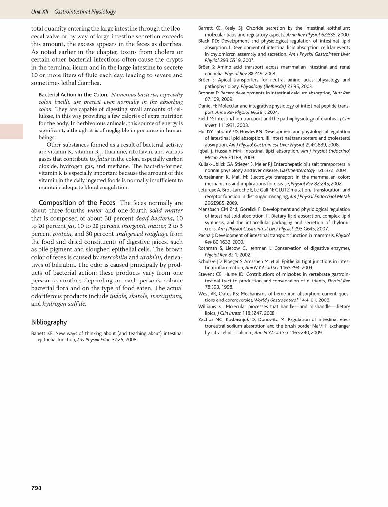
Unit XII Gastrointestinal Physiology
798
total quantity entering the large intestine through the ileo-cecal valve or by way of large intestine secretion exceeds this amount, the excess appears in the feces as diarrhea. As noted earlier in the chapter, toxins from cholera or certain other bacterial infections often cause the crypts in the terminal ileum and in the large intestine to secrete 10 or more liters of fluid each day, leading to severe and sometimes lethal diarrhea.
Bacterial Action in the Colon. Numerous bacteria, especially colon bacilli, are present even normally in the absorbing colon. They are capable of digesting small amounts of cel-lulose, in this way providing a few calories of extra nutrition for the body. In herbivorous animals, this source of energy is significant, although it is of negligible importance in human beings.
Other substances formed as a result of bacterial activity are vitamin K, vitamin B12, thiamine, riboflavin, and various gases that contribute to flatus in the colon, especially carbon dioxide, hydrogen gas, and methane. The bacteria-formed vitamin K is especially important because the amount of this vitamin in the daily ingested foods is normally insufficient to maintain adequate blood coagulation.
Composition of the Feces. The feces normally are about three-fourths water and one-fourth solid matter that is composed of about 30 percent dead bacteria, 10 to 20 percent fat, 10 to 20 percent inorganic matter, 2 to 3 percent protein, and 30 percent undigested roughage from the food and dried constituents of digestive juices, such as bile pigment and sloughed epithelial cells. The brown color of feces is caused by stercobilin and urobilin, deriva-tives of bilirubin. The odor is caused principally by prod-ucts of bacterial action; these products vary from one person to another, depending on each person’s colonic bacterial flora and on the type of food eaten. The actual odoriferous products include indole, skatole, mercaptans, and hydrogen sulfide.
Bibliography
Barrett KE: New ways of thinking about (and teaching about) intestinal epithelial function, Adv Physiol Educ 32:25, 2008.
Barrett KE, Keely SJ: Chloride secretion by the intestinal epithelium: molecular basis and regulatory aspects, Annu Rev Physiol 62:535, 2000.
Black DD: Development and physiological regulation of intestinal lipid absorption. I. Development of intestinal lipid absorption: cellular events in chylomicron assembly and secretion, Am J Physiol Gastrointest Liver Physiol 293:G519, 2007.
Bröer S: Amino acid transport across mammalian intestinal and renal epithelia, Physiol Rev 88:249, 2008.
Bröer S: Apical transporters for neutral amino acids: physiology and pathophysiology, Physiology (Bethesda) 23:95, 2008.
Bronner F: Recent developments in intestinal calcium absorption, Nutr Rev 67:109, 2009.
Daniel H: Molecular and integrative physiology of intestinal peptide trans-port, Annu Rev Physiol 66:361, 2004.
Field M: Intestinal ion transport and the pathophysiology of diarrhea, J Clin Invest 111:931, 2003.
Hui DY, Labonté ED, Howles PN: Development and physiological regulation of intestinal lipid absorption. III. Intestinal transporters and cholesterol absorption, Am J Physiol Gastrointest Liver Physiol 294:G839, 2008.
Iqbal J, Hussain MM: Intestinal lipid absorption, Am J Physiol Endocrinol Metab 296:E1183, 2009.
Kullak-Ublick GA, Stieger B, Meier PJ: Enterohepatic bile salt transporters in normal physiology and liver disease, Gastroenterology 126:322, 2004.
Kunzelmann K, Mall M: Electrolyte transport in the mammalian colon: mechanisms and implications for disease, Physiol Rev 82:245, 2002.
Leturque A, Brot-Laroche E, Le Gall M: GLUT2 mutations, translocation, and receptor function in diet sugar managing, Am J Physiol Endocrinol Metab 296:E985, 2009.
Mansbach CM 2nd, Gorelick F: Development and physiological regulation of intestinal lipid absorption. II. Dietary lipid absorption, complex lipid synthesis, and the intracellular packaging and secretion of chylomi-crons, Am J Physiol Gastrointest Liver Physiol 293:G645, 2007.
Pacha J: Development of intestinal transport function in mammals, Physiol Rev 80:1633, 2000.
Rothman S, Liebow C, Isenman L: Conservation of digestive enzymes, Physiol Rev 82:1, 2002.
Schulzke JD, Ploeger S, Amasheh M, et al: Epithelial tight junctions in intes-tinal inflammation, Ann N Y Acad Sci 1165:294, 2009.
Stevens CE, Hume ID: Contributions of microbes in vertebrate gastroin-testinal tract to production and conservation of nutrients, Physiol Rev 78:393, 1998.
West AR, Oates PS: Mechanisms of heme iron absorption: current ques-tions and controversies, World J Gastroenterol 14:4101, 2008.
Williams KJ: Molecular processes that handle—and mishandle—dietary lipids, J Clin Invest 118:3247, 2008.
Zachos NC, Kovbasnjuk O, Donowitz M: Regulation of intestinal elec-troneutral sodium absorption and the brush border Na+/H+ exchanger by intracellular calcium, Ann N Y Acad Sci 1165:240, 2009.