Postoperative Pain following Root Canal Filling with ... - MDPI
10
Journal of Clinical Medicine Review Postoperative Pain following Root Canal Filling with Bioceramic vs. Traditional Filling Techniques: A Systematic Review and Meta-Analysis of Randomized Controlled Trials Elina Mekhdieva 1 , Massimo Del Fabbro 2,3 , Mario Alovisi 1 , Allegra Comba 1 , Nicola Scotti 1 , Margherita Tumedei 2,4 , Massimo Carossa 1 , Elio Berutti 1 and Damiano Pasqualini 1, * Citation: Mekhdieva, E.; Del Fabbro, M.; Alovisi, M.; Comba, A.; Scotti, N.; Tumedei, M.; Carossa, M.; Berutti, E.; Pasqualini, D. Postoperative Pain following Root Canal Filling with Bioceramic vs. Traditional Filling Techniques: A Systematic Review and Meta-Analysis of Randomized Controlled Trials. J. Clin. Med. 2021, 10, 4509. https://doi.org/10.3390/ jcm10194509 Academic Editors: Alfredo Iandolo, Massimo Amato and Giuseppe Pantaleo Received: 12 September 2021 Accepted: 27 September 2021 Published: 29 September 2021 Publisher’s Note: MDPI stays neutral with regard to jurisdictional claims in published maps and institutional affil- iations. Copyright: © 2021 by the authors. Licensee MDPI, Basel, Switzerland. This article is an open access article distributed under the terms and conditions of the Creative Commons Attribution (CC BY) license (https:// creativecommons.org/licenses/by/ 4.0/). 1 Endodontics and Restorative Dentistry, CIR Dental School, Department of Surgical Sciences, University of Turin, 10126 Turin, Italy; [email protected] (E.M.); [email protected] (M.A.); [email protected] (A.C.); [email protected] (N.S.); [email protected] (M.C.); [email protected] (E.B.) 2 Department of Biomedical, Surgical and Dental Sciences, University of Milan, 20122 Milan, Italy; [email protected] (M.D.F.); [email protected] (M.T.) 3 IRCCS Orthopedic Institute Galeazzi, 20161 Milan, Italy 4 Department of Medical, Oral and Biotechnological Sciences, University “G. d 0 Annunzio” of Chieti-Pescara, 65122 Chieti, Italy * Correspondence: [email protected]; Tel.: +39-335-451070 or +39-011-6331569 Abstract: This meta-analysis aimed to evaluate postoperative pain (POP) following root canal filling (RCF) with gutta-percha/bioceramic sealer (BCS) vs. gutta-percha/traditional sealer (TS) techniques. Electronic databases were searched for randomized trials. Subgroup analyses were performed for analgesic intake, flare-ups, postoperative time (24/48 h), pulp status, and retreatment. The search yielded 682 records, and nine studies were selected. BCS was associated with significantly lower POP vs. TS at 24 h (P = 0.04) and 48 h (P = 0.0005). In addition, non-significant trends favoring BCS for analgesic intake at 24 h (P = 0.14), flare-ups (P = 0.24) and obturation techniques at 24 h (P = 0.41) and 48 h (P = 0.33), non-significant trends for lower POP with TS vs. BCS 24 h and 48 h in vital teeth (P = 0.50, P = 0.18, respectively), and for lower POP with BCS vs. TS in non-vital teeth at 24 h and 48 h (P = 0.16, P = 0.84, respectively). POP was numerically lower with TS vs. BCS at 24 h (P = 0.65) and 48 h after retreatment (P = 0.59). Moreover, POP did not vary between fillers when the treatment was over single (P = 0.28) or multiple visits (P = 0.50). BCS was associated with significantly lower short-term POP, and with a trend for lower analgesic intake and flare-up incidence, as compared to TS. Keywords: meta-analysis; root canal filling; postoperative pain; bioceramic sealer; analgesic intake; flare-up 1. Introduction Postoperative pain (POP) after root canal filling (RCF) affects up to 40% of patients [1]. The intensity and duration of POP vary according to multiple prognostic factors [2–4]. The filling technique is considered among the most relevant, in which warm vertical and cold lateral compaction as well as single cone are most traditionally utilized with resin-based or zinc-oxyde eugenol sealers [5,6]. The intensity and duration of postoperative pain are subjective and can be affected by many factors. In particular, by the severity of preoperative pain according to the medical history of the present diagnosis, tooth type, age, gender, etc. [2]. The intraoperative factors are also various, such as physical properties of the endodontic instrument used for the initial treatment, features of the irrigation protocol such as chemical solutions and concentrations, microbiological stability and resistance, histopathological state of the tissues surrounding the tooth, etc. [1–4]. At the final stage of root canal treatment during the obturation step, the endodontic sealer locally and directly J. Clin. Med. 2021, 10, 4509. https://doi.org/10.3390/jcm10194509 https://www.mdpi.com/journal/jcm
-
Upload
khangminh22 -
Category
Documents
-
view
3 -
download
0
Transcript of Postoperative Pain following Root Canal Filling with ... - MDPI

Journal of
Clinical Medicine
Review
Postoperative Pain following Root Canal Filling withBioceramic vs. Traditional Filling Techniques: A SystematicReview and Meta-Analysis of Randomized Controlled Trials
Elina Mekhdieva 1 , Massimo Del Fabbro 2,3 , Mario Alovisi 1 , Allegra Comba 1 , Nicola Scotti 1 ,Margherita Tumedei 2,4 , Massimo Carossa 1 , Elio Berutti 1 and Damiano Pasqualini 1,*
�����������������
Citation: Mekhdieva, E.; Del Fabbro,
M.; Alovisi, M.; Comba, A.; Scotti, N.;
Tumedei, M.; Carossa, M.; Berutti, E.;
Pasqualini, D. Postoperative Pain
following Root Canal Filling with
Bioceramic vs. Traditional Filling
Techniques: A Systematic Review and
Meta-Analysis of Randomized
Controlled Trials. J. Clin. Med. 2021,
10, 4509. https://doi.org/10.3390/
jcm10194509
Academic Editors: Alfredo Iandolo,
Massimo Amato and
Giuseppe Pantaleo
Received: 12 September 2021
Accepted: 27 September 2021
Published: 29 September 2021
Publisher’s Note: MDPI stays neutral
with regard to jurisdictional claims in
published maps and institutional affil-
iations.
Copyright: © 2021 by the authors.
Licensee MDPI, Basel, Switzerland.
This article is an open access article
distributed under the terms and
conditions of the Creative Commons
Attribution (CC BY) license (https://
creativecommons.org/licenses/by/
4.0/).
1 Endodontics and Restorative Dentistry, CIR Dental School, Department of Surgical Sciences,University of Turin, 10126 Turin, Italy; [email protected] (E.M.); [email protected] (M.A.);[email protected] (A.C.); [email protected] (N.S.); [email protected] (M.C.);[email protected] (E.B.)
2 Department of Biomedical, Surgical and Dental Sciences, University of Milan, 20122 Milan, Italy;[email protected] (M.D.F.); [email protected] (M.T.)
3 IRCCS Orthopedic Institute Galeazzi, 20161 Milan, Italy4 Department of Medical, Oral and Biotechnological Sciences, University “G. d′Annunzio” of Chieti-Pescara,
65122 Chieti, Italy* Correspondence: [email protected]; Tel.: +39-335-451070 or +39-011-6331569
Abstract: This meta-analysis aimed to evaluate postoperative pain (POP) following root canal filling(RCF) with gutta-percha/bioceramic sealer (BCS) vs. gutta-percha/traditional sealer (TS) techniques.Electronic databases were searched for randomized trials. Subgroup analyses were performed foranalgesic intake, flare-ups, postoperative time (24/48 h), pulp status, and retreatment. The searchyielded 682 records, and nine studies were selected. BCS was associated with significantly lowerPOP vs. TS at 24 h (P = 0.04) and 48 h (P = 0.0005). In addition, non-significant trends favoring BCSfor analgesic intake at 24 h (P = 0.14), flare-ups (P = 0.24) and obturation techniques at 24 h (P = 0.41)and 48 h (P = 0.33), non-significant trends for lower POP with TS vs. BCS 24 h and 48 h in vital teeth(P = 0.50, P = 0.18, respectively), and for lower POP with BCS vs. TS in non-vital teeth at 24 h and48 h (P = 0.16, P = 0.84, respectively). POP was numerically lower with TS vs. BCS at 24 h (P = 0.65)and 48 h after retreatment (P = 0.59). Moreover, POP did not vary between fillers when the treatmentwas over single (P = 0.28) or multiple visits (P = 0.50). BCS was associated with significantly lowershort-term POP, and with a trend for lower analgesic intake and flare-up incidence, as comparedto TS.
Keywords: meta-analysis; root canal filling; postoperative pain; bioceramic sealer; analgesic intake;flare-up
1. Introduction
Postoperative pain (POP) after root canal filling (RCF) affects up to 40% of patients [1].The intensity and duration of POP vary according to multiple prognostic factors [2–4]. Thefilling technique is considered among the most relevant, in which warm vertical and coldlateral compaction as well as single cone are most traditionally utilized with resin-basedor zinc-oxyde eugenol sealers [5,6]. The intensity and duration of postoperative pain aresubjective and can be affected by many factors. In particular, by the severity of preoperativepain according to the medical history of the present diagnosis, tooth type, age, gender,etc. [2]. The intraoperative factors are also various, such as physical properties of theendodontic instrument used for the initial treatment, features of the irrigation protocolsuch as chemical solutions and concentrations, microbiological stability and resistance,histopathological state of the tissues surrounding the tooth, etc. [1–4]. At the final stage ofroot canal treatment during the obturation step, the endodontic sealer locally and directly
J. Clin. Med. 2021, 10, 4509. https://doi.org/10.3390/jcm10194509 https://www.mdpi.com/journal/jcm
J. Clin. Med. 2021, 10, 4509 2 of 10
contacts with the altered periapical tissues through the apical foramen and additionallateral canals. Accordingly, the physical and chemical properties of the sealer, such aspH-level, consistency, etc., also affect the intensity of postoperative endodontic pain [1,2].The gutta-percha/bioceramic sealer (BCS) filling technique has gained popularity amongendodontists due to features that include biocompatibility (due to their similarity withbiological hydroxyapatite) and bioactive stimulation of periapical healing [7]. The settingtime (30 min for working time), sealing ability, and antimicrobial properties are all key tothe performance of endodontic sealers [8]. Premixed injectable formulations, preloadedsyringes, and moldable putty forms are all available, facilitating ease of use [9]. However,there are no robust data evaluating any potential impact of BCS vs. traditional fillingtechniques on POP among randomized controlled trials (RCTs). The aim of this systematicreview and meta-analysis was to assess the effect of the BCS filling technique comparedwith traditional filling techniques on POP in adult patients following RCF.
2. Materials and Methods2.1. Study Design
This analysis considered all the studies that evaluated POP in adult patients, followingRCF with BCS or traditional filling techniques.
Review question: How does the BCS filling technique affect the intensity of POPcompared with the resin-based sealers (RBS) filling technique in patients undergoing a rootcanal treatment?
This study complies with the Preferred Reporting Items for Systematic Reviews andMeta-Analysis Statement (PRISMA), and was carried out on the basis of the CochranePICOS formula, defined as follows: Population, adult patients of both genders (not receivinganalgesic or antibiotic medications, without long-term use of medications, not pregnant)with pulpal and/or periapical disease (without procedural errors, e.g., overfilling), who re-ceived an endodontic treatment in permanent teeth; Intervention, RCF with BCS; Comparison,RBS; Outcome, the primary outcome was the quality of life measured by the self-reportedPOP score; and Study type, RCTs. The systematic review protocol was registered withthe International Prospective Register of Systematic Reviews (PROSPERO) a priori, ID:CRD42021227248.
2.2. Search Strategy and Inclusion Criteria
A comprehensive search strategy was designed to access biomedical databases (PubMed,Springer Link, DOSS, Scopus, Nature, Wiley Online Library, Web of Science Core Collection,BMJ, Cochrane Library, Oxford scholarship online, CINAHL complete, Access medicine,Science direct), grey literature (SIGLE—information on grey literature in Europe), and aclinical trials register (clinical trial.gov). A manual search of the main endodontic journalswas also carried out (Journal of Endodontics, European Endodontic Journal, InternationalEndodontic Journal). The search terms were “postoperative pain” AND “endodonticsealer” OR “root canal treatment” in studies published from January 2010 to January 2021in English or German. The inclusion criteria were RCTs that assessed POP after RCF usingthe BCS filling technique in permanent teeth with pulpal and/or periapical disease. Theselected studies compared the impact of BCS vs. TS on POP scores following RCF. POPscores could be reported using any self-recorded pain scale. We excluded the studies thatdid not compare the individual effect of endodontic sealer on the POP level; studies thatadditionally assessed the impact of anti-inflammatory medicines and laser applications;assessed the POP level after canal overfilling; or assessed the POP level after different rootcanal preparation techniques.
2.3. Study Selection
After the removal of duplicate records, the titles and abstracts of the identified stud-ies were independently screened for eligibility by three reviewers (E.M., D.P., and M.D.F.).Consensus was achieved through discussion, where there was discordance in study selection.
J. Clin. Med. 2021, 10, 4509 3 of 10
2.4. Data Extraction
Three reviewers (E.M., D.P., and M.D.F.) independently extracted data from studiesthat met the inclusion criteria, using a standardized data collection table consisting ofstrings: References (title, authors, year of publication, country), study design, samplesize, age/sex groups, inclusion and exclusion criteria, diagnosis, pre-op status, operator,quantity of visits, glidepath, instrumentation, irrigation protocols, obturation techniqueand materials, restoration, POP assessment time and scale, analgesics intake, flare-up, etc.If multiple treatment groups were presented, the data conforming to PICO were collected.Moreover, if any information was missed, the authors were contacted through personalcommunication via e-mail. Furthermore, if there was no response for up to 5 weeks, thestudy was not included in the meta-analysis.
2.5. Quality Assessment
The quality of each RCT was assessed according to the Cochrane Risk of Bias Tool.All the domains (random sequence generation, allocation concealment, performance bias,blinding of outcome, attrition bias, reporting bias) were rated as “high”, “low” or “unclear”risk of bias. We set an additional risk of bias according to the “Operator” (expert endodon-tist: Low risk; undergraduate student: High risk). Studies were classified as overall highrisk if they contained one or more domains rated as high risk; overall moderate risk if theycontained no high-risk domain and one or more were judged as unclear; and overall lowrisk if all the domains were judged at a low risk of bias.
2.6. Meta-Analysis
The general methodology of this review followed the directions of the Cochrane Hand-book for Systematic Reviews of Interventions [10]. If possible, the odds ratio (OR)/riskratio (RR) or standardized mean differences (SMD) and their 95% confidence intervals (CI)were calculated for the quantitative data extracted from each RCT. Results from compara-ble groups of studies were pooled into a meta-analysis using Review Manager (RevMan)Software (version 5.4.1, The Cochrane Collaboration, 2020). The findings are presentedin a narrative form, if statistical pooling was not possible. The subgroup analysis wasconducted on parameters reported by at least two studies. The significance of any variationand degree of heterogeneity was determined by I2 and chi-square statistics, respectively.Publication bias tests were not conducted due to the low number of studies included.
In some cases, the included studies will present with peculiar features (for example,different inclusion criteria with respect to other studies, presence of patients with systemicconditions or asymptomatic patients). Moreover, the sensitivity analysis will be performedto assess if the exclusion of the study will affect the outcome of the analysis.
3. Results3.1. Study Selection
The search strategy identified 695 records, including 13 duplicates. The remaining682 records were screened by the title and abstract (Figure 1). In total, 656 records wereconsidered irrelevant and removed, leaving 26 studies that were assessed for eligibility byfull-text reviewing. At this stage, 17 studies were excluded [11–27], most commonly sincethey did not include a BCS (Supplementary Materials Table S1). Finally, nine studies wereincluded for systematic review. Although each of the selected studies evaluated the POPlevel following different filling techniques, the variability within the study designs and thematerials and methods employed required specific consideration during the analysis.
3.2. Study Characteristics
The characteristics of the selected RCTs are presented in Table 1. All the studies werepublished single-center RCTs that reported the characteristics of teeth, pre-operative status,diagnosis, instrumentation details and irrigation protocols, endodontic sealers and filling

J. Clin. Med. 2021, 10, 4509 4 of 10
techniques used, analgesic intake, incidence of flare-ups, number of visits, and pulp andperiapical status.
J. Clin. Med. 2021, 10, x FOR PEER REVIEW 4 of 10
and the materials and methods employed required specific consideration during the anal-ysis.
Figure 1. The preferred reporting items for systematic reviews and meta-analysis flow diagram of the search results.
3.2. Study Characteristics The characteristics of the selected RCTs are presented in Table 1. All the studies were
published single-center RCTs that reported the characteristics of teeth, pre-operative sta-tus, diagnosis, instrumentation details and irrigation protocols, endodontic sealers and filling techniques used, analgesic intake, incidence of flare-ups, number of visits, and pulp and periapical status.
Figure 1. The preferred reporting items for systematic reviews and meta-analysis flow diagram ofthe search results.
Table 1. Cochrane PICO formula.
Patients Intervention Comparison Outcome
With pulp /periapical disease
Filling withbioceramic technique
Filling withtraditional technique Postoperative pain
All the selected studies included teeth with pulp or periapical pathologies, withoutsigns of radiolucency, requiring a primary endodontic treatment. However, only three stud-ies included teeth that needed retreatment [28–30]. Four studies included teeth that wereasymptomatic pre-operatively [30–33], two studies only included symptomatic teeth [28,34],and three studies examined asymptomatic and symptomatic teeth [29,35,36]. Only one
J. Clin. Med. 2021, 10, 4509 5 of 10
study managed an endodontic treatment without local anesthesia [31]. Furthermore, threestudies assessed POP in anterior single-rooted teeth only [31–33].
The instrumentation and irrigation protocols were similar across the studies, but thefilling techniques varied: Warm vertical condensation (WVC) was utilized in five studies,single-cone technique (SCT) in three studies, carrier-based obturation in one study, andlateral condensation in one study. The resin-based sealer (RBS) was utilized as a controlgroup in all the included studies. One study deliberately carried out the filling procedureduring a second visit to exclude the influence of instrumentation stage on POP. The othereight studies evaluated POP in the context of a single visit treatment.
A variety of pain rating scales were used, including variations of the Visual AnalogScale and Verbal Rating Scale, as well as the Heft and Parker Pain Rating Scales of 0–10,0–100, 0–170 or verbal (no pain/mild pain/moderate pain/severe pain). The data werereported as either means or percentages. Four studies reported an analgesic intake andthree studies reported an incidence of flare-ups.
Remarkably, none of the included studies identified significant differences in the POPlevel, analgesic intake or incidence of flare-ups between different endodontic sealers.
3.3. Risk of Bias
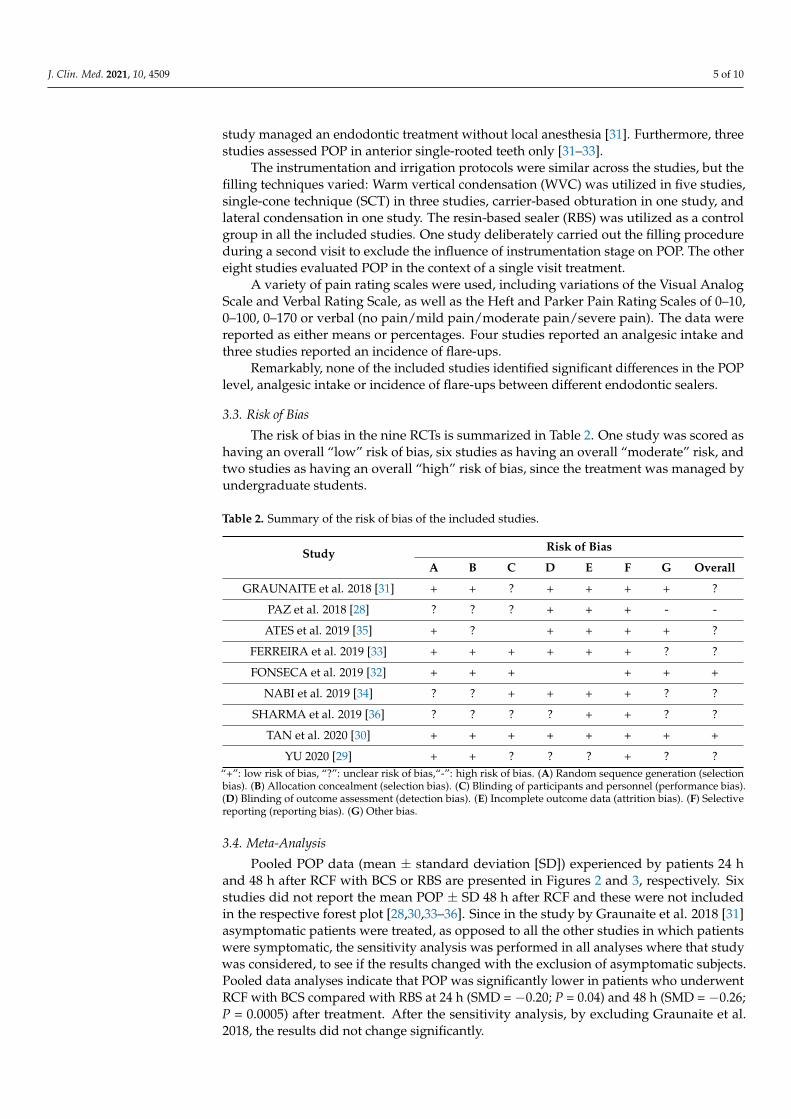
The risk of bias in the nine RCTs is summarized in Table 2. One study was scored ashaving an overall “low” risk of bias, six studies as having an overall “moderate” risk, andtwo studies as having an overall “high” risk of bias, since the treatment was managed byundergraduate students.
Table 2. Summary of the risk of bias of the included studies.
Study Risk of Bias
A B C D E F G Overall
GRAUNAITE et al. 2018 [31] + + ? + + + + ?
PAZ et al. 2018 [28] ? ? ? + + + - -
ATES et al. 2019 [35] + ? + + + + ?
FERREIRA et al. 2019 [33] + + + + + + ? ?
FONSECA et al. 2019 [32] + + + + + +
NABI et al. 2019 [34] ? ? + + + + ? ?
SHARMA et al. 2019 [36] ? ? ? ? + + ? ?
TAN et al. 2020 [30] + + + + + + + +
YU 2020 [29] + + ? ? ? + ? ?“+”: low risk of bias, “?”: unclear risk of bias,“-”: high risk of bias. (A) Random sequence generation (selectionbias). (B) Allocation concealment (selection bias). (C) Blinding of participants and personnel (performance bias).(D) Blinding of outcome assessment (detection bias). (E) Incomplete outcome data (attrition bias). (F) Selectivereporting (reporting bias). (G) Other bias.
3.4. Meta-Analysis
Pooled POP data (mean ± standard deviation [SD]) experienced by patients 24 hand 48 h after RCF with BCS or RBS are presented in Figures 2 and 3, respectively. Sixstudies did not report the mean POP ± SD 48 h after RCF and these were not includedin the respective forest plot [28,30,33–36]. Since in the study by Graunaite et al. 2018 [31]asymptomatic patients were treated, as opposed to all the other studies in which patientswere symptomatic, the sensitivity analysis was performed in all analyses where that studywas considered, to see if the results changed with the exclusion of asymptomatic subjects.Pooled data analyses indicate that POP was significantly lower in patients who underwentRCF with BCS compared with RBS at 24 h (SMD = −0.20; P = 0.04) and 48 h (SMD = −0.26;P = 0.0005) after treatment. After the sensitivity analysis, by excluding Graunaite et al.2018, the results did not change significantly.

J. Clin. Med. 2021, 10, 4509 6 of 10
J. Clin. Med. 2021, 10, x FOR PEER REVIEW 6 of 10
YU 2020 [29] + + ? ? ? + ? ? “+”: low risk of bias, “?”: unclear risk of bias,“-”: high risk of bias. (A) Random sequence genera-tion (selection bias). (B) Allocation concealment (selection bias). (C) Blinding of participants and personnel (performance bias). (D) Blinding of outcome assessment (detection bias). (E) Incomplete outcome data (attrition bias). (F) Selective reporting (reporting bias). (G) Other bias.
3.4 Meta-Analysis Pooled POP data (mean ± standard deviation [SD]) experienced by patients 24 and
48 h after RCF with BCS or RBS are presented in Figure 2 and Figure 3, respectively. Six studies did not report the mean POP ± SD 48 h after RCF and these were not included in the respective forest plot [28,30,33–36]. Since in the study by Graunaite et al. 2018 [31] asymptomatic patients were treated, as opposed to all the other studies in which patients were symptomatic, the sensitivity analysis was performed in all analyses where that study was considered, to see if the results changed with the exclusion of asymptomatic subjects. Pooled data analyses indicate that POP was significantly lower in patients who under-went RCF with BCS compared with RBS at 24 h (SMD = −0.20; P = 0.04) and 48 h (SMD = −0.26; P = 0.0005) after treatment. After the sensitivity analysis, by excluding Graunaite et al. 2018, the results did not change significantly.
Figure 2. Forest plot of POP level 24 h after RCF with BCS vs. RBS.
Figure 3. Forest plot of POP level 48 h after RCF with BCS vs. RBS.
The analgesic intake did not significantly differ between the BCS and RBS groups 24 h after RCF (RR = 0.46; P = 0.14; Figure 4). Five studies were not included in this forest plot due to a lack of available data [11–13,33,36]. The incidence of flare-up was also not signif-icantly different between the BCS and RBS groups (OR = 0.32; P = 0.24; Supplementary Materials Figure S1). After the sensitivity analysis, by excluding Graunaite et al. 2018, the results did not change significantly.
Figure 2. Forest plot of POP level 24 h after RCF with BCS vs. RBS.
J. Clin. Med. 2021, 10, x FOR PEER REVIEW 6 of 10
YU 2020 [29] + + ? ? ? + ? ? “+”: low risk of bias, “?”: unclear risk of bias,“-”: high risk of bias. (A) Random sequence genera-tion (selection bias). (B) Allocation concealment (selection bias). (C) Blinding of participants and personnel (performance bias). (D) Blinding of outcome assessment (detection bias). (E) Incomplete outcome data (attrition bias). (F) Selective reporting (reporting bias). (G) Other bias.
3.4 Meta-Analysis Pooled POP data (mean ± standard deviation [SD]) experienced by patients 24 and
48 h after RCF with BCS or RBS are presented in Figure 2 and Figure 3, respectively. Six studies did not report the mean POP ± SD 48 h after RCF and these were not included in the respective forest plot [28,30,33–36]. Since in the study by Graunaite et al. 2018 [31] asymptomatic patients were treated, as opposed to all the other studies in which patients were symptomatic, the sensitivity analysis was performed in all analyses where that study was considered, to see if the results changed with the exclusion of asymptomatic subjects. Pooled data analyses indicate that POP was significantly lower in patients who under-went RCF with BCS compared with RBS at 24 h (SMD = −0.20; P = 0.04) and 48 h (SMD = −0.26; P = 0.0005) after treatment. After the sensitivity analysis, by excluding Graunaite et al. 2018, the results did not change significantly.
Figure 2. Forest plot of POP level 24 h after RCF with BCS vs. RBS.
Figure 3. Forest plot of POP level 48 h after RCF with BCS vs. RBS.
The analgesic intake did not significantly differ between the BCS and RBS groups 24 h after RCF (RR = 0.46; P = 0.14; Figure 4). Five studies were not included in this forest plot due to a lack of available data [11–13,33,36]. The incidence of flare-up was also not signif-icantly different between the BCS and RBS groups (OR = 0.32; P = 0.24; Supplementary Materials Figure S1). After the sensitivity analysis, by excluding Graunaite et al. 2018, the results did not change significantly.
Figure 3. Forest plot of POP level 48 h after RCF with BCS vs. RBS.
The analgesic intake did not significantly differ between the BCS and RBS groups24 h after RCF (RR = 0.46; P = 0.14; Figure 4). Five studies were not included in thisforest plot due to a lack of available data [11–13,33,36]. The incidence of flare-up wasalso not significantly different between the BCS and RBS groups (OR = 0.32; P = 0.24;Supplementary Materials Figure S1). After the sensitivity analysis, by excluding Grau-naite et al. 2018, the results did not change significantly.
J. Clin. Med. 2021, 10, x FOR PEER REVIEW 7 of 10
Figure 4. Forest plot of analgesic intake 24 h after RCF with BCS vs. RBS.
The next two diagrams underline the pain prevalence and severity of the BCS group over the RBS group in 24 h (Supplementary Materials Figure S2) and 48 h (Supplementary Materials Figure S3) after RCF.
The probability of “No pain” 24 h after treatment was 1.12 × higher in the BCS group vs. the RBS group (OR = 1.12; 95% CI, 0.77–1.64; P = 0.86), while the same was observed for “Moderate pain” probability (OR = 1.21; 95% CI, 0.61–2.38; P = 0.59). There was no heterogeneity in the study effect for the BCS and RBS groups (I² = 0%; P = 0.86, and I² = 0%; P = 0.89, respectively), indicating perfect consistency in the results. The probability of “Mild pain” and “Severe pain” was 1.2 and 1.7 × higher in the RBS group vs. the BCS group, respectively (OR = 0.83; 95% CI, 0.54–1.25; P = 0.37, and OR = 0.59; 95% CI, 0.08–4.58; P = 0.62). There was also no heterogeneity in the study effect in either group (I² = 0%; P = 0.97, and I² = 0%; P = 0.61, respectively; Supplementary Materials Figure S2).
A diagram of pain characteristics in the BCS and RBS groups 48 h after treatment is presented in Supplementary Materials Figure S3. The studies that did not report pain characteristics 48 h after treatment were excluded. The probability of “No pain” 48 h after treatment was 1.21× higher in the BCS group vs. the RBS group (OR = 1.21; 95% CI, 0.60–2.42; P = 0.60; heterogeneity: I² = 18%; P = 0.30). “Mild pain” was more commonly reported in the RBS group vs. the BCS group 48 h after treatment (OR = 0.82; 95% CI, 0.40–1.68; P = 0.59; heterogeneity: I² = 1%; P = 0.40), while “Moderate pain” was equally likely in both groups 48 h after treatment (OR = 1.00; 95% CI, 0.14–7.27; P = 1.00; heterogeneity: I² = 0%; P = 0.33). “Severe pain” was reported only once in both groups (Supplementary Materials Figure S3). After the sensitivity analysis, by excluding Graunaite et al. 2018, the results did not change significantly neither for data after 24 h nor for 48 h after RCT.
The effect of the obturation technique (WVT vs. SCT) on POP based on data 24 and 48 h after RCF is presented in Supplementary Materials Figures S4 and S5. The studies that did not report 48-h data were excluded. There was a numerical difference in POP in favor of BCS for both WVT and SCT subgroups 24 h after RCF (OR = 0.85; P = 0.41). The probability of POP was numerically higher in the RBS subgroup that underwent the WVT technique (P = 0.49) in 1.2 × and lower in the BCS subgroup that underwent the SCT tech-nique (P = 0.64; Supplementary Materials Figure S4). There was evidence of lower POP in both WVT and SCT subgroups of the BCS group 48 h after RCF (P = 0.33). The probability of POP did not differ according to the obturation technique in the BCS group, but was 1.3 × higher in the RBS group 48 h after RCF (OR=0.78; P = 0.33; Supplementary Materials Figure S5).
The probability of POP by the pulp status (vital [V] and non-vital [NV] pulp) 24 and 48 h after RCF is presented in Supplementary Materials Figures S6 and S7, respectively. The studies that did not report 48-h data were excluded. There was evidence of a non-significant difference in POP in favor of RBS in the V subgroup (P = 0.50) and a trend for lower POP in NV teeth within the BCS group 24 h after RCF (P = 0.16). However, there was no statistically significant overall effect of V vs. NV pulp on POP (OR = 0.84; P = 0.45;
Figure 4. Forest plot of analgesic intake 24 h after RCF with BCS vs. RBS.
The next two diagrams underline the pain prevalence and severity of the BCS groupover the RBS group in 24 h (Supplementary Materials Figure S2) and 48 h(Supplementary Materials Figure S3) after RCF.
The probability of “No pain” 24 h after treatment was 1.12× higher in the BCS groupvs. the RBS group (OR = 1.12; 95% CI, 0.77–1.64; P = 0.86), while the same was observedfor “Moderate pain” probability (OR = 1.21; 95% CI, 0.61–2.38; P = 0.59). There was noheterogeneity in the study effect for the BCS and RBS groups (I2 = 0%; P = 0.86, and I2 = 0%;P = 0.89, respectively), indicating perfect consistency in the results. The probability of“Mild pain” and “Severe pain” was 1.2× and 1.7× higher in the RBS group vs. the BCSgroup, respectively (OR = 0.83; 95% CI, 0.54–1.25; P = 0.37, and OR = 0.59; 95% CI, 0.08–4.58;P = 0.62). There was also no heterogeneity in the study effect in either group (I2 = 0%;P = 0.97, and I2 = 0%; P = 0.61, respectively; Supplementary Materials Figure S2).

J. Clin. Med. 2021, 10, 4509 7 of 10
A diagram of pain characteristics in the BCS and RBS groups 48 h after treatmentis presented in Supplementary Materials Figure S3. The studies that did not report paincharacteristics 48 h after treatment were excluded. The probability of “No pain” 48 hafter treatment was 1.21× higher in the BCS group vs. the RBS group (OR = 1.21; 95% CI,0.60–2.42; P = 0.60; heterogeneity: I2 = 18%; P = 0.30). “Mild pain” was more commonlyreported in the RBS group vs. the BCS group 48 h after treatment (OR = 0.82; 95% CI,0.40–1.68; P = 0.59; heterogeneity: I2 = 1%; P = 0.40), while “Moderate pain” was equallylikely in both groups 48 h after treatment (OR = 1.00; 95% CI, 0.14–7.27; P = 1.00; heterogene-ity: I2 = 0%; P = 0.33). “Severe pain” was reported only once in both groups (SupplementaryMaterials Figure S3). After the sensitivity analysis, by excluding Graunaite et al. 2018, theresults did not change significantly neither for data after 24 h nor for 48 h after RCT.
The effect of the obturation technique (WVT vs. SCT) on POP based on data 24 h and48 h after RCF is presented in Supplementary Materials Figures S4 and S5. The studies thatdid not report 48-h data were excluded. There was a numerical difference in POP in favor ofBCS for both WVT and SCT subgroups 24 h after RCF (OR = 0.85; P = 0.41). The probabilityof POP was numerically higher in the RBS subgroup that underwent the WVT technique(P = 0.49) in 1.2× and lower in the BCS subgroup that underwent the SCT technique(P = 0.64; Supplementary Materials Figure S4). There was evidence of lower POP in bothWVT and SCT subgroups of the BCS group 48 h after RCF (P = 0.33). The probability of POPdid not differ according to the obturation technique in the BCS group, but was 1.3× higherin the RBS group 48 h after RCF (OR = 0.78; P = 0.33; Supplementary Materials Figure S5).
The probability of POP by the pulp status (vital [V] and non-vital [NV] pulp) 24 h and48 h after RCF is presented in Supplementary Materials Figures S6 and S7, respectively.The studies that did not report 48-h data were excluded. There was evidence of a non-significant difference in POP in favor of RBS in the V subgroup (P = 0.50) and a trend forlower POP in NV teeth within the BCS group 24 h after RCF (P = 0.16). However, therewas no statistically significant overall effect of V vs. NV pulp on POP (OR = 0.84; P = 0.45;Supplementary Materials Figure S6). The probability of POP was 2× higher in the V pulpof the BCS group, and there was evidence of lower POP in the V subgroup of the RBS group48 h after RCF (OR = 2.01; P = 0.18). There was also a non-significant trend for lower POP inNV teeth within the BCS group (OR = 0.92; P = 0.84; Supplementary Materials Figure S7).After the sensitivity analysis, by excluding Graunaite et al. 2018, the results did notchange significantly.
Pooled data from the three studies that reported the retreatment show a trend for adifference in POP in favor of RBS (OR = 1.20; P = 0.65) 24 h after RCF (Supplementary Mate-rials Figure S8). The meta-analysis determined that there was a 48% level of heterogeneitywithin the included nine studies. Only one study had analyzed a consistent number ofcases [31]. Supplementary Materials Figure S9 presents POP in retreatment groups 48 hafter RCF. Unfortunately, the limited numbers of studies and retreated cases are insufficientto determine any effect of BCS vs. RBS on POP (OR = 1.34).
There was no significant difference in POP between the BCS and RBS groups whenthe treatment was carried out over single (OR = 0.77; P = 0.28) or multiple visits 24 h afterRCF (OR = 0.81; P = 0.50; Supplementary Materials Figure S10). A lack of data precludedthe equivalent analysis of POP 48 h after RCF.
4. Discussion
This meta-analysis of nine pooled RCTs indicates that POP was significantly lowerafter RCF with BCS compared with RBS. However, none of the RCTs individually reportedany significant effect of BSC vs. RBS on POP [28–36].
In this analysis, we found that BCS was non-significantly correlated with reducedanalgesic intake vs. RBS, an observation that was also reported by one of the includedstudies [35] using the warm-obturator filling technique. However, two of the other includedRCTs [32,33] demonstrated comparable analgesic intake in the control and experimentalgroups after RCF using SCT.
J. Clin. Med. 2021, 10, 4509 8 of 10
This systematic review found a non-significant trend of reduced flare-up in the BCSgroup vs. the RBS group. This is supported by one of the included RCTs that reported asignificant reduction of flare-up following RCF with BCS vs. RBS [35]. However, anotherreported an equal occurrence between groups [31].
Regarding the warm and cold filling techniques, SCT with BCS has previously beenassociated with higher POP, while WVT with RBS has been associated with the lowest POPscores [28]. However, our pooled analysis suggests that there is a non-significant trend infavor of BCS.
Our results indicate that POP was lower in the V pulp when filled with RBS and in theNV pulp when filled with BCS. However, we found no additional background literature toplace this in context.
Our results also evidence a non-significant difference in POP in favor of RBS atretreatment. Moreover, the only RCT [31] included to report this parameter indicates nodifference between filling techniques in POP, following the retreatment procedures.
According to our results, the trend for lower POP following RCF with BCS vs. RBSfilling technique was observed across single and multiple visit treatments. However, thereare no studies in the literature to provide additional context.
The main limitations of this review are inter-study variability and inconsistency, aswell as a lack of clinically relevant outcomes. Furthermore, as mentioned, different scalesfor pain measurement were used in different studies. Though the authors made efforts toresize all the scales to a 1–10 scale, it is difficult to understand if this had a relevance in theresults. Of course, for future studies, it is recommended to use only scales for which thereis an overall consensus. Therefore, the findings presented here need to be confirmed byfurther well-designed studies and should be interpreted with caution.
5. Conclusions
Our findings suggest that the BCS filling technique may positively affect POP, whilethere was a trend of a beneficial effect for analgesic intake, incidence of flare-up, pulpstatus, and number of visits when using BCS, compared with RBS. However, due toseveral limitations in these analyses, further well-designed clinical studies are warrantedto supplement our results.
Supplementary Materials: The following are available online at https://www.mdpi.com/article/10.3390/jcm10194509/s1, Table S1: Excluded studies with reasons for exclusion, Figure S1: Forestplot of flare-up after RCF with BCS vs. RBS, Figure S2: Diagram of pain characteristics summary in asubgroup of BCS and RBS traditional filling technique groups 24 h after RCF, Figure S3: Diagram ofpain characteristics summary in a subgroup of BCS and RBS traditional filling technique groups 48 hafter RCF, Figure S4: Forest plot of POP in patients treated with warm and cold filling techniques24 h after RCF with BCS vs. RBS, Figure S5: Forest plot of POP in patients treated with warm andcold techniques 48 h after RCF with BCS vs. RBS, Figure S6: Forest plot of POP in vital and non-vitalpulp 24 h after RCF with BCS vs. RBS, Figure S7: Forest plot of POP in vital and non-vital pulp 48 hafter RCF with BCS vs. RBS, Figure S8: Forest plot of POP following retreatment 24 h after RCFwith BCS vs. RBS, Figure S9: Forest plot of POP following retreatment 48 h after RCF with BCS vs.RBS, Figure S10: Forest plot of POP 24 h after RCF with BCS vs. RBS following a single or multiplevisit treatment.
Author Contributions: Conceptualization, D.P., M.D.F. and E.B.; methodology, D.P., M.D.F. and E.B.;software, E.M., M.A., N.S., A.C., M.C. and M.T.; data analysis, E.M., M.A., N.S., A.C., M.C. and M.T.;visualization, E.M., M.A., N.S., A.C., M.C. and M.T.; supervision and project administration, D.P.,M.D.F. and E.B.; writing, E.M., D.P. and M.D.F.; reviewing and editing, D.P., M.D.F., E.M., M.A. andE.B. All authors have read and agreed to the published version of the manuscript.
Funding: This research received no external funding.
Institutional Review Board Statement: Not applicable.
Informed Consent Statement: Not applicable.

J. Clin. Med. 2021, 10, 4509 9 of 10
Conflicts of Interest: The authors declare no conflict of interest.
References1. Nosrat, A.; Dianat, O.; Verma, P.; Nixdorf, D.R.; Law, A.S. Postoperative Pain: An Analysis on Evolution of Research in
Half-Century. J. Endod. 2020, 47, 358–365. [CrossRef]2. Nagendrababu, V.; Gutmann, J.L. Factors associated with postobturation pain following single-visit nonsurgical root canal
treatment: A systematic review. Quintessence Int. 2017, 48, 193–208. [CrossRef] [PubMed]3. Comparin, D.; Moreira, E.J.L.; Souza, E.M.; De-Deus, G.; Arias, A.; Silva, E.J.N.L. Postoperative Pain after Endodontic Retreatment
Using Rotary or Reciprocating Instruments: A Randomized Clinical Trial. J. Endod. 2017, 43, 1084–1088. [CrossRef] [PubMed]4. Demenech, L.S.; de Freitas, J.V.; Tomazinho, F.S.F.; Baratto-Filho, F.; Gabardo, M.C.L. Postoperative Pain after Endodontic
Treatment under Irrigation with 8.25% Sodium Hypochlorite and Other Solutions: A Randomized Clinical Trial. J. Endod. 2020,47, 696–704. [CrossRef] [PubMed]
5. Moreira, M.S.; Anuar, A.S.N.-S.; Tedesco, T.K.; dos Santos, M.; Morimoto, S. Endodontic Treatment in Single and Multiple Visits:An Overview of Systematic Reviews. J. Endod. 2017, 43, 864–870. [CrossRef] [PubMed]
6. Sathorn, C.; Parashos, P.; Messer, H. The prevalence of postoperative pain and flare-up in single- and multiple-visit endodontictreatment: A systematic review. Int. Endod. J. 2008, 41, 91–99. [CrossRef]
7. Raghavendra, S.S.; Jadhav, G.R.; Gathani, K.M.; Kotadia, P. Bioceramics in endodontics—A review. J. Istanb. Univ. Fac. Dent. 2017,51, S128–S137. [CrossRef]
8. Komabayashi, T.; Colmenar, D.; Cvach, N.; Bhat, A.; Primus, C.; Imai, Y. Comprehensive review of current endodontic sealers.Dent. Mater. J. 2020, 39, 703–720. [CrossRef] [PubMed]
9. Vouzara, T.; Dimosiari, G.; Koulaouzidou, E.A.; Economides, N. Cytotoxicity of a New Calcium Silicate Endodontic Sealer.J. Endod. 2018, 44, 849–852. [CrossRef] [PubMed]
10. Higgins, J.P.T.; Thomas, J.; Chandler, J.; Cumpston, M.; Li, T.; Page, M.J.; Welch, V.A. Cochrane Handbook for Systematic Reviews ofInterventions, 2nd ed.; John Wiley & Sons: Chichester, UK, 2019.
11. Niang, S.O.; Bane, K.; Sarr, M.; Tourè, B.; Machtou, P. Technical quality and postoperative pain of single visit endodontic treatmentof chronic apical periodontitis filled by bioceramic sealer. IP Indian J. Conserv. Endod. 2018, 3, 92–97. [CrossRef]
12. Filipov, I.; Zagorchev, P.; Dimitrova, S.; Manchorova-Veleva, N. Postoperative pain in cold and warm endo-techniques usingbioceramic sealer. In Proceedings of the IADR Poster Session, London, UK, 25–28 July 2018.
13. Reeru, S.; Shresha, D.; Kayasatha, R. Postoperative pain and associated factors in patients undergoing single visit root canaltreatment on teeth with vital pulp. Kathmandu Univ. Med. 2018, 16, 220–223.
14. Arias, A.; de la Macorra, J.C.; Hidalgo, J.J.; Azabal, M. Predictive models of pain following root canal treatment: A prospectiveclinical study. Int. Endod. J. 2013, 46, 784–793. [CrossRef]
15. Wang, C.; Xu, P.; Ren, L.; Dong, G.; Ye, L. Comparison of post-obturation pain experience following one-visit and two-visit rootcanal treatment on teeth with vital pulps: A randomized controlled trial. Int. Endod. J. 2010, 43, 692–697. [CrossRef] [PubMed]
16. Sadaf, D.; Ahmad, M.Z. Factors associated with postoperative pain in endodontic therapy. Int. J. Biomed. Sci. 2014, 10, 243–247.17. Prashanth, M.B.; Tavane, P.N.; Abraham, S.; Chacko, L. Comparative evaluation of pain, tenderness and swelling followed by
radiographic evaluation of periapical changess at various intervals of time following single and multiple visit endodontic therapy:An in vivo study. J. Contemp. Dent. Pract. 2011, 12, 187–191. [CrossRef] [PubMed]
18. Shashirekha, G.; Jena, A.; Pattanaik, S.; Rath, J. Assessment of pain and dissolution of apically extruded sealers and their effect onthe periradicular tissues. J. Conserv. Dent. 2018, 21, 546–550. [PubMed]
19. Nunes, E.C.; Herkrath, F.J.; Suzuki, E.H.; Gualberto, E.C., Jr.; Marques, A.A.F.; Sponchiado, E.C., Jr. Comparison of the effect ofphotobiomodulation therapy and Ibuprofen on postoperative pain after endodontic retreatment: Randomized, controlled clinicalstudy. Lasers Med. Sci. 2020, 35, 971–978. [CrossRef] [PubMed]
20. Asgary, S.; Eghbal, M.J. The effect of pulpotomy using a Calcium-enriched mixture cement versus one-visit root canal therapy onpostoperative pain relief in irreversible pulpitis: A randomized clinical trial. Odontology 2010, 98, 125–133. [CrossRef] [PubMed]
21. Rao, K.N.; Kandaswamy, R.; Umashetty, G.; Rathore, V.P.S.; Hotkar, C.; Patil, B.S. Post-obturation pain following one-visit andtwo-visits root canal treatment in necrotic anterior teeth. J. Int. Oral Health 2014, 6, 28–32. [CrossRef] [PubMed]
22. Yoshinari, F.M.S.; Pereira, S.; Beraldo, D.Z.; da Silva, J.C.L.; Zafalon, E.J.; da Silva, P.G. Influence of photodinamic therapy in thecontrol of postoperative pain in endodontic treatment: A cross-sectional randomized clinical trial. Pesqui. Bras. OdontopediatriaClínica Integr. 2019, 19, e4369. [CrossRef]
23. Hepsenoglu, Y.E.; Tan, F.E.; Ozcan, M. Postoperative pain intensity after single- versus two-visit nonsurgical endodonticretreatment: A randomized clinical trial. J. Endod. 2018, 44, 1339–1346. [CrossRef] [PubMed]
24. Demirci, G.K.; Chalishkan, M.K. A prospective randomized comparative study of cold lateral condensattion versus core/gutta-percha in teeth with periapical lesions. J. Endod. 2016, 42, 206–210. [CrossRef]
25. Yaylali, I.E.; Kurnaz, S.; Tunca, Y.M. Maintaining apical patency does not increase postoperative pain in molars with necrotic pulpand apical periodontitis: A randomized controlled trial. J. Endod. 2018, 44, 335–340. [CrossRef] [PubMed]
26. Dhyani, V.K.; Chhabra, S.; Sharma, V.K.; Dhyani, A. A randomized controlled trial to evaluate the incidence of postoperative painand flare-ups in single and multiple visits root canal treatment. Med. J. Armed Forces India 2020. [CrossRef]

J. Clin. Med. 2021, 10, 4509 10 of 10
27. Gudlavalleti, B.; Patil, A.A. Comparative evaluation of postoperative pain after root canal treatment using three different sealers,Viz., Tubli-Seal EWT, Apexit Plus, AH Plus: An in-vivo study. J. Clin. Diag. Res. 2020, 14, ZC04–ZC09. [CrossRef]
28. Paz, A.; Vasconcelos, I.; Ginjeira, A. Evaluation of postoperative pain after using bioceramic materials as endodontic sealers.EC Dent. Sci. 2018, 17, 1739–1748.
29. Yu, Y.-H.; Kushnir, L.; Kohli, M.; Karabucak, B. Comparing the incidence of postoperative pain after root canal filling with warmvertical obtuation with resin-based sealer and sealer-based obturation with calcium silicate-based sealer: A prospective clinicaltrial. Clin. Oral Investig. 2021, 25, 5033–5042. [CrossRef] [PubMed]
30. Tan, H.S.G.; Lim, K.C.; Lui, J.N.; Lai, W.M.C.; Yu, V.S.H. Postobturation Pain Associated with Tricalcium Silicate and Resin-basedSealer Techniques: A Randomized Clinical Trial. J. Endod. 2021, 47, 169–177. [CrossRef] [PubMed]
31. Graunaite, I.; Skucaite, N.; Lodiene, G.; Agentiene, I.; Machiulskiene, V. Effect of resin-based and bioceramic root canal sealers onpostoperative pain. J. Endod. 2018, 44, 689–693. [CrossRef]
32. Fonseca, B.; Coelho, M.S.; da Bueno, C.E.S.; Fontana, C.E.; De Martin, A.S.; Rocha, D.G.P. Assessment of extrusion andpostoperative pain of a bioceramic and resin-based root canal sealer. Eur. J. Dent. 2019, 13, 343–348. [CrossRef]
33. De Ferreira, N.S.; Gollo, E.K.F.; Boscato, N.; Arias, A.; da Silva, E.J.N.L. Postoperative pain after root canal filling with differentendodontic sealers: A randomized clinical trial. Braz. Oral Res. 2020, 34, e069. [CrossRef] [PubMed]
34. Nabi, S.; Farooq, R.; Purra, A.; Ahmed, F. Comparison of various sealers on postoperative pain in single-visit endodontics: Arandomized clinical study. Indian J. Dent. Sci. 2019, 11, 99–102. [CrossRef]
35. Ates, A.A.; Dumani, A.; Yoldas, O.; Unal, I. Post-obturation pain following the use of carrier-based system with AH Plus or iRootSP sealers: A randomized controlled clinical trial. Clin. Oral Investig. 2019, 23, 3053–3061. [CrossRef] [PubMed]
36. Sharma, N.; Mandhotra, P.; Kumari, S.; Chandel, N. A study to compare various sealers on postoperative pain in single visitendodontics. Ann. Int. Med. Dent. Res. 2019, 6, 40–42.