Neuromodulation Advances for Seizure Control
18
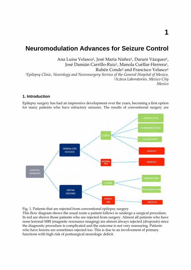
1 Neuromodulation Advances for Seizure Control Ana Luisa Velasco 1 , José María Núñez 1 , Daruni Vázquez 1 , José Damián Carrillo-Ruiz 1 , Manola Cuéllar-Herrera 1 , Rubén Conde 2 and Francisco Velasco 1 1 Epilepsy Clinic, Neurology and Neurosurgery Service of the General Hospital of Mexico, 2 Azteca Laboratories, Mexico City Mexico 1. Introduction Epilepsy surgery has had an impressive development over the years, becoming a first option for many patients who have refractory seizures. The results of conventional surgery are Fig. 1. Patients that are rejected from conventional epilepsy surgery This flow diagram shows the usual route a patient follows to undergo a surgical procedure. In red are shown those patients who are rejected from surgery. Almost all patients who have none lesional MRI (magnetic resonance imaging) are almost always rejected (dropouts) since the diagnostic procedure is complicated and the outcome is not very reassuring. Patients who have lesions are sometimes rejected too. This is due to an involvement of primary functions with high risk of postsurgical neurologic deficit.
-
Upload
independent -
Category
Documents
-
view
3 -
download
0
Transcript of Neuromodulation Advances for Seizure Control
1
Neuromodulation Advances for Seizure Control Ana Luisa Velasco1, José María Núñez1, Daruni Vázquez1,
José Damián Carrillo-Ruiz1, Manola Cuéllar-Herrera1, Rubén Conde2 and Francisco Velasco1
1Epilepsy Clinic, Neurology and Neurosurgery Service of the General Hospital of Mexico, 2Azteca Laboratories, Mexico City
Mexico
1. Introduction Epilepsy surgery has had an impressive development over the years, becoming a first option for many patients who have refractory seizures. The results of conventional surgery are
Fig. 1. Patients that are rejected from conventional epilepsy surgery This flow diagram shows the usual route a patient follows to undergo a surgical procedure. In red are shown those patients who are rejected from surgery. Almost all patients who have none lesional MRI (magnetic resonance imaging) are almost always rejected (dropouts) since the diagnostic procedure is complicated and the outcome is not very reassuring. Patients who have lesions are sometimes rejected too. This is due to an involvement of primary functions with high risk of postsurgical neurologic deficit.
Topics in Neuromodulation Treatment
4
excellent thanks to experience, and better diagnostic and surgical techniques. Nevertheless, there are a number of patients who are rejected due to several reasons, for example bilateral or multiple epileptic foci, focus involving primary functional areas of the brain, generalized seizures or non lesional imaging studies. It has been calculated that there is a 30% of patients who need surgery that have to be rejected. These patients need a surgical alternative, and neuromodulation might be the answer to many of them. (Figure 1).
Even though neuromodulation has been proposed as an ideal surgical alternative for a number of neurological disorders, it’s application for the control of epileptic seizures would seem to be the most appropriate therapeutic indication since epilepsy is considered in most cases a functional disorder. Several targets have been proposed to control seizures. Although there might be discrepancies about which target is the best, all authors agree that neuromodulation improves seizure control without deteriorating neurological functions. Many studies have been performed in different epilepsy surgery centers in the world proposing several seizure types that respond, different and stimulation modes. This chapter intends to discuss a number of questions that arise in the use of this method.
2. Who are the best candidates for neuromodulation? As with all surgical procedures that are used to treat refractory seizures, patient selection is the first step to ensure a good outcome. If we err to recognize the appropriate candidate, even the best surgery is a failure. This principle applies to neuromodulation. Furthermore, neuromodulation is expensive and needs high to be performed.
In the future, neuromodulation might become the first option when considering a surgical method for refractory epilepsy. This is due to its low surgical risk and reversible qualities. But for now that ablative surgery is less expensive and shows good results. Criteria to select a patient are the following:
Primary generalized seizures Multifocal or bilateral foci Seizures arising from eloquent areas (motor, memory and language for example)
In all the above clinical settings, conventional surgery has proven to be risky due to the fact that it can be a major surgery with high probability of infection, bleeding or loss of neural function. It is also less effective since surgeries tend to be restricted to avoid loss of function and hence residual seizures or relapses take place. If we add that there are a number of patients with non-lesional MRI, the risk and outcome is worse and patients are discarded from surgical options.
So the seizure type is decisive for the selection of the stimulation target. If we select a patient who has generalized seizures and we miss the fact that he has partial seizures with secondary generalized seizures, the patient will have a moderate improvement since generalized seizures will disappear but the partial and complex partial component will remain. So if we perform a seizure count, the number of seizures may turn out to be the same as before stimulation.
There are other considerations that we have to take into account. This procedure will need follow-up for several weeks to ensure good results. It is convenient that the patient and his caregiver understand the importance of follow-up appointments. It is desirable that the patient lives near the epilepsy center or at least has easy access to personnel that can check if the stimulator is working appropriately or to adjust the stimulation parameters. They must
Neuromodulation Advances for Seizure Control
5
be willing to return with the caregivers if stimulation fails to ensure proper function of the whole system (see follow-up section at the end of the chapter).
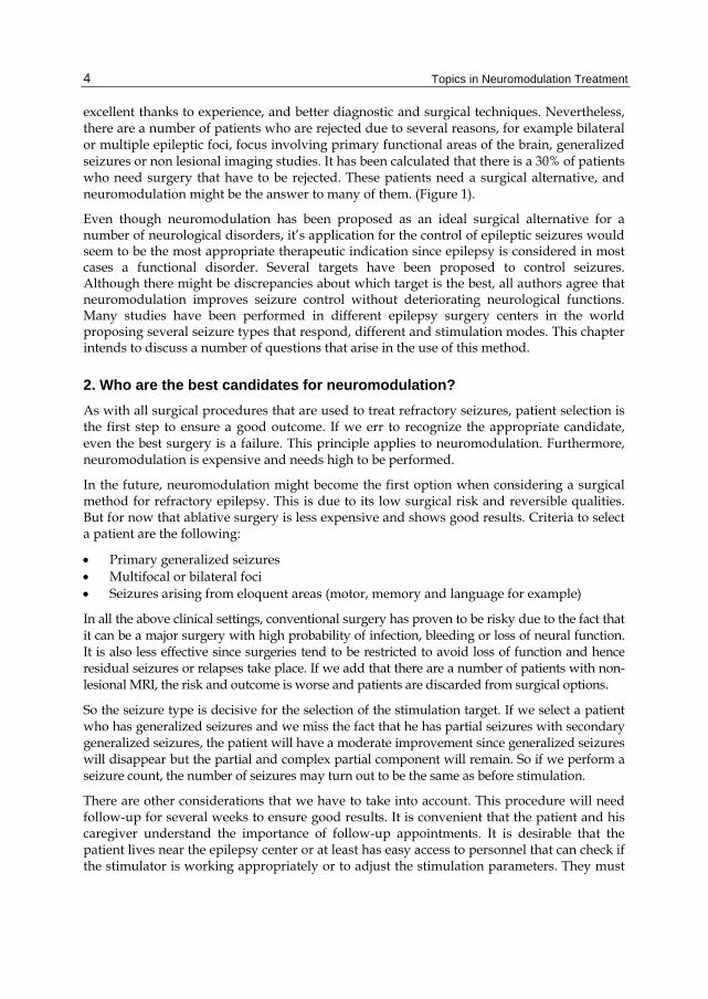
Fig. 2. Surgical evaluation of the epileptic patient This figure depicts the clinical flow starting from the moment a patient is considered a surgical candidate. Presurgical evaluation in our Epilepsy Clinic includes a detailed medical history, EEG, in some cases video EEG with or without sphenoidal electrodes, MRI, PET/SPECT, functional MRI and neuropsyhcological testing. The extrasurgical evalation refers to phase II testing that includes continuous video EEG monitoring and brain mapping with electrical stimulation. Follow up includes the postsurgical testing: EEG, MRI, neuropsychological batteries.
Patient and caregivers must also have a clear idea of what to expect. Seizures do not disappear as soon as the stimulator is turned on. There is a variable period of time to obtain seizure reduction. This period of time varies according to the stimulated target and stimulation mode. If the cerebellum or vagus nerve are chosen, the best effect will take years to be reached, if the thalamus is stimulated, seizure reduction will take from 3 to 6 months to be achieved, if the target is hippocampal focus, the time span is reduced to 2 to 6 weeks if the hippocampus shows no signs of sclerosis in the MRI. If we stimulate the cortical epileptic focus in a programmed cyclic mode, seizures diminish in a matter of days (Velasco et al 2009) and if the mode is based on a closed loop fashion seizures take months to years to decrease (Fountas and Smith 2007).
Topics in Neuromodulation Treatment
6
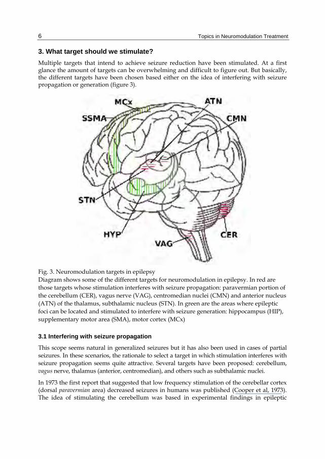
3. What target should we stimulate? Multiple targets that intend to achieve seizure reduction have been stimulated. At a first glance the amount of targets can be overwhelming and difficult to figure out. But basically, the different targets have been chosen based either on the idea of interfering with seizure propagation or generation (figure 3).
Fig. 3. Neuromodulation targets in epilepsy Diagram shows some of the different targets for neuromodulation in epilepsy. In red are those targets whose stimulation interferes with seizure propagation: paravermian portion of the cerebellum (CER), vagus nerve (VAG), centromedian nuclei (CMN) and anterior nucleus (ATN) of the thalamus, subthalamic nucleus (STN). In green are the areas where epileptic foci can be located and stimulated to interfere with seizure generation: hippocampus (HIP), supplementary motor area (SMA), motor cortex (MCx)
3.1 Interfering with seizure propagation
This scope seems natural in generalized seizures but it has also been used in cases of partial seizures. In these scenarios, the rationale to select a target in which stimulation interferes with seizure propagation seems quite attractive. Several targets have been proposed: cerebellum, vagus nerve, thalamus (anterior, centromedian), and others such as subthalamic nuclei.
In 1973 the first report that suggested that low frequency stimulation of the cerebellar cortex (dorsal paravermian area) decreased seizures in humans was published (Cooper et al, 1973). The idea of stimulating the cerebellum was based in experimental findings in epileptic
Neuromodulation Advances for Seizure Control
7
seizures induced in cats (Dow et al 1962). Even though results were controversial at first, a review of different studies with a total of 129 patients showed that 49% had significant seizure reduction, 27% being seizure free. This and other studies (Velasco et al 2005) have shown that the seizure type that best responds is primary generalized tonic clonic seizures and atypical absences and that even though there is an initial seizure reduction within the first two months of stimulation, the effect is not only maintained but there is a further decrease with time.
Vagus nerve stimulation is not very invasive since reaching the nerve at the neck level is a relatively simple procedure. It has been used in all types of refractory seizures. Its effects on seizure reduction are modest (~ 30 %). Even though the precise antiepileptic mechanism remains unclear, it appears that the thalamocortical relay neurons modulate cortical excitability, influencing seizure generation or propagation (Ben-Menachem 2002). It can produce dysphonia, headache and increases peptic ulcer and insulin dependant diabetes mellitus. It is not recommended for children under 12 years old.
According to the centro-encephalic theory by Penfield and Jasper (1954) high frequency stimulation of non-specific thalamic nuclei (such as centro-median or anterior thalamic nuclei) interferes with propagation of cortical or subcortically-initiated seizures. In 1984 Velasco et al performed the first bilateral centromedian electrode implantation in a 12-year-old boy with severe generalized seizures of the Lennox-Gastaut Syndrome. There was a considerable seizure reduction and impressive improvement in his intellectual status. Five years later they published a report of children and adults with generalized seizures of the Lennox-Gastaut Syndrome (Velasco 1989). The best results are obtained in the parvocellular portion. Neurophysiologic definition of the target is mandatory. This definition is based on electro cortical responses elicited by stimulation of the electrode contacts within different zones of the centromedian nucleus (Velasco F et al 2000, Velasco AL et al 2006). CM stimulation is more effective in generalized seizures and epilepsia partialis continua. When the patient selection as well as the anatomic and neurophysiologic criteria regarding the target localization are optimal, the results are >80% seizure reduction, some patients have become seizure free. An improvement in ability scales is also observed with no adverse effects.
As described above, the anterior nucleus is a non-specific thalamic nucleus and as such, interferes with propagation of cortical or subcortical initiated seizures but it also interferes with seizures initiated in mesial temporal structures and propagated through the fornix, mammillary body and anterior nucleus of the thalamus (Mirski and Fisher 1994). The SANTE study Group has reported the results of the bilateral stimulation of the anterior nucleus of the thalamus (Fisher et al 2010). Its best results have been obtained in complex partial and secondary generalized seizures, wich were reduced reduced by stimulation. By 2 years, there was a 56% median percent reduction in seizure frequency.
The subthalamic nucleus has been stimulated for seizure control based on the suppressive effects of pharmacological or electrical inhibition of the STN seen on different types of seizure in animal models of epilepsy (Chabardès 2002). It is considered that inhibition of the subthalamic nucleus causes activation of an endogenous epilepsy control system referred to as the nigral control of epilepsy system (Gale and Iadarola 1980). Patients with frontal lobe and myoclonic seizures are the best responders. Improvement varies from 30 to 80% (Vesper et al 2007). Mild facial twitching and paresthesias in legs and arms responded to adjustment of the stimulation parameters.
Topics in Neuromodulation Treatment
8
3.2 Interfering with seizure generation
Neuromodulation of the CM nuclei had already demonstrated anticonvulsive effects by stopping secondary tonic clonic generalized phase in patients with intractable mesial temporal lobe epilepsy but unfortunately it spared complex partial component that originated in the hippocampus (Velasco 1993a), thus patients continued to have disabling epilepsy. Based on the studies of Weiss et al (1998), Velasco et al (2000) started a project to demonstrate that the hippocampal stimulation would be a better alternative than trying to interfere with epileptic activity propagation.
Various authors (Boon et al 2007, Velasco et al 2007) have shown the beneficial effects in seizure reduction in patients with hippocampal foci stimulation; best responders are those patients whose epileptic focus can be pinpointed with hippocampal electrodes and thus stimulated directly and have non-lesional MRI. In these latter cases seizure reduction is >90% with several patients being seizure free.
Wyckhuys T et al (2007) found that high frequency stimulation of the hippocampus in kindled rats increases after-discharge thresholds with the consequent seizure reduction. There are some studies that have shown evidence that neuromodulation works by inhibiting the stimulated area. Clinical studies using neurophysiologic testing, single positron emission tomography and benzodiazepine receptor binding studies show that an inhibitory mechanism could explain seizure control (Velasco 2000, Cuéllar-Herrera 2004). Based in these observations, another challenge for epileptologists, the presence of intractable partial seizures arising from eloquent areas, was approached.
Cortical stimulation for motor seizures has been performed in two modes: an open (Velasco et al 2009) and closed loop (Sun et al 2008). The first step is to localize the epileptic focus implanting first a diagnostic grid and when done, implanting a permanent electrode directed to the focus. In case of the open loop mode, a significant (>90%) seizure decrease takes place over several days and effect persists thereafter, the stimulation mode is pre-established in a 24 hr cyclic 1 min ON, 4 min OFF. When the closed loop mode is used a seizure detection system is implanted too so that every time a seizure is detected, the stimulation therapy is delivered. In the latter mode, seizure reduction takes a longer period of time to produce a modest difference.
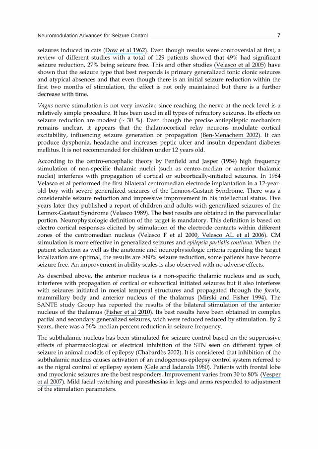
Interfering with: Target % seizure reduction Propagation Vagus nerve 30 Centromedian nuclei 80-100 Anterior nuclei 56 after 2 years Subthalamic nuclei 30-80 Generation Hippocampal foci 80-100 Motor foci 90-100
Table 1. Percentage seizure reduction This table shows the percentage of seizure reduction according to the different targets. Note that the percentages are approximate and might vary according to the different techniques explained in the text.
Neuromodulation Advances for Seizure Control
9
4. How do we set the neural stimulation? 4.1 Verifying the chosen target
No matter what target we choose we must consider that there should be a neurophysiologic rationale in our reasoning. First we have to ask ourselves if we are able to detect the precise epileptic focus to be able to interfere with seizure generation, if not, we have to go for the disruption of the epileptic activity propagation. Either way we need to make sure we are in the correct location. From the anatomic point of view this should be easy, but several problems have to be faced. In case that we decide to go for seizure propagation we must target a precise anatomic region. The localization of the target is performed by the use of the current technology: stereotactic surgery, neuro-navigation, MRI, and neurophysiology. All expertise is needed to ensure the localization. Despite all these efforts, anatomy may be disrupted. This is particularly the case of reaching thalamic targets. We prefer patients whose thalamus and brainstem are intact and symmetric. MRI is used to verify position. Even so, the best-placed electrodes must have neurophysiologic confirmation.
In the case of centromedian stimulation, detailed analysis of incremental response morphology, polarity, peak latency, and cortical distribution may aid in defining the relation of the stimulated area with specific anatomophysiologic systems within the centromedian nucleus. Electrocortical responses produced by acute electrical stimulation along the centromedian nucleus or other structures are described in detail elsewhere (Velasco M, 1996).
To perform the physiologic confirmation of the electrode position in the parvocellular portion, the three pairs of combinations of the four contacts in each DBS electrode are stimulated (0–1, 1–2, and 2–3). Unilateral stimulation is performed at 6 Hz, 1.0 ms, 320–800 μA with 10-s duration to induce recruiting responses, and unilateral high frequency is conducted as well at 60 Hz, 1.0 ms, 320–800 μA to induce focalized desynchronization and negative DC shift of the EEG baseline. Scalp distribution analysis of electrocortical responses is performed. Within CM, suprathreshold stimulation in parvocellular subnucleus induces monophasic negative waxing and waning potentials, with peak latencies from 40 to 60 ms, recorded bilaterally in frontal and central regions, with emphasis on the stimulated side. Outside this subnucleus, incremental responses remain mainly biphasic positive-negative, with 16- to 20 ms latency, fixed amplitudes for the positive component, and distribution extending more toward the posterior leads. If the electrode is localized in the posterior and basal area of the centro median nucleus within boundaries with the parafascicular nucleus induces only short-latency positive potentials at low frequency and occasionally painful sensation at high frequencies (Velasco 1998).
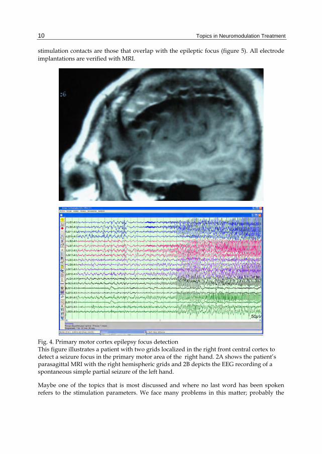
In the case of epileptic focus stimulation, localizing the precise site where it is located is mandatory; it could make the difference in seizure outcome. In our Epilepsy Clinic, patients are implanted with externalized diagnostic multicontact intracranial electrodes, and recorded outside the operating room to be able to detect spontaneous seizures. This is to ensure we know where the focus is located (figure 4). In case of hippocampal stimulation we implant bilateral hippocampal electrodes, if the focus is unilateral, the diagnostic electrodes are explanted and only a single therapeutic permanent electrode is implanted; if the patient has bilateral foci, two electrodes, one in each hippocampus is implanted. The selected
Topics in Neuromodulation Treatment
10
stimulation contacts are those that overlap with the epileptic focus (figure 5). All electrode implantations are verified with MRI.
Fig. 4. Primary motor cortex epilepsy focus detection This figure illustrates a patient with two grids localized in the right front central cortex to detect a seizure focus in the primary motor area of the right hand. 2A shows the patient’s parasagittal MRI with the right hemispheric grids and 2B depicts the EEG recording of a spontaneous simple partial seizure of the left hand.
Maybe one of the topics that is most discussed and where no last word has been spoken refers to the stimulation parameters. We face many problems in this matter; probably the
Neuromodulation Advances for Seizure Control
11
most important is the lack of sufficient financing resources that are required for the implementation of double-blinded protocols. To this problem we might add the necessity for more basic research data, multicentric studies, approval of FDA or similar agencies in different countries. Despite all this, there are some suggestions of the current parameters used for epilepsy:
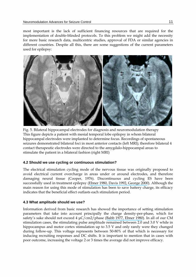
Fig. 5. Bilateral hippocampal electrodes for diagnosis and neuromodulation therapy This figure depicts a patient with mesial temporal lobe epilepsy in whom bilateral hippocampal electrodes were implanted to determine focus. Recordings of spontaneous seizures demonstrated bilateral foci in most anterior contacts (left MRI); therefore bilateral 4 contact therapeutic electrodes were directed to the amygdalo-hippocampal areas to stimulate the patient in a bilateral fashion (right MRI)
4.2 Should we use cycling or continuous stimulation?
The electrical stimulation cycling mode of the nervous tissue was originally proposed to avoid electrical current overcharge in areas under or around electrodes, and therefore damaging neural tissue (Cooper, 1976). Discontinuous and cycling ES have been successfully used in treatment epilepsy (Ebner 1980, Davis 1992, George 2000). Although the main reason for using this mode of stimulation has been to save battery charge, its efficacy indicates that the beneficial effect outlasts each stimulation period.
4.3 What amplitude should we use?
Information derived from basic research has showed the importance of setting stimulation parameters that take into account principally the charge density-per-phase, which for safety’s sake should not exceed 4 μC/cm2/phase (Babb 1977, Ebner 1980). In all of our CM stimulation cases, the stimulating pulse amplitude remained between 2.0 and 3.0 V while in hippocampus and motor cortex stimulation up to 3.5 V and only rarely were they changed during follow-up. This voltage represents between 50-80% of that which is necessary for inducing recruiting responses and DC shifts. It is important to mention that in cases with poor outcome, increasing the voltage 2 or 3 times the average did not improve efficacy.
Topics in Neuromodulation Treatment
12
4.4 Should we use high or low frequency stimulation?
Experience on neuromodulation studies in patients with movement disorders and those with pain, it is known that high frequency stimulation inhibits both. The same occurs in epilepsy; almost all studies use frequency in the range of 130 Hz. Velasco M et al (1997) demonstrated that low frequencies produced recruiting responses when stimulating the CM nuclei, and when bilateral stimulation at 3 Hz was performed in these nuclei, a typical absence seizure was reproduced. On the contrary, high frequency stimulation produced cortical inhibition of epileptic activity. In the case of neuromodulation of the subthalamic nucleus, low frequency has been used for good results (Chabardès 2002).
4.5 Open or closed loop mode?
Recent instances of clinical application of closed-loop seizure control, which are limited to stimulation with pulse trains in response to epileptiform activity, have been reviewed (Osorio et al 2001, Sun et al 2008). This method requires the implementation of a seizure detection algorithm to control the delivery of therapy using a suitable device. The theory is that stimulation therapy is provided as needed, potentially reducing the likelihood of functional disruption or habituation due to continuous treatment. It should also be better to save battery of the pulse generator reducing costs. Questions regarding the cost of a dual system (for detection and for stimulation) is one problem with this stimulation mode, we are currently dealing with the high cost of the stimulating system, most patients will not be able to pay for a detection system too. Secondly, there are currently no data that support that there is a potential likelihood of functional disruption with neuromodulation. Third, nowadays there are rechargeable batteries so the need to save energy should not be an issue. But more important is the fact that there are still many problems for precise seizure detection bringing as a result the application of unnecessary stimuli due to false positive detection or failure to stimulate if we modify seizure detection parameters to avoid the former. Probably this is the reason why studies have shown that stimulating with a closed loop mode takes much longer to reach best results. Further studies are mandatory to improve this methodology and solve issues regarding efficacy of the closed loop mode and cost of a implanting a dual system.
5. How can we make sure that neuromodulation is being adequately performed? Neuromodulation in epilepsy has certain difficulties for its long-term assessment due to several reasons:
The inherent characteristics of the symptoms (seizures) Patients do not experiment any sensations or secondary effects at all that indicate that
stimulation is being delivered The time that neuromodulation takes to show a positive effect in seizure reduction “Carry on” effect Unlike movement disorders like tremor in Parkinson’s disease, seizures appear once in
a while and are not predictable, so it is not a matter of turning the pulse generator on or off and observing if seizures disappear to know if the stimulation system is working.
Neuromodulation Advances for Seizure Control
13
Except for the dysphonia that a few patients who undergo vagus nerve stimulation experiment when the stimulator is ON, other patients do not have sensations or secondary effects that could indicate if the stimulation is taking place. Two other important observations are that stimulation takes a variable time to show its effect, this period can take from several days to months; and that, when stimulation is stopped, there is a variable period of “carry-on” effect This term refers to the observed phenomenon in which the seizure reduction is maintained for days to months after the stimulator is turned OFF, the battery depletes or the stimulation is interrupted for any reason. Seizures reappear later in a progressive manner without reaching basal (before neuromodulation) level either in number or severity. Today, the neuromodulation community accepts the “carry-on effect”.
These inherent characteristics carry the need to check the system every 6 to 12 months or when the patient returns to consult us because seizures are increasing in intensity or number. This check up consists in:
Verifying pulse generator battery current: the pulse generator computer reader can tell us if the pulse generator has more than 50% or is running low.
Checking electrode impedance (we have a comparative with our immediate post operatory measurements). If there is an increase, it could mean that that either the system extension or the electrode are broken or disconnected. In this case simple X rays are mandatory.
Perform acute stimulation trial using the internalized stimulating system to generate recruiting responses. This will allow us to know that the system is working correctly and that the brain tissue is being stimulated, if not, either the system is broken or there is something in the tissue that is preventing a correct stimulation (blood, gliosis). Also the recruiting responses distribution in the scalp EEG can show us if the electrode moved from its intended placement. Within centromedian nucleus, suprathreshold stimulation in parvocellular subnucleus induces monophasic negative waxing and waning potentials, with peak latencies from 40 to 60 ms, recorded bilaterally in frontal and central regions, with emphasis on the stimulation side (Velasco 2006). Similar responses are found in hippocampal stimulation but are localized in ipsilateral temporal region and in motor cortex stimulation localized in ipsilateral frontal region.
6. What happens to the brain function if we stimulate it?
One of the main reasons to use neuromodulation is to preserve the functional areas of the brain. Neuromodulation in cases of abnormal movements or chronic pain has proven to be effective and to preserve function. Even when some undesirable effects are present, stimulation parameters and even the stimulated contacts can be changed and the adverse effects revert. When our group started stimulation of the centromedian thalamic nuclei for seizure control, we were dealing with patients with severe epilepsy, some of them Lennox-Gastaut patients with relentless psychomotor worsening due to the amount and severity of their seizures. The surgical options were not going to spare any function. So the functional implications of neuromodulation were not our main concern and we knew that the method
Topics in Neuromodulation Treatment
14
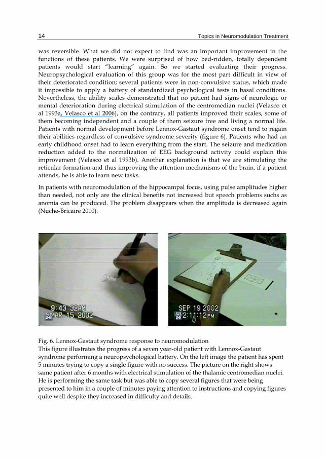
was reversible. What we did not expect to find was an important improvement in the functions of these patients. We were surprised of how bed-ridden, totally dependent patients would start “learning” again. So we started evaluating their progress. Neuropsychological evaluation of this group was for the most part difficult in view of their deteriorated condition; several patients were in non-convulsive status, which made it impossible to apply a battery of standardized psychological tests in basal conditions. Nevertheless, the ability scales demonstrated that no patient had signs of neurologic or mental deterioration during electrical stimulation of the centromedian nuclei (Velasco et al 1993a, Velasco et al 2006), on the contrary, all patients improved their scales, some of them becoming independent and a couple of them seizure free and living a normal life. Patients with normal development before Lennox-Gastaut syndrome onset tend to regain their abilities regardless of convulsive syndrome severity (figure 6). Patients who had an early childhood onset had to learn everything from the start. The seizure and medication reduction added to the normalization of EEG background activity could explain this improvement (Velasco et al 1993b). Another explanation is that we are stimulating the reticular formation and thus improving the attention mechanisms of the brain, if a patient attends, he is able to learn new tasks.
In patients with neuromodulation of the hippocampal focus, using pulse amplitudes higher than needed, not only are the clinical benefits not increased but speech problems suchs as anomia can be produced. The problem disappears when the amplitude is decreased again (Nuche-Bricaire 2010).
Fig. 6. Lennox-Gastaut syndrome response to neuromodulation This figure illustrates the progress of a seven year-old patient with Lennox-Gastaut syndrome performing a neuropsychological battery. On the left image the patient has spent 5 minutes trying to copy a single figure with no success. The picture on the right shows same patient after 6 months with electrical stimulation of the thalamic centromedian nuclei. He is performing the same task but was able to copy several figures that were being presented to him in a couple of minutes paying attention to instructions and copying figures quite well despite they increased in difficulty and details.
Neuromodulation Advances for Seizure Control
15
In regard to stimulating the epileptic focus localized in the hippocampus, the groups mentioned above that have performed neuromodulation of the hippocampal foci have not reported a worsening of the memory function. Neuropsychological batteries for memory function (Table 2) have been applied and no deterioration has been found, and possibly a tendency to improve has occurred (Velasco 2007). The same thing happens with patients with stimulation of the primary or supplementary motor cortices, no decrease in motor function has been observed (Velasco 2009). Numbers or patients are small up to now and more studies need to be performed.
Function tested Test
Attention and memory Neuropsi Attention and Memory Battery
Verbal memory Rey verbal learning
Verbal memory Digit Counting
Verbal memory Logic memory
Non verbal memory Visual reproduction
Non verbal memory Wind Mill visual spatial Bezares Test
Language dominance Dichotic listening test
Table 2. Neuropsychological battery for memory function This table illustrates the neuropsycological tests that are performed in a patient with intractable mesial temporal lobe epilepsy. In left column we point the function that testing is aimed for and on the right the specific test. This battery is accompanied with general testing as for example IQ and depression scales.
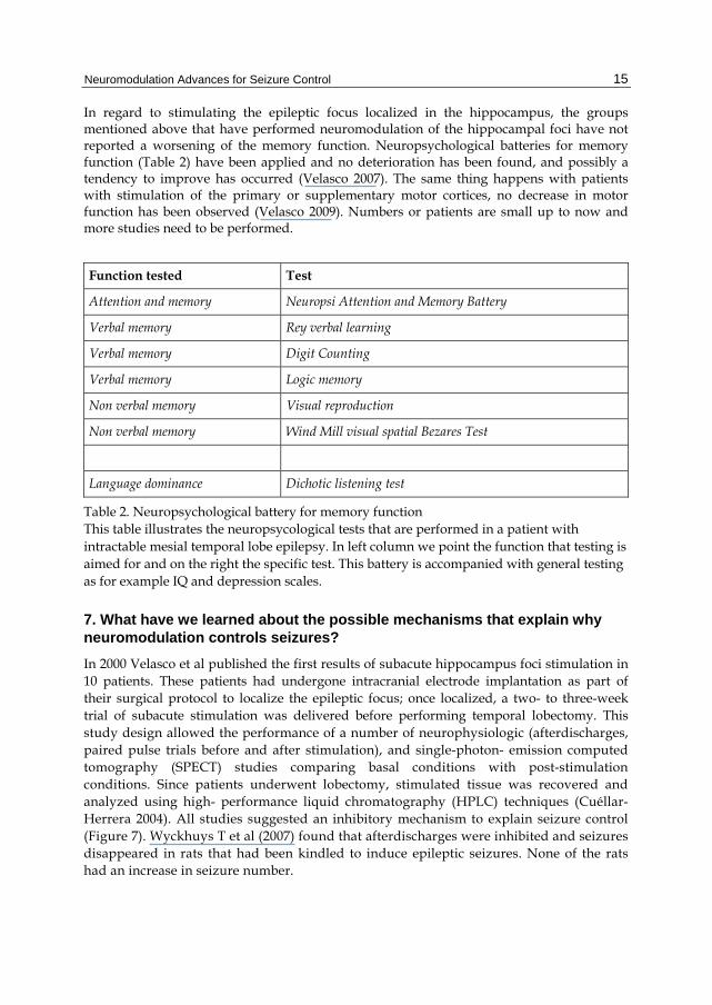
7. What have we learned about the possible mechanisms that explain why neuromodulation controls seizures? In 2000 Velasco et al published the first results of subacute hippocampus foci stimulation in 10 patients. These patients had undergone intracranial electrode implantation as part of their surgical protocol to localize the epileptic focus; once localized, a two- to three-week trial of subacute stimulation was delivered before performing temporal lobectomy. This study design allowed the performance of a number of neurophysiologic (afterdischarges, paired pulse trials before and after stimulation), and single-photon- emission computed tomography (SPECT) studies comparing basal conditions with post-stimulation conditions. Since patients underwent lobectomy, stimulated tissue was recovered and analyzed using high- performance liquid chromatography (HPLC) techniques (Cuéllar-Herrera 2004). All studies suggested an inhibitory mechanism to explain seizure control (Figure 7). Wyckhuys T et al (2007) found that afterdischarges were inhibited and seizures disappeared in rats that had been kindled to induce epileptic seizures. None of the rats had an increase in seizure number.
Topics in Neuromodulation Treatment
16
Fig. 7. Possible mechanisms that explain how neuromodulation works This figure exemplifies the possible mechanisms through which neuromodulation works in hippocampal stimulation in man. Upper image is a single emission photon computerized study in a patient who underwent neuromodulation of the hippocampus for 3 weeks. At the lower left is the recording of afterdischarges in same patients in whom we apply maximum stimulation intensity in the epileptic focus and can no longer produce a clinical seizure and barely a few spikes instead of a prolonged afterdischarge accompanied by clinical symptoms of a complex partial seizure identical to the spontaneous ones presented by the patients. The lower right shows the difference between the recovery cycles before (continuous line) and after (broken line) neuromodulation. All three studies suggest an inhibitory effect of neuromodulation of the hippocampus.
8. Conclusion This chapter shows a brief summary of the principal work that has been performed all over the world regarding neuromodulation for epilepsy. Results are encouraging; neuromodulation is a reversible surgical alternative that preserves neural function. It is capable of reducing seizure frequency and severity to a point that can be superior to what new generation antiepileptic medications have shown (Chadwik 2001, Van Rijckevorsel & Boon 2001) without their adverse effects. Patients tolerate it well, as a matter of fact, they are not even aware of being stimulated (except for vagus nerve stimulation). More concerns exist: are there other stimulation parameters worth trying (frequency, amplitude, pulse width), should they change according to the target; what is the best stimulation mode (open loop or close loop); are there other mechanisms that explain its antiepileptic effect? What will eventually happen in a longer follow up period?
Challenges are huge: exploring new targets, improving technology (better and cheaper stimulation and probably seizure detection systems, wider range of electrodes to choose from, smaller stimulating systems for younger children), improvement of surgical techniques so that we can find the focus and stimulate through the same electrode eluding unnecessary surgical procedures.
Neuromodulation Advances for Seizure Control
17
The main steps to be able to perform research in this matter have been taken; epilepsy expert groups tend to agree that neuromodulation is a field that can bring seizure relieve to patients. To be able to solve the questions and challenges, multidisciplinary, interinstitutional, worldwide projects with well thought designs are necessary.
9. References Babb, T. L., Soper, H. V., Lieb, J. P., Brown, W. J., Ottino, C. A., & Crandall, P. H. (1977).
Electrophysiological studies of long-term electrical stimulation of the cerebellum in monkeys. J Neurosurg, 47, 3, (Sept 1977), pp. 353–365, ISSN 0022-3085
Ben-Menachem, E. (2002). Vagus-nerve stimulation for the treatment of epilepsy. Lancet Neurol, 1, 8, (Dec 2002), pp. 477–482, ISSN 1474-4422
Boon, P., Vonck, K., De Herdt, V., Van Dycke, A., Goethals, M., Goossens, L., Van Zandijcke, M., De Smedt, T., Dewaele, I., Achten, R., Wadman, W., Dewaele, F., Caemaert, J., & Van Roost, D. (2007). Deep brain stimulation in patients with refractory temporal lobe epilepsy. Epilepsia, 48, 8, (Aug 2007), pp. 1551–1560, ISSN 0013-9580
Chabardes, S., Kahane, P., Minotti, L., Koudsie, A., Hirsch, E., & Benabid, A.-L. (2002). Deep brain stimulation in epilepsy with particular reference to the subthalamic nucleus. Epileptic Disord, 4 Suppl 3, (Dec 2002) pp. S83-93, ISSN 1294-9361
Cooper, I. S., Amin, I., & Gilman, S. (1973). The effect of chronic cerebellar stimulation upon epilepsy in man. Trans Am Neurol Assoc, 98, (1973) pp. 192–196, ISSN 0065-9479
Cooper, I. S., Amin, I., Riklan, M., Waltz, J. M., & Poon, T. P. (1976). Chronic cerebellar stimulation in epilepsy. Clinical and anatomical studies. Arch Neurol, 33, 8, (Aug 1976), pp. 559–570, ISSN 0003-9942
Cuellar-Herrera, M., Velasco, M., Velasco, F., Velasco, A. L., Jimenez, F., Orozco, S., Briones, M., & Rocha, L. (2004). Evaluation of GABA system and cell damage in parahippocampus of patients with temporal lobe epilepsy showing antiepileptic effects after subacute electrical stimulation. Epilepsia, 45,5, (May 2004), pp. 459–466, ISSN 0013-9580
Davis, R. & Emmonds, S. E. (1992). Cerebellar stimulation for seizure control: 17-year study. Stereotact Funct Neurosurg, 58, 1-4, (1992), pp. 200–208, ISSN 1011-6125
*Davis, R. (1998). Cerebellar stimulation for seizure control, In: Textbook of stereotactic and functional neurosurgery, Gildenberg, P., & Tasker, RR,. pp. 183-189, Publisher McGraw-Hill, ISBN 978-0070236042, Houston USA
Dow, R. S., Fernandez-Guardiola, A., & Manni, E. (1962). The influence of the cerebellum on experimental epilepsy. Electroencephalogr Clin Neurophysiol, 14, (Jun 1062), pp. 383–398, ISSN 0013-4694
Ebner, T. J., Bantli, H., & Bloedel, J. R. (1980). Effects of cerebellar stimulation on unitary activity within a chronic epileptic focus in a primate. Electroencephalogr Clin Neurophysiol, 49, 5-6 (Sept 1980), pp. 585–599, ISSN 0013-4694
Fisher, R., Salanova, V., Witt, T., Worth, R., Henry, T., Gross, R., Oommen, K., Osorio, I., Nazzaro, J., Labar, D., Kaplitt, M., Sperling, M., Sandok, E., Neal, J., Handforth, A., Stern, J., DeSalles, A., Chung, S., Shetter, A., Bergen, D., Bakay, R., Henderson, J., French, J., Baltuch, G., Rosenfeld, W., Youkilis, A., Marks, W., Garcia, P., Barbaro, N., Fountain, N., Bazil, C., Goodman, R., McKhann, G., Babu Krishnamurthy, K.,
Topics in Neuromodulation Treatment
18
Papavassiliou, S., Epstein, C., Pollard, J., Tonder, L., Grebin, J., Coffey, R., & Graves, N. (2010). Electrical stimulation of the anterior nucleus of thalamus for treatment of refractory epilepsy. Epilepsia, 51, 5,(May 2010), pp. 899–908, ISSN 0013-9580
Fountas, K. N. & Smith, J. R. (2007). A novel closed-loop stimulation system in the control of focal, medically refractory epilepsy. Acta Neurochir Suppl, 97, Pt 2, (2007), pp. 357–362, ISSN 0065-1419
Gale, K. & Iadarola, M. J. (1980). Seizure protection and increased nerve-terminal GABA: delayed effects of GABA transaminase inhibition. Science, 208, 4441, (Apr 1980), pp. 288–291, ISSN 0272-4634
George, M. S., Nahas, Z., Bohning, D. E., Lomarev, M., Denslow, S., Osenbach, R., & Ballenger, J. C. (2000). Vagus nerve stimulation: a new form of therapeutic brain stimulation. CNS Spectr, 5, 11, (Nov 2000), pp. 43–52, ISSN 1092-8529
Mirski, M. A. & Fisher, R. S. (1994). Electrical stimulation of the mammillary nuclei increases seizure threshold to pentylenetetrazol in rats. Epilepsia, 35, 6, (Nov-Dec 1994), pp. 1309–1316, ISSN 0013-9580
Nuche-Bricaire, A., Montes De Oca, M., Marcos-Ortega, M., Trejo, D., Núñez, JM., Vazquez, D. & Velasco, AL. (2010).Voltage-dependant Verbal Memory. Effects on the cognitive function through changing voltage parameters in deep brain stimulation. Proceedings of the 64th American Epilepsy Society Meeting. San Antonio, USA, Dec 2010.
Osorio, I., Frei, M. G., Manly, B. F., Sunderam, S., Bhavaraju, N. C., & Wilkinson, S. B. (2001). An introduction to contingent (closed-loop) brain electrical stimulation for seizure blockage, to ultra-short-term clinical trials, and to multidimensional statistical analysis of therapeutic efficacy. J Clin Neurophysiol, 18, 6(Nov 2001), pp. 533–544, ISSN 0736-0258
Penfield, W., & Jasper, H. (1954). Epilepsy and the functional anatomy of the human brain,: Publisher: Little Brown, ISBN 9780316698337, Boston USA
Sun, F. T., Morrell, M. J., & Wharen, R. E. J. (2008). Responsive cortical stimulation for the treatment of epilepsy. Neurotherapeutics, 5, 1, (Jan 2008), pp. 68–74, ISSN 1933-7213
Velasco, A. L., Velasco, M., Velasco, F., Menes, D., Gordon, F., Rocha, L., Briones, M., & Marquez, I. (2000). Subacute and chronic electrical stimulation of the hippocampus on intractable temporal lobe seizures: preliminary report. Arch Med Res, 31, 3(May-Jun 2000), pp. 316–328, ISSN 0188-4409
Velasco, A. L., Velasco, F., Jimenez, F., Velasco, M., Castro, G., Carrillo-Ruiz, J. D., Fanghanel, G., & Boleaga, B. (2006). Neuromodulation of the centromedian thalamic nuclei in the treatment of generalized seizures and the improvement of the quality of life in patients with Lennox-Gastaut syndrome. Epilepsia, 47, 7, (Jul 2006), pp. 1203–1212, ISSN 0013-9580
Velasco, A. L., Velasco, F., Velasco, M., Trejo, D., Castro, G., & Carrillo-Ruiz, J. D. (2007). Electrical stimulation of the hippocampal epileptic foci for seizure control: a double-blind, long-term follow-up study. Epilepsia, 48, 10, (Oct 2007), pp. 1895–1903, ISSN 0013-9580
Neuromodulation Advances for Seizure Control
19
Velasco, A. L., Velasco, F., Velasco, M., Maria Nunez, J., Trejo, D., & Garcia, I. (2009). Neuromodulation of epileptic foci in patients with non-lesional refractory motor epilepsy. Int J Neural Syst, 19, 3, (Jun 2009), pp. 139–147, ISSN 0129-0657
Velasco, AL. (2010) Developments in neurostimulation therapy for epilepsy. US Neurology, 5, 2 (Dec 2010), pp 78-81, ISSN
Velasco, F., Velasco, M., Ogarrio, C., & Fanghanel, G. (1987). Electrical stimulation of the centromedian thalamic nucleus in the treatment of convulsive seizures: a preliminary report. Epilepsia, 28, 4, (Jul-Aug 1987), pp. 421–430, ISSN 0013-9580
Velasco, F., Velasco, M., Velasco, A. L., & Jimenez, F. (1993). Effect of chronic electrical stimulation of the centromedian thalamic nuclei on various intractable seizure patterns: I. Clinical seizures and paroxysmal EEG activity. Epilepsia, 34, 6, (Nov-Dec 1993), pp. 1052–1064, ISSN 0013-9580
Velasco, F., Velasco, M., Jimenez, F., Velasco, A. L., Brito, F., Rise, M., & Carrillo-Ruiz, J. D. (2000). Predictors in the treatment of difficult-to-control seizures by electrical stimulation of the centromedian thalamic nucleus. Neurosurgery, 47, 2, (Aug 2000), pp. 295–304, ISSN 0148-396X
Velasco, F., Carrillo-Ruiz, J. D., Brito, F., Velasco, M., Velasco, A. L., Marquez, I., & Davis, R. (2005). Double-blind, randomized controlled pilot study of bilateral cerebellar stimulation for treatment of intractable motor seizures. Epilepsia, 46, 7, (Jul 2005), pp. 1071–1081, ISSN 0013-9580
Velasco, M., Velasco, F., Velasco, A. L., Lujan, M., & Vazquez del Mercado, J. (1989). Epileptiform EEG activities of the centromedian thalamic nuclei in patients with intractable partial motor, complex partial, and generalized seizures. Epilepsia, 30, 3, (May-Jun 1989), pp. 295–306, ISSN 0013-9580
Velasco, M., Velasco, F., Velasco, A. L., Velasco, G., & Jimenez, F. (1993). Effect of chronic electrical stimulation of the centromedian thalamic nuclei on various intractable seizure patterns: II. Psychological performance and background EEG activity. Epilepsia, 34, 6, (Nov-Dec 1993), pp. 1065–1074, ISSN 0013-9580
Velasco, M., Velasco, F., Velasco, A. L., Brito, F., Jimenez, F., Marquez, I., & Rojas, B. (1997). Electrocortical and behavioral responses produced by acute electrical stimulation of the human centromedian thalamic nucleus. Electroencephalogr Clin Neurophysiol, 102, 6, (Jun 1997), pp. 461–471, ISSN 0013-4694
Velasco, M., Brito, F., Jimenez, F., Gallegos, M., Velasco, A. L., & Velasco, F. (1998). Effect of fentanyl and naloxone on a thalamic induced painful response in intractable epileptic patients. Stereotact Funct Neurosurg, 71, 2, (1998), pp. 90–102, ISSN 1011-6125
Velasco, M., Velasco, F., & Velasco, A. L. (2001). Centromedian-thalamic and hippocampal electrical stimulation for the control of intractable epileptic seizures. J Clin Neurophysiol, 18, 6, (Nov 2001), pp. 495–513, ISSN 0736-0258
Vesper, J., Steinhoff, B., Rona, S., Wille, C., Bilic, S., Nikkhah, G., & Ostertag, C. (2007). Chronic high-frequency deep brain stimulation of the STN/SNr for progressive myoclonic epilepsy. Epilepsia, 48,10, (Oct 2007), pp. 1984–1989, ISSN 0013-9580
Topics in Neuromodulation Treatment
20
Weiss, S. R., Eidsath, A., Li, X. L., Heynen, T., & Post, R. M. (1998). Quenching revisited: low level direct current inhibits amygdala-kindled seizures. Exp Neurol, 154,1, (Nov 1998), pp. 185–192, ISSN
Wyckhuys, T., De Smedt, T., Claeys, P., Raedt, R., Waterschoot, L., Vonck, K., Van den Broecke, C., Mabilde, C., Leybaert, L., Wadman, W., & Boon, P. (2007). High frequency deep brain stimulation in the hippocampus modifies seizure characteristics in kindled rats. Epilepsia, 48,