The distribution of cannabinoid-induced Fos expression in rat brain: differences between the Lewis...
16
Brain Research 921 (2001) 240–255 www.elsevier.com / locate / bres Research report The distribution of cannabinoid-induced Fos expression in rat brain: differences between the Lewis and Wistar strain a, a,b c b * Jonathon C. Arnold , Ann N. Topple , Paul E. Mallet , Glenn E. Hunt , a Iain S. McGregor a Department of Psychology, Faculty of Science, University of Sydney, Sydney, NSW 2006, Australia b Department of Psychological Medicine, University of Sydney Clinical Sciences Building, Concord Hospital, Concord, NSW 2139, Australia c School of Psychology, The University of New England, Armidale, NSW 2351, Australia Accepted 24 September 2001 Abstract Previous studies have suggested that cannabis-like drugs produce mainly aversive and anxiogenic effects in Wistar strain rats, but rewarding effects in Lewis strain rats. In the present study we compared Fos expression, body temperature effects and behavioral effects elicited by the cannabinoid CB receptor agonist CP 55,940 in Lewis and Wistar rats. Both a moderate (50 mg/kg) and a high (250 1 mg/kg) dose level were used. The 250 mg / kg dose caused locomotor suppression, hypothermia and catalepsy in both strains, but with a significantly greater effect in Wistar rats. The 50 mg / kg dose provoked moderate hypothermia and locomotor suppression but in Wistar rats only. CP 55,940 caused significant Fos immunoreactivity in 24 out of 33 brain regions examined. The most dense expression was seen in the paraventricular nucleus of the hypothalamus, the islands of Calleja, the lateral septum (ventral), the central nucleus of the amygdala, the bed nucleus of the stria terminalis (lateral division) and the ventrolateral periaqueductal gray. Despite having a similar distribution of CP 55,940-induced Fos expression, Lewis rats showed less overall Fos expression than Wistars in nearly every brain region counted. This held equally true for anxiety-related brain structures (e.g. central nucleus of the amygdala, periaqueductal gray and the paraventricular nucleus of the hypothalamus) and reward-related sites (nucleus accumbens and pedunculopontine tegmental nucleus). In a further experiment, Wistar rats and Lewis rats did not differ in the amount of Fos immunoreactivity produced by cocaine (15 mg / kg). These results indicate that Lewis rats are less sensitive to the behavioral, physiological and neural effects of cannabinoids. The exact mechanism underlying this subsensitivity requires further investigation. Crown copyright 2001 Published by Elsevier Science B.V. All rights reserved. Theme: Neural basis of behaviour Topic: Drugs of abuse: opioids and others Keywords: Cannabinoid; c-fos; Strain difference; Lewis; Wistar; CP 55,940 1. Introduction predisposed to cannabis dependence is supported by data suggesting that genetic factors partly determine whether Cannabis is the most widely used illicit drug in the cannabis is experienced as euphoric or dysphoric in world and appears to have addictive potential in a subset of humans [46]. Also reflecting this genetic determination is users [32]. The notion that certain individuals may be that major strain differences have been found in the reinforcing effects of cannabinoids in rats [28,29]. Lewis strain rats appear to find the main psychoactive 9 9 constituent of marijuana, D -tetrahydrocannabinol ( D - THC), rewarding as assessed by the intracranial self- *Corresponding author. Present address: The Department of Pharma- stimulation paradigm [44]. In contrast, other rat strains cology, University of Sydney, Sydney, NSW 2006, Australia. Tel.: such as the Wistar strain appear to find cannabinoids 161-2-9351-6957; fax: 161-2-9351-3868. E-mail address: [email protected] (J.C. Arnold). aversive and anxiogenic [52,70]. These observations invite 0006-8993 / 01 / $ – see front matter Crown copyright 2001 Published by Elsevier Science B.V. All rights reserved. PII: S0006-8993(01)03127-4
Transcript of The distribution of cannabinoid-induced Fos expression in rat brain: differences between the Lewis...
Brain Research 921 (2001) 240–255www.elsevier.com/ locate /bres
Research report
The distribution of cannabinoid-induced Fos expression in rat brain:differences between the Lewis and Wistar strain
a , a,b c b*Jonathon C. Arnold , Ann N. Topple , Paul E. Mallet , Glenn E. Hunt ,aIain S. McGregor
aDepartment of Psychology, Faculty of Science, University of Sydney, Sydney, NSW 2006, AustraliabDepartment of Psychological Medicine, University of Sydney Clinical Sciences Building, Concord Hospital, Concord, NSW 2139, Australia
cSchool of Psychology, The University of New England, Armidale, NSW 2351, Australia
Accepted 24 September 2001
Abstract
Previous studies have suggested that cannabis-like drugs produce mainly aversive and anxiogenic effects in Wistar strain rats, butrewarding effects in Lewis strain rats. In the present study we compared Fos expression, body temperature effects and behavioral effectselicited by the cannabinoid CB receptor agonist CP 55,940 in Lewis and Wistar rats. Both a moderate (50 mg/kg) and a high (2501
mg/kg) dose level were used. The 250 mg/kg dose caused locomotor suppression, hypothermia and catalepsy in both strains, but with asignificantly greater effect in Wistar rats. The 50 mg/kg dose provoked moderate hypothermia and locomotor suppression but in Wistarrats only. CP 55,940 caused significant Fos immunoreactivity in 24 out of 33 brain regions examined. The most dense expression wasseen in the paraventricular nucleus of the hypothalamus, the islands of Calleja, the lateral septum (ventral), the central nucleus of theamygdala, the bed nucleus of the stria terminalis (lateral division) and the ventrolateral periaqueductal gray. Despite having a similardistribution of CP 55,940-induced Fos expression, Lewis rats showed less overall Fos expression than Wistars in nearly every brain regioncounted. This held equally true for anxiety-related brain structures (e.g. central nucleus of the amygdala, periaqueductal gray and theparaventricular nucleus of the hypothalamus) and reward-related sites (nucleus accumbens and pedunculopontine tegmental nucleus). In afurther experiment, Wistar rats and Lewis rats did not differ in the amount of Fos immunoreactivity produced by cocaine (15 mg/kg).These results indicate that Lewis rats are less sensitive to the behavioral, physiological and neural effects of cannabinoids. The exactmechanism underlying this subsensitivity requires further investigation. Crown copyright 2001 Published by Elsevier Science B.V.All rights reserved.
Theme: Neural basis of behaviour
Topic: Drugs of abuse: opioids and others
Keywords: Cannabinoid; c-fos; Strain difference; Lewis; Wistar; CP 55,940
1. Introduction predisposed to cannabis dependence is supported by datasuggesting that genetic factors partly determine whether
Cannabis is the most widely used illicit drug in the cannabis is experienced as euphoric or dysphoric inworld and appears to have addictive potential in a subset of humans [46]. Also reflecting this genetic determination isusers [32]. The notion that certain individuals may be that major strain differences have been found in the
reinforcing effects of cannabinoids in rats [28,29].Lewis strain rats appear to find the main psychoactive
9 9constituent of marijuana, D -tetrahydrocannabinol (D -THC), rewarding as assessed by the intracranial self-
*Corresponding author. Present address: The Department of Pharma-stimulation paradigm [44]. In contrast, other rat strainscology, University of Sydney, Sydney, NSW 2006, Australia. Tel.:such as the Wistar strain appear to find cannabinoids161-2-9351-6957; fax: 161-2-9351-3868.
E-mail address: [email protected] (J.C. Arnold). aversive and anxiogenic [52,70]. These observations invite
0006-8993/01/$ – see front matter Crown copyright 2001 Published by Elsevier Science B.V. All rights reserved.PI I : S0006-8993( 01 )03127-4
J.C. Arnold et al. / Brain Research 921 (2001) 240 –255 241
the hypothesis that in other rat strains, the effects of cocaine on Fos immunoreactivity in Lewis and Wistar ratscannabinoids on neural substrates mediating anxiety may provides a useful comparison to the effects of CP 55,940.outweigh their effects on neural substrates underlyingreward. In Lewis rats, however, the opposite may be true.
The central nucleus of the amygdala (CEA) appears to 2. Materials and methodsbe a critical brain region for cannabinoid-induced anxiety.
9Thus, administration of D -THC directly into the CEA had 2.1. Animalsan anxiogenic effect on mice on the elevated plus-maze[59]. Extrahypothalamic corticotropin releasing hormone The subjects were 19 inbred Lewis rats and 19 inbred(CRH) receptors such as those found in the CEA, also Wistar rats matched for age (75–90 days old). Wistar ratsappear to be important in cannabinoid-induced anxiety weighed between 370 and 550 g and Lewis rats weighed[70]. If Lewis rats are less susceptible to cannabinoid- between 350 and 430 g at the time of testing. Rats wereinduced anxiety then cannabinoids may have less of an housed in large plastic tubs in groups of eight andimpact on the CEA in these rats. This was one hypothesis maintained under a 12 h:12 h reversed light /dark cycletested in the present study in which a range of brain (lights off at 09:00 h) with food and water available adstructures were assessed for Fos immunoreactivity follow- libitum. All experiments were conducted during the darking cannabinoid administration in Lewis and Wistar rats. cycle.9Previous studies examining the effects of D -THC andits synthetic analogues on Fos immunoreactivity have
2.2. Drugsshown a consistent distribution of Fos expression follow-ing various cannabinoid receptor agonists. Fos expression
CP 55,940 (Tocris, Bristol, UK) was prepared asis typically seen in the caudate-putamen (CPU) anddescribed previously [7] and administered intraperitoneallynucleus accumbens (NAS) [50,54,55,66,69], the CEA(i.p.) 30 min prior to behavioral testing. Two doses were[50,61,69], the paraventricular nucleus of the hypo-used in this study, 50 and 250 mg/kg. The 250 mg/kg dosethalamus (PVN) [50,69], the bed nucleus of the striais a relatively high dose that resembles the potency ofterminalis (BNST) [61,69], the ventral tegmental areadoses used in previous studies assessing the effects of(VTA) [69], the lateral septum (LS) [50] and the fronto-other cannabinoid receptor agonists on Fos immuno-parietal and cingulate cortices [48,66]. However many 9reactivity in the rat brain such as D -THC (between 3.2studies have only examined a relatively small number ofand 15 mg/kg) [48,50,54,55,66], HU-210 (100 mg/kg)brain structures.[69] and anandamide (20 mg/kg) [50]. The problem withIn light of the limited anatomical detail provided bysuch doses is that they are much higher than typical humanthese studies, a more comprehensive description of Fosrecreational doses and cause profound behavioral deficitsimmunoreactivity throughout the brain was sought in thesuch as catalepsy in rodents. Thus, the current study alsocurrent study. Here we assessed the effects of the syntheticemployed a lower dose of CP 55,940 (50 mg/kg) that iscannabinoid receptor agonist CP 55,940 on Fos immuno-more relevant to doses that humans use recreationally [52].reactivity in a total of 33 brain regions. These included a
Cocaine hydrochloride (Australian Pharmaceutical In-range of reward and anxiety-related areas such as the CEA,dustries, Sydney, Australia) was dissolved in 0.9% salinethe medial prefrontal cortex (MPC), the islands of Callejaand injected at a dose of 15 mg/kg in a volume of 1 ml /kg(ICjM), the periaqueductal grey (PAG) and the pedun-immediately before behavioral testing. This dose wasculopontine tegmental nucleus (PPTg).chosen based on previous studies that have assessed theFor comparison purposes, the effects of cocaine on Foseffects of cocaine on Fos immunoreactivity [33].immunoreactivity were also examined in the present
investigation. The distribution of cocaine-induced Fosimmunoreactivity has some commonalities with that pro- 2.3. Behavioral testingduced by acute exposure to a cannabinoid receptor agonistwith Fos immunoreactivity commonly seen in the cortex, Rats from each strain were extensively handled for athe CPU, the NAS, the septum, the ICjM, and the week prior to the start of the experiment and wereamygdala [33]. randomly assigned to experimental groups. In Experiment
In terms of behavior, Lewis rats are more sensitive to 1, CP 55,940 was the drug administered while in Experi-the rewarding effects of cocaine as assessed by self- ment 2 it was cocaine. Exactly the same procedures wereadministration and conditioned place preference models used in Experiment 1 as in Experiment 2.when compared to Fischer 344 rats [41,42]. In addition, Two habituation days were given immediately prior toLewis rats are more sensitive to acutely administered the test day in order to minimise any Fos immunoreactivitycocaine where they show greater locomotor activity than due to novelty of testing, handling or injection procedures.Fischer 344 rats [13]. Thus, assessing the effects of On these habituation days the rats were subjected to
242 J.C. Arnold et al. / Brain Research 921 (2001) 240 –255
exactly the same procedures to those used on the test day cardially with 100 ml of 0.1 M phosphate-buffered salineexcept that they were given only saline injections. (PBS) followed by 250 ml of 4% paraformaldehyde in
On the test day individual rats were removed from their PBS (pH 7.3). The brains were removed and placed inhome cage and brought through to the test room in a small, paraformaldehyde overnight at 48C and stored in cold 30%enclosed rectangular tub filled with wood shavings. In sucrose for 72 h. The brains were then placed on mi-Experiment 1 all rats were given a single i.p. injection of crotome stages, frozen to 2178C and sliced at 40 mm witheither 0, 50 or 250 mg/kg CP 55,940 (n55 per group). In slices collected in PBS.Experiment 2, rats were given a single i.p. injection of 15 Free floating sections were incubated for 30 min in 1%mg/kg of cocaine (n54 per group). hydrogen peroxide in PBS and then for 30 min in 3%
Thirty minutes following injection with either CP normal horse serum in PBS. They were incubated in the55,940 or cocaine, rats were subjected to a series of tests primary c-Fos antibody (Santa Cruz Biotechnology, Santathat consisted of measuring catalepsy, axillary temperature Cruz, CA; rabbit polyclonal, specific for the amino ter-and locomotor activity. First rats were tested for catalepsy minus of c-Fos p62, non cross-reactive with FosB, Fra-1 orusing a bar test [84]. The forepaws of the rat were placed Fra-2) diluted 1:2000 in phosphate buffered horse serumon a horizontal metal bar (25 cm30.5 cm in diameter) that (0.1% bovine serum albumin, 0.2% Triton X-100, 2%was held 10 cm above the bench using a retort stand. The normal horse serum in PBS) for 72 h at 48C. Sections wereexperimenter used a stopwatch to measure the time that it washed for 30 min in PBS at room temperature and thentook the rat to move both paws on to the bench. Each rat incubated for 1 h in biotinylated anti-rabbit IgG (Vectorwas given a maximum of 2 min to do this. This procedure Laboratories, Burlingame, CA; diluted 1:500) in phosphatewas repeated twice and the average of these measures buffered horse serum. They were then washed in PBS for aformed the basis for statistical comparison. further 30 min and then incubated for 2 h in ExtrAvidin-
Rats were then tested for their axillary (underarm) horseradish peroxidase (Sigma, St Louis, MO; dilutedtemperature using a digital thermometer and thermocouple 1:1000 in phosphate buffered horse serum). After threeprobe (Yellow Spring Instruments, BAT-12). The probe 30-min washes in PBS, horseradish peroxidase activitywas placed under the rats arm for 30 s before a reading was visualised with the nickel diaminobenzidine andwas taken. Measuring axillary temperature is thought to be glucose oxidase reaction. This reaction was terminatedconsiderably less stressful than taking rectal temperature after approximately 10 min by washing in PBS. Theyet gives a reliable index of body temperature [51]. sections were then mounted onto slides, dehydrated, xylene
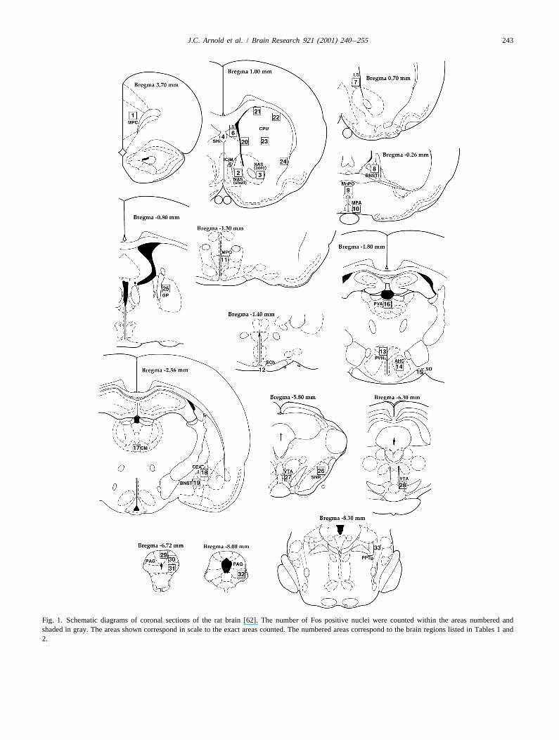
The rats were then immediately placed in one of eight cleared and coverslipped.identical chambers [30 cm (L)325.5 cm (W)350 cm (H)]for 30 min for measurement of locomotor activity. The 2.5. Counting of labeled cellsside walls and roof of the chambers were aluminum, whilethe front wall and rear wall were made of clear perspex. The nuclei of Fos immunoreactive cells followingThe cage floors consisted of 16 metal bars (0.5 cm administration of CP 55,940 (0, 50 and 250 mg/kg) werediameter, spaced 1 cm apart) connected to a high impe- quantified in 33 different brain regions or subregions withdance amplifier. When the rat moved on the grid so that reference to the rat brain atlas of Paxinos and Watson [62].contact or breaking of contact between any four bars and As described previously [21,37] quantification was per-the other 12 occurred, an activity count was recorded by a formed manually by an observer who was blind to groupMacintoshE computer running WorkbenchMacE data assignment. Counts, for the most part, were made within a
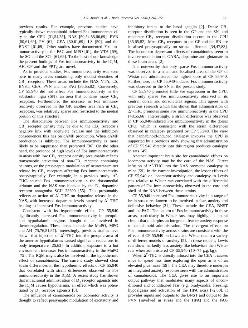
2acquisition software [52]. Each cage was encased in a graticule (0.5 mm30.5 mm) which equated to 0.25 mmwooden sound attenuation chamber that was equipped with when viewed under the 203 objective. The placement ofa fan that provided masking noise during testing. the graticule in each location is presented in Fig. 1.
Immediately after the locomotor activity test, axillary Regions in which the graticule was not used to count cellstemperature was again taken. Rats were then placed in were the ICjM, septohippocampal nucleus, suprachiasmaticindividual plastic tubs in an adjacent darkened holding nucleus (SCh), supraoptic nucleus of the hypothalamusroom for a further 60 min. On the two habituation days the (SO) and PPTg. In these cases the area was smaller thanrats were then returned to their group housing in the the area of the graticule. Thus, cells in these areas werecolony room. On the test day, the rats were removed to a counted within the regions defined in Paxinos and Watsonlaboratory where they were given an overdose of pentobar- [62]. A neuron was classified as Fos-positive when thebitone (120 mg/kg, i.p.) and perfused, prior to immuno- nucleus appeared round or oval, completely filled, and darkhistochemistry. brown or black in colour. Digitized images were produced
by a Polaroid DMC Ie camera attached to a Olympus2.4. Immunohistochemistry BX40 light microscope as previously described [21].
Fos immunoreactivity following administration of 15The immunohistochemical technique was performed as mg/kg of cocaine was quantified in eight regions using the
described previously [21,37,77]. Rats were perfused trans- same procedure as in Experiment 1. These regions were
J.C. Arnold et al. / Brain Research 921 (2001) 240 –255 243
Fig. 1. Schematic diagrams of coronal sections of the rat brain [62]. The number of Fos positive nuclei were counted within the areas numbered andshaded in gray. The areas shown correspond in scale to the exact areas counted. The numbered areas correspond to the brain regions listed in Tables 1 and2.
244 J.C. Arnold et al. / Brain Research 921 (2001) 240 –255
the: MPC, NAS (shell and core), median preoptic nucleus(MnPO), PVN, paraventricular nucleus of the thalamus(PV), CEA and dorsolateral PAG. These areas were chosenbecause they include areas where cocaine-induced Fosimmunoreactivity has been previously observed [33].
2.6. Statistical analysis
In Experiment 1, data for Fos immunoreactivity,locomotor activity, catalepsy and axillary temperature wereanalysed by two-way analysis of variance (ANOVA). Thetwo factors were strain (Lewis and Wistar) and dose of CP55,940 (vehicle, 50 and 250 mg/kg of CP 55,940). Asignificance level of P,0.05 was chosen for all statisticalcomparisons.
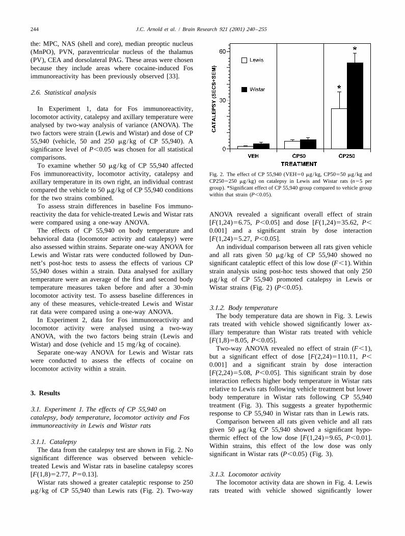
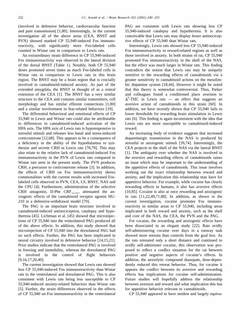
To examine whether 50 mg/kg of CP 55,940 affectedFos immunoreactivity, locomotor activity, catalepsy and Fig. 2. The effect of CP 55,940 (VEH50 mg/kg, CP50550 mg/kg and
CP2505250 mg/kg) on catalepsy in Lewis and Wistar rats (n55 peraxillary temperature in its own right, an individual contrastgroup). *Significant effect of CP 55,940 group compared to vehicle groupcompared the vehicle to 50 mg/kg of CP 55,940 conditionswithin that strain (P,0.05).
for the two strains combined.To assess strain differences in baseline Fos immuno-
reactivity the data for vehicle-treated Lewis and Wistar rats ANOVA revealed a significant overall effect of strainwere compared using a one-way ANOVA. [F(1,24)56.75, P,0.05] and dose [F(1,24)535.62, P,
The effects of CP 55,940 on body temperature and 0.001] and a significant strain by dose interactionbehavioral data (locomotor activity and catalepsy) were [F(1,24)55.27, P,0.05].also assessed within strains. Separate one-way ANOVA for An individual comparison between all rats given vehicleLewis and Wistar rats were conducted followed by Dun- and all rats given 50 mg/kg of CP 55,940 showed nonett’s post-hoc tests to assess the effects of various CP significant cataleptic effect of this low dose (F,1). Within55,940 doses within a strain. Data analysed for axillary strain analysis using post-hoc tests showed that only 250temperature were an average of the first and second body mg/kg of CP 55,940 promoted catalepsy in Lewis ortemperature measures taken before and after a 30-min Wistar strains (Fig. 2) (P,0.05).locomotor activity test. To assess baseline differences inany of these measures, vehicle-treated Lewis and Wistar
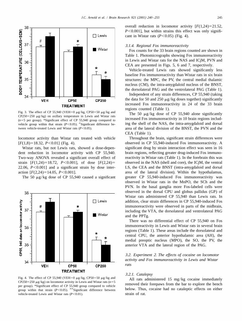
3.1.2. Body temperaturerat data were compared using a one-way ANOVA.
The body temperature data are shown in Fig. 3. LewisIn Experiment 2, data for Fos immunoreactivity and
rats treated with vehicle showed significantly lower ax-locomotor activity were analysed using a two-way
illary temperature than Wistar rats treated with vehicleANOVA, with the two factors being strain (Lewis and
[F(1,8)58.05, P,0.05].Wistar) and dose (vehicle and 15 mg/kg of cocaine).
Two-way ANOVA revealed no effect of strain (F,1),Separate one-way ANOVA for Lewis and Wistar rats
but a significant effect of dose [F(2,24)5110.11, P,were conducted to assess the effects of cocaine on
0.001] and a significant strain by dose interactionlocomotor activity within a strain.
[F(2,24)55.08, P,0.05]. This significant strain by doseinteraction reflects higher body temperature in Wistar ratsrelative to Lewis rats following vehicle treatment but lower
3. Resultsbody temperature in Wistar rats following CP 55,940treatment (Fig. 3). This suggests a greater hypothermic
3.1. Experiment 1. The effects of CP 55,940 onresponse to CP 55,940 in Wistar rats than in Lewis rats.
catalepsy, body temperature, locomotor activity and FosComparison between all rats given vehicle and all rats
immunoreactivity in Lewis and Wistar ratsgiven 50 mg/kg CP 55,940 showed a significant hypo-thermic effect of the low dose [F(1,24)59.65, P,0.01].
3.1.1. CatalepsyWithin strains, this effect of the low dose was only
The data from the catalepsy test are shown in Fig. 2. Nosignificant in Wistar rats (P,0.05) (Fig. 3).
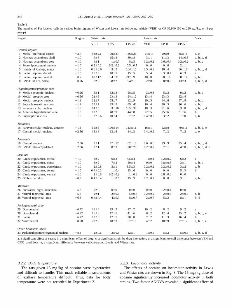
significant difference was observed between vehicle-treated Lewis and Wistar rats in baseline catalepsy scores[F(1,8)52.77, P50.13]. 3.1.3. Locomotor activity
Wistar rats showed a greater cataleptic response to 250 The locomotor activity data are shown in Fig. 4. Lewismg/kg of CP 55,940 than Lewis rats (Fig. 2). Two-way rats treated with vehicle showed significantly lower
J.C. Arnold et al. / Brain Research 921 (2001) 240 –255 245
overall reduction in locomotor activity [F(1,24)521.52,P,0.001], but within strains this effect was only signifi-cant in Wistar rats (P,0.05) (Fig. 4).
3.1.4. Regional Fos immunoreactivityFos counts for the 33 brain regions counted are shown in
Table 1. Photomicrographs showing Fos immunoreactivityin Lewis and Wistar rats for the NAS and ICjM, PVN andCEA are presented in Figs. 5, 6 and 7, respectively.
Vehicle-treated Lewis rats showed significantly lessbaseline Fos immunoreactivity than Wistar rats in six brainstructures: the MPC, the PV, the central medial thalamicnucleus (CM), the intra-amygdaloid nucleus of the BNST,the dorsolateral PAG and the ventrolateral PAG (Table 1).
Independent of any strain differences, CP 55,940 (takingthe data for 50 and 250 mg/kg doses together) significantlyincreased Fos immunoreactivity in 24 of the 33 brainregions counted (Table 1).
Fig. 3. The effect of CP 55,940 (VEH50 mg/kg, CP50550 mg/kg and The 50 mg/kg dose of CP 55,940 alone significantlyCP2505250 mg/kg) on axillary temperature in Lewis and Wistar rats
increased Fos immunoreactivity in 10 brain regions includ-(n55 per group). *Significant effect of CP 55,940 group compared to[ ing the shell of the NAS, the intra-amygdaloid and dorsalvehicle group within that strain (P,0.05). Significant difference be-
tween vehicle-treated Lewis and Wistar rats (P,0.05). area of the lateral division of the BNST, the PVN and theCEA (Table 1).
Throughout the brain, significant strain differences werelocomotor activity than Wistar rats treated with vehicleobserved in CP 55,940-induced Fos immunoreactivity. A[F(1,8)518.32, P,0.01] (Fig. 4).significant drug by strain interaction effect was seen in 16Wistar rats, but not Lewis rats, showed a dose-depen-brain regions, reflecting greater drug-induced Fos immuno-dent reduction in locomotor activity with CP 55,940.reactivity in Wistar rats (Table 1). In the forebrain this wasTwo-way ANOVA revealed a significant overall effect ofobserved in the NAS (shell and core), the ICjM, the ventralstrain [F(1,24)518.72, P,0.001], of dose [F(2,24)5LS, the CEA and the BNST (intra-amygdaloid and dorsal22.06, P,0.001] and a significant strain by dose inter-area of the lateral division). Within the hypothalamus,action [F(2,24)514.05, P,0.001].greater CP 55,940-induced Fos immunoreactivity wasThe 50 mg/kg dose of CP 55,940 caused a significantobserved in Wistar rats in the MnPO, the SCh and thePVN. In the basal ganglia more Fos-labeled cells wereobserved in the dorsal CPU and globus pallidus (GP) ofWistar rats administered CP 55,940 than Lewis rats. Inaddition, clear strain differences in CP 55,940-induced Fosimmunoreactivity were observed in parts of the midbrain,including the VTA, the dorsolateral and ventrolateral PAGand the PPTg.
There was no differential effect of CP 55,940 on Fosimmunoreactivity in Lewis and Wistar rats in several brainregions (Table 1). These areas include the dorsolateral andcentral CPU, the anterior hypothalamic area (AH), themedial preoptic nucleus (MPO), the SO, the PV, theanterior VTA and the lateral region of the PAG.
3.2. Experiment 2. The effects of cocaine on locomotoractivity and Fos immunoreactivity in Lewis and Wistarrats
3.2.1. CatalepsyFig. 4. The effect of CP 55,940 (VEH50 mg/kg, CP50550 mg/kg and All rats administered 15 mg/kg cocaine immediatelyCP2505250 mg/kg) on locomotor activity in Lewis and Wistar rats (n55
removed their forepaws from the bar to explore the benchper group). *Significant effect of CP 55,940 group compared to vehicle[[ below. Thus, cocaine had no cataleptic effects on eithergroup within that strain (P,0.05). Significant difference between
vehicle-treated Lewis and Wistar rats (P,0.01). strain of rat.
246 J.C. Arnold et al. / Brain Research 921 (2001) 240 –255
Table 1The number of Fos-labeled cells in various brain regions of Wistar and Lewis rats following vehicle (VEH) or CP 55,940 (50 or 250 mg/kg; n55 pergroup)
Region Bregma Wistar rats Lewis rats Stats
VEH CP50 CP250 VEH CP50 CP250
Frontal regions1. Medial prefrontal cortex 13.7 93623 79637 146626 24615 2969 45618 a, e2. Nucleus accumbens shell 11.0 963 1563 3968 361 1163 1460.8 a, b, c, d3. Nucleus accumbens core 11.0 461 160.7 863 0.260.2 0.660.4 0.260.2 a, b, c4. Septohippocampal nucleus 11.0 0.260.2 0.260.2 0.560.5 060 060 2615. Islands of Calleja, major 11.0 0.660.6 362 104615 0.560.3 1064 36616 a, b, c, d6. Lateral septum, dorsal 11.0 1663 1061 1565 564 360.7 663 a7. Lateral septum, ventral 10.7 63612 104631 21769 4868 68616 88614 a, b, c8. BNST lat div, dorsal 20.26 763 6862 94613 260.6 860.8 1363 a, b, c, d
Hypothalamus /preoptic area9. Median preoptic nucleus 20.26 561 1263 2865 260.8 562 962 a, b, c
10. Medial preoptic area 20.26 2166 2363 24612 1364 2363 326811. Medial preoptic nucleus 21.3 3267 5567 8269 2965 4466 5768 a, b, d12. Suprachiasmatic nucleus 21.4 2567 2969 89640 1664 2063 1666 a, b, c13. Paraventricular nucleus 21.8 1465 83616 185630 1863 3166 6368 a, b, c, d14. Anterior hypothalamic area 21.8 1968 4869 4468 2265 2566 3166 b15. Supraoptic nucleus 21.8 260.6 1064 762 0.460.2 362 160.6 a, b
Thalamus16. Paraventricular nucleus, anterior 21.8 5565 100614 113611 1661 5268 79611 a, b, d, e17. Central medial nucleus 22.56 1666 1366 1463 0.460.2 763 762 a, e
Amygdala18. Central nucleus 22.56 361 77617 82610 0.660.6 2969 2564 a, b, c, d19. BNST intra-amygdaloid 22.56 361 863 28628 0.260.2 761 460.9 a, b, c, d, e
Striatum20. Caudate /putamen, medial 11.0 862 663 8.564 160.4 0.260.2 662 a21. Caudate /putamen, dorsal 11.0 362 762 2064 060 0.860.6 361 a, b, c22. Caudate /putamen, dorsolateral 11.0 260.8 361 8.563 0.260.2 0.260.2 362 a, b23. Caudate /putamen, central 11.0 0.460.2 160.4 1366 060 060 563 b24. Caudate /putamen, ventral 11.0 160.8 0.260.2 160.3 060 0.860.6 06025. Globus pallidus 20.8 0.460.4 160.5 1563 0.260.2 060 361 a, b, c
Midbrain26. Substantia nigra, reticulata 25.8 060 060 060 060 0.260.4 06027. Ventral tegmental area 25.8 261 260.6 560.8 0.260.2 260.6 260.5 a, b28. Ventral tegmental area 26.3 0.460.4 460.9 860.7 260.7 562 861 b, d
Periaqueductal gray29. Dorsomedial 26.72 1664 1965 2767 1062 862 962 a30. Dorsolateral 26.72 1663 1765 4166 862 1264 1562 a, b, c, e31. Lateral 26.72 1263 1765 2868 762 1163 1664 b32. Ventrolateral 28.00 2263 1863 67618 462 1069 27617 a, b, c, e
Other brainstem areas33. Pedunculopontine tegmental nucleus 28.3 260.6 360.8 1261 160.5 562 560.5 a, b, c, d
a, a significant effect of strain; b, a significant effect of drug; c, a significant strain by drug interaction; d, a significant overall difference between VEH andCP50 conditions; e, a significant difference between vehicle-treated Lewis and Wistar rats.
3.2.2. Body temperature 3.2.3. Locomotor activityThe rats given 15 mg/kg of cocaine were hyperactive The effects of cocaine on locomotor activity in Lewis
and difficult to handle. This made reliable measurements and Wistar rats are shown in Fig. 8. The 15 mg/kg dose ofof axillary temperature difficult. Thus, data for body cocaine significantly increased locomotor activity in bothtemperature were not recorded in Experiment 2. strains. Two-factor ANOVA revealed a significant effect of
J.C. Arnold et al. / Brain Research 921 (2001) 240 –255 247
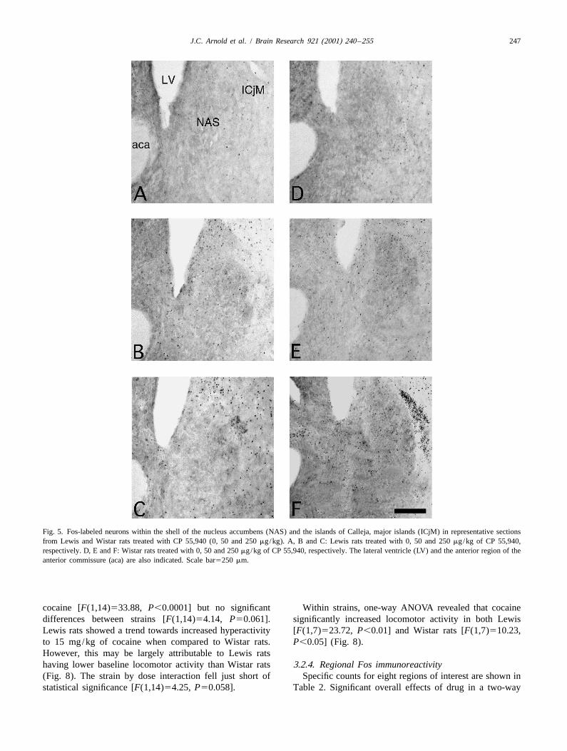
Fig. 5. Fos-labeled neurons within the shell of the nucleus accumbens (NAS) and the islands of Calleja, major islands (ICjM) in representative sectionsfrom Lewis and Wistar rats treated with CP 55,940 (0, 50 and 250 mg/kg). A, B and C: Lewis rats treated with 0, 50 and 250 mg/kg of CP 55,940,respectively. D, E and F: Wistar rats treated with 0, 50 and 250 mg/kg of CP 55,940, respectively. The lateral ventricle (LV) and the anterior region of theanterior commissure (aca) are also indicated. Scale bar5250 mm.
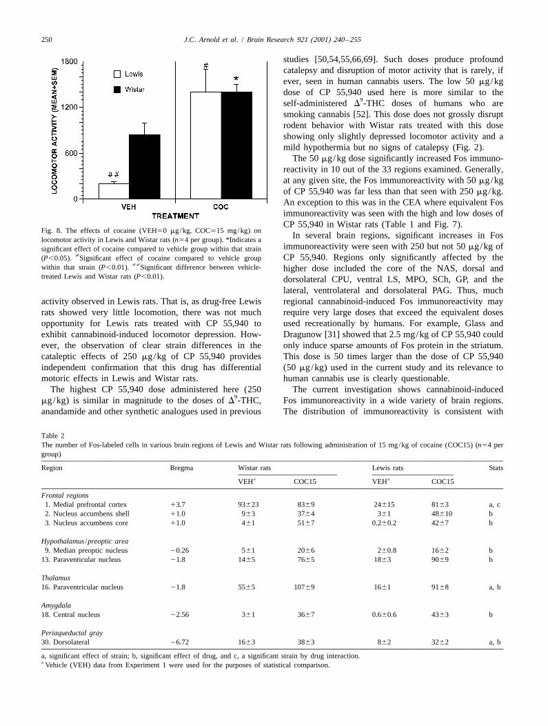
cocaine [F(1,14)533.88, P,0.0001] but no significant Within strains, one-way ANOVA revealed that cocainedifferences between strains [F(1,14)54.14, P50.061]. significantly increased locomotor activity in both LewisLewis rats showed a trend towards increased hyperactivity [F(1,7)523.72, P,0.01] and Wistar rats [F(1,7)510.23,to 15 mg/kg of cocaine when compared to Wistar rats. P,0.05] (Fig. 8).However, this may be largely attributable to Lewis ratshaving lower baseline locomotor activity than Wistar rats 3.2.4. Regional Fos immunoreactivity(Fig. 8). The strain by dose interaction fell just short of Specific counts for eight regions of interest are shown instatistical significance [F(1,14)54.25, P50.058]. Table 2. Significant overall effects of drug in a two-way
248 J.C. Arnold et al. / Brain Research 921 (2001) 240 –255
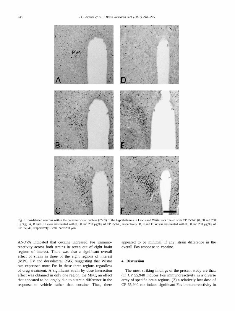
Fig. 6. Fos-labeled neurons within the paraventricular nucleus (PVN) of the hypothalamus in Lewis and Wistar rats treated with CP 55,940 (0, 50 and 250mg/kg). A, B and C: Lewis rats treated with 0, 50 and 250 mg/kg of CP 55,940, respectively. D, E and F: Wistar rats treated with 0, 50 and 250 mg/kg ofCP 55,940, respectively. Scale bar5250 mm.
ANOVA indicated that cocaine increased Fos immuno- appeared to be minimal, if any, strain difference in thereactivity across both strains in seven out of eight brain overall Fos response to cocaine.regions of interest. There was also a significant overalleffect of strain in three of the eight regions of interest(MPC, PV and dorsolateral PAG) suggesting that Wistar 4. Discussionrats expressed more Fos in these three regions regardlessof drug treatment. A significant strain by dose interaction The most striking findings of the present study are that:effect was obtained in only one region, the MPC, an effect (1) CP 55,940 induces Fos immunoreactivity in a diversethat appeared to be largely due to a strain difference in the array of specific brain regions, (2) a relatively low dose ofresponse to vehicle rather than cocaine. Thus, there CP 55,940 can induce significant Fos immunoreactivity in
J.C. Arnold et al. / Brain Research 921 (2001) 240 –255 249
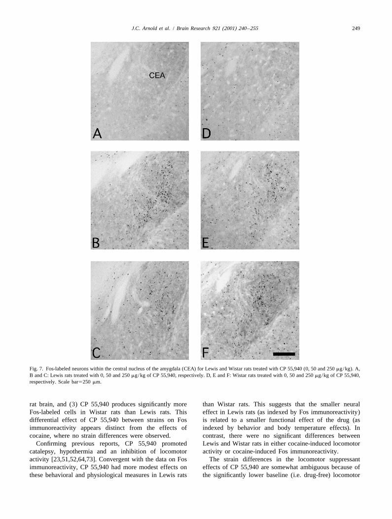
Fig. 7. Fos-labeled neurons within the central nucleus of the amygdala (CEA) for Lewis and Wistar rats treated with CP 55,940 (0, 50 and 250 mg/kg). A,B and C: Lewis rats treated with 0, 50 and 250 mg/kg of CP 55,940, respectively. D, E and F: Wistar rats treated with 0, 50 and 250 mg/kg of CP 55,940,respectively. Scale bar5250 mm.
rat brain, and (3) CP 55,940 produces significantly more than Wistar rats. This suggests that the smaller neuralFos-labeled cells in Wistar rats than Lewis rats. This effect in Lewis rats (as indexed by Fos immunoreactivity)differential effect of CP 55,940 between strains on Fos is related to a smaller functional effect of the drug (asimmunoreactivity appears distinct from the effects of indexed by behavior and body temperature effects). Incocaine, where no strain differences were observed. contrast, there were no significant differences between
Confirming previous reports, CP 55,940 promoted Lewis and Wistar rats in either cocaine-induced locomotorcatalepsy, hypothermia and an inhibition of locomotor activity or cocaine-induced Fos immunoreactivity.activity [23,51,52,64,73]. Convergent with the data on Fos The strain differences in the locomotor suppressantimmunoreactivity, CP 55,940 had more modest effects on effects of CP 55,940 are somewhat ambiguous because ofthese behavioral and physiological measures in Lewis rats the significantly lower baseline (i.e. drug-free) locomotor
250 J.C. Arnold et al. / Brain Research 921 (2001) 240 –255
studies [50,54,55,66,69]. Such doses produce profoundcatalepsy and disruption of motor activity that is rarely, ifever, seen in human cannabis users. The low 50 mg/kgdose of CP 55,940 used here is more similar to the
9self-administered D -THC doses of humans who aresmoking cannabis [52]. This dose does not grossly disruptrodent behavior with Wistar rats treated with this doseshowing only slightly depressed locomotor activity and amild hypothermia but no signs of catalepsy (Fig. 2).
The 50 mg/kg dose significantly increased Fos immuno-reactivity in 10 out of the 33 regions examined. Generally,at any given site, the Fos immunoreactivity with 50 mg/kgof CP 55,940 was far less than that seen with 250 mg/kg.An exception to this was in the CEA where equivalent Fosimmunoreactivity was seen with the high and low doses ofCP 55,940 in Wistar rats (Table 1 and Fig. 7).
Fig. 8. The effects of cocaine (VEH50 mg/kg, COC515 mg/kg) onIn several brain regions, significant increases in Foslocomotor activity in Lewis and Wistar rats (n54 per group). *Indicates a
immunoreactivity were seen with 250 but not 50 mg/kg ofsignificant effect of cocaine compared to vehicle group within that strain[ CP 55,940. Regions only significantly affected by the(P,0.05). Significant effect of cocaine compared to vehicle group
[[within that strain (P,0.01). Significant difference between vehicle- higher dose included the core of the NAS, dorsal andtreated Lewis and Wistar rats (P,0.01). dorsolateral CPU, ventral LS, MPO, SCh, GP, and the
lateral, ventrolateral and dorsolateral PAG. Thus, muchactivity observed in Lewis rats. That is, as drug-free Lewis regional cannabinoid-induced Fos immunoreactivity mayrats showed very little locomotion, there was not much require very large doses that exceed the equivalent dosesopportunity for Lewis rats treated with CP 55,940 to used recreationally by humans. For example, Glass andexhibit cannabinoid-induced locomotor depression. How- Dragunow [31] showed that 2.5 mg/kg of CP 55,940 couldever, the observation of clear strain differences in the only induce sparse amounts of Fos protein in the striatum.cataleptic effects of 250 mg/kg of CP 55,940 provides This dose is 50 times larger than the dose of CP 55,940independent confirmation that this drug has differential (50 mg/kg) used in the current study and its relevance tomotoric effects in Lewis and Wistar rats. human cannabis use is clearly questionable.
The highest CP 55,940 dose administered here (250 The current investigation shows cannabinoid-induced9
mg/kg) is similar in magnitude to the doses of D -THC, Fos immunoreactivity in a wide variety of brain regions.anandamide and other synthetic analogues used in previous The distribution of immunoreactivity is consistent with
Table 2The number of Fos-labeled cells in various brain regions of Lewis and Wistar rats following administration of 15 mg/kg of cocaine (COC15) (n54 pergroup)
Region Bregma Wistar rats Lewis rats Statsa aVEH COC15 VEH COC15
Frontal regions1. Medial prefrontal cortex 13.7 93623 8369 24615 8163 a, c2. Nucleus accumbens shell 11.0 963 3764 361 48610 b3. Nucleus accumbens core 11.0 461 5167 0.260.2 4267 b
Hypothalamus /preoptic area9. Median preoptic nucleus 20.26 561 2066 260.8 1662 b
13. Paraventicular nucleus 21.8 1465 7665 1863 9069 b
Thalamus16. Paraventricular nucleus 21.8 5565 10769 1661 9168 a, b
Amygdala18. Central nucleus 22.56 361 3667 0.660.6 4363 b
Periaqueductal gray30. Dorsolateral 26.72 1663 3863 862 3262 a, b
a, significant effect of strain; b, significant effect of drug, and c, a significant strain by drug interaction.a Vehicle (VEH) data from Experiment 1 were used for the purposes of statistical comparison.
J.C. Arnold et al. / Brain Research 921 (2001) 240 –255 251
previous results. For example, previous studies have inhibitory inputs to the basal ganglia [2]. Dense CB1
typically shown cannabinoid-induced Fos immunoreactivi- receptor distribution is seen in the GP and the SN, andty in the CPU [31,54,55], NAS [50,54,55,66,69], PVN moderate CB receptor distribution occurs in the CPU1
[50,61,69], PV [61], CEA [50,61,69], LS [50], and the [35,65,82]. Most CB receptors in the GP and the SN are1
BNST [61,69]. Other studies have documented Fos im- localised presynaptically on striatal afferents [34,47,83].munoreactivity in the PAG and MPO [61], the VTA [69], The locomotor depressant effects of cannabinoids seem tothe SO and the SCh [61,69]. To the best of our knowledge involve modulation of GABA, dopamine and glutamate inthe present findings of Fos immunoreactivity in the ICjM, these brain areas [2].AH, GP and the PPTg are novel. It is noteworthy that only sparse Fos immunoreactivity
As in previous studies, Fos immunoreactivity was seen was observed in a small and localised area of the GP ofhere in many areas containing only modest densities of Wistar rats administered the highest dose of CP 55,940.CB receptors. These areas include the NAS, VTA, LS, Furthermore, no CP 55,940-induced Fos immunoreactivity1
BNST, CEA, PVN and the PAG [35,65,82]. Conversely, was observed in the SN in the present study.CP 55,940 did not affect Fos immunoreactivity in the CP 55,940 promoted little Fos expression in the CPU,substantia nigra (SN), an area that contains many CB with only sparse Fos immunoreactivity observed in its1
receptors. Furthermore, the increase in Fos immuno- central, dorsal and dorsolateral regions. This agrees withreactivity observed in the GP, another area rich in CB previous research which has shown that administration of1
9receptors, was relatively sparse and restricted to the medial D -THC promotes some Fos immunoreactivity in the CPUportion of this structure. [48,55,66]. Interestingly, a strain difference was observed
The dissociation between Fos immunoreactivity and in CP 55,940-induced Fos immunoreactivity in the dorsalCB receptor density may be due to the CB receptor’s CPU, which is consistent with the strain differences1 1
negative link with adenylate cyclase and the inhibitory observed in catalepsy promoted by CP 55,940. The viewconsequences this has on cAMP production. When cAMP that cannabinoid-induced catalepsy involves the CPU isproduction is inhibited, Fos immunoreactivity is more supported by a previous study showing that administrationlikely to be suppressed than promoted [36]. On the other of CP 55,940 directly into this region produces catalepsyhand, the presence of high levels of Fos immunoreactivity in rats [45].in areas with low CB receptor density presumably reflects Another important brain site for cannabinoid effects on1
transynaptic activation of non-CB receptor containing locomotor activity may be the core of the NAS. Direct19neurons, or the presynaptic modulation of neurotransmitter infusion of D -THC into the NAS promoted catalepsy in
release by CB receptors affecting Fos immunoreactivity mice [59]. In the current investigation, the lesser effects of19postsynaptically. For example, in a previous study, D - CP 55,940 on locomotor activity and catalepsy in Lewis
THC-induced Fos immunoreactivity in the dorsomedial rats relative to Wistar rats correlated with the differentialstriatum and the NAS was blocked by the D dopamine pattern of Fos immunoreactivity observed in the core and1
receptor antagonist SCH 23390 [55]. This presumably shell of the NAS between these strains.9reflects an action of D -THC on dopamine release in the CP 55,940 increased Fos immunoreactivity in a range of
9NAS, with increased dopamine levels caused by D -THC brain structures known to be involved in fear, anxiety andleading to increased Fos immunoreactivity. defensive behavior [21]. These include the CEA, BNST
Consistent with its hypothermic effect, CP 55,940 and the PAG. The pattern of Fos immunoreactivity in thesesignificantly increased Fos immunoreactivity in preoptic areas, particularly in Wistar rats, may highlight a neuraland hypothalamic regions thought to be involved in circuit that underpins an integrated fear or anxiety responsethermoregulation. These areas include the MnPO, MPO to cannabinoid administration. The divergent effects onand AH [75,76,81,87]. Interestingly, previous studies have Fos immunoreactivity across strains are consistent with the
9shown that injection of D -THC into the preoptic area of effects of CP 55,940 on Lewis and Wistar rats in a varietythe anterior hypothalamus caused significant reductions in of different models of anxiety [5]. In these models, Lewisbody temperature [25,63]. In addition, exposure to a hot rats show markedly less anxiety-like behaviors than Wistarenvironment increases Fos immunoreactivity in the MnPO rats when administered CP 55,940 (10–75 mg/kg).
9[75]. The ICjM might also be involved in the hypothermic When D -THC is directly infused into the CEA it causeseffect of cannabinoids. The current study showed clear mice to spend less time exploring the open arms of anstrain differences in the hypothermic effects of CP 55,940 elevated plus maze [59]. The CEA may therefore underpinthat correlated with strain differences observed in Fos an integrated anxiety response seen with the administrationimmunoreactivity in the ICjM. A recent study has shown of cannabinoids. The CEA gives rise to an importantthat intracranial administration of D receptor agonists into output pathway that modulates many aspects of uncon-3
the ICjM causes hypothermia, an effect which was poten- ditioned and conditioned fear (e.g. bradycardia, freezing,tiated by D receptor agonists [8]. hypoalgesia and activation of the HPA axis) [72,80]. It1
The influence of cannabinoids on locomotor activity is provides inputs and outputs to the BNST and output to thethought to reflect presynaptic modulation of excitatory and PVN (involved in stress and the HPA) and the PAG
252 J.C. Arnold et al. / Brain Research 921 (2001) 240 –255
(involved in defensive behavior, cardiovascular function PAG are consistent with Lewis rats showing less CPand pain transmission) [1,80]. Interestingly, in the current 55,940-induced catalepsy and hypothermia. It is alsoinvestigation all of the above areas (CEA, BNST and conceivable that Lewis rats may display lesser antinocicep-PVN) showed marked CP 55,940-induced Fos immuno- tive effects of CP 55,940 than Wistar rats.reactivity, with significantly more Fos-labeled cells Interestingly, Lewis rats showed less CP 55,940-inducedcounted in Wistar rats in comparison to Lewis rats. Fos immunoreactivity in reward-related regions as well as
An extraordinary strain difference in CP 55,940-induced those involved in anxiety. In both strains of rat, CP 55,940Fos immunoreactivity was observed in the lateral division promoted Fos immunoreactivity in the shell of the NAS,of the dorsal BNST (Table 1). Notably, both CP 55,940 but the effect was much larger in Wistar rats. This findingdoses promoted seven times as much Fos-labeled cells in contradicts the notion that Lewis rats may be uniquelyWistar rats in comparison to Lewis rats in this brain sensitive to the rewarding effects of cannabinoids via aregion. The BNST may be a brain region that is crucially greater sensitivity to cannabinoid actions on the mesolim-involved in cannabinoid-induced anxiety. As part of the bic dopamine system [18,44]. However it might be notedextended amygdala, the BNST in thought of as a rostral that this theory is somewhat controversial. Thus, Parkerextension of the CEA [1]. The BNST has a very similar and colleagues found a conditioned place aversion to
9structure to the CEA and contains similar transmitters, cell D -THC in Lewis rats — an effect that suggests anmorphology and has similar efferent connections [1,80] aversive action of cannabinoids in this strain [60]. Inand a similar involvement in fear-related behaviors [19]. addition, we have recently shown that CP 55,940 fails to
The differential behavioral and emotional effects of CP lower thresholds for rewarding brain stimulation in Lewis55,940 in Lewis and Wistar rats could also be attributable rats [6]. This finding is again inconsistent with the idea thatto differences in cannabinoid-induced activation of the Lewis rats are more susceptible to cannabinoid-inducedHPA axis. The HPA axis of Lewis rats is hyporesponsive to reward.stressful stimuli and releases less basal and stress-induced An increasing body of evidence suggests that increasedcorticosterone [12,68]. This appears to be a consequence of dopaminergic transmission in the NAS is produced bya deficiency in the ability of the hypothalamus to syn- stressful or anxiogenic stimuli [39,74]. Interestingly, thethesise and secrete CRH in Lewis rats [78,79]. This may CEA projects to the shell of the NAS via the lateral BNSTalso relate to the relative lack of cannabinoid-induced Fos [1]. The ambiguity over whether the NAS is involved inimmunoreactivity in the PVN of Lewis rats compared to the aversive and rewarding effects of cannabinoids raisesWistar rats seen in the present study. The PVN produces an issue which may be important to the understanding ofCRH, a precursor to corticosterone release [3]. In addition, the appetitive effects of cannabinoids. This issue involvesthe effects of CRH on Fos immunoreactivity shows working out the exact relationship between reward andcommonalities with the current results with increased Fos- anxiety, and the implication this relationship may have forlabeled cells observed in PVN, CEA, LS, BNST, NAS and appetitive behavior. For example, while cocaine has clearlythe CPU [4]. Furthermore, administration of the selective rewarding effects in humans, it also has aversive effectsCRH antagonist, D-Phe CRF , attenuated the an- [10,85]. Cocaine is also at once rewarding and anxiogenic12-41
xiogenic effects of the cannabinoid receptor agonist HU- in rats [11,22,49,71,88]. In addition, as shown in the210 in a defensive-withdrawal model [70]. current investigation, cocaine promotes Fos immuno-
The PAG is an important brain structure involved in reactivity in similar areas to CP 55,940, including areascannabinoid-induced antinociception, catalepsy and hypo- implicated in both reward and anxiety, such as the shellthermia [45]. Lichtman et al. [45] showed that microinjec- and core of the NAS, the CEA, the PVN and the PAG.tions of CP 55,940 into the ventrolateral PAG produced all For cocaine, the rewarding and anxiogenic effects haveof the above effects. In addition, this study showed that been dissociated in an elegant study [22]. Rats avidlymicroinjection of CP 55,940 into the dorsolateral PAG had self-administering cocaine over days in a runway taskno such effects. Further, the PAG has been implicated in showed more retreats than controls from the goal box. Asneural circuitry involved in defensive behavior [14,15,21]. the rats retreated only a short distance and continued toPrior studies indicate that the ventrolateral PAG is involved avidly self-administer cocaine, this observation was pro-in freezing and immobility, whereas the dorsolateral PAG posed to reflect a conflict situation for the rat betweenis involved in the control of flight behaviors positive and negative aspects of cocaine’s effects. In[9,16,17,20,40]. addition, the anxiolytic compound diazepam, dose-depen-
The current investigation showed that Lewis rats showed dently reduced this retreat behavior. Thus, for cocaine itless CP 55,940-induced Fos immunoreactivity than Wistar appears the conflict between its aversive and rewardingrats in the ventrolateral and dorsolateral PAG. This is also effects has implications for cocaine self-administration.consistent with Lewis rats being less susceptible to CP Future studies will hopefully address the relationship55,940-induced anxiety-related behaviors than Wistar rats between aversion and reward and what implication this has[5]. Further, the strain differences observed in the effects for appetitive behavior relevant to cannabinoids.of CP 55,940 on Fos immunoreactivity in the ventrolateral CP 55,940 appeared to have modest and largely equiva-
J.C. Arnold et al. / Brain Research 921 (2001) 240 –255 253
lent effects on Fos immunoreactivity in the VTA of Lewis have less G proteins in a pool utilised by CB receptors.1
and Wistar rats. If anything, CP 55,940 had less of an Evidence shows that Lewis rats have less G proteins in theeffect on Fos immunoreactivity in the VTA of Lewis rats NAS in comparison to Fischer 344 rats [12]. Anotherthan Wistar rats consistent with the distinct effects that CP possibility is that pharmacokinetic properties of CP 55,94055,940 had on Fos immunoreactivity in the shell of the differ between these two strains, with Lewis rats perhapsNAS. One proposed mechanism which might mediate able to metabolise CP 55,940 more efficiently than Wistarcannabinoid-induced dopamine release in the NAS is rats. Future studies will hopefully be able to give adisinhibition of dopamine cell firing in the VTA via definitive answer to these hypotheses.inhibition of GABA interneurons [2]. This theory is basedon the finding that cannabinoids increase the spontaneousfiring of neurons within the VTA [26,27,30].
AcknowledgementsCannabinoid-induced Fos immunoreactivity in the ICjMhas not been previously reported. It is interesting to
This study was supported by an Australian Researchspeculate upon the role of the ICjM in the functionalCouncil grant to ISM and PEM and a National Health andeffects of cannabinoids. A role for the ICjM in rein-Medical Research Council grant to GEH and ISM. Inforcement appears plausible based on the anatomicaladdition, this research was also supported by an ARC-proximity and neurochemical similarity of the ICjM andsponsored postgraduate scholarship awarded to JCA. Ericathe NAS. The ICjM lies adjacent to the shell of the NAS,Campbell is gratefully acknowledged for her pilot workand like the shell of the NAS contains dopamine D and1 that helped to guide this study.D receptors [62,67]. Thus, it is possible that functional3
interactions may occur between the ICjM and the shell ofthe NAS. Interestingly, many other recreationally useddrugs promote Fos immunoreactivity in the ICjM such as ReferencesMDMA and cocaine [77,89]. Future studies might usefullyexplore whether the ICjM has a functional role in the [1] G.F. Alheid, J.S. de Olmos, C.A. Beltramino, Amygdala and
extended amygdala, in: G. Paxinos (Ed.), The Rat Nervous System,rewarding effects of drugs such as cannabis.Academic Press, San Diego, CA, 1995, pp. 495–578.The present study shows for the first time that a
[2] A. Ameri, The effects of cannabinoids on the brain, Prog. Neurobiol.cannabinoid increases Fos immunoreactivity in the PPTg.58 (1999) 315–348.
This area has been implicated in a wide range of functions [3] F.A. Antoni, M. Palkovits, G.B. Makara, E.A. Linton, P.J. Lowry,including reinforcement, reward, motor behavior, control J.Z. Kiss, Immunoreactive corticotropin-releasing hormone in the
hypothalamoinfundibular tract, Neuroendocrinology 36 (1983) 415–of sleep–wake cycles, antinociception and cognitive pro-423.cessing [38,86]. Interestingly, cannabinoids affect all of
[4] F.J. Arnold, M. De Lucas Bueno, H. Shiers, D.C. Hancock, G.I.these functions [2,24,28,29,53]. It is unlikely that theEvan, J. Herbert, Expression of c-fos in regions of the basal limbic
effects of CP 55,940 on Fos immunoreactivity in the PPTg forebrain following intracerebroventricular corticotropin-releasingare mediated via direct activation of CB receptors as there factor in unstressed or stressed male rats, Neuroscience 51 (1992)1
377–390.appear to be none in this area [35,65,82].[5] J.C. Arnold, The behavioural and neural effects of cannabinoids,The PPTg has extensive connectivity with the basal
Department of Psychology, PhD Thesis, University of Sydney,ganglia [38]. Specifically, afferent and efferent connectionsSydney, 2000, pp. 233.
between the PPTg and areas rich in CB receptors, such as1 [6] J.C. Arnold, G.E. Hunt, I.S. McGregor, Effects of the cannabinoidthe GP and the SN, have been described [38]. Recently, receptor agonist CP 55,940 and the cannabinoid receptor antagonistconverging evidence has strongly implicated the PPTg as SR 141716 on intracranial self-stimulation in Lewis rats (in press).
[7] J.C. Arnold, A.N. Topple, G.E. Hunt, I.S. McGregor, Effects ofpart of the neural circuit underlying reward [86]. Lesionspre-exposure and co-administration of the cannabinoid receptorof the PPTg reduced heroin self-administration [58],agonist CP 55,940 on behavioral sensitization to cocaine, Eur. J.
responding for rewarding brain stimulation [43] and the Pharmacol. 354 (1998) 9–16.development of conditioned place preferences to opiates, [8] S. Barik, R. de Beaurepaire, Hypothermic effects of dopamine D3stimulants or food [56,57]. The role of the PPTg in receptor agonists in the island of Calleja Magna. Potentiation by D1
activation, Pharmacol. Biochem. Behav. 60 (1998) 313–319.cannabinoid-induced reward is clearly an interesting topic[9] M.M. Behbehani, Functional characteristics of the midbrainfor further study.
periaqueductal gray, Prog. Neurobiol. 46 (1995) 575–605.Finally, it should be noted that the mechanism underly- [10] N.L. Benowitz, How toxic is cocaine?, Ciba Found. Symp. 166
ing the strain differences in cannabinoid-induced behavior (1992) 125–148.and Fos expression reported in the present study are not [11] R.J. Blanchard, M.A. Hebert, L. Dulloog, N. Kaawaloa, O. Nishi-
mura, D.C. Blanchard, Acute cocaine effects on stereotype andentirely clear. One possibility is that Lewis rats have fewerdefense: an ethoexperimental approach, Neurosci. Biobehav. Rev.central CB receptors than Wistar rats and may thus be1 23 (1998) 179–188.
subsensitive to cannabinoid effects. Alternatively, strain [12] E.S. Brodkin, W.A. Carlezon, C.N. Haile, T.A. Kosten, G.R.differences may exist in the affinity of CP 55,940 for the Heninger, E.J. Nestler, Genetic analysis of behavioral, neuroen-CB receptor. Another hypothesis is that Lewis rats may docrine, and biochemical parameters in inbred rodents — initial1
254 J.C. Arnold et al. / Brain Research 921 (2001) 240 –255
studies in Lewis and Fischer 344 rats and in A/J and C57BL/6J [34] M. Herkenham, A.B. Lynn, B.R. de Costa, E.K. Richfield, Neuronalmice, Brain Res. 805 (1998) 55–68. localization of cannabinoid receptors in the basal ganglia of the rat,
Brain Res. 547 (1991) 267–274.[13] D.M. Camp, K.E. Browman, T.E. Robinson, The effects of metham-phetamine and cocaine on motor behavior and extracellular dopa- [35] M. Herkenham, A.B. Lynn, M.R. Johnson, L.S. Melvin, B.R. demine in the ventral striatum of Lewis versus Fischer 344 rats, Brain Costa, K.C. Rice, Characterization and localization of cannabinoidRes. 668 (1994) 180–193. receptors in rat brain: a quantitative in vitro autoradiographic study,
J. Neurosci. 11 (1991) 563–583.[14] N.S. Canteras, S. Chiavegatto, L.E. Valle, L.W. Swanson, Severereduction of rat defensive behavior to a predator by discrete [36] P. Hughes, M. Dragunow, Induction of immediate-early genes andhypothalamic chemical lesions, Brain Res. Bull. 44 (1997) 297–305. the control of neurotransmitter-regulated gene expression within the
nervous system, Pharmacol. Rev. 47 (1995) 133–178.[15] N.S. Canteras, M. Goto, Fos-like immunoreactivity in theperiaqueductal gray of rats exposed to a natural predator, Neurore- [37] G.E. Hunt, I.S. McGregor, Rewarding brain stimulation inducesport 10 (1999) 413–418. only sparse fos-like immunoreactivity in dopaminergic neurons,
Neuroscience 83 (1998) 501–515.[16] P. Carrive, The periaqueductal gray and defensive behavior: func-tional representation and neuronal organization, Behav. Brain Res. [38] W.L. Inglis, P. Winn, The pedunculopontine tegmental nucleus:58 (1993) 27–47. where the striatum meets the reticular formation, Prog. Neurobiol.
47 (1995) 1–29.[17] P. Carrive, P. Leung, J. Harris, G. Paxinos, Conditioned fear tocontext is associated with increased Fos expression in the caudal [39] M.H. Joseph, A.M.J. Young, J.A. Gray, Are neurochemistry andventrolateral region of the midbrain periaqueductal gray, Neuro- reinforcement enough? Can the abuse potential of drugs be ex-science 78 (1997) 165–177. plained by common actions on a dopamine reward system in the
brain?, Hum. Psychopharmacol. 11 (1996) S55–S63.[18] J.P. Chen, W. Paredes, J.H. Lowinson, E.L. Gardner, Strain-specific9facilitation of dopamine efflux by D -tetrahydrocannabinol in the [40] K.A. Keay, R. Bandler, Deep and superficial noxious stimulation
nucleus accumbens of rat: an in vivo microdialysis study, Neurosci. increases Fos-like immunoreactivity in different regions of theLett. 129 (1991) 136–180. midbrain periaqueductal grey of the rat, Neurosci. Lett. 154 (1993)
23–26.[19] M. Davis, Are different parts of the extended amygdala involved infear versus anxiety?, Biol. Psychiatry 44 (1998) 1239–1247. [41] T.A. Kosten, M.J. Miserendino, S. Chi, E.J. Nestler, Fischer and
Lewis rat strains show differential cocaine effects in conditioned[20] B.M. De Oca, J.P. DeCola, S. Maren, M.S. Fanselow, Distinctplace preference and behavioral sensitization but not in locomotorregions of the periaqueductal gray are involved in the acquisitionactivity or conditioned taste aversion, J. Pharmacol. Exp. Ther. 269and expression of defensive responses, J. Neurosci. 18 (1998)(1994) 137–144.3426–3432.
[21] R.A. Dielenberg, G.E. Hunt, I.S. McGregor, ‘When a rat smells a [42] T.A. Kosten, M.J. Miserendino, C.N. Haile, J.L. DeCaprio, P.I.cat’: the distribution of Fos immunoreactivity in rat brain following Jatlow, E.J. Nestler, Acquisition and maintenance of intravenousexposure to a predatory odor, Neuroscience 104 (2001) 1085–1097. cocaine self-administration in Lewis and Fischer inbred rat strains,
[22] A. Ettenberg, T.D. Geist, Animal model for investigating the Brain Res. 778 (1997) 418–429.anxiogenic effects of self-administered cocaine, Psychopharmacolo- [43] M. Lepore, K.B. Franklin, N-methyl-D-aspartate lesions of thegy 103 (1991) 455–461. pedunculopontine nucleus block acquisition and impair maintenance
[23] F. Fan, D.R. Compton, S. Ward, L. Melvin, B.R. Martin, Develop- of responding reinforced with brain stimulation, Neuroscience 719ment of cross-tolerance between D -tetrahydrocannabinol, CP (1996) 147–155.
55,940 and WIN 55,212, J. Pharmacol. Exp. Ther. 271 (1994) [44] M. Lepore, X.H. Liu, V. Savage, D. Matalon, E.L. Gardner, Genetic91383–1390. differences in D -tetrahydrocannabinol-induced facilitation of brain
[24] I. Feinberg, R. Jones, J.M. Walker, C. Cavness, J. March, Effects of stimulation reward as measured by a rate-frequency curve-shift9high dosage D -tetrahydrocannabinol on sleep patterns in man, Clin. electrical brain stimulation paradigm in three different rat strains,
Pharmacol. Ther. 17 (1975) 458–466. Life Sci. 58 (1996) L365–L372.[25] A.G. Fitton, R.G. Pertwee, Changes in body temperature and oxygen [45] A.H. Lichtman, S.A. Cook, B.R. Martin, Investigation of brain sites
consumption rate of conscious mice produced by intrahypothalamic mediating cannabinoid-induced antinociception in rats: evidence9and intracerebroventricular injections of D -tetrahydrocannabinol, supporting periaqueductal gray involvement, J. Pharmacol. Exp.
Br. J. Pharmacol. 75 (1982) 409–414. Ther. 276 (1996) 585–593.9[26] E.D. French, D -Tetrahydrocannabinol excites rat VTA dopamine [46] M.J. Lyons, R. Toomey, J.M. Meyer, A.I. Green, S.A. Eisen, J.
neurons through activation of cannabinoid CB1 but not opioid Goldberg, W.R. True, M.T. Tsuang, How do genes influencereceptors, Neurosci. Lett. 226 (1997) 159–162. marijuana use? The role of subjective effects, Addiction 92 (1997)
[27] E.D. French, K. Dillon, X. Wu, Cannabinoids excite dopamine 409–417.neurons in the ventral tegmentum and substantia nigra, Neuroreport [47] P. Mailleux, J.J. Vanderhaeghen, Distribution of neuronal can-8 (1997) 649–652. nabinoid receptor in the adult rat brain: a comparative receptor
[28] E.L. Gardner, J.H. Lowinson, Marijuana’s interaction with brain binding radioautography and in situ hybridization histochemistry,reward systems: update 1991, Pharmacol. Biochem. Behav. 40 Neuroscience 48 (1992) 655–668.(1991) 571–580. [48] P. Mailleux, M. Verslype, X. Preud’homme, J.J. Vanderhaeghen,
[29] E.L. Gardner, S.R. Vorel, Cannabinoid transmission and reward- Activation of multiple transcription factor genes by tetrahydro-related events, Neurobiol. Dis. 5 (1998) 502–533. cannabinol in rat forebrain, Neuroreport 5 (1994) 1265–1268.
[30] G.L. Gessa, M.S. Mascia, M.A. Casu, G. Carta, Inhibition of [49] J.R. Mantsch, N.E. Goeders, Generalization of a restraint-inducedhippocampal acetylcholine release by cannabinoids: reversal by SR discriminative stimulus to cocaine in rats, Psychopharmacology 135141716A, Eur. J. Pharmacol. 327 (1997) R1–R2. (1998) 423–426.
[31] M. Glass, M. Dragunow, Induction of the Krox 24 transcription [50] I.S. McGregor, J.C. Arnold, M.F. Weber, A.N. Topple, G.E. Hunt, A9factor in striosomes by a cannabinoid agonist, Neuroreport 6 (1995) comparison of D -THC and anandamide induced c-fos expression in
241–244. the rat forebrain, Brain Res. 802 (1998) 19–26.[32] W. Hall, N. Solowij, Adverse effects of cannabis, Lancet 352 (1998) [51] I.S. McGregor, F.N. Dastur, R.A. McLellan, R.E. Brown, Can-
1611–1616. nabinoid modulation of rat pup ultrasonic vocalizations, Eur. J.[33] R.E. Harlan, M.M. Garcia, Drugs of abuse and immediate-early Pharmacol. 313 (1996) 43–49.
genes in the forebrain, Mol. Neurobiol. 16 (1998) 221–267. [52] I.S. McGregor, C.N. Issakidis, G. Prior, Aversive effects of the
J.C. Arnold et al. / Brain Research 921 (2001) 240 –255 255
synthetic cannabinoid CP 55,940 in rats, Pharmacol. Biochem. tenuates the acute actions of the highly potent cannabinoid receptorBehav. 53 (1996) 657–664. agonist HU-210 on defensive-withdrawal behavior in rats, J. Phar-
[53] R. Mechoulam, E. Fride, L. Hanus, T. Sheskin, T. Bisogno, V. Di macol. Exp. Ther. 276 (1996) 56–64.Marzo, M. Bayewitch, Z. Vogel, Anandamide may mediate sleep [71] R. Rogerio, R.N. Takahashi, Anxiogenic properties of cocaine in theinduction, Nature 389 (1997) 25–26. rat evaluated with the elevated plus-maze, Pharmacol. Biochem.
[54] A. Miyamoto, T. Yamamoto, M. Ohno, S. Watanabe, Desensitization Behav. 43 (1992) 631–633.of Fos protein induction in rat striatum and nucleus accumbens [72] J.B. Rosen, J. Schulkin, From normal fear to pathological anxiety,
9following repeated administration of D -tetrahydrocannabinol, Brain Psychol. Rev. 105 (1998) 325–350.Res. 763 (1997) 137–140. [73] T. Rubino, P. Massi, G. Patrini, I. Venier, G. Giagnoni, D. Parolaro,
[55] A. Miyamoto, T. Yamamoto, M. Ohno, S. Watanabe, H. Tanaka, S. Chronic CP-55,940 alters cannabinoid receptor mRNA in the rat9Morimoto, Y. Shoyama, Roles of dopamine D1 receptors in D - brain: an in situ hybridization study, Neuroreport 5 (1994) 2493–
tetrahydrocannabinol-induced expression of Fos protein in the rat 2496.brain, Brain Res. 710 (1996) 234–240. [74] J.D. Salamone, M.S. Cousins, B.J. Snyder, Behavioral functions of
[56] K. Nader, A. Bechara, D. van der Kooy, Neurobiological constraints nucleus accumbens dopamine: empirical and conceptual problemson behavioral models of motivation, Annu. Rev. Psychol. 48 (1997) with the anhedonia hypothesis, Neurosci. Biobehav. Rev. 21 (1998)85–114. 341–359.
[57] M.C. Olmstead, K.B. Franklin, Effects of pedunculopontine tegmen- [75] T.E. Scammell, K.J. Price, S.M. Sagar, Hyperthermia induces c-fostal nucleus lesions on morphine-induced conditioned place prefer- expression in the preoptic area, Brain Res. 618 (1993) 303–307.ence and analgesia in the formalin test, Neuroscience 57 (1993) [76] J.E. Smith, A.S. Jansen, M.P. Gilbey, A.D. Loewy, CNS cell groups411–418. projecting to sympathetic outflow of tail artery: neural circuits
[58] M.C. Olmstead, E.M. Munn, K.B. Franklin, R.A. Wise, Effects of involved in heat loss in the rat, Brain Res. 786 (1998) 153–164.pedunculopontine tegmental nucleus lesions on responding for [77] C.P. Stephenson, G.E. Hunt, A.N. Topple, I.S. McGregor, Theintravenous heroin under different schedules of reinforcement, J. distribution of 3,4-methylenedioxymethamphetamine ‘Ecstasy’-in-Neurosci. 18 (1998) 5035–5044. duced c-fos expression in rat brain, Neuroscience 92 (1999) 1011–
[59] E.S. Onaivi, A. Chakrabarti, E.T. Gwebu, G. Chaudhuri, Neuro- 1023.9behavioral effects of D -THC and cannabinoid (CB1) receptor gene [78] E.M. Sternberg, J.M. Hill, G.P. Chrousos, T. Kamilaris, S.J.
expression in mice, Behav. Brain Res. 72 (1996) 115–125. Listwak, P.W. Gold, R.L. Wilder, Inflammatory mediator-induced[60] L.A. Parker, T. Gillies, THC-induced place and taste aversions in hypothalamic–pituitary–adrenal axis activation is defective in strep-
Lewis and Sprague–Dawley rats, Behav. Neurosci. 109 (1995) tococcal cell wall arthritis-susceptible Lewis rats, Proc. Natl. Acad.71–78. Sci. USA 86 (1989) 2374–2378.
[61] N.A. Patel, R.L. Moldow, J.A. Patel, G.D. Wu, S.L. Chang, [79] E.M. Sternberg, W.S.d. Young, R. Bernardini, A.E. Calogero, G.P.Arachidonylethanolamide (AEA) activation of Fos proto-oncogene Chrousos, P.W. Gold, R.L. Wilder, A central nervous system defectprotein immunoreactivity in the rat brain, Brain Res. 797 (1998) in biosynthesis of corticotropin-releasing hormone is associated with225–233. susceptibility to streptococcal cell wall-induced arthritis in Lewis
[62] G. Paxinos, C. Watson, The Rat Brain in Stereotaxic Coordinates, rats, Proc. Natl. Acad. Sci. USA 86 (1989) 4771–4775.3rd Edition, Academic Press, Sydney, 1997. [80] L.W. Swanson, G.D. Petrovich, What is the amygdala?, Trends
[63] R.G. Pertwee, D. Hedley, A.S. McQueen, S.M. Gentleman, The Neurosci. 21 (1998) 323–331.9hypothermic response of mice to D -tetrahydrocannabinol is en- [81] K.A. Travis, A.K. Johnson, In vitro sensitivity of median preoptic
hanced by chlorpromazine, thioxanthenes, alpha-adrenoceptor an- neurons to angiotensin II, osmotic pressure, and temperature, Am. J.tagonists and pentolinium but not by SCH 23390 or sulpiride, Physiol. 264 (1993) R1200–1205.Neuropharmacology 27 (1988) 149–155. [82] K. Tsou, S. Brown, C. Sanudo-Pena, K. Mackie, J.M. Walker,
[64] R.G. Pertwee, L.A. Stevenson, G. Griffin, Cross-tolerance between Immunohistochemical distribution of cannabinoid CB1 receptors in9
D -tetrahydrocannabinol and the cannabimimetic agents, CP 55,940, the rat central nervous system, Neuroscience 83 (1998) 393–411.WIN 55,212-2 and anandamide, Br. J. Pharmacol. 110 (1993) [83] T.M. Westlake, A.C. Howlett, T.I. Bonner, L.A. Matsuda, M.1483–1490. Herkenham, Cannabinoid receptor binding and messenger RNA
[65] D.A.D. Pettit, M.P. Harrison, J.M. Olson, R.F. Spencer, G.A. Cabral, expression in human brain: an in vitro receptor autoradiography andImmunohistochemical localization of the neural cannabinoid re- in situ hybridization histochemistry study of normal aged andceptor in rat brain, J. Neurosci. Res. 51 (1998) 391–402. Alzheimer’s brains, Neuroscience 63 (1994) 637–652.
9 9[66] A. Porcella, G.L. Gessa, L. Pani, D -tetrahydrocannabinol increases [84] A.P. Wickens, R.G. Pertwee, D -tetrahydrocannabinol and anan-sequence-specific AP-1 DNA-binding activity and Fos-related an- damide enhances the ability of muscimol to induce catalepsy in thetigens in the rat brain, Eur. J. Neurosci. 10 (1998) 1743–1751. globus pallidus of rats, Eur. J. Pharmacol. 250 (1993) 205–208.
[67] S. Ridray, N. Griffon, V. Mignon, E. Souil, S. Carboni, J. Diaz, J.C. [85] S. Williamson, M. Gossop, B. Powis, P. Griffiths, J. Fountain, J.Schwartz, P. Sokoloff, Coexpression of dopamine D1 and D3 Strang, Adverse effects of stimulant drugs in a community sample ofreceptors in islands of Calleja and shell of nucleus accumbens of the drug users, Drug Alcohol Depend. 44 (1997) 87–94.rat: opposite and synergistic functional interactions, Eur. J. Neuro- [86] P. Winn, V.J. Brown, W.L. Inglis, On the relationships between thesci. 10 (1998) 1676–1686. striatum and the pedunculopontine tegmental nucleus, Crit. Rev.
[68] S. Rivest, C. Rivier, Stress and interleukin-1 beta-induced activation Neurobiol. 11 (1997) 241–261.of c-fos, NGFI-B and CRF gene expression in the hypothalamic [87] M. Yamada, T. Cho, N.J. Coleman, E. Richelson, Regulation ofPVN: comparison between Sprague–Dawley, Fisher-344 and Lewis daily rhythm of body temperature by neurotensin receptor in rats,rats, J. Neuroendocrinol. 6 (1994) 101–117. Res. Commun. Mol. Pathol. Pharmacol. 87 (1995) 323–332.
[69] F. Rodriguez de Fonseca, R.A. Carrera, M. Navarro, G.F. Koob, F. [88] X.M. Yang, A.L. Gorman, A.J. Dunn, N.E. Goeders, AnxiogenicWeiss, Activation of corticotropin-releasing factor in the limbic effects of acute and chronic cocaine administration: neurochemicalsystem during cannabinoid withdrawal, Science 276 (1997) 2050– and behavioral studies, Pharmacol. Biochem. Behav. 41 (1992)2054. 643–650.
[70] F. Rodriguez de Fonseca, P. Rubio, F. Menzaghi, E. Merlo-Pich, J. [89] S.T. Young, L.J. Porrino, M.J. Iadarola, Cocaine induces striatalRivier, G.F. Koob, M. Navarro, Corticotropin-releasing factor c-fos-immunoreactive proteins via dopaminergic D1 receptors, Proc.(CRF) antagonist [D-Phe12,Nle21,38,C alpha MeLeu37]CRF at- Natl. Acad. Sci. USA 88 (1991) 1291–1295.