The role of lipocortin-1 in dexamethasone-induced suppression of PGE2 and TNFα release from human...
8
1996 Stockton Press All rights reserved 0007-1188/96 $12.00 % The role of lipocortin- 1 in dexamethasone-induced suppression of PGE2 and TNFcx release from human peripheral blood mononuclear cells 'Allan W. Sudlow, *Frank Carey, *Robert Forder & Nancy J. Rothwell School of Biological Sciences, University of Manchester, Oxford Rd., Manchester, M13 9PT and *Zeneca Pharmaceuticals, Macclesfield, Cheshire, SK1O 4TG 1 Lipocortin-1 and its N-terminal derivatives exert potent inhibitory actions in various models of acute inflammation. The present study examined the ability of lipocortin (LC)-1 to suppress the release of the acute pro-inflammatory mediators, tumour necrosis factor (TNFax) and prostaglandin E2 (PGE2) from human peripheral blood mononuclear cells (PBMC) stimulated with lipopolysaccharide (LPS) or recombinant human interleukin-l# (rhIL-lB). 2 LPS (10 Mig ml-')-stimulated release of TNFa and PGE2 from PBMC was significantly inhibited by (4 h) co-incubation of the cells with 10-6 M dexamethasone (Dex), but not with 10-9 M to 10-7 M of a N-terminal fragment (amino acids 1- 188) of recombinant human LC-1 (LC-1 fragment). However, Dex suppression of LPS-stimulated TNFax and PGE2 secretion from PBMC was reversed when polyclonal antibody to LC-1 fragment (1: 10,000 dilution) was included in the medium. rhIL-lB (5 x 10⁻⁸ M)- stimulated release of TNFa and PGE2 from PBMC (after 18 h) was abolished by co-incubation of the cells with 10-7 M LC-1 fragment. 3 After incubation with Dex (4 h), cellular proteins from PBMC were immunoblotted using anti-LC-1 fragment antibody (which showed no cross-reactivity with human annexins 2 to 6). Dex caused no increase in immunoreactive (ir)LC-1 content of PBMC, although there was a three fold increase in the amount of a lower mass species with LC-1-like immunoreactivity. This was accompanied by the appearance of irLC-1 in the extracellular medium. 4 The results of the present study implicate endogenous LC-1 in glucocorticoid suppression of TNFa and PGE2 release from human PBMC and suggest an extracellular site of action for LC-1. LC-1 may also inhibit rhIL-1l,-stimulated TNFa and PGE2 secretion from PBMC. Keywords: Lipocortin-1; annexin-1; tumour necrosis factor ax; prostaglandin E2; monocyte Introduction Lipocortin-1 (LC-1), was originally identified in leukocytes as a glucocorticoid-inducible protein which potentially inhibited phospholipase A2 (PLA2) activity, and thus prevented pro-in- flammatory eicosanoid generation (Flower, 1988). LC-l is now known to belong to a family of structurally-related proteins, the lipocortins (or annexins), which bind phospholipid mem- branes in a calcium-dependent manner (Raynal & Pollard, 1994). Recombinant human (rh)LC-1 and its N-terminal peptide derivatives (amino acids 1-188 and 2-26) mimic anti-in- flammatory actions of glucocorticoids, and anti-LC-1 anti- bodies reverse glucocorticoid effects both in vivo and in vitro (Flower & Rothwell, 1994). There is growing evidence that LC- 1 may inhibit inflammation in vivo by preventing leukocyte activation and accumulation at the site of injury. Studies in the mouse have shown that intravenous injection of rhLC-1 and its N-terminal derivatives inhibit several neutrophil-dependent models of acute inflammation such as air pouch infiltration, peritonitis, skin oedema and neutropenia (Cirino et al., 1993; Perretti & Flower, 1993; Perretti et al., 1993). More recently, rhLC- I peptide 2 - 26 has been shown to mimic the suppressive actions of agents which inhibit leukocyte interaction with the endothelium, including a monoclonal antibody to a subunit of the leukocyte cell surface fl2-integrin adhesion complex (Harris et al., 1995). The majority of LC-1 identified in peripheral blood leuko- cytes is present in monocytes and neutrophils (Morand et al., 1995). Maturation of blood leukocytes to tissue macrophages may influence the steroid sensitivity of LC-1 expression, as glucocorticoid-induced increases in LC-1 in alveolar macro- phages, although not in blood leukocytes, are accompanied by inhibition of prostaglandin E2 (PGE2) release (De Caterina et al., 1993). Other studies have implicated LC-1 in glucocorti- coid suppression of PGE2 release from macrophages (Flower, 1988) and the A549 human adenocarcinoma cell line (Croxtall & Flower, 1992; 1994). Transfection of A549 cells with anti- sense DNA for an N-terminal portion of LC-1 not only blocks glucocorticoid suppression of PGE2 release, but also prevents the glucocorticoid-induced cell surface expression of newly synthesized LC-1 (Croxtall & Flower, 1994). Glucocorticoid- induced externalisation of LC-1 has also been reported in a macrophage cell line (Wu et al., 1995), ex vivo preparations of rat leukocytes (Browning et al., 1990; Peers et al., 1993) and human monocytes (Goulding et al., 1990a). In some instances, the inhibitory actions of LC-1 occur in parallel with its ex- ternalisation, suggesting an extracellular site of action for the protein (Croxtall & Flower, 1994; Wu et al., 1995). Indeed, specific, saturable binding sites for LC-1 have been identified on the surface of monocytes and neutrophils (Goulding et al., 1990b), and surface-bound LC-1 may inhibit IgG interaction with these cells (Goulding & Guyre, 1993). The ability of glucocorticoids to inhibit release of tumour necrosis factor (TNF)a and PGE2 from leukocytes in response to various stimuli, including lipopolysaccharide (LPS), is well documented (Beutler et al., 1986; Flower, 1988; Han et al., 1 Author for correspondence at present address: The Physiological Laboratory, University of Liverpool, P.O. Box 147, Liverpool L69 3BX. British Journal of Pharmacology (1996) 117, 1449-1456
Transcript of The role of lipocortin-1 in dexamethasone-induced suppression of PGE2 and TNFα release from human...
1996 Stockton Press All rights reserved 0007-1188/96 $12.00 %
The role of lipocortin- 1 in dexamethasone-induced suppression ofPGE2 and TNFcx release from human peripheral bloodmononuclear cells
'Allan W. Sudlow, *Frank Carey, *Robert Forder & Nancy J. Rothwell
School of Biological Sciences, University of Manchester, Oxford Rd., Manchester, M13 9PT and *Zeneca Pharmaceuticals,Macclesfield, Cheshire, SK1O 4TG
1 Lipocortin-1 and its N-terminal derivatives exert potent inhibitory actions in various models of acuteinflammation. The present study examined the ability of lipocortin (LC)-1 to suppress the release of theacute pro-inflammatory mediators, tumour necrosis factor (TNFax) and prostaglandin E2 (PGE2) fromhuman peripheral blood mononuclear cells (PBMC) stimulated with lipopolysaccharide (LPS) or
recombinant human interleukin-l# (rhIL-lB).2 LPS (10 Mig ml-')-stimulated release of TNFa and PGE2 from PBMC was significantly inhibited by(4 h) co-incubation of the cells with 10-6 M dexamethasone (Dex), but not with 10-9 M to 10-7 M of a
N-terminal fragment (amino acids 1- 188) of recombinant human LC-1 (LC-1 fragment). However, Dexsuppression of LPS-stimulated TNFax and PGE2 secretion from PBMC was reversed when polyclonalantibody to LC-1 fragment (1: 10,000 dilution) was included in the medium. rhIL-lB (5 x 10⁻⁸ M)-stimulated release of TNFa and PGE2 from PBMC (after 18 h) was abolished by co-incubation of thecells with 10-7 M LC-1 fragment.3 After incubation with Dex (4 h), cellular proteins from PBMC were immunoblotted using anti-LC-1fragment antibody (which showed no cross-reactivity with human annexins 2 to 6). Dex caused no
increase in immunoreactive (ir)LC-1 content of PBMC, although there was a three fold increase in theamount of a lower mass species with LC-1-like immunoreactivity. This was accompanied by theappearance of irLC-1 in the extracellular medium.4 The results of the present study implicate endogenous LC-1 in glucocorticoid suppression of TNFaand PGE2 release from human PBMC and suggest an extracellular site of action for LC-1. LC-1 may
also inhibit rhIL-1l,-stimulated TNFa and PGE2 secretion from PBMC.Keywords: Lipocortin-1; annexin-1; tumour necrosis factor ax; prostaglandin E2; monocyte
Introduction
Lipocortin-1 (LC-1), was originally identified in leukocytes asa glucocorticoid-inducible protein which potentially inhibitedphospholipase A2 (PLA2) activity, and thus prevented pro-in-flammatory eicosanoid generation (Flower, 1988). LC-l is nowknown to belong to a family of structurally-related proteins,the lipocortins (or annexins), which bind phospholipid mem-branes in a calcium-dependent manner (Raynal & Pollard,1994).
Recombinant human (rh)LC-1 and its N-terminal peptidederivatives (amino acids 1-188 and 2-26) mimic anti-in-flammatory actions of glucocorticoids, and anti-LC-1 anti-bodies reverse glucocorticoid effects both in vivo and in vitro(Flower & Rothwell, 1994). There is growing evidence that LC-1 may inhibit inflammation in vivo by preventing leukocyteactivation and accumulation at the site of injury. Studies in themouse have shown that intravenous injection of rhLC-1 and itsN-terminal derivatives inhibit several neutrophil-dependentmodels of acute inflammation such as air pouch infiltration,peritonitis, skin oedema and neutropenia (Cirino et al., 1993;Perretti & Flower, 1993; Perretti et al., 1993). More recently,rhLC- I peptide 2 - 26 has been shown to mimic the suppressiveactions of agents which inhibit leukocyte interaction with theendothelium, including a monoclonal antibody to a subunit ofthe leukocyte cell surface fl2-integrin adhesion complex (Harriset al., 1995).
The majority of LC-1 identified in peripheral blood leuko-cytes is present in monocytes and neutrophils (Morand et al.,1995). Maturation of blood leukocytes to tissue macrophagesmay influence the steroid sensitivity of LC-1 expression, asglucocorticoid-induced increases in LC-1 in alveolar macro-phages, although not in blood leukocytes, are accompanied byinhibition of prostaglandin E2 (PGE2) release (De Caterina etal., 1993). Other studies have implicated LC-1 in glucocorti-coid suppression of PGE2 release from macrophages (Flower,1988) and the A549 human adenocarcinoma cell line (Croxtall& Flower, 1992; 1994). Transfection of A549 cells with anti-sense DNA for an N-terminal portion of LC-1 not only blocksglucocorticoid suppression of PGE2 release, but also preventsthe glucocorticoid-induced cell surface expression of newlysynthesized LC-1 (Croxtall & Flower, 1994). Glucocorticoid-induced externalisation of LC-1 has also been reported in amacrophage cell line (Wu et al., 1995), ex vivo preparations ofrat leukocytes (Browning et al., 1990; Peers et al., 1993) andhuman monocytes (Goulding et al., 1990a). In some instances,the inhibitory actions of LC-1 occur in parallel with its ex-ternalisation, suggesting an extracellular site of action for theprotein (Croxtall & Flower, 1994; Wu et al., 1995). Indeed,specific, saturable binding sites for LC-1 have been identifiedon the surface of monocytes and neutrophils (Goulding et al.,1990b), and surface-bound LC-1 may inhibit IgG interactionwith these cells (Goulding & Guyre, 1993).
The ability of glucocorticoids to inhibit release of tumournecrosis factor (TNF)a and PGE2 from leukocytes in responseto various stimuli, including lipopolysaccharide (LPS), is welldocumented (Beutler et al., 1986; Flower, 1988; Han et al.,
1 Author for correspondence at present address: The PhysiologicalLaboratory, University of Liverpool, P.O. Box 147, Liverpool L693BX.
British Journal of Pharmacology (1996) 117, 1449-1456

A.W. Sudlow et al LC-1 and release of PGE2 and TNFo
1990). Although release of TNFcL from PBMC is important inactivation of local inflammatory responses such as neutrophilmigration (Baumann & Gauldie, 1994), there is little in-formation on the effects of LC-1 on cytokine release fromPBMC. In this study, we examine a role for LC-1 as an in-hibitor of LPS- and interleukin (IL)-lfl-stimulated secretion ofTNFa and PGE2 from adherent cells isolated from humanblood (peripheral blood mononuclear cells, PBMC).
Methods
Preparation and incubation of PBMC
Human blood was collected into 3.2% aqueous trisodium ci-trate (10 ml per 100 ml blood) under sterile conditions, anddiluted two fold with RPMI-1640 medium with 2 mM L-glu-tamine and gentamycin 10 ,ug ml- . Volumes of diluted bloodwere layered onto Ficoll-Hypaque (<20 pg ml-' endotoxin;Pharmacia, Sweden) and centrifuged at 1200 g for 25 min at22°C. The leukocyte layer was removed, diluted three fold withice-cold RMPI-1640 medium supplemented with 2% heat in-activated foetal calf serum (FCS) and centrifuged at 1200 g for10 min at 4°C. The cell pellets were resuspended in a smallervolume of ice-cold RMPI-1640 medium (with 2% FCS) andcentrifuged as before. This washing step was repeated threetimes before the cells were resuspended in 10 ml medium (withFCS) and aliquots taken to count cell number in a haemo-cytometer and to assess cell viability by exclusion of (0.1%)Trypan blue. The suspension was diluted with medium to therequired seeding density (1 to 5 x 10⁶ cells ml- ') and plated outinto either 96-well cell culture plates (200 ,ul aliquots) or 24-well cell culture plates (1 ml aliquots). The cells were main-tained under a humidified atmosphere of 5% CO2, 95% air at37°C for 2 h. The non-adherent cells were washed away byrepeated aspiration and replacement with fresh medium,leaving a population of cells which were predominatly (> 85%)monocytes. After overnight incubation at 37°C, the adherentcells were washed as before, and medium with drugs or vehicleadded. After further incubation, medium was removed fromthe cells to a 96-well microtitre plate (EIA immunoassay plate;Costar, U.S.A.) either prepared for TNFa assay (see below), orfor storage at - 20°C until required for PGE2 assay.
Assay of TNFoc
Ninety-six well plates were incubated overnight at 4°C with a1: 5000 dilution of polyclonal sheep anti-human TNFa anti-body in coating buffer (0.5 M NaHCO3-Na2CO3 buffer, pH 9.6with 0.02% w/v sodium azide as preservative) or coating bufferalone (to estimate non-specific binding (NSB)). Plates were
washed three times with a PBS-Tween-BSA (PBST) buffer(137 mM NaCl, 2 mM KH2PO4, 20 mM Na2HPO4, 0.05% v/vTween-20; 0.1% BSA; pH 7.4) and incubated with 1% BSA incoating buffer for 30 min at 22°C. Subsequent incubationswere interspersed with extensive washes in PBST; 100,l or
200 ,l (depending on the requirement of sample for PGE2assay) of sample or standard (or medium alone, NSB) wasadded to wells and plates incubated overnight at 4°C. Re-combinant human TNFx diluted in medium over the range0.01 to 5 ng ml-1 was assayed in triplicate. A 1: 5000 dilutionof anti-human TNFoc rabbit polyclonal antibody in PBST wasadded to wells and the plates incubated at 22°C for 2 h, beforefurther incubation with a 1: 4000 dilution of goat anti-rabbitIgG horseradish peroxidase conjugate (affinity purified; ICNBiochemicals, U.K.) in PBST (with 1% BSA) for 2 h at 22°C.Substrate (4% urea hydrogen peroxide solution) was added tochromogen (0.1 mg ml-' tetramethylbenzidine in 0.1 M so-
dium acetate trihydrate; 0.1 M citric acid to pH 6) in the ratio1: 250 and 200 yl added to all wells. Plates were incubated inthe dark for 30 min at 22°C and the reaction stopped by ad-dition of 50 1l of 2 M sulphuric acid. Optical density (OD) ofchromogen was measured at 450 nm with a SLT plate reader
(SLT, Austria) linked to a computer running Soft 2000 Soft-ware (Tecan, U.K.). This software was used to constructstandard curves and read off sample OD values to calculateTNFa concentration. The limit of assay sensitivity (the meanvalue for 0 pg ml-' TNFa + 2.5 x standard deviation of mean,n = 4 to 6 per assay) varied between 25 and 30 pg ml-l TNFox,with medium from PBMC diluting out in parallel to TNFastandards up to 5000 pg ml- .
Assay of PGE2
The concentration of PGE2 in the incubation medium fromhuman PBMC was measured by a highly specific radio-immunoassay (RIA) which has been previously described(Haworth & Carey, 1986). Briefly, PGE2 standards were as-sayed in triplicate and (100 Ml) samples in duplicate with a1: 10,000 dilution of anti-PGE2 antibody. Dextran-coatedcharcoal was used to separate bound radiolabelled PGE2(0.005 1Ci per tube [3H]-PGE2, specific activity 200 Cimmol-1, Amersham, U.K.) from free and, after addition ofscintillant (PCS scintillant, Amersham, U.K.) to the super-natant, radioactivity was counted for 4 min using a rack betacounter (LKB, Finland) programmed with an appropriatequench curve. The RIA was routinely sensitive to 5 pg ml-'PGE2 and linear over 10 to 1000 pg ml-1 PGE2, with mediumfrom PBMC diluting out in parallel to PGE2 standards overthis range.
Immunoblotting for LC-I
After incubation in serum-free medium (RPMI-1640 mediumsupplemented as described above but without FCS), PBMCwere harvested into HEPES-buffered Tyrode solution (com-position, mM: NaCl 140, KCl 5, MgCl2 3, glucose 10, HEPES5, pH 7.2), washed twice by centrifugation at 5000 g for 2 min,solubilized in dissociation buffer (70 mM SDS; 20% glycerol;1.5% 2-mercapto-ethanol; 0.01% bromophenol blue; 50 mMTris/4 M HCl, pH 8.3) and heated to 80°C for 2 min. Any cellswere removed from medium taken from PBMC (1.5 ml from24-well plates) by centrifugation at 5000 g for 2 min, the su-pernatant fractions desalted by dialysis (5 kDa exclusion tub-ing in 3 volumes of 5 litres distilled water over 6 h at 4°C),concentrated by freeze-drying under vacuum for 20 h and so-lubilized in dissociation buffer as described above. Sampleswere loaded on to 12.5% polyacrylamide slab gels and sepa-rated by electrophoresis (SDS PAGE). Following separation,proteins were transferred to nitrocellulose membrane using aFast Blot B34 electroblotter (Biometra, Germany), washed inTris-buffer saline (TBS:20 mM Tris/HCl pH 7.5; 0.5 M NaCl),incubated for 1 h at 22°C in TBS with 3% gelatin and thenincubated overnight at 22°C with a 1: 200 dilution of anti-LC-1 polyclonal antibody in TBS with 1% gelatin. After washingin TBS, blots were incubated with a 1: 2500 dilution of goatanti-rabbit IgG horseradish peroxidase conjugate (BioRad,U.S.A.) in TBS (with 1% gelatin) for 2 h at 22°C and im-munoreactive protein was visualized either by incubation insubstrate solution (60 mg BioRad HRP colour reagent in20 ml ice-cold methanol added to 60 Ml 30% hydrogen per-oxide solution in 100 ml 20 mM TBS) or by enhanced chemi-luminescence (ECL; Amersham). Immunoblots were scannedwith a laser densitometer (Desage, Germany) and im-munoreactive band intensity measured either by reflectance(blots) or absorbance (ECL films) at 580 nm.
Materials
Unless stated otherwise, cell culture media and apparatus wereobtained from Gibco Laboratories, Paisley, U.K., and otherreagents from the Sigma Chemical Co., Poole, U.K. Anti-bodies recombinant human TNFa (British Biotechnology,Cambrige, U.K.) were raised in sheep and rabbits, antibodiesto PGE2 in rabbits (Haworth & Carey, 1986), and were used aswhole plasma fractions of blood from immunized animals
1450

A.W. Sudlow et al LC-I and release of PGE2 and TNFa
collected into 3.2% aqueous trisodium citrate (10 ml per100 ml blood). A fragment of recombinant human lipocortin-lconsisting of amino acids 1-188 (LC-1 fragment) was syn-thesized as described by Carey et al. (1990), and comprised0.42 mg ml-' protein ( > 99% purity; < 12.5 endotoxin unitsml-' in the Limulus lysate assay, TechGen, U.K.) in 25 mMTris, pH 8. LC-1 fragment was conjugated to bovine thyr-oglobulin using N-(dimethylaminopropyl)-NA-ethylcarbodii-mide, dialysed, concentrated by freeze-drying, reconstituted inadjuvant and injected (intramuscularly) into the hind limb of aNew Zealand white rabbit in a manner similar to that de-scribed by Forder & Carey (1983). Anti-LC-1 antibodies (awhole blood plasma fraction from an immunized rabbit) werediluted in sterile phosphate-buffered saline (PBS, pH 7.4) be-fore use. For immunoblotting, rhLC-1, recombinant humanannexin-2, and purified human annexin-3 were generouslydonated by Dr J. Browning (Biogen, Boston, U.S.A.). Re-combinant human annexin-5 and a recombinant human an-nexin mixture (LC-1, annexins 2, 4, 6 and p1 1) were purchasedfrom Zymed Laboratories (San Francisco, U.S.A.).
Recombinant human (rh)IL-lf (3 x 10⁷ iu mg -'; <0.3 en-dotoxin units mg-') donated by Dr A. Shaw (Glaxo, Swit-zerland), and a single batch of bacterial endotoxin fromEscherichia coli L3012 (phenol extract), referred to here aslipopolysaccharide (LPS), were diluted in sterile RPMI-1640medium (<0.5 endotoxin units ml-') with 2% heat inactivatedFCS. Dexamethasone 21-phosphate (disodium salt) was di-luted in sterile PBS to the required dexamethasone equivalentconcentration.
250
200
E 1500.
L 100z
50
0
30-
L. 20-E0.w
(D 10-0-
0-
a
U N.........D...
UND UND
b
PBS Dex106M
Statistical analysis
Assay data are expressed as mean + the standard error of themean (s.e.mean), with the number of observations in each groupindicated in parentheses i.e. (n =). Comparison data from twotreatment groups was made by Student's two-tailed t test. Whencomparison of data from more than two treatment groups was
required, one or two factor analysis of variance (ANOVA) was
used as appropriate. Scheffe's multiple comparison test was usedpost hoc to one-way ANOVA. A two-tailed probability of lessthan 5% (i.e. P< 0.05) was taken as statistically significant. Iftheconcentration ofTNFa in medium from control incubates wasbelow the limit of assay sensitivity, this treatment group was
excluded from the statistical analysis.
Results
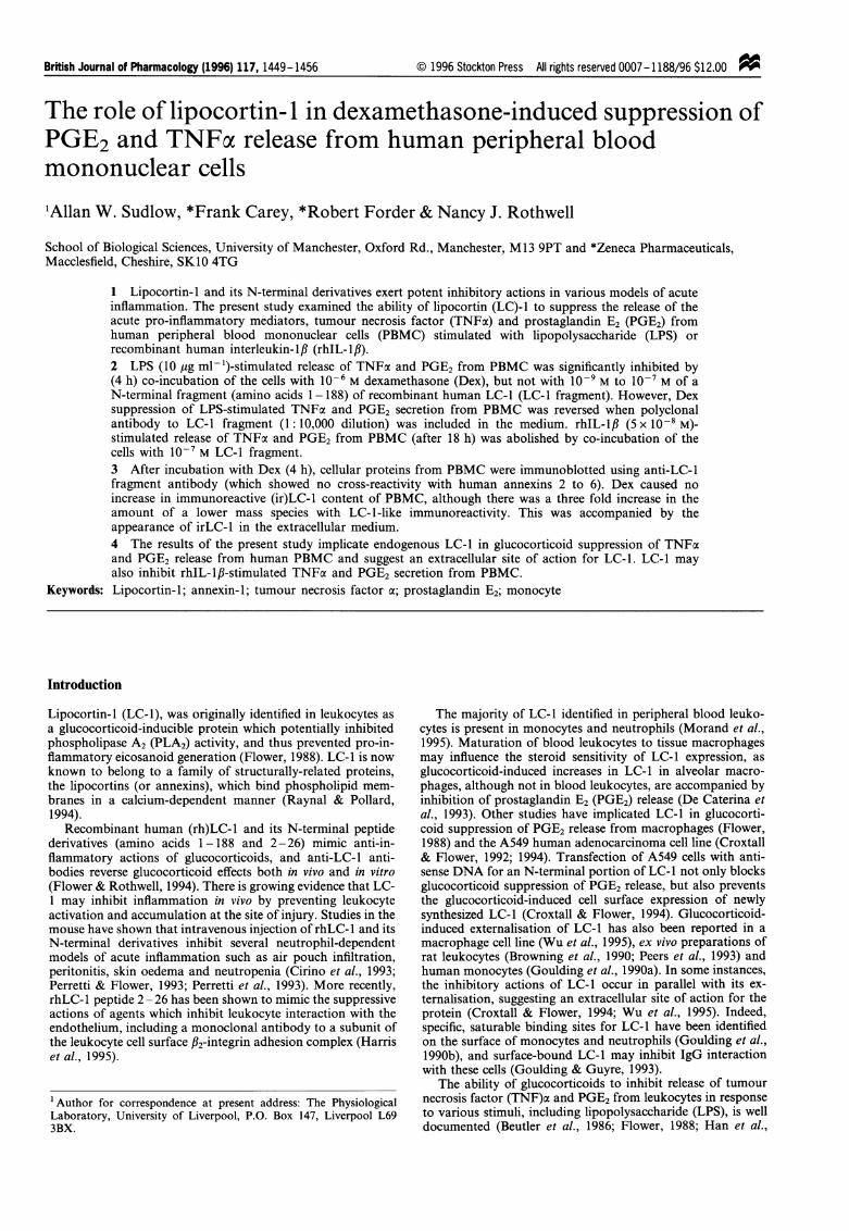
LPS (10 ig ml-') stimulation of TNFa release from PBMCwas inhibited by 77% (P<0.001) when cells were pre-in-cubated for 15 min, and then co-incubated for 4 h with 10-6 Mdexamethasone (Dex) (Figure la). TNFa release from PBMCincubated for 4 h in the absence of LPS was below the limit ofassay sensitivity, 25 pg ml-' (Figure la). LPS caused a twofold increase (P<0.001) in PGE2 release from the same cells,which Dex inhibited by 30% (P<0.01) (Figure lb). After 4 hincubation with 10-9 M to 10-7 M LC-1 fragment or vehicle(Tris) alone, TNFx concentrations in medium from PBMCwere below assay sensitivity, 25 pg ml-' (Figure 2a) and PGE2concentrations were not significantly altered (P>0.05) (Figure2b). LPS-stimulated TNFa release from PBMC was not sig-nificantly altered (P>0.05) by the presence of 10-9 M to10-7 M LC-1 fragment (Figure 2a), nor did these concentra-tions of LC-1 fragment inhibit LPS-stimulated (P<0.001) re-
lease of PGE2 from the same cells (Figure 2b). In an additionalexperiment (data not shown), 10-9 M to 10-7 M LC-1 frag-ment failed to inhibit the release of TNFcx and PGE2 fromPBMC stimulated with 10 ng ml-' LPS.
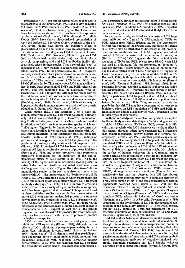
The cross-reactivity of a 1: 200 dilution of anti-LC-1 poly-clonal antibody with rhLC-1, human annexins 2 to 6 and theannexin-2 subunit pll was assessed by immunoblotting (Fig-ure 3). Anti-LC-1 antibody did not detect 50 ng of purifiedannexin-3 (lane A), recombinant human annexin-2 (lane C) or
Figure 1 Dexamethasone suppression of LPS-stimulated release ofTNFx and PGE2 from PBMC: PBMC (106) were pre-incubated with106M dexamethasone (Dex) or vehicle (PBS) for 15min, beforeaddition of lI0gmlP- LPS (solid column) or medium (open column)and a further 4 h incubation. Medium from cells was assayed for (a)TNFa and (b) PGE2 content as described in methods. Values shownare means + s.e.mean (n = 8). In (a), Dex inhibited LPS-stimulatedrelease of TNFa (***P<0.001, Student's unpaired t test), assay
sensitivity (dotted line) was 25 pgml-' (UND denotes TNFaconcentrations were below assay detection limit). In (b), LPSstimulation of PGE2 release (***P<0.001, two-way ANOVA) wasinhibited by Dex (**P<0.01, two-way ANOVA).
recombinant human annexin-5 (lane F), but did detect 50 ng ofrhLC-1 and LC-1 fragment (lanes D and E). When a mixtureof rhLC-1, human annexins 2, 4, 6 and the annexin-2 subunitp11 was immunoblotted, a single immunoreactive species was
detected (lane B) with a molecular mass equivalent to that ofrhLC-1 (lane D).
After 4 h incubation with Dex or vehicle (PBS), PBMCcellular protein (Figure 4a) and extracellular medium (Figure4b) were immunoblotted with a 1: 200 dilution of anti-LC-lfragment antibody. Immunoblotting ofPBMC cellular proteinrevealed, in addition to immunoreactive (ir)LC-1 (the upperband in Figure 4a), an immunoreactive species with a mole-cular mass approximately 7 kDa less than that of irLC-1 (thelower band in Figure 4a). Incubation of PBMC with Dex didnot enhance the band intensity for irLC-1 when compared toPBS, although the band intensity for the lower mass im-munoreactive protein was increased three fold by the presenceof Dex (Figure 4a). Immunoblotting of serum-free mediumtaken off the same PBMC revealed the presence of irLC-l onlyin medium from cells incubated with Dex (Figure 4b). Inaddition, medium from Dex-treated PBMC contained thelower molecular mass immunoreactive species describedabove, and an immunoreactive species with a molecular mass
approximately 6 kDa greater than that of irLC-l (Figure 4b).Similar results were obtained when PBMC and extracellularmedium were immunoblotted for LC-1 content after 18 h in-cubation with or without Dex (data not shown).
LPS-stimulated TNFa release (P<0.001) from PBMC was
suppressed by 73% (P<0.001) by Dex, although inclusion of a
**
PBS Dex 104 M
1451

A.W. Sudlow et al LC-I and release of PGE2 and TNFa
kDa
110 -
84 -
47 -UND UND UND UND
33-
24
16 -
I T IT
10 00 1IV- T T- rl
A B C D E F kDa
-110-84
-47
-33
-24
- 16
Figure 2 LC-l fragment does not inhibit LPS-stimulated release ofTNFa and PGE2 from PBMC: PBMC (106) were pre-incubated with10-9M to 10-7 LC-l fragment or vehicle (Tris) for 15min, beforeaddition of 10 ig mlV- LPS (solid column) or medium (open column)and a further 4 h incubation. Medium from cells was assayed for (a)TNFx and (b) PGE2 content as described previously. Values shownare means+ s.e.mean (n = 8). In (a), LC-1 fragment failed to inhibitLPS-stimulated TNFa release (P> 0.05, one-way ANOVA), assaysensitivity (dotted line) was 25 pg ml - (UND denotes TNFxconcentrations were below assay detection limit). In (b), LPSstimulation of PGE2 release (***P<0.001, one-way ANOVA) wasnot significantly altered by LC-1 fragment (P> 0.05, one-wayANOVA).
1: 10,000 (final) dilution of anti-LC-1 fragment antibody in themedium partially reversed (P<0.001) this inhibition to 26%(Figure 5a). When a 1: 10,000 dilution of control antibody wasincluded in the medium, Dex suppression (64%) of LPS-sti-mulated TNFo release was not significantly altered (P>0.05)(Figure 5a). LPS-stimulated release of PGE2 from the samecells (P<0.001) was inhibited by 54% (P<0.001) by Dex, andthis suppression was reduced (P <0.001) to 8% when anti-LC-1 fragment antibody was present in the medium (Figure 5b).Co-incubation with an equivalent dilution of control antibodydid not significantly alter (P> 0.05) Dex suppression (50%) ofLPS-stimulated PGE2 release from cells.
The effect of LC-1 fragment on rhIL-l/3-stimulated releaseof TNFx and PGE2 from PBMC was investigated. After 18 h,mean TNFa concentration in medium from vehicle-treatedPBMC (41 pg ml-') was reduced to below the limit of assaysensitivity (30 pg ml-') in the presence of 10-9 M or 10-7 MLC-l fragment alone (Figure 6a). A separate study (data notshown) demonstrated that neither 10-7 M LC-1 fragment norits vehicle (Tris), altered the ability of the TNFox assay to detecthuman TNFoc over the standard curve range of 10 to5000 pg ml-'. After 18 h, 5 x 10⁻⁹ M and 5 x 10⁻⁸ M rhIL-,Benhanced TNFoc secretion from PBMC approximately two fold(P <0.01 and P< 0.001 respectively) (Figure 6a). There was nostimulation of TNFa release from PBMC using the aboveconcentrations of rhIL-1,B after 4 h, and the apparent rhIL- I/B-stimulation ofTNFa secretion was not stastically significant at6 or 12 h (P>0.05, data not shown). Co-incubation of rhIL-1/3-stimulated PBMC with 10-9 M and 10-7 M LC-1 fragmentrendered TNFa undetectable (Figure 6a). PGE2 release fromunstimulated PBMC (after 18 h) was considerably higher thanthat seen after 4 h (Figure 6b compared with Figures Ib, 2band 5b), and was not significantly altered (P> 0.05) by I0-9 M
Figure 3 Cross-reactivity of anti-LC- 1 fragment polyclonal antibodywith rhLC-1 and human annexins 2 to 6: rhLC-1 and recombinanthuman or purified human annexins 2 to 6 were immunoblotted with1:200 dilution of anti-LC-1 fragment polyclonal antibody asdescribed in methods. Lanes were (A) 50ng purified annexin-3; (B)25 ng each of rhLC- 1, recombinant annexins 2, 4, 6 and the annexin-2subunit p11; (C) 50 ng annexin-2; and (D) 50 ng rhLC-1. On aseparate immunoblot lanes were (E) 50ng each of rhLC-1 and LC-1fragment; and (F) 50ng recombinant annexin-5. Molecular weightmarker proteins are as indicated. For each lane, the results shown arerepresentative of at least two immunoblotting experiments using anti-LC-l fragment antibody.
kDa47-
33-
kDa47-
33-
a
PBS Oex
b200 ngrhLC-1 Dex
200 ngrhLC-1 PBS Dex
PBS Dex PBS
Figure 4 Dexamethasone induces externalization of LC- 1 immuno-reactivity from PBMC: (a) 106 PBMC were incubated in serum-freemedium with 10-6M dexamethasone (Dex) or vehicle (PBS) for 4h.Cells were harvested, pooled (2 x 10⁶), solubilized, cellular proteins(50,ug per track) separated by SDS PAGE and immunoblotted with1: 200 dilution of anti-LC-1 fragment polyclonal antibody, with 20 ngrhLC- 1 included on the blot as a control. Duplicates for eachtreatment and molecular weight markers are as indicated. (b)Medium from the same PBMC was pooled (2 x 1.5 ml), desalted,concentrated by freeze-drying, extracellular protein solubilized in(200,u1) dissociation buffer and duplicate (100pl1) samples separatedby SDS PAGE and immunoblotted as described above. Theimmunoblots shown in (a) and (b) are both representative of twoexperiments.
or i0' M LC-1 fragment. The two fold increase (P<0.05) inrelease of PGE2 from PBMC stimulated with 5 x 10⁻⁸ M rhIL-1/ was reversed (P<0.05) by 10-7 M LC-1 fragment (Figure6b).
a200
150
CDa 100
U-Z 50
0
30'b
20
10
E
CLa_
o0LC-1fragment:
1452

A.W. Sudlow et a! LC-I and release of PGE2 and TNFa1
I~~~~~~~~~~~~~~~~~~100 -
80 -
*** *** ***
o0
**
I UND UND UND
*b
01)0 0 0 0> T
Dex 106 M: - - + + +
Antibody: - - - anti-LC-1 Control
Figure 5 Anti-LC-l fragment antibody reverses dexamethasonesuppression of LPS-stimulated release of TNFa and PGE2 fromPBMC: PBMC (106) were pre-incubated with 10-6M dexamethasone(Dex) or vehicle (PBS) and a 1:10,000 (final) dilution of anti-LC-1fragment or control antibody for 15 min, before addition of10 pgml-1 LPS (solid column) or medium (open column) and a
further 4h incubation. Medium from cells was assayed for (a) TNFaand (b) PGE2 content as described previously. Values shown are
means+s.e.mean (n=6). Dex suppression (***P<0.001, one-way
ANOVA) of both LPS-stimulated (a) TNFa release (***P<0.001,one-way ANOVA) and (b) PGE2 release (***P<0.001, one-wayANOVA) was partially reversed by anti-LC-1 fragment antibody(***P<0.001, one-way ANOVA). Assay sensitivity (dotted line) forTNFoc was 25pgml-'.
Discussion
The acute release of pro-inflammatory mediators such as ei-cosanoids and cytokines from activated tissue macrophages isof primary importance for development of local inflammatoryresponses. Once released, cytokines such as TNFcx act on
stromal cells at the reactive site to elicit the release of a sec-
ondary wave of cytokines (Baumann & Gauldie, 1994). Severalstudies have shown the LC-1 and its N-terminal derived pep-tides can mimic the potent anti-inflammatory actions of glu-cocorticoids (Flower & Rothwell, 1994), but few haveexamined the effect of LC-1 on cytokine release. The presentstudy investigated the ability of LC-1 to suppress the release ofTNFac and PGE2 from PBMC stimulated with LPS or rhIL-1I#.
Concentrations of TNFa secreted by 'unstimulated' PBMC(i.e. in the absence of LPS or rhIL-1fl) were either below or
near the limit of assay sensitivity. The very low concentrationsof TNFcx released from PBMC in the absence of a drug sti-mulus has been noted previously (e.g. Schindler et al., 1990).Haskill et al. (1988) have demonstrated that isolation andadherence of PBMC could induce TNFax gene transcriptionbut that actual secretion of TNFax requires a secondary sti-mulus such as LPS.
Dex inhibition of LPS-stimulated PGE2 release from PBMCwas significant, but less pronounced than inhibition of TNFa
Figure 6 LC-l fragment suppression of IL-lfl-stimulated release ofTNFa and PGE2 from PBMC: PBMC (2 x 10⁵) were pre-incubatedwith 10-9M or 10-7M LC-1 fragment or vehicle (Tris) for 15 min,before addition of 5 x 10⁻⁸M (hatched column) or 5 x 10⁻⁹M IL-1I3(solid column), or medium (open column), and a futher 18 hincubation. Medium from cells was assayed for (a) TNFa and (b)PGE2 content as described previously. Values shown are means
+s.e.mean (n=8). In (a), IL-1/3 stimulation of TNFa release(**P<0.01, ***P<0.001, one-way ANOVA) was abolished by LC-1 fragment, assay sensitivity (dotted line) was 30 pgml-l (UNDdenotes TNFa concentrations were below assay detection limit). In(b), 5 x 10⁻⁸M IL-I# stimulation of PGE2 release (*P<0.05, one-way
ANOVA) was reversed by 10-7M LC-l fragment (*P<0.05, one-way
ANOVA).
release (Figure lb). Glucocorticoid suppression of TNFaproduction from LPS-stimulated PBMC involves inhibition ofboth TNFa gene transcription and, via a glucocorticoid-inducible protein, mRNA translation (Beutler et al., 1986; Hanet al., 1990). Glucocorticoid suppression of PGE2 productioninvolves inhibition of phospholipase activity (Flower, 1988)and suppression of cyclo-oxygena3e-2 (Cox-2) expression atthe level of translation (Bailey, 1991) and possibly transcrip-tion (Newman et al., 1994).
After incubation with Dex for 4 h (or 18 h, data notshown), LC-l expression in PBMC was not markedly altered,although that of a lower mass protein was enhanced ap-proximately three fold (Figure 4a). This was accompanied bythe Dex-induced appearance of LC-l (and lower and highermass species with LC-1-like immunoreactivity) in the extra-cellular medium (Figure 4b). The high levels of irLC-1 presentin PBMC incubated with PBS suggests that LC-1 expressionmay have already been maximal such that exposure to Dexelicited no clear increase in LC-l cell content (Figure 4a).Failure of glucocorticoid to induce LC-1 expression in bothperipheral blood leukocytes (Morand et al., 1995) and acti-vated monocytes (Browning et al., 1990) has been noted pre-viously. In addition, isolation and adherence of PBMC can
induce expression of a range of cellular proteins associatedwith the acute immune response (Haskill et al., 1988) and Peerset al. (1993) have demonstrated a non-glucocorticoid me-
chanism for increased expression of LC-1 in activated leuko-cytes from adrenalectomized rats.
400-1
300-
200-
100-
E0.
U-
z
E0.
U-z
60
40'
20
O-
I
0.
(9a-
350
300
250
m 200
W 150(9100
50
0LC-1fragment:
*
0 0
I I
I I
F7
II
T T T
1453
a
A.W. Sudlow et al LC-1 and release of PGE2 and TNFa
Extracellular LC- 1 can appear within hours of exposure toglucocorticoid in vivo (Peers et al., 1993) and in vitro (Croxtall& Flower, 1992; 1994; Peers et al., 1993; Taylor et al., 1993;Wu et al., 1995), as observed here (Figure 4b). There is evi-dence for translational control of extracellular LC-1 expressionby glucocorticoid (Taylor et al., 1993), although Croxtall &Flower (1994) have shown glucocorticoid up-regulation ofA549 cell surface LC-1 involves de novo synthesis of the pro-tein. Several studies have shown that inhibitory effects ofglucocorticoid on cells and tissue in vitro are accompanied bythe externalization of endogenous LC-1 (Croxtall & Flower,1992; Taylor et al., 1993; Wu et al., 1995). Exogenous LC-1(and its N-terminal peptide derivatives) can mimic gluco-corticoid suppression, and anti-LC-1 antibodies inhibit glu-cocorticoid effects in these studies. Thus a pericellular 'pool' ofendogenous LC-1 may mediate glucocorticoid actions (Crox-tall & Flower, 1992). In the present study, anti-LC-1 fragmentantibody (which neutralises glucocorticoid actions both in vivoand in vitro, Flower & Rothwell, 1994) reversed Dex sup-pression of LPS-stimulated release of TNFcx and PGE2 fromPBMC (Figure 5a, b). Thus endogenous LC-1 may mediate, atleast in part, Dex suppression of TNFa and PGE2 release fromPBMC, and this inhibition may be associated with ex-ternalization of LC-1 (Figure 4b). Although the mechanism forthis inhibition is not yet known, others have demonstratedsurface binding sites for LC- 1 on mouse and human monocytes(Goulding et al., 1990b; Perretti et al., 1993) which may beimportant for the immunosuppressive activity of the protein(Goulding & Guyre, 1993; Perretti et al., 1993).When human rhLC-1 and annexins 2 to 6 were im-
munoblotted with an anti-LC-1 fragment polyclonal antibody,only rhLC-1 was detected (Figure 3). However, immunoblot-ting PBMC cellular protein or extracellular medium revealedin addition to LC-1, an immunoreactive species which was7 kDa lighter (Figure 4a, b). Immunoblotting experiments byothers have identified lower molecular mass species with LC-1like immunoreactivity in the subcellular fractions from leu-kocytes (Smith et al., 1990; Peers et al., 1993). Findings ofprevious studies suggest that these LC-1-like proteins are theproducts of proteolytic degradation of full sequence LC-1(Flower, 1988). Proteolysed irLC-1 has been detected in mac-rophage cell lysates despite the presence of protease inhibitors(Peers et al., 1993), and in vivo, proteolysis may represent apermissive mechanism for the inhibition of the anti-in-flammatory effects of LC-1 (Smith et al., 1990). As to theidentity of the higher mass immunoreactive species present inextracellular medium (with an estimated molecular mass6 kDa greater than irLC-1) (Figure 4b), other tissue/cell im-munoblotting studies in the past have detected similar massspecies with LC-1-like immunoreactivity (Pepinsky et al., 1989;Ando et al., 1991), including a study in which macrophage-like(U937 cell line) cell lysate was blotted with anti-LC-1 fragmentantibody (Murphy et al., 1992). LC-1 is known to cross-linkwith itself to form a variety of higher molecular mass species,and it has been suggested that the 40 - 45 kDa species detectedin these published studies may result from cross-linking ofnative LC-1 with another unidentified protein or a fragmentderived from in situ proteolysis of native LC-1 (Pepinsky et al.,1989; Ando et al., 1991; Murphy et al., 1992). In Figure 4b, thedifferences in the estimated mass of upper and lower species incomparison to irLC-1 suggest at least part of the peptidefragment that was cleaved off to produce the lower mass spe-cies, may have associated with the native protein to producethe higher mass species.
LC-1 has been implicated as a mediator of glucocorticoidinhibition of PLA2 activity (Flower, 1988), although the spe-cificity of LC-1 inhibition of phospholipase activity, in parti-cular PLA2 inhibition, is controversial (Raynal & Pollard,1994). Parente et al. (1984) demonstrated that partially-pur-ified lipocortins (39 to 44 kDa) inhibited PGE2 secretion fromleukocytes stimulated with heat-killed Bordetella pertusis.More recently, Bailey (1991) has suggested that LC-1 mediatesthe translational component of glucocorticoid suppression of
Cox-2 expression, although this does not seem to be the case inA549 cells (Newman et al., 1994) or a macrophage cell line(Wu et al., 1995). In addition, Morand et al. (1993) reportedthat LC-1 did not inhibit LPS-stimulated IL-1l3 release fromhuman monocytes.
In the present study, we failed to demonstrate LC-1 frag-ment inhibition of (10 pg ml-') LPS-stimulated TNFo orPGE2 release from PBMC (Figures 2a, b). The discrepancybetween the findings of the present study and those of Parenteet al. (1984) may be attributed to differences in cell prepara-tion, culture conditions, use of LC-1 fragment instead ofpurified LC-1 or the nature of the stimulus. However, as(10-7 M to 10-9 M) LC-1 fragment also failed to inhibit sti-mulation of TNFx and PGE2 release from PBMC when LPSwas used at a thousand fold less concentration (10 ng ml-',data not shown), it seems unlikely that lack of inhibition wasdue to a dose-related effect of LPS. Although LC-1 fragment isknown to mimic many of the actions of rhLC-1 (Flower &Rothwell, 1994), both agents exhibit different activity profilesin several in vivo and in vitro models of inflammation (Perrettiet al., 1993; Flower & Rothwell, 1994; Perretti, 1994). In ex-periments involving cytokine-stimulated leukocyte activationand accumulation, LC- 1 fragment has been shown to be con-siderably less potent than rhLC-1 (Peretti, 1994) and in onestudy in particular, LC-1 fragment, unlike rhLC-1, exhibitedonly partial inhibition of IL-lI#-induced neutrophil accumu-lation (Perretti et al., 1993). Thus, we cannot exclude thepossibility that rhLC-1 may have demonstrated at least someinhibitory effect on LPS stimulation of TNFa and PGE2 re-lease, although limited availability of rhLC-1 precluded its usein these types of experiments.
Without knowledge of the mechanism by which, as impliedby the antibody reversal data (Figures Sa, b), endogenous LC-1 inhibits LPS-stimulated TNFoa and PGE2 release, it is diffi-cult to say precisely why LC-1 fragment was without effect inthis regard, although others have suggested LC-1 fragmentsmay exhibit inconsistent activity because of N-terminal het-erogeneity (Perretti et al., 1993). Another possible explanationis that the mechanism by which LC-1 fragment inhibits IL-,lB-stimulated TNFoa and PGE2 release (Figures 6a, b) is differentfrom that by which endogenous LC-1 inhibits LPS stimulation.Perretti (1994) has suggested a model in which, LC-1 binds tothe surface of human leukocytes via a C-terminal region(amino acids 246 to 254) in order to exert its anti-inflammatoryactions. This region is absent from LC-1 fragment and impliesthat the LC-1 fragment inhibition of IL-l, stimulation ob-served here (Figures 6a, b), may involve a different mechanism.
The magnitude of rhIL-lf-stimulated TNFa release fromPBMC, although statistically significant (Figure 6a), wasconsiderably less than that observed with LPS (see above).rhIL-l1B has been reported previously to stimulate secretion ofTNFa from human PBMC at the same concentrations used inexperiments described here (Ikejima et al., 1990), but theconcurrent release of IL-6 may feedback to inhibit TNFa se-cretion (Schindler et al., 1990). IL-1If up-regulates PLA2 ac-tivity in various cell types (Dower et al., 1992), and may alsostimulate PGE2 release via induction of Cox-2 expression(Newman et al., 1994). In A549 cells, Newman et al. (1994)demonstrated the involvement of LC-1 in glucocorticoid sup-pression of the stimulatory actions of IL-I# on PGE2 release,but not Cox-2 expression. Thus, the mechanism for LC-1fragment inhibition of rhIL-lfl-stimulated TNFax and PGE2secretion (Figures 6a, b) is, as yet, unclear.
rhLC-1 and its N-terminal derivatives inhibit several neu-trophil-dependent in vivo models of acute inflammation, al-though this inhibition is not stimulus-specific and occurs inresponse to various inflammatory stimuli including IL-i, IL-6and IL-8 (Perretti & Flower, 1993; 1994). Injection of LC-1directly into the site of inflammation is not as effective, butpassive immunisation of mice with anti-LC-1 antiserum abol-ishes glucocorticoid inhibition of cytokine-stimulated neu-trophil migration, suggesting that LC-1 inhibits leukocyteactivation prior to tissue infiltration (Perretti & Flower, 1993;
1454

A.W. Sudlow et al LC-I and release of PGE2 and TNFz 1455
1994). rhLC-l peptide 2-26 mimics the inhibitory effects invivo of a monoclonal antibody to the CDl lbI32-integrin ad-hesion complex subunit and other agents that prevent leuko-cyte margination and adhesion, suggesting that LC-1suppresses neutrophil migration by interfering with the adhe-sion cascade during leukocyte activation (Harris et al., 1995).This inhibition of leukocyte activation by extracellular LC-lmay explain the suppressive actions of the protein in othermodels of acute inflammation (e.g. Cirino et al., 1989).
In the current study, we have shown that glucocorticoidinduces the externalization of LC-l from PBMC, and proposethat this endogenous extracellular LC-l mediates glucocorti-
coid inhibition of LPS-stimulated PGE2 and TNFa releasefrom the cells. Whether this represents another facet of LC-linhibition of leukocyte activation is not yet clear, althoughpreliminary evidence suggests that LC-l may also suppressrelease of these pro-inflammatory mediators in response toother stimuli such as IL-I/3.
This work was funded by the BBSRC and Zeneca Pharmaceuticals.We are grateful to Dr J. Browning of Biogen for his gifts ofrecombinant human LC-1, annexin-2 and purified human annexin-3.
References
ANDO, Y., IMAMURA, S., OWADA, M.K. & KANNAGI, R. (1991).Calcium-induced intracellular cross-linking of lipocortin-1 bytissue transglutaminase in A431 cells. J. Biol. Chem., 266, 1101 -1108.
BAILEY, J.M. (1991). New mechanisms for effects of anti-inflamma-tory glucocorticoids. Biofactors, 3, 97-102.
BAUMANN, H. & GAULDIE, J. (1994). The acute phase response.Immunol. Today, 15, 74-80.
BEUTLER, B., KROCHIN, N., MILSARK, I.W., LUEDKE, C. &CERAMI, A. (1986). Control of cachectin (tumor necrosis factor)synthesis: mechanisms of endotoxin resistance. Science, 232,977-979.
BROWNING, J.L., WARD, M.P., WALLNER, B.P. & PEPINSKY, R.B.(1990). Studies on the structural properties of lipocortin-1 andthe regulation of its synthesis by steroids. Prog. Clin. Biol. Res.,349, 27-45.
CAREY, F., FORDER, R., EDGE, M.D., GREENE, A.R., HORAN, M.A.,STRIJBOS, P.J.L.M. & ROTHWELL, N.J. (1990). Lipocortin-1fragment modifies pyrogenic actions of cytokines in rats. Am. J.Physiol., 259, 266-269.
CIRINO, G., CICALA, C., SORRENTINO, L., CILIBERTO, G., ARPAIA,G., PERRETTI, M. & FLOWER, R.J. (1993). Anti-inflammatoryactions of an N-terminal peptide from human lipocortin- 1. Br. J.Pharmacol., 108, 573-574.
CIRINO, G., PEERS, S.H., FLOWER, R.J., BROWNING, J.L. &PEPINSKY, R.B. (1989). Human recombinant lipocortin-1 hasacute local anti-inflammatory properties in the rat paw edematest. Proc. Nati. Acad. Sci. U.S.A., 86, 3428-3432.
CROXTALL, J.D. & FLOWER, R.J. (1992). Lipocortin-1 mediatesdexamethasone-induced growth arrest of the A549 lung adeno-carcinoma cell line. Proc. Natl. Acad. Sci. U.S.A., 89, 3571-3575.
CROXTALL, J.D. & FLOWER, R.J. (1994). Antisense oligonucleotidesto human lipocortin-1 inhibit glucocorticoid-induced inhibitionofA549 cell growth and eicosanoid release. Biochem. Pharmacol.,48, 1729-1734.
DE CATERINA, R., SICARI, R., GIANNESSI, D., PAGGIARO, P.L.,PAOLETTI, P., LAZZERNI, G., BERNINI, W., SOLITO, E. &PARENTE, L. (1993). Macrophage-specific eicosanoid synthesisinhibition and lipocortin-1 induction by glucocorticoids. J. Appl.Physiol., 75, 2368-2375.
DOWER, S.K., SIMS, J.E., CERRETI, D.P. & BIRD, T.A. (1992). Theinterleukin-1 system; receptors, ligands and signals. Chem.Immunol., 51, 33-64.
FLOWER, R.J. (1988). Lipocortin and the mechanism of action of theglucocorticoids. Br. J. Pharmacol., 94, 987-1015.
FLOWER, R.J. & ROTHWELL, N.J. (1994). Lipocortin-1: cellularmechanisms and clinical relevance. Trends Pharmacol. Sci., 15,71-76.
FORDER, R.A. & CAREY, F. (1983). Measurement of human venousplasma prostacyclin and metabolites by radioimmunoassay; areappraisal. Prostaglandins Leukot. Med., 12, 323 -346.
GOULDING, N.J., GODOLPHIN, J.L., SHARLAND, P.R., PEERS, S.H.,SAMPSON, M., MADDISON, P.J. & FLOWER, R.J. (1990a). Anti-inflammatory lipocortin-l production by peripheral bloodleucocytes in response to hydrocortisone. Lancet, 335, 1416-1418.
GOULDING, N.J. & GUYRE, P.M. (1993). Lipocortin-1 binding tohuman leukocytes correlates with its ability to inhibit IgGinteractions with Fcy receptors. Biochem. Biophys. Res.Commun., 192, 351- 358.
GOULDING, N.J., LUYING, P. & GUYRE, P.M. (1990b). Character-istics of lipocortin-1 binding to the surface of human peripheralblood leucocytes. Biochem. Soc. Trans., 18, 1237-1238.
HAN, J., THOMPSON, P. & BEUTLER, B. (1990). Dexamethasone andpentoxifylline inhibit endotoxin-induced cachectin/tumor necro-sis factor synthesis at separate points in the signaling pathway. J.Exp. Med., 172, 391-394.
HARRIS, J.G., FLOWER, R.J. & PERRETTI, M. (1995). Alteration ofneutrophil trafficking by a lipocortinl N-terminus peptide. Eur.J. Pharmacol., 279, 149- 157.
HASKILL, S., JOHNSON, C., EIERMAN, D., BECKER, S. & WARREN,K. (1988). Adherence induces selective mRNA expression ofmonocyte mediators and proto-oncogenes. J. Immunol., 140,1690-1694.
HAWORTH, D. & CAREY, F. (1986). Thromboxane synthesisinhibition: implications for prostaglandin endoperoxidemetabolism. Prostaglandins, 31, 33-45.
IKEJIMA, T., OKUSAWA, S., GHEZZI, P., VAN DER MEER, J.W. &DINARELLO, C.A. (1990). Interleukin-1 induces tumor necrosisfactor (TNF) in human peripheral blood mononuclear cells invitro and a circulating TNF-like activity in rabbits. J. Infect. Dis.,162, 215-223.
MORAND, E.F., HUTCHINSON, P., HARGREAVES, A., GOULDING,N.J., BOYCE, N.W. & HOLDSWORTH, S.R. (1995). Detection ofintracellular LC-1 in human leukocyte subsets. Clin. Immunol.Immunopathol., 76, 195-202.
MORAND, E.F., RICKARD, D. & GOULDING, N.J. (1993). Lack ofinvolvement of lipocortin- 1 in dexamethasone suppression of IL-1 release. Med. Inflamm., 2, 49- 52.
MURPHY, C.T., PEERS, S.H., FORDER, R.A., FLOWER, R.J., CAREY,F. & WESTWICK, J. (1992). Evidence for the presence and locationof annexins in human platelets. Biochem. Biophys. Res. Commun.,189, 1739-1746.
NEWMAN, S.P., FLOWER, R.J. & CROXTALL, J.D. (1994). Dex-amethasone suppression of IL-lfl-induced cyclooxygenase-2expression is not mediated by lipocortin-1 in A549 cells. Biochem.Biophys. Res. Commun., 202, 931 -939.
PARENTE, L., DI ROSA, M., FLOWER, R.J., GHIARA, P., MELI, R.,PERSICO, P., SALMON, J.A. & WOOD, J.N. (1984). Relationshipbetween the anti-phospholipase and the anti-inflammatory effectsof glucocorticoid-induced proteins. Eur. J. Pharmacol., 99, 233 -239.
PEERS, S.H., SMILLIE, F., ELDERFIELD, A.J. & FLOWER, R.J. (1993).Glucocorticoid- and non-glucocorticoid induction of lipocortins(annexins)- 1 and -2 in rat peritoneal leucocytes in vivo. Br. J.Pharmacol., 108, 66-72.
PEPINSKY, R.B., SINCLAIR, L.K., CHOW, E.P. & O'BRINE-GRECO, B.(1989). A dimeric form of lipocortin-1 in human placenta.Biochem. J., 263, 97- 103.
PERRETTI, M. (1994). Lipocortin-derived peptides. Biochem.Pharmacol., 47, 931-938.
PERRETTI, M., AHLUWALIA, A., HARRIS, J.G., GOULDING, N.J. &FLOWER, R.J. (1993). Lipocortin-1 fragments inhibit neutrophilaccumulation and neutrophil-dependent edema in the mouse. J.Immunol., 151, 4306-4314.
PERRETTI, M. & FLOWER, R.J. (1993). Modulation of IL-1-inducedneutrophil migration by dexamethasone and lipocortin. J.Immunol., 150, 1-8.
PERRETTI, M. & FLOWER, R.J. (1994). Cytokines, glucocorticoidsand lipocortins in the control of neutrophil migration. Pharma-col. Res., 30, 53-59.

1456 A.W. Sudlow et al LC-1 and release of PGE2 and TNFa
RAYNAL, P. & POLLARD, H.B. (1994). Annexins: the problem ofassessing the biological role for a gene family of multifunctionalcalcium- and phospholipid-binding proteins. Biochem. Biophys.Acta., 1197, 63-93.
SCHINDLER, R., MANCILLA, J., ENDRES, S., GHORBANI, R.,CLARK, S.C. & DINARELLO, C.A. (1990). Correlations andinteractions in the production of interleukin-6 (IL-6), IL-1 andtumour necrosis factor (TNF) in human blood mononuclearcells; IL-6 suppresses IL-1 and TNF. Blood, 75, 40-47.
SMITH, S.F., TETLEY, T.D., GUZ, A. & FLOWER, R.J. (1990).Detection of lipocortin-1 in human lung lavage fluid: lipocortindegradation as a possible proteolytic mechanism in the control ofinflammatory mediators and inflammation. Environ. HealthPersp., 85, 35 - 144.
TAYLOR, A.D., COWELL, A.M., FLOWER, R.J. & BUCKINGHAM, J.C.(1993). Lipocortin-l mediates an early inhibitory action ofglucocorticoid on the secretion of ACTH by the rat anteriorpituitary gland in vitro. Neuroendocrinol., 58, 430-439.
WU, C.C., CROXTALL, J.D., PERRETTI, M., BRYANT, C.E., THIE-MERMANN, C., FLOWER, R.J. & VANE, R.J. (1995). Lipocortin 1mediates the inhibition by dexamethasone of the induction byendotoxin of nitric oxide synthase in the rat. Proc. Nati. Acad.Sci. U.S.A., 92, 3473-3477.
(Received September 5, 1995Revised December 4, 1995
Accepted December 11, 1995)