Isolated ileal interposition in enteroendocrine L cells differentiation
45
UNIVERSIDADE FEDERAL DE SÃO PAULO (FEDERAL UNIVERSITY OF SÃO PAULO) ESCOLA PAULISTA DE MEDICINA (SCHOOL OF MEDICINE) UNIFESP – EPM PROGRAMA DE PÓS-GRADUAÇÃO EM MEDICINA TRANSLACIONAL (POST-GRADUATION PROGRAM IN TRANSLATIONAL MEDICINE) Coordinator: Prof. Dr. José Alberto Neder Serafini Ph.D. Research Project Bianca Marigliani Co-Workers: Lucas Pedroso Fernandes Ferreira Leal, Karina Ferreira Neves, Linda Omar Alves Bernardes, Gilmara Silva Aguiar Yamaguchi, Wellington Cardia, João Luiz Cansanção Azevedo, Otávio Cansanção Azevedo, Gustavo Peixoto Soares Miguel. Isolated ileal interposition in enteroendocrine L cells differentiation. Advisor: Prof. Dr. João Luiz Moreira Coutinho de Azevedo Co-Advisors: Prof. Dr. Ismael Dale Cotrim Guerreiro da Silva Profa. Dra. Valderez Bastos Valero Lapchick Prof. Dr. Sang Won Han SÃO PAULO, BRAZIL - 2011 Nature Precedings : doi:10.1038/npre.2011.6614.1 : Posted 17 Nov 2011
-
Upload
independent -
Category
Documents
-
view
0 -
download
0
Transcript of Isolated ileal interposition in enteroendocrine L cells differentiation
UNIVERSIDADE FEDERAL DE SÃO PAULO (FEDERAL UNIVERSITY OF SÃO PAULO)
ESCOLA PAULISTA DE MEDICINA (SCHOOL OF MEDICINE)
UNIFESP – EPM
PROGRAMA DE PÓS-GRADUAÇÃO EM MEDICINA TRANSLACIONAL (POST-GRADUATION PROGRAM IN TRANSLATIONAL MEDICINE)
Coordinator: Prof. Dr. José Alberto Neder Serafini
Ph.D. Research Project
Bianca Marigliani
Co-Workers: Lucas Pedroso Fernandes Ferreira Leal, Karina Ferreira Neves, Linda Omar Alves Bernardes, Gilmara Silva Aguiar Yamaguchi,
Wellington Cardia, João Luiz Cansanção Azevedo, Otávio Cansanção Azevedo, Gustavo Peixoto Soares Miguel.
Isolated ileal interposition in enteroendocrine L cells differentiation.
Advisor: Prof. Dr. João Luiz Moreira Coutinho de Azevedo
Co-Advisors:
Prof. Dr. Ismael Dale Cotrim Guerreiro da Silva Profa. Dra. Valderez Bastos Valero Lapchick
Prof. Dr. Sang Won Han
SÃO PAULO, BRAZIL - 2011
Nat
ure
Pre
cedi
ngs
: doi
:10.
1038
/npr
e.20
11.6
614.
1 : P
oste
d 17
Nov
201
1
ABSTRACT
INTRODUCTION: Due to the progressive nature of type 2 diabetes, its complexity and drug
treatment perpetuity, there is currently a search for surgical procedures that can promote
euglycemia also in non-obese patients. Diabetic patients glycemic control can be achieved by
increasing the blood concentration of GLP-1, a hormone produced by L cells that are more
densely concentrated in the terminal ileum. Early and extended improvement of diabetes in
patients submitted to bariatric surgeries awakened the necessity of investigating the isolated
ileal interposition as surgical alternative for the treatment of diabetes. The interposition of this
ileal segment to a more anterior region (proximal jejunum) can promote a greater stimulation
of the L cells by poorly digested food, increasing the production of GLP-1 and reflecting on
glycemic control. However, in order to consolidate the ileal interposition as a surgical
treatment of diabetes it is necessary that the interposed ileum keep the same differentiation
rate into L cells for a long period to justify the intervention.
AIMS: To investigate the isolated ileal interposition influence on the differentiation of
intestinal precursor cells into enteroendocrine L cells over time.
METHODS: Twelve 12-week-old male Wistar rats (Rattus norvegicus albinus) of the WAB strain
(heterogeneous) will be used. All animals will receive a high-calorie, high-fat diet for 16 weeks
or more until they develop glucose dysmetabolism confirmed by glycemic test. They will be
divided into two groups of 10 animals each: the isolated ileal interposition group (GI) and the
control group (GC), comprising animals that will not be submitted to any surgical intervention.
Blood samples will be collected under anesthesia at the weeks 12, 26, 36 and 44 for the
determination of serum levels of glucose, insulin, GLP-1, glucagon, C-peptide and glycosilated
hemoglobin. The insulin tolerance test will be performed and insulin resistance will be
calculated. For the comparative analysis of the ileal precursor cells differentiation into
enteroendocrine cells among the two groups, the following intestinal fragments will be
collected after euthanasia: interposed ileum and remaining ileum from GI, jejunum and ileum
from GC. These fragments will be analyzed by imunofluorescence and also by Real Time PCR
using PCR Arrays for target genes including the main ones related to stem cell, stem cell
singnalling, diabetes, Wnt and Notch signaling pathways and other genes and pathways
involved in the differentiation of intestinal precursor cells into enteroendocrine cells,
especially GLP-1-producing L cells that play important role in euglycemia.
Nat
ure
Pre
cedi
ngs
: doi
:10.
1038
/npr
e.20
11.6
614.
1 : P
oste
d 17
Nov
201
1
1. INTRODUCTION
Diabetes is one of the most common chronic diseases and a growing global epidemic
(Danaei et al., 2011).1 Type 2 diabetes (T2D) is a serious metabolic disease characterised by
high glucose levels. Complications from T2D usually result in poor quality of life, disability, and
early death.2 Despite efforts to control glycaemia in diabetes patients, no therapeutic
approach has significantly impacted the progression of this disease (Tahrani et al., 2010).3
There is, however, a simple and reversible surgical procedure that might become a valid
therapeutic alternative in non-obese patients. This procedure, isolated ileal interposition (III),
results in a dramatic increase in the incretin glucagon-like peptide 1 (GLP-1).
Epidemiology and aetiopathogenesis of diabetes
In 2000, diabetes caused the death of about three million people.4 In 2010, the number of
affected individuals was approximately 285 million, and this number is projected to increase
54% by 2030. A much greater increase in the number of adults with diabetes is projected in
underdeveloped countries than in developed countries: the number of affected individuals is
projected to increase approximately 70% in underdeveloped countries and 20% in developed
countries.1 T2D, also known as non-insulin-dependent diabetes, is responsible for 90% to 95%
of diabetes cases.5
T2D is a progressive heterogeneous disease that mainly involves peripheral insulin
resistance and gradual dysfunction of pancreatic beta cells.2,6 It develops when pancreatic
islets can no longer maintain insulinaemia at levels sufficient to overcome peripheral tissue
resistance. Throughout the course of the disease, diabetes passes through intermediate
stages. Initially, diabetes is treated only with modifications in lifestyle and specific diet. Later in
the disease, patients are treated with drugs that promote insulin secretion. As insulin
deficiency unavoidably increases, however, the combination of therapeutic oral agents often
fails to control hyperglycaemia efficiently, and the use of insulin is eventually required.6-9
In the long run, patients with T2D have an increased risk of complications that are a
substantial cause of morbidity and mortality, including macrovascular disease, nephropathy,
retinopathy, and neuropathy.10 Further complications of diabetes include ischemia of the limbs
resulting in amputation, dental problems, and pregnancy disorders.5 The risk of developing
diabetes-associated complications is related to the duration of diabetes and the level of
glycaemic control.11
Nat
ure
Pre
cedi
ngs
: doi
:10.
1038
/npr
e.20
11.6
614.
1 : P
oste
d 17
Nov
201
1
Although 90% of T2D cases can be attributed to excessive weight,12 its incidence is growing
among individuals with body mass index (BMI) in the normal and overweight ranges.13,14
Treatment of diabetes
T2D is a multifaceted condition requiring an integrated and individualised approach to
each patient’s care that can prove quite challenging (Freeman, 2010).15 Treatment must
initially be based on substantial lifestyle changes, including diet adjustment and regular
physical activity. In most patients, these behavioural measures eventually do not suffice to
keep glycaemia within appropriate levels, and oral antidiabetic agents are added.17 Currently,
there are several drug classes with different mechanisms of action that might be used singly or
in various therapeutic combinations.16 Many patients eventually require combinations of two
or more oral drugs.17-19 Despite these treatments, glycaemia increases over time as the disease
progresses, requiring the use of insulin in combination with oral agents and eventual full
insulinisation. Although these interventions might decrease peripheral blood glycaemia, none
of these actions is effective in stopping disease progression.9 Intensification of insulin therapy
is the most appropriate intervention used to attempt to achieve normoglycaemia and reduce
complications through early, strict, persistent, and effective control of glycaemia.16,20,21 Thus,
insulin is currently the only effective conservative therapeutic option for achieving metabolic
control.6,7,22
However, the use of external insulin is not easy to manage. Attaining optimal
glycaemic levels, i.e., glycaemia levels as close as possible to those of a non-diabetic
individual23 in order to prevent complications, still poses a major challenge in clinical
practice.2,21 Due to the limitations of most of the available therapeutic measures, only a small
fraction of the diabetic population meets therapeutic goals.23 These limitations include poor
compliance with diets, resistance to physical exercise programmes, limited efficacy and
significant side effects of current therapeutic agents, delays in insulin therapy onset, and
patient aversion to the multiple parenteral injection regimes required for the administration of
insulin.21-24 Successful insulin therapy requires accurate information, motivation, high
socioeconomic and cultural levels, high adherence and learning ability, availability of
resources, and participation of and support by a multiprofessional team.
Some antidiabetic drugs accelerate beta cell apoptosis,2 whereas others reduce bone
mineral density and promote weight gain due to volume expansion and oedema, potentially
causing or exacerbating heart failure and triggering ischemic cardiac events.17,18 Some insulin
Nat
ure
Pre
cedi
ngs
: doi
:10.
1038
/npr
e.20
11.6
614.
1 : P
oste
d 17
Nov
201
1

analogues are associated with a more physiological pattern of recovery; these drugs are more
flexible to use and convenient to prescribe, and they allow greater freedom in diet while still
providing improved quality of life.6,7,25Formulations eliminating the need for subcutaneous
injections might correct some limitations of typical insulin therapy, thus improving glycaemic
control and increasing patient quality of life.21 Some formulations that allow non-invasive
administration of insulin are in the testing phase, including the inhalable insulin powders
Exubera (Pfizer), Technosphere (MannKind), Aerdose (Aerogen), BAI (Kos), Alveair (Coremed),
and Bio-Air (BioSante). Therapy selection must take into account tolerability, non-glycaemic
effects of antidiabetic agents, effects on associated comorbidities, and cost (Stolar et al.,
2008).17
The limitations of conventional treatments that fail to preserve pancreatic beta cell
function over time have resulted in a critical need to find new means to attain appropriate
glycaemic control and avoid or delay the need for additional measures (Hansen et al., 2010).2
Thus, antidiabetic treatments seeking to preserve beta cell function and integrity and halt T2D
progression are clearly needed.8 More effective measures based on the disease
aetiopathogenesis are necessary; these measures must control both fasting and postprandial
glycaemia.22
A new approach to T2D treatment involves the use of therapies based on incretins
such as dipeptydil peptidase-4 (DPP-4) inhibitors and GLP-1 receptor agonists, which are GLP-1
analogues and bind to GLP-1 receptors on pancreatic beta cells to inactivate them.2,3 Both
groups of drugs have proven safe and effective in reducing glycaemia and have yielded
favourable effects regarding weight, lipid profile, and blood pressure.9,17,26-28 Both are
associated with insulin release and glucose-dependant glucagon suppression with consequent
low hypoglycaemia risk. Experimental studies showed that these therapies prolong the
survival, delay the dysfunction, and promote the regeneration of pancreatic beta cells and thus
theoretically hold the potential to halt T2D progression.3,9,28
Although these studies have demonstrated the therapeutic promise of DPP-4 and GLP-
1 analogues, the high cost of these new agents and the lack of studies on their long-term
safety must be considered. Nausea, headache, acute pancreatitis, upper airway infection,
depression, severe hypoglycaemia, and skin allergic reactions have been reported with the use
of these drugs. Some individuals also exhibit moderate reduction of glycated haemoglobin
levels when compared to patients treated with insulin and older agents. Moreover, the
Nat
ure
Pre
cedi
ngs
: doi
:10.
1038
/npr
e.20
11.6
614.
1 : P
oste
d 17
Nov
201
1
development of carcinomas has been associated with the use of these agents in guinea pigs;
however, this finding has not been confirmed in humans.18,19,28
In addition to the wide variety of pharmacological options for multidisciplinary
treatment aimed at weight loss and glycaemic control, bariatric and metabolic surgical
techniques can be included among the therapeutic approaches to T2D and insulin resistance.
These surgical interventions have been shown to provide long-term control of T2D.29,30
Surgical interventions
Bariatric surgery can improve and eventually completely reverse obesity-associated
comorbidities in 70% to 100% of patients,31 thus increasing their life expectancy,32 partially
reversing hypothalamic dysfunction, and increasing the anti-inflammatory activity of the
cerebrospinal fluid.33
Improved glycaemic control is observed months after adjustable gastric band surgery,
and improvement is faster and more complete with ROUX-en-Y bypass. Both strategies can
improve or even cure T2D, potentially through different mechanisms (Meijer et al., 2011).34
Vertical gastrectomy with or without contention ring and Roux-en-Y gastrojejunal bypass –
thought to be the gold standard surgical intervention in the treatment of morbid obesity – are
known to achieve the goals of weight loss and control of comorbidities and to maintain these
goals over time.35 This control of comorbidities is usually attributed to body mass reduction;
however, a potentially glycaemia-controlling endocrine effect has been observed even before
any significant weight loss.36 After gastrojejunal bypass, levels of substances directly secreted
by the bowel such as GLP-1 were found to be elevated in the peripheral blood; these
substances can stimulate insulin production by pancreatic beta cells, facilitate insulin-mediated
glucose transport into cells, and induce a feeling of satiety.37
Roux-en-Y gastrojejunal bypass favours the stimulation of GLP-1-producing cells by
foods arriving at the distal portions of the small intestine incompletely digested, as food transit
is diverted to the proximal jejunum.38 Jejunal bypass and other highly effective bariatric and
metabolic surgical interventions deliver nutrient-rich chyme to the distal bowel earlier than
normal. Its arrival directly to the ileum activates a negative feedback mechanism known as the
“ileal brake”,39 which involves neuronal and endocrine mechanisms that influence stomach
voiding, intestinal motility, and satiety.40
Nat
ure
Pre
cedi
ngs
: doi
:10.
1038
/npr
e.20
11.6
614.
1 : P
oste
d 17
Nov
201
1
As early as 1998, it was already thought that T2D might be an anterior bowel disease.41
Currently, several clinical, physiological, biological, anthropological, epidemiological,
anatomical, and evolutionary lines of evidences together with surgical results have confirmed
that the proximal small intestine – whose size was appropriate for our ancestral environment –
might have become too large due to the modern industrialised diet. Consumers of a rich and
modified modern diet developed a much larger anterior intestine (jejunum) than desired. A
shorter jejunum prevent ingested food from being fully absorbed in the proximal portion of
the intestine, allowing it to arrive at the distal intestine (ileum) almost in natura in order to
stimulate L-type endocrine cells to produce substances such as GLP-1 that promote insulin
production by the endocrine pancreas, facilitate glucidic metabolism in peripheral tissues, and
induce satiety through selective hypothalamic inhibition.42
The genesis of disruptions in glucose metabolism involves a multifaceted range of
intimately intertwined factors. Of these factors, hormones are the most significant; of
particular interest is GLP-1, which is produced by enteroendocrine L-cells of the small intestine,
which are more densely concentrated at the terminal ileum.32 GLP-1 is produced by tissue-
specific post-translational processing of its precursors, namely, the peptide proglucagon, by
pro-hormone convertase enzymes.43 Post-translational modifications of the glucagon gene give
rise to five different products in the bowel: glycentin, oxyntomodulin (OXM), intervening
peptide-1 (IP-1), GLP-1, and GLP-2.44 Proglucagon is processed in bowel L-cells by PC1/3 pro-
hormone convertase.43 GLP-1 secretion occurs in response to stimuli generated by nutrients
with an incretin effect.45 Incretins are hormones secreted into the blood circulation by the
gastrointestinal tract in response to intake of certain nutrients. This results in increased insulin
production and consequent glucose uptake. GLP-1 has well-defined functions, such as
stimulation of glucose-dependent insulin secretion, upregulation of insulin gene transcription,
induction of Langerhans islet beta cell neogenesis and proliferation, inhibition of beta cell
apoptosis, increased phenotypic differentiation of beta cells, stimulation of somatostatin
production, and reduction of glucagon production.46,47
Advances in neurogastroenterology have provided a better understanding of
gastrointestinal physiology. Therefore, bariatric surgery intervention modalities evolved into
the current mixed procedures that take into account neurohormonal and metabolic factors in
addition to restriction and dysabsorption features. Thus, the term baroendocrine surgery is
increasingly used, especially in the treatment of T2D.38,40 Currently, mixed (restrictive and
dysabsorptive) bariatric surgery is the most efficacious treatment in patients with morbid
Nat
ure
Pre
cedi
ngs
: doi
:10.
1038
/npr
e.20
11.6
614.
1 : P
oste
d 17
Nov
201
1

obesity, resulting in significant improvement of associated comorbidities (Melissas, 2008).49 In
studies of these interventions, enhanced GLP-1 release and improved glycaemic control have
been observed even before significant weight loss, demonstrating that the control of diabetes
might be related to hormonal effects secondary to the surgical technique performed.47,50
Isolated ileal interposition
In the early 1980s, enhanced GLP-1 release had already been shown to suffice for body
weight control in obese rats treated with the interposition of a 5- or 10-cm terminal ileum
fragment.51 The effects of this surgery are illustrated in Figure 1.
Figure 1. Effects of ileal interposition on glucose control. (1) Dietary nutrients enter the bowel lumen and stimulate neuroendocrine cells causing (2) early and long-lasting GLP-1 release, (4) which will affect gastrointestinal motility and stomach voiding. (5) GLP-1 is an incretin and will also mediate insulin secretion and endocrine pancreas protection.
52
Another study in rats showed hypertrophy of transposed ileum together with
increased serum GLP-1 levels.53 In humans, GLP-1 increased after jejunoileal and
biliopancreatic bypass in morbidly obese individuals.54 Another study published in 1998
showed that patients exhibited high levels of GLP-1 even 20 years after jejunoileal bypass.55
Nat
ure
Pre
cedi
ngs
: doi
:10.
1038
/npr
e.20
11.6
614.
1 : P
oste
d 17
Nov
201
1
Together with a study published in 1999, these findings suggested that ileal interposition might
be used as a treatment for T2D.56 Fast and permanent improvements in glucose control were
observed in patients immediately after bariatric surgery, and surgery eliminated the need for
glycaemia-controlling drugs in most cases.57,58 The beneficial effects of bariatric surgery on
glycaemic control also depend on the duration of disease59 and the type of surgical
intervention; interventions based solely on stomach restriction, such as adjustable gastric
bands, proved to be less effective in improving T2D than procedures involving substantial
amounts of intestinal bypass55,60,61 or significant increases in digestive transit speed, such as
vertical gastrectomy.62,63
Vertical gastrectomy is aimed mainly at achieving weight loss. It is a restrictive
procedure due to the significant reduction in stomach reservoir capacity; moreover, it is
considered a metabolic procedure, as it results in reduced circulating levels of the orexigenic
hormone ghrelin, which is produced at the fundus and greater curvature of the stomach.62,63
The good long-term postoperative64 glycaemic control achieved by vertical gastrectomy63
might be due to the arrival of incompletely digested food at the distal ileum as a result of
increased voiding speed after surgery, which in turn results in increased GLP-1 secretion by L-
cells.66
Ileal interposition has frequently been performed in obese and non-obese humans;
however, it is never performed in isolation. Promising results were recently obtained in
humans using techniques combining vertical gastrectomy with the interposition of a segment
of distal ileum in the trajectory of the proximal jejunum.67-72 This procedure can induce early
satiety along with benefits to glucidic metabolism and cause short- and long-term weight loss.
In non-obese diabetic patients, vertical gastrectomy combined with ileal interposition
effectively controlled T2D.67-71 Analysis of the effects of vertical gastrectomy combined with
ileal interposition on humans 6 and 18 months after the procedure demonstrated T2D
remission in 80% of patients, who were freed from treatment with hypoglycaemic agents or
diet. The remaining 20% of patients showed significant improvement despite the need to
continue oral treatment (Tinoco, 2011).72
Despite these findings, however, there is a conceptual problem in proposing to
perform vertical gastrectomy in non-obese diabetic patients: the metabolic benefits reported
in the literature67-22 were probably due almost exclusively to ileal interposition rather than to
the partial gastric restriction procedure (vertical gastrectomy).
Nat
ure
Pre
cedi
ngs
: doi
:10.
1038
/npr
e.20
11.6
614.
1 : P
oste
d 17
Nov
201
1

The early postoperative improvement in glucose metabolism observed in obese and
diabetic patients subjected to vertical gastrectomy alone63 is most likely due to the increase in
intestinal transit speed induced by intervention,66 which favours quicker arrival of undigested
food to the terminal ileum, where it stimulates L-cells to produce endogenous GLP-1. We
conclude that in these non-obese patients with glucidic dysmetabolism, vertical gastrectomy
improves glucose metabolism, but this effect is only due to the acceleration of gastrointestinal
transit, which causes incompletely digested food to arrive at the terminal ileum, where it
triggers GLP-1 production by L-cells. Other consequences of vertical gastrectomy, such as the
restriction caused by making the gastric reservoir a small-calibre tube, and the anorexigenic
effect caused by reducing levels of the orexigenic hormone ghrelin,63 arise from the removal of
the fundus and greater curvature of the stomach, where X/A-type neuroendocrine cells are
more concentrated. However, vertical gastrectomy is a major surgical procedure that is not
without significant complications, such as torpid evolution of fistulas and reflux esophagitis.
There is no reason to apply this procedure to non-obese diabetic patients, in whom isolated
ileal interposition may be highly effective. Ileal interposition in rats involves placing a 10- to 20-
cm segment of distal ileum with its nerves and vessels intact into the proximal jejunum,73
resulting in significant hyperplasia, hypertrophy, and even full “jejunisation” of the transposed
ileum.74-78
Isolated ileal transposition proved efficacious in correcting dysmetabolism in several
studies of experimental animals;51,79,80 however, in the context of bariatric and metabolic
surgery, this surgical modality involving only ileal interposition has not been assessed in
humans.
Increased synthesis and release of GLP-1 can be attributed to increased stimulation of
L-cells located in the interposed ileum segment in response to the presence of a greater
amount of partially digested food, resulting in direct effects on glucidic metabolism (Patriti et
al., 2007).81 Serum levels of GLP-1 increase in response to alimentary stimulation, resulting in a
satiating effect on the central nervous system,82 reduced fat absorption by the gastrointestinal
tract,83 and reduced gastric84 and intestinal motility.85 The most remarkable effects of GLP-1
are reduced peripheral insulin resistance, decreased apoptosis of pancreatic beta cells,
increased differentiation of primitive pancreatic canaliculus cells into adult beta cells, and
increased proliferation of beta cells.86,87
GLP-1 is the incretin hormone most associated with the antidiabetic effects of bariatric
surgery. Stimulation of ileal L-cells to cleave proglucagon and release GLP-1 seems to be the
Nat
ure
Pre
cedi
ngs
: doi
:10.
1038
/npr
e.20
11.6
614.
1 : P
oste
d 17
Nov
201
1
most effective means of inducing the incretin effect in diabetic patients subjected to bariatric
surgery. Several techniques might be applied to achieve this effect. All of these techniques are
derived from the hindgut theory, which states that contact of partially digested food with the
ileum corrects the deleterious effects of “empty ileum syndrome” caused by the lack of L-cell
stimulation.61,81,88 The results of the simple interposition of an ileum segment into the proximal
segments of the small intestine are the strongest arguments supporting this hypothesis. A
study of interposition in experimental animals subjected to a model of diet-induced obesity
showed a significant increase in GLP-1 levels (Strader, 2006).52 Similarly, interposition of a 50-
cm ileum segment distal to Treitz’s angle in combination with vertical gastrectomy resulted in
improved diabetes symptoms in a clinical trial.89
In addition to the “ileal brake” (a reaction that decreases proximal gastrointestinal
transit motility and enterohormone90,91 production following ileal interposition surgery), ileal
interposition also improved glucose tolerance in experimental, euglycaemic rats.52,92
These findings suggest that isolated ileal interposition might be a valid alternative for
the treatment of diabetes. Due to the intestines’ great capacity for adaptation, further
research is necessary to justify ileal interposition as a surgical treatment for diabetes.
Specifically, studies addressing the ability of interposed ileum L-cells to continue to
differentiate with a density similar to intact ileum; to fulfil their functions, including GLP-1
production, over time; and to contribute to the metabolic control of glucose levels will be
necessary.
Cell differentiation and intestinal adaptation
Cells of the small intestine constantly proliferate and differentiate, and they are able to
adapt after injury, inflammation, or resection.93 The intestine is known to be able to adapt
morphologically and functionally in response to internal and external stimuli. Intestinal
adaptation, also known as enteroplasticity, is a complex and multifaceted process that serves
as a paradigm for gene-environment interactions. Adaptation can occur after the loss of a
small portion of intestine, in diabetes, with age, or due to malnutrition.94-97 Increased nutrient
absorption after intestinal resection compensates for absorption surface loss and minimises
malabsorption98 by increasing crypt depth, villus length, enterocyte proliferation, and
absorption of electrolytes, glucose, and amino acids.99,100
Several studies aiming to elucidate the basis of the adaptation response showed that
digestive and absorptive properties are increased coordinately with the expression of
Nat
ure
Pre
cedi
ngs
: doi
:10.
1038
/npr
e.20
11.6
614.
1 : P
oste
d 17
Nov
201
1

enteroendocrine genes.101-103 Peptides derived from proglucagon, an adaptation response
marker, act as humoural mediators; ileal levels of proglucagon increase after small intestine
resection. Proglucagon messenger RNA (mRNA) levels increase specifically in the ileum. This
increase is immediate and persists for up to three weeks after resection.104 Multiple complex
factors, including humoural factors such as the growth factors insulin-like growth factor 1 (IGF-
1), peptide YY (PYY), epidermal growth factor (EGF), and GLP-2, biliopancreatic secretions, and
nutritional factors (glutamine, arginine, fatty acids, and triglycerides), also influence the
mechanism of intestinal adaptation.93
Dietary components supply continual signals inducing the expression of genes that
influence intestinal adaptation.105 The amount and type of dietary fat consumed can influence
intestinal function.106 Carbohydrates can induce the intestinal adaptation response by
increasing hexose transporter levels in order to increase sugar absorption.107 Alterations in the
amount of ingested protein induce adaptations in the transport of non-essential amino
acids.108 Biliary acids solubilise fat and participate in complex hormone metabolism by
activating nuclear receptors, which control the transcription of genes also involved in glucose
metabolism, and the cell surface receptor TGR5, which modulates energy expenditure in
muscle and adipose cells. TGR5 has been shown to be expressed by enteroendocrine GLP-1-
secreting L-cells. TGR5 activation by biliary acids results in intestinal secretion of GLP-1, thus
improving post-prandial tolerance to insulin in mice, which might have implications for T2D
treatment (Knop, 2010).109 TGR5, GPR119, and GPR120 are G protein-coupled receptors known
to mediate the release of GLP-1 by L-cells.110-113
The proglucagon gene encodes two hormones with important functions and opposite
effects on glucose homeostasis: glucagon, which is expressed in pancreatic islets, and GLP-1,
which is expressed in the bowels. The tissue-specific regulation of proglucagon expression
remains poorly understood. In endocrine cell lines, the glucagon promoter is stimulated by
beta-catenin, which is the main effector of the Wnt signalling pathway. GLP-1 synthesis and
mRNA expression are activated by the inhibition of glycogen synthase kinase 3β, which is the
main negative modulator of the Wnt pathway. In addition to demonstrating a specific
mechanism for the regulation of proglucagon expression in intestinal endocrine cells, this
study suggests that tissue-specific expression of the factors TF and TF-4 plays a role in the
diverse Wnt signalling pathways.114
Several hormones, such as glucocorticoids and the growth hormones IGF-1, EGF,
keratinocyte growth factor (KGF), leptin, ghrelin, and GLP-2, can modify intestinal shape and
Nat
ure
Pre
cedi
ngs
: doi
:10.
1038
/npr
e.20
11.6
614.
1 : P
oste
d 17
Nov
201
1
function. It is clear that genes regulating cell cycle, proliferation, differentiation, and apoptosis
are important components of the adaptation process (Drozdowski et al., 2009).97 Several
studies in humans and laboratory animals showed that after massive resection of the proximal
small intestine, the remaining ileum exhibits morphological and functional adaptation in an
attempt to preserve nutritional health by increasing ileal absorption of dietary nutrients. The
authors of these studies concluded that the ileal adaptation mechanisms for peptide
absorption are mediated by cell proliferation, i.e., villus hyperplasia and intestinal dilatation,
which serves to increase the absorption surface.115
Gastrointestinal epithelial cells are under constant regenerative pressure. To maintain
homeostasis, there must be a balance among cell apoptosis, senescence, proliferation, and
differentiation. The maintenance of this balance is attributed to gastrointestinal stem cells.
These cells are able to replicate and give rise to cells identical to themselves and to cells that
will differentiate into each of the different cell types present in this tissue.116 Small numbers of
intestinal stem cells (between one and three) are found at the bottom of each of the
Lieberkühn crypts, which are the invaginations that make up the intestine’s proliferative
component. These cells give rise to a transient population of progenitor cells, which divide
quickly while migrating along villi towards the bowel lumen. During migration, these cells
commit to one of three different cell lineages: secretory (goblet cells), absorptive
(enterocytes), or enteroendocrine. This is a continual process in which the most differentiated
cells are replaced at the top of the villi every four or five days. Another type of secretory cell,
called Paneth cells, differentiates at the bottom of the crypts (Figure 2).117
Nat
ure
Pre
cedi
ngs
: doi
:10.
1038
/npr
e.20
11.6
614.
1 : P
oste
d 17
Nov
201
1
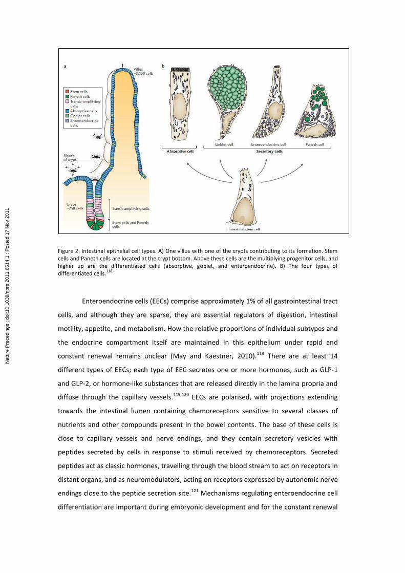
Figure 2. Intestinal epithelial cell types. A) One villus with one of the crypts contributing to its formation. Stem cells and Paneth cells are located at the crypt bottom. Above these cells are the multiplying progenitor cells, and higher up are the differentiated cells (absorptive, goblet, and enteroendocrine). B) The four types of differentiated cells.
118
Enteroendocrine cells (EECs) comprise approximately 1% of all gastrointestinal tract
cells, and although they are sparse, they are essential regulators of digestion, intestinal
motility, appetite, and metabolism. How the relative proportions of individual subtypes and
the endocrine compartment itself are maintained in this epithelium under rapid and
constant renewal remains unclear (May and Kaestner, 2010).119 There are at least 14
different types of EECs; each type of EEC secretes one or more hormones, such as GLP-1
and GLP-2, or hormone-like substances that are released directly in the lamina propria and
diffuse through the capillary vessels.119,120 EECs are polarised, with projections extending
towards the intestinal lumen containing chemoreceptors sensitive to several classes of
nutrients and other compounds present in the bowel contents. The base of these cells is
close to capillary vessels and nerve endings, and they contain secretory vesicles with
peptides secreted by cells in response to stimuli received by chemoreceptors. Secreted
peptides act as classic hormones, travelling through the blood stream to act on receptors in
distant organs, and as neuromodulators, acting on receptors expressed by autonomic nerve
endings close to the peptide secretion site.121 Mechanisms regulating enteroendocrine cell
differentiation are important during embryonic development and for the constant renewal
Nat
ure
Pre
cedi
ngs
: doi
:10.
1038
/npr
e.20
11.6
614.
1 : P
oste
d 17
Nov
201
1

of the intestinal epithelium in the adult. The identification of transcription factors and DNA
regulating elements that contribute to the specific genetic profile of each cell type is
increasing the understanding of the different networks controlling the spatial and temporal
activation of enteroendocrine differentiation programmes.122 EECs secrete several
regulatory molecules that control physiological and homeostatic functions, mainly post-
prandial secretion and motility, and act as sensors of lumen contents.123 Although EECs
secrete transcriptional regulators, they differentiate from pluripotent stem cells located in
crypts. Their origin is therefore endodermal as with all epithelial mucosa cell types.124-126
The Wnt signalling pathway plays an important role in the proliferative activity of
the normal intestinal crypt.127 The Lgr5/GPR49 gene is only expressed in the stem cell
compartment located at the crypt bottom, demonstrating that all epithelial lines derive
from Ldr5-expressing intestinal stem cells.125 EEC spatial orientation along the crypt-villus
axis is known to be closely associated with differentiation. Although most EECs differentiate
and migrate towards the villus top, a recent study showed that a small EEC subpopulation
that migrates towards or remains at the crypt bottom expresses stem cell markers and
post-mitotic endocrine markers.128 A fourth type of secretory cell, referred to as tuft cells,
was recently described. The differentiation of tuft cells depends on the transcription factors
Atoh1/Math1 but not on other factors, distinguishing them from EECs, Paneth cells, and
goblet cells. These cells are the main source of endogenous intestinal opioids, and they are
the only type of epithelial cell expressing cyclooxygenase enzymes, suggesting an important
role in intestinal epithelial physiopathology.129
The Notch signalling pathway plays a crucial role in enteroendocrine differentiation.
Notch is a transmembrane receptor protein that mediates cell-to-cell communication and
coordinates a signalling cascade. Notch receives signals at the cell surface and functions in
the nucleus to regulate gene expression.130 In mammals, the main components of this
pathway are ligands (Delta and Jagged), receptors (Notch 1, 2, 3, and 4), and a transcription
factor (RBP-Jk) that activates the target genes. Ligand interaction promotes proteolytic
cleavage of Notch, thus releasing the Notch intracellular domain from the cell surface.131
Intracellular Notch is transported to the nucleus, where it interacts with RBP-Jk to form a
complex that binds and activates the promoters of hairy/enhancer of split (HES) gene family
members. Hes1 or another HES family member then represses bHLH pro-endocrine
transcription factors such as neurogenin 3 (Ngn3) by binding their promoters or enhancers
(Figure 3).119,132,133 Hes1 is crucial for maintaining a reservoir of undifferentiated endocrine
Nat
ure
Pre
cedi
ngs
: doi
:10.
1038
/npr
e.20
11.6
614.
1 : P
oste
d 17
Nov
201
1
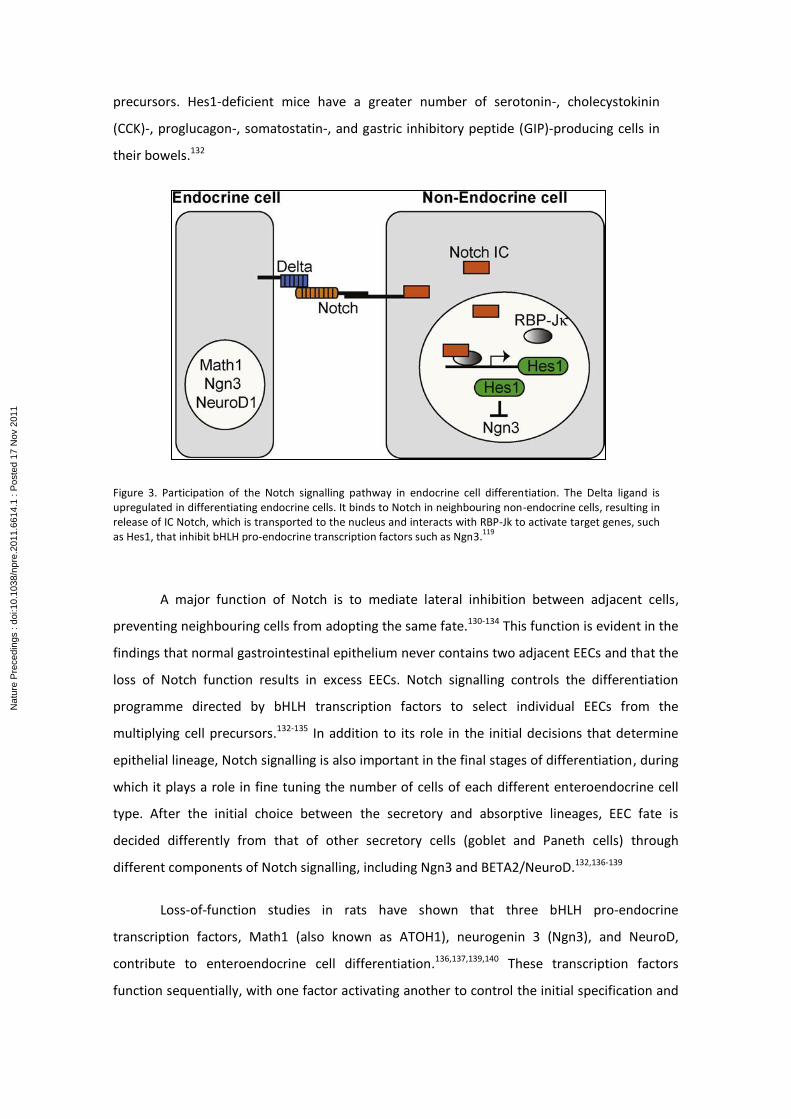
precursors. Hes1-deficient mice have a greater number of serotonin-, cholecystokinin
(CCK)-, proglucagon-, somatostatin-, and gastric inhibitory peptide (GIP)-producing cells in
their bowels.132
Figure 3. Participation of the Notch signalling pathway in endocrine cell differentiation. The Delta ligand is upregulated in differentiating endocrine cells. It binds to Notch in neighbouring non-endocrine cells, resulting in release of IC Notch, which is transported to the nucleus and interacts with RBP-Jk to activate target genes, such as Hes1, that inhibit bHLH pro-endocrine transcription factors such as Ngn3.
119
A major function of Notch is to mediate lateral inhibition between adjacent cells,
preventing neighbouring cells from adopting the same fate.130-134 This function is evident in the
findings that normal gastrointestinal epithelium never contains two adjacent EECs and that the
loss of Notch function results in excess EECs. Notch signalling controls the differentiation
programme directed by bHLH transcription factors to select individual EECs from the
multiplying cell precursors.132-135 In addition to its role in the initial decisions that determine
epithelial lineage, Notch signalling is also important in the final stages of differentiation, during
which it plays a role in fine tuning the number of cells of each different enteroendocrine cell
type. After the initial choice between the secretory and absorptive lineages, EEC fate is
decided differently from that of other secretory cells (goblet and Paneth cells) through
different components of Notch signalling, including Ngn3 and BETA2/NeuroD.132,136-139
Loss-of-function studies in rats have shown that three bHLH pro-endocrine
transcription factors, Math1 (also known as ATOH1), neurogenin 3 (Ngn3), and NeuroD,
contribute to enteroendocrine cell differentiation.136,137,139,140 These transcription factors
function sequentially, with one factor activating another to control the initial specification and
Nat
ure
Pre
cedi
ngs
: doi
:10.
1038
/npr
e.20
11.6
614.
1 : P
oste
d 17
Nov
201
1

final differentiation of EECs.119 Math1 is necessary for the initial specification of all three
intestinal secretory lines (goblet, Paneth, and EEC).138 Later on, expression of lineage-specific
transcription factors, such as Sox9 for Paneth cells, Klf4 for Goblet cells, and Ngn3/NeuroD for
EECs, is necessary.136,141,142
Figure 4. Overview of enteroendocrine differentiation in the intestinal epithelium. Lgr5-expressing stem cells in the
crypts give rise to the four intestinal epithelial cell types. Expression of Math1 is restricted to the secretory lineage,
and Hes1 is restricted to the absorptive lineage. After this initial specification is established, lineage-specific
transcription factors, such as Sox9 for Paneth cells, Klf4 for goblet cells, and Gf1/Ngn3/NeuroD for EECs, are
necessary for differentiation into each specific secretory lineage.119
Some studies show that intestinal precursors preferentially differentiate into
enterocytes in the intestine of adult Math1 knockout rats, thus confirming the importance of
this gene in maintaining the balance between enterocytes and EECs.143 In mice and humans, all
intestinal EECs require Ngn3,136 which acts downstream of the Math 1 transcription factor.
Ngn3 is a bHLH transcription factor that is activated on embryonic day 11.5.144 Its expression
persists in the adult bowels and the glandular portion of the stomach.136
Genetic analyses of NeuroD provided the first link between Notch signalling and EEC
differentiation.139 In contrast to Ngn3, NeuroD expression is restricted to a subgroup of
secretin-producing EECs.139,145 NeuroD acts downstream of Ngn3, as demonstrated by the lack
of NeuroD expression in Ngn3-deficient mice.146 Notch signalling also controls the transcription
Nat
ure
Pre
cedi
ngs
: doi
:10.
1038
/npr
e.20
11.6
614.
1 : P
oste
d 17
Nov
201
1

of Hes1, which encodes a bHLH transcriptional repressor that inhibits the activity of the bHLH
transcriptional activators mentioned above.147 Hes1 is crucial for maintaining pro-endocrine
reservoirs in an undifferentiated state. Several bHLH factors are up-regulated in rats in the
absence of Hes1, resulting in early differentiation of enteroendocrine subtypes and high
numbers of serotonin-, CCK-, proglucagon-, somatostatin-, and GIP-positive cells in the
intestine.132
Ngn3 and NeuroD specify the differentiation of EECs such as L-cells, in which Px4 and
Pax6 play important roles in final differentiation.148 A diet rich in oligofructose, a non-digestible
carbohydrate, was shown to double the number of GLP-1-producing L-cells in the proximal
colon of rats through a mechanism involving up-regulation of Ngn3 and NeuroD. This study
suggests that the final products of oligofructose fermentation, such as acetate, propionate,
and butyrate, might be involved in the induction of L-cell differentiation. Butyrate is thought to
regulate intestinal cell differentiation, as it was shown in vitro to increase the expression of the
glucagon gene in immortalised L-cells. Moreover, butyrate infusion in the colon in vivo
increases proglucagon and GLP-1 levels.149,150
The transcription factors Foxa1 and Foxa2 are expressed throughout the
gastrointestinal epithelium from embryo to adult. Mice lacking these factors also lack GLP-1-
and GLP-2-expressing cells (L-cells), and they have reduced numbers of somatostatin-
expressing D-cells and PYY-expressing L-cells. The mRNA levels of glucagon, somatostatin, PYY,
and the transcription factors Isl-1 and Pax6 were reduced in the small intestine of these
animals, demonstrating that Foxa1 and Foxa2 are involved in the network of transcription
factors regulating enteroendocrine lineage differentiation.151
Transcription factors other than bHLH factors also seem to play important roles in EEC
differentiation. In addition to the early EEC differentiation observed in Hes1-deficient animals,
Pax4, Pax6, Nkx2.2, and Isl-1 genes were activated in the intestine.132 Instead of controlling EEC
global differentiation like bHLH genes, these factors seem to play important roles in the fine
control of specification decisions among enteroendocrine populations.119 LIM homeodomain
genes encode a family of transcription regulating proteins involved in the control of several
aspects of embryonic development and are responsible for some human diseases. Islet-1 (Isl-1)
is expressed in gastrointestinal epithelium on approximately embryonic day 10, and in adults,
its expression is restricted to EEC subgroups. Expression analysis suggests that Isl-1 might play
an important role in the final differentiation and/or maintenance of mature enteroendocrine
subtypes in the gastrointestinal epithelium.152
Nat
ure
Pre
cedi
ngs
: doi
:10.
1038
/npr
e.20
11.6
614.
1 : P
oste
d 17
Nov
201
1
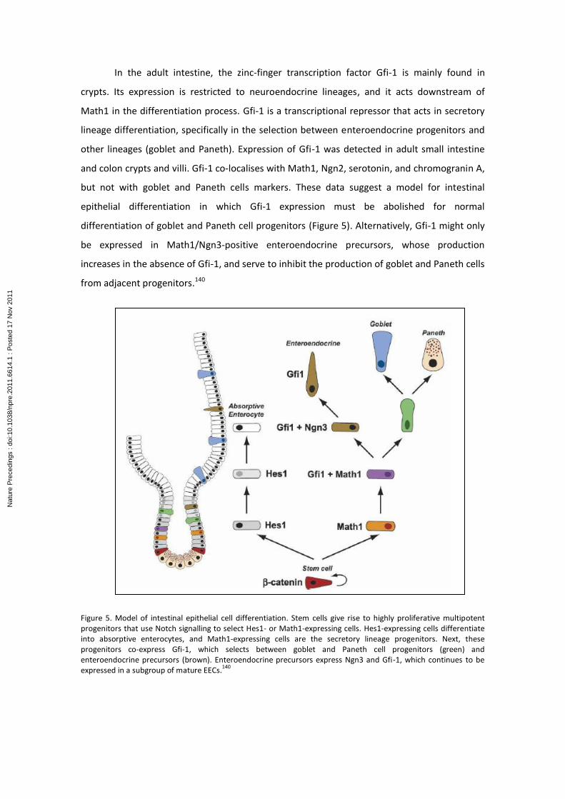
In the adult intestine, the zinc-finger transcription factor Gfi-1 is mainly found in
crypts. Its expression is restricted to neuroendocrine lineages, and it acts downstream of
Math1 in the differentiation process. Gfi-1 is a transcriptional repressor that acts in secretory
lineage differentiation, specifically in the selection between enteroendocrine progenitors and
other lineages (goblet and Paneth). Expression of Gfi-1 was detected in adult small intestine
and colon crypts and villi. Gfi-1 co-localises with Math1, Ngn2, serotonin, and chromogranin A,
but not with goblet and Paneth cells markers. These data suggest a model for intestinal
epithelial differentiation in which Gfi-1 expression must be abolished for normal
differentiation of goblet and Paneth cell progenitors (Figure 5). Alternatively, Gfi-1 might only
be expressed in Math1/Ngn3-positive enteroendocrine precursors, whose production
increases in the absence of Gfi-1, and serve to inhibit the production of goblet and Paneth cells
from adjacent progenitors.140
Figure 5. Model of intestinal epithelial cell differentiation. Stem cells give rise to highly proliferative multipotent progenitors that use Notch signalling to select Hes1- or Math1-expressing cells. Hes1-expressing cells differentiate into absorptive enterocytes, and Math1-expressing cells are the secretory lineage progenitors. Next, these progenitors co-express Gfi-1, which selects between goblet and Paneth cell progenitors (green) and enteroendocrine precursors (brown). Enteroendocrine precursors express Ngn3 and Gfi-1, which continues to be expressed in a subgroup of mature EECs.
140
Nat
ure
Pre
cedi
ngs
: doi
:10.
1038
/npr
e.20
11.6
614.
1 : P
oste
d 17
Nov
201
1
Another zinc-finger protein, Insm1 or IA-1, is found in proliferating cells, many of which
co-express NeuroD1 and chromogranin A.153 Insm1 has been shown to control the
differentiation of specific intestinal enteroendocrine lineages and acts downstream of Notch
and Ngn3 after the initial specification of enteroendocrine precursors.119 Several homeobox
genes, including Isl-1, Pdx1, Nkx6.1, and Nkx2.2, have also been shown to participate in EEC
differentiation.154
Two paired box (Pax) genes, Pax 4 and Pax 6, are linked to pancreatic and intestinal
endocrine cell differentiation.155,156 Deletion of Pax4 in the duodenum disrupts the
differentiation of serotonin-, secretin-, CCK-, GIP-, and PYY-expressing cells; however, there is
no significant reduction in the number of EECs in the ileum and colon of Pax4-deficient mice.
There is a severe reduction of somatostatin- and gastrin-expressing cells in the distal stomach
of Pax6-deficient mice; however, serotonin-expressing cells are not affected. The number of
GIP-expressing cells in the duodenum of Pax6-deficient mice is reduced.157 Pax6 is crucial for
proglucagon gene expression in intestinal epithelium. It is expressed in intestinal EECs and
binds to G1 and G3 elements in the proglucagon promoter to activate its transcription.158,159
Pax6 might also bind to the promoter and induce production of PC 1/3 pro-hormone
convertase, an enzyme essential for the conversion of proinsulin into insulin160 and of
proglucagon into GLP-1.43 Proglucagon processing is accomplished by PC1 in L-cells and by PC2
in pancreatic alpha cells to produce glucagon. Published data suggest that these tissue-specific
processing mechanisms are due to differential expression of PC1 and PC2.113,161,162 GIP, another
important incretin, might be expressed alone or together with GLP-1. Cells expressing GIP and
GLP-1 are Pax6- and Pdx1-positive, and cells expressing only GLP-1 are Pax6-positive and Pdx1-
negative. This suggests that the presence of Pdx1 determines whether gastrointestinal L-cells
will co-express GIP.163
Most Nkx6.1-expressing cells also express serotonin, and some express gastrin. Nkx6.1
is not expressed in Pdx1-deficient mice, indicating that Pdx1 acts upstream of Nkx6.1 in
enteroendocrine differentiation.164 Some enteroendocrine hormone-producing intestinal
populations are reduced in Nkx2.2-deficient mice, including serotonin-, CCK-, GIP-, and gastrin-
producing cells; however, the number of ghrelin-expressing cells is greater, and the numbers
of Paneth cells, goblet cells, and enterocytes are unaltered. It seems that Nkx2.2 functions
upstream of Pax6 in regulating the fate of intestinal cells, as Pax6 mRNA and protein levels are
reduced in Nkx2.2-deficient cells. Specification of ghrelin-expressing cells is thought to occur at
the expense of other intestinal enteroendocrine cells; however, the total number of EECs
Nat
ure
Pre
cedi
ngs
: doi
:10.
1038
/npr
e.20
11.6
614.
1 : P
oste
d 17
Nov
201
1
remains the same.165 Nkx6.3 knockout mice show a severe and selective reduction in gastrin-
secreting G-cells and an increase in somatostatin-secreting D-cells; however, these animals
express normal levels of the Pdx1, Pax6, and Ngn3 transcription factors, which are needed for
G-cell development.166 These results suggest that Nkx6.3 acts independently from other
transcription factors in G-cell differentiation.119
GATA transcription factors regulate proliferation, differentiation, and gene expression
in several organs. In the small intestine, they are needed for crypt cell proliferation, secretory
lineage differentiation, and gene expression in absorptive enterocytes. GATA4 is expressed in
the proximal region in 85% of the small intestine and regulates the jejuno-ileal gradient of
gene expression in absorptive enterocytes. GATA6 is co-expressed with GATA4, but it is also
expressed in the ileum. Deletion of GATA6 in the ileum causes decreased crypt cell
proliferation, reduces the number of Paneth cells and EECs, increases the number of crypt
goblet cells, and alters the expression of specific genes in absorptive enterocytes. Deletion of
GATA4 and GATA6 in rats results in a jejunum and ileum phenotype almost identical to that of
the GATA6-deficient ileum, suggesting that GATA4 and GATA6 share some functions.167
GATA4 is a zinc-finger transcription factor involved in jejunum gene expression.
GATA4-deficient mice exhibit a dramatically decreased ability to absorb dietary cholesterol and
fat. A study comparing global gene expression profiles in wild-type jejunum and ileum and
GATA4-deficient jejunum demonstrated a 53% decrease in the expression of jejunum-specific
genes and a 47% increase in the expression of ileum-specific genes in GATA4-deficient jejunum
samples. Changes included decreased expression of mRNA encoding lipid and cholesterol
transporters and increased expression of mRNA encoding proteins involved in bile absorption.
This study showed that GATA4 is crucial for jejunum function and plays an essential role in the
determination of jejunal versus ileal identity. It has been shown that GATA4 or GATA6 is
necessary for secretory progenitors to commit to a neuroendocrine lineage.167-169
Immunohistochemical analysis of mouse small intestine revealed that GATA4 is only expressed
in villus enterocytes. GATA5 was not detected in enterocytes, but it was detected in other
lineages. High levels of GATA6 were only detected in the neuroendocrine lineage. Together,
these results suggest that GATA transcription factors may play different roles in the
differentiation and/or allocation and maintenance of small intestine lineages.179
Some transcription factors and signalling molecules regulate the expression of the
glucagon gene. The homeoprotein Cdx-2 activates the glucagon gene promoter in pancreatic
and intestinal proglucagon-producing cell lineages.171 The Pax-6 protein158 and the kinase A
Nat
ure
Pre
cedi
ngs
: doi
:10.
1038
/npr
e.20
11.6
614.
1 : P
oste
d 17
Nov
201
1

signalling pathway172 are involved in the regulation of glucagon gene expression in pancreatic
and intestinal glucagon-producing cell lineages, primary pancreatic islets, and intestinal cell
cultures.114
Studies of intestinal adaptation have examined genes possibly involved in this process
using differential display polymerase chain reaction (DD-PCR) and cDNA microarray.
Examination of the patterns of gene expression in animals during the adaptation process has
revealed that some metabolism-related genes are increased at least two-fold after resection.
Changes in the expression of genes encoding ion channels, transport proteins, transcriptions
factors, DNA-binding proteins, receptors, and cytoskeleton proteins were also observed.173,174
One study using DD-PCR showed that adaptation after resection results from the up-regulation
of genes not previously related to the adaptation response. Genes that exhibited differential
regulation after resection were divided into three categories: amino acid transport, protein
transport, and signal transduction molecules. This same study suggested that analyses using
reverse transcriptase polymerase chain reaction (RT-PCR) can more precisely define the
magnitude of the adaptation response.93
To study adaptation after 50% resection of the mouse intestine, cDNA microarrays
were used to characterise the expression of individual genes and global gene expression
patterns in the remaining ileum. The analysis of these microarrays revealed changes in the
expression of several genes, including those involved in cell cycle regulation, apoptosis, DNA
synthesis, and transcriptional regulation. Expression patterns were consistent with the
increase in cell proliferation and apoptosis observed during intestinal adaptation; however,
verification by RT-PCR and Northern blot are still necessary.173 Another study examined gene
regulation after resection in mice using cDNA microarrays and showed that 27 genes were
altered after resection; these genes included sprr2, which increased almost five-fold and had
not previously been linked to intestinal adaptation.174
Very little is known about what guides region-specific expression of hormones in the
bowels. Despite the discovery of the function of transcription factors important for
neuroendocrine cell differentiation, it is still not known how these factors direct and control
gene expression in the different enteroendocrine cell types. Nor is it known to what degree
these transcription factors control hormone expression in adults.119
The differentiation mechanism of L-cells and the genetic and/or environmental factors
that might influence the proliferation rate of this intestinal cell subtype, which is crucial for
Nat
ure
Pre
cedi
ngs
: doi
:10.
1038
/npr
e.20
11.6
614.
1 : P
oste
d 17
Nov
201
1
maintaining normoglycaemia, remain unknown. Thus, comparison of intact and interposed
ileum in terms of the differential expression of genes related to the differentiation of intestinal
stem cells into each of the intestinal epithelial lineages, and specifically to the differentiation
of neuroendocrine GLP-1-producing L-cells, is essential for the study of ileal interposition as a
possible surgical treatment of T2D.
2. AIMS
Overall
To assess the effects of isolated ileal interposition on intestinal stem cells of rats with diet-
induced glucidic dysmetabolism.
Specific
To investigate changes in the relative expression of genes involved in intestinal stem
cell differentiation into enteroendocrine cells after isolated ileal interposition.
To analyse changes in the number and function of L-cells after isolated ileal
interposition.
3. METHODS
3.1. Research Ethics Committee
This research project will be presented to the Experimental Research Ethics Committee
of the Federal University of São Paulo – São Paulo School of Medicine (UNIFESP/EPM).
3.2. Experimental procedures
Experimental procedures will be performed according to guidelines in the manual
“Care and Handling of Laboratory Animals”, Lapchick, Mattaria, Ko; Atheneu, 2009, CDD
636.0885.
All animals will be provided the same diet and will be subjected to surgery when they
become dysmetabolic. Blood samples will be collected from all animals on the days of surgery
and euthanasia for biochemical analysis. After euthanasia, samples will be collected from the
interposed and remaining ileum of the interposition group and from the jejunum and ileum of
the control group. These samples will be analysed using molecular biology and
Nat
ure
Pre
cedi
ngs
: doi
:10.
1038
/npr
e.20
11.6
614.
1 : P
oste
d 17
Nov
201
1
immunofluorescence techniques to compare changes in the small intestine segment under
study relative to the characteristic endocrine pattern of the ileum.
3.3. Sample
Twenty male Wistar – 2BAW heterogeneous male rats (Rattus norvegicus albinus) aged
12 weeks and weighing between 250 and 280 g will be used. The animals will be supplied by
the Laboratory of Animal Experimentation at the Institute of Pharmacology and Molecular
Biology of the Federal University of São Paulo – São Paulo School of Medicine (UNIFESP/EPM),
where they will be housed throughout the study.
Animals will be housed in individual cages and kept for 32 weeks at a controlled
ambient temperature of 23 2C, relative humidity of 55 15%, and automatic 12-hour light–
dark cycle (06:00 / 18:00) (Timer-Kienzle).
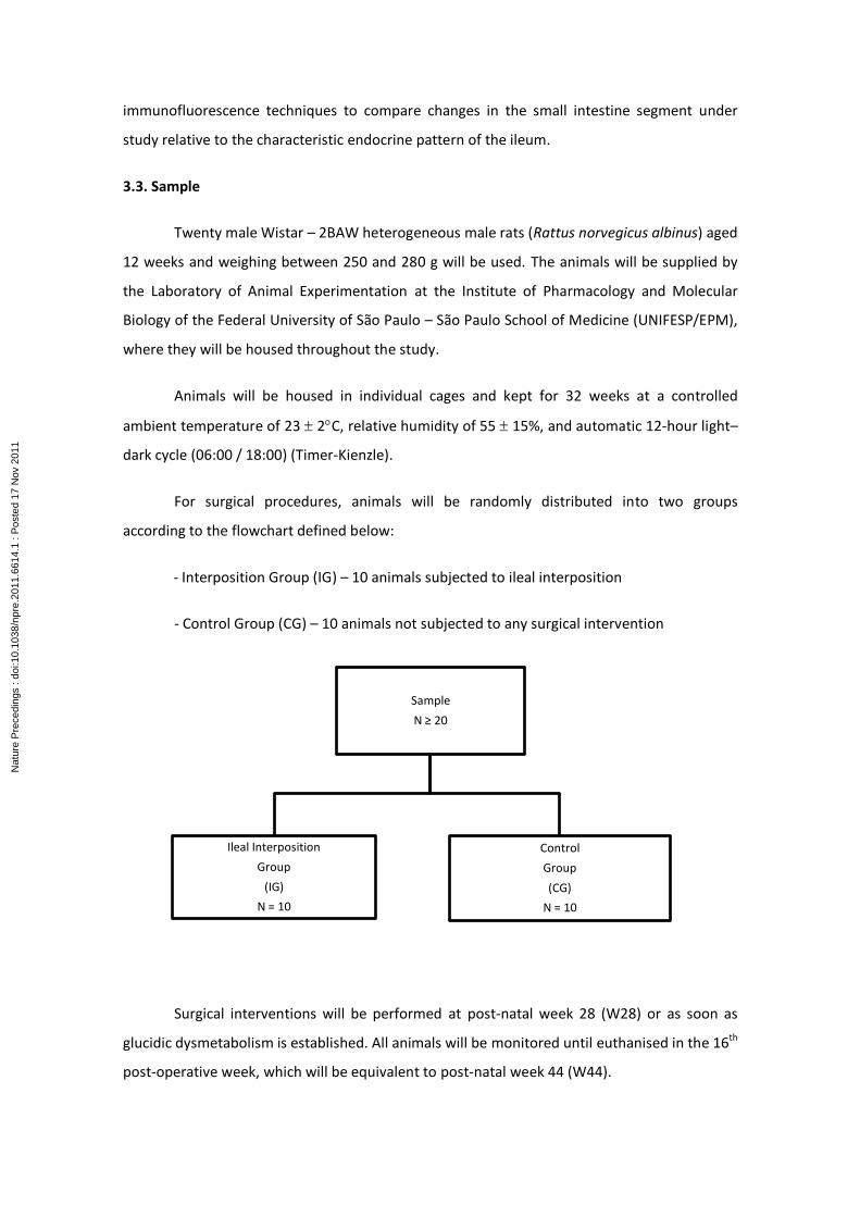
For surgical procedures, animals will be randomly distributed into two groups
according to the flowchart defined below:
- Interposition Group (IG) – 10 animals subjected to ileal interposition
- Control Group (CG) – 10 animals not subjected to any surgical intervention
Surgical interventions will be performed at post-natal week 28 (W28) or as soon as
glucidic dysmetabolism is established. All animals will be monitored until euthanised in the 16th
post-operative week, which will be equivalent to post-natal week 44 (W44).
Sample
N ≥ 20
Ileal Interposition
Group
(IG)
N = 10
Control
Group
(CG)
N = 10
Nat
ure
Pre
cedi
ngs
: doi
:10.
1038
/npr
e.20
11.6
614.
1 : P
oste
d 17
Nov
201
1
3.4. Diet
Throughout the experiment, all animals will be provided a lipid-rich pelletised
hypercaloric diet (HD) ad libitum in alternating cycles of four feed types (1, 2, 3, and 4) to
stimulate intake. The experimental feed will be alternated every 24 hours, and non-ingested
amounts will be measured. Consumption of these diets will promote obesity in animals, which
will exhibit characteristics commonly associated with human obesity, such as insulin
resistance, hyperglycaemia, hyperinsulinaemia, dyslipidaemia, and liver steatosis. All groups
will have free access to water.
Experimental feeds 1, 2, 3, and 4 will be industrially prepared by Rhoster®, Araçoiaba
da Serra – SP according to guidelines in Requirements of the Laboratory Rat recommended by
the National Academy of Sciences and will consist of standard rat feed with protein, vitamin,
and mineral supplements. Additional hyperenergetic ingredients used in hypercaloric
experimental diets will be (in grams per kilogram):
- HD1 – standard ration, 355; toasted peanuts, 176; casein, 123; corn oil, 82; chocolate,
88; corn biscuits, 176; and vitamins and minerals.
- HD2 – standard ration, 439; toasted peanuts, 218; casein 129; corn oil, 61; chips, 153;
and vitamins and minerals.
- HD3 - standard ration, 371; toasted peanuts, 185; casein, 99; corn oil, 68; pasta, 185;
grated cheese, 92; and vitamins and minerals.
- HD4 - standard ration, 359; toasted peanuts, 179; casein 105; corn oil, 80; condensed
milk, 161; wafer biscuits, 116; and vitamins and minerals.
The macronutrient composition of the standard and hypercaloric-hyperlipidic
experimental feeds prepared and analysed in the laboratory by Rhoster®, Araçoiaba da Serra –
SP is described in Table 1.
Nat
ure
Pre
cedi
ngs
: doi
:10.
1038
/npr
e.20
11.6
614.
1 : P
oste
d 17
Nov
201
1
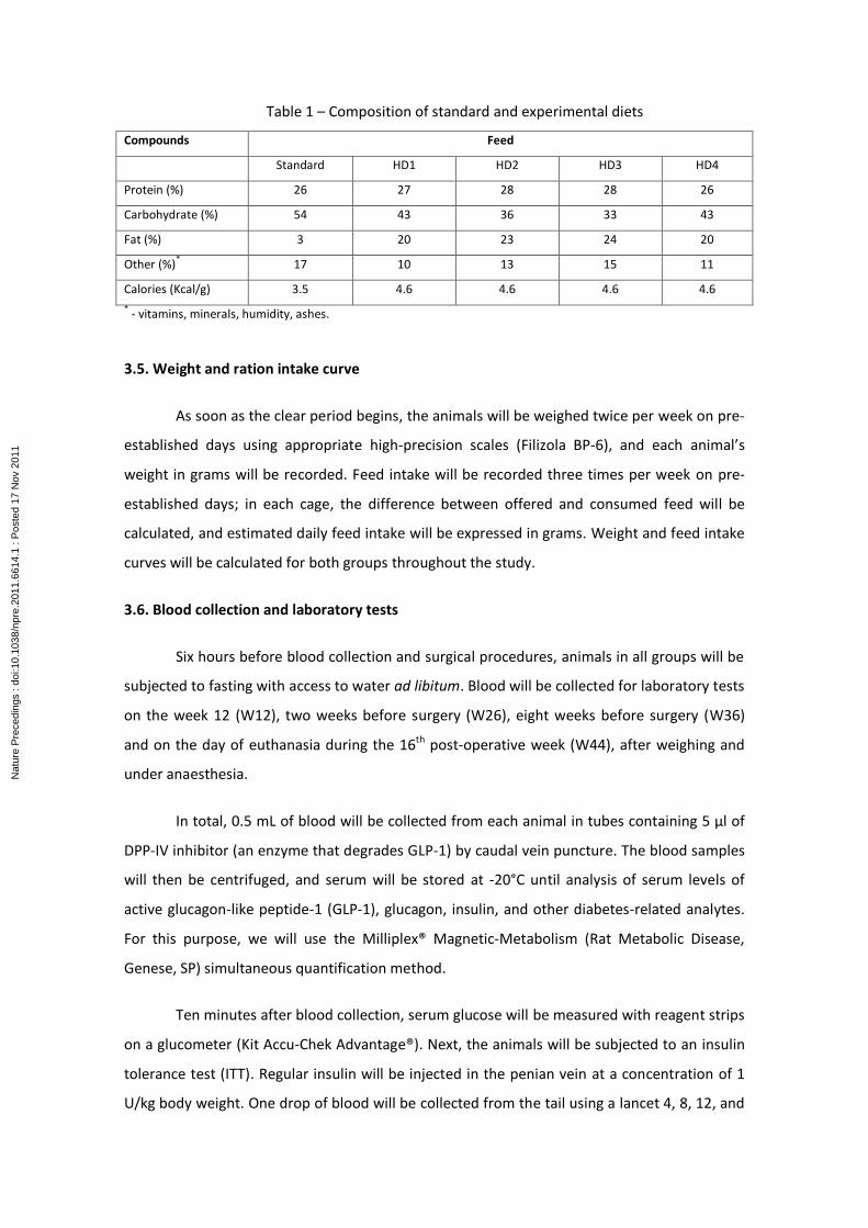
Table 1 – Composition of standard and experimental diets
Compounds Feed
Standard HD1 HD2 HD3 HD4
Protein (%) 26 27 28 28 26
Carbohydrate (%) 54 43 36 33 43
Fat (%) 3 20 23 24 20
Other (%)*
17 10 13 15 11
Calories (Kcal/g) 3.5 4.6 4.6 4.6 4.6
* - vitamins, minerals, humidity, ashes.
3.5. Weight and ration intake curve
As soon as the clear period begins, the animals will be weighed twice per week on pre-
established days using appropriate high-precision scales (Filizola BP-6), and each animal’s
weight in grams will be recorded. Feed intake will be recorded three times per week on pre-
established days; in each cage, the difference between offered and consumed feed will be
calculated, and estimated daily feed intake will be expressed in grams. Weight and feed intake
curves will be calculated for both groups throughout the study.
3.6. Blood collection and laboratory tests
Six hours before blood collection and surgical procedures, animals in all groups will be
subjected to fasting with access to water ad libitum. Blood will be collected for laboratory tests
on the week 12 (W12), two weeks before surgery (W26), eight weeks before surgery (W36)
and on the day of euthanasia during the 16th post-operative week (W44), after weighing and
under anaesthesia.
In total, 0.5 mL of blood will be collected from each animal in tubes containing 5 μl of
DPP-IV inhibitor (an enzyme that degrades GLP-1) by caudal vein puncture. The blood samples
will then be centrifuged, and serum will be stored at -20°C until analysis of serum levels of
active glucagon-like peptide-1 (GLP-1), glucagon, insulin, and other diabetes-related analytes.
For this purpose, we will use the Milliplex® Magnetic-Metabolism (Rat Metabolic Disease,
Genese, SP) simultaneous quantification method.
Ten minutes after blood collection, serum glucose will be measured with reagent strips
on a glucometer (Kit Accu-Chek Advantage®). Next, the animals will be subjected to an insulin
tolerance test (ITT). Regular insulin will be injected in the penian vein at a concentration of 1
U/kg body weight. One drop of blood will be collected from the tail using a lancet 4, 8, 12, and
Nat
ure
Pre
cedi
ngs
: doi
:10.
1038
/npr
e.20
11.6
614.
1 : P
oste
d 17
Nov
201
1
16 minutes after insulin injection for the detection of glycaemia. The results will be recorded
for later analysis.
Serum glucose will also be measured weekly on pre-established days throughout the
study (W14 to W44) using reagent strips and a glucometer (Kit Accu-Chek Advantage®).
3.7. Anaesthesia, weighing, and antibiotic prophylaxis
Before the onset of anaesthesia, all animals will be weighed on high-precision scales
(Filizola BP-6), and their weights will be recorded in grams. In both groups (IG and CG), general
anaesthesia will be induced with halothane to collect tail blood. Dissociative anaesthesia will
then be induced by simultaneously injecting the animals intramuscularly with Zoletil 50®
(tiletamine+zolazepam) at a dose of 20 mg/kg and Fentanil® at a dose of 0.025 mg/kg to
perform the insulin tolerance test and surgical intervention.170
Each animal will be maintained on spontaneous ventilation during the procedure, and
the anaesthetic plane will be checked periodically every 45 minutes by assessing the auricular
and interdigital reflexes, which are abolished during anaesthesia. When these reflexes
reappear, one-third of the initial anaesthetic dose will be administered. The total amount of
anaesthetics used will be recorded at the end of the surgical intervention.
Antibiotic prophylaxis will be performed by intramuscular injection of cefoxitin at a
dose of 50 mg/kg body weight soon after anaesthesia for surgery.
3.8. Surgical procedures
Rats in the IG group will be subjected to surgery upon developing diet-induced glucidic
dysmetabolism, which should occur around 28 weeks of life (W28). All Halsted asepsis and
antisepsis principles will be applied, and sterile microsurgical tools will be used.
The anaesthetised rats will be placed in horizontal dorsal decubitus on the surgical
table with the legs and tail duly immobilised and secured with tape. Antisepsis of the
abdominal area will be performed with chlorhexidine in an aqueous vehicle.
A sterilised fenestrated surgical drape will be placed on each animal’s abdominal area,
and a 5-cm long median longitudinal incision will be made in the wall with a disposable scalpel
containing a number 15 blade.
Nat
ure
Pre
cedi
ngs
: doi
:10.
1038
/npr
e.20
11.6
614.
1 : P
oste
d 17
Nov
201
1
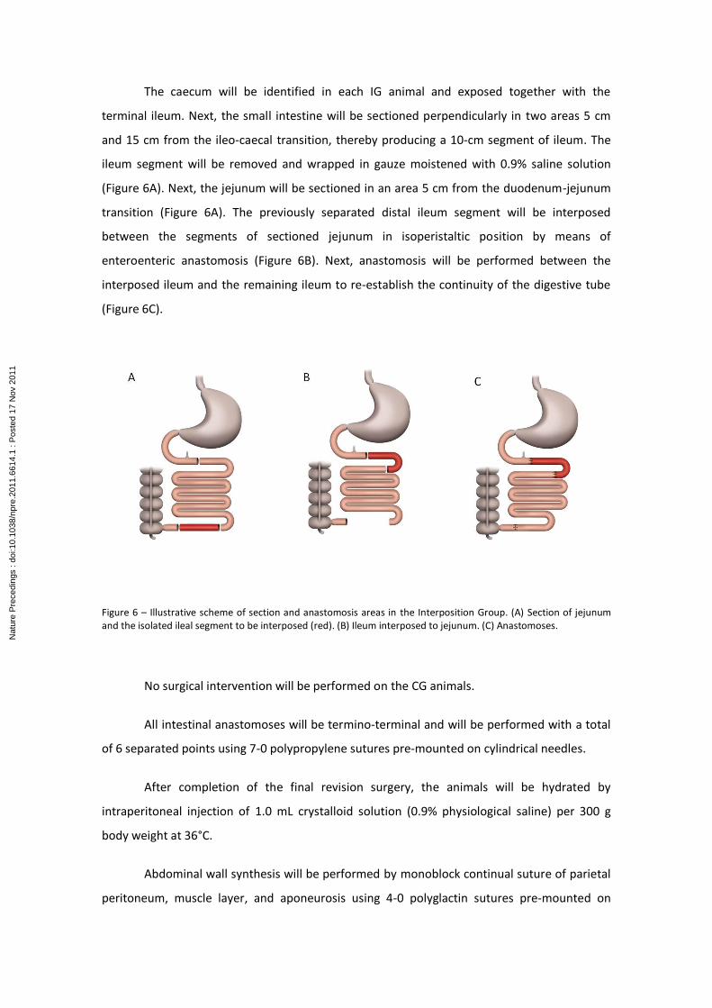
The caecum will be identified in each IG animal and exposed together with the
terminal ileum. Next, the small intestine will be sectioned perpendicularly in two areas 5 cm
and 15 cm from the ileo-caecal transition, thereby producing a 10-cm segment of ileum. The
ileum segment will be removed and wrapped in gauze moistened with 0.9% saline solution
(Figure 6A). Next, the jejunum will be sectioned in an area 5 cm from the duodenum-jejunum
transition (Figure 6A). The previously separated distal ileum segment will be interposed
between the segments of sectioned jejunum in isoperistaltic position by means of
enteroenteric anastomosis (Figure 6B). Next, anastomosis will be performed between the
interposed ileum and the remaining ileum to re-establish the continuity of the digestive tube
(Figure 6C).
Figure 6 – Illustrative scheme of section and anastomosis areas in the Interposition Group. (A) Section of jejunum and the isolated ileal segment to be interposed (red). (B) Ileum interposed to jejunum. (C) Anastomoses.
No surgical intervention will be performed on the CG animals.
All intestinal anastomoses will be termino-terminal and will be performed with a total
of 6 separated points using 7-0 polypropylene sutures pre-mounted on cylindrical needles.
After completion of the final revision surgery, the animals will be hydrated by
intraperitoneal injection of 1.0 mL crystalloid solution (0.9% physiological saline) per 300 g
body weight at 36°C.
Abdominal wall synthesis will be performed by monoblock continual suture of parietal
peritoneum, muscle layer, and aponeurosis using 4-0 polyglactin sutures pre-mounted on
Nat
ure
Pre
cedi
ngs
: doi
:10.
1038
/npr
e.20
11.6
614.
1 : P
oste
d 17
Nov
201
1
cylindrical needles. In the skin, continual suture with inverted stitches will be performed using
4-0 polyglactin sutures pre-mounted on cylindrical needles.
3.9. Postoperative assessment
Animals will be kept warm and observed until full recovery from anaesthesia, when
they will be placed in individual cages and transported to the housing room in the same
laboratory and under the same environmental conditions as before surgery.
Before recovery from anaesthesia, animals will be given analgesic by gavage (0.5 mL of
3 g/mL dipyrone in water). Water will be reintroduced ad libitum as soon as animals recover
from anaesthesia. Dipyrone (1 g/100 mL in water) will be given during the first 72 post-
operative hours. Access to food will be allowed 12 hours after surgery.
The animals will be assessed and monitored until the 16th post-operative week (W44).
3.10. Euthanasia
On the 44th post-natal week, the fasting, anaesthesia, weighing, and blood collection
for laboratory tests will be repeated. Euthanasia will then be performed by decapitation
according to the guidelines of the Brazilian Society of Laboratory Animals Science/Brazilian
College of Animal Experimentation – SCAL-COBEA.
3.11. Study parameters
- Body Weight: as soon as the clear cycle starts, each animal in each group (IG and CG) will be
weighed on high-precision scales, and the weight will be recorded in grams (W1, W2, W3…,
W32).
- Feed Intake: all animals will be given feed diurnally at a known amount sufficient for their
daily needs; three times per week, non-ingested feed will be weighed on adequate scales, and
daily feed intake will be calculated from the difference between supplied and remaining feed.
- Biochemical Tests: serum levels of active GLP-1 (GLP-1), glucose (GLI), insulin (INS), and
glucagon (GLU) will be measured in all animals on week 12 (W12), two weeks before surgery
(W26), eight weeks after surgery (W36) and euthanasia (W44).
- Insulin Resistance: on the days of surgery and euthanasia, insulin resistance (IR) will be
calculated using the HOMA-IR indirect test (Homeostasis Model Assessment Insulin Resistance)
Nat
ure
Pre
cedi
ngs
: doi
:10.
1038
/npr
e.20
11.6
614.
1 : P
oste
d 17
Nov
201
1
from the product of serum insulin (mU/mL), glycaemia (mg/mL), and the constant 0.05551
divided by 22.5.
- Insulin Tolerance Test (ITT): will be performed in all animals on the day of study onset (W12),
two weeks before surgery (S26), eight weeks after surgery (W36), and euthanasia (W44). The
rate of glucose removal will be calculated using the formula In2/t1/2; t1/2 will be calculated
for glucose from the minimum slope of the regression curve during the phase of linear decay
of serum glucose concentration using PRISMA software.
3.12. Tissue collection
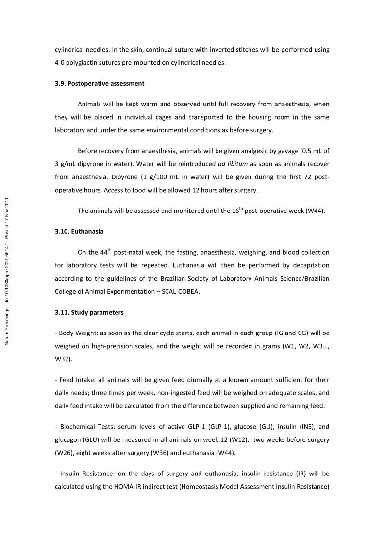
After euthanasia, the following fragments of intestinal tissue will be collected for
histological analysis by immunofluorescence and gene expression analysis by molecular
biology:
- Interposition Group: median segment of transposed ileum and segment adjacent to
ileal anastomosis (ileum remaining in normal position) (Figure 7A).
- Control Group: ileum segment and jejunum segment (Figure 7B).
Figure 7 – Illustrative scheme of tissue collection for analysis. (A) Interposition Group: interposed ileum segment (purple) and remaining ileum adjacent to ileal anastomosis (green). (B) Control Group: segment of jejunum (blue) and ileum (yellow).
Nat
ure
Pre
cedi
ngs
: doi
:10.
1038
/npr
e.20
11.6
614.
1 : P
oste
d 17
Nov
201
1
3.13. Histological analysis by immunofluorescence
One-centimetre tissue samples for histological analysis by immunofluorescence will be
cryopreserved in increasing sucrose concentrations (10% and 20% for 4 hours at 4°C and 30%
for 12 hours at 4°C), frozen in liquid nitrogen, and kept at -80°C until analysis. Three- to five-
micron-thick sections will be cut from the tissues using a cryostat (Leica) and mounted on
silanated slides (BMF).
To block nonspecific staining, the slides will be incubated in phosphate-buffered saline
(PBS) pH 7.4 + 1% bovine serum albumin (BSA) for 30 minutes at room temperature. For
binding with primary antibody, the slides will be incubated with specific antibody (anti-GLP-1
and others, Abcam) diluted in 1% PBS/albumin buffer (Sigma A9647) for 1 hour at 37°C or for
16-18 hours at 4°C in a humid chamber. Next, they will be washed three times with 1%
PBS/albumin for 5-10 minutes and incubated with a fluorophore-conjugated secondary
antibody (FITC, Rhodamine, or Alexa Fluor 488, Invitrogen) for 90 minutes at 4°C in a humid
chamber following the manufacturer's instructions. After incubation, the slides will be washed
three times with 1% PBS/albumin for 5-10 minutes and incubated with the second primary
antibody, and the incubation and washing steps described above will be repeated. Finally, the
slides will be incubated with 4',6-diamidino-2-phenylindole (DAPI, Invitrogen) for 30 minutes at
37°C, mounted on 1% glycerol-coated microscopy slides, and stored refrigerated and protected
from light until analysis.
Immunoexpression of GLP-1, an L-cell marker, and other relevant genes will be
assessed under a fluorescence microscope using appropriate filters for each fluorochrome.
3.14. Molecular biology: studying relative gene expression
3.14.1. Preparation of total RNA
Collected segments (interposed and remaining ileum in the IG, jejunum and ileum in
the CG) will be immediately immersed in RNA stabilisation reagent (RNAlater RNA Stabilization
Reagent, Qiagen) and stored in tubes (RNAlater Tissue Protect Tubes, Qiagen) at -20°C
according to the manufacturer’s instructions until analysis.
To isolate total RNA, we will homogenise each sample using QIAzol reagent (QIAzol
Lysis Reagent, Qiagen) on a Polytron tissue processor. After precipitation with ethanol, total
RNA will be isolated using a commercial kit (RNeasy Mini Kit, Qiagen), and genomic DNA will be
Nat
ure
Pre
cedi
ngs
: doi
:10.
1038
/npr
e.20
11.6
614.
1 : P
oste
d 17
Nov
201
1
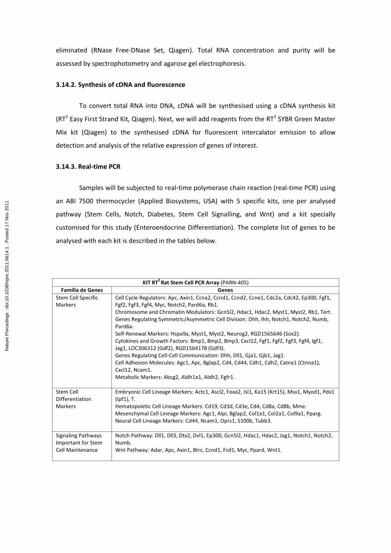
eliminated (RNase Free-DNase Set, Qiagen). Total RNA concentration and purity will be
assessed by spectrophotometry and agarose gel electrophoresis.
3.14.2. Synthesis of cDNA and fluorescence
To convert total RNA into DNA, cDNA will be synthesised using a cDNA synthesis kit
(RT2 Easy First Strand Kit, Qiagen). Next, we will add reagents from the RT2 SYBR Green Master
Mix kit (Qiagen) to the synthesised cDNA for fluorescent intercalator emission to allow
detection and analysis of the relative expression of genes of interest.
3.14.3. Real-time PCR
Samples will be subjected to real-time polymerase chain reaction (real-time PCR) using
an ABI 7500 thermocycler (Applied Biosystems, USA) with 5 specific kits, one per analysed
pathway (Stem Cells, Notch, Diabetes, Stem Cell Signalling, and Wnt) and a kit specially
customised for this study (Enteroendocrine Differentiation). The complete list of genes to be
analysed with each kit is described in the tables below.
KIT RT2
Rat Stem Cell PCR Array (PARN-405)
Família de Genes Genes
Stem Cell Specific Markers
Cell Cycle Regulators: Apc, Axin1, Ccna2, Ccnd1, Ccnd2, Ccne1, Cdc2a, Cdc42, Ep300, Fgf1, Fgf2, Fgf3, Fgf4, Myc, Notch2, Pard6a, Rb1. Chromosome and Chromatin Modulators: Gcn5l2, Hdac1, Hdac2, Myst1, Myst2, Rb1, Tert. Genes Regulating Symmetric/Asymmetric Cell Division: Dhh, Ihh, Notch1, Notch2, Numb, Pard6a. Self-Renewal Markers: Hspa9a, Myst1, Myst2, Neurog2, RGD1565646 (Sox2). Cytokines and Growth Factors: Bmp1, Bmp2, Bmp3, Cxcl12, Fgf1, Fgf2, Fgf3, Fgf4, Igf1, Jag1, LOC306312 (Gdf2), RGD1564178 (Gdf3). Genes Regulating Cell-Cell Communication: Dhh, Dll1, Gja1, Gjb1, Jag1. Cell Adhesion Molecules: Agc1, Apc, Bglap2, Cd4, Cd44, Cdh1, Cdh2, Catna1 (Ctnna1), Cxcl12, Ncam1. Metabolic Markers: Abcg2, Aldh1a1, Aldh2, Fgfr1.
Stem Cell Differentiation Markers
Embryonic Cell Lineage Markers: Actc1, Ascl2, Foxa2, Isl1, Ka15 (Krt15), Msx1, Myod1, Pdx1 (Ipf1), T. Hematopoietic Cell Lineage Markers: Cd19, Cd3d, Cd3e, Cd4, Cd8a, Cd8b, Mme. Mesenchymal Cell Lineage Markers: Agc1, Alpi, Bglap2, Col1a1, Col2a1, Col9a1, Pparg. Neural Cell Lineage Markers: Cd44, Ncam1, Oprs1, S100b, Tubb3.
Signaling Pathways Important for Stem Cell Maintenance
Notch Pathway: Dll1, Dll3, Dtx2, Dvl1, Ep300, Gcn5l2, Hdac1, Hdac2, Jag1, Notch1, Notch2, Numb. Wnt Pathway: Adar, Apc, Axin1, Btrc, Ccnd1, Fzd1, Myc, Ppard, Wnt1.
Nat
ure
Pre
cedi
ngs
: doi
:10.
1038
/npr
e.20
11.6
614.
1 : P
oste
d 17
Nov
201
1
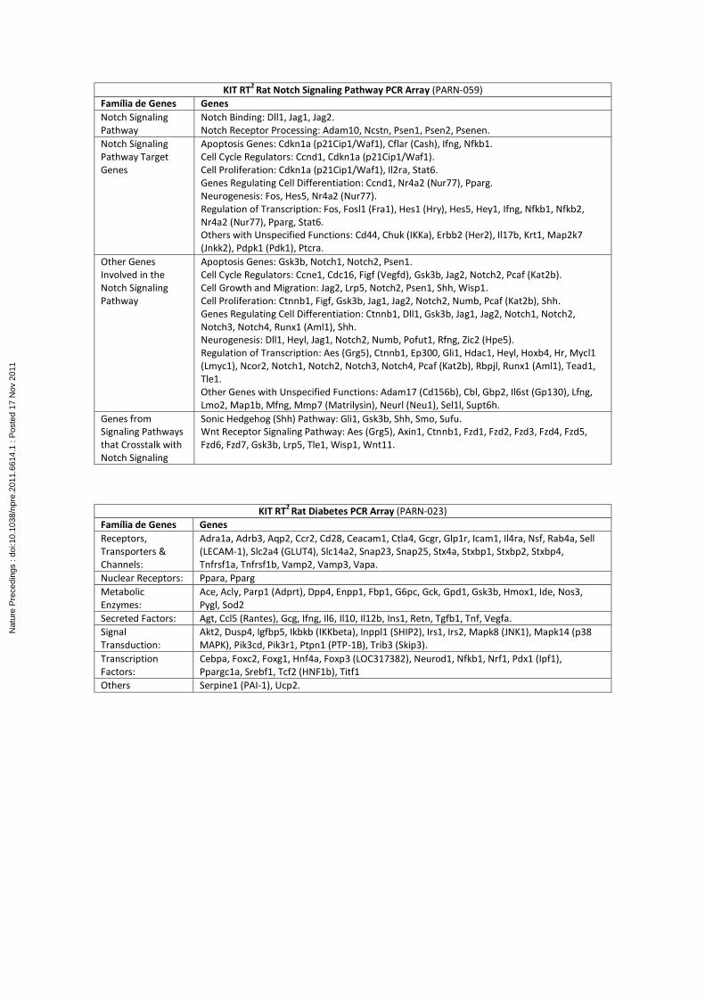
KIT RT2
Rat Notch Signaling Pathway PCR Array (PARN-059)
Família de Genes Genes
Notch Signaling Pathway
Notch Binding: Dll1, Jag1, Jag2. Notch Receptor Processing: Adam10, Ncstn, Psen1, Psen2, Psenen.
Notch Signaling Pathway Target Genes
Apoptosis Genes: Cdkn1a (p21Cip1/Waf1), Cflar (Cash), Ifng, Nfkb1. Cell Cycle Regulators: Ccnd1, Cdkn1a (p21Cip1/Waf1). Cell Proliferation: Cdkn1a (p21Cip1/Waf1), Il2ra, Stat6. Genes Regulating Cell Differentiation: Ccnd1, Nr4a2 (Nur77), Pparg. Neurogenesis: Fos, Hes5, Nr4a2 (Nur77). Regulation of Transcription: Fos, Fosl1 (Fra1), Hes1 (Hry), Hes5, Hey1, Ifng, Nfkb1, Nfkb2, Nr4a2 (Nur77), Pparg, Stat6. Others with Unspecified Functions: Cd44, Chuk (IKKa), Erbb2 (Her2), Il17b, Krt1, Map2k7 (Jnkk2), Pdpk1 (Pdk1), Ptcra.
Other Genes Involved in the Notch Signaling Pathway
Apoptosis Genes: Gsk3b, Notch1, Notch2, Psen1. Cell Cycle Regulators: Ccne1, Cdc16, Figf (Vegfd), Gsk3b, Jag2, Notch2, Pcaf (Kat2b). Cell Growth and Migration: Jag2, Lrp5, Notch2, Psen1, Shh, Wisp1. Cell Proliferation: Ctnnb1, Figf, Gsk3b, Jag1, Jag2, Notch2, Numb, Pcaf (Kat2b), Shh. Genes Regulating Cell Differentiation: Ctnnb1, Dll1, Gsk3b, Jag1, Jag2, Notch1, Notch2, Notch3, Notch4, Runx1 (Aml1), Shh. Neurogenesis: Dll1, Heyl, Jag1, Notch2, Numb, Pofut1, Rfng, Zic2 (Hpe5). Regulation of Transcription: Aes (Grg5), Ctnnb1, Ep300, Gli1, Hdac1, Heyl, Hoxb4, Hr, Mycl1 (Lmyc1), Ncor2, Notch1, Notch2, Notch3, Notch4, Pcaf (Kat2b), Rbpjl, Runx1 (Aml1), Tead1, Tle1. Other Genes with Unspecified Functions: Adam17 (Cd156b), Cbl, Gbp2, Il6st (Gp130), Lfng, Lmo2, Map1b, Mfng, Mmp7 (Matrilysin), Neurl (Neu1), Sel1l, Supt6h.
Genes from Signaling Pathways that Crosstalk with Notch Signaling
Sonic Hedgehog (Shh) Pathway: Gli1, Gsk3b, Shh, Smo, Sufu. Wnt Receptor Signaling Pathway: Aes (Grg5), Axin1, Ctnnb1, Fzd1, Fzd2, Fzd3, Fzd4, Fzd5, Fzd6, Fzd7, Gsk3b, Lrp5, Tle1, Wisp1, Wnt11.
KIT RT2
Rat Diabetes PCR Array (PARN-023)
Família de Genes Genes
Receptors, Transporters & Channels:
Adra1a, Adrb3, Aqp2, Ccr2, Cd28, Ceacam1, Ctla4, Gcgr, Glp1r, Icam1, Il4ra, Nsf, Rab4a, Sell (LECAM-1), Slc2a4 (GLUT4), Slc14a2, Snap23, Snap25, Stx4a, Stxbp1, Stxbp2, Stxbp4, Tnfrsf1a, Tnfrsf1b, Vamp2, Vamp3, Vapa.
Nuclear Receptors: Ppara, Pparg
Metabolic Enzymes:
Ace, Acly, Parp1 (Adprt), Dpp4, Enpp1, Fbp1, G6pc, Gck, Gpd1, Gsk3b, Hmox1, Ide, Nos3, Pygl, Sod2
Secreted Factors: Agt, Ccl5 (Rantes), Gcg, Ifng, Il6, Il10, Il12b, Ins1, Retn, Tgfb1, Tnf, Vegfa.
Signal Transduction:
Akt2, Dusp4, Igfbp5, Ikbkb (IKKbeta), Inppl1 (SHIP2), Irs1, Irs2, Mapk8 (JNK1), Mapk14 (p38 MAPK), Pik3cd, Pik3r1, Ptpn1 (PTP-1B), Trib3 (Skip3).
Transcription Factors:
Cebpa, Foxc2, Foxg1, Hnf4a, Foxp3 (LOC317382), Neurod1, Nfkb1, Nrf1, Pdx1 (Ipf1), Ppargc1a, Srebf1, Tcf2 (HNF1b), Titf1
Others Serpine1 (PAI-1), Ucp2.
Nat
ure
Pre
cedi
ngs
: doi
:10.
1038
/npr
e.20
11.6
614.
1 : P
oste
d 17
Nov
201
1
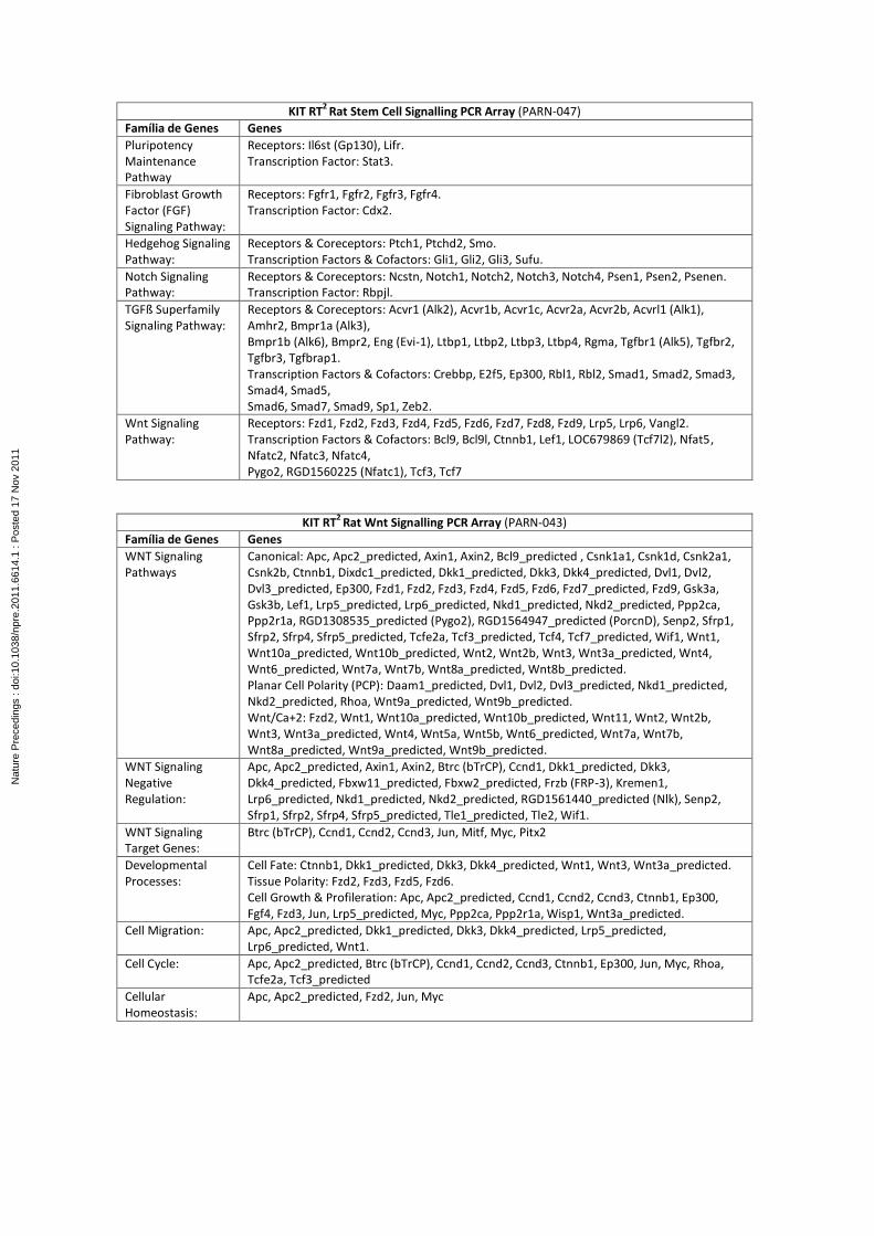
KIT RT2
Rat Stem Cell Signalling PCR Array (PARN-047)
Família de Genes Genes
Pluripotency Maintenance Pathway
Receptors: Il6st (Gp130), Lifr. Transcription Factor: Stat3.
Fibroblast Growth Factor (FGF) Signaling Pathway:
Receptors: Fgfr1, Fgfr2, Fgfr3, Fgfr4. Transcription Factor: Cdx2.
Hedgehog Signaling Pathway:
Receptors & Coreceptors: Ptch1, Ptchd2, Smo. Transcription Factors & Cofactors: Gli1, Gli2, Gli3, Sufu.
Notch Signaling Pathway:
Receptors & Coreceptors: Ncstn, Notch1, Notch2, Notch3, Notch4, Psen1, Psen2, Psenen. Transcription Factor: Rbpjl.
TGFß Superfamily Signaling Pathway:
Receptors & Coreceptors: Acvr1 (Alk2), Acvr1b, Acvr1c, Acvr2a, Acvr2b, Acvrl1 (Alk1), Amhr2, Bmpr1a (Alk3), Bmpr1b (Alk6), Bmpr2, Eng (Evi-1), Ltbp1, Ltbp2, Ltbp3, Ltbp4, Rgma, Tgfbr1 (Alk5), Tgfbr2, Tgfbr3, Tgfbrap1. Transcription Factors & Cofactors: Crebbp, E2f5, Ep300, Rbl1, Rbl2, Smad1, Smad2, Smad3, Smad4, Smad5, Smad6, Smad7, Smad9, Sp1, Zeb2.
Wnt Signaling Pathway:
Receptors: Fzd1, Fzd2, Fzd3, Fzd4, Fzd5, Fzd6, Fzd7, Fzd8, Fzd9, Lrp5, Lrp6, Vangl2. Transcription Factors & Cofactors: Bcl9, Bcl9l, Ctnnb1, Lef1, LOC679869 (Tcf7l2), Nfat5, Nfatc2, Nfatc3, Nfatc4, Pygo2, RGD1560225 (Nfatc1), Tcf3, Tcf7
KIT RT2
Rat Wnt Signalling PCR Array (PARN-043)
Família de Genes Genes
WNT Signaling Pathways
Canonical: Apc, Apc2_predicted, Axin1, Axin2, Bcl9_predicted , Csnk1a1, Csnk1d, Csnk2a1, Csnk2b, Ctnnb1, Dixdc1_predicted, Dkk1_predicted, Dkk3, Dkk4_predicted, Dvl1, Dvl2, Dvl3_predicted, Ep300, Fzd1, Fzd2, Fzd3, Fzd4, Fzd5, Fzd6, Fzd7_predicted, Fzd9, Gsk3a, Gsk3b, Lef1, Lrp5_predicted, Lrp6_predicted, Nkd1_predicted, Nkd2_predicted, Ppp2ca, Ppp2r1a, RGD1308535_predicted (Pygo2), RGD1564947_predicted (PorcnD), Senp2, Sfrp1, Sfrp2, Sfrp4, Sfrp5_predicted, Tcfe2a, Tcf3_predicted, Tcf4, Tcf7_predicted, Wif1, Wnt1, Wnt10a_predicted, Wnt10b_predicted, Wnt2, Wnt2b, Wnt3, Wnt3a_predicted, Wnt4, Wnt6_predicted, Wnt7a, Wnt7b, Wnt8a_predicted, Wnt8b_predicted. Planar Cell Polarity (PCP): Daam1_predicted, Dvl1, Dvl2, Dvl3_predicted, Nkd1_predicted, Nkd2_predicted, Rhoa, Wnt9a_predicted, Wnt9b_predicted. Wnt/Ca+2: Fzd2, Wnt1, Wnt10a_predicted, Wnt10b_predicted, Wnt11, Wnt2, Wnt2b, Wnt3, Wnt3a_predicted, Wnt4, Wnt5a, Wnt5b, Wnt6_predicted, Wnt7a, Wnt7b, Wnt8a_predicted, Wnt9a_predicted, Wnt9b_predicted.
WNT Signaling Negative Regulation:
Apc, Apc2_predicted, Axin1, Axin2, Btrc (bTrCP), Ccnd1, Dkk1_predicted, Dkk3, Dkk4_predicted, Fbxw11_predicted, Fbxw2_predicted, Frzb (FRP-3), Kremen1, Lrp6_predicted, Nkd1_predicted, Nkd2_predicted, RGD1561440_predicted (Nlk), Senp2, Sfrp1, Sfrp2, Sfrp4, Sfrp5_predicted, Tle1_predicted, Tle2, Wif1.
WNT Signaling Target Genes:
Btrc (bTrCP), Ccnd1, Ccnd2, Ccnd3, Jun, Mitf, Myc, Pitx2
Developmental Processes:
Cell Fate: Ctnnb1, Dkk1_predicted, Dkk3, Dkk4_predicted, Wnt1, Wnt3, Wnt3a_predicted. Tissue Polarity: Fzd2, Fzd3, Fzd5, Fzd6. Cell Growth & Profileration: Apc, Apc2_predicted, Ccnd1, Ccnd2, Ccnd3, Ctnnb1, Ep300, Fgf4, Fzd3, Jun, Lrp5_predicted, Myc, Ppp2ca, Ppp2r1a, Wisp1, Wnt3a_predicted.
Cell Migration: Apc, Apc2_predicted, Dkk1_predicted, Dkk3, Dkk4_predicted, Lrp5_predicted, Lrp6_predicted, Wnt1.
Cell Cycle: Apc, Apc2_predicted, Btrc (bTrCP), Ccnd1, Ccnd2, Ccnd3, Ctnnb1, Ep300, Jun, Myc, Rhoa, Tcfe2a, Tcf3_predicted
Cellular Homeostasis:
Apc, Apc2_predicted, Fzd2, Jun, Myc
Nat
ure
Pre
cedi
ngs
: doi
:10.
1038
/npr
e.20
11.6
614.
1 : P
oste
d 17
Nov
201
1

KIT RT2
Rat Customized Enteroendocrine Differentiation PCR Array
Genes
ATOH1 (ou Math1)
Ngn3, BETA2/NeuroD, Pax4, Pax6, Nkx2.2, Nkx6.1, Isl-1, Insm1 (ou IA-1), Pdx1, CCK
Foxa1, Foxa2
Gfi-1
GATA4
GATA5
GATA6
Proglucagon, Glucagon, GLP-1, GLP1-r, GLP-2, PYY, TGR5, GPR-119, GPR-120, PC-1, GIP
TCF-4, Glicentin, Oxyntomodulin
Other genes related to enteroendocrine differentiation
3.14.4. Gene expression
The relative expression of genes of interest will be analysed in real-time by using the
PCR equipment software Abi Prism 7000 Sequence Detection System version 1.6 to compare
gene expression levels among tissue fragments from the different groups of animals.
During real-time PCR, fluorescence increases with every PCR cycle until reaching a
threshold where all samples can be compared. The threshold corresponds to the time-point
used for fluorescence analysis. This time-point is defined by the researcher and must be within
the range in which fluorescence generated by sample amplification becomes significantly
higher than background fluorescence.
The threshold is defined during the PCR exponential phase, when the amount of
product formed can be used to calculate the initial concentration of template strands (cDNA)
amplified by the reaction. The exact cycle when fluorescence crosses the threshold is called
the threshold cycle (Ct). More concentrated samples (with greater numbers of initial template
strands) reach the threshold faster and have lower Ct values. When PCR efficiency is close to
100%, the number of copies generated increases exponentially and doubles with each reaction
cycle.
The number of initial template strands can be calculated from the Ct. The equation
Ns=No(1+E)n describes exponential PCR amplification, where “Ns” in the number of template
strands at a given reaction cycle, “No” is the number of initial template strands, “E” is
efficiency of the amplification reaction, and “n” is the number of cycles. Assuming that
reaction efficiency is 100% (log E=1) and “n” is a selected amplification point identical for all
samples, we can calculate the number of initial template strands using the following equation:
N0=Nsx2-Ct.
Nat
ure
Pre
cedi
ngs
: doi
:10.
1038
/npr
e.20
11.6
614.
1 : P
oste
d 17
Nov
201
1

The method of analysing gene expression by real-time PCR proposed here represents a
relative quantification of genes of interest because it requires an endogenous control (a gene
whose expression does not exhibit statistically significant variation among the analysed
samples). Relative quantification of a given sample by real-time PCR results from the
relationship between the Ct of the gene of interest (i) and the Ct of the endogenous control
(e):
Nsi=Noi x 2-Cti (gene of interest) Nse=Noe x 2-Cte (endogenous gene)
Because Nsi/Nse is equal to the normalised number of molecules of the gene of
interest relative to the number of endogenous control molecules that we will call X, and Nse
and Nsi is a constant that we will name K, the equation becomes:
X=K.2-Ct
Relative quantification of gene expression is used to describe changes in the
expression of a gene of interest in a group of samples relative to a control group. In this study,
we will compare ileum and jejunum samples in their normal localisations with interposed
ileum samples and with ileum samples from animals in the sham-treated group. Thus, the
value of X will be measured under two different conditions, and by dividing the value of X
under condition A by the value of X under condition B, we obtain 2-Ct , as shown by the
following equation:
This method will be used in our study to calculate the relative expression levels of
genes of interest. The relative expression levels of genes of interest will be compared among
the different segments from different groups of animals (IG ileum, SG ileum, CG jejunum, and
ileum).
3.14.5. Statistical analysis
The variables will be summarised for each study group using the pertinent descriptive
statistics: absolute (n) and relative (%) frequency, or mean, standard deviation (s.d.), median,
maximum, and minimum values. The data will be analysed using parametric or non-parametric
tests according to the observed distribution.
Nat
ure
Pre
cedi
ngs
: doi
:10.
1038
/npr
e.20
11.6
614.
1 : P
oste
d 17
Nov
201
1

For normally distributed data, analysis of variance will be applied with two fixed
factors: groups (4 levels: IG, SG, CG1, and CG2) and assessment time-point (3 levels: Week 12
(W12), Week 20 (W20), and Week 28 (W28)). For the variables assessed weekly, the test will
have 16 levels corresponding to each week. For non-normally distributed data, the Mann-
Whitney test will be applied to compare techniques within each assessment, and the Friedman
test will be applied to related samples to compare variations within each technique. The
presence of an association between qualitative variables will be assessed by the chi-squared
test or Fisher’s exact test. The level of significance will be set to 0.05 (α = 5%), and p values
lower than this value will be considered significant.
4. REFERENCES
1. Global Burden of Metabolic Risk Factors of Chronic Diseases Collaborating Group (Blood Glucose). National, regional, and global trends in fasting plasma glucose and diabetes prevalence since 1980: systematic analysis of health examination surveys and epidemiological studies with 370 country-years and 2·7 million participants. Lancet. 2011;378(9785):31-40. 2. Hansen KB, Vilsboll T, Knop FK. Incretin mimetics: a novel therapeutic option for patients with type 2 diabetes – a review. Diabetes Metab Syndr Obes. 2010;3:155-63. 3. Tahrani AA, Piya MK, Kennedy A, Barnett AH. Glycaemic control in type 2 diabetes: targets and new therapies. Pharmacol Therapeut. 2010;125(2):328-61. 4. Roglic G, Unwin N, Bennett PH, Mathers C, Tuomilehto J, Nag S, Connolly V, King H. The burden of mortality attributable to diabetes: realistic estimates for the year 2000. Diabetes Care. 2005;28(9):2130-5. 5. Centers for Disease Control and Prevention. National Diabetes Fact Sheet: national estimates and general information on diabetes and prediabetes in the United States, 2011 Atlanta, GA: U.S. Department of Health and Human Services, Centers for Disease Control and Prevention, 2011. Available at: http://www.cdc.gov/diabetes/pubs/pdf/ndfs_2011.pdf 6. Tibaldi J, Rakel R. Why, when and how to initiate insulin therapy in patients with type 2 diabetes. Int J Clin Pract. 2007;61(4):633-44. 7. Meneghini L. Demonstrating strategies for initiation of insulin therapy: matching the right insulin to the right patient. Int J Clin Pract. 2008;62(8):1255-64. 8. Campbell RK. Type 2 diabetes: where we are today: an overview of disease burden, current treatments, and treatment strategies. J Am Pharm Assoc. 2009;49:3-9. 9. Horton ES. Can newer therapies delay the progression of type 2 diabetes mellitus? Endocr Pract. 2008;14(5):625-38. 10. Calcutt NA, Cooper ME, Kern TS, Schmidt AM. Therapies for hyperglycaemia-induced diabetic complications: from animal models to clinical trials. Nat Rev Drug Discov. 2009;8(5):417-29. 11. Turner R, Holman R. Lessons from UK prospective diabetes study. Diabetes Res Clin Pract. 1995;28:S151-7. 12. Hemmingsen B, Lund S, Gluud C, Vaag A, Almdal T, Hemmingsen C, Wetterslev J. Targeting intensive glycaemic control versus targeting conventional glycaemic control for type 2 diabetes mellitus. Cochrane database of systematic reviews (Online). 2011;6:CD008143. Available at: http://www2.cochrane.org/reviews/en/ab008143.html 13. Hossain P, Kawar B, El Nahas M. Obesity and diabetes in the developing world - a growing challenge. New Engl J Med. 2007;356(3):213-5.
Nat
ure
Pre
cedi
ngs
: doi
:10.
1038
/npr
e.20
11.6
614.
1 : P
oste
d 17
Nov
201
1
14. Mokdad AH, Ford ES, Bowman BA, Dietz WH, Vinicor F, Bales VS, Marks JS. Prevalence of obesity, diabetes, and obesity-related health risk factors, 2001. JAMA-J Am Med Assoc. 2003;289(1):76-9. 15. Freeman JS. New therapeutic options: management strategies to optimize glycemic control. J Am Osteopath Assoc. 2010;110(3Suppl2):S15-20. 16. Blonde L. Current antihyperglycemic treatment guidelines and algorithms for patients with type 2 diabetes mellitus. Am J Med. 2010;123(3):S12-8. 17. Stolar MW, Hoogwerf BJ, Boyle PJ, Gorshow SM, Wales D. Managing type 2 diabetes: going beyond glycemic control. J Manag Care Pharm. 2008;14(5SupplB): S2-19. Available at: http://www.amcp.org/data/jmcp/JMCPSuppBJune08Web.pdf 18. Krentz AJ, Patel MB, Bailey CJ. New drugs for type 2 diabetes mellitus: what is their place in therapy? Drugs. 2008;68(15):2131-62. 19. VanDeKoppel S, Choe H, Sweet B. Managed care perspective on three new agents for type 2 diabetes. J Manag Care Pharm. 2008;14(4):363-80. 20. Vinik A. Advancing therapy in type 2 diabetes mellitus with early, comprehensive progression from oral agents to insulin therapy. Clin Ther. 2007;29(6):1236-53. 21. Del Prato S. Unlocking the opportunity of tight glycaemic control. Diabetes Obes Metab. 2005;7:S1-4. 22. Fleury-Milfort E. Practical strategies to improve treatment of type 2 diabetes. J Am Acad Nurse Pract. 2008;20(6):295-304. 23. Liebl A. Challenges in optimal metabolic control of diabetes. Diabetes Metab Res Rev. 2002;18(S3):S36-41. 24. Kunt T, Snoek F. Barriers to insulin initiation and intensification and how to overcome them. Int J Clin Pract. 2009;63:6-10. 25. Heinemann L. Overcoming obstacles: new management options. Eur J Endocrinol. 2004;151(2):T23-7. 26. Brunton S. Beyond glycemic control: treating the entire type 2 diabetes disorder. Postgrad Med. 2009;121(5):68-81. 27. Peters AL. Patient and treatment perspectives: Revisiting the link between type 2 diabetes, weight gain, and cardiovascular risk. Clev Clin J Med. 2009;76(5):S20-7. 28. Hansen KB, Vilsboll T, Knop FK. Incretin mimetics: a novel therapeutic option for patients with type 2 diabetes – a review. Diabetes Metab Syndr Obes. 2010;3:155-63. 29. Czupryniak L, Wiszniewski M, Szyma ski D, Paw owski M, Loba J, Strzelczyk J. Long-term results of gastric bypass surgery in morbidly obese type 1 diabetes patients. Obes Surg. 2010;20(4):506-8. 30. Rubino F, Gagner M. Potential of surgery for curing type 2 diabetes mellitus. Ann Surg. 2002;236(5):554-9. 31. Buchwald H, Avidor Y, Braunwald E. Bariatric Surgery: A Systematic Review. JAMA-J Am Med Assoc. 2004;292(14):1724-37. 32. Santry HP, Gillen DL, Lauderdale DS. Trends in bariatric surgical procedures. Jama. 2005;294(15):1909-17. 33. van de Sande-Lee S, Pereira FRS, Cintra DE, Fernandes PT, Cardoso AR, Garlipp CR, Chaim EA, Pareja JC, Geloneze B, Li LM, Cendes F, Velloso LA. Partial reversibility of hypothalamic dysfunction and changes in brain activity after body mass reduction in obese subjects. Diabetes. 2011;60(6):1699-704. 34. Meijer RI, van Wagensveld BA, Siegert CE, Eringa EC, Serne EH, Smulders YM. Bariatric surgery as a novel treatment for type 2 diabetes mellitus: a systematic review. Arch Surg. 2011;146(6):744-50. 35. Pories WJ, Swanson MS, MacDonald KG, Long SB, Morris PG, Brown BM, Barakat HA, deRamon RA, Israel G, Dolezal JM, Dohm L. Who would have thought it? An operation proves to be the most effective therapy for adult-onset diabetes mellitus. Ann Surg. 1995;222(3):339-50.
Nat
ure
Pre
cedi
ngs
: doi
:10.
1038
/npr
e.20
11.6
614.
1 : P
oste
d 17
Nov
201
1

36. Rubino F, Gagner M, Gentileschi P, Kini S, Fukuyama S, Feng J, Diamond E.. The early effect of the Roux-en-Y gastric bypass on hormones involved in body weight regulation and glucose metabolism. Ann Surg. 2004;240(2):236-42 37. Reinehr T, Roth CL, Schernthaner GH, Kopp HP, Kriwanek S, Schernthaner G. Peptide YY and glucagon-like peptide-1 in morbidly obese patients before and after surgically induced weight loss. Obes Surg. 2007;17(12):1571-7. 38. Strader AD, Vahl TP, Jandacek RJ, Woods SC, D’Alessio DA, Seeley RJ. Weight loss through ileal transposition is accompanied by increased ileal hormone secretion and synthesis in rats. AmJ Physiol-Endoc M. 2005;288(2):E447-53. 39. Näslund E, Hellströrn PM, Kral JG. The gut and food intake: an update for surgeons. J Gastrointest Surg. 2001;5(5):556-67. 40. Van Citters GW, Lin HC. The ileal brake: a fifteen-year progress report. Curr Gastroenterol Rep. 1999;1(5):404-9. 41. Hickey MS, Pories WJ, MacDonald Jr KG, Cory KA, Dohm GL, Swanson MS, Israel RG, Barakat HA, Considine RV, Caro JF, Houmard JA. A new paradigm for type 2 diabetes mellitus: could it be a disease of the foregut? Ann Surg. 1998;227(5):637-43. 42. Santoro S. Is the metabolic syndrome a disease of the foregut? Yes, excessive foregut. Ann Surg. 2008;247(6):1074-5. 43. Dhanvantari S, Seidah NG, Brubaker PL. Role of prohormone convertases in the tissue-specific processing of proglucagon. Mol Endocrinol. 1996;10(4):342-55. 44. Baggio LL, Drucker DJ. Biology of incretins: GLP-1 and GIP. Gastroenterology. 2007;132(6):2131-57. 45. Holst JJ. The physiology of glucagon-like peptide 1. Physiol Rev. 2007;87(4):1409-39. 46. Drucker DJ. Glucagon-like peptides: regulators of cell proliferation, differentiation, and apoptosis. Mol Endocrinol. 2003;17(2):161-71. 47. Pournaras DJ, Le Roux CW. Obesity, gut hormones, and bariatric surgery. World J Surg. 2009;33(10):1983-8. 48. Rubino F, Marescaux J. Effect of duodenal–jejunal exclusion in a non-obese animal model of type 2 diabetes: a new perspective for an old disease. Ann Surg. 2004;239(1):1-11. 49. Melissas J. IFSO guidelines for safety, quality, and excellence in bariatric surgery. Obes Surg. 2008;18(5):497-500. 50. Laferrere B, Teixeira J, McGinty J, Tran H, Egger JR, Colarusso A, Kovack B, Bawa B, Koshy N, Lee H, Yapp K, Olivan B. Effect of weight loss by gastric bypass surgery versus hypocaloric diet on glucose and incretin levels in patients with type 2 diabetes. J Clin Endocr Metab. 2008;93(7):2479-85. 51. Koopmans H, Sclafani A, Fichtner C, Aravich P. The effects of ileal transposition on food intake and body weight loss in VMH-obese rats. The Am J Clin Nutr. 1982;35(2):284-93. 52. Strader AD. Ileal transposition provides insight into the effectiveness of gastric bypass surgery. Physiol Behav. 2006;88(3):277-82. 53. Ferri GL, Koopmans H, Ghatei M, Vezzadini P, Labo G, Bloom SR, Polak JM. Ileal enteroglucagon cells after ileal-duodenal transposition in the rat. Digestion. 1983;26(1):10-6. 54. Sarson D, Scopinaro N, Bloom S. Gut hormone changes after jejunoileal (JIB) or biliopancreatic (BPB) bypass surgery for morbid obesity. Int J Obesity. 1981;5(5):471-80. 55. Näslund E, Backman L, Juul Holst J, Theodorsson E, Hellström PM. Importance of small bowel peptides for the improved glucose metabolism 20 years after jejunoileal bypass for obesity. Obes Surg. 1998;8(3):253-60. 56. Mason EE. Ileal transposition and enteroglucagon/GLP-1 in obesity (and diabetic?) surgery. Obes Surg. 1999;9(3):223-8. 57. De Luis D, Pacheco D, Izaola O, Romero A, Marcos J, Pelaz J, Barrera A, Cabezas G, Terroba MC, Cuellar L, Anta A. Early clinical and surgical results of biliopancreatic diversion. Obes Surg. 2005;15(6):799-802.
Nat
ure
Pre
cedi
ngs
: doi
:10.
1038
/npr
e.20
11.6
614.
1 : P
oste
d 17
Nov
201
1
58. Wickremesekera K, Miller G, Naotunne TDS, Knowles G, Stubbs RS. Loss of insulin resistance after Roux-en-Y gastric bypass surgery: a time course study. Obes Surg. 2005;15(4):474-81. 59. Schauer PR, Burguera B, Ikramuddin S, Cottam D, Gourash W, Hamad G, Eid GM, Mattar S, Ramanathan R, Barinas-Mitchel E, Rao RH, Kuller L, Kelley D. Effect of laparoscopic Roux-en Y gastric bypass on type 2 diabetes mellitus. Ann Surg. 2003;238(4):467-84. 60. Weber M, Müller MK, Bucher T, Wildi S, Dindo D, Horber F, Hauser R, Clavien PA. Laparoscopic gastric bypass is superior to laparoscopic gastric banding for treatment of morbid obesity. Ann Surg. 2004;240(6):975-82. 61. Scopinaro N, Marinari GM, Camerini GB, Papadia FS, Adami GF. Specific effects of biliopancreatic diversion on the major components of metabolic syndrome. Diabetes Care. 2005;28(10):2406-11. 62. Cohen R, Uzzan B, Bihan H, Khochtali I, Reach G, Catheline JM. Ghrelin levels and sleeve gastrectomy in super-super-obesity. Obes Surg. 2005;15(10):1501-2. 63. Bohdjalian A, Langer FB, Shakeri-Leidenmühler S, Gfrerer L, Ludvik B, Zacherl J, Prager G. Sleeve gastrectomy as sole and definitive bariatric procedure: 5-year results for weight loss and ghrelin. Obes Surg. 2010;20(5):535-40. 64. Vidal J, Ibarzabal A, Romero F, Delgado S, Momblán D, Flores L, Lacy A. Type 2 diabetes mellitus and the metabolic syndrome following sleeve gastrectomy in severely obese subjects. Obes Surg. 2008;18(9):1077-82. 65. Melissas J, Daskalakis M, Koukouraki S, Askoxylakis I, Metaxari M, Dimitriadis E, Stathaki M, Papadakis JA. Sleeve gastrectomy - a “food limiting” operation. Obes Surg. 2008;18(10):1251-6. 66. Braghetto I, Davanzo C, Korn O, Csendes A, Valladares H, Herrera E, Gonzales P, Papapietro K. Scintigraphic evaluation of gastric emptying in obese patients submitted to sleeve gastrectomy compared to normal subjects. Obes Surg. 2009;19(11):1515-21 67. De Paula AL, Stival AR, Macedo A, Ribamar J, Mancini M, Halpern A, Vencio S. Prospective randomized controlled trial comparing 2 versions of laparoscopic ileal interposition associated with sleeve gastrectomy for patients with type 2 diabetes with BMI 21–34 kg/m2. Surg Obes Relat Dis. 2010;6(3):296-304. 68. De Paula AL, Macedo ALV, Prudente AS, Queiroz L, Schraibman V, Pinus J. Laparoscopic sleeve gastrectomy with ileal interposition (“neuroendocrine brake”)—pilot study of a new operation. Surg Obes Relat Dis. 2006;2(4):464-7. 69. De Paula AL, Macedo ALV, Schraibman V, Mota B, Vencio S. Hormonal evaluation following laparoscopic treatment of type 2 diabetes mellitus patients with BMI 20–34. Surg Endosc. 2009;23(8):1724-32. 70. De Paula A, Macedo ALV, Mota B, Schraibman V. Laparoscopic ileal interposition associated to a diverted sleeve gastrectomy is an effective operation for the treatment of type 2 diabetes mellitus patients with BMI 21–29. Surg Endosc. 2009;23(6):1313-20. 71. De Paula A, Macedo ALV, Rassi N, Vencio S, Machado C, Mota BR, Silva LQ, Halpern A, Schraibman V. Laparoscopic treatment of metabolic syndrome in patients with type 2 diabetes mellitus. Surg Endosc. 2008;22(12):2670-8 72. Tinoco A, El-Kadre L, Aquiar L, Tinoco R, Savassi-Rocha P. Short-term and mid-term control of type 2 diabetes mellitus by laparoscopic sleeve gastrectomy with ileal interposition. World J Surg. 2011;35(10):2238-44. 73. Koopmans H, Sclafani A. Control of body weight by lower gut signals. Int J Obesity. 1981;5(5):491-5. 74. Smithy W, Cuadros C, Johnson H, Kral J. Effects of ileal interposition on body weight and intestinal morphology in dogs. Int J Obesity. 1986;10(6):453-60. 75. Kotler DP, Koopmans H. Preservation of intestinal structure and function despite weight loss produced by ileal transposition in rats. Physiol Behav. 1984;32(3):423-7.
Nat
ure
Pre
cedi
ngs
: doi
:10.
1038
/npr
e.20
11.6
614.
1 : P
oste
d 17
Nov
201
1

76. Chu KU, Tsuchiya T, Ishizuka J, Uchida T, Townsend Jr CM, Thompson JC. Trophic response of gut and pancreas after ileojejunal transposition. Ann Surg. 1995;221(3):249-56. 77. Tsuchiya T, Ishizuka J, Sato K, Shimoda I, Rajaraman S, Uchida T, Townsend CM Jr, Thompson JC. Effect of ileojejunal transposition on the growth of the GI tract and pancreas in young and aged rats. J Gerontol A Biol Sci Med Sci. 1995;50(3):M155-61. 78. Menge H, Robinson J. Functional and structural characteristics of the rat intestinal mucosa following ileo-jejunal transposition. Acta Hepatogastroenterol. 1978;25(2):150-4. 79. Patriti A, Facchiano E, Annetti C, Aisa MC, Galli F, Fanelli C, Donini A. Early improvement of glucose tolerance after ileal transposition in a non-obese type 2 diabetes rat model. Obes Surg. 2005;15(9):1258-64. 80. Strader AD, Vahl TP, Jandacek RJ, Woods SC, D’Alessio DA, Seeley RJ. Weight loss through ileal transposition is accompanied by increased ileal hormone secretion and synthesis in rats. Am J Physiol Endocrinol Metab. 2005;288(2):E447-53. 81. Patriti A, Aisa MC, Annetti C, Sidoni A, Galli F, Ferri I, Gullà N, Donini A. How the hindgut can cure type 2 diabetes. Ileal transposition improves glucose metabolism and beta-cell function in Goto-kakizaki rats through an enhanced Proglucagon gene expression and L-cell number. Surgery. 2007;142(1):74-85. 82. Pannacciulli N, Le DSNT, Salbe AD, Chen K, Reiman EM, Tataranni PA, Krakoff J. Postprandial glucagon-like peptide-1 (GLP-1) response is positively associated with changes in neuronal activity of brain areas implicated in satiety and food intake regulation in humans. Neuroimage. 2007;35(2):511-7. 83. Meier J, Gethmann A, Götze O, Gallwitz B, Holst J, Schmidt WE, Nauck MA. Glucagon-like peptide 1 abolishes the postprandial rise in triglyceride concentrations and lowers levels of non-esterified fatty acids in humans. Diabetologia. 2006;49(3):452-8. 84. Nagell CF, Wettergren A, Ørskov C, Holst JJ. Inhibitory effect of GLP-1 on gastric motility persists after vagal deafferentation in pigs. Scand J Gastroenterol. 2006;41(6):667-72. 85. Tolessa T, Gutniak M, Holst JJ, Efendic S, Hellström PM. Inhibitory effect of glucagon-like peptide-1 on small bowel motility. Fasting but not fed motility inhibited via nitric oxide independently of insulin and somatostatin. J Clin Invest. 1998;102(4):764-74. 86. Farilla L, Hui H, Bertolotto C, Kang E, Bulotta A, Di Mario U, Perfetti R. Glucagon-like peptide-1 promotes islet cell growth and inhibits apoptosis in Zucker diabetic rats. Endocrinology. 2002;143(11):4397-408. 87. Farilla L, Bulotta A, Hirshberg B, Li Calzi S, Khoury N, Noushmehr H, Bertolotto C, Di Mario U, Harlan DM, Perfetti R. Glucagon-like peptide 1 inhibits cell apoptosis and improves glucose responsiveness of freshly isolated human islets. Endocrinology. 2003;144(12):5149-58. 88. Valverde I, Puente J, Martín-Duce A, Molina L, Lozano O, Sancho V, Malaisse WJ, Villañueva-Peñacarrillo ML. Changes in glucagon-like peptide-1 (GLP-1) secretion after biliopancreatic diversion or vertical banded gastroplasty in obese subjects. Obes Surg. 2005;15(3):387-97. 89. De Paula AL, Macedo ALV, Prudente AS, Queiroz L, Schraibman V, Pinus J. Laparoscopic sleeve gastrectomy with ileal interposition (“neuroendocrine brake”) - pilot study of a new operation. Surg Obes Relat Dis. 2006;2(4):464-7. 90. Cummings DE, Shannon MH. Ghrelin and gastric bypass: is there a hormonal contribution to surgical weight loss? J Clin Endocrinol Metab.2003;88(7):2999-3002. 91. Strader AD, Woods SC. Gastrointestinal hormones and food intake. Gastroenterology. 2005;128(1):175-91. 92. Strader AD, Clausen TR, Goodin SZ, Wendt D. Ileal interposition improves glucose tolerance in low dose streptozotocin-treated diabetic and euglycemic rats. Obes Surg. 2009;19(1):96-104. 93. Baksheev L, Fuller PJ. Gene expression in the adapting small bowel after massive small bowel resection. J Gastroenterol. 2006;41(11):1041-52.
Nat
ure
Pre
cedi
ngs
: doi
:10.
1038
/npr
e.20
11.6
614.
1 : P
oste
d 17
Nov
201
1
94. Thomson A, Wild G. Adaptation of Intestinal Nutrient Transport in Health and Disease. Part I. Digest Dis Sci. 1997;42(3):453-69. 95. Thomson A, Wild G. Adaptation of Intestinal Nutrient Transport in Health and Disease. Part II. Digest Dis Sci. 1997;42(3):470-88. 96. Ferraris RP, Carey HV. Intestinal transport during fasting and malnutrition. Annu Rev Nutr. 2000;20(1):195-219. 97. Drozdowski LA, Clandinin MT, Thomson AB. Morphological, kinetic, membrane biochemical and genetic aspects of intestinal enteroplasticity. World J Gastroenterol. 2009;15(7):774-87. 98. Sturm A, Layer P, Goebell H, Dignass AU. Short-bowel syndrome: an update on the therapeutic approach. Scand J Gastroenterol. 1997;32(4):289-96. 99. Wolvekamp M, Heineman E, Taylor R, Fuller P. Towards understanding the process of intestinal adaptation. Digest Dis. 1996;14(1):59-72. 100. O’Connor TP, Lam MM, Diamond J. Magnitude of functional adaptation after intestinal resection. Am J Physiol. 1999;276(5):R1265-75. 101. Tavakkolizadeh A, Whang EE. Understanding and augmenting human intestinal adaptation: a call for more clinical research. JPEN J Parenter Enteral Nutr. 2002;26(4):251-5. 102. Dowling R. Small bowel adaptation and its regulation. Scand J Gastroenterol. 1982;74:53-74. 103. Helmrath MA, VanderKolk WE, Can G, Erwin CR, Warner BW. Intestinal adaptation following massive small bowel resection in the mouse. J Am Coll Surg. 1996;183(5):441-9. 104. Taylor RG, Beveridge DJ, Fuller PJ. Expression of ileal glucagon and peptide tyrosine-tyrosine genes. Response to inhibition of polyamine synthesis in the presence of massive small-bowel resection. Biochem J. 1992;286(Pt 3):737-41. 105. Jump DB, Clarke SD. Regulation of gene expression by dietary fat. Annu Rev Nutr. 1999;19(1):63-90. 106. Keelan M, Cheeseman CI, Clandinin MT, Thomson AB. Intestinal morphology and transport after ileal resection in rat is modified by dietary fatty acids. Clin Invest Med. 1996;19(2):63-70. 107. Diamond JM, Karasov WH, Cary C, Enders D, Yung R. Effect of dietary carbohydrate on monosaccharide uptake by mouse small intestine in vitro. J Physiol. 1984;349(1):419-40. 108. Ziegler TR, Mantell MP, Chow JC, Rombeau JL, Smith RJ. Gut adaptation and the insulin-like growth factor system: regulation by glutamine and IGF-I administration. Am J Physiol. 1996;271(5):G866-75. 109. Knop FK. Bile-induced secretion of glucagon-like peptide-1: pathophysiological implications in type 2 diabetes? Am J Physiol Endocrinol Metab. 2010;299(1):E10-3. 110. Chu ZL, Jones RM, He H, Carroll C, Gutierrez V, Lucman A, Moloney M, Gao H, Mondala H, Bagnol D, Unett D, Liang Y, Demarest K, Semple G, Behan DP, Leonard J. A role for beta-cell-expressed G protein-coupled receptor 119 in glycemic control by enhancing glucose-dependent insulin release. Endocrinology. 2007;148(6):2601-9. 111. Hirasawa A, Tsumaya K, Awaji T, Katsuma S, Adachi T, Yamada M, Sugimoto Y, Miyazaki S, Tsujimoto G. Free fatty acids regulate gut incretin glucagon-like peptide-1 secretion through GPR120. Nat Med. 2005;11(1):90-4. 112. Katsuma S, Hirasawa A, Tsujimoto G. Bile acids promote glucagon-like peptide-1 secretion through TGR5 in a murine enteroendocrine cell line STC-1. Biochem Biophys Res Commun. 2005;329(1):386-90. 113. Whalley N, Pritchard L, Smith DM, White A. Processing of proglucagon to GLP-1 in pancreatic alpha cells: is this a paracrine mechanism enabling GLP-1 to act on beta cells? J Endocrinol. 2011;211(1):99-106. 114. Yi F, Brubaker PL, Jin T. TCF-4 mediates cell type-specific regulation of proglucagon gene expression by -catenin and glycogen synthase kinase-3. J Biol Chem. 2005;280(2):1457-64.
Nat
ure
Pre
cedi
ngs
: doi
:10.
1038
/npr
e.20
11.6
614.
1 : P
oste
d 17
Nov
201
1
115. Qandeel HG, Alonso F, Hernandez DJ, Madhavan S, Duenes JA, Zheng Y, Sarr MG. Peptide absorption after massive proximal small bowel resection: Mechanisms of ileal adaptation. J Gastrointest Surg. 2011;15(9):1537-47. 116. Brittan M, Wright N. The gastrointestinal stem cell. Cell Proliferat.2004;37(1):35-53. 117. Sancho E, Batlle E, Clevers H. Live and let die in the intestinal epithelium. Curr Opin Cell Biol. 2003;15(6):763-70. 118. Crosnier C, Stamataki D, Lewis J. Organizing cell renewal in the intestine: stem cells, signals and combinatorial control. Nat Rev Genet. 2006;7(5):349-59. 119. May CL, Kaestner KH. Gut endocrine cell development. Mol Cell Endocrinol. 2010;323(1):70-5. 120. Rindi G, Leiter AB, Kopin AS, Bordi C, Solcia E. The “normal” endocrine cell of the gut: changing concepts and new evidences. Ann NY Acad Sci. 2004;1014(1):1-12. 121. Gutierrez-Aguilar R, Woods SC. Nutrition and L and K-enteroendocrine cells. Curr Opin Endocrinol Diab Obes. 2011;18(1):35-41. 122. Lee CS, Kaestner KH. Development of gut endocrine cells. Best Pract Res Clin Endocrinol Metab. 2004;18(4):453-62. 123. Moran GW, Leslie FC, Levison SE, McLaughlin JT. Review: Enteroendocrine cells: Neglected players in gastrointestinal disorders? Therap Adv Gastroenterol. 2008;1(1):51-60. 124. Barker N, Van De Wetering M, Clevers H. The intestinal stem cell. Genes Dev. 2008;22(14):1856-64. 125. Barker N, Clevers H. Tracking down the stem cells of the intestine: strategies to identify adult stem cells. Gastroenterology. 2007;133(6):1755-60. 126. Barker N, Van Es JH, Kuipers J, Kujala P, Van Den Born M, Cozijnsen M, Haegebarth A, Korving J, Begthel H, Peters PJ, Clevers H. Identification of stem cells in small intestine and colon by marker gene Lgr5. Nature. 2007;449(7165):1003-7. 127. Hoffman J, Kuhnert F, Davis CR, Kuo CJ. Wnts as essential growth factors for the adult small intestine and colon. Cell cycle. 2004;3(5):554-7. 128. Sei Y, Lu X, Liou A, Zhao X, Wank SA. A stem cell marker-expressing subset of enteroendocrine cells resides at the crypt base in the small intestine. Am J Physiol Gastrointest Liver Physiol. 2011;300(2):G345-56. 129. Gerbe F, van Es JH, Makrini L, Brulin B, Mellitzer G, Robine S, Romagnolo B, Shroyer NF, Bourgaux JF, Pignodel C, Clevers H, Jay P. Distinct ATOH1 and Neurog3 requirements define tuft cells as a new secretory cell type in the intestinal epithelium. J Cell Biol. 2011;192(5):767-80. 130. Lai EC. Notch signaling: control of cell communication and cell fate. Development. 2004;131(5):965-73. 131. Ray WJ, Yao M, Mumm J, Schroeter EH, Saftig P, Wolfe M, Selkoe DJ, Kopan R, Goate AM. Cell surface presenilin-1 participates in the -secretase-like proteolysis of Notch. J Biol Chem. 1999;274(51):36801-7. 132. Jensen J, Pedersen EE, Galante P, Hald J, Heller RS, Ishibashi M, Kageyama R, Guillemont F, Serup P, Madsen OD. Control of endodermal endocrine development by Hes-1. Nat Genet. 2000;24(1):36-44. 133. van den Brink GR, de Santa Barbara P, Roberts DJ. Epithelial Cell Differentiation-a Mather of Choice. Science. 2001;294(5549):2115-6. 134. Artavanis-Tsakonas S, Rand MD, Lake RJ. Notch signaling: cell fate control and signal integration in development. Science. 1999;284(5415):770-6. 135. Åsa Apelqvist HL, Lukas Sommer PB, David JA, Tasuku Honjo MH, de Angelis UL. Notch signalling controls pancreatic cell differentiation. Nature. 1999;400(6747):877-81. 136. Jenny M, Uhl C, Roche C, Duluc I, Guillermin V, Guillemot F, Jensen J, Kedinger M, Gradwohl G. Neurogenin3 is differentially required for endocrine cell fate specification in the intestinal and gastric epithelium. EMBO J. 2002;21(23):6338-47.
Nat
ure
Pre
cedi
ngs
: doi
:10.
1038
/npr
e.20
11.6
614.
1 : P
oste
d 17
Nov
201
1
137. Lee CS, Perreault N, Brestelli JE, Kaestner KH. Neurogenin 3 is essential for the proper specification of gastric enteroendocrine cells and the maintenance of gastric epithelial cell identity. Genes Dev. 2002;16(12):1488-97. 138. Yang Q, Bermingham NA, Finegold MJ, Zoghbi HY. Requirement of Math1 for secretory cell lineage commitment in the mouse intestine. Science. 2001;294(5549):2155-8. 139. Naya FJ, Huang HP, Qiu Y, Mutoh H, DeMayo FJ, Leiter AB, Tsai MJ. Diabetes, defective pancreatic morphogenesis, and abnormal enteroendocrine differentiation in BETA2/neuroD-deficient mice. Genes Dev. 1997;11(18):2323-34. 140. Shroyer NF, Wallis D, Venken KJT, Bellen HJ, Zoghbi HY. Gfi1 functions downstream of Math1 to control intestinal secretory cell subtype allocation and differentiation. Genes Dev. 2005;19(20):2412-7. 141. Mori-Akiyama Y, van den Born M, van Es JH, Hamilton SR, Adams HP, Zhang J, Clevers H, de Crombrugghe B. SOX9 is required for the differentiation of paneth cells in the intestinal epithelium. Gastroenterology. 2007;133(2):539-46. 142. Katz JP, Perreault N, Goldstein BG, Lee CS, Labosky PA, Yang VW, Kaestner KH. The zinc-finger transcription factor Klf4 is required for terminal differentiation of goblet cells in the colon. Development. 2002;129(11):2619-28. 143. Shroyer NF, Helmrath MA, Wang VYC, Antalffy B, Henning SJ, Zoghbi HY. Intestine-specific ablation of mouse atonal homolog 1 (Math1) reveals a role in cellular homeostasis. Gastroenterology. 2007;132(7):2478-88. 144. Schwitzgebel VM, Scheel DW, Conners JR, Kalamaras J, Lee JE, Anderson DJ, Sussel L, Johnson JD, german MS. Expression of neurogenin3 reveals an islet cell precursor population in the pancreas. Development. 2000;127(16):3533-42. 145. Mutoh H, Fung BP, Naya FJ, Tsai MJ, Nishitani J, Leiter AB. The basic helix–loop–helix transcription factor BETA2/NeuroD is expressed in mammalian enteroendocrine cells and activates secretin gene expression. Proc Natl Acad Sci USA. 1997;94(8):3560-4. 146. Huang HP, Liu M, El-Hodiri HM, Chu K, Jamrich M, Tsai MJ. Regulation of the pancreatic islet-specific gene BETA2 (neuroD) by neurogenin 3. Mol Cell Biol. 2000;20(9):3292-307. 147. Jarriault S, Brou C, Logeat F, Schroeter EH, Kopan R, Israel A. Signalling downstream of activated mammalian Notch. Nature. 1995; 377:355-8. 148. Fujita Y, Cheung AT, Kieffer TJ. Harnessing the gut to treat diabetes. Pediatr Diabetes. 2004;5:57-69. 149. Cani PD, Hoste S, Guiot Y, Delzenne NM. Dietary non-digestible carbohydrates promote L-cell differentiation in the proximal colon of rats. Br J Nutr. 2007;98(1):32-7. 150. Roberfroid M, Delzenne N. Dietary fructans. Annu Rev Nutr. 1998;18(1):117-43. 151. Ye DZ, Kaestner KH. Foxa1 and Foxa2 control the differentiation of goblet and enteroendocrine L-and D-cells in mice. Gastroenterology. 2009;137(6):2052-62. 152. Das P, May CL. Expression analysis of the Islet-1 gene in the developing and adult gastrointestinal tract. Gene Expr Patterns. 2011;11(3-4):244-54. 153. Gierl MS, Karoulias N, Wende H, Strehle M, Birchmeier C. The Zinc-finger factor Insm1 (IA-1) is essential for the development of pancreatic cells and intestinal endocrine cells. Genes Dev. 2006;20(17):2465-78. 154. Leonard J, Serup P, Gonzalez G, Edlund T, Montminy M. The LIM family transcription factor Isl-1 requires cAMP response element binding protein to promote somatostatin expression in pancreatic islet cells. Proc Natl Acad Sci USA. 1992;89(14):6247-51. 155. Sosa-Pineda B, Chowdhury K, Torres M, Oliver G, Gruss P. The Pax4 gene is essential for differentiation of insulin-producing cells in the mammalian pancreas. Nature. 1997;386(6623):399-402. 156. St-Onge L, Sosa-Pineda B, Chowdhury K, Mansouri A, Gruss P. Pax6 is required for differentiation of glucagon-producing cells in mouse pancreas. Nature. 1997;387(6631):406-9. 157. Larsson LI, St-Onge L, Hougaard DM, Sosa-Pineda B, Gruss P. Pax 4 and 6 regulate gastrointestinal endocrine cell development. Mech Develop. 1998;79(1-2):153-9.
Nat
ure
Pre
cedi
ngs
: doi
:10.
1038
/npr
e.20
11.6
614.
1 : P
oste
d 17
Nov
201
1

158. Trinh DK, Zhang K, Hossain M, Brubaker PL, Drucker DJ. Pax-6 activates endogenous proglucagon gene expression in the rodent gastrointestinal epithelium. Diabetes. 2003;52(2):425-33. 159. Hill ME, Asa SL, Drucker DJ. Essential requirement for Pax6 in control of enteroendocrine proglucagon gene transcription. Mol Endocrinol. 1999;13(9):1474-86. 160. Wen JH, Chen YT, Song SJ, Ding J, Gao Y, Hu QK, Feng RP, Liu YZ, Ren GC, Zhang CY, Hong TP, Gao X, Li LS. Paired box 6 (PAX6) regulates glucose metabolism via proinsulin processing mediated by prohormone convertase 1/3 (PC1/3). Diabetologia. 2009;52(3):504-13. 161. Tucker JD, Dhanvantari S, Brubaker PL. Proglucagon processing in islet and intestinal cell lines. Regul Peptides. 1996;62(1):29-35. 162. Rouille Y, Westermark G, Martin SK, Steiner DF. Proglucagon is processed to glucagon by prohormone convertase PC2 in alpha TC1-6 cells. Proc Natl Acad Sci USA. 1994;91(8):3242-6. 163. Fujita Y, Chui JWY, King DS, Zhang T, Seufert J, Pownall S, Cheung AT, Kieffer TJ. Pax6 and Pdx1 are required for production of glucose-dependent insulinotropic polypeptide in proglucagon-expressing L cells. Am J Physiol Endocrionol Metab. 2008;295(3):E648-57. 164. Oster A, Jensen J, Edlund H, Larsson LI. Homeobox gene product Nkx 6.1 immunoreactivity in nuclei of endocrine cells of rat and mouse stomach. J Histochem Cytochem. 1998;46(6):717-21. 165. Desai S, Loomis Z, Pugh-Bernard A, Schrunk J, Doyle MJ, Minic A, McCoy E, Sussel L. Nkx2. 2 regulates cell fate choice in the enteroendocrine cell lineages of the intestine. Dev Biol. 2008;313(1):58-66. 166. Choi MY, Romer AI, Wang Y, Wu MP, Ito S, Leiter AB, Shivdasani RA. Requirement of the tissue-restricted homeodomain transcription factor Nkx6. 3 in differentiation of gastrin-producing G cells in the stomach antrum. Mol Cell Biol. 2008;28(10):3208-18. 167. Beuling E, Baffour-Awuah NYA, Stapleton KA, Aronson BE, Noah TK, Shroyer NF, Duncan SA, Fleet JC, Krasinski SD. GATA factors regulate proliferation, differentiation, and gene expression in small intestine of mature mice. Gastroenterology. 2011;140(4):1219-29. 168. Battle MA, Bondow BJ, Iverson MA, Adams SJ, Jandacek RJ, Tso P, Duncan SA. GATA4 is essential for jejunal function in mice. Gastroenterology. 2008;135(5):1676-86. 169. Bosse T, Piaseckyj CM, Burghard E, Fialkovich JJ, Rajagopal S, Pu WT, Krasinski SD. Gata4 is essential for the maintenance of jejunal-ileal identities in the adult mouse small intestine. Mol Cell Biol. 2006;26(23):9060-70. 170. Dusing MR, Wiginton DA. Epithelial lineages of the small intestine have unique patterns of GATA expression. J Mol Histol. 2005;36(1):15-24. 171. Jin T, Drucker DJ. Activation of proglucagon gene transcription through a novel promoter element by the caudal-related homeodomain protein cdx-2/3. Mol Cell Biol. 1996;16(1):19-28. 172. Drucker DJ, Jin T, Asa SL, Young TA, Brubaker PL. Activation of proglucagon gene transcription by protein kinase-A in a novel mouse enteroendocrine cell line. Mol Endocrinol. 1994;8(12):1646-55. 173. Erwin CR, Falcone RA, Stern LE, Kemp CJ, Warner BW. Analysis of intestinal adaptation gene expression by cDNA expression arrays. JPEN J Parenter Enteral Nutr. 2000;24(6):311-6. 174. Stern LE, Erwin CR, Falcone RA. cDNA microarray analysis of adapting bowel after intestinal resection. J Pediatr Surg. 2001;36(1):190-5.
Nat
ure
Pre
cedi
ngs
: doi
:10.
1038
/npr
e.20
11.6
614.
1 : P
oste
d 17
Nov
201
1