Assessment of Southern blot ribotyping for differentiation of Leptospira strains isolated from field...
10
Assessment of Southern blot ribotyping for differentiation of Leptospira strains isolated from field rats Uraiwan Kositanont a, ⁎ , Kamonnaree Chotinantakul a , Duangporn Phulsuksombati b , Chanwit Tribuddharat a a Department of Microbiology, Faculty of Medicine Siriraj Hospital, Mahidol University, Bangkok 10700, Thailand b Department of Veterinary Medicine, Armed Forces Research Institute of Medical Sciences, Bangkok 10400, Thailand Received 10 October 2006; received in revised form 8 January 2007; accepted 24 January 2007 Available online 2 February 2007 Abstract A Southern blot ribotyping based on EcoRV and HindIII digestion with two 16S and 23S rDNA probes for differentiating 27 Leptospira serovars was developed. The results between ribotyping and serotyping among 40 leptospiral strains isolated from field rats trapped in the northeastern region of Thailand during 1999–2000, were compared. A combination of Southern blot ribotyping, using EcoRV or HindIII digestion with both 16S and 23S rDNA as the probes, successfully typed 27 Leptospira serovars into 24 ribotypes with the discriminatory index (D) values of 0.99. The 16S- and 23S-EcoRV ribopatterns produced 17 and 9 profiles, respectively, with D values of 0.95 and 0.63, respectively. Ribopatterns of HindIII from both specific probes yielded 17 patterns. The D values of 16S- and 23S-HindIII ribopatterns were 0.94 and 0.93, respectively. With EcoRV digestion, the 16S rDNA probe was more discriminative than the 23S rDNA probe for differentiating Leptospira serovars. Moreover, the 16S-EcoRV (11 profiles), 16S-HindIII (11 profiles), and 23S-HindIII (10 profiles) ribopatterns produced higher numbers of distinct and unique profiles than the 23S-EcoRV (5 profiles). The results showed 100% concordance between ribotyping and serotyping, leading to all 40 isolates being successfully typed. The current study revealed that ribotyping as a quick and powerful tool for differentiating Leptospira serovars, has potential value in epidemiological studies. © 2007 Elsevier B.V. All rights reserved. Keywords: Leptospira; Ribotyping; Serovar identification; Southern blot ribotyping 1. Introduction Leptospirosis is a globally important zoonotic disease, caused by spirochetes belonging to the genus Leptospira (Faine, 1988; Levett, 2001). Knowledge of local serovars and their maintenance hosts is essential for understanding the epidemiology of the disease in a region and for targeted vaccine development. The taxonomy of Leptospira con- tinues to evolve in its implications for diagnosis and epide- miology. There are two major ways to classify Leptospira. The first is the phenotypic method, which classifies lep- tospires into serovars by agglutination after cross-absorption with homologous antigen (Terpstra, 1992). More than 250 leptospiral serovars have been recorded and antigenically related serovars are grouped into 24 serogroups. Leptospira are classified into 17 genospecies, defined as being at least 70% DNA-related and whose related DNA sequences contain at most 5% unpaired bases (divergence) (Brenner et al., 1999). Serological typing by cross agglutinin absorption test (CAAT) is the taxonomic standard and has been used extensively in many laboratories, but requires maintenance and handling of live cultures of leptospires, which is laborious (Lupidi et al., 1991). Therefore, molecular genotyping has become more valuable in epidemiological studies. DNA:DNA hybridization demonstrated genetic differences between serogroups and serovars in species (Brenner et al., 1999). Although this technique is very useful in differentiation, it is not appropriate for laboratory service due to the use of many probes and other complications. Hence, the search for alternative methods of diagnosis and identification of isolates has been focused on the other DNA-based techniques. The restriction endonuclease analysis (REA) and arbitrarily primed PCR (AP-PCR) have been used for characterization of leptospiral serovars but the banding patterns have been very Journal of Microbiological Methods 69 (2007) 288 – 297 www.elsevier.com/locate/jmicmeth ⁎ Corresponding author. Tel.: +662 4197068; fax: +662 4184148. E-mail address: [email protected] (U. Kositanont). 0167-7012/$ - see front matter © 2007 Elsevier B.V. All rights reserved. doi:10.1016/j.mimet.2007.01.012
Transcript of Assessment of Southern blot ribotyping for differentiation of Leptospira strains isolated from field...

(2007) 288–297www.elsevier.com/locate/jmicmeth
Journal of Microbiological Methods 69
Assessment of Southern blot ribotyping for differentiation of Leptospirastrains isolated from field rats
Uraiwan Kositanont a,⁎, Kamonnaree Chotinantakul a,Duangporn Phulsuksombati b, Chanwit Tribuddharat a
a Department of Microbiology, Faculty of Medicine Siriraj Hospital, Mahidol University, Bangkok 10700, Thailandb Department of Veterinary Medicine, Armed Forces Research Institute of Medical Sciences, Bangkok 10400, Thailand
Received 10 October 2006; received in revised form 8 January 2007; accepted 24 January 2007Available online 2 February 2007
Abstract
A Southern blot ribotyping based on EcoRV and HindIII digestion with two 16S and 23S rDNA probes for differentiating 27 Leptospiraserovars was developed. The results between ribotyping and serotyping among 40 leptospiral strains isolated from field rats trapped in thenortheastern region of Thailand during 1999–2000, were compared. A combination of Southern blot ribotyping, using EcoRV or HindIIIdigestion with both 16S and 23S rDNA as the probes, successfully typed 27 Leptospira serovars into 24 ribotypes with the discriminatory index(D) values of 0.99. The 16S- and 23S-EcoRV ribopatterns produced 17 and 9 profiles, respectively, with D values of 0.95 and 0.63, respectively.Ribopatterns of HindIII from both specific probes yielded 17 patterns. The D values of 16S- and 23S-HindIII ribopatterns were 0.94 and 0.93,respectively. With EcoRV digestion, the 16S rDNA probe was more discriminative than the 23S rDNA probe for differentiating Leptospiraserovars. Moreover, the 16S-EcoRV (11 profiles), 16S-HindIII (11 profiles), and 23S-HindIII (10 profiles) ribopatterns produced higher numbersof distinct and unique profiles than the 23S-EcoRV (5 profiles). The results showed 100% concordance between ribotyping and serotyping,leading to all 40 isolates being successfully typed. The current study revealed that ribotyping as a quick and powerful tool for differentiatingLeptospira serovars, has potential value in epidemiological studies.© 2007 Elsevier B.V. All rights reserved.
Keywords: Leptospira; Ribotyping; Serovar identification; Southern blot ribotyping
1. Introduction
Leptospirosis is a globally important zoonotic disease,caused by spirochetes belonging to the genus Leptospira(Faine, 1988; Levett, 2001). Knowledge of local serovarsand their maintenance hosts is essential for understandingthe epidemiology of the disease in a region and for targetedvaccine development. The taxonomy of Leptospira con-tinues to evolve in its implications for diagnosis and epide-miology. There are two major ways to classify Leptospira.The first is the phenotypic method, which classifies lep-tospires into serovars by agglutination after cross-absorptionwith homologous antigen (Terpstra, 1992). More than 250leptospiral serovars have been recorded and antigenicallyrelated serovars are grouped into 24 serogroups. Leptospira
⁎ Corresponding author. Tel.: +662 4197068; fax: +662 4184148.E-mail address: [email protected] (U. Kositanont).
0167-7012/$ - see front matter © 2007 Elsevier B.V. All rights reserved.doi:10.1016/j.mimet.2007.01.012
are classified into 17 genospecies, defined as being at least70% DNA-related and whose related DNA sequences containat most 5% unpaired bases (divergence) (Brenner et al., 1999).
Serological typing by cross agglutinin absorption test (CAAT)is the taxonomic standard and has been used extensively in manylaboratories, but requires maintenance and handling of livecultures of leptospires, which is laborious (Lupidi et al., 1991).Therefore, molecular genotyping has become more valuable inepidemiological studies. DNA:DNA hybridization demonstratedgenetic differences between serogroups and serovars in species(Brenner et al., 1999). Although this technique is very useful indifferentiation, it is not appropriate for laboratory service due tothe use of many probes and other complications. Hence, thesearch for alternative methods of diagnosis and identification ofisolates has been focused on the other DNA-based techniques.
The restriction endonuclease analysis (REA) and arbitrarilyprimed PCR (AP-PCR) have been used for characterization ofleptospiral serovars but the banding patterns have been very

289U. Kositanont et al. / Journal of Microbiological Methods 69 (2007) 288–297
complex and difficult to interpret (Ciceroni et al., 2002; Elliset al., 1991). Pulsed-field gel electrophoresis (PFGE), althoughit provides a reliable and reproducible identification of Lep-tospira serovars, is time-consuming and requires specializedprocessing and equipment. Nucleic acid hybridization tech-nique shows more distinguishable patterns and is easier tointerpret than REA. The usefulness of 16S and 23S rDNA fromEscherichia coli probes in Southern blot hybridization ofEcoRI-digested leptospiral DNA gave unique profiles formany serovars, (Perolat et al., 1990). Moreover, ribotypingillustrated accurate discrimination between the serovar Hardjo(strain Hardjoprajitno from L. interrogans) and strain Sponse-lee (genotype Hardjobovis from L. borgpetersenii) (Perolatet al., 1994). A range of other probes has been used to generaterestriction fragment length polymorphisms (RFLPs) to classifysome serovars (Pacciarini et al., 1992; Van Eys et al., 1991;Zuerner and Bolin, 1990). In addition, the species-specificDNA probes produced by AP-PCR could identify the threepathogenic species (Leptospira interrogans sensu stricto,L. borgpetersenii, and L. kirschneri) by dot blot hybridization(Letocart et al., 1997). Insertion sequence elements have beenused as genomic markers for typing Leptospira serovars suchas IS1500 (Zuerner and Bolin, 1997) and IS1533 (Zuerneret al., 1993a; Zuerner et al., 1995). Genomic DNA from someserovars tested with the IS1500-based assay (Zuerner andBolin, 1997) failed to yield a detectable hybridization patternwhile the IS1533-based assay showed a small unique bandingpattern. This was due to the finding that several serovars had a
Table 1Serogroups, serovars, strains, and species used in this study
Leptospira No. Serogroup Serov
Pathogenic 1 Australis Austra2 Bangk3 Bratis4 Autumnalis Sutum5 Racha6 Ballum Ballum7 Bataviae Batav8 Canicola Canic9 Cellidoni Cellid10 Djasiman Djasim11 Grippotyphosa Gripp12 Hebdomadis Hebdo13 Icterohaemorrhagiae Copen14 Ictero15 Javanica Javan16 Louisiana Saigo17 Panama Panam18 Pomona Pomo19 Pyrogenes Pyrog20 Zanon21 Ranarum Ranar22 Sarmin Sarmi23 Sejroe Hardjo24 Sejroe25 Wolff26 Tarassovi Tarass
Saprophytic 27 Semaranga Patoc
⁎ This species has been classified as a pathogenic species according to the InternLeptospira, Gravekamp et al. (1993) and Kositanont et al. (2006).
few full or partial copies of IS1533 (Zuerner et al., 1993a). Thepurpose of this study was to increase the efficiency ofribotyping by using two 16S and 23S rDNA probes and twoEcoRV and HindIII digestion for differentiating Leptospiraserovars and to compare the ribotyping results of 40 Leptos-pira strains with serotyping methods.
2. Materials and methods
2.1. Leptospira reference strains
Twenty-seven serovars of Leptospira representing 20serogroups and 7 Leptospira species, supplied by NationalInstitute of Health (NIH), Thailand and National Institute ofAnimal Health, Thailand, are shown in Table 1.
2.2. Leptospira isolates
Forty Leptospira isolates from field rats in the northeasternregions of Thailand during 1999–2000 were supplied by theArmed Forces Research Institute ofMedical Sciences (AFRIMS),Bangkok, Thailand. Serovars of these isolates were typed bymicroscopic agglutination test (MAT) (Faine, 1988),monoclonal antibody coated latex test, cross agglutinin absorp-tion test (CAAT) (Lupidi et al., 1991), and immunoblotting(Niwetpathomwat and Doungchawee, 2006). All strains wereconfirmed as pathogenic or saprophytic Leptospira serovars byusing PCR as described elsewhere (Kositanont et al., 2006).
ar Strain Species
lis Ballico L. interrogansok Bangkok D 92 L. interroganslava Jez Bratislava L. interrogansnalis Akiyami A L. interrogansmati Rachmat L. interrogans
MUS 127 L. borgpeterseniiiae Van Tienam L. interrogansola Hound Utrech IV L. interrogansoni Celledoni L. weiliian Djasiman L. interrogans
otyphosa Moskva V L. kirschenerimadis Hebdomadis L. interroganshegeni M 20 L. interroganshaemorrhagiae RGA L. interrogansica Veldrat Bataviae 46 L. borgpeterseniin L 79 L. interrogansa CZ 214 K L. noguchiina Pomona L. interrogansenes Salinem L. interrogansi Zanoni L. interrogansum ICF L. meyeri⁎
n Sarmin L. weiliiHardjoprajitno L. interrogansM84 L. borgpetersenii
i 3750 L. interrogansovi Perepelicin L. borgpetersenii
Patoc I L. biflexa
ational Committee on Systemic Bacteriology, Subcommittee on Taxonomy of

290 U. Kositanont et al. / Journal of Microbiological Methods 69 (2007) 288–297
2.3. Culture conditions and preparation of chromosomal DNA
Leptospires were grown to a stationary phase in 7–14 daysat 29 °C in liquid medium of Ellinghausen–McCullough–Johnson–Harris (EMJH) (Ellighahausen and McCullough,
Fig. 1. Southern blot hybridization and schematic presentation of 16S-EcoRV (A),reference Leptospira strains. Lane M, 1 kb plus ladder, lanes 1–27; serovars AustraCellidoni, Djasiman, Grippotyphosa, Hebdomadis, Copenhageni, IcterohaemorrhagHardjo, Sejroe, Wolffi, Tarassovi, and Patoc, respectively. Sizes (in kilobase pairs)kilobase pairs) are shown on the lines in schematic presentation.
1965) with shaking. Genomic DNA was extracted by DNAdetection kit (QIAamp DNA mini kit, Qiagen, Germany)according to the manufacturer's recommendations. DNAconcentration was measured by UV spectrophotometry. TheDNA preparations were further used in ribotyping, specific
23S-EcoRV (B), 16S-HindIII (C), and 23S-HindIII (D) ribopatterns among 27lis, Bangkok, Bratislava, Autumnalis, Rachamati, Ballum, Bataviae, Canicola,iae, Javanica, Saigon, Panama, Pomona, Pyrogenes, Zanoni, Ranarum, Sarmin,are indicated on the left in Southern blot hybridization and fragment sizes (in

Fig. 1 (continued ).
291U. Kositanont et al. / Journal of Microbiological Methods 69 (2007) 288–297
probe preparations, and PCR-based identification of patho-genic and non-pathogenic leptospires. All extracted DNAsamples were kept at −20 °C until used.
2.4. Preparation of DNA probes
Amplification products of 968 and 874 bp generated from16S rDNA and 23S rDNA of L. interrogans serovar
Icterohaemorrhagiae, respectively, were used as probes insouthern blot hybridization for ribotyping analysis. Thesequences of primers for 16S rDNA were primer 16F (5′-ACCATGCAGCACCTGTGAAG-3′) and primer 16R(5′-TGCAAGTCAAGCGGAGTAGC-3′) at a location rela-tive to the genome of L. interrogans serovar Canicola (GenBankaccession number X17547) as described elsewhere (Zuerneret al., 1993b). The PCR reaction was performed in 50 μl of

Table 2Summarized results of 16S-EcoRV (16E), 23S-EcoRV (23E), 16S-HindIII(16H), 23S-HindIII (23H), and combination of ribopatterns obtained from 27reference Leptospira strains
Ribotype Serovar Combinationof ribopatterns16E–23E–16H–23H
16E 23E 16H 23H
R1 Australis 1–1–1–1 − − + −R2 Bangkok 2–2–2–2 − − − −R3 Bratislava 3–1–3–1 − − + −R4 Autumnalis 4–2–4–3 + − − −R5 Rachamati 5–2–4–4 − − − −R6 Ballum 6–3–5–5 + + − +R7 Bataviae 7–2–6–3 + − + −R8 Canicola 2–2–2–6 − − − +R9 Cellidoni 8–4–5–7 + − − +R10 Djasiman 9–2–7–3 + − + −R11 Grippotyphosa 10–2–8–2 − − − −R12 Hebdomadis 3–2–9–3 − − − −R13 Wolffi − − − −
Copenhegeni 11–2–10–8 − − − −Icterohaemorrhagiae − − − −
R14 Javanica 12–4–11–9 + − + +R15 Saigon 13–5–12–10 + + + +R16 Panama 1–2–2–11 − − − −
Pomona − − − −R17 Pyrogenes 10–2–13–3 − − + −R18 Zanoni 10–6–2–8 − + − −R19 Ranarum 5–2–2–12 − − − +R20 Sarmin 14–7–14–13 + + + +R21 Hardjo 1–2–15–4 − − + −R22 Sejroe 15–4–16–14 + − + +R23 Tarassovi 16–4–8–15 + − − +R24 Patoc 17–8–17–16 + + + +Discriminatoryindex value
0.99 0.95 0.63 0.94 0.93
+ = ability to identify by matching to unique profile.− = inability to identify by matching to unique profile.
292 U. Kositanont et al. / Journal of Microbiological Methods 69 (2007) 288–297
reaction mixture containing 100 ng of DNA sample, 2.5 mM ofMgCl2, 25 pmol of each primers, 200 μM of dNTPs, 1× PCRBuffer and 1 unit of Taq polymerase. The temperature profilewas as follows: 25 cycles of 94 °C for 1min, 50 °C for 1min, and72 °C for 2 min, then followed by a final extension at 72 °C for7 min.
A 23S rDNA probe was prepared by using LepF1 (5′-GTTACCAAGCACAAGATTAG-3′) sense and LepR1 (5′-TAGTCCCGATTACATTTTC-3′) antisense primers as de-scribed elsewhere (Kositanont et al., 2006). The PCR reactionwas performed in 50 μl of reaction mixture containing 5 μl ofDNA sample, 4.0 mM of MgCl2, 20 pmol of each primer,200 μM of dNTPs, 1× PCR Buffer and 1 unit of Taqpolymerase. The temperature profile was as follows: 94 °Cfor 5 min in the first cycle, then 30 cycles of 94 °C for 35 s(denaturation), 50 °C for 1 min (annealing), and 72 °C for 1 min(extension), and a final extension on 72 °C for 7 min.
Both amplification products were purified using a QIAquickGel Extraction Kit (Qiagen, Germany) according to themanufacturer's instructions. The probes were labeled by usinga random primer kit with digoxigenin-11-dUTP (Roche,Germany) according to the manufacturer's instructions. Thelabeled probes were stored at −20 °C until used.
2.5. DNA preparation for ribotyping
Five hundred ng to one μg of genomic DNA sample weredigested with either 1.5 units of EcoRVor 1.5 units of HindIIIrestriction enzymes at 37 °C overnight. The digested DNAwas subject to gel electrophoresis. The gels were soaked in0.25 M HCl solution which were gently shaken and were thenput in denaturation solution (1.5 M NaCl, 0.5 M NaOH). Thegels were washed with distilled water and soaked inneutralization solution (1.5 M NaCl, 0.5 M tris HCl pH7.0). Electrophoretically separated DNA fragments weretransferred to a Hybond N+ nylon membrane (Amersham,UK) by the downward capillary transfer method. DNA wastransferred to nylon membrane After DNA transferring, themembranes were submerged in a 2 × SSC solution for 5 min,dried at room temperature, and stored at 4 °C in a dry placeuntil used.
2.6. Hybridization and immunological detection
Hybridization was carried out in a hybridization oven (MO-HYR1, Major Science, Taiwan). The membranes were prehy-bridized for 30min inDig EasyHyb reagent. ADIG-labeledDNAprobe (about 25 ng/ml DIG Easy Hyb reagent) was denatured byboiling for 5 min and was rapidly cooled in ice/water.Hybridization was carried out overnight with a heat-denaturedprobe. The membranes were washed twice with 2 × SSC, 0.1%SDS at 37 °C. Then, themembraneswere washed twicewith 0.5 ×SSC, 0.1% SDS (pre-warmed to wash temperature) at 65 °C.
After hybridization and stringency washes, the membraneswere rinsed in washing buffer (0.1 M Maleic acid, 0.15 MNaCl pH 7.5, 0.3% (v/v) Tween 20) and blocked in 1×blocking solution. The membranes were incubated with
antibody solution (Anti-Digoxigenin-Alkaline phosphatase1:100,000 in blocking solution). Then, the membranes werewashed twice with washing buffer. After washing, the mem-branes were equilibrated in detection buffer (0.1 M Tris–HCl,0.1 M NaCl pH 9.5). Detection was archived by usingdisodium 3-(4-methoxyspiro {1,2-dioxetane-3,29-(59-chloro)tricycle [3.3.1.13,7] decan}-4-yl) phenyl phosphate (CSPD)as a substrate. After exposure of the membranes to X-ray film(AGFA, Germany), the films were developed in a KodakRPX-OMAT M6B automatic film processor. To ensure thereproducibility of the fingerprint patterns, all strains weretested for three times.
2.7. Analysis of the data
Ribopatterns were scanned into Tag Image File Format(TIFF) and analyzed with the GeneTools analysis software(Syngene, USA) for estimation of fragment molecular weights(MW) compared with 1 kilobase pair (kb) plus DNA ladder(Invitrogen, USA) as a standard size reference.
The discriminatory power of a typing method is its abilityto distinguish between unrelated strains. It is determined by

293U. Kositanont et al. / Journal of Microbiological Methods 69 (2007) 288–297
using the Simpson's index of diversity (discrimination index,D). D value was calculated for each technique (Hunter andGaston, 1988), in which value between 1.0 and 0 may beobtained, whereby the value of 1.0 denotes that all strains aredifferentiated into individual types and the value of 0 assignsall strains studied to the same type.
3. Results
3.1. Ribopatterns of 27 Leptospira reference strains
3.1.1. 16S-EcoRV ribopatternAnalysis of the 16S-EcoRV ribopattern allowed delineation
of 17 different profiles as 16E/1–16E/17 (Fig. 1A). Elevenunique profiles which revealed clearly marked heterogeneitybetween member serovars, were 16E/4 (serovar Autumnalis),16E/6 (serovar Ballum), 16E/7 (serovar Bataviae), 16E/8 (serovarCellidoni), 16E/9 (serovar Djasiman), 16E/12 (serovar Javanica),16E/13 (serovar Saigon), 16E/14 (serovar Sarmin), 16E/15(serovar Sejroe), 16E/16 (serovar Tarassovi), and 16E/17(serovar Patoc). Other 6 profiles could be differentiated intogroup of serovars consisting of 16E/1 (serovars Australis, Panama,Pomona, and Hardjo), 16E/2 (serovars Bangkok and Canicola),
Fig. 2. Representative Southern blot hybridization analysis of 16S-EcoRV (A), 23Leptospira strains isolated from field rats. Lane M, 1 kb plus ladder; and lanes 1Autumnalis; lanes 3–5, 8, 10, and 13, serovar Zanoni; lanes 6 and 7, serovar Austra
16E/3 (serovars Bratislava, Hebdomadis, and Wolffi), 16E/5(serovars Rachamati and Ranarum), 16E/10 (serovars Grippoty-phosa, Pyrogenes, andZanoni), and 16E/11 (serovarsCopenhageniand Icterohaemorrhagiae). Profiles 16E/1, 16E/3, and 16E/13exhibited 3 bandswhereas the other profiles displayed 4 bandswiththe exception of the profile 16E/12 which presented 5 bands. Theprofiles yielded fragments in the 1.6–17.5 kb size range. SerovarPatoc contained the unique fragment at MW of approximately1.6 kb which differed from the other pathogenic serovars.
3.1.2. 23S-EcoRV ribopatternThe 23S-EcoRV ribopattern produced 8 profiles designated as
23E/1–8 (Fig. 1B). Five unique profiles were found in serovarsBallum (23E/3), Saigon (23E/5), Zanoni (23E/6), Sarmin (23E/7),and Patoc (23E/8). Sixteen serovars clustered in the profile 23E/2contained only one band while the others contained 2 bands. Theprofiles exhibited DNA bands which ranged from 3.6 to 18.9 kb.No common band was observed in this ribopattern.
3.1.3. 16S-HindIII ribopatternFig. 1C shows 17 profiles as assigned as 16H/1–17 of 16S-
HindIII ribopattern. All profiles gave 2 fragments exceptserovar Bataviae (16H/6) which contained only one band.
S-EcoRV (B), 16S-HindIII (C), and 23S-HindIII (D) ribopatterns among 40–14; example profiles from 14 leptospiral strains: lanes 1, 2, and 9, serovarlis; lanes 11, 12, and 14, serovar Bataviae.

294 U. Kositanont et al. / Journal of Microbiological Methods 69 (2007) 288–297
Eleven unique profiles 16H/1 (serovars Australis), 16H/3(serovar Bratislava), 16H/6 (serovar Bataviae), 16H/7 (serovarDjasiman), 16H/11 (serovar Javanica), 16H/12 (serovar Sai-gon), 16H/13 (serovar Pyrogenes), 16H/14 (serovar Sarmin),16H/15 (serovar Hardjo), 16H/16 (serovar Sejroe), and 16H/17(serovar Patoc) were observed. The range of size was 3.1–16.8 kb. No common band was found.
3.1.4. 23S-HindIII ribopatternThe 23S-HindIII ribopattern generated 16 profiles which
were designated as 23H/1–16 (Fig. 1D). Ten serovars producedunique patterns, which were 23H/5 (serovar Ballum), 23H/6(serovar Canicola), 23H/7 (serovar Cellidoni), 23H/9 (serovarJavanica), 23H/10 (serovar Saigon), 23H/12 (serovar Ranarum),23H/13 (serovar Sarmin), 23H/14 (serovar Sejroe), 23H/15(Tarassovi), and 23H/16 (serovar Patoc). Eleven profilespresented 5 bands which were 23H/1 (serovars Australis andBratislava), 23H/2 (serovars Bangkok and Grippotyphosa),23H/3 (serovars Autumnalis, Bataviae, Djasiman, Hebdomadis,Pyrogenes, and Wolffi), 23H/5 (serovars Rachamati andHardjo), 23H/6 (serovar Ballum), 23H/8 (serovar Cellidoni),23H/9 (serovars Copenhageni, Icterohaemorrhagiae, andZanoni), 23H/11 (serovar Saigon), 23H/13 (serovar Ranarum),and 23H/15 (serovar Sejroe). Profiles 23H/7 (serovar Canicola),23H/10 (serovar Javanica), and 23H/16 (serovar Tarassovi)generated 4 bands, whereas profiles 23H/14 (serovar Sarmin)and 23H/17 (serovar Patoc) showed 3 and 2 bands, respectively.TheMWranged from 0.3 to 15.6 kb. A common band was foundat approximately 0.3 kb, in all pathogenic leptospires used in thisstudy, while 2 fragments of 0.60 kb and 0.35 kb were found innon-pathogenic strain (serovar Patoc).
3.2. Combination of Southern blot ribotyping using bothEcoRV and HindIII with both 16S and 23S rDNA probes
The D index values of the 16S-EcoRV, 23S-EcoRV, 16S-HindIII, 23S-HindIII, and combination of both EcoRV and
Table 3Results of Southern blot ribotyping compared with serological typing of leptospiral
Serologicaltyping
Ribotyping
16S-EcoRV 23S-EcoRV 16S
Profile Serovar Profile Serovar Pro
Autumnalis(n=4)
16E/4 Autumnalis 23E/2 Autumnalis, Bangkok,Bataviae, Canicola,Copenhageni, Djasiman,Grippotyphosa, Hardjo,Hebdomadis
6H
Bataviae(n=8)
16E/7 Bataviae
Pyrogenes(n=26)
16E/10 Grippotyphosa,Zanoni, Pyrogenes
Icterohaemorrhagiae,Panama, Pomona,Rachamati, Ranarum,Woffi, Pyrogenes
Australis(n=2)
– untypeable – untypeable 16H
HindIII ribopatterns were 0.95, 0.63, 0.94, 0.93, and 0.99,respectively, (Table 2). Combination of ribopatterns with both16S and 23S rDNA probes yielded 24 ribotypes. There were 21serovars that could be differentiated solely in this study. Theresult also showed that 3 groups of serovars Hebdomadis andWolffi (R13), serovars Copenhageni and Icterohaemorrhagiae(R14), and serovars Panama and Pomona (R17) could not bedifferentiated from each other.
3.3. Comparison of serovar identification of leptospiresisolated from field rats between Southern blot ribotyping andserological typing
Ribopatterns of 40 leptospiral strains isolated from field ratsare shown in Fig. 2 and Table 3. By 16S-EcoRV (Fig. 2A andTable 3), 8 isolates (serovar Bataviae) and 4 isolates (serovarAutumnalis) gave the matched profiles with the reference strainsand serological typing results. Twenty-six isolates (serovarPyrogenes) produced profile 16E/10 (serovars Grippotyphosa,Pyrogenes, and Zanoni). Two isolates (serovar Australis) werefound to be untypeable by using 16S-EcoRV ribopattern.
All of the isolates were grouped into profile 23E/2 (serovarsAutumnalis, Bangkok, Bataviae, Canicola, Copenhageni, Djasi-man, Grippotyphosa, Hardjo, Hebdomadis, Icterohaemorrha-giae, Panama, Pomona, Rachamati, Ranarum, and Wolffi) byusing 23S-EcoRV (Fig. 2B and Table 3).
From analysis of the 16S-HindIII ribopattern (Fig. 2C andTable 3), 2 isolates (serovar Australis) and 8 isolates (serovarBataviae) were identified with the same serological typingresults. Four isolates (serovar Autumnalis) yielded profile 16H/4 (serovars Autumnalis and Rachamati) while 26 isolates(serovar Pyrogenes) were found untypeable.
The 23S-HindIII ribopattern showed only one profile 23H/3(serovars Autumnalis, Bataviae, Djasiman, Hebdomadis, andWolffi) of all the 40 isolates (Fig. 2D and Table 3). However, theresults showed 100% concordance between combination ofribotyping and serotyping (Table 3).
strains isolated from field rats
-HindIII 23S-HindIII Combination
file Serovar Profile Serovar Ribotype Serovar
/4 Autumnalis,Rachamati
23H/3 R4 Autumnalis
Autumnalis, Bataviae,Djasiman, Hebdomadis,Woffii, Pyrogenes
R7 Bataviae
R17 Pyrogenes
/1 Australis – untypeable R1 Australis
295U. Kositanont et al. / Journal of Microbiological Methods 69 (2007) 288–297
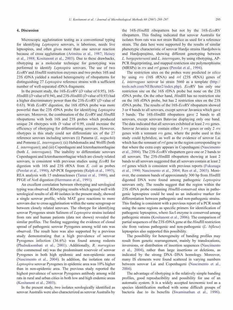
4. Discussion
Microscopic agglutination testing as a conventional typingfor identifying Leptospira serovars, is laborious, needs liveleptospires, and often gives more than one serovar reactionbecause of cross agglutination (Arimitsu et al., 1987; Heiseyet al., 1988; Kositanont et al., 2003). Due to these drawbacks,ribotyping as a molecular technique for genotyping wasperformed to identify Leptospira serovars. The use of twoEcoRVand HindIII restriction enzymes and two probes 16S and23S rDNA yielded a marked heterogeneity of ribopatterns fordistinguishing 27 Leptospira reference strains with a sufficientnumber of well-separated rDNA-fragments.
In the present study, the 16S-EcoRV (D value of 0.95), 16S-HindIII (D value of 0.94), and 23S-HindIII (D value of 0.93) hada higher discriminatory power than the 23S-EcoRV (D value of0.63). With EcoRV digestion, the 16S rDNA probe was morepowerful than the 23S rDNA probe for identifying Leptospiraserovars. Moreover, the combination of the EcoRVand HindIIIribopatterns with both 16S and 23S probes which producedunique 24 ribotypes with D value of 0.99, could increase theefficiency of ribotyping for differentiating serovars. However,ribotypes in this study could not differentiate six of the 27reference serovars including serovars (i) Panama (L. noguchii)and Pomona (L. interrogans); (ii) Hebdomadis and Wolffi (bothL. interrogans); and (iii) Copenhageni and Icterohaemorrhagiae(both L. interrogans). The inability to differentiate serovarsCopenhageni and Icterohaemorrhagiae which are closely relatedserovars, is consistent with previous studies using EcoRI fordigestion with 16S and 23S rRNA from E. coli as probes(Perolat et al., 1990), AP-PCR fingerprints (Ralph et al., 1993),REA analysis with 15 endonucleases (Tamai et al., 1988), andPFGE of NotI digestion (Herrmann et al., 1991).
An excellent correlation between ribotyping and serologicaltyping was observed. Ribotyping results which agreed well withserological results of all 40 isolates in the present study, showeda single serovar profile, while MAT gave reactions to moreserovars due to cross agglutination within the same serogroup orbetween closely related serovars. The ribotype for identifyingserovar Pyrogenes strain Salinem of Leptospira strains isolatedfrom rats and human patients (data not shown) revealed thesimilar profiles. The finding suggesting the evidence of clonalspread of pathogenic serovar Pyrogenes among wild rats wasobserved. The result here was also supported by a previousstudy demonstrating that a high prevalence of serovarPyrogenes infection (36.6%) was found among rodents(Phulsuksombati et al., 2001). Additionally, R. norvegicus(the commensal rat) was the predominant reservoir of serovarPyrogenes in both high epidemic and non-epidemic areas(Nascimento et al., 2004). In addition, the isolation rate ofLeptospira serovar Pyrogenes in epidemic area was 10% higherthan in non-epidemic area. The previous study reported thehighest prevalence of serovar Pyrogenes antibody among wildrats in rural and urban cities in both low and high endemic areas(Kositanont et al., 2003).
In the present study, two isolates serologically identified asserovar Australis were also characterized as serovar Australis by
the 16S-HindIII ribopattern but not by the 16S-EcoRVribopattern. This finding indicated that serovar Australis forisolates from rats was not strain Ballico as used for a referencestrain. The data here were supported by the results of similarphenotypic characteristic of serovar Hardjo strains Hardjobovisand Hardjoprajitno, showing different genotyping betweenL. borgepetersenii and L. interrogans, by using ribotyping, AP-PCR fingerprinting, and mapped restriction site polymorphisms(MRSPs) in rrs and rrl genes (Perolat et al., 1994).
The restriction sites on the probes were predicted in silicoby using rrs (16S rRNA) and rrl (23S rRNA) genes ofL. interrogans serovar lai strain 5660 as a template (http://tools.neb.com/NEBcutter2/index.php). EcoRV has only onerestriction site on the 16S rDNA probe but none on the 23SrDNA probe. On the other hand, HindIII has no restriction siteon the 16S rDNA probe, but has 2 restriction sites on the 23SrDNA probe. The results of the 16S-EcoRV ribopattern showed3 or 4 bands to all serovars, except serovar Javanica which gave5 bands. The 16S-HindIII ribopattern gave 2 bands to allserovars, except serovars Bataviae displaying only one band.The data indicated that all serovars exhibited at least 2 rrs genes.Serovar Javanica may contain either 3 rrs genes or only 2 rrsgenes with a remnant rrs gene, where the probe used in thisstudy could hybridize, as well as the evidence of serovar Laiwhich has the remnant of rrl gene in the region corresponding tothat where the extra copy appears in Copenhageni (Nascimentoet al., 2004). The 23S-EcoRV ribopattern gave one or 2 bands toall serovars. The 23S-HindIII ribopattern showing at least 2bands to all serovars suggested that all serovars contain at least 2rrl genes which is consistent with previous reports (Fukunagaet al., 1990; Nascimento et al., 2004; Ren et al., 2003). More-over, the common bands of approximately 300 bp from HindIIIdigested DNA were found among pathogenic Leptospiraserovars only. The results suggest that the region within the23S rDNA probe containing HindIII-conserved sites in patho-genic leptospires could be used as an indicative marker fordifferentiation between pathogenic and non-pathogenic strains.This finding is consistent with a previous report of a PCR resultusing the same regions as specific primers for identification ofpathogenic leptospires, where SacI enzyme is conserved amongpathogenic strains (Kositanont et al., 2006). The comparison ofpartial sequences of the 23S rDNA gene at theHindIII restrictionsite from various pathogenic and non-pathogenic (L. biflexa)leptospries also supported this possibility.
The possibility for heterogeneity of banding profiles mayresult from genetic rearrangement, mainly by translocations,inversions, or distribution of insertion sequences (Nascimentoet al., 2004), rather than large insertions or deletions, asindicated by the strong DNA–DNA homology. Moreover,many IS elements were found scattered in varying numbersbetween serovar Lai and Copenhageni (Nascimento et al.,2004).
The advantage of ribotyping is the relatively simple bandingprofiles, good reproducibility and possibility for use of anautomatic system. It is a widely accepted taxonomic tool as aspecies identification method with some difficult groups ofbacteria due to its benefits (Swaminathan et al., 1996).

296 U. Kositanont et al. / Journal of Microbiological Methods 69 (2007) 288–297
Ribotyping is a useful molecular technique for molecular typingto identify isolates of Leptospira species.
Acknowledgments
This work was partly supported by a Thesis Grant, from theFaculty of Graduate Studies, and a Siriraj Graduate ThesisScholarship, from the Faculty of Medicine Siriraj Hospital,Mahidol University, Thailand.
The authors gratefully acknowledge Dr. Lee D Smythe,WHO/FAO/OIE Collaborating Centre for Reference & Re-search on Leptospirosis, Centre for Public Health Sciences,Queensland Health Scientific Services, Australia, and MajorNoppadol Sangjun, Department of Veterinary Medicine,AFRIMS for serotyping the Leptospira isolates from rats byusing CAAT and MAT, respectively. We would like to thankAssoc. Prof. Pattama Ekpo, Department of Immunology,Faculty of Medicine Siriraj Hospital, Mahidol University,Thailand, for providing serological results of monoclonalantibody testing; and Assoc. Prof. Dr. Galayanee Doungcha-wee, Department of Pathobiology, Faculty of Science, MahidolUniversity, Thailand, for her help in immunoblotting technique.We wish to thank Assis. Prof. Chanin Limwong, Department ofMedicine, Faculty of Medicine Siriraj Hospital, MahidolUniversity, Thailand, for his kind offer of Syngene SoftwareAnalysis. We are indebted to Mrs. Pimjai Naigowit andDr. Duangchai Suwanchareon of the National Institute of Health(NIH), Thailand, and National Institute of Animal Health,Thailand, for providing reference Leptospira strains. We alsothank Ms. Patcharee Prasajak for excellent technical skill inpreparing the figures. We wish to thank Dr. Lee D Smythe for hisvaluable critical advice pertaining to the manuscript.
References
Arimitsu, Y., Kobayashi, S., Matuhasi, T., Suzuki, H., Yamaji, Y., Suprasert, S.,Supawadee, J., 1987. Epidemiological studies on leptospirosis in ChiangMai (Thailand). Epidemiol. Infect. 98, 97–100.
Brenner, D.J., Kaufmann, A.F., Sulzer, K.R., Steigerwalt, A.G., Rogers, F.C.,Weyant, R.S., 1999. Further determination of DNA relatedness betweenserogroups and serovars in the family Leptospiraceae with a proposal forLeptospira alexanderi sp. nov. and four new Leptospira genomospecies.Int. J. Syst. Bacteriol. 49, 839–858.
Ciceroni, L., Ciarrocchi, S., Ciervo, A., Petrucca, A., Pinto, A., Calderaro, A.,Viani, I., Galati, L., Dettori, G., Chezzi, C., 2002. Differentiation ofleptospires of the serogroup Pomona by monoclonal antibodies, pulsed-fieldgel electrophoresis and arbitrarily primed polymerase chain reaction. Res.Microbiol. 153, 37–44.
Ellighahausen, H.C., McCullough, W.G., 1965. Nutrition of Leptospira pomonaand growth of 13 other serotypes: fractionation of oleic albumin complex anda medium of bovine albumin and polysorbate 80. Am. J. Vet. Res. 26, 45–51.
Ellis, W.A., Montgomery, J.M., Thiermann, A.B., 1991. Restriction endonu-clease analysis as a taxonomic tool in the study of pig isolates belonging tothe Australis serogroup of Leptospira interrogans. J. Clin. Microbiol. 29,957–961.
Faine, S., 1988. Leptospirosis. In: Balows, A., Hausler, W.J., Ohashi, M.,Turano, A. (Eds.), Laboratory Diagnosis of Infectious Diseases, Principlesand Practice. Springer-Verlag, New York, pp. 344–352.
Fukunaga, M., Masuzawa, T., Okuzako, N., Mifuchi, I., Yanagihara, Y., 1990.Linkage of ribosomal RNA genes in Leptospira. Microbiol. Immunol. 34,565–573.
Gravekamp, C., Van de Kemp, H., Franzen, M., Carrington, D., Schoone, G.J.,Van Eys, G.J., Everard, C.O., Hartskeerl, R.A., Terpstra, W.J., 1993.Detection of seven species of pathogenic leptospires by PCR using two setsof primers. J. Gen. Microbiol. 139, 1691–1700.
Heisey, G.B., Nimmanitya, S., Karnchanachetanee, C., Tingpalapong, M.,Samransamruajkit, S., Hansukjariya, P., Elwell, M.R., 1988. Epidemiologyand characterization of leptospirosis at an urban and provincial site inThailand. Southeast Asian J. Trop. Med. Public Health 19, 317–322.
Herrmann, J.L., Baril, C., Bellenger, E., Perolat, P., Baranton, G., Saint Girons,I., 1991. Genome conservation in isolates of Leptospira interrogans.J. Bacteriol. 173, 7582–7588.
Hunter, P.R., Gaston, M.A., 1988. Numerical index of the discriminatory abilityof typing systems: an application of Simpson's index of diversity. J. Clin.Microbiol. 26, 2465–2466.
Kositanont, U., Naigowit, P., Imvithaya, A., Singchai, C., Puthavathana, P.,2003. Prevalence of antibodies to Leptospira serovars in rodents and shrewstrapped in low and high endemic areas in Thailand. J. Med. Assoc. Thai. 86,136–142.
Kositanont, U., Rugsasuk, S., Leelaporn, A., Phulsuksombati, D., Tantitanawat,S., Naigowit, P., 2006. Detection and differentiation between pathogenic andsaprophytic Leptospira species by multiplex polymerase chain reaction.Diagn. Microbiol. Infect. Dis. 57 (2) (Feb), 117–122.
Letocart, M., Baranton, G., Perolat, P., 1997. Rapid identification of pathogenicLeptospira species (Leptospira interrogans, L. borgpetersenii, andL. kirschneri) with species-specific DNA probes produced by arbitrarilyprimed PCR. J. Clin. Microbiol. 35, 248–253.
Levett, P.N., 2001. Leptospirosis. Clin. Microbiol. Rev. 14, 296–326.Lupidi, R., Cinco, M., Balanzin, D., Delprete, E., Varaldo, P.E., 1991.
Serological follow-up of patients involved in a localized outbreak ofleptospirosis. J. Clin. Microbiol. 29, 805–809.
Nascimento, A.L., Ko, A.I., Martins, E.A., Monteiro-Vitorello, C.B., Ho, P.L.,Haake, D.A., Verjovski-Almeida, S., Hartskeerl, R.A., Marques, M.V.,Oliveira, M.C., Menck, C.F., Leite, L.C., Carrer, H., Coutinho, L.L.,Degrave, W.M., Dellagostin, O.A., El-Dorry, H., Ferro, E.S., Ferro, M.I.,Furlan, L.R., Gamberini, M., Giglioti, E.A., Goes-Neto, A., Goldman, G.H.,Goldman, M.H., Harakava, R., Jeronimo, S.M., Junqueira-de-Azevedo, I.L.,Kimura, E.T., Kuramae, E.E., Lemos, E.G., Lemos, M.V., Marino, C.L.,Nunes, L.R., de Oliveira, R.C., Pereira, G.G., Reis, M.S., Schriefer, A.,Siqueira, W.J., Sommer, P., Tsai, S.M., Simpson, A.J., Ferro, J.A., Camargo,L.E., Kitajima, J.P., Setubal, J.C., Van Sluys, M.A., 2004. Comparativegenomics of two Leptospira interrogans serovars reveals novel insights intophysiology and pathogenesis. J. Bacteriol. 186, 2164–2172.
Niwetpathomwat, A., Doungchawee, G., 2006. Western immunoblot analysisusing a ten Leptospira serovars combined antigen for serodiagnosis ofleptospirosis. Southeast Asian J. Trop. Med. Public Health 37, 309–311.
Pacciarini, M.L., Savio, M.L., Tagliabue, S., Rossi, C., 1992. Repetitivesequences cloned from Leptospira interrogans serovar Hardjo genotypeHardjoprajitno and their application to serovar identification. J. Clin.Microbiol. 30, 1243–1249.
Perolat, P., Grimont, F., Regnault, B., Grimont, P.A., Fournie, E., Thevenet, H.,Baranton, G., 1990. rRNA gene restriction patterns of Leptospira: amolecular typing system. Res. Microbiol. 141, 159–171.
Perolat, P., Merien, F., Ellis, W.A., Baranton, G., 1994. Characterization ofLeptospira isolates from serovar Hardjo by ribotyping, arbitrarily primedPCR, and mapped restriction site polymorphisms. J. Clin. Microbiol. 32,1949–1957.
Phulsuksombati, D., Sangjun, N., Khoprasert, Y., Kingnate, D., Tangkamakul,W., 2001. Leptospires in rodents, northeastern region 1999–2000. J. HealthSci. 10, 516–525.
Ralph, D., McClelland, M., Welsh, J., Baranton, G., Perolat, P., 1993. Leptospiraspecies categorized by arbitrarily primed polymerase chain reaction (PCR)and by mapped restriction polymorphisms in PCR-amplified rRNA genes.J. Bacteriol. 175, 973–981.
Ren, S.X., Fu, G., Jiang, X.G., Zeng, R.,Miao, Y.G., Xu, H., Zhang, Y.X., Xiong,H., Lu, G., Lu, L.F., Jiang, H.O., Jia, J., Tu, Y.F., Jiang, J.X., Gu, W.Y.,Zhang, Y.O., Cai, Z., Sheng, H.H., Yin, H.F., Zhang, Y., Zhu, G.F., Wan, M.,Huang, H.L., Oian, Z., Wang, S.Y., Ma, W., Yao, Z.J., Shen, Y., Oiang, B.O.,Xia, O.C., Guo, X.K., Danchin, A., Saint Girons, I., Somerville, R.L., Wen,

297U. Kositanont et al. / Journal of Microbiological Methods 69 (2007) 288–297
Y.M., Shi, M.H., Chen, Z., Xu, J.G., Zhao, G.P., 2003. Unique physiologicaland pathogenic features of Leptospira interrogans revealed by whole-genome sequencing. Nature 422, 888–893.
Swaminathan, B., Matar, G.M., Reeves, M.W., Graves, L.M., Ajello, G., Bibb,W.F., Helsel, L.O., Morales, M., Dronavalli, H., el-Swify, M., DeWitt, W.,Hunter, S.B., 1996.Molecular subtyping ofNeisseriameningitides serogroupB: comparison of five methods. J. Clin. Microbiol. 34, 1468–1473.
Tamai, T., Sada, E., Kobayashi, Y., 1988. Restriction endonuclease DNAanalysis of Leptospira interrogans serovars Icterohaemorrhagiae andCopenhageni. Microbiol. Immunol. 32, 887–894.
Terpstra, W.J., 1992. Typing Leptospira from the perspective of a referencelaboratory. Acta Leiden 60, 79–87.
Van Eys, G.J., Gerritsen, M.J., Korver, H., Schoone, G.J., Kroon, C.C., Terpstra,W.J., 1991. Characterization of serovars of the genus Leptospira by DNAhybridization with Hardjobovis and Icterohaemorrhagiae recombinantprobes with special attention to serogroup Sejroe. J. Clin. Microbiol. 29,1042–1048.
Zuerner, R.L., Bolin, C.A., 1990. Nucleic acid probe characterizes Leptospirainterrogans serovars by restriction fragment length polymorphisms. Vet.Microbiol. 24, 355–366.
Zuerner, R.L., Bolin, C.A., 1997. Differentiation of Leptospira interrogansisolates by IS1500 hybridization and PCR assays. J. Clin. Microbiol. 35,2612–2617.
Zuerner, R.L., Ellis, W.A., Bolin, C.A., Montgomery, J.M., 1993a. Restrictionfragment length polymorphisms distinguish Leptospira borgpeterseniiserovar hardjo type hardjo-bovis isolates from different geographicallocations. J. Clin. Microbiol. 31, 578–583.
Zuerner, R.L., Herrmann, J.L., Saint Girons, I., 1993b. Comparison of geneticmaps for two Leptospira interrogans serovars provides evidence for twochromosomes and intraspecies heterogeneity. J. Bacteriol. 175, 5445–5451.
Zuerner, R.L., Alt, D., Bolin, C.A., 1995. IS1533-based PCR assay foridentification of Leptospira interrogans sensu lato serovars. J. Clin.Microbiol. 33, 3284–3289.