Identification of Collagenase as a Critical Virulence Factor for Invasiveness and Transmission of...
11
MAJOR ARTICLE Identification of Collagenase as a Critical Virulence Factor for Invasiveness and Transmission of Pathogenic Leptospira Species Kokouvi Kassegne, 1,2,a Weilin Hu, 1,2,a David M. Ojcius, 3 Dexter Sun, 4 Yumei Ge, 1,2 Jinfang Zhao, 5 X. Frank Yang, 6 Lanjuan Li, 1 and Jie Yan 1,2 1 Division of Basic Medical Microbiology, State Key Laboratory for Diagnosis and Treatment of Infectious Diseases, First Affiliated Hospital, Zhejiang University School of Medicine, 2 Department of Medical Microbiology and Parasitology, Zhejiang University School of Medicine, and 5 Department of Clinical Laboratory, Zhejiang Traditional Chinese Medical Hospital, Hangzhou, China; 3 Health Sciences Research Institute and School of Natural Sciences, University of California, Merced, 4 Department of Neurology and Neuroscience, New York Presbyterian Hospital and Hospital for Special Surgery, Cornell University Weill Medical College, New York, and 6 Department of Microbiology and Immunology, Indiana University School of Medicine, Indianapolis Background. Leptospirosis is a global zoonotic disease. Transmission of Leptospira from animals to humans occurs through contact with water contaminated with leptospire-containing urine of infected animals. However, the molecular basis for the invasiveness of Leptospira and transmission of leptospirosis remains unknown. Methods. Activity of Leptospira interrogans strain Lai colA gene product (ColA) to hydrolyze different collagen- ic substrates was determined by spectrophotometry. Expression and secretion of ColA during infection were detect- ed by reverse-transcription quantitative polymerase chain reaction and Western blot assay. The colA gene–deleted (ΔcolA) and colA gene–complemented (CΔcolA) mutants were generated to determine the roles of ColA in transcy- tosis in vitro and virulence in hamsters. Results. Recombinant or native ColA hydrolyzed all the tested substrates in which type III collagen was the fa- vorite substrate with 2.16 mg/mL K m and 35.6 h −1 K cat values. Coincubation of the spirochete with HUVEC or HEK293 cells directly caused the significant elevation of ColA expression and secretion. Compared with wild-type strain, ΔcolA mutant displayed much-attenuated transcytosis through HEK293 and HUVEC monolayers, and less leptospires in blood, lung, liver, kidney and urine and 25-fold-decreased 50% lethal dose and milder histopathologi- cal injury in hamsters. Conclusions. The product of colA gene is a collagenase as a crucial virulence factor in the invasiveness and transmission of L. interrogans. Keywords. Leptospira; Collagenase; Invasiveness; Tissue injury; Transmission. Leptospirosis caused by pathogenic Leptospira species is a global zoonotic infectious disease [1], especially in Southeast Asia and South America [2, 3]. In North America and Europe, leptospirosis is considered an emerging infectious disease because of frequent case reports and several outbreaks in recent years [4–7]. Many animals can serve as hosts of pathogenic Lepto- spira species, including dogs and livestock [8]. After being shed in urine of the infected animals, the spiro- chete survives for long periods of time in moist soil and natural bodies of water [8, 9]. After contact with the skin and mucosa of individuals, the leptospires in the con- taminated soil or water invade the human body prompt- ly to cause septicemia, followed by a rapid spread into lungs, liver, and kidneys to cause tissue injury [9]. Lepto- spirosis is clinically characterized by high fever, myalgia, hemorrhage, jaundice, renal impairment, and septic shock [10]. In the convalescence stage, patients also Received 2 January 2013; accepted 15 May 2013; electronically published 25 November 2013. a K. K. and W. H. contributed equally to this work. Correspondence: Jie Yan Prof, MD., Department of Medical Microbiology and Parasitology, Zhejiang University School of Medicine, Hangzhou, Zhejiang 310058, PR China ([email protected]). The Journal of Infectious Diseases 2014;209:1105–15 © The Author 2013. Published by Oxford University Press on behalf of the Infectious Diseases Society of America. All rights reserved. For Permissions, please e-mail: [email protected]. DOI: 10.1093/infdis/jit659 ColA as a Virulence Factor of Leptospira • JID 2014:209 (1 April) • 1105 at California Digital Library - Office of the President on September 21, 2015 http://jid.oxfordjournals.org/ Downloaded from
-
Upload
independent -
Category
Documents
-
view
1 -
download
0
Transcript of Identification of Collagenase as a Critical Virulence Factor for Invasiveness and Transmission of...

M A J O R A R T I C L E
Identification of Collagenase as a CriticalVirulence Factor for Invasiveness andTransmission of Pathogenic Leptospira Species
Kokouvi Kassegne,1,2,a Weilin Hu,1,2,a David M. Ojcius,3 Dexter Sun,4 Yumei Ge,1,2 Jinfang Zhao,5 X. Frank Yang,6
Lanjuan Li,1 and Jie Yan1,2
1Division of Basic Medical Microbiology, State Key Laboratory for Diagnosis and Treatment of Infectious Diseases, First Affiliated Hospital, ZhejiangUniversity School of Medicine, 2Department of Medical Microbiology and Parasitology, Zhejiang University School of Medicine, and 5Department ofClinical Laboratory, Zhejiang Traditional Chinese Medical Hospital, Hangzhou, China; 3Health Sciences Research Institute and School of NaturalSciences, University of California, Merced, 4Department of Neurology and Neuroscience, New York Presbyterian Hospital and Hospital for SpecialSurgery, Cornell University Weill Medical College, New York, and 6Department of Microbiology and Immunology, Indiana University School of Medicine,Indianapolis
Background. Leptospirosis is a global zoonotic disease. Transmission of Leptospira from animals to humansoccurs through contact with water contaminated with leptospire-containing urine of infected animals. However, themolecular basis for the invasiveness of Leptospira and transmission of leptospirosis remains unknown.
Methods. Activity of Leptospira interrogans strain Lai colA gene product (ColA) to hydrolyze different collagen-ic substrates was determined by spectrophotometry. Expression and secretion of ColA during infection were detect-ed by reverse-transcription quantitative polymerase chain reaction and Western blot assay. The colA gene–deleted(ΔcolA) and colA gene–complemented (CΔcolA) mutants were generated to determine the roles of ColA in transcy-tosis in vitro and virulence in hamsters.
Results. Recombinant or native ColA hydrolyzed all the tested substrates in which type III collagen was the fa-vorite substrate with 2.16 mg/mL Km and 35.6 h−1 Kcat values. Coincubation of the spirochete with HUVEC orHEK293 cells directly caused the significant elevation of ColA expression and secretion. Compared with wild-typestrain, ΔcolA mutant displayed much-attenuated transcytosis through HEK293 and HUVEC monolayers, and lessleptospires in blood, lung, liver, kidney and urine and 25-fold-decreased 50% lethal dose and milder histopathologi-cal injury in hamsters.
Conclusions. The product of colA gene is a collagenase as a crucial virulence factor in the invasiveness andtransmission of L. interrogans.
Keywords. Leptospira; Collagenase; Invasiveness; Tissue injury; Transmission.
Leptospirosis caused by pathogenic Leptospira speciesis a global zoonotic infectious disease [1], especially inSoutheast Asia and South America [2, 3]. In NorthAmerica and Europe, leptospirosis is considered an
emerging infectious disease because of frequent casereports and several outbreaks in recent years [4–7].
Many animals can serve as hosts of pathogenic Lepto-spira species, including dogs and livestock [8]. Afterbeing shed in urine of the infected animals, the spiro-chete survives for long periods of time in moist soil andnatural bodies of water [8, 9]. After contact with the skinand mucosa of individuals, the leptospires in the con-taminated soil or water invade the human body prompt-ly to cause septicemia, followed by a rapid spread intolungs, liver, and kidneys to cause tissue injury [9]. Lepto-spirosis is clinically characterized by high fever, myalgia,hemorrhage, jaundice, renal impairment, and septicshock [10]. In the convalescence stage, patients also
Received 2 January 2013; accepted 15 May 2013; electronically published 25November 2013.
aK. K. and W. H. contributed equally to this work.Correspondence: Jie Yan Prof, MD., Department of Medical Microbiology and
Parasitology, Zhejiang University School of Medicine, Hangzhou, Zhejiang 310058,PR China ([email protected]).
The Journal of Infectious Diseases 2014;209:1105–15© The Author 2013. Published by Oxford University Press on behalf of the InfectiousDiseases Society of America. All rights reserved. For Permissions, please e-mail:[email protected]: 10.1093/infdis/jit659
ColA as a Virulence Factor of Leptospira • JID 2014:209 (1 April) • 1105
at California D
igital Library - O
ffice of the President on September 21, 2015
http://jid.oxfordjournals.org/D
ownloaded from

discharge leptospires in the urine for about 1 week [8–10]. Untilnow, however, little has been known about the molecular basisfor the powerful invasiveness and transmission of pathogenicLeptospira species.
Previous reports revealed that a collagenase of Clostridiumhistolyticum contributes to damage of cells and spread intissues of hosts [11, 12]. Collagenases of Clostridium perfrin-gens, Vibrio parahaemolyticus, and Fusobacterium nucleatumact as virulence factors for invasiveness and tissue injury [13–15]. These data indicate that bacterial collagenases play impor-tant roles in invasiveness and transmission during infection.
Leptospira interrogans is the most common genotype inpathogenic Leptospira species [1–7]. Several serogroups of L. in-terrogans are epidemic in China, but L. interrogans serogroupIcterohaemorrhagiae serovar Lai is responsible for leptospirosisin about 70% of patients [3, 16]. The chromosomal DNA ofL. interrogans serovar Lai strain Lai includes is a single colAgene that is predicted to encode a collagenase [17]. However,the collagenolytic activity and pathogenic role of product of thegene have not been characterized.
In the current study, we determined the distribution of colAgene in L. interrogans strains belonging to different serogroupsthat prevalent in China as well as the ability of the colA geneproduct of L. interrogans strain Lai to hydrolyze different collagen-ic substrates. Next, we investigated the expression and secretion ofColA protein in the spirochete during human renal epithelial andblood vessel endothelial cell infection. Finally, we generated a colAgene–deleted mutant to further evaluate the role of ColA proteinin the invasiveness and transmission of the spirochete in cellmonolayers and animals. The results of our study identify ColA asa novel and crucial virulence factor of L. interrogans.
MATERIALS ANDMETHODS
AnimalMale Syrian hamsters (mean weight ± standard deviation [SD],25 ± 2 g per animal) were provided by the ExperimentalAnimal Center of Zhejiang University. The animal experimen-tal protocols were approved by the Ethics Committee forAnimal Experiment of Zhejiang University.
Leptospiral Strain, Cell Lines, and CultureSeven pathogenic L. interrogans strains and 2 nonpathogenicLeptospira biflexa strains belonging to different serogroupsor serovars (see Supplementary Information) were cultivatedat 28°C in Ellinghausen-McCullough-Johnson-Harris (EMJH)medium [18]. A human renal tubular epithelial cell line (HEK293)and a human umbilical vein endothelial cell line (HUVEC) weremaintained in Dulbecco’s modified Eagle’s medium (DMEM)medium (Gibco), supplemented with 10% fetal calf serum (FCS;Gibco), 100 U/mL penicillin, and 100 µg/mL streptomycin at 37°C in an atmosphere of 5% carbon dioxide (CO2).
Detection of colA Gene, Preparation of Recombinant andNative ColA Proteins, and Preparation of Antiserum andImmunoglobulin GSee the Supplementary Information for details about detectionof the colA gene in different leptospiral strains by polymerasechain reaction (PCR) and sequencing; preparation of the re-combinant ColA (rColA) and native ColA proteins from L. in-terrogans strain Lai; and preparation of rabbit antiserum andimmunoglobulin G (IgG) antibodies against rColA.
Collagen Hydrolysis TestOne milliliter of Calcium binding buffer (CBB) (0.4 mmol/Lcalcium chloride and 50 mmol/L Tris-HCl; pH 7.4) containing5 mg of native bovine type I or II collagen, human type III orIV collagen (Sigma), or synthetic substrate Azocoll or Pzpeptide (Sigma), was mixed with 50 or 100 µg of rColA or 100µg of native ColA protein. The mixtures were incubated at 37°Cfor 5 hours. Hydrolysis of the substrates was measured by spec-trophotometry at an optical density at 570 nm (collagen hydro-lysis), 520 nm, or 320 nm (Azocoll or Pz peptide hydrolysis)[19–21]. In addition, collagenolytic ability of the rColA waschecked by sodium dodecyl sulfate–polyacrylamide gel electro-phoresis using 100 µg of rColA for hydrolysis with 5 mg of typeIII collagen and 200 µg of rColA for hydrolysis with 5 mg oftype I, II, or IV collagen. Moreover, solutions of 0.2 mol/Lsodium phosphate with 0.15 mol/L sodium chloride (pH 7.0)or 0.2 mol/L sodium borate with 0.15 mol/L sodium chloride(pH 7.2) were used to evaluate the influence of different bufferson hydrolytic activity of the rColA [19–21].
Collagen Hydrolysis Inhibition TestThe rColA or native ColA protein (100 µg) was pretreated with5 µmol/L collagenase inhibitor I (Millipore) for a 30-minute in-cubation at 37°C in CBB buffer [22]. The ability of the ColAproteins before or after treatment with the inhibitor to hydro-lyze the 4 collagens was determined as already described.
Determination of Km and Kcat ValuesThe collagen hydrolysis test demonstrated type III collagen asthe preferred substrate of rColA and native ColA protein. Todetermine the enzyme kinetic parameters (Km and Kcat), 0.25,0.5, 1, 2, 3, 4, or 5 mg of type III collagen was mixed with 100µg of rColA in 1 mL of CBB buffer, and the hydrolysis of typeIII collagen was performed as already described. The Km andKcat values of rColA to hydrolyze type III collagen were calcu-lated by means of double reciprocal Lineweaver-Burk plot [23].
Measurement of Transcription and Expression of colA GeneDuring InfectionFreshly cultured L. interrogans strain Lai was collected by using17 200 g centrifugation at 15°C for 15 minutes. The harvestedleptospires were counted with a Petroff-Hausser countingchamber (Fisher Scientific) under a dark-field microscope [24].
1106 • JID 2014:209 (1 April) • Kassegne et al
at California D
igital Library - O
ffice of the President on September 21, 2015
http://jid.oxfordjournals.org/D
ownloaded from

HUVEC and HEK293 cells (1 × 10⁶ per well) were seeded in6-well culture plates (Corning) for a 12-hour incubation in anatmosphere of 5% CO2. After being washed with phosphate-buffered saline (PBS), the cell monolayers were infected withthe spirochete (1 × 10⁸) at 37°C for 0.5, 1, 2, 4, 8, 12 or 24 hours[25–27]. Media from the leptospire cell cocultures were collect-ed by a 15-minute centrifugation at 17 200 g. The spirochete(1 × 10⁸) was inoculated into the media or in 8% FCS-DMEMfor incubation at 37°C for the same durations. On the otherhand, the cocultures were treated with 0.05% sodium deoxy-cholate–PBS to lyse cells [28], followed by a short centrifuga-tion at 500 g to remove cell debris. The supernatants as well asthe leptospiral cultures in the different media were centrifugedat 17 200 g for 15 minutes (4°C) to precipitate leptospires.
Total leptospiral RNA was extracted using TRIzol reagent(Sigma) and digestion with RNase-Free DNase (TaKaRa).Complementary DNA (cDNA) was synthesized from the totalRNA by reverse transcription (RT) using a cDNA Synthesis Kit(TaKaRa). Using the cDNA as template, the colA messengerRNA (mRNA) was measured with real-time fluorescence quan-titative PCR (qPCR) using a SYBR Ex-Taq Kit (TaKaRa). Inthe RT-qPCR, 16S RNA gene of the spirochete was used as theinner reference [29]. The RT-qPCR data were analyzed usingthe ΔΔCt model and a randomization test with REST 2005software [30].
The HUVEC or HEK293 monolayer (1 × 10⁶ per well) wasinfected with the spirochete (1 × 10⁸) for 1, 2, 4, 8, or 12 hours.Incubation of the spirochete in the media from the leptospirecell cocultures or in 8% FCS-DMEM, and lysis of the cells andprecipitation of leptospires were performed as already de-scribed. The leptospires were ultrasonically broken and thencentrifuged at 500 g for 15 minutes. The supernatants were col-lected to detect protein concentrations using a Protein Concen-tration Assay Kit (Bio-Rad). Using rabbit anti-rColA IgG as theprimary antibody and horseradish peroxidase–conjugated goatanti-rabbit IgG (Jackson ImmunoResearch) as the secondaryantibody, the ColA in the protein specimens was detected withWestern blot assay. The immunoblotting signals were quanti-fied by densitometry (gray scale determination) using an ImageAnalyzor (Bio-Rad). In this assay, a leptospiral membrane lipo-protein (LipL41) was used as the control [29, 31].
Determination of ColA Secretion During InfectionHUVEC or HEK293 cell monolayer (1 × 10⁶ per well) was in-fected with L. interrogans strain Lai (1 × 10⁸) for 1, 2, 4, 8, or 12hours. After lysis of the cells, the supernatants were harvestedby using 17 200 g centrifugation at 4°C for 15 minutes. Mean-while, the supernatant from the spirochete (1 × 10⁸) in EMJHmedium or incubated in 8% FCS-DMEM for the indicated timeswas also collected by centrifugation, as described. Total pro-teins in all the supernatants were extracted by trichloroaceticacid precipitation [32]. With rabbit anti-rColA IgG used as the
primary antibody and horseradish peroxidase–conjugated goatanti-rabbit IgG (Jackson ImmunoResearch) as the secondaryantibody, ColA protein in the protein specimens was detectedby means of Western blot assay. The immunoblotting signalswere quantified as described. In the assay, a leptospiral cyto-plasmic protein (FliY) and a secretory hemolysin (Sph2) wereused as the controls [32, 33].
Generation and Identification of colA Gene–Deleted mutantand colA Gene–Complemented MutantsSee the Supplementary Information for details about the gener-ation and identification of the colA gene–deleted (ΔcolA) andcolA gene–complemented (CΔcolA) mutants.
Transwell migration AssayHUVEC or HEK293 cells (1 × 10⁶ cells per well) were seeded inthe upper compartment of a 12-well Transwell plate (Corning)and then incubated for 48 hours in an atmosphere of 5% CO2
at 37°C to form tight cell monolayers. Transendothelial or epi-thelial electric resistance (TEER) of the cell monolayers wasmonitored using a Millicell electric resistance indicator (Milli-pore); TEER values >200 Ω/cm2 indicated the integrity of cellmonolayer [34]. The HUVEC or HEK293 cell monolayers wasinfected with L. interrogans strain Lai (1 × 10⁸) for 1, 2, 4, 8, 12,24, or 48 hours at 37°C. The leptospires that transmigratedfrom the upper into lower compartments were counted withPetroff-Hausser counting chamber (Fisher Scientific) under adark-field microscope [24]. In the assay, the spirochete(1 × 10⁸) through the upper compartments without cells wasused as the controls.
Determination of ColAVirulence in HamstersSyrian hamsters were intraperitoneally injected with 104, 105,106, 107, or 108 leptospires of the ΔcolA or CΔcolA mutant orwild-type L. interrogans strain Lai, with 8 animals used pergroup [35]. Eight negative control animals were intraperitoneal-ly injected with EMJH medium. The animals were monitoredtwice daily, and their survival was recorded within 14 days afterchallenge to calculate the 50% lethal dose using Warren probitanalysis [28]. In addition, hamsters (n = 6) were infected witheach of the mutant and wild-type strains, as already described.Lung, liver, and kidney specimens were collected on days 3 and7 after challenge for histopathological examination after hema-toxylin-eosin staining.
Determining the Role of ColA in Leptospiral Invasiveness inHamstersSyrian hamsters (n = 54) were challenged with 1 × 10⁶ lepto-spires of the ΔcolA or CΔcolA mutant or wild-type L. interrog-ans strain Lai, as described above. Heart blood specimens werecollected on day 1 or 3, lung, liver, and kidney on day 3 or 7,and urine specimens on day 7 or 14 after infection [36]. Thesections of lung, liver, and kidney specimens were prepared for
ColA as a Virulence Factor of Leptospira • JID 2014:209 (1 April) • 1107
at California D
igital Library - O
ffice of the President on September 21, 2015
http://jid.oxfordjournals.org/D
ownloaded from

Figure 1. Distribution of colA gene, preparation of recombinant ColA (rColA) and native ColA protein and characterization of the colA gene–deleted(ΔcolA) and colA gene–complemented (CΔcolA) mutants. A, Distribution of colA gene in different pathogenic Leptospira interrogans and nonpathogenicLeptospira biflexa strains. Lane M, DNA marker. Lane 1, Blank control. Lanes 2–8, Amplicons of colA gene (2667 base pairs [bp]) from L. interrogansserovar Lai strain Lai, serovar Grippotyphosa strain Lin-6, serovar Autumnalis strain Lin-4, serovar Pomona strain Luo, serovar Hebdomadis strain P7,serovar Australis strain 65-9, and serovar Canicola strain Lin, respectively. Lanes 9–10, Negative polymerase chain reaction (PCR) results for colA gene de-tection of L. biflexa serovar Patoc strain Patoc-1 and serovar Adamana strain CH-11. B, Expression of rColA and purification of rColA and native ColAprotein. Lane M, Protein marker. Lane 1, Blank control (wild-type pET42a). Lanes 2–3, Expressed and purified rColA protein, respectively. Lane 4, NativeColA protein extracted using immunoprecipitation resin. C, Identification of ΔcolA and CΔcolA mutants by PCR. Lane M, DNA marker. Lane 1, Blankcontrol. Lane 2, Amplicon (2589 bp) of the signal peptide–absent colA gene from L. interrogans strain Lai for recombinant expression. Lane 3, Amplicon(3569 bp) of the colA-kan-colA segment (3329 bp) plus 2 extending regions (120 bp each) for characterization of the ΔcolA mutant. Lane 4, Amplicon(5314 bp) of the 5′ arm-colA-spc-3′ arm (5074 bp) plus 2 extending regions (120 bp each) for characterization of the CΔcolA mutant. D, Schematic diagramof the sequencing result for ΔcolA mutant. Positions of PCR primers used are marked below. E, Schematic diagram of the sequencing result of CΔcolAmutant. Positions of PCR primers used are marked below.
1108 • JID 2014:209 (1 April) • Kassegne et al
at California D
igital Library - O
ffice of the President on September 21, 2015
http://jid.oxfordjournals.org/D
ownloaded from

examination of leptospires in tissues after silver staining [36].In addition, 5 mg of the tissue specimens was homogenized inan ice bath and then added with 5 mL of PBS. The mixtureswere centrifuged at 300 g for 10 minutes (4°C) to collect super-natants. Next, 0.1 mL of the supernatant or blood specimenswas inoculated into 1 mL of Korthof medium for a 24-hourincubation and then spread on EMJH agar plates for a 3-weekincubation to count colony-forming units [36]. The urine spec-imens were centrifuged at 250 g for 5 minutes to remove partic-ulate matters, followed by a 15-minute centrifugation at 17 200g to precipitate leptospires. For counting leptospires, the pelletswere suspended at the 1:1 or 1:100 volume ratio of PBS to theurine specimens, as described above [24].
Statistical AnalysisData from a minimum of 3 experiments were averaged and pre-sented as means ± SDs. Statistical significance was determinedwith 1-way analysis of variance followed by the Dunnett
multiple-comparison test, with differences considered signifi-cant at P < .05.
RESULTS
Sequencing and Expression of colA GeneAll the tested 7 pathogenic L. interrogans strains, but not the 2nonpathogenic L. biflexa strains, have the colA gene (Figure 1A)with 99.4%–100% amino acid sequence identity compared withthat in the report (data not shown) [17]. The Escherichia coliBL21DE3pET42a-colA expressed rColA, and the purified rColA ornative ColA protein showed a single band in gel after electro-phoresis (Figure 1B).
Characterization of ΔcolA and CΔcolAMutantsThe ΔcolA and CΔcolA mutants could grow persistently inEMJH medium with typical shape and motility and similargrowth kinetics compared with the wild-type strain (data notshown). The PCR (Figure 1C) and sequencing data (Figure 1D
Figure 2. Collagenase activity of recombinant ColA (rColA) and native ColA proteins from Leptospira interrogans strain Lai. A, Ability of rColA and nativeColA proteins to hydrolyze different collagenase substrates, determined with spectrophotometry. Bars show means ± standard deviations (SDs) of 3 inde-pendent experiments. In this test, CBB buffer was used. The hydrolysis of type I, II, III, or IV collagen and synthetic Azocoll or Pz peptide substrate wasmeasured by spectrophotometry at OD570 (optical density at 570 nm), OD520 or OD320, respectively. *P < .05 (vs hydrolytic activity of 50 µg of rColA). B, At-tenuated collagenolytic ability of rColA and native ColA proteins after treatment with collagenase inhibitor I. Bars show means ± SDs of 3 independent ex-periments. The buffer and spectrophotometric examination were as in A. *P < .05 (vs hydrolytic activity of rColA and native ColA protein before treatmentwith collagenase inhibitor I). C, Ability of rColA to hydrolyse native collagens, demonstrated by sodium dodecyl sulfate–polyacrylamide gel electrophoresis.In this test, 100 µg of rColA was used for 5 mg of type III collagen hydrolysis, and 200 µg of rColA for 5 mg of type I, II, or IV collagen hydrolysis and CBBbuffer. Lane M, Protein marker. Lanes C1–C4, Type I, II, III, or IV collagen before hydrolysis. Lanes T1–T4, Hydrolysis of type I, II, III, or IV collagen after in-cubation with rColA. D, Influence of different reactive conditions on collagenase activity of rColA. Bars show means ± SDs of 3 independent experiments.The hydrolysis of type I-IV collagens, synthetic Azocoll and Pz peptide substrate was measured by spectrophotometry at OD570, OD520 and OD320, respec-tively. *P < .05 (vs hydrolytic activity of rColA in the CBB buffer). CaCl2, calcium chloride; NaCl, sodium chloride. E, Km and Kcat values of rColA hydrolyzingtype III collagen in CBB buffer. In this assay, 100 µg of rColA and different concentrations of type III collagen (0.25, 0.5, 1, 2, 4, or 5 mg) were used. The hy-drolysis of type III collagen was measured by spectrophotometry at OD570. [S] = substrates, V = velocity of substrate hydrolysis.The image showed theability of rColA to hydrolyze substrate with different concentrations per hour.
ColA as a Virulence Factor of Leptospira • JID 2014:209 (1 April) • 1109
at California D
igital Library - O
ffice of the President on September 21, 2015
http://jid.oxfordjournals.org/D
ownloaded from

Figure 3. Increase in colA gene expression and ColA protein secretion during infection. A, Increase in Leptospira interrogans strain Lai colA mRNAlevels during infection of HUVEC or HEK293 cells for the indicated times. Bars show means ± standard deviations (SDs) of 3 independent experiments. Thevalues at 0 hour indicate the colA messenger RNA (mRNA) levels in the spirochete before infection or incubation, set as 1.0. *P < .05 (vs colA mRNAlevels in the spirochete before infection or incubation [0 hour]); †P < .05 (vs the colA mRNA levels in the spirochete incubated in Dulbecco’s modifiedEagle’s medium (DMEM) or in media from leptospire cell cocultures for the indicated times). B, Increase in ColA protein expression in L. interrogans strainLai during infection of HUVEC or HEK293 cells for the indicated times. The immunoblotting results at 0 hour indicate the ColA protein expression in the spi-rochete before infection or incubation. ColA-1 indicates the expressed ColA protein in the spirochete during infection of HUVEC or HEK293 cells; ColA-2, ex-pression of ColA protein in the spirochete during incubation in the media from leptospire-HUVEC or leptospire-HEK293 cocultures; ColA-3, expression ofColA protein in the spirochete during incubation in 2.5% fetal calf serum (FCS)–DMEM medium. LipL41 is a leptospiral outer membrane lipoprotein usedas the control. C, Quantification of immunoblotting bands (by gray scale determination) reflecting ColA protein expression levels during infection of hostcells for the indicated times (statistical data from experiments as shown in B). Bars show means ± SDs of 3 independent experiments. Gray scale values ofimmunoblotting bands reflect the ColA expression levels in L. interrogans strain Lai before infection or incubation (0 hour). *P < .05 (vs ColA expressionlevels [gray scale values] in the spirochete before infection or incubation [0 hour]). D, Enhancement of ColA secretion of L. interrogans strain Lai during in-fection of HUVEC or HEK293 cells for the indicated times. The immunoblotting results at 0 hour indicate no secretory ColA protein in the supernatant fromculture of the spirochete in Ellinghausen-McCullough-Johnson-Harris (EMJH) medium. FliY is a leptospiral cytoplasmic protein and Sph2 a leptospiral se-cretory hemolysin, used as controls. The DMEM lane indicates the ColA protein secretion in the supernatants from cultures of the spirochete in 2.5% FCS-DMEM medium. E, Quantification of immunoblotting bands (by gray scale determination) reflecting the secretion of ColA protein during infection of hostcells for the indicated times (statistical data from experiments such as shown in D). Bars show means ± SDs of 3 independent experiments. The values at0 hour indicate the gray scale values, reflecting no secretion of ColA protein in the supernatant from culture of L. interrogans strain Lai in EMJH medium.*P < .05 (vs the secretion level [gray scale value] of ColA protein in the supernatant from culture of the spirochete in EMJH medium [0 hour]).
1110 • JID 2014:209 (1 April) • Kassegne et al
at California D
igital Library - O
ffice of the President on September 21, 2015
http://jid.oxfordjournals.org/D
ownloaded from

and 1E) confirmed the colA gene deletion in the ΔcolA mutantand the colA gene complementation in the CΔcolA mutant.The RT-qPCR and Western blot assays also proved the absenceor presence of colA mRNA and ColA protein in the ΔcolA orCΔcolAmutant (data not shown).
Collagenase Activity of rColA and Native ColA ProteinBoth the rColA and native ColA proteins from L. interrogansstrain Lai had the similar ability to hydrolyze all the 4 nativecollagens and 2 synthetic substrates, but the type III collagenwas the preferred substrate, with the highest hydrolytic ratio(Figure 2A). However, the collagenolytic ability was blocked bycollagenase inhibitor I (Figure 2B). The ability of rColA to hy-drolyze 4 collagens was shown in Figure 2C. In the CBB buffer,the rColA displayed a higher collagenolytic ability than inthe other 2 buffers (Figure 2D). The Km and Kcat values ofrColA for hydrolysis of type III collagen in CBB buffer were2.16 mg/mL and 35.6 hours−1, respectively (Figure 2E).
Elevation of ColA Expression During InfectionWild-type L. interrogans strain Lai in DMEM medium or in themedia from leptospire cell cocultures showed a small colAmRNA level elevation compared with that in EMJH medium,but a rapid and dramatic colAmRNA level elevation in the spiro-chete was found during infection of HUVEC or HEK293 cells(Figure 3A). The ColA protein expression in the spirochete fromEMJH medium was relatively low, and the incubation of the spi-rochete with DMEM medium or the media from leptospire cellcocultures did not up-regulate the expression of ColA protein(Figure 3B and 3C). When the spirochete was incubated with thetwo cell types, the expression levels of ColA protein were signifi-cantly elevated in a time dependent manner (Figure 3B and 3C).The data suggest that the colA gene is required by L. interrogans
during infection and that direct leptospire cell interaction causesthe elevation of ColA protein expression.
Enhancement of ColA Secretion During InfectionThe ColA protein of wild-type L. interrogans strain Lai was un-detectable in EMJH medium and the incubation of the spiro-chete in DMEM medium also did not cause the secretion ofColA protein (Figure 3D and 3E). When the spirochete was in-cubated with HUVEC or HEK293 cells, the ColA protein wasdetectable in the supernatants from the cocultures and the se-cretion levels of ColA protein were significantly elevated in atime-dependent manner (Figure 3D and 3E). The findingssuggest that the collagenase encoded by colA gene of L. inter-rogans plays a direct role during infection and that leptospirecell interaction induces the secretion of collagenase.
Attenuated Transcytosis of ΔcolAMutantThe Transwell migration assay demonstrated that transcytosis ofthe ΔcolAmutant through the HUVEC or HEK293 cell monolay-er was significantly attenuated compared with the CΔcolAmutantor wild-type L. interrogans strain Lai (Figure 4A). The high TEERvalues (205–216 Ω/cm2) of the 2 cell monolayers in the whole in-fection process indicated that the integrity of cell monolayers wasnot damaged (Figure 4B) [34]. The data suggest that the ColAprotein is a powerful invasive enzyme of L. interrogans.
Reduced Leptospire Loading in Tissues and Discharge in Urineof ΔcolAMutantThere were noticeably fewer leptospires in the lung, liver ,andkidney tissues from the ΔcolA mutant–infected hamsters thanfrom the hamsters infected with the CΔcolAmutant or wild-typeL. interrogans strain Lai strain (Figure 5A). Similarly, significantfewer leptospiral colonies were isolated from the lung, liver,kidney, or blood specimens from the ΔcolA mutant–infected
Figure 4. Attenuated ability for transcytosis of colA gene–deleted (ΔcolA) mutant. A, Transcytosis of ΔcolA and colA gene–complemented (CΔcolA)mutants and wild-type Leptospira interrogans strain Lai through HUVEC and HEK293 cell monolayers for the indicated times. *P < .05 (vs numbers ofCΔcolA mutant or wild-type L. interrogans strain Lai through HUVEC and HEK293 cell monolayers). B, Electric resistance change in HUVEC or HEK293 cellmonolayers infected with ΔcolA or CΔcolA mutant or wild-type L. interrogans strain Lai for the indicated times.
ColA as a Virulence Factor of Leptospira • JID 2014:209 (1 April) • 1111
at California D
igital Library - O
ffice of the President on September 21, 2015
http://jid.oxfordjournals.org/D
ownloaded from

hamsters than from the CΔcolA mutant or wild-type strain–infected hamsters (Figure 5B and 5C). In particular, comparedwith hamsters infected with the CΔcolA mutant or wild-typestrain, those infected with the ΔcolAmutant had many fewer lep-tospires in urine (Figure 5D and 5E). The data indicate that thecollagenase expressed by the colA gene plays an important role inthe dissemination in vivo and discharge into urine of L. interrog-ans during the infection of hosts.
Attenuated Virulence of ΔcolAMutant in HamstersThe 50% lethal dose in hamsters within 14 days after challengewas 2.4 × 10⁷ leptospires for the ΔcolA mutant, 1.0 × 10⁶
leptospires for the CΔcolA mutant, and 0.94 × 10⁶ leptospires forwild-type L. interrogans strain Lai. When 1 × 10⁶ leptospires wereused as the challenge dose per animal, the wild-type strain orCΔcolA mutant caused more serious pathological injury in thelung, liver, and kidney of the infected hamsters than the ΔcolAmutant (Figure 6). The data indicate that the collagenase encodedby colA gene contributes to the virulence of L. interrogans.
DISCUSSION
Virulence of bacterial pathogens is dependent on their inva-siveness and toxins. Invasiveness reflects the ability of bacteria
Figure 5. Reduced load in tissues and discharge in urine of colA gene–deleted (ΔcolA) mutant in hamsters. A, Leptospires in the lung, liver, and kidney ofthe hamsters infected with ΔcolA or colA gene–complemented (CΔcolA) mutant or wild-type Leptospira interrogans strain Lai for the indicated times. The lepto-spires in the tissue specimens were stained with a silver staining method. Arrows indicate leptospires in the tissues. B, Colony counts in lung, liver, and kidneyspecimens from hamsters infected with ΔcolA or CΔcolA mutant or wild-type L. interrogans strain Lai for the indicated times. Bars show means ± standard devi-ations (SDs) of colonies from 3 separate samples of ≥3 animals. *P < .05 (vs colony numbers in the lung, liver, and kidney specimens of the hamsters infectedwith the CΔcolA mutant or wild-type strain). C, Colony counts in blood specimens from hamsters infected with ΔcolA or CΔcolA mutant or wild-type L. interrog-ans strain Lai for the indicated times. Bars show means ± SDs of colonies from 3 separate samples of ≥3 animals. *P < .05 (vs colony numbers in the peripheralblood specimens of the hamsters infected with the CΔcolA mutant or wild-type strain). D, Leptospires in urine specimens of the hamsters infected with ΔcolAor CΔcolA mutant or wild-type L. interrogans strain Lai for the indicated times. All urine specimens were centrifuged at 17 200 g for 15 minutes at 4°C to precip-itate leptospires. To count leptospires in urine, the leptospire-containing pellets from urine specimens collected on day 7 after challenge were suspended inphosphate-buffered saline and urine specimens at a 1:100 volume ratio, and the pellets from urine specimens collected on day 14 after the challenge were sus-pended in phosphate-buffered saline with the same volume of urine specimens. E, Counts of leptospires of ΔcolA or CΔcolA mutant or wild-type L. interrogansstrain Lai in urine specimens of hamsters for the indicated times. Bars show means ± SDs of colonies from 3 separate samples of ≥3 animals. *P < .05 (vs lepto-spires in urine specimens from the hamsters infected with the CΔcolA mutant or wild-type strain).
1112 • JID 2014:209 (1 April) • Kassegne et al
at California D
igital Library - O
ffice of the President on September 21, 2015
http://jid.oxfordjournals.org/D
ownloaded from

to adhere to host cells, penetrate through the epithelial barrier,invade the bloodstream, and spread into different tissues andorgans [37]. Collagenase is a common invasive enzyme, playingan important role in the invasiveness of bacteria [11, 38, 39].Pathogenic Leptospira species have powerful invasiveness, en-abling the pathogen to quickly invade the human body andspread into internal organs [3, 7, 36, 40]. Previous studies re-vealed that pathogenic Leptospira strains, but not nonpatho-genic strains, could transmigrate across vascular endothelialand renal epithelial cell monolayers [34]. These data indicatethat invasiveness is required by pathogenic Leptospira speciesto cause disease.
Collagens serve as the major structural component of nearlyall mammal tissues [41–43]. Type I collagen exists in the extra-cellular matrix of cells in the corium layer, and type II collagenis only found in cartilage and vitreum [41]. Type III collagen isabundant in the vessel walls, corium, lung, heart, and gastroin-testinal tract [42], and type IV collagen is distributed in thevessel wall as a component of the basal membrane [43]. Thus,type I, III, and IV collagens participate in forming epithelialand endothelial barriers against the invasion and disseminationof pathogens, including L. interrogans.
No collagenase-encoding genes could be found in the geno-mic DNA of nonpathogenic L. biflexa [44]. However, several
Figure 6. Histopathological damage in hamsters infected with colA gene–deleted (ΔcolA) or colA gene–complemented (CΔcolA) mutant or wild-typeLeptospira interrogans strain Lai. Lung, liver, and kidney specimens of hamsters were collected on day 7 after challenge with the ΔcolA or CΔcolA mutantor wild-type L. interrogans strain Lai. Serious congestion and multiple focal necrosis of nephric tubular epithelia in kidney, visible hemorrhaging, and in-flammatory cell infiltration in lung, and extensive hepatocyte necrosis in liver occurred in the hamsters infected with the CΔcolA mutant or wild-type strain(106 leptospires per animal). Conversely, mild cellular edema in kidney, inflammatory cell infiltration in lung, and slight granular degeneration in liveroccurred in the hamsters infected with the same amount (106 leptospires) of the ΔcolA mutant.
ColA as a Virulence Factor of Leptospira • JID 2014:209 (1 April) • 1113
at California D
igital Library - O
ffice of the President on September 21, 2015
http://jid.oxfordjournals.org/D
ownloaded from
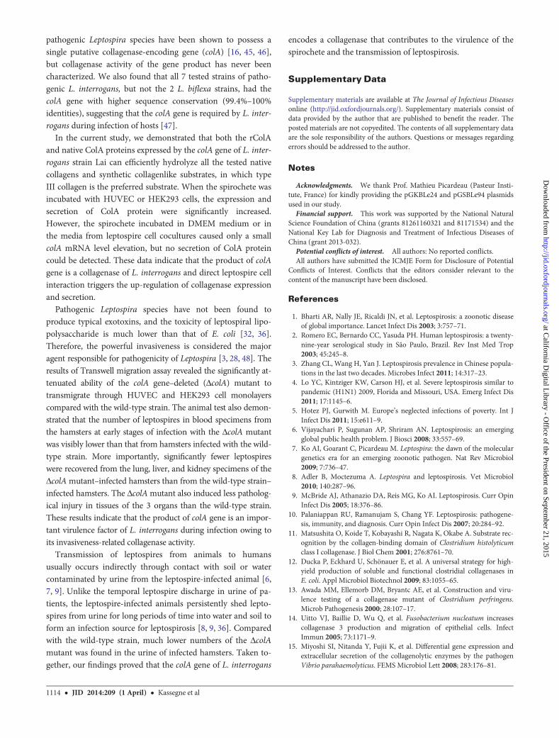
pathogenic Leptospira species have been shown to possess asingle putative collagenase-encoding gene (colA) [16, 45, 46],but collagenase activity of the gene product has never beencharacterized. We also found that all 7 tested strains of patho-genic L. interrogans, but not the 2 L. biflexa strains, had thecolA gene with higher sequence conservation (99.4%–100%identities), suggesting that the colA gene is required by L. inter-rogans during infection of hosts [47].
In the current study, we demonstrated that both the rColAand native ColA proteins expressed by the colA gene of L. inter-rogans strain Lai can efficiently hydrolyze all the tested nativecollagens and synthetic collagenlike substrates, in which typeIII collagen is the preferred substrate. When the spirochete wasincubated with HUVEC or HEK293 cells, the expression andsecretion of ColA protein were significantly increased.However, the spirochete incubated in DMEM medium or inthe media from leptospire cell cocultures caused only a smallcolA mRNA level elevation, but no secretion of ColA proteincould be detected. These data indicate that the product of colAgene is a collagenase of L. interrogans and direct leptospire cellinteraction triggers the up-regulation of collagenase expressionand secretion.
Pathogenic Leptospira species have not been found toproduce typical exotoxins, and the toxicity of leptospiral lipo-polysaccharide is much lower than that of E. coli [32, 36].Therefore, the powerful invasiveness is considered the majoragent responsible for pathogenicity of Leptospira [3, 28, 48]. Theresults of Transwell migration assay revealed the significantly at-tenuated ability of the colA gene–deleted (ΔcolA) mutant totransmigrate through HUVEC and HEK293 cell monolayerscompared with the wild-type strain. The animal test also demon-strated that the number of leptospires in blood specimens fromthe hamsters at early stages of infection with the ΔcolA mutantwas visibly lower than that from hamsters infected with the wild-type strain. More importantly, significantly fewer leptospireswere recovered from the lung, liver, and kidney specimens of theΔcolAmutant–infected hamsters than from the wild-type strain–infected hamsters. The ΔcolAmutant also induced less patholog-ical injury in tissues of the 3 organs than the wild-type strain.These results indicate that the product of colA gene is an impor-tant virulence factor of L. interrogans during infection owing toits invasiveness-related collagenase activity.
Transmission of leptospires from animals to humansusually occurs indirectly through contact with soil or watercontaminated by urine from the leptospire-infected animal [6,7, 9]. Unlike the temporal leptospire discharge in urine of pa-tients, the leptospire-infected animals persistently shed lepto-spires from urine for long periods of time into water and soil toform an infection source for leptospirosis [8, 9, 36]. Comparedwith the wild-type strain, much lower numbers of the ΔcolAmutant was found in the urine of infected hamsters. Taken to-gether, our findings proved that the colA gene of L. interrogans
encodes a collagenase that contributes to the virulence of thespirochete and the transmission of leptospirosis.
Supplementary Data
Supplementary materials are available at The Journal of Infectious Diseasesonline (http://jid.oxfordjournals.org/). Supplementary materials consist ofdata provided by the author that are published to benefit the reader. Theposted materials are not copyedited. The contents of all supplementary dataare the sole responsibility of the authors. Questions or messages regardingerrors should be addressed to the author.
Notes
Acknowledgments. We thank Prof. Mathieu Picardeau (Pasteur Insti-tute, France) for kindly providing the pGKBLe24 and pGSBLe94 plasmidsused in our study.Financial support. This work was supported by the National Natural
Science Foundation of China (grants 81261160321 and 81171534) and theNational Key Lab for Diagnosis and Treatment of Infectious Diseases ofChina (grant 2013-032).Potential conflicts of interest. All authors: No reported conflicts.All authors have submitted the ICMJE Form for Disclosure of Potential
Conflicts of Interest. Conflicts that the editors consider relevant to thecontent of the manuscript have been disclosed.
References
1. Bharti AR, Nally JE, Ricaldi JN, et al. Leptospirosis: a zoonotic diseaseof global importance. Lancet Infect Dis 2003; 3:757–71.
2. Romero EC, Bernardo CC, Yasuda PH. Human leptospirosis: a twenty-nine-year serological study in São Paulo, Brazil. Rev Inst Med Trop2003; 45:245–8.
3. Zhang CL, Wang H, Yan J. Leptospirosis prevalence in Chinese popula-tions in the last two decades. Microbes Infect 2011; 14:317–23.
4. Lo YC, Kintziger KW, Carson HJ, et al. Severe leptospirosis similar topandemic (H1N1) 2009, Florida and Missouri, USA. Emerg Infect Dis2011; 17:1145–6.
5. Hotez PJ, Gurwith M. Europe’s neglected infections of poverty. Int JInfect Dis 2011; 15:e611–9.
6. Vijayachari P, Sugunan AP, Shriram AN. Leptospirosis: an emergingglobal public health problem. J Biosci 2008; 33:557–69.
7. Ko AI, Goarant C, Picardeau M. Leptospira: the dawn of the moleculargenetics era for an emerging zoonotic pathogen. Nat Rev Microbiol2009; 7:736–47.
8. Adler B, Moctezuma A. Leptospira and leptospirosis. Vet Microbiol2010; 140:287–96.
9. McBride AJ, Athanazio DA, Reis MG, Ko AI. Leptospirosis. Curr OpinInfect Dis 2005; 18:376–86.
10. Palaniappan RU, Ramanujam S, Chang YF. Leptospirosis: pathogene-sis, immunity, and diagnosis. Curr Opin Infect Dis 2007; 20:284–92.
11. Matsushita O, Koide T, Kobayashi R, Nagata K, Okabe A. Substrate rec-ognition by the collagen-binding domain of Clostridium histolyticumclass I collagenase. J Biol Chem 2001; 276:8761–70.
12. Ducka P, Eckhard U, Schönauer E, et al. A universal strategy for high-yield production of soluble and functional clostridial collagenases inE. coli. Appl Microbiol Biotechnol 2009; 83:1055–65.
13. Awada MM, Ellemorb DM, Bryantc AE, et al. Construction and viru-lence testing of a collagenase mutant of Clostridium perfringens.Microb Pathogenesis 2000; 28:107–17.
14. Uitto VJ, Baillie D, Wu Q, et al. Fusobacterium nucleatum increasescollagenase 3 production and migration of epithelial cells. InfectImmun 2005; 73:1171–9.
15. Miyoshi SI, Nitanda Y, Fujii K, et al. Differential gene expression andextracellular secretion of the collagenolytic enzymes by the pathogenVibrio parahaemolyticus. FEMS Microbiol Lett 2008; 283:176–81.
1114 • JID 2014:209 (1 April) • Kassegne et al
at California D
igital Library - O
ffice of the President on September 21, 2015
http://jid.oxfordjournals.org/D
ownloaded from

16. Dong HY, Hu Y, Xue F, et al. Characterization of the ompL1 gene ofpathogenic Leptospira species in China and cross-immunogenicity ofthe OmpL1 protein. BMCMicrobiol 2008; 8:223–35.
17. Ren SX, Fu G, Jiang XG, et al. Unique physiological and pathogenic fea-tures of Leptospira interrogans revealed by whole-genome sequencing.Nature 2003; 422:888–93.
18. Lin XA, Sun AH, Ruan P, Zhang Z, Yan J. Characterization of con-served combined T and B cell epitopes in Leptospira interrogans majorouter membrane proteins OmpL1 and LipL41. BMC Microbiol 2011;11:21–6.
19. Jung CM, Matsushita O, Katayama S, Minami J, Sakurai J, Okabe A.Identification of metal ligands in the Clostridium histolyticum ColHcollagenase. J Bacteriol 1999; 181:2816–22.
20. Matsushita O, Yoshihara K, Katayama SI, Minami J, Okabe A. Purifica-tion and characterization of a Clostridium perfringens 120-kilodaltoncollagenase and nucleotide sequence of the corresponding gene. J Bac-teriol 1994; 176:149–56.
21. Wünsch E, Heidrich HG. Zur quantitativen bestimmung der kollage-nase. Hoppe-Seyler Z Physiol Chem 1963; 333:149–51.
22. Odake S, Okayama T, Obata M, et al. Vertebrate collagenase inhibitor I,tripeptidyl hydroxamic acids. Chem Pharm Bull 1990; 38:1007–11.
23. Yu MS, Lee CY. Expression and characterization of the prtV gene en-coding a collagenase from Vibrio parahaemolyticus in Escherichia coli.Microbiology 1999; 145:143–50.
24. Schreier S, Triampo W, Doungchawee G, Triampo D, Chadsuthi S.Leptospirosis research: fast, easy and reliable enumeration of mobileleptospires. Biol Res 2009; 42:5–12.
25. Li LW, Ojcius DM, Yan J. Comparison of invasion of fibroblasts andmacrophages by high- and low-virulence Leptospira strains: Coloniza-tion of the host-cell nucleus and induction of necrosis by the virulentstrain. Arch Microbiol 2007; 188:591–8.
26. Liu YY, Zheng W, Li LW, Mao YF, Yan J. Pathogenesis of leptospirosis:interaction of Leptospira interrogans with in vitro cultured mammaliancells. Med Microbiol Immunol 2007; 196:233–9.
27. Jin DD, Ojcius DM, Sun D, et al. Leptospira interrogans induces apo-ptosis in macrophages via caspase-8- and caspase-3-dependent path-ways. Infect Immun 2009; 77:799–809.
28. Zhang L, Zhang CL, Ojcius DM, et al. Identification of Mce as a criticalouter membrane protein of pathogenic Leptospira species responsiblefor RGD motif-dependent adherence and host-cell invasion. Mol Mi-crobiol 2012; 83:1006–23.
29. Carrillo-Casas EM, Hernández-Castro R, Suárez-Güemes F, de la Peña-Moctezuma A. Selection of the internal control gene for real-timequantitative RT-PCR assays in temperature treated Leptospira. CurrMicrobiol 2008; 56:539–46.
30. Pfaffl MW, Horgan GW, Dempfle L. Relative expression software tool(REST) for group-wise comparison and statistical analysis of relativeexpression results in real-time PCR. Nucleic Acids Res 2002; 30:e36.
31. Luo DJ, Xue F, Ojcius DM, et al. Protein typing of major outer mem-brane lipoproteins from Chinese pathogenic Leptospira spp. and char-acterization of their immunogenicity. Vaccine 2010; 28:243–55.
32. Wang H, Wu YF, Ojcius DM, et al. Leptospiral hemolysins induceproinflammatory cytokines through Toll-like receptor 2- and 4-mediat-ed JNK and NF-κB signaling pathways. PLoS One 2012; 7:e42266.
33. Liao SM, Sun AH, Ojcius DM, Wu SL, Zhao JF, Yan J. Inactivation ofthe fliY gene encoding a flagellar motor switch protein attenuates mo-bility and virulence of Leptospira interrogans strain Lai. BMCMicrobiol2009; 9:253–63.
34. Martinez-Lopez DG, Fahey M, Coburn J. Responses of human endo-thelial cells to pathogenic and non-pathogenic Leptospira species. PLoSNeglect Trop Dis 2010; 4:e918.
35. Ristow P, Bourhy P, da Cruz-McBride FW, et al. The OmpA-likeprotein Loa22 is essential for leptospiral virulence. PLoS Pathog 2007;3:894–903.
36. Faine S, Adler B, Bolin C, Perolat P. Leptospira and leptospirosis. 2nded. Melbourne, Australia: MediSci, 1999.
37. Sansonetti PJ. Bacterial pathogens, from adherence to invasion: com-parative strategies. Med Microbiol Immunol 1993; 182:223–32.
38. Awad MM, Ellemor DM, Bryant AE, et al. Construction and virulencetesting of a collagenase mutant of Clostridium perfringens. MicrobPathog 2000; 28:107–17.
39. Han HJ, Taki T, Kondo H, Hirono I, Aoki T. Pathogenic potential of acollagenase gene from Aeromonas veronii. Can J Microbiol 2008; 54:1–10.
40. Li SJ, Ojcius DM, Liao SM, et al. Replication or death: distinct fates ofpathogenic Leptospira strain Lai within macrophages of human ormouse origin. Innate Immun 2010; 16:80–92.
41. Zhu Y, Oganesian A, Keene DR, Sandell LJ. Type IIA procollagen con-taining the cysteine-rich animo propeptide is deposited in the extracel-lular matrix of prechondrogenic tissue and binds to TGF-beta 1 andBMP-2. Cell Biol 1999; 144:1069–80.
42. De paepe A, Malfait F. Bleeding and bruising in patients with Ehlers-Danlos syndrome and other collagen vascular disorders. Br J Haematol2004; 127:491–500.
43. Kühn K. Basement membrane (type IV) collagen. Matrix Biol 1995;14:439–45.
44. Picardeau M, Bulach DM, Bouchier C, et al. Genome sequence of thesaprophyte Leptospira biflexa provides insights into the evolution ofLeptospira and the pathogenesis of leptospirosis. PLoS One 2008; 3:e1607.
45. Nascimento AL, Ko AI, Martins EA, et al. Comparative genomics oftwo Leptospira interrogans serovars reveals novel insights into physiolo-gy and pathogenesis. J Bacteriol 2004; 86:2164–72.
46. Bulach DM, Zuerner RL, Wilson P, et al. Genome reduction in Lepto-spira borgpetersenii reflects limited transmission potential. Proc NatlAcad Sci U S A 2006; 103:14560–5.
47. Xue F, Yan J, Picardeau M. Evolution and pathogenesis of Leptospiraspp.: Lessons learned from the genomes. Microbes Infect 2009; 11:328–33.
48. Davis JM, Haake DA, Ramakrishnan L. Leptospira interrogans stablyinfects zebrafish embryos, altering phagocyte behavior and homing tospecific tissues. PLoS NTD 2009; 3:e463.
ColA as a Virulence Factor of Leptospira • JID 2014:209 (1 April) • 1115
at California D
igital Library - O
ffice of the President on September 21, 2015
http://jid.oxfordjournals.org/D
ownloaded from