Exploitation of Astrocytes by Glioma Cells to Facilitate Invasiveness: a Mechanism Involving Matrix...
10
Exploitation of Astrocytes by Glioma Cells to Facilitate Invasiveness: A Mechanism Involving Matrix Metalloproteinase-2 and the Urokinase-Type Plasminogen Activator–Plasmin Cascade Duc M. Le, 1 Arnaud Besson, 1 Darrin K. Fogg, 2 Kyu-Sil Choi, 2 David M. Waisman, 2 Cynthia G. Goodyer, 5 Barry Rewcastle, 3 and V. Wee Yong 1,4 Departments of 1 Oncology, 2 Medical Science, 3 Pathology, and 4 Clinical Neurosciences, University of Calgary, Calgary, Alberta, Canada, and 5 McGill University, Montreal Children’s Hospital Research Institute, Montreal, Quebec, Canada The presence of reactive astrocytes around glioma cells in the CNS suggests the possibility that these two cell types could be interacting. We addressed whether glioma cells use the astrocyte environment to modulate matrix metalloproteinase-2 (MMP-2), a proteolytic enzyme implicated in the invasiveness of glioma cells. We found that astrocytes in culture produce significant amounts of the pro-form of MMP-2 but undetectable levels of active MMP-2. However, after coculture with the U251N glioma line, astrocyte pro-MMP-2 was converted to the active form. The mechanism of pro-MMP-2 activation in glioma–astrocyte coculture was investigated and was found to involve the urokinase-type plasminogen activator (uPA)–plasmin cascade whereby uPA bound to uPA receptor (uPAR), leading to the conversion of plasminogen to plasmin. The latter cleaved pro-MMP-2 to generate its active form. Furthermore, key components (i.e., uPAR, uPA, and pro-MMP-2) were contributed principally by astrocytes, whereas the U251N glioma cells provided plasminogen. In correspondence with this biochemical cascade, the transmigration of U251N cells through Boyden invasion chambers coated with an extracellular matrix barrier was increased significantly in the presence of astrocytes, and this was inhibited by agents that disrupted the uPA–plasmin cascade. Finally, using resected human glioblastoma specimens, we found that tumor cells, but not astrocytes, expressed plasminogen in situ. We conclude that glioma cells exploit their astrocyte environment to activate MMP-2 and that this leads to the increased invasiveness of glioma cells. Key words: astrocytes; brain tumors; glioma; metalloproteinases; metastasis; plasmin Introduction Gliomas are brain neoplasms that account for 50% of tumors that arise within the CNS. The more malignant forms, compris- ing anaplastic astrocytomas and the high-grade glioblastoma multiforme (GBM), may invade diffusely throughout the CNS to form foci of new growth, rendering surgery, radiation, and che- motherapies inadequate as treatment modalities. Understanding the mechanisms that regulate glioma invasiveness is critical to improve the prognosis of patients with incurable gliomas. The family of matrix metalloproteinases (MMPs) is impli- cated in tumor invasion and metastasis (Sternlicht and Werb, 2001). In addition, other features of tumor evolution, including survival, growth, and angiogenesis, also may be dependent on MMPs (Chambers and Matrisian, 1997). MMP members are classified into subgroups on the basis of structure and substrate preference (Yong et al., 2001). One category, the gelatinases (MMP-2 and MMP-9), has been associated intimately with tu- mor invasiveness and metastasis because of its potent ability to degrade the type IV collagen present in basement membrane that surrounds blood vessels. Elevated levels of gelatinases are found in many types of human cancers (Stetler-Stevenson et al., 1993). With respect to gliomas, the upregulation of MMP-2 and MMP-9 has been observed in cell lines and resected specimens (Rutka et al., 1995; Saxena et al., 1995; Sawaya et al., 1996; Forsyth et al., 1998, 1999; Lampert et al., 1998; Beliveau et al., 1999; Pa- genstecher et al., 2001; VanMeter et al., 2001; Wild-Bode et al., 2001). In vivo the expression of MMP-2, determined by in situ hybridization or immunohistochemistry, is principally in glioma cells, whereas MMP-9 expression tends to be predominant in regions of angiogenesis (for review, see Forsyth et al., 2001). These analyses implicate MMP-2 in the growth or invasiveness of gliomas in situ, whereas MMP-9 may be associated with angio- genesis. In support, there is a high correlation between the capac- ity of cultured glioma cell lines to invade across an artificial base- ment membrane, Matrigel, and their expression of MMP-2 (Uhm et al., 1996). Inhibitors of metalloproteinases reduce the invasiveness of glioma cell lines in vitro (Uhm et al., 1996; Tonn et al., 1999) and attenuate the growth of glioma cells implanted into the flank of severe combined immunodeficient (SCID) mice (Price et al., 1999). MMPs are produced by cells as pro-enzymes, and they require Received April 15, 2002; revised Feb. 12, 2003; accepted Feb. 19, 2003. D.M.L. was supported by studentship awards from the National Science and Engineering Research Council of Canada and the Alberta Heritage Foundation for Medical Research. We thank the Canadian Institutes for Health Research for support of operating funds. Correspondence should be addressed to Dr. V. Wee Yong, Departments of Oncology and Clinical Neurosciences, University of Calgary, 3330 Hospital Drive, Calgary, Alberta T2N 4N1, Canada. E-mail: [email protected]. Copyright © 2003 Society for Neuroscience 0270-6474/03/234034-10$15.00/0 4034 • The Journal of Neuroscience, May 15, 2003 • 23(10):4034 – 4043
Transcript of Exploitation of Astrocytes by Glioma Cells to Facilitate Invasiveness: a Mechanism Involving Matrix...
Exploitation of Astrocytes by Glioma Cells to FacilitateInvasiveness: A Mechanism Involving MatrixMetalloproteinase-2 and the Urokinase-Type PlasminogenActivator–Plasmin Cascade
Duc M. Le,1 Arnaud Besson,1 Darrin K. Fogg,2 Kyu-Sil Choi,2 David M. Waisman,2 Cynthia G. Goodyer,5
Barry Rewcastle,3 and V. Wee Yong1,4
Departments of 1Oncology, 2Medical Science, 3Pathology, and 4Clinical Neurosciences, University of Calgary, Calgary, Alberta, Canada, and 5McGillUniversity, Montreal Children’s Hospital Research Institute, Montreal, Quebec, Canada
The presence of reactive astrocytes around glioma cells in the CNS suggests the possibility that these two cell types could be interacting.We addressed whether glioma cells use the astrocyte environment to modulate matrix metalloproteinase-2 (MMP-2), a proteolyticenzyme implicated in the invasiveness of glioma cells. We found that astrocytes in culture produce significant amounts of the pro-formof MMP-2 but undetectable levels of active MMP-2. However, after coculture with the U251N glioma line, astrocyte pro-MMP-2 wasconverted to the active form. The mechanism of pro-MMP-2 activation in glioma–astrocyte coculture was investigated and was found toinvolve the urokinase-type plasminogen activator (uPA)–plasmin cascade whereby uPA bound to uPA receptor (uPAR), leading to theconversion of plasminogen to plasmin. The latter cleaved pro-MMP-2 to generate its active form. Furthermore, key components (i.e.,uPAR, uPA, and pro-MMP-2) were contributed principally by astrocytes, whereas the U251N glioma cells provided plasminogen. Incorrespondence with this biochemical cascade, the transmigration of U251N cells through Boyden invasion chambers coated with anextracellular matrix barrier was increased significantly in the presence of astrocytes, and this was inhibited by agents that disrupted theuPA–plasmin cascade. Finally, using resected human glioblastoma specimens, we found that tumor cells, but not astrocytes, expressedplasminogen in situ. We conclude that glioma cells exploit their astrocyte environment to activate MMP-2 and that this leads to theincreased invasiveness of glioma cells.
Key words: astrocytes; brain tumors; glioma; metalloproteinases; metastasis; plasmin
IntroductionGliomas are brain neoplasms that account for �50% of tumorsthat arise within the CNS. The more malignant forms, compris-ing anaplastic astrocytomas and the high-grade glioblastomamultiforme (GBM), may invade diffusely throughout the CNS toform foci of new growth, rendering surgery, radiation, and che-motherapies inadequate as treatment modalities. Understandingthe mechanisms that regulate glioma invasiveness is critical toimprove the prognosis of patients with incurable gliomas.
The family of matrix metalloproteinases (MMPs) is impli-cated in tumor invasion and metastasis (Sternlicht and Werb,2001). In addition, other features of tumor evolution, includingsurvival, growth, and angiogenesis, also may be dependent onMMPs (Chambers and Matrisian, 1997). MMP members areclassified into subgroups on the basis of structure and substratepreference (Yong et al., 2001). One category, the gelatinases(MMP-2 and MMP-9), has been associated intimately with tu-
mor invasiveness and metastasis because of its potent ability todegrade the type IV collagen present in basement membrane thatsurrounds blood vessels. Elevated levels of gelatinases are foundin many types of human cancers (Stetler-Stevenson et al., 1993).
With respect to gliomas, the upregulation of MMP-2 andMMP-9 has been observed in cell lines and resected specimens(Rutka et al., 1995; Saxena et al., 1995; Sawaya et al., 1996; Forsythet al., 1998, 1999; Lampert et al., 1998; Beliveau et al., 1999; Pa-genstecher et al., 2001; VanMeter et al., 2001; Wild-Bode et al.,2001). In vivo the expression of MMP-2, determined by in situhybridization or immunohistochemistry, is principally in gliomacells, whereas MMP-9 expression tends to be predominant inregions of angiogenesis (for review, see Forsyth et al., 2001).These analyses implicate MMP-2 in the growth or invasiveness ofgliomas in situ, whereas MMP-9 may be associated with angio-genesis. In support, there is a high correlation between the capac-ity of cultured glioma cell lines to invade across an artificial base-ment membrane, Matrigel, and their expression of MMP-2(Uhm et al., 1996). Inhibitors of metalloproteinases reduce theinvasiveness of glioma cell lines in vitro (Uhm et al., 1996; Tonn etal., 1999) and attenuate the growth of glioma cells implanted intothe flank of severe combined immunodeficient (SCID) mice(Price et al., 1999).
MMPs are produced by cells as pro-enzymes, and they require
Received April 15, 2002; revised Feb. 12, 2003; accepted Feb. 19, 2003.D.M.L. was supported by studentship awards from the National Science and Engineering Research Council of
Canada and the Alberta Heritage Foundation for Medical Research. We thank the Canadian Institutes for HealthResearch for support of operating funds.
Correspondence should be addressed to Dr. V. Wee Yong, Departments of Oncology and Clinical Neurosciences,University of Calgary, 3330 Hospital Drive, Calgary, Alberta T2N 4N1, Canada. E-mail: [email protected] © 2003 Society for Neuroscience 0270-6474/03/234034-10$15.00/0
4034 • The Journal of Neuroscience, May 15, 2003 • 23(10):4034 – 4043
further processing to generate the active enzyme. The activationof pro-MMPs is facilitated by active forms of other MMPs or byother groups of proteinases (Nagase, 1997). For example, a serineproteinase, plasmin, can activate gelatinases without the need forany intermediaries (Mazzieri et al., 1997).
The mechanism by which gliomas modulate their MMP-2content or activity remains unclear. Because glioma cells producefactors that promote the proliferation (Couldwell et al., 1992)and motility (Lal et al., 1996) of astrocytes, we have tested thehypothesis that glioma cells exploit their astrocyte environmentfor MMP-2 production and activation and that the consequenceof this interaction is the increased ability of glioma cells to invade.We also have addressed the mechanisms of the glioma exploita-tion of astrocytes for invasion.
Materials and MethodsDetection of reactive astrocytes in proximity to glioma cells in situ. Speci-mens obtained from surgical resections of human brain tumors wereimmersion-fixed in 10% neutral buffered formalin and subsequentlywere embedded in paraffin. Tissue sections were cut (5 �m) and pro-cessed for immunoreactivity for glial fibrillary acidic protein (GFAP), anastrocyte-specific marker. In brief, first the sections were deparaffinized(xylene, 3�; 95% ethanol, 3�) for 20 min. Blocking of endogenousperoxidase activity was achieved by incubating sections in 1% H2O2 inmethanol for 15 min. Antigen recovery ensued by incubating sections in0.05% trypsin and 2% CaCl2, pH 7.5, at 37°C for 20 min. After beingblocked with nonimmune serum, the sections were incubated with theprimary antibody (rabbit anti-GFAP, Dako, Mississauga, Ontario) for 3hr in a humidity chamber at room temperature. After the sections wererinsed, a secondary antibody was applied. Sections were treated withavidin– biotin–peroxidase (ABC) complex (Vector Laboratories, Burl-ington, Ontario), and immunoreactivity was visualized by using diami-nobenzidine (DAB) as the chromogen. Sections were counterstainedwith hematoxylin and examined with a light microscope.
To implant human glioma cells into the brain of mice, we anesthetizedSCID NOD mice with ketamine (200 mg/kg, i.p.) and xylazine (10 mg/kg, i.p.) and secured them in a stereotactic apparatus. After a midlineincision exposed the skull, a 0.9 mm burr hole was made 1 mm lateral tothe midline, half-way between the lambda and bregma sutures. U87 orU251N cells (100,000 cells) suspended in 2 �l of saline were injected intothe mouse brain with a 5 �l Hamilton syringe. The needle was removedslowly, and the skin was sutured. Mice were given 2 ml of saline (injectedsubcutaneously) and were allowed to recover on a heating pad. On day 25after surgery the mice were killed, and brains were harvested andimmersion-fixed in 10% neutral buffered formalin. After fixation thebrains were embedded in paraffin, and 5-�m-thick coronal sections wereused for GFAP immunoreactivity.
To attempt colocalization of plasminogen on glioma cells or astrocytesin situ, we used a human brain tumor specimen. Specifically, a paraffin-embedded specimen containing glioblastoma cells that were GFAP-negative was used to allow for their differentiation from neighboringGFAP-positive astrocytes. After deparaffinization, antigen recovery, andblocking as described above, 6 �m sections were incubated with a poly-clonal rabbit anti-GFAP (Dako), followed by anti-rabbit Ig Cy3 (1:500;Molecular Probes, Leiden, The Netherlands). Then a mouse monoclonalantibody to human plasminogen (number 3644, 10 �g/ml; AmericanDiagnostica, Greenwich, CT) and anti-mouse Ig Alexa 488 were used.Finally, the sections were incubated with Hoechst 33852 (Sigma, St.Louis, MO) for 10 min to label all cell nuclei.
Cell culture. The U251N and U87 cell lines are multiply passaged hu-man glioblastoma cells (Ishii et al., 1999), the use for which has beennoted previously (Besson and Yong, 2000); we have selected these forstudy because these human lines grow well in the brains of immunocom-promised mice. Human fetal astrocytes were cultured from brains ob-tained from therapeutic abortions. The use of these specimens was ap-proved by institutional human ethics committees. Astrocytes of �99%purity were prepared as described in detail previously (Vecil et al., 2000).
Glioma cells or astrocytes were maintained in MEM supplemented with10% fetal calf serum, 1� nonessential amino acids, 1 mM sodium pyru-vate, 2 mM glutamine, and 0.5% dextrose. All culture medium constitu-ents were obtained from Invitrogen (Burlington, Ontario). When cellswere used in experiments to measure the content or activity of MMP-2,the medium was changed over to an RPMI-based serum-free but hor-monally supplemented medium (M2; Boutros et al., 1997) containing0.5% dextrose, 25 �g/ml insulin, 100 �g/ml transferrin, 20 nM proges-terone, 50 �M putrescine, and 30 nM sodium selenite. M2 serum-freemedium was necessary because fetal calf serum contains a variety ofMMPs that would obscure MMP-2 produced by cells.
In experiments to detect MMP-2, unless otherwise stated, 1 millionhuman fetal astrocytes (HFA) or U251N glioma cells were cultured alonein six-well culture plates or were cocultured (500,000 cells for each celltype) for 24 hr. The volume of medium was 1.5 ml/well. For each exper-iment two separate culture duplicates were used per condition (thusaccounting for duplicate lanes in zymography; see Results). When inhib-itors were used to test the requirement of various protease subclasses toactivate MMP-2, these were added immediately after coculture of thecells. The following inhibitors were used: pepstatin A, E-64 [trans-epoxysuccinyl-L-leucylamido-(4-guanidino)-butane], aprotinin (fromSigma-Aldrich, Oakville, CA), �2-antiplasmin (Calbiochem, La Jolla,CA), and recombinant human tissue inhibitors of metalloproteinases(TIMPs) -1 or -2 (generously provided by Dr. Dylan Edwards, Universityof East Anglia, Norwich, UK). These inhibitors were used at concentra-tions described to be effective in culture (Uhm et al., 1996).
Analyses of MMP-2 level and activity. As with all MMPs, MMP-2 existsin a pro- and active form. To detect both forms simultaneously, we useda gelatin zymography protocol (Uhm et al., 1996). Because the gelati-nases are secreted proteases, the amount of pro- and active MMP-2 pro-duced by cells was examined by assaying the cell-conditioned mediumafter 24 hr of exposure. The cell-conditioned medium was mixed with4� gel-loading buffer; 80 �l was electrophoresed in a 10% SDS-gel con-taining 1 mg/ml gelatin. The gel was incubated overnight in rinse buffer(50 mM Tris, pH 7.5, 5 mM CaCl2, and 2.5% Triton X-100) at roomtemperature to wash away the SDS, thus allowing the gelatinases to re-nature. Then the gel was placed in a reaction buffer (50 mM Tris, pH 7.5,and 5 mM CaCl2) for 18 hr to facilitate gelatin degradation by gelatinases.Next the gel was incubated for at least 4 hr in Coomassie blue stainsolution. The gel was destained by 10% isopropanol and 10% acetic acid.Areas of gelatinase activity, given the paucity of gelatin in these zones,were observed as clear bands against a dark background. The locations ofpro- and active MMP-2 were based on molecular weight determinationsand by confirmations with the resolved pattern of recombinant humanMMP-2 (Oncogene, Cambridge, MA). Previous experiments that usedMMP-2 antibodies to deplete this gelatinase from conditioned mediahave confirmed the locations of pro- and active MMP-2 (Uhm et al.,1996). It should be noted that, although pro-MMP-2 is an inactive zy-mogen in its native state, by virtue of the cysteine residue in the propep-tide interacting with zinc atom at the catalytic site, the dissociation of thecysteine–zinc interaction during electrophoresis allows the pro-MMP-2to degrade gelatin and to be identifiable as a white band in zymograms.
Although zymography is based on the ability of gelatinases to degradegelatin, the resultant size of the band of gelatin degradation is a directreflection of the amounts of pro- and active gelatinases rather than theirintrinsic enzyme activity. Thus to measure MMP-2 enzyme activity di-rectly, we used an MMP-2 fluorogenic substrate, MCA-Pro-Leu-Ala-Nva-Dpa-Ala-Arg-NH2 (Calbiochem). When cleaved by active MMP-2,the quenched fluorescence was emitted. Then 50 �l of 24 hr cell-conditioned medium was added to individual wells of a 96-well cultureplate. MMP-2 substrate (50 �l of 50 �M stock solution) was added to thewells. Fluorescence was monitored for 1 hr in a PerkinElmer Life Sciences(Emeryville, CA) HTS 7000 Bioassay Reader (excitation wavelength of325 nm and emission wavelength of 393 nm) and expressed as relativefluorescence units.
The total level of MMP-2 (pro- and active) also was measured by usingan MMP-2 ELISA (Oncogene) according to the manufacturer’s instruc-tions. Medium conditioned for 24 hr by the cells was collected, and 50 �lwas added for analysis.
Le et al. • Glioma–Astrocyte Interactions J. Neurosci., May 15, 2003 • 23(10):4034 – 4043 • 4035
Plasmin activity assay. Because plasmin activity was low in cells insix-well plates, 100 mm dishes were seeded with astrocytes (5 millioncells/dish), U251N cells (5 million cells/dish) or astrocyte–U251N cocul-tures (2.5 million cells/dish of each cell type). Serum-free M2 medium (4ml/dish) was used, and the cells were incubated at 37°C for 24 hr. Eachconditioned medium was collected and cleared by centrifugation. Thesupernatant was concentrated centrifugally 100-fold with Ultrafree cen-trifugal filter tubes (Millipore, Bedford, MA) that retained molecules�10 kDa. Concentrated supernatant (0.5 ml) was added to 24-well cul-ture plates, followed by 20 �l (0.2 mM) of AFC-80 (7-amino-4-trifluoromethy coumarin: Z-Ala-Lys-Lys) plasmin substrate (EnzymeSystems Products, Livermore, CA). Fluorescence was monitored for 1 hr(excitation wavelength of 405 nm and emission wavelength of 535 nm)and expressed as relative fluorescence units.
uPAR immunofluorescence. Astrocytes (10,000 cells) or U251N cells(10,000 cells) were seeded onto 12 mm glass coverslips. After adherenceand morphological differentiation the cells were untreated or treatedwith 10 U/ml phospholipase C� (PI-PLC; Molecular Probes, Leiden,Netherlands) for 1 hr at 37°C. PI-PLC was removed, and the cells wereincubated at 37°C overnight before being fixed with 2% paraformalde-hyde for 20 min. The primary antibody (1:500 dilution of mouse anti-uPAR; American Diagnostica) was added to the coverslip and incubatedat room temperature for 1 hr. Coverslips were rinsed three times withPBS. Secondary antibody (1:500 dilution of goat anti-mouse Alexa 488)was applied for 1 hr at room temperature. Cells then were permeabilizedwith 0.2% Triton X-100 for 3 min. Phalloidin (1:1000 dilution; Molecu-lar Probes), staining filamentous actin to highlight cell boundaries, wasapplied for 1 hr at room temperature. After being rinsed with PBS, thecoverslips were mounted onto glass slides. Sections were viewed with anOlympus confocal laser-scanning microscope, and images were acquiredwith the Olympus Fluoview program (version 3.0).
Western blotting experiments. The 100 mm dishes were seeded withastrocytes (5 million cells), U251N cells (5 million cells), or astrocyte–U251N cells (2.5 million cells of each cell type) in M2 medium andincubated at 37°C for 24 hr. Samples were performed in duplicate. Con-ditioned medium was collected, cleared by centrifugation, and concen-trated 100-fold as described earlier. To obtain cell lysates, we rinsedculture dishes three times with PBS. Cells then were exposed to 200 �llysis buffer [containing (in mM): 50 HEPES, pH 7.5, 50 NaCl, 1 EDTA, 2.5EGTA plus 0.1% Tween-20, 1% Nonidet P-40, 0.2% SDS, 10% glycerolcomplemented with (in mM) 1 DTT, 10 �-glycerophosphate, 1 NaF, 0.1sodium orthovanadate, 1 PMSF, and (in �g/ml) 10 leupeptin, 10 apro-tinin, 10 pepstatin A]. A 26 gauge needle was used to homogenize thelysate, which then was left on ice for 10 min. The lysate was centrifuged(10,000 rpm) for 10 min, and the supernatant was kept. The total proteincontent per sample was analyzed by Bradford protein assay (Bio-Rad,Hercules, CA). Protein loading buffer (4�, 200 mM Tris-HCl, pH 7.8,400 mM DTT, 8% SDS, 0.4% bromophenol blue, and 40% glycerol) wasadded to each sample. Equal amounts (150 �g) of protein from celllysates or equal volumes of conditioned media were subjected to 8%SDS-PAGE. Then the proteins were transferred onto a polyvinylidenedifluoride (PDVF) membrane (Millipore). Membranes were blocked in ablocking solution (PBS containing 0.5% Tween-20 and 5% poweredmilk) and incubated with primary antibodies (1:500 dilution of anti-uPAR) overnight at 4°C. Finally, the membranes were rinsed with PBScontaining 0.1% Tween-20 and incubated in secondary antibody(1:10,000 dilution) for 1 hr at room temperature. ECL (Amersham Bio-sciences, Uppsala, Sweden) was used for detection.
Invasion assays. In vitro invasion assays were performed in 8 �m poresize Matrigel-coated invasion chambers (Becton Dickinson, Bedford,MA) as described previously (Uhm et al., 1996). Astrocytes (5000 cells)were seeded onto the top compartment of each chamber (placed in 24-well culture plates) and incubated at 37°C for 4 hr. Then U251N cells(25,000 cells) suspended in M2 were plated onto the astrocytes. Becauseastrocytes also may invade across the Matrigel barrier, the U251N gliomacells were stably transfected with a LacZ construct so that glioma cellscould be differentiated from astrocytes on the basis of galactosidase ac-tivity. After G418 selection and histochemical staining (see below), allglioma cells were �-galactosidase-positive (data not shown).
Medium conditioned by NIH3T3 fibroblasts was placed in the bottomwell of the invasion chambers to act as a directional chemoattractantforce for glioma invasion. The entire chamber setup, in triplicate for eachexperimental condition, was incubated for 68 hr at 37°C. Then the cellswere fixed with 0.4% paraformaldehyde for 15 min. �-Galactosidasestaining was performed by first rinsing the invasion chambers with phos-phate buffer (77 mM Na2HPO4; 23 mM NaH2PO4, adjusted to pH 7.4) for1 min, followed by fixation with the �-gal fixation buffer (0.25% glutar-aldehyde, 0.1 M Na2HPO4, 0.1 M NaH2PO, 2 mM MgCl2, and 125 mM
EGTA) for 15 min. After being washed three times with a wash buffer (0.1M Na2HPO4; 0.1 M NaH2PO4, 2 mM MgCl2, 0.01% desoxycholic acid, and0.02% IGEPAL; 15 min each time), the chambers were left overnight inthe �-gal staining buffer [containing 0.1 M Na2HPO4, 0.1 M NaH2PO4,and (in mM) 2 MgCl2, 5 K4Fe(CN)6-3 H2O, 5 K3Fe(CN)6, 1 spermidineplus 0.01% desoxycholic acid and 1 mg/ml X-gal] at 37°C. Cells wererefixed, in 95% ethanol, for 20 min. A cotton-tipped swab was applied tothe top surface of the membrane to remove cells that did not transmigrateacross the invasion chamber. Each membrane then was cut out andmounted onto glass slides. Cells that stained blue (i.e., glioma) werecounted with a light microscope.
MTT cell viability assays. Viability of cells was determined with the3-(4,5-dimethylthiazol-2-yl)-2,5-diphenyltetrazolium bromide thia-zolyl blue (MTT) assay. Astrocytes (10,000 cells) or U251N cells (10,000cells) were seeded in wells of a 96-well culture plate. These were incu-bated, in triplicate, with test factors at 37°C for 24 hr. MTT (20 �l; 0.5mg/ml) was added per well for an additional 1 hr at 37°C. Wells wererinsed twice with PBS; 100% DMSO (200 �l) was applied to each well tosolubilize the MTT color. Absorbance of each sample was measured by aspectrophotometer at a wavelength of 550 nm and expressed as relativeabsorbance units.
ResultsAstrocytes become reactive in the vicinity of glioma cellsin situWhen the CNS is subjected to a variety of insults, astrocytes un-dergo typical reactive changes in a phenomenon called astroglio-sis (Yong, 1998). Similarly, in the proximity of tumor cells insections from patients with glioma, reactive astrocytes were de-tected by increased GFAP immunoreactivity (Fig. 1). Reactiveastrocytes abutted glioma cells that had infiltrated some distancefrom the primary tumor mass (Fig. 1A, from a patient with ma-lignant astrocytoma) and also were found within a tumor mass ofanother type of glioma, oligodendroglioma (Fig. 1B).
To confirm the occurrence of reactive astrocytes in the pres-ence of neoplastic growth, we implanted U87 or U251N gliomacells into the brains of SCID mice. These cell lines were chosen fortheir capacity to readily grow intracerebrally. At 25 d after im-plantation a significant mass of tumor cells was observed (Fig.1C). Astrocytes at the interface of glioma and brain parenchymahad an increase in GFAP immunoreactivity, indicating theirreactivity.
Activation of pro-MMP-2 occurs inglioma–astrocyte cocultureThe observation that astrocytes become reactive when encoun-tering glioma cells in situ suggests the possibility that the two celltypes could be interacting. Specifically, we used tissue cultureparadigms to examine whether glioma cells exploit astrocytes tofacilitate their invasion. The levels of pro- and active MMP-2secreted by astrocytes and U251N cells were examined first intheir 24 hr conditioned media by gelatin zymography. Astrocyte-conditioned medium contained a high level of pro-MMP-2 butnegligible amounts of active MMP-2. The medium collectedfrom U251N cells contained low but detectable levels of both pro-and active MMP-2 (Fig. 2A). Interestingly, when astrocytes were
4036 • J. Neurosci., May 15, 2003 • 23(10):4034 – 4043 Le et al. • Glioma–Astrocyte Interactions
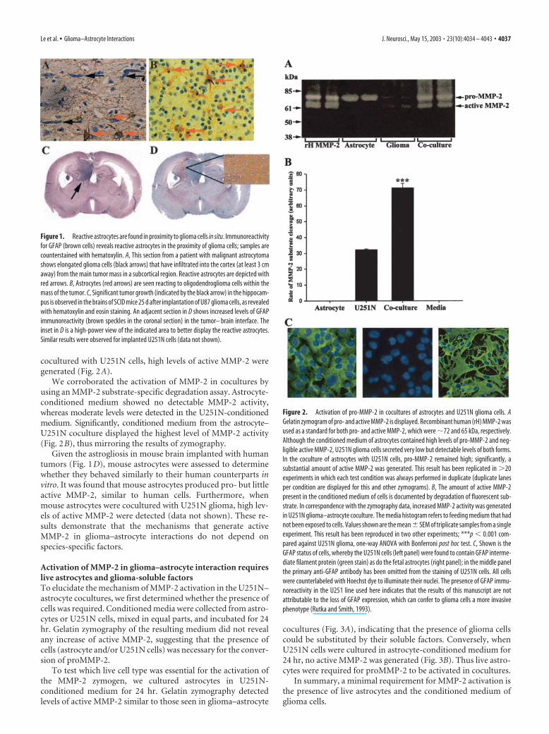
cocultured with U251N cells, high levels of active MMP-2 weregenerated (Fig. 2A).
We corroborated the activation of MMP-2 in cocultures byusing an MMP-2 substrate-specific degradation assay. Astrocyte-conditioned medium showed no detectable MMP-2 activity,whereas moderate levels were detected in the U251N-conditionedmedium. Significantly, conditioned medium from the astrocyte–U251N coculture displayed the highest level of MMP-2 activity(Fig. 2B), thus mirroring the results of zymography.
Given the astrogliosis in mouse brain implanted with humantumors (Fig. 1D), mouse astrocytes were assessed to determinewhether they behaved similarly to their human counterparts invitro. It was found that mouse astrocytes produced pro- but littleactive MMP-2, similar to human cells. Furthermore, whenmouse astrocytes were cocultured with U251N glioma, high lev-els of active MMP-2 were detected (data not shown). These re-sults demonstrate that the mechanisms that generate activeMMP-2 in glioma–astrocyte interactions do not depend onspecies-specific factors.
Activation of MMP-2 in glioma–astrocyte interaction requireslive astrocytes and glioma-soluble factorsTo elucidate the mechanism of MMP-2 activation in the U251N–astrocyte cocultures, we first determined whether the presence ofcells was required. Conditioned media were collected from astro-cytes or U251N cells, mixed in equal parts, and incubated for 24hr. Gelatin zymography of the resulting medium did not revealany increase of active MMP-2, suggesting that the presence ofcells (astrocyte and/or U251N cells) was necessary for the conver-sion of proMMP-2.
To test which live cell type was essential for the activation ofthe MMP-2 zymogen, we cultured astrocytes in U251N-conditioned medium for 24 hr. Gelatin zymography detectedlevels of active MMP-2 similar to those seen in glioma–astrocyte
cocultures (Fig. 3A), indicating that the presence of glioma cellscould be substituted by their soluble factors. Conversely, whenU251N cells were cultured in astrocyte-conditioned medium for24 hr, no active MMP-2 was generated (Fig. 3B). Thus live astro-cytes were required for proMMP-2 to be activated in cocultures.
In summary, a minimal requirement for MMP-2 activation isthe presence of live astrocytes and the conditioned medium ofglioma cells.
Figure 1. Reactive astrocytes are found in proximity to glioma cells in situ. Immunoreactivityfor GFAP (brown cells) reveals reactive astrocytes in the proximity of glioma cells; samples arecounterstained with hematoxylin. A, This section from a patient with malignant astrocytomashows elongated glioma cells (black arrows) that have infiltrated into the cortex (at least 3 cmaway) from the main tumor mass in a subcortical region. Reactive astrocytes are depicted withred arrows. B, Astrocytes (red arrows) are seen reacting to oligodendroglioma cells within themass of the tumor. C, Significant tumor growth (indicated by the black arrow) in the hippocam-pus is observed in the brains of SCID mice 25 d after implantation of U87 glioma cells, as revealedwith hematoxylin and eosin staining. An adjacent section in D shows increased levels of GFAPimmunoreactivity (brown speckles in the coronal section) in the tumor– brain interface. Theinset in D is a high-power view of the indicated area to better display the reactive astrocytes.Similar results were observed for implanted U251N cells (data not shown).
Figure 2. Activation of pro-MMP-2 in cocultures of astrocytes and U251N glioma cells. AGelatin zymogram of pro- and active MMP-2 is displayed. Recombinant human (rH) MMP-2 wasused as a standard for both pro- and active MMP-2, which were �72 and 65 kDa, respectively.Although the conditioned medium of astrocytes contained high levels of pro-MMP-2 and neg-ligible active MMP-2, U251N glioma cells secreted very low but detectable levels of both forms.In the coculture of astrocytes with U251N cells, pro-MMP-2 remained high; significantly, asubstantial amount of active MMP-2 was generated. This result has been replicated in �20experiments in which each test condition was always performed in duplicate (duplicate lanesper condition are displayed for this and other zymograms). B, The amount of active MMP-2present in the conditioned medium of cells is documented by degradation of fluorescent sub-strate. In correspondence with the zymography data, increased MMP-2 activity was generatedin U251N glioma–astrocyte coculture. The media histogram refers to feeding medium that hadnot been exposed to cells. Values shown are the mean � SEM of triplicate samples from a singleexperiment. This result has been reproduced in two other experiments; ***p � 0.001 com-pared against U251N glioma, one-way ANOVA with Bonferroni post hoc test. C, Shown is theGFAP status of cells, whereby the U251N cells (left panel) were found to contain GFAP interme-diate filament protein (green stain) as do the fetal astrocytes (right panel); in the middle panelthe primary anti-GFAP antibody has been omitted from the staining of U251N cells. All cellswere counterlabeled with Hoechst dye to illuminate their nuclei. The presence of GFAP immu-noreactivity in the U251 line used here indicates that the results of this manuscript are notattributable to the loss of GFAP expression, which can confer to glioma cells a more invasivephenotype (Rutka and Smith, 1993).
Le et al. • Glioma–Astrocyte Interactions J. Neurosci., May 15, 2003 • 23(10):4034 – 4043 • 4037
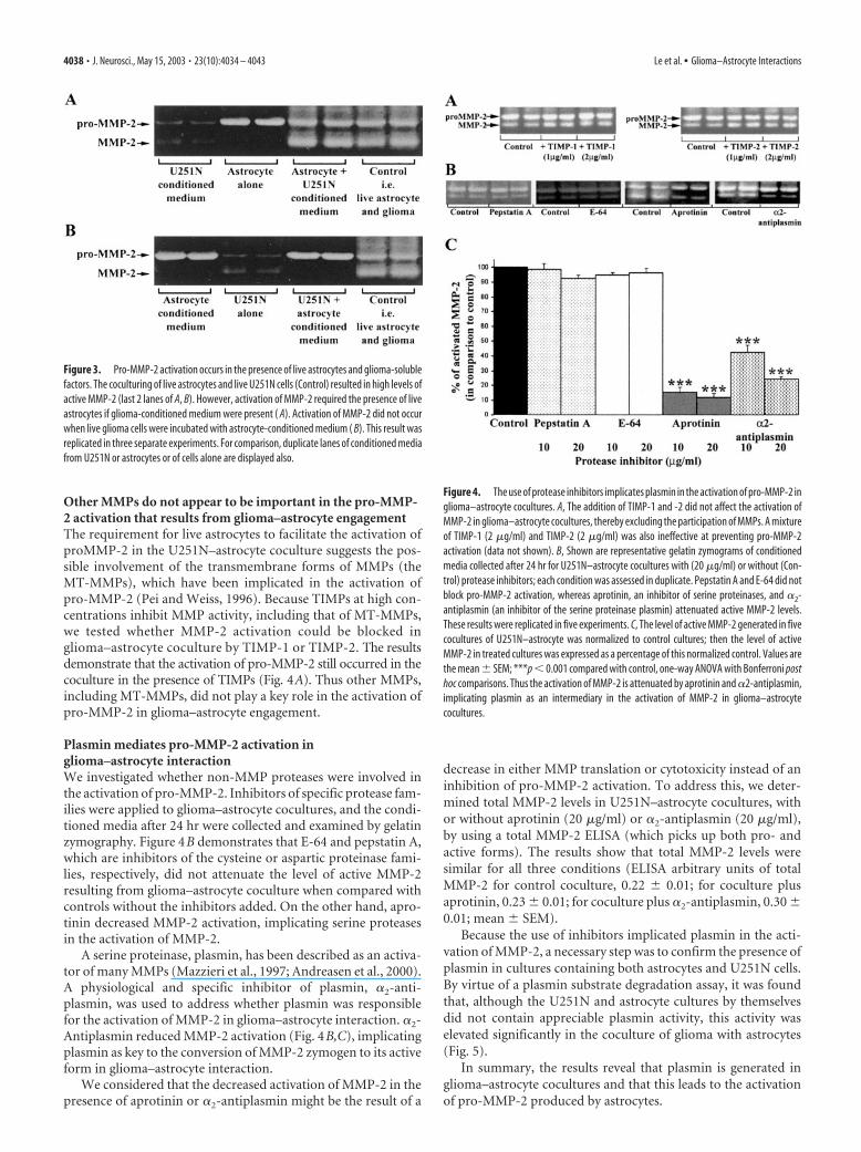
Other MMPs do not appear to be important in the pro-MMP-2 activation that results from glioma–astrocyte engagementThe requirement for live astrocytes to facilitate the activation ofproMMP-2 in the U251N–astrocyte coculture suggests the pos-sible involvement of the transmembrane forms of MMPs (theMT-MMPs), which have been implicated in the activation ofpro-MMP-2 (Pei and Weiss, 1996). Because TIMPs at high con-centrations inhibit MMP activity, including that of MT-MMPs,we tested whether MMP-2 activation could be blocked inglioma–astrocyte coculture by TIMP-1 or TIMP-2. The resultsdemonstrate that the activation of pro-MMP-2 still occurred in thecoculture in the presence of TIMPs (Fig. 4A). Thus other MMPs,including MT-MMPs, did not play a key role in the activation ofpro-MMP-2 in glioma–astrocyte engagement.
Plasmin mediates pro-MMP-2 activation inglioma–astrocyte interactionWe investigated whether non-MMP proteases were involved inthe activation of pro-MMP-2. Inhibitors of specific protease fam-ilies were applied to glioma–astrocyte cocultures, and the condi-tioned media after 24 hr were collected and examined by gelatinzymography. Figure 4B demonstrates that E-64 and pepstatin A,which are inhibitors of the cysteine or aspartic proteinase fami-lies, respectively, did not attenuate the level of active MMP-2resulting from glioma–astrocyte coculture when compared withcontrols without the inhibitors added. On the other hand, apro-tinin decreased MMP-2 activation, implicating serine proteasesin the activation of MMP-2.
A serine proteinase, plasmin, has been described as an activa-tor of many MMPs (Mazzieri et al., 1997; Andreasen et al., 2000).A physiological and specific inhibitor of plasmin, �2-anti-plasmin, was used to address whether plasmin was responsiblefor the activation of MMP-2 in glioma–astrocyte interaction. �2-Antiplasmin reduced MMP-2 activation (Fig. 4B,C), implicatingplasmin as key to the conversion of MMP-2 zymogen to its activeform in glioma–astrocyte interaction.
We considered that the decreased activation of MMP-2 in thepresence of aprotinin or �2-antiplasmin might be the result of a
decrease in either MMP translation or cytotoxicity instead of aninhibition of pro-MMP-2 activation. To address this, we deter-mined total MMP-2 levels in U251N–astrocyte cocultures, withor without aprotinin (20 �g/ml) or �2-antiplasmin (20 �g/ml),by using a total MMP-2 ELISA (which picks up both pro- andactive forms). The results show that total MMP-2 levels weresimilar for all three conditions (ELISA arbitrary units of totalMMP-2 for control coculture, 0.22 � 0.01; for coculture plusaprotinin, 0.23 � 0.01; for coculture plus �2-antiplasmin, 0.30 �0.01; mean � SEM).
Because the use of inhibitors implicated plasmin in the acti-vation of MMP-2, a necessary step was to confirm the presence ofplasmin in cultures containing both astrocytes and U251N cells.By virtue of a plasmin substrate degradation assay, it was foundthat, although the U251N and astrocyte cultures by themselvesdid not contain appreciable plasmin activity, this activity waselevated significantly in the coculture of glioma with astrocytes(Fig. 5).
In summary, the results reveal that plasmin is generated inglioma–astrocyte cocultures and that this leads to the activationof pro-MMP-2 produced by astrocytes.
Figure 3. Pro-MMP-2 activation occurs in the presence of live astrocytes and glioma-solublefactors. The coculturing of live astrocytes and live U251N cells (Control) resulted in high levels ofactive MMP-2 (last 2 lanes of A, B). However, activation of MMP-2 required the presence of liveastrocytes if glioma-conditioned medium were present ( A). Activation of MMP-2 did not occurwhen live glioma cells were incubated with astrocyte-conditioned medium ( B). This result wasreplicated in three separate experiments. For comparison, duplicate lanes of conditioned mediafrom U251N or astrocytes or of cells alone are displayed also.
Figure 4. The use of protease inhibitors implicates plasmin in the activation of pro-MMP-2 inglioma–astrocyte cocultures. A, The addition of TIMP-1 and -2 did not affect the activation ofMMP-2 in glioma–astrocyte cocultures, thereby excluding the participation of MMPs. A mixtureof TIMP-1 (2 �g/ml) and TIMP-2 (2 �g/ml) was also ineffective at preventing pro-MMP-2activation (data not shown). B, Shown are representative gelatin zymograms of conditionedmedia collected after 24 hr for U251N–astrocyte cocultures with (20 �g/ml) or without (Con-trol) protease inhibitors; each condition was assessed in duplicate. Pepstatin A and E-64 did notblock pro-MMP-2 activation, whereas aprotinin, an inhibitor of serine proteinases, and �2-antiplasmin (an inhibitor of the serine proteinase plasmin) attenuated active MMP-2 levels.These results were replicated in five experiments. C, The level of active MMP-2 generated in fivecocultures of U251N–astrocyte was normalized to control cultures; then the level of activeMMP-2 in treated cultures was expressed as a percentage of this normalized control. Values arethe mean � SEM; ***p � 0.001 compared with control, one-way ANOVA with Bonferroni posthoc comparisons. Thus the activation of MMP-2 is attenuated by aprotinin and �2-antiplasmin,implicating plasmin as an intermediary in the activation of MMP-2 in glioma–astrocytecocultures.
4038 • J. Neurosci., May 15, 2003 • 23(10):4034 – 4043 Le et al. • Glioma–Astrocyte Interactions
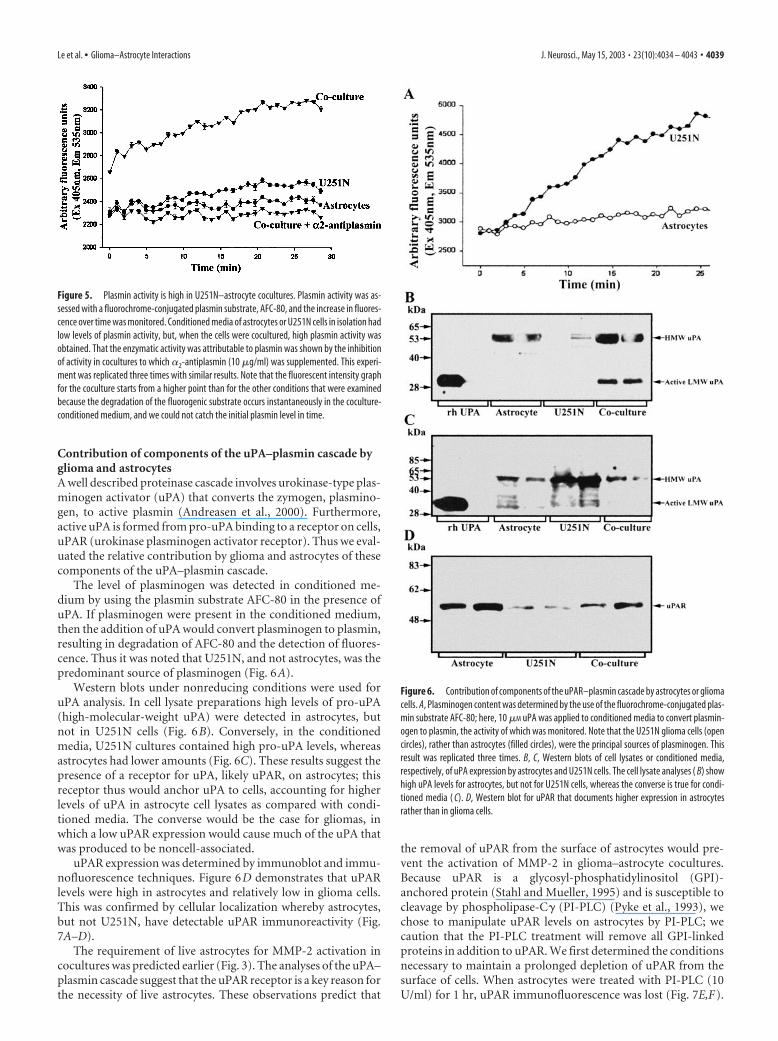
Contribution of components of the uPA–plasmin cascade byglioma and astrocytesA well described proteinase cascade involves urokinase-type plas-minogen activator (uPA) that converts the zymogen, plasmino-gen, to active plasmin (Andreasen et al., 2000). Furthermore,active uPA is formed from pro-uPA binding to a receptor on cells,uPAR (urokinase plasminogen activator receptor). Thus we eval-uated the relative contribution by glioma and astrocytes of thesecomponents of the uPA–plasmin cascade.
The level of plasminogen was detected in conditioned me-dium by using the plasmin substrate AFC-80 in the presence ofuPA. If plasminogen were present in the conditioned medium,then the addition of uPA would convert plasminogen to plasmin,resulting in degradation of AFC-80 and the detection of fluores-cence. Thus it was noted that U251N, and not astrocytes, was thepredominant source of plasminogen (Fig. 6A).
Western blots under nonreducing conditions were used foruPA analysis. In cell lysate preparations high levels of pro-uPA(high-molecular-weight uPA) were detected in astrocytes, butnot in U251N cells (Fig. 6B). Conversely, in the conditionedmedia, U251N cultures contained high pro-uPA levels, whereasastrocytes had lower amounts (Fig. 6C). These results suggest thepresence of a receptor for uPA, likely uPAR, on astrocytes; thisreceptor thus would anchor uPA to cells, accounting for higherlevels of uPA in astrocyte cell lysates as compared with condi-tioned media. The converse would be the case for gliomas, inwhich a low uPAR expression would cause much of the uPA thatwas produced to be noncell-associated.
uPAR expression was determined by immunoblot and immu-nofluorescence techniques. Figure 6D demonstrates that uPARlevels were high in astrocytes and relatively low in glioma cells.This was confirmed by cellular localization whereby astrocytes,but not U251N, have detectable uPAR immunoreactivity (Fig.7A–D).
The requirement of live astrocytes for MMP-2 activation incocultures was predicted earlier (Fig. 3). The analyses of the uPA–plasmin cascade suggest that the uPAR receptor is a key reason forthe necessity of live astrocytes. These observations predict that
the removal of uPAR from the surface of astrocytes would pre-vent the activation of MMP-2 in glioma–astrocyte cocultures.Because uPAR is a glycosyl-phosphatidylinositol (GPI)-anchored protein (Stahl and Mueller, 1995) and is susceptible tocleavage by phospholipase-C� (PI-PLC) (Pyke et al., 1993), wechose to manipulate uPAR levels on astrocytes by PI-PLC; wecaution that the PI-PLC treatment will remove all GPI-linkedproteins in addition to uPAR. We first determined the conditionsnecessary to maintain a prolonged depletion of uPAR from thesurface of cells. When astrocytes were treated with PI-PLC (10U/ml) for 1 hr, uPAR immunofluorescence was lost (Fig. 7E,F).
Figure 5. Plasmin activity is high in U251N–astrocyte cocultures. Plasmin activity was as-sessed with a fluorochrome-conjugated plasmin substrate, AFC-80, and the increase in fluores-cence over time was monitored. Conditioned media of astrocytes or U251N cells in isolation hadlow levels of plasmin activity, but, when the cells were cocultured, high plasmin activity wasobtained. That the enzymatic activity was attributable to plasmin was shown by the inhibitionof activity in cocultures to which �2-antiplasmin (10 �g/ml) was supplemented. This experi-ment was replicated three times with similar results. Note that the fluorescent intensity graphfor the coculture starts from a higher point than for the other conditions that were examinedbecause the degradation of the fluorogenic substrate occurs instantaneously in the coculture-conditioned medium, and we could not catch the initial plasmin level in time.
Figure 6. Contribution of components of the uPAR–plasmin cascade by astrocytes or gliomacells. A, Plasminogen content was determined by the use of the fluorochrome-conjugated plas-min substrate AFC-80; here, 10 �M uPA was applied to conditioned media to convert plasmin-ogen to plasmin, the activity of which was monitored. Note that the U251N glioma cells (opencircles), rather than astrocytes (filled circles), were the principal sources of plasminogen. Thisresult was replicated three times. B, C, Western blots of cell lysates or conditioned media,respectively, of uPA expression by astrocytes and U251N cells. The cell lysate analyses ( B) showhigh uPA levels for astrocytes, but not for U251N cells, whereas the converse is true for condi-tioned media ( C). D, Western blot for uPAR that documents higher expression in astrocytesrather than in glioma cells.
Le et al. • Glioma–Astrocyte Interactions J. Neurosci., May 15, 2003 • 23(10):4034 – 4043 • 4039
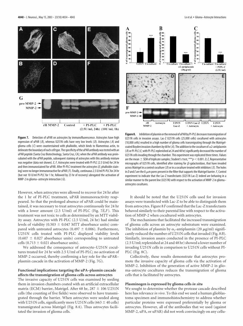
However, when astrocytes were allowed to recover for 24 hr afterthe 1 hr of PI-PLC treatment, uPAR immunoreactivity reap-peared. So that the prolonged absence of uPAR could be main-tained, it was necessary to treat astrocytes continuously for 24 hrwith a lower amount (2.5 U/ml) of PI-PLC (Fig. 7E,F). Thistreatment was not toxic to cells as determined by an MTT viabil-ity assay. Astrocytes with PI-PLC (2.5 U/ml, 24 hr) had similarlevels of viability (0.503 � 0.047 MTT absorbance units) com-pared with untreated astrocytes (0.497 � 0.006). Furthermore,U251N cells treated with PI-PLC displayed viability levels(0.607 � 0.027 absorbance units) corresponding to untreatedcells (0.713 � 0.021 absorbance units).
We addressed the consequence of astrocyte–U251N cocul-tures treated for 24 hr with 2.5 U/ml of PI-PLC; no activation ofMMP-2 occurred, thereby confirming a key role for the uPAR–plasmin cascade in the activation of MMP-2 (Fig. 7G).
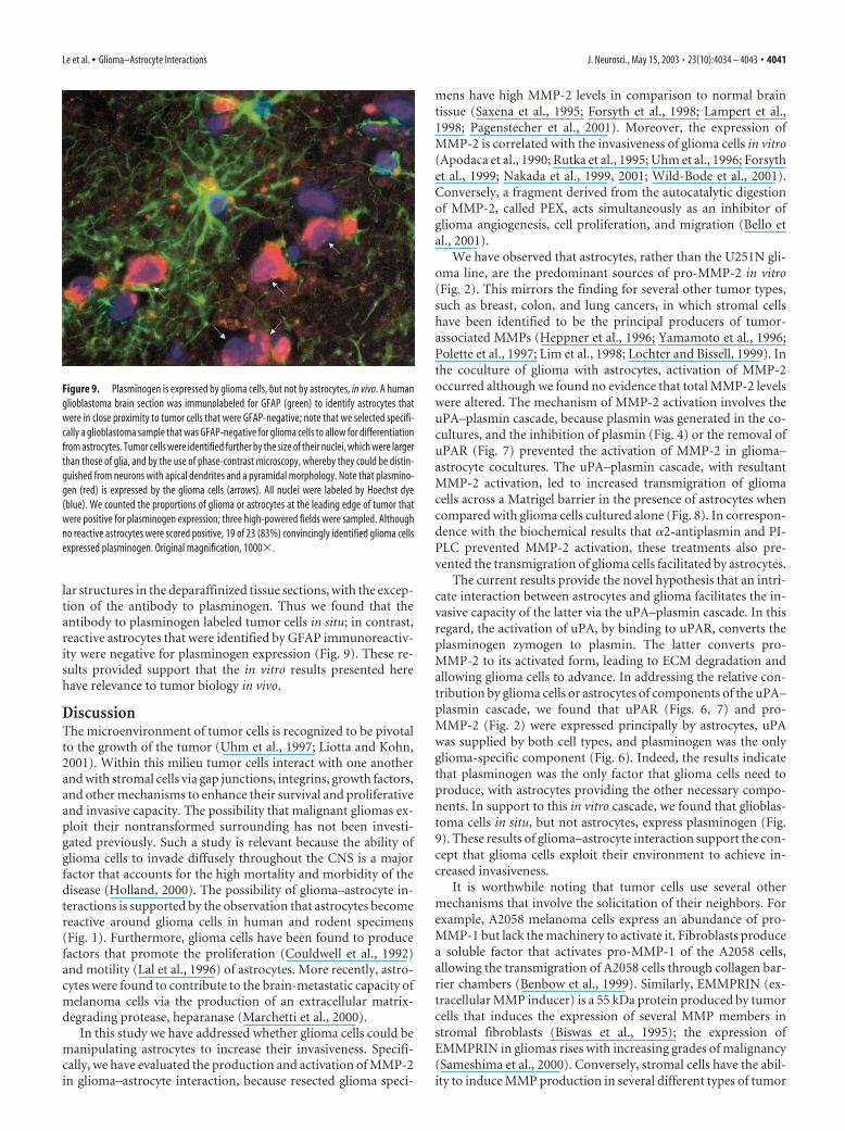
Functional implications: targeting the uPA–plasmin cascadeaffects the transmigration of glioma cells across astrocytesThe invasive capacity of U251N cells was examined by seedingthem in invasion chambers coated with an artificial extracellularmatrix (ECM) barrier, Matrigel. After 68 hr, 287 � 106 U251Ncells (the counting of five fields) were observed to have transmi-grated through the barrier. When astrocytes were seeded alongwith U251N cells, significantly more U251N cells (663 � 40 cells)transmigrated across Matrigel (Fig. 8A). Thus astrocytes facili-tated the invasion of glioma cells.
It should be noted that the U251N cells used for invasionassays were transfected with Lac-Z to be able to distinguish themfrom astrocytes. Figure 8F confirmed that the Lac-Z transfectantsbehaved similarly to their parental line with respect to the activa-tion of MMP-2 when cocultured with astrocytes.
The mechanisms that facilitated the increased transmigrationof glioma cells across an astrocyte substratum were examined.The inhibition of plasmin by �2-antiplasmin (20 �g/ml) signifi-cantly reduced the number of U251N cells that invaded (Fig. 8B).Similarly, invasion assays conducted in the presence of PI-PLC(2.5 U/ml; replenished at 24 and 48 hr) showed a lower number ofinvading U251N cells in comparison to U251N cells without PI-PLC (Fig. 8C).
Collectively, these results demonstrate that astrocytes pro-mote the invasive capacity of glioma cells via the activation ofMMP-2. Inhibition of the generation of active MMP-2 in glio-ma–astrocyte cocultures reduces the transmigration of gliomacells that is facilitated by astrocytes.
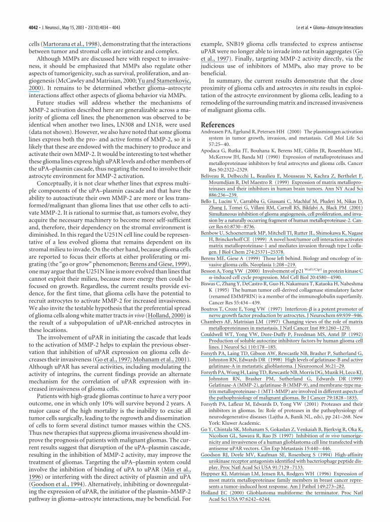
Plasminogen is expressed by glioma cells in situWe sought to determine whether the protease cascade describedhere has relevance in vivo. To this end we used a human glioblas-toma specimen and immunohistochemistry to address whetherparticular proteins were expressed preferentially by glioma orastrocytes. However, all of the antibodies that we used (againstMMP-2, uPA, or uPAR) did not work convincingly on any cellu-
Figure 7. Detection of uPAR on astrocytes by immunofluorescence. Astrocytes have highexpression of uPAR ( B), whereas U251N cells have very low levels ( D). Astrocytes ( A) andglioma cells ( C) were counterstained with phalloidin, which binds to filamentous actin, todelineate the boundary of each cell type. The specificity of the uPAR antibody was tested with anuPAR peptide (Santa Cruz Biotechnology, Santa Cruz, CA); when the uPAR antibody was prein-cubated with the uPAR peptide, subsequent staining of astrocytes with this antibody mixturewas negative (data not shown). E, F, Astrocytes were treated with PI-PLC (2.5 U/ml) for 24 hrand then immunostained for uPAR. After PI-PLC treatment the astrocytes (E; phalloidin stain-ing) were no longer immunoreactive for uPAR ( F). Finally, continuous 2.5 U/ml PI-PLC for 24 hr(but not 10 U/ml PI-PLC for 1 hr, followed by 23 hr of recovery) abrogated the activation ofMMP-2 in glioma–astrocyte interaction ( G).
Figure 8. Inhibition of plasmin or the removal of uPAR by PI-PLC decreases transmigration ofU251N cells in invasion assays. Lac-Z U251N cells (25,000 cells) cocultured with astrocytes(10,000 cells) resulted in a high number of glioma cells transmigrating through the Matrigel-coated Boyden invasion chambers by 68 hr ( A). The addition to the coculture of �2-antiplasmin( B) or PI-PLC (C; with PI-PLC replenished at 24 and 48 hr) significantly decreased the number ofU251N cells invading through the chamber. This experiment was replicated three times. Valuesare the mean � SEM of triplicate samples; Student’s t test, ***p � 0.001. D, E, Representativemicrographs of U251N cells, identified after staining for �-galactosidase, that have invadedacross Matrigel in a control coculture ( D) or in a coculture treated with inhibitors ( E). The holesin D and E are the 8 �m pores present in the filter that supports the Matrigel barrier. F, Controlexperiment to indicate that the Lac-Z transfectants (U251N Lac-Z) indeed are behaving in asimilar manner to the parent line (U251N) with respect to the activation of MMP-2 in glioma–astrocytes cocultures.
4040 • J. Neurosci., May 15, 2003 • 23(10):4034 – 4043 Le et al. • Glioma–Astrocyte Interactions
lar structures in the deparaffinized tissue sections, with the excep-tion of the antibody to plasminogen. Thus we found that theantibody to plasminogen labeled tumor cells in situ; in contrast,reactive astrocytes that were identified by GFAP immunoreactiv-ity were negative for plasminogen expression (Fig. 9). These re-sults provided support that the in vitro results presented herehave relevance to tumor biology in vivo.
DiscussionThe microenvironment of tumor cells is recognized to be pivotalto the growth of the tumor (Uhm et al., 1997; Liotta and Kohn,2001). Within this milieu tumor cells interact with one anotherand with stromal cells via gap junctions, integrins, growth factors,and other mechanisms to enhance their survival and proliferativeand invasive capacity. The possibility that malignant gliomas ex-ploit their nontransformed surrounding has not been investi-gated previously. Such a study is relevant because the ability ofglioma cells to invade diffusely throughout the CNS is a majorfactor that accounts for the high mortality and morbidity of thedisease (Holland, 2000). The possibility of glioma–astrocyte in-teractions is supported by the observation that astrocytes becomereactive around glioma cells in human and rodent specimens(Fig. 1). Furthermore, glioma cells have been found to producefactors that promote the proliferation (Couldwell et al., 1992)and motility (Lal et al., 1996) of astrocytes. More recently, astro-cytes were found to contribute to the brain-metastatic capacity ofmelanoma cells via the production of an extracellular matrix-degrading protease, heparanase (Marchetti et al., 2000).
In this study we have addressed whether glioma cells could bemanipulating astrocytes to increase their invasiveness. Specifi-cally, we have evaluated the production and activation of MMP-2in glioma–astrocyte interaction, because resected glioma speci-
mens have high MMP-2 levels in comparison to normal braintissue (Saxena et al., 1995; Forsyth et al., 1998; Lampert et al.,1998; Pagenstecher et al., 2001). Moreover, the expression ofMMP-2 is correlated with the invasiveness of glioma cells in vitro(Apodaca et al., 1990; Rutka et al., 1995; Uhm et al., 1996; Forsythet al., 1999; Nakada et al., 1999, 2001; Wild-Bode et al., 2001).Conversely, a fragment derived from the autocatalytic digestionof MMP-2, called PEX, acts simultaneously as an inhibitor ofglioma angiogenesis, cell proliferation, and migration (Bello etal., 2001).
We have observed that astrocytes, rather than the U251N gli-oma line, are the predominant sources of pro-MMP-2 in vitro(Fig. 2). This mirrors the finding for several other tumor types,such as breast, colon, and lung cancers, in which stromal cellshave been identified to be the principal producers of tumor-associated MMPs (Heppner et al., 1996; Yamamoto et al., 1996;Polette et al., 1997; Lim et al., 1998; Lochter and Bissell, 1999). Inthe coculture of glioma with astrocytes, activation of MMP-2occurred although we found no evidence that total MMP-2 levelswere altered. The mechanism of MMP-2 activation involves theuPA–plasmin cascade, because plasmin was generated in the co-cultures, and the inhibition of plasmin (Fig. 4) or the removal ofuPAR (Fig. 7) prevented the activation of MMP-2 in glioma–astrocyte cocultures. The uPA–plasmin cascade, with resultantMMP-2 activation, led to increased transmigration of gliomacells across a Matrigel barrier in the presence of astrocytes whencompared with glioma cells cultured alone (Fig. 8). In correspon-dence with the biochemical results that �2-antiplasmin and PI-PLC prevented MMP-2 activation, these treatments also pre-vented the transmigration of glioma cells facilitated by astrocytes.
The current results provide the novel hypothesis that an intri-cate interaction between astrocytes and glioma facilitates the in-vasive capacity of the latter via the uPA–plasmin cascade. In thisregard, the activation of uPA, by binding to uPAR, converts theplasminogen zymogen to plasmin. The latter converts pro-MMP-2 to its activated form, leading to ECM degradation andallowing glioma cells to advance. In addressing the relative con-tribution by glioma cells or astrocytes of components of the uPA–plasmin cascade, we found that uPAR (Figs. 6, 7) and pro-MMP-2 (Fig. 2) were expressed principally by astrocytes, uPAwas supplied by both cell types, and plasminogen was the onlyglioma-specific component (Fig. 6). Indeed, the results indicatethat plasminogen was the only factor that glioma cells need toproduce, with astrocytes providing the other necessary compo-nents. In support to this in vitro cascade, we found that glioblas-toma cells in situ, but not astrocytes, express plasminogen (Fig.9). These results of glioma–astrocyte interaction support the con-cept that glioma cells exploit their environment to achieve in-creased invasiveness.
It is worthwhile noting that tumor cells use several othermechanisms that involve the solicitation of their neighbors. Forexample, A2058 melanoma cells express an abundance of pro-MMP-1 but lack the machinery to activate it. Fibroblasts producea soluble factor that activates pro-MMP-1 of the A2058 cells,allowing the transmigration of A2058 cells through collagen bar-rier chambers (Benbow et al., 1999). Similarly, EMMPRIN (ex-tracellular MMP inducer) is a 55 kDa protein produced by tumorcells that induces the expression of several MMP members instromal fibroblasts (Biswas et al., 1995); the expression ofEMMPRIN in gliomas rises with increasing grades of malignancy(Sameshima et al., 2000). Conversely, stromal cells have the abil-ity to induce MMP production in several different types of tumor
Figure 9. Plasminogen is expressed by glioma cells, but not by astrocytes, in vivo. A humanglioblastoma brain section was immunolabeled for GFAP (green) to identify astrocytes thatwere in close proximity to tumor cells that were GFAP-negative; note that we selected specifi-cally a glioblastoma sample that was GFAP-negative for glioma cells to allow for differentiationfrom astrocytes. Tumor cells were identified further by the size of their nuclei, which were largerthan those of glia, and by the use of phase-contrast microscopy, whereby they could be distin-guished from neurons with apical dendrites and a pyramidal morphology. Note that plasmino-gen (red) is expressed by the glioma cells (arrows). All nuclei were labeled by Hoechst dye(blue). We counted the proportions of glioma or astrocytes at the leading edge of tumor thatwere positive for plasminogen expression; three high-powered fields were sampled. Althoughno reactive astrocytes were scored positive, 19 of 23 (83%) convincingly identified glioma cellsexpressed plasminogen. Original magnification, 1000�.
Le et al. • Glioma–Astrocyte Interactions J. Neurosci., May 15, 2003 • 23(10):4034 – 4043 • 4041
cells (Martorana et al., 1998), demonstrating that the interactionsbetween tumor and stromal cells are intricate and complex.
Although MMPs are discussed here with respect to invasive-ness, it should be emphasized that MMPs also regulate otheraspects of tumorigenicity, such as survival, proliferation, and an-giogenesis (McCawley and Matrisian, 2000; Yu and Stamenkovic,2000). It remains to be determined whether glioma–astrocyteinteractions affect other aspects of glioma behavior via MMPs.
Future studies will address whether the mechanisms ofMMP-2 activation described here are generalizable across a ma-jority of glioma cell lines; the phenomenon was observed to beidentical when another two lines, LN308 and LN18, were used(data not shown). However, we also have noted that some gliomalines express both the pro- and active forms of MMP-2, so it islikely that these are endowed with the machinery to produce andactivate their own MMP-2. It would be interesting to test whetherthese glioma lines express high uPAR levels and other members ofthe uPA–plasmin cascade, thus negating the need to involve theirastrocyte environment for MMP-2 activation.
Conceptually, it is not clear whether lines that express multi-ple components of the uPA–plasmin cascade and that have theability to autoactivate their own MMP-2 are more or less trans-formed/malignant than glioma lines that use other cells to acti-vate MMP-2. It is rational to surmise that, as tumors evolve, theyacquire the necessary machinery to become more self-sufficientand, therefore, their dependency on the stromal environment isdiminished. In this regard the U251N cell line could be represen-tative of a less evolved glioma that remains dependent on itsstromal milieu to invade. On the other hand, because glioma cellsare reported to focus their efforts at either proliferating or mi-grating (the “go or grow” phenomenon; Berens and Giese, 1999),one may argue that the U251N line is more evolved than lines thatcannot exploit their milieu, because more energy then could befocused on growth. Regardless, the current results provide evi-dence, for the first time, that glioma cells have the potential torecruit astrocytes to activate MMP-2 for increased invasiveness.We also invite the testable hypothesis that the preferential spreadof glioma cells along white matter tracts in vivo (Holland, 2000) isthe result of a subpopulation of uPAR-enriched astrocytes inthese locations.
The involvement of uPAR in initiating the cascade that leadsto the activation of MMP-2 helps to explain the previous obser-vation that inhibition of uPAR expression on glioma cells de-creases their invasiveness (Go et al., 1997; Mohanam et al., 2001).Although uPAR has several activities, including modulating theactivity of integrins, the current findings provide an alternatemechanism for the correlation of uPAR expression with in-creased invasiveness of glioma cells.
Patients with high-grade gliomas continue to have a very pooroutcome, one in which only 10% will survive beyond 2 years. Amajor cause of the high mortality is the inability to excise alltumor cells surgically, leading to the regrowth and disseminationof cells to form several distinct tumor masses within the CNS.Thus new therapies that suppress glioma invasiveness should im-prove the prognosis of patients with malignant gliomas. The cur-rent results suggest that disruption of the uPA–plasmin cascade,resulting in the inhibition of MMP-2 activity, may improve thetreatment of gliomas. Targeting the uPA–plasmin system couldinvolve the inhibition of binding of uPA to uPAR (Min et al.,1996) or interfering with the direct activity of plasmin and uPA(Goodson et al., 1994). Alternatively, inhibiting or downregulat-ing the expression of uPAR, the initiator of the plasmin–MMP-2pathway in glioma–astrocyte interactions, may be beneficial. For
example, SNB19 glioma cells transfected to express antisenseuPAR were no longer able to invade into rat brain aggregates (Goet al., 1997). Finally, targeting MMP-2 activity directly, via thejudicious use of inhibitors of MMPs, also may prove to bebeneficial.
In summary, the current results demonstrate that the closeproximity of glioma cells and astrocytes in situ results in exploi-tation of the astrocyte environment by glioma cells, leading to aremodeling of the surrounding matrix and increased invasivenessof malignant glioma cells.
ReferencesAndreasen PA, Egelund R, Petersen HH (2000) The plasminogen activation
system in tumor growth, invasion, and metastasis. Cell Mol Life Sci57:25– 40.
Apodaca G, Rutka JT, Bouhana K, Berens ME, Giblin JR, Rosenblum ML,McKerrow JH, Banda MJ (1990) Expression of metalloproteinases andmetalloproteinase inhibitors by fetal astrocytes and glioma cells. CancerRes 50:2322–2329.
Beliveau R, Delbecchi L, Beaulieu E, Mousseau N, Kachra Z, Berthelet F,Moumdjian R, Del Maestro R (1999) Expression of matrix metallopro-teinases and their inhibitors in human brain tumors. Ann NY Acad Sci886:236 –239.
Bello L, Lucini V, Carrabba G, Giussani C, Machluf M, Pluderi M, Nikas D,Zhang J, Tomei G, Villani RM, Carroll RS, Bikfalvi A, Black PM (2001)Simultaneous inhibition of glioma angiogenesis, cell proliferation, and inva-sion by a naturally occurring fragment of human metalloproteinase-2. Can-cer Res 61:8730–8736.
Benbow U, Schoenermark MP, Mitchell TI, Rutter JL, Shimokawa K, NagaseH, Brinckerhoff CE (1999) A novel host/tumor cell interaction activatesmatrix metalloproteinase-1 and mediates invasion through type I colla-gen. J Biol Chem 274:25371–25378.
Berens ME, Giese A (1999) Those left behind. Biology and oncology of in-vasive glioma cells. Neoplasia 1:208 –219.
Besson A, Yong VW (2000) Involvement of p21 Waf1/Cip1 in protein kinase C�-induced cell cycle progression. Mol Cell Biol 20:4580 – 4590.
Biswas C, Zhang Y, DeCastro R, Guo H, Nakamura T, Kataoka H, NabeshmaK (1995) The human tumor cell-derived collagenase stimulatory factor(renamed EMMPRIN) is a member of the immunoglobulin superfamily.Cancer Res 55:434 – 439.
Boutros T, Croze E, Yong VW (1997) Interferon-� is a potent promoter ofnerve growth factor production by astrocytes. J Neurochem 69:939 –946.
Chambers AF, Matrisian LM (1997) Changing views of the role of matrixmetalloproteinases in metastasis. J Natl Cancer Inst 89:1260 –1270.
Couldwell WT, Yong VW, Dore-Duffy P, Freedman MS, Antel JP (1992)Production of soluble autocrine inhibitory factors by human glioma celllines. J Neurol Sci 110:178 –185.
Forsyth PA, Laing TD, Gibson AW, Rewcastle NB, Brasher P, Sutherland G,Johnston RN, Edwards DR (1998) High levels of gelatinase-B and activegelatinase-A in metastatic glioblastoma. J Neurooncol 36:21–29.
Forsyth PA, Wong H, Laing TD, Rewcastle NB, Morris DG, Muzik H, Leco KJ,Johnston RN, Brasher PM, Sutherland G, Edwards DR (1999)Gelatinase-A (MMP-2), gelatinase-B (MMP-9), and membrane-type ma-trix metalloproteinase-1 (MT1-MMP) are involved in different aspects ofthe pathophysiology of malignant gliomas. Br J Cancer 79:1828 –1835.
Forsyth PA, Lafleur M, Edwards D, Yong VW (2001) Proteases and theirinhibitors in gliomas. In: Role of proteases in the pathophysiology ofneurodegenerative diseases (Lajtha A, Banik NL, eds), pp 241–268. NewYork: Kluwer Academic.
Go Y, Chintala SK, Mohanam S, Gokaslan Z, Venkaiah B, Bjerkvig R, Oka K,Nicolson GL, Sawaya R, Rao JS (1997) Inhibition of in vivo tumorige-nicity and invasiveness of a human glioblastoma cell line transfected withantisense uPAR vectors. Clin Exp Metastasis 15:440 – 446.
Goodson RJ, Doyle MV, Kaufman SE, Rosenberg S (1994) High-affinityurokinase receptor antagonists identified with bacteriophage peptide dis-play. Proc Natl Acad Sci USA 91:7129 –7133.
Heppner KJ, Matrisian LM, Jensen RA, Rodgers WH (1996) Expression ofmost matrix metalloproteinase family members in breast cancer repre-sents a tumor-induced host response. Am J Pathol 149:273–282.
Holland EC (2000) Glioblastoma multiforme: the terminator. Proc NatlAcad Sci USA 97:6242– 6244.
4042 • J. Neurosci., May 15, 2003 • 23(10):4034 – 4043 Le et al. • Glioma–Astrocyte Interactions
Ishii N, Maier D, Merlo A, Tada M, Sawamura Y, Diserens AC, Van Meir EG(1999) Frequent coalterations of TP53, p16/CDKN2A, p14ARF, PTENtumor suppressor genes in human glioma cell lines. Brain Pathol9:469 – 479.
Lal PG, Ghirnikar RS, Eng LF (1996) Astrocyte–astrocytoma cell line inter-actions in culture. J Neurosci Res 44:216 –222.
Lampert K, Machein U, Machein MR, Conca W, Peter HH, Volk B (1998)Expression of matrix metalloproteinases and their tissue inhibitors inhuman brain tumors. Am J Pathol 153:429 – 437.
Lim M, Martinez T, Jablons D, Cameron R, Guo H, Toole BP, Li JD, BasbaumC (1998) Tumor-derived EMMPRIN stimulates collagenase transcrip-tion through MAPK. FEBS Lett 441:88 –92.
Liotta LA, Kohn EC (2001) The microenvironment of the tumour– hostinterface. Nature 411:375–379.
Lochter A, Bissell MJ (1999) An odyssey from breast to bone: multi-stepcontrol of mammary metastases and osteolysis by matrix metalloprotein-ases. APMIS 107:128 –136.
Marchetti D, Li J, Shen R (2000) Astrocytes contribute to the brain-metastatic specificity of melanoma cells by producing heparanase. CancerRes 60:4764 – 4770.
Martorana AM, Zheng G, Crowe TC, O’Grady RL, Lyons JG (1998) Epithe-lial cells up-regulate matrix metalloproteinases in cells within the samemammary carcinoma that have undergone an epithelial–mesenchymaltransition. Cancer Res 58:4970 – 4979.
Mazzieri R, Masiero L, Zanetta L, Monea S, Onisto M, Garbisa S, Mignatti P(1997) Control of type IV collagenase activity by components of theurokinase–plasmin system: a regulatory mechanism with cell-bound re-actants. EMBO J 16:2319 –2332.
McCawley LJ, Matrisian LM (2000) Matrix metalloproteinases: multifunc-tional contributors to tumor progression. Mol Med Today 6:149 –158.
Min HY, Doyle LV, Vitt CR, Zandonella CL, Stratton-Thomas JR, ShumanMA, Rosenberg S (1996) Urokinase receptor antagonists inhibit angio-genesis and primary tumor growth in syngeneic mice. Cancer Res56:2428 –2433.
Mohanam S, Jasti SL, Kondraganti SR, Chandrasekar N, Kin Y, Fuller GN,Rao JS (2001) Stable transfection of urokinase-type plasminogen activa-tor antisense construct modulates invasion of human glioblastoma cells.Clin Cancer Res 7:2519 –2526.
Nagase H (1997) Activation mechanisms of matrix metalloproteinases. BiolChem 378:151–160.
Nakada M, Nakamra H, Ikeda E, Fujimoto N, Yamashita J, Sato M, Seiki Y(1999) Expression and tissue localization of membrane-type 1, 2, and 3matrix metalloproteinases in human astrocytic tumors. Am J Pathol154:417– 428.
Nakada M, Kita D, Futami K, Yamashita J, Fujimoto N, Sato H, Okada Y(2001) Roles of membrane-type 1 matrix metalloproteinase and tissueinhibitor of metalloproteinases-2 in invasion and dissemination of hu-man malignant glioma. J Neurosurg 94:464 – 473.
Pagenstecher A, Wussler EM, Opdenakker G, Volk B, Campbell IL (2001)Distinct expression patterns and levels of enzymatic activity of matrixmetalloproteinases and their inhibitors in primary brain tumors. J Neu-ropathol Exp Neurol 60:598 – 612.
Pei D, Weiss S (1996) Transmembrane-deletion mutants of the membrane-type matrix metalloproteinase-1 process pro-gelatinase A and expressintrinsic matrix-degrading activity. J Biol Chem 271:9135–9140.
Polette M, Gilles C, Marchand V, Lorenzato M, Toole B, Tournier JM, ZuckerS, Birembaut P (1997) Tumor collagenase stimulatory factor (TCSF)expression and localization in human lung and breast cancers. J Histo-chem Cytochem 45:703–709.
Price A, Shi Q, Morris D, Wilcox ME, Brasher PMA, Rewcastle NB, ShalinskyD, Appelt K, Johnston RN, Yong VW, Edwards D, Forsyth P (1999)Marked inhibition of tumor growth in a malignant glioma tumor modelby a novel synthetic matrix metalloproteinase inhibitor AG3340. ClinCancer Res 5:845– 854.
Pyke C, Eriksen J, Solberg H, Nielsen BS, Kristensen P, Lund LR, Dano K(1993) An alternatively spliced variant of mRNA for the human receptorfor urokinase plasminogen activator. FEBS Lett 326:69 –74.
Rutka JT, Smith SL (1993) Transfection of human astrocytoma cells withglial fibrillary acidic protein complementary DNA: analysis of expression,proliferation, and tumorigenicity. Cancer Res 53:3624 –3631.
Rutka JT, Matsuzawa K, Hubbard SL, Fukuyama K, Becker LE, Stetler-Stevenson W, Edwards DR, Dirks PB (1995) Expression of TIMP-1,TIMP-2, 72- and 92-kDa type IV collagenase transcripts in human astro-cytoma cell lines: correlation with astrocytoma cell invasiveness. Int JOncol 6:877– 884.
Sameshima T, Nabeshima K, Toole BP, Yokogami K, Okada Y, Goya T,Koono M, Wakisaka S (2000) Expression of EMMPRIN (CD147), a cellsurface inducer of matrix metalloproteinases, in normal human brain andgliomas. Int J Cancer 88:21–27.
Sawaya RE, Yamamoto M, Gokaslan ZL, Wang SW, Mohanam S, Fuller GN,McCutcheon, Stetler-Stevenson WG, Nicolson GL, Rao JS (1996) Ex-pression and localization of 72 kDa type IV collagenase (MMP-2) inhuman malignant gliomas in vivo. Clin Exp Metastasis 14:35– 42.
Saxena A, Robertson JT, Kufta C (1995) Increased expression of gelatinase Aand TIMP-2 in primary human glioblastomas. Int J Oncol 7:469 – 473.
Stahl A, Mueller BM (1995) The urokinase-type plasminogen activator re-ceptor, a GPI-linked protein, is localized in caveolae. J Cell Biol129:335–344.
Sternlicht MD, Werb Z (2001) How matrix metalloproteinases regulate cellbehavior. Annu Rev Cell Dev Biol 17:463–516.
Stetler-Stevenson WG, Aznavoorian S, Liotta LA (1993) Tumor cell inter-actions with the extracellular matrix during invasion and metastasis.Annu Rev Cell Biol 9:541–573.
Tonn JC, Kerkau S, Hanke A, Bouterfa H, Mueller JG, Wagner S, Vince GH,Roosen K (1999) Effect of synthetic matrix-metalloproteinase inhibi-tors on invasive capacity and proliferation of human malignant gliomas invitro. Int J Cancer 80:764 –772.
Uhm JH, Dooley NP, Villemure JG, Yong VW (1996) Glioma invasion invitro: regulation by matrix metalloprotease-2 and protein kinase C. ClinExp Metastasis 14:421– 433.
Uhm JH, Dooley NP, Villemure JG, Yong VW (1997) Mechanisms of gli-oma invasion: role of matrix metalloproteinases. Can J Neurol Sci24:3–15.
VanMeter TE, Rooprai HK, Kibble MM, Fillmore HL, Broaddus WC, Pilk-ington GJ (2001) The role of matrix metalloproteinase genes in gliomainvasion: co-dependent and interactive proteolysis. J Neurooncol53:213–235.
Vecil GG, Larsen PH, Herx LM, Besson A, Goodyer CG, Yong VW (2000)Interleukin-1 is a key regulator of matrix metalloproteinase-9 in humanneurons in culture and following mouse brain trauma in vivo. J NeurosciRes 61:212–224.
Wild-Bode C, Weller M, Wick W (2001) Molecular determinants of gliomacell migration and invasion. J Neurosurg 94:978 –984.
Yamamoto M, Mohanam S, Sawaya R, Fuller GN, Seiki M, Sato H, GokaslanZL, Liotta LA, Nicolson GL, Rao JS (1996) Differential expression ofmembrane-type matrix metalloproteinase and its correlation with gelati-nase A activation in human malignant brain tumors in vivo and in vitro.Cancer Res 56:384 –392.
Yong VW (1998) Response of astrocytes and oligodendrocytes to injury.Ment Retard Dev Disabil Res Rev 4:193–199.
Yong VW, Power C, Forsyth P, Edwards DR (2001) Metalloproteinases inbiology and pathology of the nervous system. Nat Rev Neurosci2:502–511.
Yu Q, Stamenkovic I (2000) Cell surface-localized matrix metalloproteinase-9proteolytically activates TGF-� and promotes tumor invasion and angiogen-esis. Genes Dev 14:163–176.
Le et al. • Glioma–Astrocyte Interactions J. Neurosci., May 15, 2003 • 23(10):4034 – 4043 • 4043