Tissue plasminogen activator and urokinase plasminogen activator in human epileptogenic pathologies
Upload
nottinghamCategory
view
0download
0
BioMed CentralBMC Molecular Biology
ss
Open AcceResearch articleCharacterisation of urokinase plasminogen activator receptor variants in human airway and peripheral cellsCeri E Stewart* and Ian SayersAddress: Division of Therapeutics and Molecular Medicine, Nottingham Respiratory Biomedical Research Unit, University of Nottingham, Queen's Medical Centre, Nottingham, NG7 2UH, UK
Email: Ceri E Stewart* - [email protected]; Ian Sayers - [email protected]
* Corresponding author
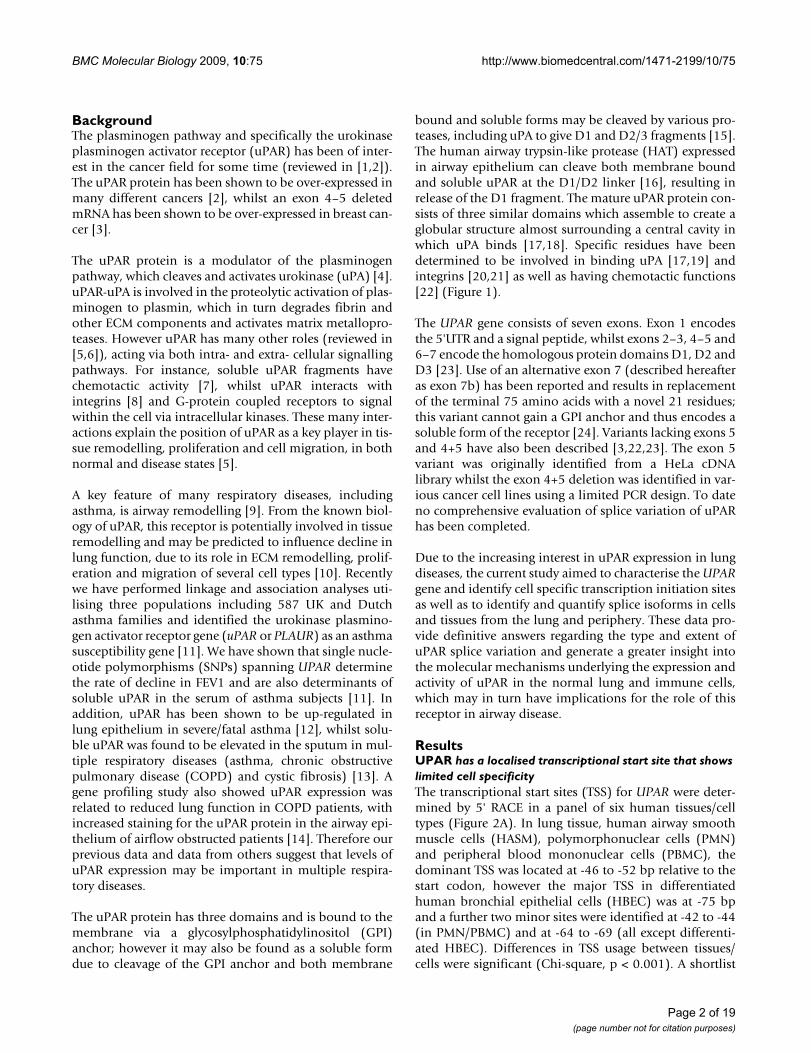
AbstractBackground: Expression of the urokinase plasminogen activator receptor (UPAR) has been shownto have clinical relevance in various cancers. We have recently identified UPAR as an asthmasusceptibility gene and there is evidence to suggest that uPAR may be upregulated in lung diseasessuch as COPD and asthma. uPAR is a key receptor involved in the formation of the serine proteaseplasmin by interacting with uPA and has been implicated in many physiological processes includingproliferation and migration. The current aim was to determine key regulatory regions and splicevariants of UPAR and quantify its expression in primary human tissues and cells (including lung,bronchial epithelium (HBEC), airway smooth muscle (HASM) and peripheral cells).
Results: Using Rapid Amplification of cDNA Ends (RACE) a conserved transcription start site (-42 to -77 relative to ATG) was identified and multiple transcription factor binding sites predicted.Seven major splice variants were identified (>5% total expression), including multiple exondeletions and an alternative exon 7b (encoding a truncated, soluble, 229aa protein). Variants weredifferentially expressed, with a high proportion of E7b usage in lung tissue and structural cells (55–87% of transcripts), whereas classical exon 7 (encoding the GPI-linked protein) was preferentiallyexpressed in peripheral cells (~80% of transcripts), often with exon 6 or 5+6 deletions. Real-timePCR confirmed expression of uPAR mRNA in lung, as well as airway and peripheral cell types with~50–100 fold greater expression in peripheral cells versus airway cells and confirmed RACE data.Protein analysis confirmed expression of multiple different forms of uPAR in the same cells as wellas expression of soluble uPAR in cell supernatants. The pattern of expression did not directlyreflect that seen at the mRNA level, indicating that post-translational mechanisms of regulation mayalso play an important role.
Conclusion: We have identified multiple uPAR isoforms in the lung and immune cells and shownthat expression is cell specific. These data provide a novel mechanism for uPAR regulation, asdifferent exon splicing may determine uPAR function e.g. alternative E7b results in a soluble isoformdue to the loss of the GPI anchor and exon deletions may affect uPA (ligand) and/or integrin bindingand therefore influence downstream pathways. Expression of different isoforms within the lungshould be taken into consideration in studies of uPAR in respiratory disease.
Published: 28 July 2009
BMC Molecular Biology 2009, 10:75 doi:10.1186/1471-2199-10-75
Received: 4 December 2008Accepted: 28 July 2009
This article is available from: http://www.biomedcentral.com/1471-2199/10/75
© 2009 Stewart and Sayers; licensee BioMed Central Ltd. This is an Open Access article distributed under the terms of the Creative Commons Attribution License (http://creativecommons.org/licenses/by/2.0), which permits unrestricted use, distribution, and reproduction in any medium, provided the original work is properly cited.
Page 1 of 19(page number not for citation purposes)
BMC Molecular Biology 2009, 10:75 http://www.biomedcentral.com/1471-2199/10/75
BackgroundThe plasminogen pathway and specifically the urokinaseplasminogen activator receptor (uPAR) has been of inter-est in the cancer field for some time (reviewed in [1,2]).The uPAR protein has been shown to be over-expressed inmany different cancers [2], whilst an exon 4–5 deletedmRNA has been shown to be over-expressed in breast can-cer [3].
The uPAR protein is a modulator of the plasminogenpathway, which cleaves and activates urokinase (uPA) [4].uPAR-uPA is involved in the proteolytic activation of plas-minogen to plasmin, which in turn degrades fibrin andother ECM components and activates matrix metallopro-teases. However uPAR has many other roles (reviewed in[5,6]), acting via both intra- and extra- cellular signallingpathways. For instance, soluble uPAR fragments havechemotactic activity [7], whilst uPAR interacts withintegrins [8] and G-protein coupled receptors to signalwithin the cell via intracellular kinases. These many inter-actions explain the position of uPAR as a key player in tis-sue remodelling, proliferation and cell migration, in bothnormal and disease states [5].
A key feature of many respiratory diseases, includingasthma, is airway remodelling [9]. From the known biol-ogy of uPAR, this receptor is potentially involved in tissueremodelling and may be predicted to influence decline inlung function, due to its role in ECM remodelling, prolif-eration and migration of several cell types [10]. Recentlywe have performed linkage and association analyses uti-lising three populations including 587 UK and Dutchasthma families and identified the urokinase plasmino-gen activator receptor gene (uPAR or PLAUR) as an asthmasusceptibility gene [11]. We have shown that single nucle-otide polymorphisms (SNPs) spanning UPAR determinethe rate of decline in FEV1 and are also determinants ofsoluble uPAR in the serum of asthma subjects [11]. Inaddition, uPAR has been shown to be up-regulated inlung epithelium in severe/fatal asthma [12], whilst solu-ble uPAR was found to be elevated in the sputum in mul-tiple respiratory diseases (asthma, chronic obstructivepulmonary disease (COPD) and cystic fibrosis) [13]. Agene profiling study also showed uPAR expression wasrelated to reduced lung function in COPD patients, withincreased staining for the uPAR protein in the airway epi-thelium of airflow obstructed patients [14]. Therefore ourprevious data and data from others suggest that levels ofuPAR expression may be important in multiple respira-tory diseases.
The uPAR protein has three domains and is bound to themembrane via a glycosylphosphatidylinositol (GPI)anchor; however it may also be found as a soluble formdue to cleavage of the GPI anchor and both membrane
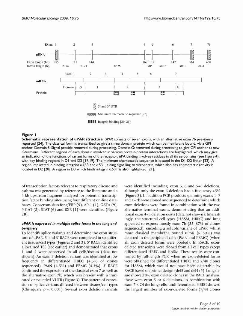
bound and soluble forms may be cleaved by various pro-teases, including uPA to give D1 and D2/3 fragments [15].The human airway trypsin-like protease (HAT) expressedin airway epithelium can cleave both membrane boundand soluble uPAR at the D1/D2 linker [16], resulting inrelease of the D1 fragment. The mature uPAR protein con-sists of three similar domains which assemble to create aglobular structure almost surrounding a central cavity inwhich uPA binds [17,18]. Specific residues have beendetermined to be involved in binding uPA [17,19] andintegrins [20,21] as well as having chemotactic functions[22] (Figure 1).
The UPAR gene consists of seven exons. Exon 1 encodesthe 5'UTR and a signal peptide, whilst exons 2–3, 4–5 and6–7 encode the homologous protein domains D1, D2 andD3 [23]. Use of an alternative exon 7 (described hereafteras exon 7b) has been reported and results in replacementof the terminal 75 amino acids with a novel 21 residues;this variant cannot gain a GPI anchor and thus encodes asoluble form of the receptor [24]. Variants lacking exons 5and 4+5 have also been described [3,22,23]. The exon 5variant was originally identified from a HeLa cDNAlibrary whilst the exon 4+5 deletion was identified in var-ious cancer cell lines using a limited PCR design. To dateno comprehensive evaluation of splice variation of uPARhas been completed.
Due to the increasing interest in uPAR expression in lungdiseases, the current study aimed to characterise the UPARgene and identify cell specific transcription initiation sitesas well as to identify and quantify splice isoforms in cellsand tissues from the lung and periphery. These data pro-vide definitive answers regarding the type and extent ofuPAR splice variation and generate a greater insight intothe molecular mechanisms underlying the expression andactivity of uPAR in the normal lung and immune cells,which may in turn have implications for the role of thisreceptor in airway disease.
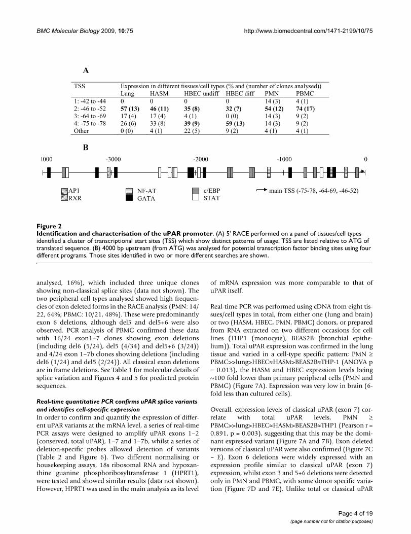
ResultsUPAR has a localised transcriptional start site that shows limited cell specificityThe transcriptional start sites (TSS) for UPAR were deter-mined by 5' RACE in a panel of six human tissues/celltypes (Figure 2A). In lung tissue, human airway smoothmuscle cells (HASM), polymorphonuclear cells (PMN)and peripheral blood mononuclear cells (PBMC), thedominant TSS was located at -46 to -52 bp relative to thestart codon, however the major TSS in differentiatedhuman bronchial epithelial cells (HBEC) was at -75 bpand a further two minor sites were identified at -42 to -44(in PMN/PBMC) and at -64 to -69 (all except differenti-ated HBEC). Differences in TSS usage between tissues/cells were significant (Chi-square, p < 0.001). A shortlist
Page 2 of 19(page number not for citation purposes)
BMC Molecular Biology 2009, 10:75 http://www.biomedcentral.com/1471-2199/10/75
of transcription factors relevant to respiratory disease andasthma was generated by reference to the literature and a4 kb upstream fragment analysed for potential transcrip-tion factor binding sites using four different on-line data-bases. Consensus sites for c/EBP (9), AP-1 (1), GATA (9),NF-AT (2), STAT (6) and RXR (1) were identified (Figure2B).
uPAR is expressed in multiple splice forms in the lung and peripheryTo identify splice variants and determine the exon struc-ture of uPAR, 5' and 3' RACE were completed in six differ-ent tissues/cell types (Figures 2 and 3). 5' RACE identifieda localised TSS (see earlier) and demonstrated that exons1 and 2 were conserved in all cells/tissues (data notshown). An exon 3 deletion variant was identified at lowfrequency in differentiated HBEC (4.5% of clonessequenced), PMN (4.5%) and PBMC (4.3%). 3' RACEconfirmed the expression of the classical exon 7 as well asthe alternative exon 7b, which was present with a trun-cated or extended 3'UTR (Figure 3). The pattern of expres-sion of splice variants differed between tissues/cell types(Chi-square p < 0.001). Several exon deletion variants
were identified including; exon 5, 6 and 5+6 deletions,although only the exon 6 deletion had a frequency >5%(Figure 3). In addition PCR products spanning exons 1–7and 1–7b were cloned and sequenced to determine whichexon deletions were found in combination with the twoalternative terminal exons, demonstrating that an addi-tional exon 4+5 deletion exists (data not shown). Interest-ingly, the structural cell types (HASM, HBEC) and lungappeared to express mostly exon 7b (55–87% of clonessequenced), encoding a soluble variant of uPAR, whilstmore classical membrane bound uPAR (≈ 80%) wasdetected in the peripheral cells (PMN and PBMC) (whenall exon deleted forms were pooled). In RACE, exon-deleted transcripts were cloned from all cell types exceptdifferentiated HBEC and HASM. These results were con-firmed by full-length PCR, when no exon-deleted formswere obtained for differentiated HBEC and 2/48 clonesfor HASM, which would not have been detectable byRACE based on primer design (del3 and del4+5). Lung tis-sue showed 8% exon deleted clones in the RACE analysis;these were exon 5 or 6 deletions, in combination withexon 7b. Of the lung cells, undifferentiated HBEC showedthe largest number of exon-deleted forms (7/44 clones
Schematic representation of uPAR structureFigure 1Schematic representation of uPAR structure. UPAR consists of seven exons, with an alternative exon 7b previously reported [24]. The classical form is transcribed to give a three domain protein which can be membrane bound, via a GPI anchor. Domain S: Signal peptide removed during processing, Domain G: removed during processing to give GPI anchor at new C-terminus. Different regions of each domain involved in various protein-protein interactions are highlighted, which may give an indication of the functions of variant forms of the receptor. uPA binding involves residues in all three domains (see Figure 4), with key binding regions in D1 and D2 [17,19]. The minimum chemotactic sequence is located in the D1-D2 linker [22], A region implicated in binding integrins α1β3 and α5β1, aiding signalling to vitronectin, which also has chemotactic activity is located in D2 [20]. A region in D3 which binds integrin α5β1 is also highlighted [21].
�
�������������������� ���
��������� � � ��� ��� �� ���
��������� ��� �� �� �� ��
������������������������ ���� ���� � � ��� ���� � �� ���� ��!��������������� ����� ��� � ��� "��� � �� ���� ���
������� �� � � �� �� � �� ���
�#���$�#�%&'� ��(����)��*������*��*�+�,)��*��-.�� ����!������$����-�/��.�
Page 3 of 19(page number not for citation purposes)
BMC Molecular Biology 2009, 10:75 http://www.biomedcentral.com/1471-2199/10/75
analysed, 16%), which included three unique clonesshowing non-classical splice sites (data not shown). Thetwo peripheral cell types analysed showed high frequen-cies of exon deleted forms in the RACE analysis (PMN: 14/22, 64%; PBMC: 10/21, 48%). These were predominantlyexon 6 deletions, although del5 and del5+6 were alsoobserved. PCR analysis of PBMC confirmed these datawith 16/24 exon1–7 clones showing exon deletions(including del6 (5/24), del5 (4/34) and del5+6 (3/24))and 4/24 exon 1–7b clones showing deletions (includingdel6 (1/24) and del5 (2/24)). All classical exon deletionsare in frame deletions. See Table 1 for molecular details ofsplice variation and Figures 4 and 5 for predicted proteinsequences.
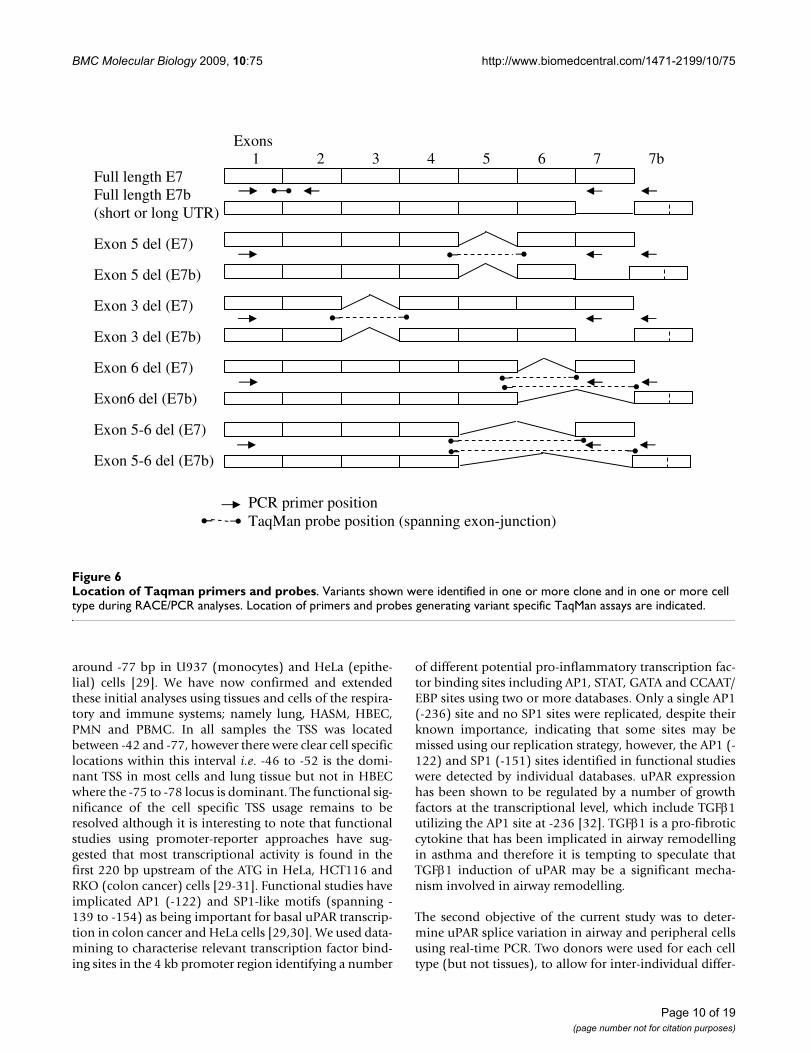
Real-time quantitative PCR confirms uPAR splice variants and identifies cell-specific expressionIn order to confirm and quantify the expression of differ-ent uPAR variants at the mRNA level, a series of real-timePCR assays were designed to amplify uPAR exons 1–2(conserved, total uPAR), 1–7 and 1–7b, whilst a series ofdeletion-specific probes allowed detection of variants(Table 2 and Figure 6). Two different normalising orhousekeeping assays, 18s ribosomal RNA and hypoxan-thine guanine phosphoribosyltransferase 1 (HPRT1),were tested and showed similar results (data not shown).However, HPRT1 was used in the main analysis as its level
of mRNA expression was more comparable to that ofuPAR itself.
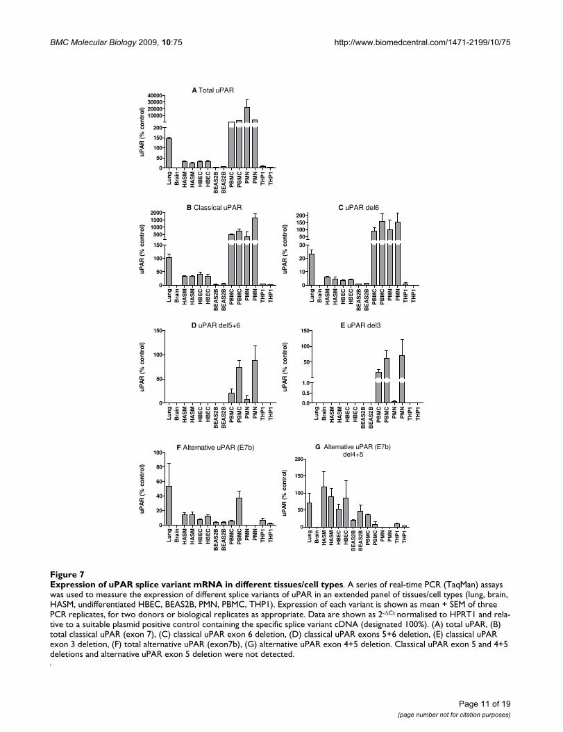
Real-time PCR was performed using cDNA from eight tis-sues/cell types in total, from either one (lung and brain)or two (HASM, HBEC, PMN, PBMC) donors, or preparedfrom RNA extracted on two different occasions for celllines (THP1 (monocyte), BEAS2B (bronchial epithe-lium)). Total uPAR expression was confirmed in the lungtissue and varied in a cell-type specific pattern; PMN ≥PBMC>>lung>HBEC≈HASM>BEAS2B≈THP-1 (ANOVA p= 0.013), the HASM and HBEC expression levels being~100 fold lower than primary peripheral cells (PMN andPBMC) (Figure 7A). Expression was very low in brain (6-fold less than cultured cells).
Overall, expression levels of classical uPAR (exon 7) cor-relate with total uPAR levels, PMN ≥PBMC>>lung>HBEC≈HASM>BEAS2B≈THP1 (Pearson r =0.891, p = 0.003), suggesting that this may be the domi-nant expressed variant (Figure 7A and 7B). Exon deletedversions of classical uPAR were also confirmed (Figure 7C– E). Exon 6 deletions were widely expressed with anexpression profile similar to classical uPAR (exon 7)expression, whilst exon 3 and 5+6 deletions were detectedonly in PMN and PBMC, with some donor specific varia-tion (Figure 7D and 7E). Unlike total or classical uPAR
Identification and characterisation of the uPAR promoterFigure 2Identification and characterisation of the uPAR promoter. (A) 5' RACE performed on a panel of tissues/cell types identified a cluster of transcriptional start sites (TSS) which show distinct patterns of usage. TSS are listed relative to ATG of translated sequence. (B) 4000 bp upstream (from ATG) was analysed for potential transcription factor binding sites using four different programs. Those sites identified in two or more different searches are shown.
� 0��
���� ������������������������������������������������������ ���������������������!!�"��� #�$� #%�&�������� #%�&������ �$�� �%$&�
'(�)*+����)**� ,� ,� ,� ,� '*��-!� *��'!�+(�)*.����)/+� �������� ������� ������ ������� �������� ��������-(�).*����).0� '1��*!� '1��*!� *��'!� ,��,!� '*��-!� 0��+!�*(�)1/����)12� +.��.!� --��2!� ������� �������� '*��-!� 0��+!�3�4��� ,��,!� *��'!� ++��/!� 0��+!� *��'!� *��'!�
�1��
� � ���
*,,,� )-,,,� )+,,,� )',,,� ,�
�'� 5 �
�6)��7��
���%��������
����������)1/)128�).*).08�)*.)/+!��
Page 4 of 19(page number not for citation purposes)
BMC Molecular Biology 2009, 10:75 http://www.biomedcentral.com/1471-2199/10/75
which were much more highly expressed in peripheralcells, alternative uPAR (E7b) was expressed inlung>PBMC>HASM≈HBEC>THP1≈BEAS2B (Figure 7F).Expression of this variant did not correlate with totaluPAR expression. Exon 4+5 deletions were detected in allcells expressing uPAR(E7b) with expressionHASM>lung≈HBEC>BEAS2B≈PBMC>THP1 (Figure 7G).Classical uPAR exon 5 and 4+5 deletions and alternativeuPAR exon 5 deletion were not detected (data notshown).
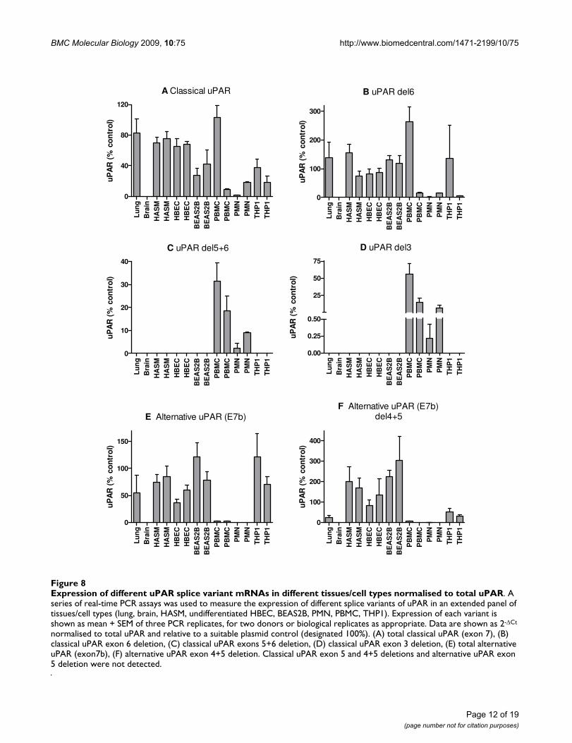
Normalisation of these data for total uPAR expressionallowed direct comparison with our previous RACE datawhich highlighted proportional expression of differentsplice variants in tissues/cells (Figure 8). The correlationbetween RACE data (Figure 3) and classical uPAR propor-tional expression (Figure 8A) was not clear as it may beexpected that the proportional expression of this isoformshould be elevated in e.g. PBMC/PMN vs. lung/HASM/HBEC as shown in the absolute expression data (Figure7A). However, uPAR(E7b) was proportionally highlyexpressed in cultured cells (THP1 and BEAS2B) > lung,HASM and HBEC (about 2/3×) >> (1/50×) PBMC (notdetected in PMN) (ANOVA not significant) (Figure 8E).These data correspond with RACE data (Figure 3), suggest-ing lower proportional expression of this variant in pri-mary peripheral cells versus lung cells and highlighteddifferences between cultured cell lines (BEAS2B, THP1)and the equivalent primary cells i.e. HBEC and PBMC.Comparison of the proportional expression of the exon4+5 deleted uPAR(E7b) form to total uPAR(E7b) expres-sion, suggests lower expression in lung, PBMC and THP1than HASM, HBEC and BEAS2B (Figure 8F), potentiallysuggesting that this splicing event occurs preferentially instructural cells. The predicted protein sequences of allconfirmed splice variants are shown in Figures 4 and 5.
Protein analysis confirms the existence of multiple forms of uPARTo determine whether the variation in uPAR isoforms atthe mRNA level was reflected at the protein level, total celllysates were assayed using two uPAR specific antibodies inWestern blots. Lysates from two donors or extractions(cell lines) were assayed (except for commercial lysates:PMN and PBMC). Representative results are shown in Fig-ure 9. Western blots performed with either domain 1 ordomain 2 specific antibodies detected multiple proteinsin all samples (Figure 9A and 9B, summarised in Table 3).Controls using no primary antibody or an isotype controlshowed no binding to either a THP1 cell lysate or recom-binant uPAR (data not shown). Loading was evaluatedusing β-actin and was equivalent for key comparisons e.g.between PBMC and THP1 and between HASM, HBEC andBEAS2B (Figure 9C).
Predicted sizes for all variants detected by real-time PCRsuggested that multiple variants between 60–100% of thefull length classical uPAR should be detected in all celltypes (Table 4). It should be noted that additional mRNAspecies were detected by PCR and RACE which were notanalysed by real-time PCR, therefore more species mayactually be present in cells. However, the abundance ofthese transcripts in RACE was low (<5%). The epitope ofthe D1 antibody is located at amino acids 52–60 (exon 2–3) therefore this antibody will detect all variants exceptexon 3 deletions and D2/3 proteolysis fragments. Theepitope of the D2 antibody is located at amino acids 125–132 (exon 4) therefore this antibody will not detect exon4 deleted variants or D1 proteolysis fragments. Glycosyla-tion of uPAR occurs at asparginine residues located atamino acids 52 (located in D1, exon3), 162 (D2, exon 5),172 (D2, exon 5) and 200 (D3, exon 6) and shows sitespecific heterogeneity, generating multiple differentmolecular weight forms [25]. This may result in a widerange of proteins on Western blotting.
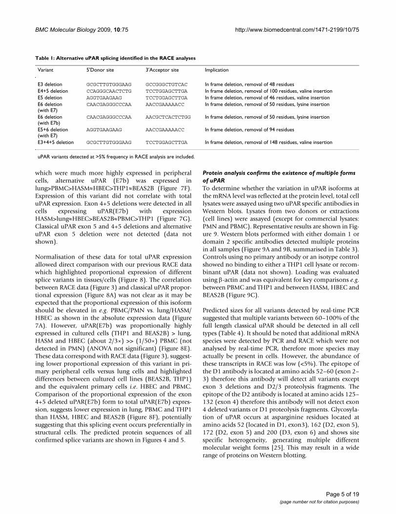
Table 1: Alternative uPAR splicing identified in the RACE analyses
Variant 5'Donor site 3'Acceptor site Implication
E3 deletion GCGCTTGTGGGAAG GCCGGGCTGTCAC In frame deletion, removal of 48 residuesE4+5 deletion CCAGGGCAACTCTG TCCTGGAGCTTGA In frame deletion, removal of 100 residues, valine insertionE5 deletion AGGTGAAGAAG TCCTGGAGCTTGA In frame deletion, removal of 46 residues, valine insertionE6 deletion(with E7)
CAACGAGGGCCCAA AACCGAAAAACC In frame deletion, removal of 50 residues, lysine insertion
E6 deletion(with E7b)
CAACGAGGGCCCAA AACGCTCACTCTGG In frame deletion, removal of 50 residues, lysine insertion
E5+6 deletion(with E7)
AGGTGAAGAAG AACCGAAAAACC In frame deletion, removal of 94 residues
E3+4+5 deletion GCGCTTGTGGGAAG TCCTGGAGCTTGA In frame deletion, removal of 148 residues, valine insertion
uPAR variants detected at >5% frequency in RACE analysis are included.
Page 5 of 19(page number not for citation purposes)

BMC Molecular Biology 2009, 10:75 http://www.biomedcentral.com/1471-2199/10/75
Full-length uPAR protein has a predicted molecularweight of 31 kDa, however differential glycosylation pat-terns result in a size range of 40–60 kDa. RecombinantuPAR expressed in a mouse melanoma cell line wasincluded and was detected between 45–60 kDa with bothantibodies. The D2 specific antibody also detected ahigher molecular weight protein (70–75 kDa), possiblyrepresenting a dimerisation or aggregation product. Alower molecular weight protein was also observed (30–35kDa), which probably corresponds to a D2/3 proteolyticfragment.
The D1 antibody detected fewer proteins than the D2 anti-body (including for the recombinant protein) (Figure9A). D1 proteolytic fragments (expected molecular weight10–20 kDa) were not detected, probably due to their rela-tively small molecular weight. The highest molecularweight protein seen in all cell types (60 kDa) probablyrepresents full-length classical uPAR as it correspondsmost closely to the band observed for recombinant uPARand by others [26-28]. Proteins are seen around 50 kDafor all except PMN and PBMC cells, suggesting that thesemay represent the alternative exon 7b form (expected
Identification of 3' variants in a panel of tissues/cell typesFigure 3Identification of 3' variants in a panel of tissues/cell types. 3' RACE was performed on a panel of tissues/cell types using a uPAR-specific forward primer located in exon 4. Frequencies of the most common variants (>5% overall expression) in each cell type based on numbers of RACE clones are summarised (A) and their structures shown (B). "Other" variants include both alternative termination sites and splice variants. Grey blocks highlight 3' UTR in the terminal exon.
A
Expression in different tissues/cell types (% and (number of clones analysed)) Expressed form
Lung HASM HBEC undiff HBEC diff PMN PBMC 1: E7b short 54 (20) 18 (6) 66 (29) 67 (14) 14 (3) 24 (5) 2: E7 (classical) 24 (9) 15 (5) 7 (3) 14 (3) 14 (3) 19 (4) 3: E7b long 3 (1) 56 (19) 11 (5) 19 (4) 5 (1) 0 4: E6 del, E7 0 0 2 (1) 0 45 (10) 33 (7) Others 19 (7) 12 (4) 9 (4) 0 23 (5) 24 (5)
B
1
2
3
4
Frequently expressed variants (>5% total)
273-277 bp
563-564 bp
443-453 bp
563-564 bp
Exon: 4 5 6 7 7b Exon length (bp): 162 135 147 564 430
Page 6 of 19(page number not for citation purposes)
BMC Molecular Biology 2009, 10:75 http://www.biomedcentral.com/1471-2199/10/75
~90% of the mass of classical uPAR). The slightly smallerfragments detected in PMN and PBMC cells only (desig-nated 45 kDa) may represent single exon (e.g. exon 6)deletions of classical uPAR (expected ~80% of the mass ofclassical uPAR). A protein at 37 kDa is detected in all celltypes except HBEC, being particularly strongly expressedin PMN and PBMC. This could represent dual exon dele-tions (e.g. classical uPAR, exon 5+6 deletion), although itspattern of expression does not completely correspond tothat seen at the mRNA level. The smallest protein seen (30kDa) may represent an alternative exon 7b, exon 4+5deleted form (expected ~55% of the mass of classicaluPAR), although this might be expected to be expressed ina greater number of cell types.
The D2 antibody detects three proteins in the recom-binant sample, probably reflecting a dimerisation, full-length uPAR and a D2/3 proteolytic fragment (Figure 9B).The large (75 kDa) protein is detected in the majority ofcell types, being most strongly expressed in PBMC andTHP1 cells. HBEC lysates showed less abundant proteinsat lower molecular weights than other structural celllysates, reflecting the pattern using the D1 antibody,although overall it is difficult to make direct comparisonsbetween the two blots.
The expression of soluble PAR was detected in the super-natants of all cells tested (Figure 9D). Overall, higher lev-els of soluble uPAR were observed in supernatants fromBEAS2B and THP1 cells compared to HASM and HBEC.Interestingly, in the proportional mRNA level analyses,
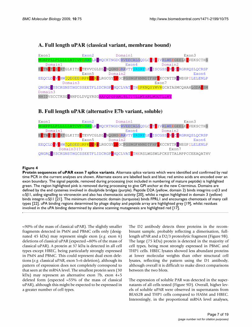
Protein sequences of uPAR exon 7 splice variantsFigure 4Protein sequences of uPAR exon 7 splice variants. Alternate splice variants which were identified and confirmed by real time PCR in the current analyses are shown. Alternate exons are labelled back and blue; red amino acids are encoded over an exon boundary. The signal peptide, removed during processing (not included in numbering of mature peptide) is highlighted green. The region highlighted pink is removed during processing to give GPI anchor at the new C-terminus. Domains are defined by the end cysteines involved in disulphide bridges (purple). Peptide D2A (yellow, domain 2) binds integrins αvβ3 and α5β1, aiding signalling to vitronectin and also has chemotactic activity [20], whilst a region highlighted in domain 3 (yellow) binds integrin α5β1 [21]. The minimum chemotactic domain (turquoise) binds FPRL1 and encourages chemotaxis of many cell types [22]. uPA binding regions determined by phage display and peptide array are highlighted grey [19], whilst residues involved in the uPA binding determined by alanine scanning mutagenesis are highlighted red [17].
A. Full length uPAR (classical variant, membrane bound) Exon1 Exon2 Domain1 Exon3 MGHPPLLPLLLLLHTCVPASWGLRCMQCKTNGDCRVEECALGQDLCRTTIVRLWEEGEELELVEKSCTHS Domain1 Exon4 Domain2 EKTNRTLSYRTGLKITSLTEVVCGLDLCNQGNSGRAVTYSRSRYLECISCGSSDMSCERGRHQSLQCRSP Exon5 Domain2 Exon6 EEQCLDVVTHWIQEGEEGRPKDDRHLRGCGYLPGCPGSNGFHNNDTFHFLKCCNTTKCNEGPILELENLP Domain3 Exon7 QNGRQCYSCKGNSTHGCSSEETFLIDCRGPMNQCLVATGTHEPKNQSYMVRGCATASMCQHAHLGDAFSM Domain3 NHIDVSCCTKSGCNHPDLDVQYRSGAAPQPGPAHLSLTITLLMTARLWGGTLLWT
B. Full length uPAR (alternative E7b variant, soluble) Exon1 Exon2 Domain1 Exon3 MGHPPLLPLLLLLHTCVPASWGLRCMQCKTNGDCRVEECALGQDLCRTTIVRLWEEGEELELVEKSCTHS Domain1 Exon4 Domain2 EKTNRTLSYRTGLKITSLTEVVCGLDLCNQGNSGRAVTYSRSRYLECISCGSSDMSCERGRHQSLQCRSP Exon5 Domain2 Exon6 EEQCLDVVTHWIQEGEEGRPKDDRHLRGCGYLPGCPGSNGFHNNDTFHFLKCCNTTKCNEGPILELENLP Domain3(?) Exon7 QNGRQCYSCKGNSTHGCSSEETFLIDCRGPMNQCLVATGTHERSLWGSWLPCKSTTALRPPCCEEAQATHV
Page 7 of 19(page number not for citation purposes)
BMC Molecular Biology 2009, 10:75 http://www.biomedcentral.com/1471-2199/10/75
Page 8 of 19(page number not for citation purposes)
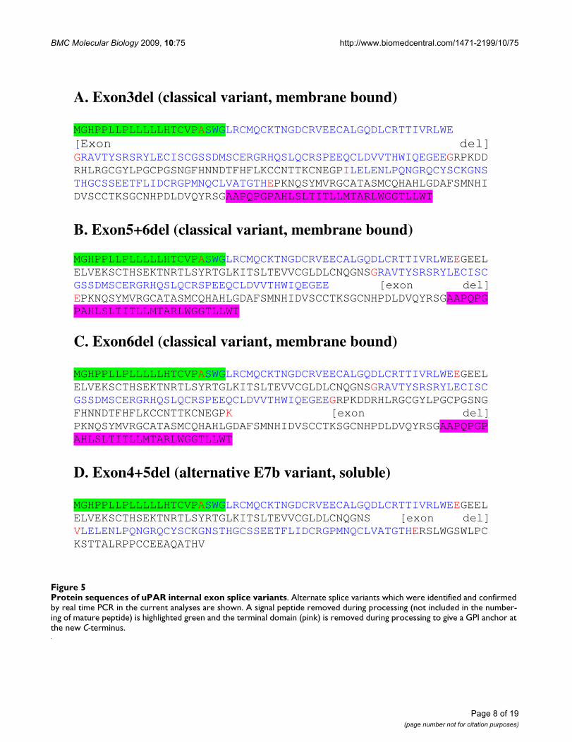
Protein sequences of uPAR internal exon splice variantsFigure 5Protein sequences of uPAR internal exon splice variants. Alternate splice variants which were identified and confirmed by real time PCR in the current analyses are shown. A signal peptide removed during processing (not included in the number-ing of mature peptide) is highlighted green and the terminal domain (pink) is removed during processing to give a GPI anchor at the new C-terminus.
A. Exon3del (classical variant, membrane bound) MGHPPLLPLLLLLHTCVPASWGLRCMQCKTNGDCRVEECALGQDLCRTTIVRLWE [Exon del] GRAVTYSRSRYLECISCGSSDMSCERGRHQSLQCRSPEEQCLDVVTHWIQEGEEGRPKDDRHLRGCGYLPGCPGSNGFHNNDTFHFLKCCNTTKCNEGPILELENLPQNGRQCYSCKGNSTHGCSSEETFLIDCRGPMNQCLVATGTHEPKNQSYMVRGCATASMCQHAHLGDAFSMNHIDVSCCTKSGCNHPDLDVQYRSGAAPQPGPAHLSLTITLLMTARLWGGTLLWT
B. Exon5+6del (classical variant, membrane bound) MGHPPLLPLLLLLHTCVPASWGLRCMQCKTNGDCRVEECALGQDLCRTTIVRLWEEGEELELVEKSCTHSEKTNRTLSYRTGLKITSLTEVVCGLDLCNQGNSGRAVTYSRSRYLECISCGSSDMSCERGRHQSLQCRSPEEQCLDVVTHWIQEGEE [exon del] EPKNQSYMVRGCATASMCQHAHLGDAFSMNHIDVSCCTKSGCNHPDLDVQYRSGAAPQPGPAHLSLTITLLMTARLWGGTLLWT
C. Exon6del (classical variant, membrane bound) MGHPPLLPLLLLLHTCVPASWGLRCMQCKTNGDCRVEECALGQDLCRTTIVRLWEEGEELELVEKSCTHSEKTNRTLSYRTGLKITSLTEVVCGLDLCNQGNSGRAVTYSRSRYLECISCGSSDMSCERGRHQSLQCRSPEEQCLDVVTHWIQEGEEGRPKDDRHLRGCGYLPGCPGSNGFHNNDTFHFLKCCNTTKCNEGPK [exon del] PKNQSYMVRGCATASMCQHAHLGDAFSMNHIDVSCCTKSGCNHPDLDVQYRSGAAPQPGPAHLSLTITLLMTARLWGGTLLWT
D. Exon4+5del (alternative E7b variant, soluble) MGHPPLLPLLLLLHTCVPASWGLRCMQCKTNGDCRVEECALGQDLCRTTIVRLWEEGEELELVEKSCTHSEKTNRTLSYRTGLKITSLTEVVCGLDLCNQGNS [exon del] VLELENLPQNGRQCYSCKGNSTHGCSSEETFLIDCRGPMNQCLVATGTHERSLWGSWLPCKSTTALRPPCCEEAQATHV
BMC Molecular Biology 2009, 10:75 http://www.biomedcentral.com/1471-2199/10/75
BEAS2B and THP1 were shown to express a greater pro-portion of the alternative, soluble form (exon 7b) com-pared to HASM and HBEC (Figure 8E).
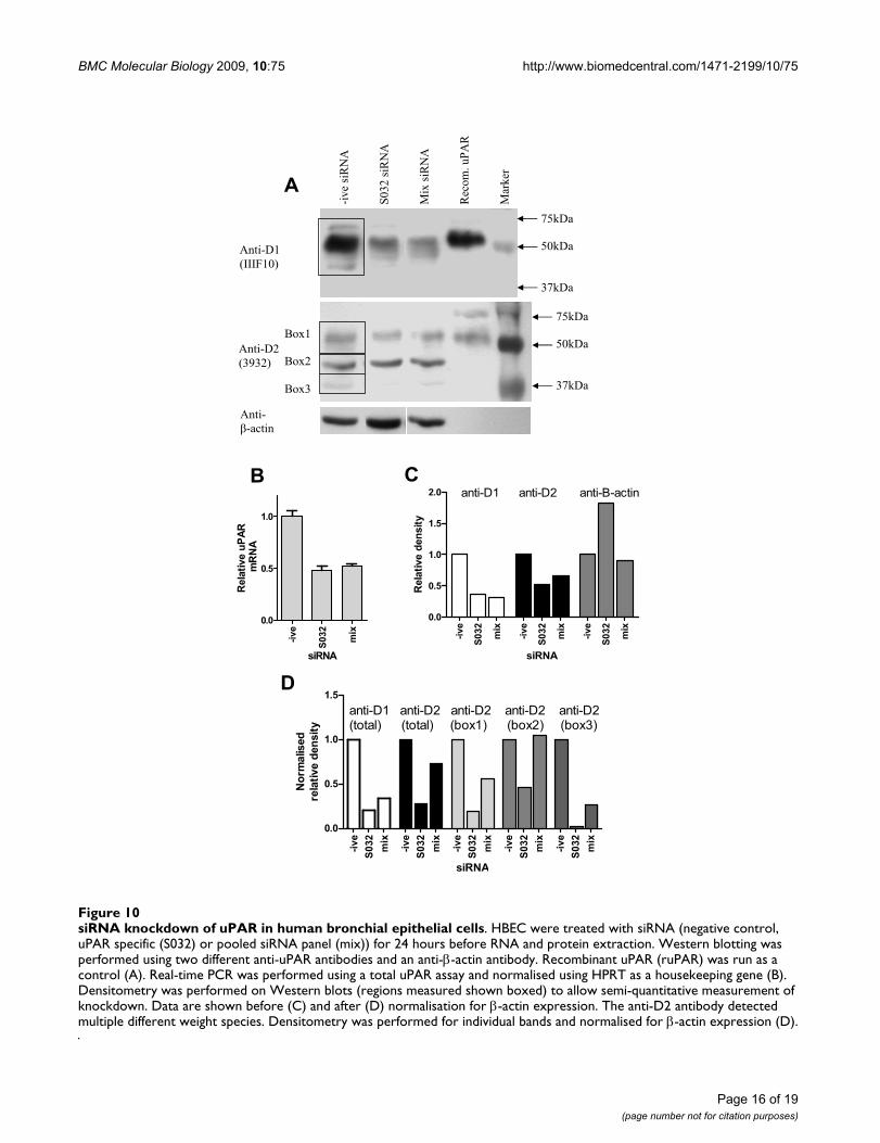
siRNA confirms the specificity of anti-uPAR Western blotsWestern blotting using two monoclonal antibodies foruPAR detected multiple bands. To confirm that thesebands represent uPAR variants, siRNA was performed inundifferentiated HBEC cells. Cells were treated withsiRNA (negative control, uPAR-specific (S032) or a uPAR-specific panel (mix)) for 24 hours before RNA and proteinwere extracted and analysed by real-time PCR and West-ern blotting (Figure 10A). Total uPAR mRNA expressionwas measured by real-time PCR, showing about 50%knockdown with both siRNA strategies (Figure 10B).Total uPAR protein expression detected by the domain 1(IIIF10) and domain 2 (3932) antibodies is also reduced(Figure 10A) which was confirmed by densitometry anal-ysis, before and after normalisation to β-actin (Figures10C and 10D respectively). The domain 2 specific anti-body detected three main species, which were all shownto be knocked down at the protein level by at least onesiRNA approach using densitometry (Figure 10D).
DiscussionThe expression and functional activity of uPAR is of inter-est in both cancer and respiratory disease. The current
study aimed to characterise the key regulatory regions ofUPAR, identify all common splice variation and quantifyexpression in the normal lung and in specific lung andperipheral cells at the mRNA and protein levels. We haveidentified a localised TSS with some cell specificity andconfirmed expression of uPAR in lung and peripheralcells. At the mRNA level, multiple uPAR splice variantswere identified including alternative E7 (E7b) and dele-tions of E3, E5+6 and E6, and their patterns of expressionin different tissues/cell types characterised. Primaryperipheral cells (PMN and PBMC) expressed multipleexon deleted forms of membrane bound uPAR, whilstlung cells including epithelium and airway smooth mus-cle expressed a greater proportion of an alternative solubleuPAR with and without an exon 4+5 deletion. Proteinanalyses confirmed expression of multiple differentiallyexpressed forms of uPAR in all cell extracts, and solubleuPAR was detected in the supernatants of cultured HASM,HBEC, THP1 and BEAS2B cells. Specificity of the Westernblotting analyses was confirmed by siRNA. Our data pro-vide a novel insight into the molecular mechanisms thatpotentially regulate uPAR expression and activity in theairways and the periphery which has implications regard-ing the potential role of uPAR in airway disease biology.
Previous analyses in cell lines suggested the TSS of UPARis 52 bp upstream of the ATG, with a minor start site
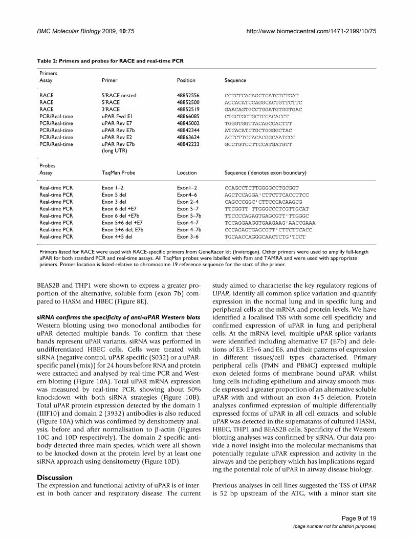
Table 2: Primers and probes for RACE and real-time PCR
PrimersAssay Primer Position Sequence
RACE 5'RACE nested 48852556 CCTCTCACAGCTCATGTCTGATRACE 5'RACE 48852500 ACCACATCCAGGCACTGTTCTTCRACE 3'RACE 48852519 GAACAGTGCCTGGATGTGGTGACPCR/Real-time uPAR Fwd E1 48866085 CTGCTGCTGCTCCACACCTPCR/Real-time uPAR Rev E7 48845002 TGGGTGGTTACAGCCACTTTPCR/Real-time uPAR Rev E7b 48842344 ATCACATCTGCTGGGGCTACPCR/Real-time uPAR Rev E2 48863624 ACTCTTCCACACGGCAATCCCPCR/Real-time uPAR Rev E7b
(long UTR)48842223 GCCTGTCCTTCCATGATGTT
ProbesAssay TaqMan Probe Location Sequence ('denotes exon boundary)
Real-time PCR Exon 1–2 Exon1–2 CCAGCCTCTTGGGGCCTGCGGTReal-time PCR Exon 5 del Exon4–6 AGCTCCAGGA'CTTCTTCACCTTCCReal-time PCR Exon 3 del Exon 2–4 CAGCCCGGC'CTTCCCACAAGCGReal-time PCR Exon 6 del +E7 Exon 5–7 TTCGGTT'TTGGGCCCTCGTTGCATReal-time PCR Exon 6 del +E7b Exon 5–7b TTCCCCAGAGTGAGCGTT'TTGGGCReal-time PCR Exon 5+6 del +E7 Exon 4–7 TCCAGGAAGGTGAAGAAG'AACCGAAAReal-time PCR Exon 5+6 del; E7b Exon 4–7b CCCAGAGTGAGCGTT'CTTCTTCACCReal-time PCR Exon 4+5 del Exon 3–6 TGCAACCAGGGCAACTCTG'TCCT
Primers listed for RACE were used with RACE-specific primers from GeneRacer kit (Invitrogen). Other primers were used to amplify full-length uPAR for both standard PCR and real-time assays. All TaqMan probes were labelled with Fam and TAMRA and were used with appropriate primers. Primer location is listed relative to chromosome 19 reference sequence for the start of the primer.
Page 9 of 19(page number not for citation purposes)
BMC Molecular Biology 2009, 10:75 http://www.biomedcentral.com/1471-2199/10/75
around -77 bp in U937 (monocytes) and HeLa (epithe-lial) cells [29]. We have now confirmed and extendedthese initial analyses using tissues and cells of the respira-tory and immune systems; namely lung, HASM, HBEC,PMN and PBMC. In all samples the TSS was locatedbetween -42 and -77, however there were clear cell specificlocations within this interval i.e. -46 to -52 is the domi-nant TSS in most cells and lung tissue but not in HBECwhere the -75 to -78 locus is dominant. The functional sig-nificance of the cell specific TSS usage remains to beresolved although it is interesting to note that functionalstudies using promoter-reporter approaches have sug-gested that most transcriptional activity is found in thefirst 220 bp upstream of the ATG in HeLa, HCT116 andRKO (colon cancer) cells [29-31]. Functional studies haveimplicated AP1 (-122) and SP1-like motifs (spanning -139 to -154) as being important for basal uPAR transcrip-tion in colon cancer and HeLa cells [29,30]. We used data-mining to characterise relevant transcription factor bind-ing sites in the 4 kb promoter region identifying a number
of different potential pro-inflammatory transcription fac-tor binding sites including AP1, STAT, GATA and CCAAT/EBP sites using two or more databases. Only a single AP1(-236) site and no SP1 sites were replicated, despite theirknown importance, indicating that some sites may bemissed using our replication strategy, however, the AP1 (-122) and SP1 (-151) sites identified in functional studieswere detected by individual databases. uPAR expressionhas been shown to be regulated by a number of growthfactors at the transcriptional level, which include TGFβ1utilizing the AP1 site at -236 [32]. TGFβ1 is a pro-fibroticcytokine that has been implicated in airway remodellingin asthma and therefore it is tempting to speculate thatTGFβ1 induction of uPAR may be a significant mecha-nism involved in airway remodelling.
The second objective of the current study was to deter-mine uPAR splice variation in airway and peripheral cellsusing real-time PCR. Two donors were used for each celltype (but not tissues), to allow for inter-individual differ-
Location of Taqman primers and probesFigure 6Location of Taqman primers and probes. Variants shown were identified in one or more clone and in one or more cell type during RACE/PCR analyses. Location of primers and probes generating variant specific TaqMan assays are indicated.
Exons
1 2 3 4 5 6 7 7b Full length E7 Full length E7b (short or long UTR)
Exon 5 del (E7)
Exon 5 del (E7b)
Exon 3 del (E7)
Exon 3 del (E7b)
Exon 6 del (E7)
Exon6 del (E7b)
Exon 5-6 del (E7)
Exon 5-6 del (E7b)
PCR primer position TaqMan probe position (spanning exon-junction)
Page 10 of 19(page number not for citation purposes)
BMC Molecular Biology 2009, 10:75 http://www.biomedcentral.com/1471-2199/10/75
Page 11 of 19(page number not for citation purposes)
Expression of uPAR splice variant mRNA in different tissues/cell typesFigure 7Expression of uPAR splice variant mRNA in different tissues/cell types. A series of real-time PCR (TaqMan) assays was used to measure the expression of different splice variants of uPAR in an extended panel of tissues/cell types (lung, brain, HASM, undifferentiated HBEC, BEAS2B, PMN, PBMC, THP1). Expression of each variant is shown as mean + SEM of three PCR replicates, for two donors or biological replicates as appropriate. Data are shown as 2-ΔCt normalised to HPRT1 and rela-tive to a suitable plasmid positive control containing the specific splice variant cDNA (designated 100%). (A) total uPAR, (B) total classical uPAR (exon 7), (C) classical uPAR exon 6 deletion, (D) classical uPAR exons 5+6 deletion, (E) classical uPAR exon 3 deletion, (F) total alternative uPAR (exon7b), (G) alternative uPAR exon 4+5 deletion. Classical uPAR exon 5 and 4+5 deletions and alternative uPAR exon 5 deletion were not detected.
A Total uPAR
Lu
ng
Bra
inH
AS
MH
AS
MH
BE
CH
BE
CB
EA
S2B
BE
AS
2BP
BM
CP
BM
CP
MN
PM
NT
HP
1T
HP
1
0
50
100
150
200
10000200003000040000
uP
AR
(%
co
ntr
ol)
B Classical uPAR
Lu
ng
Bra
inH
AS
MH
AS
MH
BE
CH
BE
CB
EA
S2B
BE
AS
2BP
BM
CP
BM
CP
MN
PM
NT
HP
1T
HP
1
0
50
100
150
500100015002000
uP
AR
(%
con
tro
l)
C uPAR del6
Lu
ng
Bra
inH
AS
MH
AS
MH
BE
CH
BE
CB
EA
S2B
BE
AS
2BP
BM
CP
BM
CP
MN
PM
NT
HP
1T
HP
1
0
10
20
30
50100150200
uP
AR
(%
con
tro
l)
D uPAR del5+6
Lu
ng
Bra
inH
AS
MH
AS
MH
BE
CH
BE
CB
EA
S2B
BE
AS
2BP
BM
CP
BM
CP
MN
PM
NT
HP
1T
HP
1
0
50
100
150
uP
AR
(%
co
ntr
ol)
E uPAR del3L
un
gB
rain
HA
SM
HA
SM
HB
EC
HB
EC
BE
AS
2BB
EA
S2B
PB
MC
PB
MC
PM
NP
MN
TH
P1
TH
P1
0.0
0.5
1.0
50
100
150
uPA
R (
% c
on
tro
l)
F Alternative uPAR (E7b)
Lu
ng
Bra
inH
AS
MH
AS
MH
BE
CH
BE
CB
EA
S2B
BE
AS
2BP
BM
CP
BM
CP
MN
PM
NT
HP
1T
HP
1
0
20
40
60
80
100
uP
AR
(%
co
ntr
ol)
G Alternative uPAR (E7b)del4+5
Lung
Bra
inH
AS
MH
AS
MH
BE
CH
BE
CB
EA
S2B
BE
AS
2BP
BM
CP
BM
CP
MN
PM
NT
HP
1T
HP
1
0
50
100
150
200
uPA
R (
% c
ontr
ol)
BMC Molecular Biology 2009, 10:75 http://www.biomedcentral.com/1471-2199/10/75
Page 12 of 19(page number not for citation purposes)
Expression of different uPAR splice variant mRNAs in different tissues/cell types normalised to total uPARFigure 8Expression of different uPAR splice variant mRNAs in different tissues/cell types normalised to total uPAR. A series of real-time PCR assays was used to measure the expression of different splice variants of uPAR in an extended panel of tissues/cell types (lung, brain, HASM, undifferentiated HBEC, BEAS2B, PMN, PBMC, THP1). Expression of each variant is shown as mean + SEM of three PCR replicates, for two donors or biological replicates as appropriate. Data are shown as 2-ΔCt
normalised to total uPAR and relative to a suitable plasmid control (designated 100%). (A) total classical uPAR (exon 7), (B) classical uPAR exon 6 deletion, (C) classical uPAR exons 5+6 deletion, (D) classical uPAR exon 3 deletion, (E) total alternative uPAR (exon7b), (F) alternative uPAR exon 4+5 deletion. Classical uPAR exon 5 and 4+5 deletions and alternative uPAR exon 5 deletion were not detected.
A Classical uPAR
Lung
Bra
inH
AS
MH
AS
MH
BE
CH
BE
CB
EA
S2B
BE
AS
2BP
BM
CP
BM
CP
MN
PM
NT
HP
1T
HP
1
0
40
80
120uP
AR
(%
con
trol
)
B uPAR del6
Lun
gB
rain
HA
SM
HA
SM
HB
EC
HB
EC
BE
AS
2BB
EA
S2B
PB
MC
PB
MC
PM
NP
MN
THP
1TH
P1
0
100
200
300
uPA
R (
% c
ontr
ol)
C uPAR del5+6
Lun
gB
rain
HA
SM
HA
SM
HB
EC
HB
EC
BE
AS
2BB
EA
S2B
PB
MC
PB
MC
PM
NP
MN
THP
1TH
P1
0
10
20
30
40
uPA
R (
% c
ontr
ol)
D uPAR del3
Lung
Bra
inH
AS
MH
AS
MH
BE
CH
BE
CB
EA
S2B
BE
AS
2BP
BM
CP
BM
CP
MN
PM
NTH
P1
THP
1
0.00
0.25
0.50
25
50
75
uPA
R (
% c
ont
rol)
E Alternative uPAR (E7b)
Lun
gB
rain
HA
SM
HA
SM
HB
EC
HB
EC
BE
AS
2BB
EA
S2B
PB
MC
PB
MC
PM
NP
MN
THP
1TH
P1
0
50
100
150
uP
AR
(%
con
trol
)
F Alternative uPAR (E7b)del4+5
Lun
gB
rain
HA
SM
HA
SM
HB
EC
HB
EC
BE
AS
2BB
EA
S2B
PB
MC
PB
MC
PM
NP
MN
THP
1TH
P1
0
100
200
300
400
uP
AR
(%
con
trol
)
BMC Molecular Biology 2009, 10:75 http://www.biomedcentral.com/1471-2199/10/75
ences. We observed expression of the previously identifieduPAR E7b soluble splice variant [24], which is predictedto result in the loss of the GPI anchor, in the lung andperiphery. Interestingly the primary peripheral cellsexpressed low levels of this variant (as a fraction of totaluPAR) compared to the lung tissue and cells. This suggeststhat peripheral cells could retain more of their uPAR at thecell membrane (GPI-bound) whilst lung structural cellsproduce more soluble, secreted uPAR. Binding of uPA tomembrane bound uPAR initiates many intracellular sig-nalling pathways, resulting in outcomes including differ-entiation, proliferation and cell motility [5], whilstsoluble uPAR can act as a chemoattractant for hematopoi-etic cells [33]. It is therefore possible that airway cellsmight express more soluble uPAR to allow them to attractcells into the airways, whilst the peripheral cells expressmainly the surface form of the receptor to allow them torespond to external signals.
In addition to the exon7 variants we also identified aseries of internal exon deletion variants expressed withinthe context of both the membrane bound and solublereceptor. These included exon 3, 4+5, 5+6 and 6 deletions.Exon deleted forms of classical uPAR were found most fre-quently in the primary peripheral cells, PMN and PBMC.Exon 6 deletion was the most widely expressed. Loss ofexon 6 would disrupt the structure of D3, particularly as itincludes one of the key cysteines involved in disulphidebonding. This would be expected to lead to reduced uPAbinding affinity [19] as two amino acid residues shown tobe involved in uPA binding are located in this exon [17].Expression of an exon 4+5 deleted variant of uPAR haspreviously been described and was shown to be associatedwith shorter disease-free survival in breast cancers using areal-time PCR strategy [3], however the assay used did notdistinguish between exon 7 variants. In the present study,
we did not detect this deletion in combination with clas-sical exon 7 using real-time PCR (although a single clonewas obtained from PBMC by PCR), whereas in combina-tion with alternative exon 7b this variant was detected inmost tissues/cell types, notably the structural cells HASM,HBEC and BEAS2B. Based on the known structure ofuPAR, this variant might express a soluble form of thereceptor lacking the key chemotactic sequence [22] andD2, which includes integrin binding domains [20]. SomeuPA binding ability conferred by D1 may be retained [19]although many uPA binding residues will be lost [17] andthe structure of the protein [18] will be compromised. It ispossible that this form might act as a dominant-negativeinhibitor of uPAR action, by sequestering uPA and pre-venting its binding to full length membrane-bound or sol-uble forms of the receptor.
Repeated epithelial wounding/repair leading to airwayremodelling has been implicated in asthma pathogenesis[34]. Epithelial cells have the capacity to repair by cellspreading, migration and proliferation, all integrin-dependent processes. Integrins have therefore been sug-gested to play a prominent role in wound repair in asthmaand the expression of α3β1 (laminin receptor) and α5β1(fibronectin receptor) integrins have been shown to beupregulated at epithelial wound edges [35,36]. uPARinteracts with integrins (mainly α3β1 and α5β1) and hasbeen shown to influence cell adhesion and migration onECM proteins [37,38]. Therefore the identification ofsplice variants that lose this ability to bind integrins (i.e.exon 5 deletions) may be of significance in normal anddisease mechanisms.
At the protein level, multiple forms of uPAR were detectedin cell lysates. Overall, our data suggest that multipleforms of uPAR are expressed in the panel of cells studied
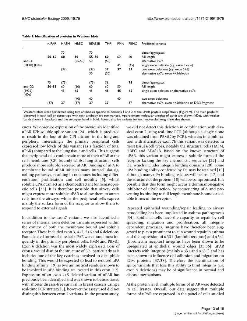
Table 3: Identification of proteins in Western blots
ruPAR HASM HBEC BEAS2B THP1 PMN PBMC Predicted variants
70 70 dimer/aggregates50–60 60 60 55–60 60 60 60 full length
anti-D1 (55-50) 50 (50) alternative ex7b(IIIF10) (kDa) 45 (45) single exon deletion (e.g. exon 3 or 6)
(37) (37) 37 37 37 two exon deletions (e.g. exon 5+6)30 (30) alternative ex7b, exon 4+5deletion
75 (75) (75) 75 75 dimer/aggregatesanti-D2 50–55 60 (60) 60 60 50 full length(3932)(kDa)
45 45 45 45 45 45 single exon deletion or alternative ex7b
(40) 40 40 two exon deletions(37) 37 (37) 37 37 37 alternative ex7b, exon 4+5deletion or D2/3 fragment
Western blots were performed using two antibodies specific to domains 1 and 2 of the uPAR protein respectively (Figure 9). The main proteins observed in each cell or tissue type with each antibody are summarised. Approximate molecular weights of bands are shown (kDa), with weaker bands shown in brackets and the strongest band in bold. Potential splice variants for each molecular weight are also shown.
Page 13 of 19(page number not for citation purposes)
BMC Molecular Biology 2009, 10:75 http://www.biomedcentral.com/1471-2199/10/75
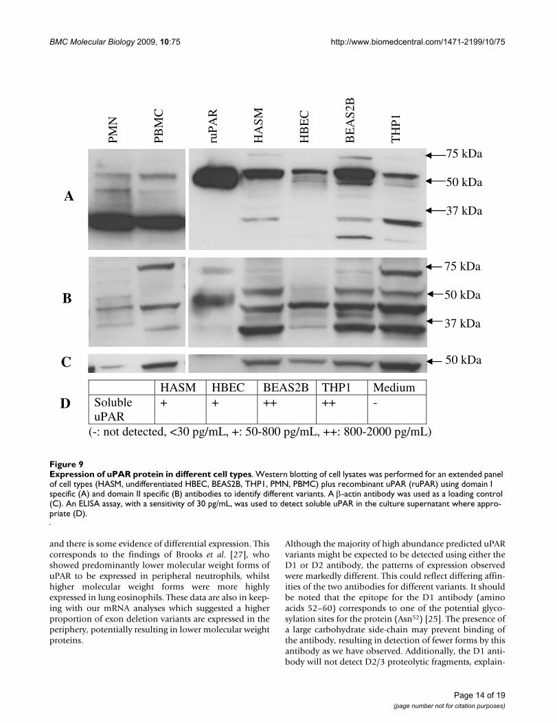
and there is some evidence of differential expression. Thiscorresponds to the findings of Brooks et al. [27], whoshowed predominantly lower molecular weight forms ofuPAR to be expressed in peripheral neutrophils, whilsthigher molecular weight forms were more highlyexpressed in lung eosinophils. These data are also in keep-ing with our mRNA analyses which suggested a higherproportion of exon deletion variants are expressed in theperiphery, potentially resulting in lower molecular weightproteins.
Although the majority of high abundance predicted uPARvariants might be expected to be detected using either theD1 or D2 antibody, the patterns of expression observedwere markedly different. This could reflect differing affin-ities of the two antibodies for different variants. It shouldbe noted that the epitope for the D1 antibody (aminoacids 52–60) corresponds to one of the potential glyco-sylation sites for the protein (Asn52) [25]. The presence ofa large carbohydrate side-chain may prevent binding ofthe antibody, resulting in detection of fewer forms by thisantibody as we have observed. Additionally, the D1 anti-body will not detect D2/3 proteolytic fragments, explain-
Expression of uPAR protein in different cell typesFigure 9Expression of uPAR protein in different cell types. Western blotting of cell lysates was performed for an extended panel of cell types (HASM, undifferentiated HBEC, BEAS2B, THP1, PMN, PBMC) plus recombinant uPAR (ruPAR) using domain I specific (A) and domain II specific (B) antibodies to identify different variants. A β-actin antibody was used as a loading control (C). An ELISA assay, with a sensitivity of 30 pg/mL, was used to detect soluble uPAR in the culture supernatant where appro-priate (D).
HASM HBEC BEAS2B THP1 Medium Soluble uPAR
+ + ++ ++ -
(-: not detected, <30 pg/mL, +: 50-800 pg/mL, ++: 800-2000 pg/mL)
PM
N
PB
MC
ruP
AR
HA
SM
HB
EC
BE
AS
2B
TH
P1
75 kDa 50 kDa 37 kDa
75 kDa 50 kDa 37 kDa
50 kDa
A
B
C
D
Page 14 of 19(page number not for citation purposes)
BMC Molecular Biology 2009, 10:75 http://www.biomedcentral.com/1471-2199/10/75
ing the presence of fewer low molecular weight proteinsdetected using this antibody. An attempt has been madeto mirror patterns of protein expression as determined byWestern blot to real-time PCR results, with some successfor the D1 antibody results e.g. the presence of a 50 kDaprotein potentially representing the alternative exon7bobserved in all cell types except PMN and PBMC, as wellas a 45 kDa form seen only in PBMC and PMN which mayrepresent single exon deletions of classical uPAR. Con-cordance between the two assays may be affected by dif-fering efficiencies of the real-time PCR assays or antibodyspecificity. However, it will also reflect genuine biologicaldifferences, as post-translational control mechanismsincluding glycosylation and proteolytic cleavage play akey role in uPAR protein maturation [15,25]. Our datareflect this complex regulatory pathway.
All cultured cells tested showed expression of solubleuPAR. This assay does not distinguish between classicaluPAR released from its GPI anchor, proteolytic fragmentsand alternatively spliced (exon 7b) soluble uPAR. How-ever, all of these cell lines expressed relatively high pro-portions of alternative uPAR(E7b) mRNA and the level ofmRNA correlates with soluble uPAR protein expressione.g. BEAS2B and THP1 had elevated alternativeuPAR(E7b) mRNA and soluble uPAR protein. Therefore,the elevated levels of uPAR in the sputum of asthma andCOPD subjects may reflect expression of soluble uPARfrom both epithelial and smooth muscle cells.
ConclusionWe have identified the key promoter region of UPAR inthe airway and highlighted potential regulatory transcrip-tion factor binding sites. We have identified multiplesplice variants with potentially different functional activ-ity including soluble expression and/or the loss of keybinding domains, such as those involved in ligand bind-ing or integrin interactions. The expression patterns ofthese splice variants in airway and peripheral cells providean insight into the molecular mechanisms regulatinguPAR expression and activity. These data highlight theimportance of uPAR splice variants, which should betaken into consideration in future work to determine therole of uPAR in respiratory disease.
MethodsCell culture, preparation of total RNA and cell protein lysates and siRNA knock-downPrimary human airway smooth muscle (HASM) cells wereisolated and prepared as described previously [39] andcultured in DMEM + 10% foetal calf serum (FCS). Airwaysmooth muscle cells from two individuals were used.BEAS-2B (airway epithelial cells) were cultured in DMEM+ 10% FCS, whilst THP1 (monocyte) cells (ATCC) werecultured in modified RPMI 1640 medium + 10% FCS +0.05 mM β-mercaptoethanol. Human bronchial epithe-lial cells (HBEC) from two donors were obtained fromLonza (Wokingham, UK) and cultured in bronchial epi-thelial growth medium (BEGM). Cells were differentiatedat an air-liquid interface using bronchial epithelial differ-entiation medium (BEDM) as described previously [40].
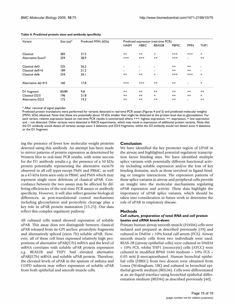
Table 4: Predicted protein sizes and antibody specificity
Variant Size (aa)* Predicted MWt (kDa) Predicted expression (real-time PCR)HASM HBEC BEAS2B PBMC PMN THP1
Classical 283 31.5 ++ ++ + +++ +++ +Alternative Exon7 259 28.9 +++ +++ ++ +++ - ++
Classical del3 235 26.2 - - - ++ ++ -Classical del5+6 189 21.2 - - - ++ ++ -Classical del6 234 26.1 ++ ++ + +++ +++ +
Alternative del 4+5 160 17.8 +++ +++ ++ ++ - +
D1 fragment 83/89 9.8 ++ ++ ++ ++ ++ ++Classical D2/3 196 21.8 ++ ++ + ++ ++ +Alternative D2/3 172 19.2 ++ ++ ++ ++ - ++
* After removal of signal peptidesPredicted protein translations were performed for variants detected in real-time PCR assays (Figures 4 and 5) and predicted molecular weights (MWt, kDa) obtained. Note that these are potentially about 10 kDa smaller than might be detected at the protein level due to glycosylation. For each variant, relative expression based on real-time PCR results is summarised where +++: highest expression, ++: expression, + low expression and -: not detected. Other variants were detected in RACE experiments, which may result in expression of additional protein variants. Note that the D1 antibody would detect all variants except exon 3 deletions and D2/3 fragments, whilst the D2 antibody would not detect exon 4 deletions or the D1 fragment.
Page 15 of 19(page number not for citation purposes)
BMC Molecular Biology 2009, 10:75 http://www.biomedcentral.com/1471-2199/10/75
Page 16 of 19(page number not for citation purposes)
siRNA knockdown of uPAR in human bronchial epithelial cellsFigure 10siRNA knockdown of uPAR in human bronchial epithelial cells. HBEC were treated with siRNA (negative control, uPAR specific (S032) or pooled siRNA panel (mix)) for 24 hours before RNA and protein extraction. Western blotting was performed using two different anti-uPAR antibodies and an anti-β-actin antibody. Recombinant uPAR (ruPAR) was run as a control (A). Real-time PCR was performed using a total uPAR assay and normalised using HPRT as a housekeeping gene (B). Densitometry was performed on Western blots (regions measured shown boxed) to allow semi-quantitative measurement of knockdown. Data are shown before (C) and after (D) normalisation for β-actin expression. The anti-D2 antibody detected multiple different weight species. Densitometry was performed for individual bands and normalised for β-actin expression (D).
����
�
��
��
����
��9�
���� ��
�:�
�:
�:�
�� ��
�����9�����
�� ��
����
��9�
���� ��
��9�
���� ��
��9�
���� ��
�:�
�:
�:�
�:
�:� �������������������������
�� ��
�����9���������
��
��9�
���� ��
��9�
���� ��
��9�
���� ��
��9�
���� ��
��9�
���� ��
�:�
�:
�:�
�:������������������������������� ������ ������ ����� ���� �����
�� ��
����������
������9���������
�
2�3��
+�'4
�
��
�+�'4
�
(��
�+�'4
�
'�*
��5�)
6 '
�
(�!
7�!�
���2���� 8����
���2���"��
���2�92�*����
��
���������������
�� ��
��
��7������7������7���
��7������7������7���
BMC Molecular Biology 2009, 10:75 http://www.biomedcentral.com/1471-2199/10/75
For downstream analysis, cells were lysed and RNAextracted using silica columns (RNeasy mini kit, Qiagen,Crawley, UK). For protein analysis, cell lysates were pre-pared using cytobuster buffer (Novagen, Merck, Notting-ham, UK) with benzonase (Merck) and complete proteaseinhibitor (Roche, Welwyn Garden City, UK), as directedby the manufacturer. Ethical approval for the use of pri-mary cells was obtained from the Nottingham UniversityHospitals local ethical committee.
uPAR was targeted for knockdown in undifferentiatedHBEC cells using siRNA specific to exon 1 (S032) or apanel of four siRNAs specific to exons 1–2, 7 and 7b(mix). Silencer select negative control #1 was used as acontrol. siRNA sequences available on request. Silencerselect siRNAs were obtained from Applied Biosystems(Warrington, UK). Cells were plated in 6-well plates at100,000 cells per well and transfected after 48 hours with10 nM total siRNA using 5 μl Liopfectamine 2000 asdirected by the manufacturer (Invitrogen, Paisley, UK).
Peripheral cell preparationWhole blood was collected from healthy volunteers (45ml). Red blood cells were removed using dextran sedi-mentation, before separating the neutrophils (PMN) andmononuclear cells (PBMC) by centrifugation in a discon-tinuous percoll gradient. Slides were prepared by cytospinfor characterisation of cell populations. PMN were >95%pure (some eosinophils and basophils), whilst PBMCconsisted mostly of lymphocytes (44%) and monocytes(39%) with the remainder eosinophils and neutrophils asdefined by morphology. Ethical approval was obtainedfrom the Nottingham University Hospitals local ethicalcommittee. RNA was prepared as described previously.RNA and lysates from PMN and PBMC were also obtainedcommercially (3 H Biomedical, Uppsala, Sweden).
Rapid amplification of cDNA ends (RACE)RACE-ready cDNA was synthesised from total RNA (1 μg)(PMN, PBMC, HASM, differentiated and undifferentiatedHBEC and commercially obtained lung (Ambion,Applied Biosystems)) using the GeneRacer kit as directed(Invitrogen). 5' and 3' RACE used GeneRacer primers anduPAR exon 4 specific nested primers for amplification(Table 2). Full-length uPAR PCR products generated usingprimers specific to exon 1 and 7 or 7b were also cloned tofurther clarify the exon structure of the gene. Both RACEproducts and PCR products were sequenced using BigDye3.1 (Applied Biosystems) in conjunction with an ABI 310DNA sequencer. Sequences were aligned to the humandatabase using BLAST but also compared to known uPARtranscripts and each other using EMBL-EBI's multiplealignment tool, ClustalW http://www.ebi.ac.uk/clustalw.Alignments were made against reference sequences
NM_002659.2 (classical E7) and NM_001005376 (alter-native E7b).
Real-time PCRmRNA levels of total uPAR and splice variants were quan-tified using a series of real-time PCR assays. Ampliconsspanning exons 1–2, 1–7 and 1–7b were used in combi-nation with a series of splice-variant specific FAM/TAMRAlabelled TaqMan probes to define up to 16 different vari-ants (Table 2, Figure 6). For each splice variant, PCR prod-ucts were cloned and sequenced to generate a positivecontrol and standard. cDNA was synthesised using Super-script II (Invitrogen) and random hexamer primers as perinstructions. RNA (0.7 μg) was used in a 20 μl reversetranscription reaction. Real-time PCR was performedusing TaqMan gene expression master mix (Applied Bio-systems) and 18s ribosomal RNA or HPRT1 endogenouscontrol (Applied Biosystems). Data was corrected usingthe housekeeper (18s or HPRT1) and the 2-ΔCt methodand normalised to a variant specific positive control plas-mid. Alternative splice variants were also normalised tototal uPAR to identify proportional expression and allowcomparison to RACE data.
Western blottingLysates of cultured cells and commercially obtained PMNand PBMC protein lysates were separated by 10% SDS-PAGE under reducing conditions. Proteins were trans-ferred to a PVDF membrane and probed using two anti-uPAR antibodies; uPAR D1 monoclonal: IIIF10 (SantaCruz Biotechnology, Heidelberg, Germany) 0.25 μg/ml(1:400) and uPAR D2 monoclonal: 3932 (American Diag-nostica, Axis-Shield UK, Kimbolton, UK) 2.5 μg/ml(1:200) with an appropriate HRP-conjugated secondaryantibody (R&D systems, Abingdon, UK) (1:1000). Bind-ing was visualised by enhanced chemiluminescence (ECL,Amersham Biosciences, GE healthcare, Little Chalfont,UK). Membranes were stripped using Restore Westernblot stripping buffer (Thermo Fisher Scientific, Perbio Sci-ence UK, Cramlington, UK) and re-probed for β-actinexpression (ab8227, Abcam, Cambridge, UK, 1:5000) as aloading control. Antibody dilutions were based on manu-facturers' recommendations. Densitometry of proteinbands was completed using ImageJ 1.41 http://rsbweb.nih.gov/ij/.
Enzyme-linked immunosorbent assay (ELISA)The uPAR Duoset ELISA kit (R&D systems) was used toquantify soluble uPAR in cell culture supernatants, fol-lowing the manufacturer's recommendations. uPAR wasquantified using tetramethylbenzidine (R&D systems)and a MultiscanEX plate reader (Thermo Fisher Scientific)at 450 nm with 570 nm background subtraction. Thedetection limit of the assay was 30 pg/mL sample.
Page 17 of 19(page number not for citation purposes)
BMC Molecular Biology 2009, 10:75 http://www.biomedcentral.com/1471-2199/10/75
In silico promoter analysisTranscription factors relevant to respiratory disease andasthma were identified by reference to the literature (AP1,NFKB, NF-AT, CREB, STAT, C/EBP, GR, RXR, SRC-1, TIF-2, p/CAF, CBP, GATA)[41,42]. A 4000 bp fragmentupstream of the transcriptional start site was analysed forpotential transcription factor binding sites using four dif-ferent algorithms available online, as described previously[39]. Results of these searches were compared and thosebinding sites identified in two or more different searcheswere included in a final summary.
Statistical analysisDifferences between RACE results were evaluated usingcontingency tables (Chi-square test). Differences inexpression in real-time PCR experiments were evaluatedusing ANOVA and Tukey's post hoc test. Data were ana-lysed using Prism v.5.01 (GraphPad software, La Jolla,CA). P < 0.05 was considered significant.
AbbreviationsCOPD: chronic obstructive pulmonary disease; D1–3:domain 1–3 (of uPAR protein); ECM: extracellular matrix;ELISA: enzyme-linked immunosorbent assay; GPI: glyco-sylphosphatidylinositol; HASM: human airway smoothmuscle cell; HBEC: human bronchial epithelial cell;HPRT1: hypoxanthine guanine phosphoribosyltrans-ferase 1; PBMC: peripheral blood mononuclear cell; PCR:polymerase chain reaction; PMN: polymorphonuclear cell(neutrophil); RACE: rapid amplification of cDNA ends;SEM: standard error of the mean; TSS: transcriptional startsite; uPA: urokinase plasminogen activator (PLAU); uPAR:urokinase plasminogen activator receptor (PLAUR); UTR:untranslated region.
Authors' contributionsCS participated in the design of the study, performed thebenchwork and statistical analyses and drafted the manu-script. IS conceived the study, participated in its designand coordination and helped to draft the manuscript. Allauthors read and approved the final manuscript.
AcknowledgementsThis work was funded by the Medical Research Council UK (New Investi-gator Award to IS). The authors would like to thank Sam Wadsworth and Ling-Yang Cheung for culturing HBECs and Asif Tulah for culturing BEAS2B.
References1. Laufs S, Schumacher J, Allgayer H: Urokinase-receptor (u-PAR):
an essential player in multiple games of cancer: a review onits role in tumor progression, invasion, metastasis, prolifera-tion/dormancy, clinical outcome and minimal residual dis-ease. Cell Cycle 2006, 5(16):1760-1771.
2. Mazar AP: Urokinase plasminogen activator receptor choreo-graphs multiple ligand interactions: implications for tumorprogression and therapy. Clin Cancer Res 2008,14(18):5649-5655.
3. Luther T, Kotzsch M, Meye A, Langerholc T, Fussel S, Olbricht N,Albrecht S, Ockert D, Muehlenweg B, Friedrich K, et al.: Identifica-tion of a novel urokinase receptor splice variant and its prog-nostic relevance in breast cancer. Thromb Haemost 2003,89(4):705-717.
4. Collen D: The plasminogen (fibrinolytic) system. Thromb Hae-most 1999, 82(2):259-270.
5. Blasi F, Carmeliet P: uPAR: a versatile signallingorchestrator.Nat Rev Mol Cell Biol 2002, 3(12):932-943.
6. Crippa MP: Urokinase-type plasminogen activator. Int J BiochemCell Biol 2007, 39(4):690-694.
7. Ploug M, Ellis V: Structure-function relationships in the recep-tor for urokinase-type plasminogen activator. Comparisonto other members of the Ly-6 family and snake venom alpha-neurotoxins. FEBS Lett 1994, 349(2):163-168.
8. Chapman HA, Wei Y: Protease crosstalk with integrins: theurokinase receptor paradigm. Thromb Haemost 2001,86(1):124-129.
9. James AL, Wenzel S: Clinical relevance of airway remodelling inairway diseases. Eur Respir J 2007, 30(1):134-155.
10. Kucharewicz I, Kowal K, Buczko W, Bodzenta-Lukaszyk A: The plas-min system in airway remodeling. Thromb Res 2003, 112(1–2):1-7.
11. Barton SJ, Koppelman GH, Vonk JM, Browning CA, Nolte IM, StewartCE, Bainbridge S, Mutch S, Rose-Zerilli MJ, Postma DS, et al.: PLAURpolymorphisms are associated with asthma, PLAUR levelsand lung function decline. J Allergy Clin Immunol 2009,123(6):1391-400.
12. Chu EK, Cheng J, Foley JS, Mecham BH, Owen CA, Haley KJ, MarianiTJ, Kohane IS, Tschumperlin DJ, Drazen JM: Induction of the plas-minogen activator system by mechanical stimulation ofhuman bronchial epithelial cells. Am J Respir Cell Mol Biol 2006,35(6):628-638.
13. Xiao W, Hsu YP, Ishizaka A, Kirikae T, Moss RB: Sputum catheli-cidin, urokinase plasminogen activation system compo-nents, and cytokines discriminate cystic fibrosis, COPD, andasthma inflammation. Chest 2005, 128(4):2316-2326.
14. Wang IM, Stepaniants S, Boie Y, Mortimer JR, Kennedy B, Elliott M,Hayashi S, Loy L, Coulter S, Cervino S, et al.: Gene expression pro-filing in patients with chronic obstructive pulmonary diseaseand lung cancer. Am J Respir Crit Care Med 2008, 177(4):402-411.
15. Hoyer-Hansen G, Lund IK: Urokinase receptor variants in tissueand body fluids. Adv Clin Chem 2007, 44:65-102.
16. Beaufort N, Leduc D, Eguchi H, Mengele K, Hellmann D, Masegi T,Kamimura T, Yasuoka S, Fend F, Chignard M, et al.: The human air-way trypsin-like protease modulates the urokinase receptor(uPAR, CD87) structure and functions. Am J Physiol Lung Cell MolPhysiol 2007, 292(5):L1263-72.
17. Gardsvoll H, Gilquin B, Le Du MH, Menez A, Jorgensen TJ, Ploug M:Characterization of the functional epitope on the urokinasereceptor. Complete alanine scanning mutagenesis supple-mented by chemical cross-linking. J Biol Chem 2006,281(28):19260-19272.
18. Llinas P, Le Du MH, Gardsvoll H, Dano K, Ploug M, Gilquin B, SturaEA, Menez A: Crystal structure of the human urokinase plas-minogen activator receptor bound to an antagonist peptide.Embo J 2005, 24(9):1655-1663.
19. Liang OD, Chavakis T, Kanse SM, Preissner KT: Ligand bindingregions in the receptor for urokinase-type plasminogen acti-vator. J Biol Chem 2001, 276(31):28946-28953.
20. Degryse B, Resnati M, Czekay RP, Loskutoff DJ, Blasi F: Domain 2 ofthe urokinase receptor contains an integrin-interactingepitope with intrinsic signaling activity: generation of a newintegrin inhibitor. J Biol Chem 2005, 280(26):24792-24803.
21. Chaurasia P, Aguirre-Ghiso JA, Liang OD, Gardsvoll H, Ploug M,Ossowski L: A region in urokinase plasminogen receptordomain III controlling a functional association withalpha5beta1 integrin and tumor growth. J Biol Chem 2006,281(21):14852-14863.
22. Fazioli F, Resnati M, Sidenius N, Higashimoto Y, Appella E, Blasi F: Aurokinase-sensitive region of the human urokinase receptoris responsible for its chemotactic activity. EMBO J 1997,16(24):7279-7286.
23. Casey JR, Petranka JG, Kottra J, Fleenor DE, Rosse WF: The struc-ture of the urokinase-type plasminogen activator receptorgene. Blood 1994, 84(4):1151-1156.
Page 18 of 19(page number not for citation purposes)
BMC Molecular Biology 2009, 10:75 http://www.biomedcentral.com/1471-2199/10/75
Publish with BioMed Central and every scientist can read your work free of charge
"BioMed Central will be the most significant development for disseminating the results of biomedical research in our lifetime."
Sir Paul Nurse, Cancer Research UK
Your research papers will be:
available free of charge to the entire biomedical community
peer reviewed and published immediately upon acceptance
cited in PubMed and archived on PubMed Central
yours — you keep the copyright
Submit your manuscript here:http://www.biomedcentral.com/info/publishing_adv.asp
BioMedcentral
24. Pyke C, Eriksen J, Solberg H, Nielsen BS, Kristensen P, Lund LR, DanoK: An alternatively spliced variant of mRNA for the humanreceptor for urokinase plasminogen activator. FEBS Lett 1993,326(1–3):69-74.
25. Ploug M, Rahbek-Nielsen H, Nielsen PF, Roepstorff P, Dano K: Glyc-osylation profile of a recombinant urokinase-type plasmino-gen activator receptor expressed in Chinese hamster ovarycells. J Biol Chem 1998, 273(22):13933-13943.
26. Shetty S, Idell S: Urokinase induces expression of its own recep-tor in Beas2B lung epithelial cells. J Biol Chem 2001,276(27):24549-24556.
27. Brooks AM, Bates ME, Vrtis RF, Jarjour NN, Bertics PJ, Sedgwick JB:Urokinase-type plasminogen activator modulates airwayeosinophil adhesion in asthma. Am J Respir Cell Mol Biol 2006,35(4):503-511.
28. Bernstein AM, Twining SS, Warejcka DJ, Tall E, Masur SK: Urokinasereceptor cleavage: a crucial step in fibroblast-to-myofibrob-last differentiation. Mol Biol Cell 2007, 18(7):2716-2727.
29. Soravia E, Grebe A, De Luca P, Helin K, Suh TT, Degen JL, Blasi F: Aconserved TATA-less proximal promoter drives basal tran-scription from the urokinase-type plasminogen activatorreceptor gene. Blood 1995, 86(2):624-635.
30. Dang J, Boyd D, Wang H, Allgayer H, Doe WF, Wang Y: A regionbetween -141 and -61 bp containing a proximal AP-1 is essen-tial for constitutive expression of urokinase-type plasmino-gen activator receptor. Eur J Biochem 1999, 264(1):92-99.
31. Lengyel E, Wang H, Stepp E, Juarez J, Wang Y, Doe W, Pfarr CM,Boyd D: Requirement of an upstream AP-1 motif for the con-stitutive and phorbol ester-inducible expression of the urok-inase-type plasminogen activator receptor gene. J Biol Chem1996, 271(38):23176-23184.
32. Yue J, Sun B, Liu G, Mulder KM: Requirement of TGF-beta recep-tor-dependent activation of c-Jun N-terminal kinases (JNKs)/stress-activated protein kinases (Sapks) for TGF-beta up-regulation of the urokinase-type plasminogen activatorreceptor. J Cell Physiol 2004, 199(2):284-292.
33. Selleri C, Montuori N, Ricci P, Visconte V, Carriero MV, Sidenius N,Serio B, Blasi F, Rotoli B, Rossi G, et al.: Involvement of the urok-inase-type plasminogen activator receptor in hematopoieticstem cell mobilization. Blood 2005, 105(5):2198-2205.
34. Holgate ST: Epithelium dysfunction in asthma. J Allergy ClinImmunol 2007, 120(6):1233-1244.
35. Larjava H, Salo T, Haapasalmi K, Kramer RH, Heino J: Expression ofintegrins and basement membrane components by woundkeratinocytes. J Clin Invest 1993, 92(3):1425-1435.
36. Sheppard D: Functions of pulmonary epithelial integrins: fromdevelopment to disease. Physiol Rev 2003, 83(3):673-686.
37. Preissner KT, Kanse SM, May AE: Urokinase receptor: a molecu-lar organizer in cellular communication. Curr Opin Cell Biol2000, 12(5):621-628.
38. Wei Y, Lukashev M, Simon DI, Bodary SC, Rosenberg S, Doyle MV,Chapman HA: Regulation of integrin function by the uroki-nasereceptor. Science 1996, 273(5281):1551-1555.
39. Smith N, Browning CA, Duroudier N, Stewart C, Peel S, Swan C, HallIP, Sayers I: Salmeterol and cytokines modulate inositol-phos-phate signalling in human airway smooth muscle cells viaregulation at the receptorlocus. Respir Res 2007, 8:68.
40. Wadsworth SJ, Nijmeh HS, Hall IP: Glucocorticoids increaserepair potential in a novel in vitro human airway epithelialwounding model. J Clin Immunol 2006, 26(4):376-387.
41. Barnes PJ, Adcock IM: Transcription factors and asthma. EurRespir J 1998, 12(1):221-234.
42. Rahman I, MacNee W: Role of transcription factors in inflam-matory lung diseases. Thorax 1998, 53(7):601-612.
Page 19 of 19(page number not for citation purposes)
Copyright © 2022 FDOKUMEN