Extravascular lung water after pneumonectomy followed by ventilator-induced lung injury: A-277
Upload
independentCategory
view
0download
0
Soluble Urokinase Plasminogen Activator Receptor Level Is anIndependent Predictor of the Presence and Severity of CoronaryArtery Disease and of Future Adverse EventsDanny J. Eapen, MD; Pankaj Manocha, MD; Nima Ghasemzedah, MD; Riyaz S. Patel, MD; Hatem Al Kassem,MD; Muhammad Hammadah, MD;Emir Veledar, PhD; Ngoc-Anh Le, PhD; Tomasz Pielak, BS; ChristianW. Thorball, BS; Aristea Velegraki, PhD; Dimitrios T. Kremastinos,MD, PhD;Stamatios Lerakis, MD; Laurence Sperling, MD; Arshed A. Quyyumi, MD
Introduction-—Soluble urokinase plasminogen activator receptor (suPAR) is an emerging inflammatory and immune biomarker.Whether suPAR level predicts the presence and the severity of coronary artery disease (CAD), and of incident death and myocardialinfarction (MI) in subjects with suspected CAD, is unknown.
Methods and Results-—We measured plasma suPAR levels in 3367 subjects (67% with CAD) recruited in the Emory CardiovascularBiobank and followed them for adverse cardiovascular (CV) outcomes of death and MI over a mean 2.1�1.1 years. Presence ofangiographic CAD (≥50% stenosis in ≥1 coronary artery) and its severity were quantitated using the Gensini score. Cox’sproportional hazard survival and discrimination analyses were performed with models adjusted for established CV risk factors andC-reactive protein levels. Elevated suPAR levels were independently associated with the presence of CAD (P<0.0001) and itsseverity (P<0.0001). A plasma suPAR level ≥3.5 ng/mL (cutoff by Youden’s index) predicted future risk of MI (hazard ratio [HR]=3.2; P<0.0001), cardiac death (HR=2.62; P<0.0001), and the combined endpoint of death and MI (HR=1.9; P<0.0001), even afteradjustment of covariates. The C-statistic for a model based on traditional risk factors was improved from 0.72 to 0.74 (P=0.008)with the addition of suPAR.
Conclusion-—Elevated levels of plasma suPAR are associated with the presence and severity of CAD and are independentpredictors of death and MI in patients with suspected or known CAD. ( J Am Heart Assoc. 2014;3:e001118 doi: 10.1161/JAHA.114.001118)
Key Words: biomarker • cardiovascular outcomes • coronary heart disease • C-reactive protein • inflammation
T he development of atherosclerosis and subsequentplaque rupture is a highly complex process that involves
interplay of multiple mechanistic pathways, including thoseinvolving inflammation and immunity. Soluble urokinaseplasminogen activator receptor (suPAR), an emerging biomar-ker that represents activation of both of these pathways, has
been implicated in atherogenesis by orchestrating cellularadhesion, migration, and proliferation during development ofatherosclerotic plaque.1 Increased plasma and intraplaquesuPAR levels have been observed in subjects with symptom-atic carotid plaques, suggesting that suPAR may be associ-ated with the vulnerable plaque phenotype.2 Increased suPARlevels have also been associated with incident cardiovascular(CV) disease (CVD) in the “healthy” population, even afteradjustment for Framingham risk variables and C-reactiveprotein (CRP).3,4 Moreover, in individuals presenting with ST-elevation myocardial infarction (STEMI), elevated suPAR levelwas an independent predictor of all-cause mortality andrecurrent myocardial infarction (MI).5
Whether suPAR levels predict presence, absence, andseverity of coronary artery disease (CAD), and whether thelevel is a measure of long-term risk of adverse CVD events insubjects at risk for CAD remains unknown, and is the aim ofthis study. We hypothesized that higher suPAR levels will beassociated with the presence and severity of CAD and withincident adverse CVD events.
From the Emory Clinical Cardiovascular Research Institute, Emory UniversitySchool of Medicine, Atlanta, GA (D.J.E., P.M., N.G., R.S.P., H.A.K., M.H., E.V., N.-A.L., S.L., L.S., A.A.Q.); Department of Medicine, Cardiff University, Cardiff,UK (R.S.P.); Clinical Research Center, Copenhagen University Hospital,Copenhagen, Denmark (C.W.T., T.P.); Medical School of Athens, Athens, Greece(A.V., D.T.K., S.L.).
Correspondence to: Arshed A. Quyyumi, MD, FRCP, Emory ClinicalCardiovascular Research Institute, 1462 Clifton Rd NE, Suite 507, Atlanta,GA 30322. E-mail: [email protected]
Received June 3, 2014; accepted August 21, 2014.
ª 2014 The Authors. Published on behalf of the American Heart Association,Inc., by Wiley Blackwell. This is an open access article under the terms of theCreative Commons Attribution-NonCommercial License, which permits use,distribution and reproduction in any medium, provided the original work isproperly cited and is not used for commercial purposes.
DOI: 10.1161/JAHA.114.001118 Journal of the American Heart Association 1
ORIGINAL RESEARCH
by guest on November 27, 2014http://jaha.ahajournals.org/Downloaded from
Methods
Study PopulationStudy participants were recruited as part of the EmoryCardiology Biobank and consisted of 3763 patients enrolledbefore undergoing elective or emergent cardiac catheteriza-tion across 3 Emory Healthcare sites between 2003 and2009. Subjects with congenital heart disease, severe valvularheart disease, severe anemia, recent blood transfusion,myocarditis, active inflammatory diseases, and cancer werenot enrolled in the Biobank registry. Patients aged 20 to90 years were interviewed to collect information on demo-graphic characteristics, medical history, medication use, andbehavioral (lifestyle) habits. Risk-factor prevalence wasdetermined by physician diagnosis and/or treatment forhypertension (HTN), hyperlipidemia, and diabetes. Smokingwas classified as nonsmoker or “ever smoked” if there was alifetime history of smoking at least 100 cigarettes. Bloodpressure (BP), heart rate, weight, and height were measured.Medical records were reviewed to confirm self-reportedhistory of MI and history of revascularization. After excludingindividuals with heart transplantation (n=170), as well asthose with missing or incomplete angiographic data or serumand plasma samples (n=226), 3367 subjects were eligible.The study was approved by the institutional review board atEmory University (Atlanta, GA). All subjects provided writteninformed consent at the time of enrollment.
Outcomes and Follow-upFollow-up was conducted between 1 and 5 years for deter-mination of the primary composite endpoint of all-cause deathand nonfatal MI, as well as the secondary endpoint of cardiacdeath. This was performed by personnel blinded to thebiomarker data through telephone interview, chart review, andlinkage with the Social Security Death Index and staterecords. Cause of death was adjudicated by 2 cardiologistswith a third arbitrator in case of disagreement. Cardiac deathwas defined as death attributable to an ischemic CV cause(fatal MI) or sudden death resulting from an unknown, butpresumed, CV cause in high-risk patients. Medical recordswere accessed or requested to validate all self-reportedevents, including MI, which was again defined using standardinternational criteria for diagnosis of MI.6
Identification of CAD and Severity ScoringAll coronary angiograms were scored for luminal narrowingusing a modified American Heart Association/AmericanCollege of Cardiology classification.7 Patients were desig-nated as having angiographically smooth normal coronary
arteries, nonsignificant CAD (visible plaque resulting in <50%luminal stenosis), or significant CAD (at least 1 majorepicardial vessel with ≥50% stenosis). Those with a historyof coronary artery bypass grafting (CABG) were all labeled ashaving significant CAD. The severity of their CAD wascalculated based on angiographic disease of their nativevessels before revascularization. Quantitative angiographicscoring was performed using the Gensini score that quantifiesCAD severity by a nonlinear points system for degree ofluminal narrowing. The score has prognostic significance.8
Sample CollectionFasting arterial blood samples for serum and plasma weredrawn at cardiac catheterization and stored at �80°C (mean,4.9 years). Serum high-sensitivity (hs)CRP measurementswere determined using a sandwich immunoassay by First-Mark, Inc. (San Diego, CA), and plasma levels of suPAR weremeasured using commercially available kits according tothe manufacturer’s instructions (suPARnostic kit; ViroGates,Copenhagen, Denmark). Minimum detectable hsCRP andsuPAR was 0.1 mg/L and 0.1 ng/mL, respectively. BothCRP and suPAR have previously been shown to be stable infrozen samples.910
Statistical AnalysesContinuous variables are presented as means (�SD) andcategorical variables as proportions (%). The Student t test, 1-way ANOVA, and Cochran-Mantel-Haenszel’s chi-squared testwere used, as appropriate. Mann-Whitney’s U or Kruskall-Wallis’ nonparametric tests were performed on non-normallydistributed variables. The relationship between biomarkersand outcomes was determined using the Cox’s proportionalhazards regression in unadjusted models and in modelsadjusted for established risk factors that include clinicallyrelevant covariates for CVD outcomes (age at baseline,gender, race, diagnosis of HTN, diabetes, dyslipidemia, use ofstatins, aspirin, clopidogrel, previous history of MI, acute MI atpresentation, estimated glomerular filtration rate (eGFR),Gensini score, hsCRP, body mass index (BMI), left ventricular(LV) ejection fraction (LVEF), history of CABG, and smokingstatus. Continuous variables were normalized by calculatingtheir respective z-scores ([value in individual – mean value inthe study population]/SD). Missing covariate data for the fullyadjusted model (range, 0% to 3%) was imputed and sensitivityanalysis with unimputed data found results to be similar. Theproportional hazards assumption for Cox’s models wasevaluated by plots of Schoenfeld residuals and formal testing(a chi-square test calculated as the sum of Schoenfeldresiduals). No significant violations of the assumption werefound.
DOI: 10.1161/JAHA.114.001118 Journal of the American Heart Association 2
suPAR and CAD Eapen et alORIG
INALRESEARCH
by guest on November 27, 2014http://jaha.ahajournals.org/Downloaded from
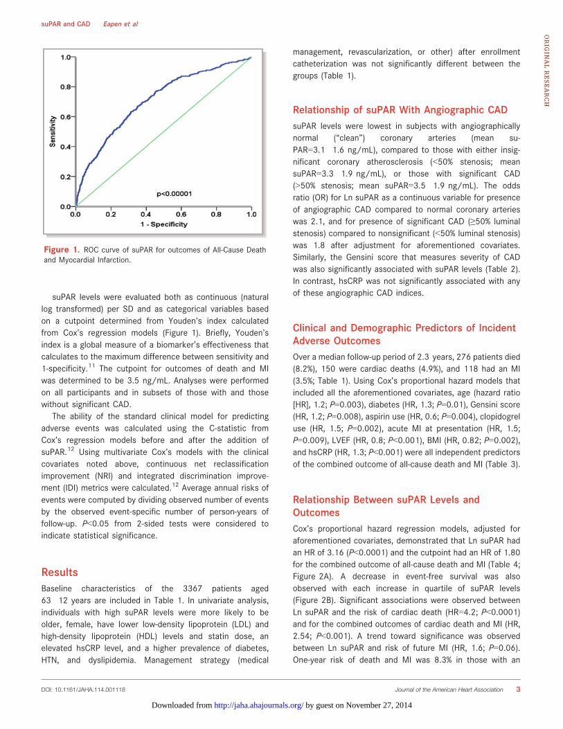
suPAR levels were evaluated both as continuous (naturallog transformed) per SD and as categorical variables basedon a cutpoint determined from Youden’s index calculatedfrom Cox’s regression models (Figure 1). Briefly, Youden’sindex is a global measure of a biomarker’s effectiveness thatcalculates to the maximum difference between sensitivity and1-specificity.11 The cutpoint for outcomes of death and MIwas determined to be 3.5 ng/mL. Analyses were performedon all participants and in subsets of those with and thosewithout significant CAD.
The ability of the standard clinical model for predictingadverse events was calculated using the C-statistic fromCox’s regression models before and after the addition ofsuPAR.12 Using multivariate Cox’s models with the clinicalcovariates noted above, continuous net reclassificationimprovement (NRI) and integrated discrimination improve-ment (IDI) metrics were calculated.12 Average annual risks ofevents were computed by dividing observed number of eventsby the observed event-specific number of person-years offollow-up. P<0.05 from 2-sided tests were considered toindicate statistical significance.
ResultsBaseline characteristics of the 3367 patients aged63�12 years are included in Table 1. In univariate analysis,individuals with high suPAR levels were more likely to beolder, female, have lower low-density lipoprotein (LDL) andhigh-density lipoprotein (HDL) levels and statin dose, anelevated hsCRP level, and a higher prevalence of diabetes,HTN, and dyslipidemia. Management strategy (medical
management, revascularization, or other) after enrollmentcatheterization was not significantly different between thegroups (Table 1).
Relationship of suPAR With Angiographic CADsuPAR levels were lowest in subjects with angiographicallynormal (“clean”) coronary arteries (mean su-PAR=3.1�1.6 ng/mL), compared to those with either insig-nificant coronary atherosclerosis (<50% stenosis; meansuPAR=3.3�1.9 ng/mL), or those with significant CAD(>50% stenosis; mean suPAR=3.5�1.9 ng/mL). The oddsratio (OR) for Ln suPAR as a continuous variable for presenceof angiographic CAD compared to normal coronary arterieswas 2.1, and for presence of significant CAD (≥50% luminalstenosis) compared to nonsignificant (<50% luminal stenosis)was 1.8 after adjustment for aforementioned covariates.Similarly, the Gensini score that measures severity of CADwas also significantly associated with suPAR levels (Table 2).In contrast, hsCRP was not significantly associated with anyof these angiographic CAD indices.
Clinical and Demographic Predictors of IncidentAdverse OutcomesOver a median follow-up period of 2.3 years, 276 patients died(8.2%), 150 were cardiac deaths (4.9%), and 118 had an MI(3.5%; Table 1). Using Cox’s proportional hazard models thatincluded all the aforementioned covariates, age (hazard ratio[HR], 1.2; P=0.003), diabetes (HR, 1.3; P=0.01), Gensini score(HR, 1.2; P=0.008), aspirin use (HR, 0.6; P=0.004), clopidogreluse (HR, 1.5; P=0.002), acute MI at presentation (HR, 1.5;P=0.009), LVEF (HR, 0.8; P<0.001), BMI (HR, 0.82; P=0.002),and hsCRP (HR, 1.3; P<0.001) were all independent predictorsof the combined outcome of all-cause death and MI (Table 3).
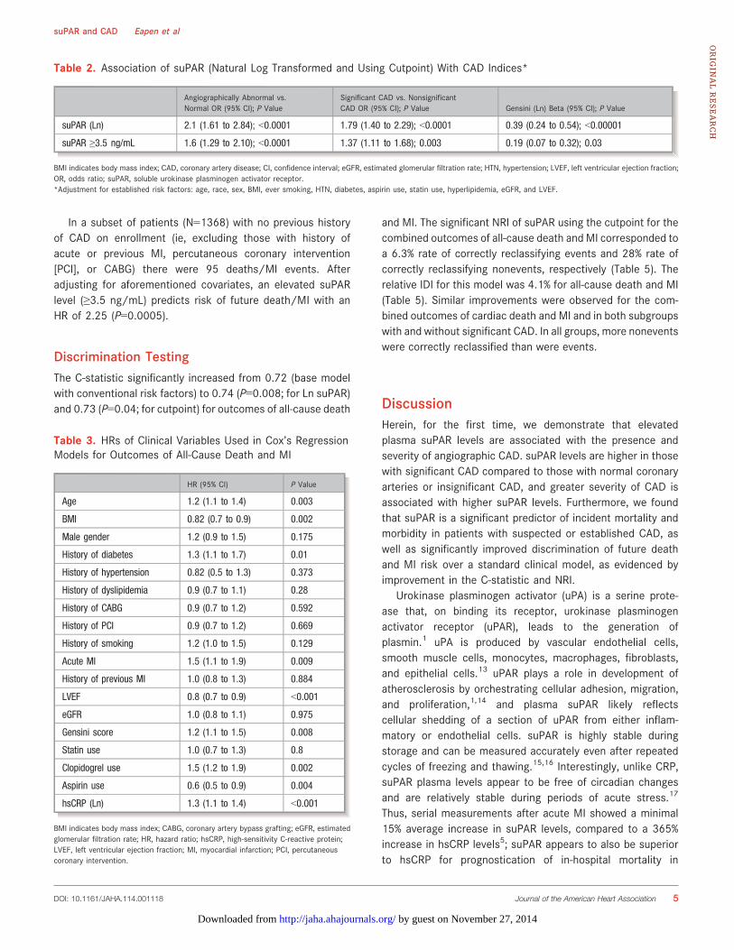
Relationship Between suPAR Levels andOutcomesCox’s proportional hazard regression models, adjusted foraforementioned covariates, demonstrated that Ln suPAR hadan HR of 3.16 (P<0.0001) and the cutpoint had an HR of 1.80for the combined outcome of all-cause death and MI (Table 4;Figure 2A). A decrease in event-free survival was alsoobserved with each increase in quartile of suPAR levels(Figure 2B). Significant associations were observed betweenLn suPAR and the risk of cardiac death (HR=4.2; P<0.0001)and for the combined outcomes of cardiac death and MI (HR,2.54; P<0.001). A trend toward significance was observedbetween Ln suPAR and risk of future MI (HR, 1.6; P=0.06).One-year risk of death and MI was 8.3% in those with an
Figure 1. ROC curve of suPAR for outcomes of All-Cause Deathand Myocardial Infarction.
DOI: 10.1161/JAHA.114.001118 Journal of the American Heart Association 3
suPAR and CAD Eapen et alORIG
INALRESEARCH
by guest on November 27, 2014http://jaha.ahajournals.org/Downloaded from
elevated suPAR level (≥3.5 ng/mL), compared to 2.8% inthose without an elevated level.
In the prespecified subgroups with nonsignificant CAD(<50% stenosis; n=1044) and in those with significant CAD(≥50% stenosis; n=2323), suPAR levels predicted combinedoutcomes of all-cause death and MI or cardiac death and MI
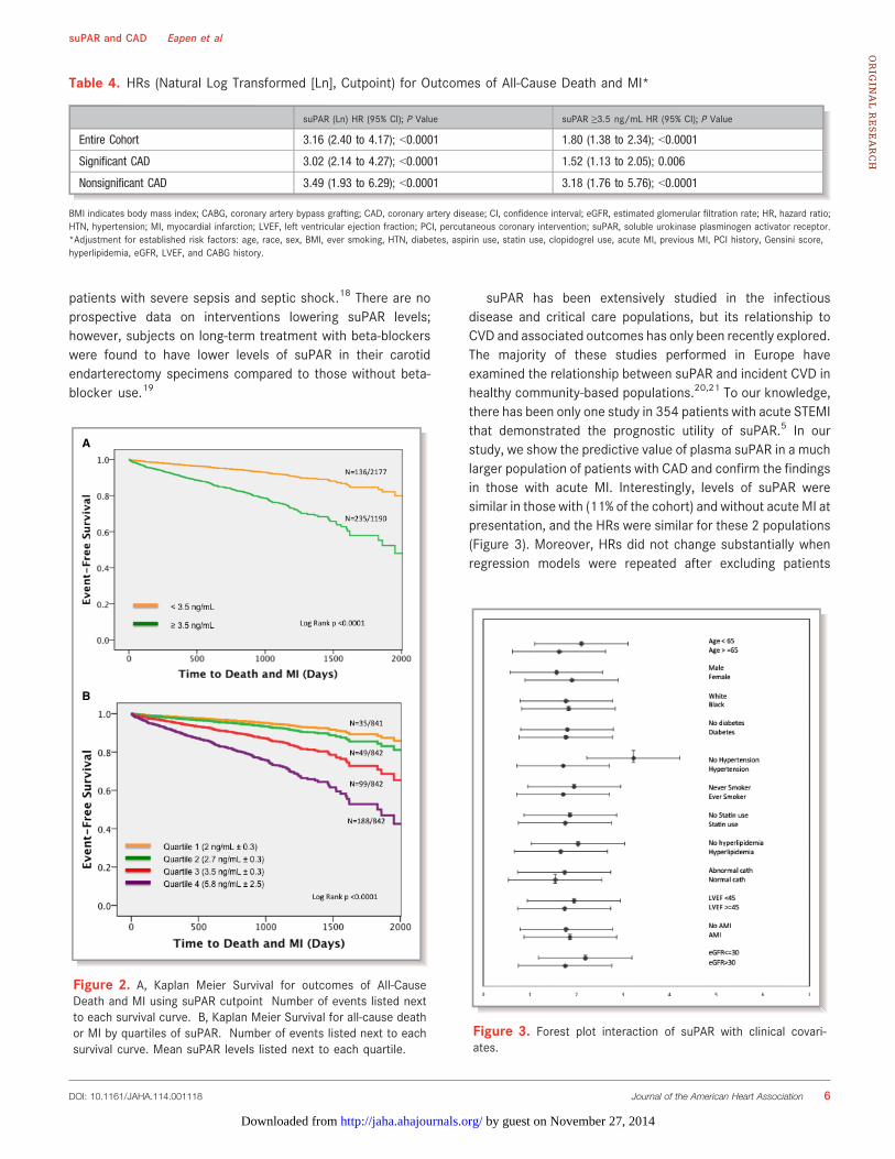
(Table 4). Total death/MI rates were 5.2%, 6.2%, and 12.8%for angiographically normal, nonsignificant, and significantgroups, respectively. We found no significant heterogeneity inthe HR based on age, gender, race, and presence of individualrisk factors, presentation with acute MI, severity of CAD, andeGFR values (Figure 3).
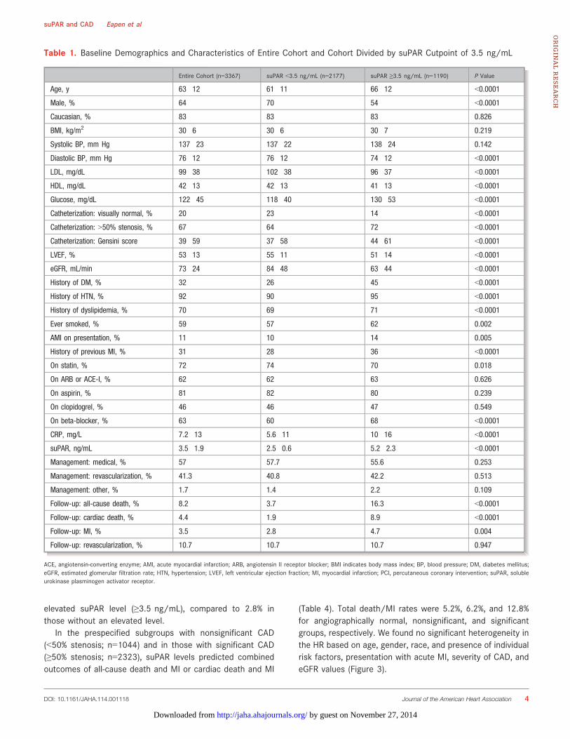
Table 1. Baseline Demographics and Characteristics of Entire Cohort and Cohort Divided by suPAR Cutpoint of 3.5 ng/mL
Entire Cohort (n=3367) suPAR <3.5 ng/mL (n=2177) suPAR ≥3.5 ng/mL (n=1190) P Value
Age, y 63�12 61�11 66�12 <0.0001
Male, % 64 70 54 <0.0001
Caucasian, % 83 83 83 0.826
BMI, kg/m2 30�6 30�6 30�7 0.219
Systolic BP, mm Hg 137�23 137�22 138�24 0.142
Diastolic BP, mm Hg 76�12 76�12 74�12 <0.0001
LDL, mg/dL 99�38 102�38 96�37 <0.0001
HDL, mg/dL 42�13 42�13 41�13 <0.0001
Glucose, mg/dL 122�45 118�40 130�53 <0.0001
Catheterization: visually normal, % 20 23 14 <0.0001
Catheterization: >50% stenosis, % 67 64 72 <0.0001
Catheterization: Gensini score 39�59 37�58 44�61 <0.0001
LVEF, % 53�13 55�11 51�14 <0.0001
eGFR, mL/min 73�24 84�48 63�44 <0.0001
History of DM, % 32 26 45 <0.0001
History of HTN, % 92 90 95 <0.0001
History of dyslipidemia, % 70 69 71 <0.0001
Ever smoked, % 59 57 62 0.002
AMI on presentation, % 11 10 14 0.005
History of previous MI, % 31 28 36 <0.0001
On statin, % 72 74 70 0.018
On ARB or ACE-I, % 62 62 63 0.626
On aspirin, % 81 82 80 0.239
On clopidogrel, % 46 46 47 0.549
On beta-blocker, % 63 60 68 <0.0001
CRP, mg/L 7.2�13 5.6�11 10�16 <0.0001
suPAR, ng/mL 3.5�1.9 2.5�0.6 5.2�2.3 <0.0001
Management: medical, % 57 57.7 55.6 0.253
Management: revascularization, % 41.3 40.8 42.2 0.513
Management: other, % 1.7 1.4 2.2 0.109
Follow-up: all-cause death, % 8.2 3.7 16.3 <0.0001
Follow-up: cardiac death, % 4.4 1.9 8.9 <0.0001
Follow-up: MI, % 3.5 2.8 4.7 0.004
Follow-up: revascularization, % 10.7 10.7 10.7 0.947
ACE, angiotensin-converting enzyme; AMI, acute myocardial infarction; ARB, angiotensin II receptor blocker; BMI indicates body mass index; BP, blood pressure; DM, diabetes mellitus;eGFR, estimated glomerular filtration rate; HTN, hypertension; LVEF, left ventricular ejection fraction; MI, myocardial infarction; PCI, percutaneous coronary intervention; suPAR, solubleurokinase plasminogen activator receptor.
DOI: 10.1161/JAHA.114.001118 Journal of the American Heart Association 4
suPAR and CAD Eapen et alORIG
INALRESEARCH
by guest on November 27, 2014http://jaha.ahajournals.org/Downloaded from
In a subset of patients (N=1368) with no previous historyof CAD on enrollment (ie, excluding those with history ofacute or previous MI, percutaneous coronary intervention[PCI], or CABG) there were 95 deaths/MI events. Afteradjusting for aforementioned covariates, an elevated suPARlevel (≥3.5 ng/mL) predicts risk of future death/MI with anHR of 2.25 (P=0.0005).
Discrimination TestingThe C-statistic significantly increased from 0.72 (base modelwith conventional risk factors) to 0.74 (P=0.008; for Ln suPAR)and 0.73 (P=0.04; for cutpoint) for outcomes of all-cause death
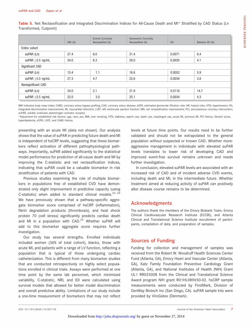
and MI. The significant NRI of suPAR using the cutpoint for thecombined outcomes of all-cause death and MI corresponded toa 6.3% rate of correctly reclassifying events and 28% rate ofcorrectly reclassifying nonevents, respectively (Table 5). Therelative IDI for this model was 4.1% for all-cause death and MI(Table 5). Similar improvements were observed for the com-bined outcomes of cardiac death and MI and in both subgroupswith and without significant CAD. In all groups, more noneventswere correctly reclassified than were events.
DiscussionHerein, for the first time, we demonstrate that elevatedplasma suPAR levels are associated with the presence andseverity of angiographic CAD. suPAR levels are higher in thosewith significant CAD compared to those with normal coronaryarteries or insignificant CAD, and greater severity of CAD isassociated with higher suPAR levels. Furthermore, we foundthat suPAR is a significant predictor of incident mortality andmorbidity in patients with suspected or established CAD, aswell as significantly improved discrimination of future deathand MI risk over a standard clinical model, as evidenced byimprovement in the C-statistic and NRI.
Urokinase plasminogen activator (uPA) is a serine prote-ase that, on binding its receptor, urokinase plasminogenactivator receptor (uPAR), leads to the generation ofplasmin.1 uPA is produced by vascular endothelial cells,smooth muscle cells, monocytes, macrophages, fibroblasts,and epithelial cells.13 uPAR plays a role in development ofatherosclerosis by orchestrating cellular adhesion, migration,and proliferation,1,14 and plasma suPAR likely reflectscellular shedding of a section of uPAR from either inflam-matory or endothelial cells. suPAR is highly stable duringstorage and can be measured accurately even after repeatedcycles of freezing and thawing.15,16 Interestingly, unlike CRP,suPAR plasma levels appear to be free of circadian changesand are relatively stable during periods of acute stress.17
Thus, serial measurements after acute MI showed a minimal15% average increase in suPAR levels, compared to a 365%increase in hsCRP levels5; suPAR appears to also be superiorto hsCRP for prognostication of in-hospital mortality in
Table 2. Association of suPAR (Natural Log Transformed and Using Cutpoint) With CAD Indices*
Angiographically Abnormal vs.Normal OR (95% CI); P Value
Significant CAD vs. NonsignificantCAD OR (95% CI); P Value Gensini (Ln) Beta (95% CI); P Value
suPAR (Ln) 2.1 (1.61 to 2.84); <0.0001 1.79 (1.40 to 2.29); <0.0001 0.39 (0.24 to 0.54); <0.00001
suPAR ≥3.5 ng/mL 1.6 (1.29 to 2.10); <0.0001 1.37 (1.11 to 1.68); 0.003 0.19 (0.07 to 0.32); 0.03
BMI indicates body mass index; CAD, coronary artery disease; CI, confidence interval; eGFR, estimated glomerular filtration rate; HTN, hypertension; LVEF, left ventricular ejection fraction;OR, odds ratio; suPAR, soluble urokinase plasminogen activator receptor.*Adjustment for established risk factors: age, race, sex, BMI, ever smoking, HTN, diabetes, aspirin use, statin use, hyperlipidemia, eGFR, and LVEF.
Table 3. HRs of Clinical Variables Used in Cox’s RegressionModels for Outcomes of All-Cause Death and MI
HR (95% CI) P Value
Age 1.2 (1.1 to 1.4) 0.003
BMI 0.82 (0.7 to 0.9) 0.002
Male gender 1.2 (0.9 to 1.5) 0.175
History of diabetes 1.3 (1.1 to 1.7) 0.01
History of hypertension 0.82 (0.5 to 1.3) 0.373
History of dyslipidemia 0.9 (0.7 to 1.1) 0.28
History of CABG 0.9 (0.7 to 1.2) 0.592
History of PCI 0.9 (0.7 to 1.2) 0.669
History of smoking 1.2 (1.0 to 1.5) 0.129
Acute MI 1.5 (1.1 to 1.9) 0.009
History of previous MI 1.0 (0.8 to 1.3) 0.884
LVEF 0.8 (0.7 to 0.9) <0.001
eGFR 1.0 (0.8 to 1.1) 0.975
Gensini score 1.2 (1.1 to 1.5) 0.008
Statin use 1.0 (0.7 to 1.3) 0.8
Clopidogrel use 1.5 (1.2 to 1.9) 0.002
Aspirin use 0.6 (0.5 to 0.9) 0.004
hsCRP (Ln) 1.3 (1.1 to 1.4) <0.001
BMI indicates body mass index; CABG, coronary artery bypass grafting; eGFR, estimatedglomerular filtration rate; HR, hazard ratio; hsCRP, high-sensitivity C-reactive protein;LVEF, left ventricular ejection fraction; MI, myocardial infarction; PCI, percutaneouscoronary intervention.
DOI: 10.1161/JAHA.114.001118 Journal of the American Heart Association 5
suPAR and CAD Eapen et alORIG
INALRESEARCH
by guest on November 27, 2014http://jaha.ahajournals.org/Downloaded from
patients with severe sepsis and septic shock.18 There are noprospective data on interventions lowering suPAR levels;however, subjects on long-term treatment with beta-blockerswere found to have lower levels of suPAR in their carotidendarterectomy specimens compared to those without beta-blocker use.19
suPAR has been extensively studied in the infectiousdisease and critical care populations, but its relationship toCVD and associated outcomes has only been recently explored.The majority of these studies performed in Europe haveexamined the relationship between suPAR and incident CVD inhealthy community-based populations.20,21 To our knowledge,there has been only one study in 354 patients with acute STEMIthat demonstrated the prognostic utility of suPAR.5 In ourstudy, we show the predictive value of plasma suPAR in a muchlarger population of patients with CAD and confirm the findingsin those with acute MI. Interestingly, levels of suPAR weresimilar in those with (11% of the cohort) and without acute MI atpresentation, and the HRs were similar for these 2 populations(Figure 3). Moreover, HRs did not change substantially whenregression models were repeated after excluding patients
A
B
Figure 2. A, Kaplan Meier Survival for outcomes of All-CauseDeath and MI using suPAR cutpoint Number of events listed nextto each survival curve. B, Kaplan Meier Survival for all-cause deathor MI by quartiles of suPAR. Number of events listed next to eachsurvival curve. Mean suPAR levels listed next to each quartile.
Figure 3. Forest plot interaction of suPAR with clinical covari-ates.
Table 4. HRs (Natural Log Transformed [Ln], Cutpoint) for Outcomes of All-Cause Death and MI*
suPAR (Ln) HR (95% CI); P Value suPAR ≥3.5 ng/mL HR (95% CI); P Value
Entire Cohort 3.16 (2.40 to 4.17); <0.0001 1.80 (1.38 to 2.34); <0.0001
Significant CAD 3.02 (2.14 to 4.27); <0.0001 1.52 (1.13 to 2.05); 0.006
Nonsignificant CAD 3.49 (1.93 to 6.29); <0.0001 3.18 (1.76 to 5.76); <0.0001
BMI indicates body mass index; CABG, coronary artery bypass grafting; CAD, coronary artery disease; CI, confidence interval; eGFR, estimated glomerular filtration rate; HR, hazard ratio;HTN, hypertension; MI, myocardial infarction; LVEF, left ventricular ejection fraction; PCI, percutaneous coronary intervention; suPAR, soluble urokinase plasminogen activator receptor.*Adjustment for established risk factors: age, race, sex, BMI, ever smoking, HTN, diabetes, aspirin use, statin use, clopidogrel use, acute MI, previous MI, PCI history, Gensini score,hyperlipidemia, eGFR, LVEF, and CABG history.
DOI: 10.1161/JAHA.114.001118 Journal of the American Heart Association 6
suPAR and CAD Eapen et alORIG
INALRESEARCH
by guest on November 27, 2014http://jaha.ahajournals.org/Downloaded from
presenting with an acute MI (data not shown). Our analysisshows that the value of suPAR in predicting future death and MIis independent of hsCRP levels, suggesting that these biomar-kers reflect activation of different pathophysiological path-ways. Importantly, suPAR added significantly to the statisticalmodel performance for prediction of all-cause death and MI byimproving the C-statistic and net reclassification indices,indicating that suPAR could be a valuable biomarker in riskstratification of patients with CAD.
Previous studies examining the role of multiple biomar-kers in populations free of established CVD have demon-strated only slight improvement in predictive capacity (usingC-statistic) when added to standard clinical models.22–24
We have previously shown that a pathway-specific aggre-gate biomarker score comprised of hsCRP (inflammation),fibrin degradation products (thrombosis), and heat shockprotein 70 (cell stress) significantly predicts cardiac deathand MI in a population with CAD.25 Whether suPAR willadd to this biomarker aggregate score requires furtherinvestigation.
Our study has several strengths. Enrolled individualsincluded women (36% of total cohort), blacks, those withacute MI, and patients with a range of LV function, reflecting apopulation that is typical of those undergoing cardiaccatheterization. This is different from many biomarker studiesthat are conducted retrospectively on highly select popula-tions enrolled in clinical trials. Assays were performed at onetime point by the same lab personnel, which minimizedvariability. C-statistic, NRI, and IDI were calculated usingsurvival models that allowed for better model discriminationand overall predictive ability. Limitations of our study includea one-time measurement of biomarkers that may not reflect
levels at future time points. Our results need to be furthervalidated and should not be extrapolated to the generalpopulation without suspected or known CAD. Whether more-aggressive management in individuals with elevated suPARlevels translates to lower risk of developing CAD andimproved event-free survival remains unknown and needsfurther investigation.
In conclusion, elevated suPAR levels are associated with anincreased risk of CAD and of incident adverse CVD events,including death and MI, in the intermediate future. Whethertreatment aimed at reducing activity of suPAR can positivelyalter disease course remains to be determined.
AcknowledgmentsThe authors thank the members of the Emory Biobank Team, EmoryClinical Cardiovascular Research Institute (ECCRI), and AtlantaClinical and Translational Science Institute recruitment of partici-pants, compilation of data, and preparation of samples.
Sources of FundingFunding for collection and management of samples wasreceived from the Robert W. Woodruff Health Sciences CenterFund (Atlanta, GA), Emory Heart and Vascular Center (Atlanta,GA), Katz Family Foundation Preventive Cardiology Grant(Atlanta, GA), and National Institutes of Health (NIH) GrantUL1 RR025008 from the Clinical and Translational ScienceAward program NIH grant R01HL089650-02. hsCRP samplemeasurements were conducted by FirstMark, Division ofGenWay Biotech Inc (San Diego, CA). suPAR sample kits wereprovided by ViroGates (Denmark).
Table 5. Net Reclassification and Integrated Discrimination Indices for All-Cause Death and MI* Stratified by CAD Status (LnTransformed, Cutpoint)
NRI (%)Events CorrectlyReclassified (%)
Nonevents CorrectlyReclassified (%) IDI Relative IDI (%)
Entire cohort
suPAR (Ln) 27.4 6.0 21.4 0.0071 8.4
suPAR ≥3.5 ng/mL 34.0 6.3 28.0 0.0035 4.1
Significant CAD
suPAR (Ln) 15.4 �1.1 16.6 0.0052 5.9
suPAR ≥3.5 ng/mL 27.3 4.7 22.6 0.0034 3.8
Nonsignificant CAD
suPAR (Ln) 24.0 2.1 21.9 0.0116 14.7
suPAR ≥3.5 ng/mL 22.0 �3.0 25.1 0.0034 4.3
BMI indicates body mass index; CABG, coronary artery bypass grafting; CAD, coronary artery disease; eGFR, estimated glomerular filtration rate; HR, hazard ratio; HTN, hypertension; IDI,integrated discrimination improvement; MI, myocardial infarction; LVEF, left ventricular ejection fraction; NRI, net reclassification improvement; PCI, percutaneous coronary intervention;suPAR, soluble urokinase plasminogen activator receptor.*Adjustment for established risk factors: age, race, sex, BMI, ever smoking, HTN, diabetes, aspirin use, statin use, clopidogrel use, acute MI, previous MI, PCI history, Gensini score,hyperlipidemia, eGFR, LVEF, and CABG history.
DOI: 10.1161/JAHA.114.001118 Journal of the American Heart Association 7
suPAR and CAD Eapen et alORIG
INALRESEARCH
by guest on November 27, 2014http://jaha.ahajournals.org/Downloaded from
DisclosuresPielak and Thorball are employed by ViroGates (Denmark).Eapen and Quyyumi had full access to all of the data in thestudy and take responsibility for the integrity of the data andthe accuracy of the data analysis.
References1. Fuhrman B. The urokinase system in the pathogenesis of atherosclerosis.
Atherosclerosis. 2012;222:8–14.
2. Edsfeldt A, Nitulescu M, Grufman H, Gronberg C, Persson A, Nilsson M,Persson M, Bjorkbacka H, Goncalves I. Soluble urokinase plasminogenactivator receptor is associated with inflammation in the vulnerable humanatherosclerotic plaque. Stroke. 2012;43:3305–3312.
3. Eugen-Olsen J, Andersen O, Linneberg A, Ladelund S, Hansen TW, Langkilde A,Petersen J, Pielak T, Moller LN, Jeppesen J, Lyngbaek S, Fenger M, Olsen MH,Hildebrandt PR, Borch-Johnsen K, Jorgensen T, Haugaard SB. Circulatingsoluble urokinase plasminogen activator receptor predicts cancer, cardiovas-cular disease, diabetes and mortality in the general population. J Intern Med.2010;268:296–308.
4. Lyngbaek S, Marott JL, Sehestedt T, Hansen TW, Olsen MH, Andersen O,Linneberg A, Haugaard SB, Eugen-Olsen J, Hansen PR, Jeppesen J. Cardiovas-cular risk prediction in the general population with use of suPAR, crp, andframingham risk score. Int J Cardiol. 2013;167:2904–2911.
5. Lyngbaek S, Marott JL, Moller DV, Christiansen M, Iversen KK, ClemmensenPM, Eugen-Olsen J, Jeppesen JL, Hansen PR. Usefulness of soluble urokinaseplasminogen activator receptor to predict repeat myocardial infarction andmortality in patients with st-segment elevation myocardial infarction under-going primary percutaneous intervention. Am J Cardiol. 2012;110:1756–1763.
6. Thygesen K, Alpert JS, Jaffe AS, Simoons ML, Chaitman BR, White HD,Thygesen K, Alpert JS, White HD, Jaffe AS, Katus HA, Apple FS, Lindahl B,Morrow DA, Chaitman BR, Clemmensen PM, Johanson P, Hod H, Underwood R,Bax JJ, Bonow JJ, Pinto F, Gibbons RJ, Fox KA, Atar D, Newby LK, Galvani M,Hamm CW, Uretsky BF, Steg PG, Wijns W, Bassand JP, Menasche P, Ravkilde J,Ohman EM, Antman EM, Wallentin LC, Armstrong PW, Simoons ML, Januzzi JL,Nieminen MS, Gheorghiade M, Filippatos G, Luepker RV, Fortmann SP,Rosamond WD, Levy D, Wood D, Smith SC, Hu D, Lopez-Sendon JL, RobertsonRM, Weaver D, Tendera M, Bove AA, Parkhomenko AN, Vasilieva EJ, Mendis S,Bax JJ, Baumgartner H, Ceconi C, Dean V, Deaton C, Fagard R, Funck-BrentanoC, Hasdai D, Hoes A, Kirchhof P, Knuuti J, Kolh P, McDonagh T, Moulin C,Popescu BA, Reiner Z, Sechtem U, Sirnes PA, Tendera M, Torbicki A, VahanianA, Windecker S, Morais J, Aguiar C, Almahmeed W, Arnar DO, Barili F, BlochKD, Bolger AF, Botker HE, Bozkurt B, Bugiardini R, Cannon C, de Lemos J, EberliFR, Escobar E, Hlatky M, James S, Kern KB, Moliterno DJ, Mueller C, NeskovicAN, Pieske BM, Schulman SP, Storey RF, Taubert KA, Vranckx P, Wagner DR.Third universal definition of myocardial infarction. J Am Coll Cardiol.2012;60:1581–1598.
7. Austen WG, Edwards JE, Frye RL, Gensini GG, Gott VL, Griffith LS, McGoon DC,Murphy ML, Roe BB. A reporting system on patients evaluated for coronaryartery disease. Report of the ad hoc committee for grading of coronary arterydisease, council on cardiovascular surgery, american heart association.Circulation. 1975;51:5–40.
8. Gensini G. Coronary Arteriography. New York, NY: Futura Publishing Co; 1975.
9. Jeppesen J, Hansen TW, Olsen MH, Rasmussen S, Ibsen H, Torp-Pedersen C,Hildebrandt PR, Madsbad S. C-reactive protein, insulin resistance and risk of
cardiovascular disease: a population-based study. Eur J Cardiovasc PrevRehabil. 2008;15:594–598.
10. Andersen O, Eugen-Olsen J, Kofoed K, Iversen J, Haugaard SB. Solubleurokinase plasminogen activator receptor is a marker of dysmetabolism in hiv-infected patients receiving highly active antiretroviral therapy. J Med Virol.2008;80:209–216.
11. Youden WJ. Index for rating diagnostic tests. Cancer. 1950;3:32–35.
12. Pencina MJ, D’Agostino RB, D’Agostino RB, Vasan RS. Evaluating the addedpredictive ability of a new marker: from area under the roc curve toreclassification and beyond. Stat Med. 2008;27:157–172.
13. Andreasen PA, Egelund R, Petersen HH. The plasminogen activation systemin tumor growth, invasion, and metastasis. Cell Mol Life Sci. 2000;57:25–40.
14. Waltz DA, Fujita RM, Yang X, Natkin L, Zhuo S, Gerard CJ, Rosenberg S,Chapman HA. Nonproteolytic role for the urokinase receptor in cellularmigration in vivo. Am J Respir Cell Mol Biol. 2000;22:316–322.
15. Kofoed K, Schneider UV, Scheel T, Andersen O, Eugen-Olsen J. Developmentand validation of a multiplex add-on assay for sepsis biomarkers using xmaptechnology. Clin Chem. 2006;52:1284–1293.
16. Riisbro R, Christensen IJ, Hogdall C, Brunner N, Hogdall E. Soluble urokinaseplasminogen activator receptor measurements: influence of sample handling.Int J Biol Markers. 2001;16:233–239.
17. Thuno M, Macho B, Eugen-Olsen J. SuPAR: the molecular crystal ball. DisMarkers. 2009;27:157–172.
18. Suberviola B, Castellanos-Ortega A, Ruiz Ruiz A, Lopez-Hoyos M, SantibanezM. Hospital mortality prognostication in sepsis using the new biomarkerssuPAR and proadm in a single determination on icu admission. Intensive CareMed. 2013;39:1945–1952.
19. Asciutto G, Edsfeldt A, Dias NV, Nilsson J, Prehn C, Adamski J, Goncalves I.Treatment with beta-blockers is associated with lower levels of lp-pla2 andsuPAR in carotid plaques. Cardiovas Pathol. 2013;32:438–443.
20. Kjellman A, Akre O, Gustafsson O, Hoyer-Hansen G, Lilja H, Norming U,Piironen T, Tornblom M. Soluble urokinase plasminogen activator receptor as aprognostic marker in men participating in prostate cancer screening. J InternMed. 2011;269:299–305.
21. Persson M, Engstrom G, Bjorkbacka H, Hedblad B. Soluble urokinaseplasminogen activator receptor in plasma is associated with incidence ofcvd. Results from the malmo diet and cancer study. Atherosclerosis.2012;220:502–505.
22. St-Pierre AC, Cantin B, Bergeron J, Pirro M, Dagenais GR, Despres JP,Lamarche B. Inflammatory markers and long-term risk of ischemic heartdisease in men a 13-year follow-up of the quebec cardiovascular study.Atherosclerosis. 2005;182:315–321.
23. Wang TJ, Gona P, Larson MG, Tofler GH, Levy D, Newton-Cheh C, Jacques PF,Rifai N, Selhub J, Robins SJ, Benjamin EJ, D’Agostino RB, Vasan RS. Multiplebiomarkers for the prediction of first major cardiovascular events and death. NEngl J Med. 2006;355:2631–2639.
24. Kim HC, Greenland P, Rossouw JE, Manson JE, Cochrane BB, Lasser NL,Limacher MC, Lloyd-Jones DM, Margolis KL, Robinson JG. Multimarkerprediction of coronary heart disease risk: the women’s health initiative.J Am Coll Cardiol. 2010;55:2080–2091
25. Eapen DJ, Manocha P, Patel RS, Hammadah M, Veledar E, Wassel C,Nanjundappa RA, Sikora S, Malayter D, Wilson PW, Sperling L, Quyyumi AA,Epstein SE. Aggregate risk score based on markers of inflammation, cellstress, and coagulation is an independent predictor of adverse cardiovascularoutcomes. J Am Coll Cardiol. 2013;62:329–337.
DOI: 10.1161/JAHA.114.001118 Journal of the American Heart Association 8
suPAR and CAD Eapen et alORIG
INALRESEARCH
by guest on November 27, 2014http://jaha.ahajournals.org/Downloaded from
Velegraki, Dimitrios T. Kremastinos, Stamatios Lerakis, Laurence Sperling and Arshed A. QuyyumiMuhammad Hammadah, Emir Veledar, Ngoc-Anh Le, Tomasz Pielak, Christian W. Thorball, Aristea
Danny J. Eapen, Pankaj Manocha, Nima Ghasemzedah, Riyaz S. Patel, Hatem Al Kassem,Presence and Severity of Coronary Artery Disease and of Future Adverse Events
Soluble Urokinase Plasminogen Activator Receptor Level Is an Independent Predictor of the
Online ISSN: 2047-9980 Dallas, TX 75231
is published by the American Heart Association, 7272 Greenville Avenue,Journal of the American Heart AssociationThe doi: 10.1161/JAHA.114.001118
2014;3:e001118; originally published October 23, 2014;J Am Heart Assoc.
http://jaha.ahajournals.org/content/3/5/e001118World Wide Web at:
The online version of this article, along with updated information and services, is located on the
for more information. http://jaha.ahajournals.orgAccess publication. Visit the Journal at
is an online only OpenJournal of the American Heart AssociationSubscriptions, Permissions, and Reprints: The
by guest on November 27, 2014http://jaha.ahajournals.org/Downloaded from
Copyright © 2022 FDOKUMEN