Optimality conditions on fields in microstructures and controllable differential schemes
Upload
independentCategory
view
1download
0
Dynamic Article LinksC<Soft Matter
Cite this: Soft Matter, 2012, 8, 2644
www.rsc.org/softmatter PAPER
PEG-urokinase nanogels with enhanced stability and controllable bioactivity†
Hui Tan,a Haiqiang Jin,b Hongcheng Mei,c Lijun Zhu,a Wei Wei,a Qian Wang,a Fuxin Liang,a
Chengliang Zhang,a Jiaoli Li,a Xiaozhong Qu,*a Dihua Shangguan,c Yining Huangb and Zhenzhong Yang*a
Received 31st October 2011, Accepted 5th December 2011
DOI: 10.1039/c2sm07072c
Protein nanogels were synthesized via a one-step reaction procedure by crosslinking urokinase with
benzaldehyde bifunctionalized poly(ethylene glycol). The crosslinked architecture significantly
enhances the stability of urokinase against enzyme degradation in comparison with the core–shell
structural PEGylated proteins. Meanwhile, bioactivity of the urokinase incorporated in the nanogels
can be adjusted by varying the chain length of the corsslinking polymer. With a shorter crosslinker the
bioactivity of the uPA nanogels is seriously restricted under physiological conditions. However, the
restricted bioactivity can be completely launched by either enlarging the mesh size of the nanogel by
using longer crosslinkers, or treating the nanogels in endosomal conditions to dissociate the nanogel
structure due to the reversible conjugation chemistry.
Introduction
Polymer–protein conjugates, e.g. PEGylated therapeutic
proteins, have gained increasing interest aimed at decreasing
proteolytic degradation, prolonging circulation duration and
improving the solubility and stability.1,2 The conjugates are
normally synthesized by grafting functionalized polymers onto
the protein surface, or in situ polymerization from activated
proteins.3 The polymer moiety generates an ‘‘umbrella-like
effect’’ that can avoid the penetration of surrounding ‘‘harmful’’
bioactives like proteinase.4–6 However the steric hindrance would
significantly reduce the specific activity of the proteins.7,8
Therefore, how to balance the protective effect and the resultant
bioactivity becomes a challenge in protein conjugation. So far,
many studies focus on the structure–property relationship of the
conjugated polymers, e.g. the number, length and architecture
(linear, branched, or dendrimeric) of the polymer chains.9,10 For
example, polymers with a branched structure were found to give
a better protective effect hence longer circulation half-time to the
polymer–protein conjugates than the linear ones.11,12 On the
other hand, in comparison to the randomly modified proteins,
the synthesis of protein–polymer conjugates via site-specific
modification is considered more efficient to control the
aState Key Laboratory of Polymer Physics and Chemistry, Institute ofChemistry, Chinese Academy of Sciences, Beijing, 100190, China.E-mail: [email protected]; [email protected]; Fax: +86-10-62559373;Tel: +86-10-82619206bPeking University First Hospital, Beijing, 100034, ChinacCAS Key Laboratory of Analytical Chemistry for Living Biosystems,Institute of Chemistry, Chinese Academy of Sciences, Beijing, 100190,China
† Electronic Supplementary Information (ESI) available: 1H NMRspectra, CD spectra and CLSM images. See DOI: 10.1039/c2sm07072c/
2644 | Soft Matter, 2012, 8, 2644–2650
bioactivity and reduce the modification level of the proteins.13
For instance, the site-specific conjugation enhances the effect of
polymer size on the shielding extent to targeting substrate.4 With
stimuli responsive polymers, the well defined protein–polymer
conjugates are able to change their properties upon external
stimulus. As such, streptavidin–PNiPAm conjugates show
characteristic of temperature dependent biotin binding.14 The
biotin binding affinity was favored below the LCST of PNiPAm,
whereas it was reduced at temperatures higher than the LCST
due to the collapse of the polymer chain around the active site
of streptavidin. This strategy has been exploited to prepare
thermo-, pH and photo-responsive protein–polymer particles
with switching activity.15,16 More recently, another approach was
proposed to control the protein protection and bioactivity via the
encapsulation of proteins inside a crosslinked polymer matrix.
The protein can be protected for more than 3 h against
proteinase. Meanwhile the single protein nanocapsule possesses
higher activity towards small substrates if the shell is non-
degradable, while for a degradable capsule layer, they react with
large substrates along with the shell degradation.17 Although the
performance is rather promising, the system suffers from a multi-
step synthesis and a lack of adaptability due to the permanent
modification of protein which may also restrict the applications
in vivo.
Herein, we report an alternative approach to simultaneously
improve the stability and control the bioactivity of proteins by
the construction of crosslinked protein–polymer nanoparticles,
i.e. protein nanogels. Polymer–protein nanoparticles have
attracted attention, because of various potential applications
including biosensing, bioseparation and drug/gene delivery.16 So
far the methods for preparing protein–polymer nanoparticles
include the conjugation of protein to polymeric or hybrid
nanoparticles,18–21 and the self-assembly of synthesized
This journal is ª The Royal Society of Chemistry 2012
amphiphilic protein–polymer conjugates.22–24 In our work, the
protein nanogels were synthesized by in situ crosslinking between
protein molecules with a polymer crosslinker, i.e. poly(ethylene
glycol) (PEG) with benzaldehyde end groups. The crosslinker
reacts with the lysine residues of protein forming a dynamic
benzoic-imine linkage.25,26 The protein–polymer nanogels are
stable under physiological condition and labile at mild acidic pHs
which is promising for the intracellular delivery of proteins.
Besides, the nanogels possess two more interesting facts that may
favor the future development of protein–polymer systems.
Firstly, in comparison with the surface modified single protein,
a crosslinked structure greatly enhances the stability of the
protein nanogels against proteolytic degradation. Secondly, the
bioactivity of the protein nanogels is controllable by adjusting
the molecular weight of the crosslinking polymer (Scheme 1).
Experimental
Materials
Benzaldehyde capped polyethylene glycol (OHC-PEG-CHO)
(MW ¼ 600 Da and 2000 Da) and benzaldehyde capped methoxy
poly(ethylene glycol) (mPEG-CHO) (MW ¼ 750 Da and
2000 Da) were synthesized according to our previous work.25,26
Urokinase-type plasminogen activator (uPA) was obtained from
Livzon Pharmaceutical Group Inc., China. Chromogenic
substrate of uPA (pyroGLU-GLY-ARG-pNA.HCl, MW ¼499 Da) and the analysis kit were purchased from Chemicon
International Inc., USA. Chymotrypsin (MW � 25 kDa) was
purchased from Sigma-Aldrich, USA. Solvents and other
compounds were obtained from the Beijing Chemical Reagents
Company, China.
Synthesis of PEG–uPA conjugates
Two kinds of PEG–uPA conjugate, i.e. uPA nanogel and core–
shell structured PEGylated uPA, were synthesized. Typically,
uPA (60 mg, 1.22 mmol) and mPEG-CHO or OHC-PEG-CHO
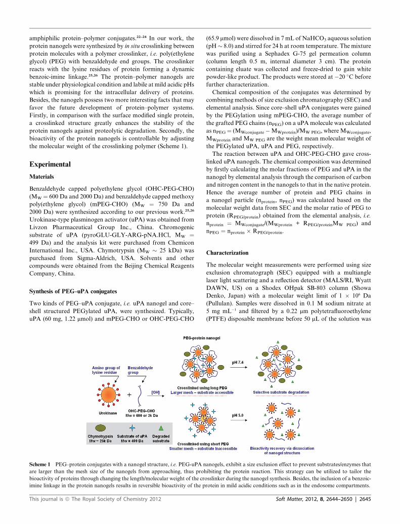
Scheme 1 PEG–protein conjugates with a nanogel structure, i.e. PEG-uPA n
are larger than the mesh size of the nanogels from approaching, thus proh
bioactivity of proteins through changing the length/molecular weight of the cro
imine linkage in the protein nanogels results in reversible bioactivity of the p
This journal is ª The Royal Society of Chemistry 2012
(65.9 mmol) were dissolved in 7 mL of NaHCO3 aqueous solution
(pH� 8.0) and stirred for 24 h at room temperature. The mixture
was purified using a Sephadex G-75 gel permeation column
(column length 0.5 m, internal diameter 3 cm). The protein
containing eluate was collected and freeze-dried to gain white
powder-like product. The products were stored at �20 �C before
further characterization.
Chemical composition of the conjugates was determined by
combining methods of size exclusion chromatography (SEC) and
elemental analysis. Since core–shell uPA conjugates were gained
by the PEGylation using mPEG-CHO, the average number of
the grafted PEG chains (nPEG) on a uPAmolecule was calculated
as nPEG¼ (MWconjugate �MWprotein)/MW PEG, where MWconjugate,
MWprotein and MW PEG are the weight mean molecular weight of
the PEGylated uPA, uPA and PEG, respectively.
The reaction between uPA and OHC-PEG-CHO gave cross-
linked uPA nanogels. The chemical composition was determined
by firstly calculating the molar fractions of PEG and uPA in the
nanogel by elemental analysis through the comparison of carbon
and nitrogen content in the nanogels to that in the native protein.
Hence the average number of protein and PEG chains in
a nanogel particle (nprotein, nPEG) was calculated based on the
molecular weight data from SEC and the molar ratio of PEG to
protein (RPEG/protein) obtained from the elemental analysis, i.e.
nprotein ¼ MWconjugate/(MWprotein + RPEG/proteinMW PEG) and
nPEG ¼ nprotein � RPEG/protein.
Characterization
The molecular weight measurements were performed using size
exclusion chromatograph (SEC) equipped with a multiangle
laser light scattering and a reflection detector (MALS/RI, Wyatt
DAWN, US) on a Shodex OHpak SB-803 column (Showa
Denko, Japan) with a molecular weight limit of 1 � 106 Da
(Pullulan). Samples were dissolved in 0.1 M sodium nitrate at
5 mg mL�1 and filtered by a 0.22 mm polytetrafluoroethylene
(PTFE) disposable membrane before 50 mL of the solution was
anogels, exhibit a size exclusion effect to prevent substrates/enzymes that
ibiting the protein reaction. This strategy can be utilized to tailor the
sslinker during the nanogel synthesis. Besides, the inclusion of a benzoic-
rotein in mild acidic conditions such as in the endosome compartments.
Soft Matter, 2012, 8, 2644–2650 | 2645
injected into the column equilibrated at 37 �C and eluted with the
0.1 M sodium nitrate (containing 0.02% of sodium azide) at
a flow rate of 1 mL min�1. The dn/dc value of the sample solu-
tions was conducted using an Optilab rEX interferometric
refractometer. Elemental analysis was carried out using a Perkin-
Elmer 2400 analyzer. 1H NMR was performed on a Bruker AV
400 MHz spectrometer (Bruker Corporation, Switzerland) by
dissolving the samples in deuterated water (D2O) at required pH
adjusted using DCl and/or NaOD solution. The size and zeta
potential of the PEG-protein conjugates was measured using
a Zetasizer (Nano Series, Malvern Instruments, UK) in aqueous
solution at required pHs with a fixed concentration of 0.5 mg
mL�1. The morphology of the conjugates was observed on
a JEOL 1011 transmission electron microscope (TEM) at
200 kV. The samples were negatively stained using 0.5 wt%
phosphotungstic acid (PTA).
Biodegradation of PEG–uPA conjugates by proteinase
The biodegradation of the PEG–uPA conjugates was evaluated
by incubating the conjugates with chymotrypsin in vitro. 1 mL
solution of PEG-uPA conjugates containing an equiv of 52 IU
uPA was placed in 4 mL vials to mix with 5 IU (0.125 mg) of
chymotrypsin and the mixtures were held at 37 �C. At predicted
time points, 50 mL of the mixtures were directly injected into the
SEC to test the concentration of survived conjugates, while an
amount of the incubating solution was heated at 100 �C for 5 min
to stop the reaction. Then, the pH of the solution was adjusted to
5 using a small volume of 2 M HCl and left to stand for 4 h to
cleave the benzoic-imine bond in the conjugates and release the
uPA. The resultant solution was subsequently neutralized by
adding an equal amount of 2 M NaOH in order to analyze the
integrality of the released uPA using SDS-PAGE. The electro-
phoresis was carried out at room temperature in a vertical slab
gel apparatus. The column contained a separating gel (7.5%
polyacrylamide, 0.75 � 8 � 6 cm), overlaid with a stacking gel
(3% polyacrylamide, 0.75 � 8 � 2.5 cm). The samples were
mixed with loading buffer (containing 0.25 M Tris HCl (pH 6.8),
0.5 M DTT, 10% SDS, 50% glycerol and 0.5% bromophenol
blue) in a ratio of 1 : 1 v/v and loaded to the column together
with the protein molecular weight markers. A potential of 60 V
was applied until the dye front entered the separating gel then the
potential was increased to 110 V. Protein bands were visualized
by sensitive silver staining.
Enzymatic activity of uPA conjugates
A chromogenic substrate (pyroGLU-GLY-ARG-pNA.HCl),
which can be cleaved by uPA to generate a colored product, was
used for testing the bioactivity of uPA conjugates. uPA stan-
dards, uPA conjugate solutions (pH 7.4) and the protein conju-
gates after an acid-dissociation (pH 5.0 for 4 h) were pipetted
into 96-well plates in which the amount of uPA is equal in each
well (10 IU). The volume of the solutions in the wells was
adjusted to be 180 mL by adding 20 mL of assay buffer (pH¼ 7.4)
and sufficient amount of water. Then 20 mL of the chromogenic
substrate solution (2.5 mg mL�1) was added to the wells and the
well plates were incubated at 37 �C. Optical density of the well
plates at 405 nm (OD405) was recorded periodically using
2646 | Soft Matter, 2012, 8, 2644–2650
a microplate reader (Versamax Tunable Microplate Reader).
The experiments were conducted in triplicate.
Results and discussion
Synthesis of PEG–uPA nanogels and core–shell PEGylated uPA
Poly(ethylene glycol)–urokinase nanogels were synthesized by
conjugating the amino groups of uPA with benzaldehyde
bifunctionalized PEG (OHC-PEG-CHO). For comparison,
core–shell structural PEGylated uPAs (mPEG–uPA) were also
prepared by reacting with benzaldehyde monofunctionalized
mPEG (mPEG-CHO). The synthesis involves a one-step reaction
in aqueous media at basic pH (e.g. 7.5–8). For the synthesis of
polymer–protein conjugates, aldehyde functionalized polymers
were frequently applied for targeting the lysine residues of
proteins,27–29 however a further reduction of the produced imine
bond is normally necessary in order to strengthen the linkage.15,16
In our present work, the use of benzaldehyde capped PEG can
generate sufficiently stable PEG–protein conjugates under
physiological conditions (37 �C, pH 7.4) due to the formation of
benzoic-imine linkage.25 This is confirmed by 1H NMR from
the disappearance of the aldehdye proton peak at ca. 10 ppm in
the D2O solution of the uPA conjugates (Fig. S1, ESI†). The
PEGylation with mPEG-CHO gave core–shell structural
PEGylated uPAs (mPEG-uPA), recognized by a limited increase
of particle size compared to the native uPA (Table 1). The PEG
substitution level was evaluated based on size exclusion chro-
matography (SEC) and further confirmed by elemental analysis.
As listed in Table 1, these calculations are in good agreement,
showing that with a benzaldehyde to lysine feed ratio of
2 : 1 mol/mol, an average of 13–17 PEG chains were grafted onto
each uPAmolecule in the products which is slightly influenced by
the molecular weight of the mPEG-CHO, i.e. shorter polymer
chains result in higher substitution due to a steric favor. The
substitution level is around 50–60% to the overall 27 lysine
residues in a uPA molecule. On the contrary, the reaction of uPA
with bifunctionalized PEG (OHC-PEG-CHO) leads to larger
particles, as revealed in the TEM images (Fig. 1). The particles
contain multiple protein and PEG chains (Fig. 2a, Table 1),
indicating the formation of a crosslinked structure, i.e. nanogels.
Compared to the mPEG-uPAs, the chemical composition and
the size of the PEG–uPA nanogels are more seriously influenced
by the molecular weight (MW) of the PEG, i.e. 600 or 2000 Da
(Table 1). With the same excess amount of feed PEG, an average
of 42 and 30 PEG chains in each PEG600–uPA and PEG2000–
uPA particle are found, with which 4 and 6 uPA are crosselinked
respectively. A weak aldehyde peak is observed in the 1H NMR
spectra of PEG–uPA nanogels (Fig. S2, ESI†), indicating that
a few OHC-PEG-CHO molecules are grafted onto the proteins
with only one end. Although the calculated PEGylation level of
the uPA nanogels is ca. 10 and 5 PEG per uPA molecule, lower
than that of the mPEG-uPAs, the result can still imply the
existence of intra-crosslinking PEG chains on protein surfaces in
addition to the bridging PEG chains across different protein
molecules, since such a structure should be entropically favored.
As we previously reported, the formation of the benzoic-imine
bond is reversible upon pH variation.25,26 In this work, the drop
of pH value from neutral to mild acidic, e.g. from 7.4 to 5.0,
This journal is ª The Royal Society of Chemistry 2012
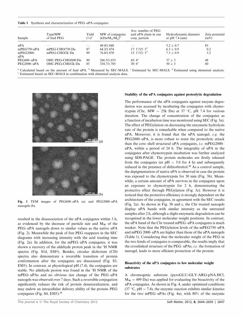
Table 1 Synthesis and characterization of PEG–uPA conjugates
SampleType/MWof feed PEG
Yield(%)a
MW of conjugates(kDa/Mw/Mn)
b
Ave. number of PEGand uPA chain in oneconj. particle
Hydrodynamic diameterat pH 7.4 (nm)
Zeta potential(mV)
uPA — — 49.0/1.048 — 5.2 � 0.7 85mPEG750-uPA mPEG-CHO/750 Da 87 64.2/1.074 17: 1c/15: 1d 6.3 � 0.9 0.2mPEG2000–uPA
mPEG-CHO/2k Da 89 76.6/1.070 13: 1c/12: 1d 7.3 � 0.9 3.2
PEG600–uPA OHC-PEG-CHO/600 Da 90 266.5/1.653 42: 4e 37 � 3 48PEG2000–uPA OHC-PEG-CHO/2k Da 85 334.7/1.705 30: 6e 50 � 3 60
a Calculated based on the amount of feed uPA. b Measured by SEC-MALS. c Estimated by SEC-MALS. d Estimated using elemental analysis.e Estimated based on SEC-MALS in combination with elemental analysis data.
Fig. 1 TEM images of PEG600–uPA (a) and PEG2000–uPA
nanogels (b).
resulted in the disassociation of the uPA conjugates within 3 h,
as evidenced by the decrease of particle size and MW of the
PEG–uPA nanogels down to similar values as the native uPA
(Fig. 2). Meanwhile the peak of free PEG reappears in the SEC
diagrams with increasing intensity with the acid treating time
(Fig. 2a). In addition, for the mPEG–uPA conjugates, it was
shown a recovery of the aldehyde proton peak in the 1H NMR
spectra (Fig. S1d, ESI†). Besides, circular dichroism (CD)
spectra also demonstrate a reversible transition of protein
conformation after the conjugates are dissociated (Fig. S3,
ESI†). In contrast, at physiological pH (7.4), the conjugates are
stable. No aldehyde proton was found in the 1H NMR of the
mPEG–uPAs and no obvious size change of the PEG–uPA
nanogels was observed over 7 days. Such a reversible conjugation
significantly reduces the risk of protein denaturalization, and
may endow an intracellular delivery ability of the protein–PEG
conjugates (Fig. S4, ESI†).25
This journal is ª The Royal Society of Chemistry 2012
Stability of the uPA conjugates against proteolytic degradation
The performance of the uPA conjugates against enzyme degra-
dation was accessed by incubating the conjugates with chymo-
trypsin (Chr, MW � 25k Da) at 37 �C, pH 7.4 for various
duration. The change of concentration of the conjugates as
a function of incubation time was monitored using SEC (Fig. 3a).
The effect of PEGylation on decreasing the enzymatic hydrolysis
rate of the protein is remarkable when compared to the native
uPA. Moreover, it is found that the uPA nanogel, e.g. the
PEG2000–uPA, is more robust to resist the proteolytic attack
than the core–shell structural uPA conjugates, i.e. mPEG2000–
uPA, within a period of 24 h. The integrality of uPA in the
conjugates after chymotrypsin incubation was further analyzed
using SDS-PAGE. The protein molecules are firstly released
from the conjugates (at pH ¼ 5.0 for 4 h) and subsequently
reduced in the presence of dithiothreitol.30 As a control sample,
the depigmentation of native uPA is observed in case the protein
was exposed to the chymotrypsin for 30 min (Fig. 3b). Mean-
while, a certain amount of uPA survives in the conjugates upon
an exposure to chymotrypsin for 2 h, demonstrating the
protective effect through PEGylation (Fig. 3c). However it is
noticed that the protective efficiency is strongly dependent on the
architecture of the conjugates, in agreement with the SEC results
(Fig. 2a). As shown in Fig. 3b and c, the Chr treated nanogels
display uPA bands with similar intensity as the untreated
samples after 2 h, although a slight enzymatic degradation can be
recognized in the lower molecular weight positions. In contrast,
the uPA band of the Chr treated mPEG–uPA conjugates is much
weaker. Note that the PEGylation levels of the mPEG750–uPA
and mPEG 2000–uPA are higher than those of the uPA nanogels
(Table 1). Considering that the molecular weight of the PEG in
the two kinds of conjugates is comparable, the results imply that
the crosslinked structure of the PEG–uPAs, i.e. the formation of
nanogel, leads to more efficient protection of the protein.
Bioactivity of the uPA conjugates to low molecular weight
substrates
A chromogenic substrate (pyroGLU-GLY-ARG-pNA.HCl,
MW ¼ 499 Da) was applied for evaluating the bioactivity of the
uPA conjugates. As shown in Fig. 4, under optimized conditions
(37 �C, pH ¼ 7.4), the enzymic reaction exhibits similar kinetics
for the two mPEG–uPAs (Fig. 4a), with 80% of the enzymic
Soft Matter, 2012, 8, 2644–2650 | 2647

Fig. 2 (a) SEC-RI diagrams of the PEG600–uPA nanogel and the core–
shell structural mPEG750–uPA in comparison with the diagrams of
native uPA, OHC-PEG600-CHO and mPEG750-CHO; and the chro-
matographic diagrams of the PEG600–uPA nanogels upon a treatment at
acidic pH (5.0) for various periods. The evolution of particle size (b) and
molecular weight (c) of the PEG600–uPA nanogels during the acidic
treatment were monitored using a zetasizer and SEC-MALS as a function
of time.
Fig. 3 SEC peak intensity (with RI detector) of uPA and different PEG–
uPA conjugates after incubating with chymotrypsin for 2–24 h (a). SDS-
PAGE investigation on the uPA released from the PEG-uPA conjugates
after the incubation with chymotrypsin (Chr) for 30 min (b) and 2 h (c).
The conjugates were dissociated by an acidic treatment in order to release
the uPA before the SDS-PAGE test.
activity with respect to the native protein during reaction for
100 h (Fig. 4c). The results imply that the polymer layer on the
mPEG–uPAs has no effect on preventing the low molecular
weight substrate from penetrating to the protein core, irre-
spective of the length of the polymer within a molecular weight
range 750–2k Da, although it has shown a moderate protection
against the binding of chymotrypsin (Fig. 3). It is reasonable
to attribute the bioactivity loss of the mPEG–uPAs (�20%
2648 | Soft Matter, 2012, 8, 2644–2650
compared to native uPA) to the denaturalization of protein
during the synthesis procedure, because a dissociation of the
conjugates did not improve the bioactivity of the released uPA in
the samples (Fig. 4c).
In comparison, the bioactivity of uPA nanogels is sensitively
related to the molecular weight of the crosslinker, i.e. the
OHC-PEG-CHO, where the nanogels exhibit huge difference in
reacting with the small substrate in case the MW of the PEG
alternated from 600 to 2k Da (Fig. 4b). While 80% of the enzymic
activity is obtained from the PEG2000–uPA, similar to that of
the mPEG–uPAs, only 11% bioactivity is displayed by the
PEG600-uPA nanogels (Fig. 4c). Besides, as shown in Fig. 4c, the
restrained bioactivity of the uPA in the PEG600–uPA nanogel
can be fully recovered by breaking the crosslinked structure.
It is commonly accepted that the lysine residue is not included
in the active site of uPA.31 Therefore the stability and bioactivity
change of the uPA conjugates would be mainly attributed to the
shielding effect from the PEG chains. Typically, the protective
effect of the polymer layer to the surface modified protein
depends on the extent of steric resistance against the approaching
of guest molecules. In the current work, for the surface
PEGylated uPA, approximately 13–17 mPEG chains, with MW
¼ 750 and 2k Da, are grafted onto a protein molecule. The zeta
potential of the PEGylated uPAs is close to neutral (Table 1),
implying that the protein surface is relatively well-masked by the
polymer chains. On the other hand, it is expected that the
shielding of PEG chains on the PEG–uPA nanogels would be
worse because the PEGylation level of the nanogels is lower. This
is demonstrated by the zeta potential of the uPA nanogels which
is very positive as the native uPA (Table 1). However beyond the
This journal is ª The Royal Society of Chemistry 2012

Fig. 4 Time dependent optical density at 405 nm generated from the
cleavage of pyroGLU-GLY-ARG-pNA.HCl caused by the core–shell
structural mPEG–uPA conjugates (a) and the PEG–uPA nanogels (b) at pH
7.4. Relative bioactivity of the uPA conjugates (black columns) and the
dissociated uPA conjugates (free uPA, red columns) to the native uPA (c).
The dissociated uPA conjugates were gained by an acidic treatment of uPA
conjugates at pH 5.0. And the acid treatment was also applied to the native
uPA which was used as a control sample. The acidic solutions were titrated
back to the optimized condition (pH ¼ 7.4) before the bioactivity test. No
auto-hydrolysis of the substrate was observed under the experimental
conditions. Statistics analysis: Student’s t-test, * the statistics difference is
significant, p<0.0001;NS, the statisticsdifference isnot significant (p>0.05).
prediction, the uPA nanogels exhibit much better stability
against proteinase than the mPEG–uPAs (Fig. 3). Meanwhile,
the bioactivity of the PEG–uPA nanogels shows a strong
This journal is ª The Royal Society of Chemistry 2012
dependence on the molecular weight of the crosslinker polymer,
which was not found on the surface PEGylated mPEG–uPAs
(Fig. 4). Furthermore, it is noticed that the nanogels conjugated
by lower molecular weight PEG are more resistive to the
surrounding molecules, which has not been experienced from the
surface modification of single proteins. Therefore it is inferred
that the protective mechanism of the PEG–uPA nanogels is
different from the shielding effect of the surface PEGylated
proteins.
By a crosslinking reaction the uPA forms a 3D network
(Scheme 1), thus the protective effect for the nanogels is mainly
influenced by the mesh size instead of the surface density of the
polymer layer as revealed from the PEGylated single-protein
conjugates.9,10,32–34 Essentially the mesh size of a polymer
network is determined by the length of the crosslinker as well as
the degree of crosslinking. Thus the uPA nanogel crosslinked
with low MW PEG, e.g. the PEG600–uPA, should exhibit low
activity to the substrate because their MW values are comparable
(600 vs. 499 Da). The size exclusion can efficiently prevent the
penetration of the substrate molecules into the network hence
limit their binding to the active sites of the protein. The low
amplitude of activity of the PEG600–uPA to the substrate might
come from the binding at the exterior surface of the nanogel
particles. Furthermore, the extension of PEG chains to a proper
length can enlarge the mesh size of the uPA nanogels which will
enable the penetration of the substrate molecules and meanwhile
can keep prohibiting the approach of larger-sized proteinase.
This is demonstrated by the property of the PEG2000–uPA.
While its bioactivity to the substrate is preserved as high as the
free proteins (Fig. 4), the PEG2000–uPA nanogel can still resist
the binding by the chymotrypsin (Fig. 3).
Conclusions
In this study, core–shell structured PEGylated uPA conjugates
and PEG-crosslinked uPA nanogels were synthesized to
demonstrate that the architecture of polymer–protein conjugates
plays an important role on the protective efficiency as well as the
bioactivity of the protein in the resultant system. The results
imply that the bioactivity of protein is controllable through the
construction of protein nanogels, by varying the molecular
weight of the crosslinker. The PEG–uPA nanogels with shorter
crosslinker exhibit a selective activity to the smaller sized
substrate of uPA while they can prevent the binding of the larger
sized chymotrypsin. Besides, the restrained bioactivity of the
PEG–uPA nanogels can be switched on in a mild acidic envi-
ronment, referring to the endosomal condition of cells, due to the
reversibility of the conjugating chemistry in the nanogel
synthesis. This might offer a possible way for the intracellular
delivery of proteins by PEGylation using the benzoic-imine
linkage.
Acknowledgements
This work is financially supported by the National Natural
Science Foundation of China (50873108, 50853001, 50733004)
and the Knowledge Innovation Program of the Chinese
Academy of Sciences (KJCX2-YW-H19).
Soft Matter, 2012, 8, 2644–2650 | 2649

Notes and references
1 F. M. Veronese and J. M. Harris, Adv. Drug Delivery Rev., 2002, 54,453.
2 M. J. Roberts, M. D. Bentley and J. M. Harris, Adv. Drug DeliveryRev., 2002, 54, 459.
3 F. Biedermann, U. Rauwald, J. M. Zayed and O. A. Scherman,Chem.Sci., 2011, 2, 279.
4 Z. L. Ding, R. B. Fong, C. J. Long, P. S. Stayton and A. S. Hoffman,Nature, 2001, 411, 59.
5 P. Esposito, L. Barbero, P. Caccia, P. Caliceti, M. D’Antonio,G. Piquet and F. M. Veronese, Adv. Drug Delivery Rev., 2003, 55,1279.
6 K. D. Hinds and S. W. Kim, Adv. Drug Delivery Rev., 2002, 54, 505.7 G. Pasut and F. M. Veronese, Prog. Polym. Sci., 2007, 32, 933.8 F. M. Veronese, Biomaterials, 2001, 22, 405.9 C. Yang, D. N. Lu and Z. Liu, Biochemistry, 2011, 50, 2585.10 M. A. Gauthier and H. A. Klok, Polym. Chem., 2010, 1, 1352.11 L. Tao, J. Q. Liu and T. P. Davis, Biomacromolecules, 2009, 10, 2847.12 L. Tao, J. Xu, D. Cell and T. P. Davis, Macromolecules, 2010, 43,
3721.13 T. Shimoboji, E. Larenas, T. Fowler, A. S. Hoffman and
P. S. Stayton, Bioconjugate Chem., 2003, 14, 517.14 Z. Ding, C. J. Long, Y. Hayashi, E. V. Bulmus, A. S. Hoffman and
P. S. Stayton, Bioconjugate Chem., 1999, 10, 395.15 C. Boyer, X. Huang, M. R. Whittaker, V. Bulmus and T. P. Davis,
Soft Matter, 2011, 7, 1599.16 M. A. Gauthier and H. A. Klok, Chem. Commun., 2008, 2591.17 M. Yan, J. J. Du, Z. Gu, M. Liang, Y. F. Hu, W. J. Zhang,
S. Priceman, L. L. Wu, Z. H. Zhou, Z. Liu, T. Segura, Y. Tang andY. F. Lu, Nat. Nanotechnol., 2010, 5, 48.
18 E. S. Lee, D. Kim, Y. S. Youn, K. T. Oh and Y. H. Bae, Angew.Chem., Int. Ed., 2008, 47, 2418.
19 Z. Jia, J. Liu, C. Boyer, T. P. Davis and V. Bulmus,Biomacromolecules, 2009, 10, 3253.
2650 | Soft Matter, 2012, 8, 2644–2650
20 H. Mattoussi, J. M. Mauro, E. R. Goldman, G. P. Anderson,V. C. Sundar, F. V. Mikulec and M. G. Bawendi, J. Am. Chem.Soc., 2000, 122, 12142.
21 W. Wang, Y. Xu, D. Wang and Z. Li, J. Am. Chem. Soc., 2009, 131,12892.
22 A. J. Dirks, R. J. M. Nolte and J. J. L. M. Cornelissen, Adv. Mater.,2008, 20, 3953.
23 I. C. Reynhout, J. J. L. M. Cornelissen and R. J. M. Nolte, J. Am.Chem. Soc., 2007, 129, 2327.
24 J. W. Bae, E. Lee, K. M. Park and K. D. Park,Macromolecules, 2009,42, 3437.
25 C. Ding, J. Gu, X. Qu and Z. Yang, Bioconjugate Chem., 2009, 20,1163.
26 L. Zhao, L. Zhu, Q. Wang, J. Li, C. Zhang, J. Liu, X. Qu, G. He,Y. Lu and Z. Yang, Soft Matter, 2011, 7, 6144.
27 L. Tao, G. Mantovani, F. Lecolley and D. M. Haddleton, J. Am.Chem. Soc., 2004, 126, 13220.
28 S. M. Chamow, T. P. Kogan, M. Venuti, T. Gadek, R. J. Harris,D. H. Peers, J. Mordenti, S. Shak and A. Ashkenazi, BioconjugateChem., 1994, 5, 133.
29 H. Y. Shao, M. M. Crnogorac, T. Kong, S. Y. Chen, J. M. Williams,J. M. Tack, V. Gueriguian, E. N. Cagle, M. Carnevali, D. Tumelty,X. Paliard, L. P. Miranda, J. A. Bradburne andG. G. Kochendoerfer, J. Am. Chem. Soc., 2005, 127, 1350.
30 U. K. Laemmli, Nature, 1970, 227, 680.31 E. B. Ong, A. J. Johnson and G. Schoellmann, Biochim. Biophys.
Acta., 1976, 429, 252.32 S. J. Bell, C. M. Fam, E. A. Chlipala, S. J. Carlson, J. I. Lee,
M. S. Rosendahl, D. H. Doherty and G. N. Cox, BioconjugateChem., 2008, 19, 299.
33 M. S. Rosendahl, D. H. Doherty, D. J. Smith, S. J. Carlson,E. A. Chlipala and G. N. Cox, Bioconjugate Chem., 2005, 16,200.
34 R. Duncan, H. R. P. Gilbert, R. J. Carbajo and M. J. Vicent,Biomacromolecules, 2008, 9, 1146.
This journal is ª The Royal Society of Chemistry 2012
Copyright © 2022 FDOKUMEN