Deregulation of the signaling pathways controlling urokinase production. Its relationship with the...
10
Eur. J. Biochem. 263, 295–304 (1999) q FEBS 1999 REVIEW ARTICLE Deregulation of the signaling pathways controlling urokinase production Its relationship with the invasive phenotype Julio A. Aguirre Ghiso 1 , Daniel F. Alonso 2 , Eduardo F. Farı ´as 3 , Daniel E. Gomez 2 and Elisa Bal de Kier Joffe ` 3 1 Division of Medical Oncology, Department of Medicine, Mount Sinai School of Medicine, New York, USA; 2 Laboratory of Molecular Oncology, Department of Science and Technology, Quilmes National University, Buenos Aires; and 3 Department of Cell Biology, Research Area, Institute of Oncology ‘Angel H. Roffo’, University of Buenos Aires, Argentina We review the evidence in support of the notion that, upon experimental oncogenic transformation or in spontaneous human cancers, mitogenesis and expression of urokinase (uPA) and its receptor (uPAR) are activated through common signaling complexes and pathways. It is well documented that uPA, uPAR or metalloproteinases (MMPs) are overexpressed in tumor cells of mesenchymal or epithelial origin and these molecules are required for tumor invasion and metastasis. Furthermore, oncogenic stimuli, which may render the transformed cells tumorigenic and metastatic in vivo, activate, in a constitutive fashion, the extracellular-regulated kinases (Erk 1 and 2) classical mitogenic pathway and others such as the NH 2 -Jun-kinase (Jnk). Cells from human tumors or oncogene-transformed cells overexpress uPA and uPAR, and also show a sustained activation of the above- mentioned signaling modules. In this paper we show that the classical mitogenic pathway involving Ras-Erk, PKC-Erk or Rac-JNK, among others, is activated by growth factors or endogenously by oncogenes, and constitutively activates uPA and uPAR expression. All the data obtained from human tumors or experimental systems, incorporated into a general model, indicate that oncogenic stimuli lead to the constitutive activation of mitogenesis and uPA and its receptor expression, through the activation of the same classical and nonclassical signaling complexes and pathways that regulate cell proliferation. We also discuss contrasting points of view. For instance, what governs the differential regulation of mitogenesis and the signal that leads to protease overexpression in a way that allows normal cells during physiological events to respond to growth factors, and proliferate without overexpressing extracellular matrix (ECM) proteases? Or how can cells remodel their microenvironment without proliferating? What restrains benign tumors from overexpressing tumor-associated proteases when they certainly have the mitogenic signal fully activated? This may occur by the differential regulation of transcriptional programs and recent reports reviewed in this paper may provide an insight into how this occurs at the signaling and transcriptional levels. Keywords: intracellular signaling; invasion; mitogenesis; oncogenes; proteases; transformation; tumorigenesis; uPA; uPAR. THE ROLE OF PROTEASES IN TUMOR INVASION AND METASTASIS Tumor invasion and metastasis is a multistep process that involves several tumor-derived and host-derived factors. In order to reach a distant organ and develop new tumor foci, tumor cells must exit the primary tumor, intravasate, dissemi- nate through the arterial or lymphatic system, disseminate, attach to the endothelium of the target organ, extravasate and finally organize and grow as a new metastatic focus [1]. Tumor cells generally exhibit several characteristics such as anchorage- independent growth, high migratory capacity, the ability to produce high amounts of tumor proteases and enhanced proliferation [2–4]. All of these characteristics are involved in one or more steps of the metastatic cascade. Proteases are expressed by normal cells in tissue remodeling events [5,6] and also during pathological events such as tumor invasion and metastasis [7–10]. The main difference between normal or pathological situations resides in the way in which the expression of these proteases is controlled, and in the levels of expression compared with their normal counterparts. Some of the proteases associated with the invasive capacity of tumor cells are cysteine proteases or aspartic acid proteases (such as cathepsins L, B and D, respectively [11]); metalloproteinases (MMPs [12]) and, in particular, the serine proteases, urokinase- type plasminogen activator (uPA) and plasmin [7,13]. Several human tumors including mammary, lung, bladder, kidney, colorectal, stomach, brain, ovary, endometrium and melanoma, were all found to overexpress uPA [14]. Correspondence to J. Aguirre Ghiso, Atran Building, Room 610 (Box 1178), Division of Medical Oncology, Department of Medicine, Mount Sinai School of Medicine, One Gustave L. Levy Place, New York, NY 10029-6574, USA. Fax: +1 212 996 5787; Tel.: +1 212 241 3194; E-mail: [email protected] Abbreviations: bFGF, basic fibroblast growth factor; CSF-1, colony- stimulating factor-1; ECM, extracellular matrix; EGF, epidermal growth factor; Erk, extracellular regulated kinase; GPI, glycophosphatidylinositol; HGF/SF, hepatocyte growth factor/scatter factor; IGF, insulin-like growth factor; JNK, NH 2 Jun kinase; LPA, lysophosphatidic acid; MMPs, matrix metalloproteinases; N-RTK, nonreceptor tyrosine kinases; PA, phosphatidic acid; PAI, plasminogen activator inhibitor; PI3K, phosphatydilinositol 3 kinase; PKC, protein kinase C; PLC, phospholipase C; PLD, phospholipase D; PMA, phorbol-12-myristate 13-acetate RTK, receptor tyrosine kinases; SAPK, stress-activated protein kinase; TIMPs, tissue inhibitors of metalloproteinases; TPA, 12-O-tetradecanoylphorbol 13-acetate; uPA, urokinase-type plasminogen activator; uPAR, uPA receptor; VEGF, vascular endothelial growth factor. (Received 16 February 1999, accepted 23 April 1999)
-
Upload
independent -
Category
Documents
-
view
0 -
download
0
Transcript of Deregulation of the signaling pathways controlling urokinase production. Its relationship with the...
Eur. J. Biochem. 263, 295±304 (1999) q FEBS 1999
R E V I E W A R T I C L E
Deregulation of the signaling pathways controlling urokinase productionIts relationship with the invasive phenotype
Julio A. Aguirre Ghiso1, Daniel F. Alonso2, Eduardo F. FarõÂas3, Daniel E. Gomez2 and Elisa Bal de Kier JoffeÁ 3
1Division of Medical Oncology, Department of Medicine, Mount Sinai School of Medicine, New York, USA; 2Laboratory of Molecular Oncology,
Department of Science and Technology, Quilmes National University, Buenos Aires; and 3Department of Cell Biology, Research Area,
Institute of Oncology `Angel H. Roffo', University of Buenos Aires, Argentina
We review the evidence in support of the notion that, upon experimental oncogenic transformation or in
spontaneous human cancers, mitogenesis and expression of urokinase (uPA) and its receptor (uPAR) are activated
through common signaling complexes and pathways. It is well documented that uPA, uPAR or metalloproteinases
(MMPs) are overexpressed in tumor cells of mesenchymal or epithelial origin and these molecules are required
for tumor invasion and metastasis. Furthermore, oncogenic stimuli, which may render the transformed cells
tumorigenic and metastatic in vivo, activate, in a constitutive fashion, the extracellular-regulated kinases (Erk 1
and 2) classical mitogenic pathway and others such as the NH2-Jun-kinase (Jnk). Cells from human tumors or
oncogene-transformed cells overexpress uPA and uPAR, and also show a sustained activation of the above-
mentioned signaling modules. In this paper we show that the classical mitogenic pathway involving Ras-Erk,
PKC-Erk or Rac-JNK, among others, is activated by growth factors or endogenously by oncogenes, and
constitutively activates uPA and uPAR expression. All the data obtained from human tumors or experimental
systems, incorporated into a general model, indicate that oncogenic stimuli lead to the constitutive activation of
mitogenesis and uPA and its receptor expression, through the activation of the same classical and nonclassical
signaling complexes and pathways that regulate cell proliferation. We also discuss contrasting points of view. For
instance, what governs the differential regulation of mitogenesis and the signal that leads to protease
overexpression in a way that allows normal cells during physiological events to respond to growth factors, and
proliferate without overexpressing extracellular matrix (ECM) proteases? Or how can cells remodel their
microenvironment without proliferating? What restrains benign tumors from overexpressing tumor-associated
proteases when they certainly have the mitogenic signal fully activated? This may occur by the differential
regulation of transcriptional programs and recent reports reviewed in this paper may provide an insight into how
this occurs at the signaling and transcriptional levels.
Keywords: intracellular signaling; invasion; mitogenesis; oncogenes; proteases; transformation; tumorigenesis;
uPA; uPAR.
T H E R O L E O F P R O T E A S E S I N T U M O RI N V A S I O N A N D M E T A S T A S I S
Tumor invasion and metastasis is a multistep process thatinvolves several tumor-derived and host-derived factors. In
order to reach a distant organ and develop new tumor foci,tumor cells must exit the primary tumor, intravasate, dissemi-nate through the arterial or lymphatic system, disseminate,attach to the endothelium of the target organ, extravasate andfinally organize and grow as a new metastatic focus [1]. Tumorcells generally exhibit several characteristics such as anchorage-independent growth, high migratory capacity, the ability toproduce high amounts of tumor proteases and enhancedproliferation [2±4]. All of these characteristics are involvedin one or more steps of the metastatic cascade.
Proteases are expressed by normal cells in tissue remodelingevents [5,6] and also during pathological events such as tumorinvasion and metastasis [7±10]. The main difference betweennormal or pathological situations resides in the way in whichthe expression of these proteases is controlled, and in the levelsof expression compared with their normal counterparts. Someof the proteases associated with the invasive capacity of tumorcells are cysteine proteases or aspartic acid proteases (such ascathepsins L, B and D, respectively [11]); metalloproteinases(MMPs [12]) and, in particular, the serine proteases, urokinase-type plasminogen activator (uPA) and plasmin [7,13]. Severalhuman tumors including mammary, lung, bladder, kidney,colorectal, stomach, brain, ovary, endometrium and melanoma,were all found to overexpress uPA [14].
Correspondence to J. Aguirre Ghiso, Atran Building, Room 610 (Box
1178), Division of Medical Oncology, Department of Medicine, Mount
Sinai School of Medicine, One Gustave L. Levy Place, New York,
NY 10029-6574, USA. Fax: +1 212 996 5787; Tel.: +1 212 241 3194;
E-mail: [email protected]
Abbreviations: bFGF, basic fibroblast growth factor; CSF-1, colony-
stimulating factor-1; ECM, extracellular matrix; EGF, epidermal growth
factor; Erk, extracellular regulated kinase; GPI, glycophosphatidylinositol;
HGF/SF, hepatocyte growth factor/scatter factor; IGF, insulin-like growth
factor; JNK, NH2 Jun kinase; LPA, lysophosphatidic acid; MMPs, matrix
metalloproteinases; N-RTK, nonreceptor tyrosine kinases; PA, phosphatidic
acid; PAI, plasminogen activator inhibitor; PI3K, phosphatydilinositol 3
kinase; PKC, protein kinase C; PLC, phospholipase C; PLD, phospholipase
D; PMA, phorbol-12-myristate 13-acetate RTK, receptor tyrosine kinases;
SAPK, stress-activated protein kinase; TIMPs, tissue inhibitors of
metalloproteinases; TPA, 12-O-tetradecanoylphorbol 13-acetate; uPA,
urokinase-type plasminogen activator; uPAR, uPA receptor; VEGF, vascular
endothelial growth factor.
(Received 16 February 1999, accepted 23 April 1999)
296 J. A. Aguirre Ghiso et al. (Eur. J. Biochem. 263) q FEBS 1999
All the above-mentioned proteases may promote the degra-dation of extracellular matrices during the different steps of themetastatic cascade. This process enables tumor cells to reachthe blood vessels from the primary tumor site throughintravasation, to disseminate and to develop metastatic nodules[1]. It has been widely described that the inhibition of proteaseexpression [15] or enzymatic activity dramatically reduces theability of tumor cells to exhibit an invasive and metastaticphenotype [16±18]. In addition, a balance exists between tumorproteases and their inhibitors such as plasminogen activatorinhibitors (PAIs) [19] and MMP inhibitors (TIMPs) [1]. Thus,the ability of tumor cells to express an invasive phenotype willdepend, in part, on the balance between proteases and theirinhibitors. In this regard, it has been described for severalexperimental and human tumors that the balance betweenproteases and their inhibitors is usually shifted towards aconstitutive pericellular proteolysis (high proteases/low inhibi-tors) [19]. It is possible that differential regulation of theexpression of proteases and their inhibitors by intracellularsignaling pathways, altered in tumor cells but not in normalcells, results in the above-mentioned imbalance in pericellularproteolysis. In the present paper we focus mainly on the presentknowledge regarding the signaling pathways that mediate theoverproduction of uPA and its receptor in tumor cells and theirrelationship with the signaling pathways controlling transfor-mation and mitogenesis.
u P A - D E P E N D E N T P R O T E O LY S I S
uPA is a serine protease of 45±55 kDa (depending on thespecies analyzed) that exists in a proenzyme form (pro-uPA)which, upon activation, activates plasminogen to the activeserine-protease plasmin. Pro-uPA is converted from a single
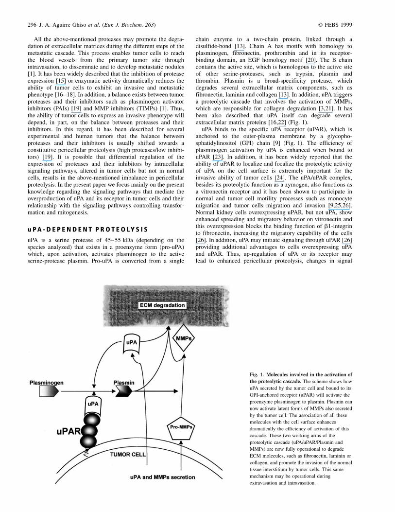
chain enzyme to a two-chain protein, linked through adisulfide-bond [13]. Chain A has motifs with homology toplasminogen, fibronectin, prothrombin and in its receptor-binding domain, an EGF homology motif [20]. The B chaincontains the active site, which is homologous to the active siteof other serine-proteases, such as trypsin, plasmin andthrombin. Plasmin is a broad-specificity protease, whichdegrades several extracellular matrix components, such asfibronectin, laminin and collagen [13]. In addition, uPA triggersa proteolytic cascade that involves the activation of MMPs,which are responsible for collagen degradation [3,21]. It hasbeen also described that uPA itself can degrade severalextracellular matrix proteins [16,22] (Fig. 1).
uPA binds to the specific uPA receptor (uPAR), which isanchored to the outer-plasma membrane by a glycopho-sphatidylinositol (GPI) chain [9] (Fig. 1). The efficiency ofplasminogen activation by uPA is enhanced when bound touPAR [23]. In addition, it has been widely reported that theability of uPAR to localize and focalize the proteolytic activityof uPA on the cell surface is extremely important for theinvasive ability of tumor cells [24]. The uPA/uPAR complex,besides its proteolytic function as a zymogen, also functions asa vitronectin receptor and it has been shown to participate innormal and tumor cell motility processes such as monocytemigration and tumor cells migration and invasion [9,25,26].Normal kidney cells overexpressing uPAR, but not uPA, showenhanced spreading and migratory behavior on vitronectin andthis overexpression blocks the binding function of b1-integrinto fibronectin, increasing the migratory capability of the cells[26]. In addition, uPA may initiate signaling through uPAR [26]providing additional advantages to cells overexpressing uPAand uPAR. Thus, up-regulation of uPA or its receptor maylead to enhanced pericellular proteolysis, changes in signal
Fig. 1. Molecules involved in the activation of
the proteolytic cascade. The scheme shows how
uPA secreted by the tumor cell and bound to its
GPI-anchored receptor (uPAR) will activate the
proenzyme plasminogen to plasmin. Plasmin can
now activate latent forms of MMPs also secreted
by the tumor cell. The association of all these
molecules with the cell surface enhances
dramatically the efficiency of activation of this
cascade. These two working arms of the
proteolytic cascade (uPA/uPAR/Plasmin and
MMPs) are now fully operational to degrade
ECM molecules, such as fibronectin, laminin or
collagen, and promote the invasion of the normal
tissue interstitium by tumor cells. This same
mechanism may be operational during
extravasation and intravasation.
q FEBS 1999 Intracellular signaling and uPA expression in tumor cells (Eur. J. Biochem. 263) 297
transduction and increased migratory behavior, which amongother changes may result in the acquisition of the invasivephenotype by tumor cells [27]
I N T R A C E L L U L A R S I G N A L I N G P A T H W AY SA S S O C I A T E D W I T H T R A N S F O R M A T I O N
It is known that upon transformation normal intracellularsignaling pathways are altered [28]. In this regard, transforma-tion by oncogenes such as v-Src or v-Ras, among others, leadsthrough aberrant signaling events to altered gene expressionand to striking differences in tumor cell morphology and loss ofparental cell shape [28,29].
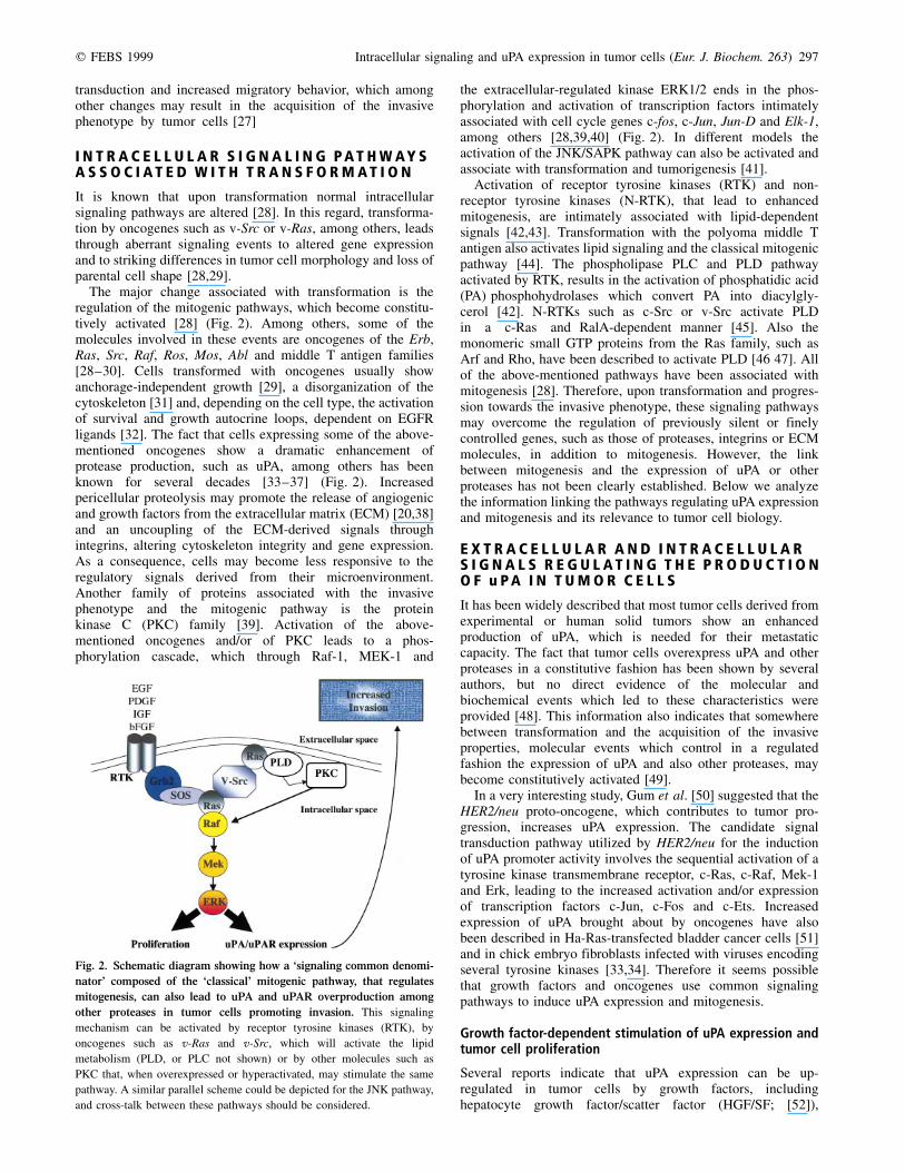
The major change associated with transformation is theregulation of the mitogenic pathways, which become constitu-tively activated [28] (Fig. 2). Among others, some of themolecules involved in these events are oncogenes of the Erb,Ras, Src, Raf, Ros, Mos, Abl and middle T antigen families[28±30]. Cells transformed with oncogenes usually showanchorage-independent growth [29], a disorganization of thecytoskeleton [31] and, depending on the cell type, the activationof survival and growth autocrine loops, dependent on EGFRligands [32]. The fact that cells expressing some of the above-mentioned oncogenes show a dramatic enhancement ofprotease production, such as uPA, among others has beenknown for several decades [33±37] (Fig. 2). Increasedpericellular proteolysis may promote the release of angiogenicand growth factors from the extracellular matrix (ECM) [20,38]and an uncoupling of the ECM-derived signals throughintegrins, altering cytoskeleton integrity and gene expression.As a consequence, cells may become less responsive to theregulatory signals derived from their microenvironment.Another family of proteins associated with the invasivephenotype and the mitogenic pathway is the proteinkinase C (PKC) family [39]. Activation of the above-mentioned oncogenes and/or of PKC leads to a phos-phorylation cascade, which through Raf-1, MEK-1 and
the extracellular-regulated kinase ERK1/2 ends in the phos-phorylation and activation of transcription factors intimatelyassociated with cell cycle genes c-fos, c-Jun, Jun-D and Elk-1,among others [28,39,40] (Fig. 2). In different models theactivation of the JNK/SAPK pathway can also be activated andassociate with transformation and tumorigenesis [41].
Activation of receptor tyrosine kinases (RTK) and non-receptor tyrosine kinases (N-RTK), that lead to enhancedmitogenesis, are intimately associated with lipid-dependentsignals [42,43]. Transformation with the polyoma middle Tantigen also activates lipid signaling and the classical mitogenicpathway [44]. The phospholipase PLC and PLD pathwayactivated by RTK, results in the activation of phosphatidic acid(PA) phosphohydrolases which convert PA into diacylgly-cerol [42]. N-RTKs such as c-Src or v-Src activate PLDin a c-Ras and RalA-dependent manner [45]. Also themonomeric small GTP proteins from the Ras family, such asArf and Rho, have been described to activate PLD [46 47]. Allof the above-mentioned pathways have been associated withmitogenesis [28]. Therefore, upon transformation and progres-sion towards the invasive phenotype, these signaling pathwaysmay overcome the regulation of previously silent or finelycontrolled genes, such as those of proteases, integrins or ECMmolecules, in addition to mitogenesis. However, the linkbetween mitogenesis and the expression of uPA or otherproteases has not been clearly established. Below we analyzethe information linking the pathways regulating uPA expressionand mitogenesis and its relevance to tumor cell biology.
E X T R A C E L L U L A R A N D I N T R A C E L L U L A RS I G N A L S R E G U L A T I N G T H E P R O D U C T I O NO F u P A I N T U M O R C E L L S
It has been widely described that most tumor cells derived fromexperimental or human solid tumors show an enhancedproduction of uPA, which is needed for their metastaticcapacity. The fact that tumor cells overexpress uPA and otherproteases in a constitutive fashion has been shown by severalauthors, but no direct evidence of the molecular andbiochemical events which led to these characteristics wereprovided [48]. This information also indicates that somewherebetween transformation and the acquisition of the invasiveproperties, molecular events which control in a regulatedfashion the expression of uPA and also other proteases, maybecome constitutively activated [49].
In a very interesting study, Gum et al. [50] suggested that theHER2/neu proto-oncogene, which contributes to tumor pro-gression, increases uPA expression. The candidate signaltransduction pathway utilized by HER2/neu for the inductionof uPA promoter activity involves the sequential activation of atyrosine kinase transmembrane receptor, c-Ras, c-Raf, Mek-1and Erk, leading to the increased activation and/or expressionof transcription factors c-Jun, c-Fos and c-Ets. Increasedexpression of uPA brought about by oncogenes have alsobeen described in Ha-Ras-transfected bladder cancer cells [51]and in chick embryo fibroblasts infected with viruses encodingseveral tyrosine kinases [33,34]. Therefore it seems possiblethat growth factors and oncogenes use common signalingpathways to induce uPA expression and mitogenesis.
Growth factor-dependent stimulation of uPA expression andtumor cell proliferation
Several reports indicate that uPA expression can be up-regulated in tumor cells by growth factors, includinghepatocyte growth factor/scatter factor (HGF/SF; [52]),
Fig. 2. Schematic diagram showing how a `signaling common denomi-
nator' composed of the `classical' mitogenic pathway, that regulates
mitogenesis, can also lead to uPA and uPAR overproduction among
other proteases in tumor cells promoting invasion. This signaling
mechanism can be activated by receptor tyrosine kinases (RTK), by
oncogenes such as v-Ras and v-Src, which will activate the lipid
metabolism (PLD, or PLC not shown) or by other molecules such as
PKC that, when overexpressed or hyperactivated, may stimulate the same
pathway. A similar parallel scheme could be depicted for the JNK pathway,
and cross-talk between these pathways should be considered.
298 J. A. Aguirre Ghiso et al. (Eur. J. Biochem. 263) q FEBS 1999
vascular endothelial growth factor (VEGF; [53]) epidermalgrowth factor (EGF; [48,49]), insulin-like growth factors I andII (IGF-I and IGF-II; [54]), basic fibroblast growth factor(bFGF; [55]), lysophosphatidic acid (LPA; [56]), colonystimulating factor-1 (CSF-1; [57,58]), vasopressin [59], andthrombin [60], among others. Most of these exogenous signals,through their receptors, activate PLC, PKC, PLD, Ral, Ras, Raf,Mek-1 and Erk1/2 [28,39,61] and also members of the Rho,Rac and Cdc42 family of small GTP-binding proteins[46,62,63]. In a mammary tumor model and other cell linesIGF-I and IGF-II and EGF, which activate RTKs, show anincrease of uPA production [49,54]. These cells were found toconstitutively produce IGFs and express receptors for thesegrowth factors [54]. Therefore, a positive autocrine loopmechanism by IGFs or by other growth factors such asEGFR ligands may be one of the mechanisms by whichtumor cells maintain a positive feedback on the up-regulation ofuPA [49,64]. Although growth factor-dependent activation ofall the signaling molecules and pathways described is wellunderstood for mitogenesis, until recently less insight hadbeen gained regarding their involvement in the up-regulation of uPA or other proteases. However, taken togetherthis information shows that the growth factors that willstimulate mitogenesis induce uPA expression by activatingthe same signaling components of the mitogenic pathways.
Growth factor activity has been attributed to uPA [65±67].uPA bound to its receptor activates intracellular signalingthrough a still-unknown adapter protein, b1/2-integrins beingthe best candidates [27,68±70]. uPAR overexpression seems togive additional advantages to tumor cells, besides enhancingplasminogen activation through uPA [24]. uPA binding to uPARactivates several tyrosine kinases from the Src family (Fyn,Lck, Hck, Yes; 71) and may also stimulate the Jak/Stat [69] andMAPK pathways [70,72]. It is noteworthy that the signalingpathways activated by uPA/uPAR seem to be the samepathways that induce their own expression. Thus, it is possiblethat overexpression of the uPA/uPAR system in tumor cellsleads to a signaling loop and/or activation of additionalmechanisms dependent on these molecules that contribute toenhanced pericellular proteolysis, migration, survival andproliferation.
Intracellular signaling pathways controlling uPAoverexpression and mitogenesis
Since the early 1970s it has been well established thatoncogenic virus transformation leads to enhanced plasminogenactivator activity [33], but the signaling modules operating andwhether they were the same as those controlling mitogenesiswas not known. In the last 4 years a great deal of informationhas appeared regarding the intracellular signals controlling notonly uPA and its receptor expression, but also the expression ofother tumor proteases such as MMPs. As mentioned above theserine-protease uPA is subject to regulation by hormones,phorbol esters and oncogenic transformation. The uPA gene isorganized in 11 exons and is 6.4 kb long [73]. Thehexanucleotide sequence GGCGGG, previously found atsimilar regions in several viral and eukaryotic promoters andshown to be essential for promoter activity [74], is repeatedthree times between the CAAT and the TATA boxes. The uPApromoter has been described to contain a TPA responsiveelement [75]. It has been reported that uPA expression can beup-regulated by phorbol esters and that transfection of severalPKC isoforms activate uPA gene transcription ([75,76]; Fig. 2).We have recently described that the constitutively activated
signal up-regulating uPA expression in murine mammary tumorcells was dependent on a PKC and PLD pathway [49]. Inaddition, we have also described that EGF-induced expressionof uPA is dependent on PLD [49]. Furthermore, Connolly andRose [77] have recently shown that when mammary tumorcells producing low levels of uPA are transfected withPKCa, an isoform highly correlated with the invasivephenotype [78], uPA expression is up-regulated constitutively.Interestingly, PKC and tyrosine kinases have been describedas also regulating uPA and MMP expression in glioma celllines [79].
Several isoforms of PKC can directly activate a PLD activityand the serine-threonine kinase c-Raf [28,42,80]. In this regard,Lengyel et al. [81] recently described that squamous cellcarcinoma cells expressing a constitutively activated c-Rafshow an up-regulation of uPA transcription through theactivation of Erk-1, which requires the transcription factorc-Jun and AP-1/PEA-3 transcription motifs as reported byothers [82]. In addition, Jun-D-binding and Jun-C-binding AP-1motifs have been described to regulate uPAR expressionthrough Erk-1-dependent signaling [83]. Similarly, De Petroet al. [84] have described that the activation of uPA genetranscription in human hepatocellular carcinoma is presum-ably due to activation of c-Jun, since the uPA gene containsa transcriptional enhancer with two sites for AP-1 transcrip-tion factors.
It has been shown that the specific inhibitor of Mek-1,PD098059, leads to a down-regulation of the secreted uPAactivity and transcription [85]. Also, Ha-Ras and v-Mosinduction of uPA expression is mediated through the Erkpathway [37]. The polyoma middle T antigen associates withseveral of the signaling molecules of the classical mitogenicpathway. In this regard, it has been described that NIH 3T3cells transformed with middle T antigen show enhancement ofuPA expression through a signaling pathway involving c-Src,Sos, c-Ras, c-Raf and Erk [44]. Similarly, uPA expressionstimulated by the Trp-Met oncogene seems to be mediated bythe Sos/Grb2 signaling complex and also by PI3-K [86],although growth-factor-induced uPA expression has never beenlinked to PI3-K. Information showing that direct phos-phorylation of c-Raf by PKC or direct interaction of c-Ras orv-Ras with c-Raf lead to the up-regulation of uPA is stillmissing.
Taken together, these results strongly indicate that theclassical mitogenic pathway, dependent on PLD, PKC,Grb2/SOS/c-Ras, c-Raf, Mek1 and Erk1 is involved in theup-regulation of not only uPA but also uPAR expression. Fromthe previous two sections we learn that in experimental models,human spontaneous tumors growth factors, oncogenes, orwhatever genetic changes may activate the classical mitogenicpathway will also activate uPA and uPAR expression. Thus,transformation may lead to the constitutive activation of the cellcycle as well as to the constitutive up-regulation of uPA and itsreceptor, leading the cells to express a highly invasive andproliferative phenotype (Fig. 2).
Novel signaling pathways controlling uPA expression andmitogenesis
Most of the information gathered describes the oncogene-induced expression of uPA through the classical mitogenicpathway (Ras±Raf±Mek±Erk). However, oncogenes such asv-Src or v-Ras among others, activate other signalingeffector molecules besides those mentioned [87]. Transfor-mation of NIH 3T3 fibroblasts with v-Src or v-Ras leads to a
q FEBS 1999 Intracellular signaling and uPA expression in tumor cells (Eur. J. Biochem. 263) 299
constitutive up-regulation of uPA expression [34,35] andactivation of PLD (hPLD1) through the activation of Ral-A, asmall GTPase of the Ras superfamily [45]. We have foundin this model that a dominant negative mutant of Ral A(S28N-RalA), described as specifically blocking PLDactivation by v-Src and v-Ras [45], reduces the up-regulationof uPA induced by v-Src or v-Ras [88]. The RalA pathway hasbeen described as facilitating cell transformation induced bythese oncogenes [89]. In addition, the S28N mutant of Ralblocks v-Src and v-Ras induced tumorigenesis in vivo. Inaccordance with this result a novel oncogene, Rsc (rabbitsquamous cell carcinoma), a Ral-GDS homolog, also inducestumorigenesis in vivo [90]. These data indicate thatnovel signaling pathways controlling cell proliferation andtransformation also participate in the up-regulation of uPAexpression.
The involvement of other signals in uPA expression in tumorcells has been reported. Activation of JNK/SAPK depends onRac, Cdc42 and Ras [63]. Constitutive activation of JNK bythese small GTPases or by bg subunits induce activation ofmitogenesis and tumorigenesis [63]. uPA and uPAR over-expression is responsive to a Rac-JNK pathway in squamouscell carcinoma cells [91]. UV-induced activation of JNKinduces apoptosis and also leads to enhanced uPA transcription[92], although the cellular role of uPA induced by UV was notdescribed. Therefore it seems that the JNK-SAPK pathway,which may function as a mitogenic (or apoptotic) pathway [63],can also induce uPA and uPAR expression.
The treatment of tumor cells with inhibitors of proteinphosphatases, such as Na-orthovanadate or okadaic acid,up-regulates uPA transcription through the signaling com-ponents of the classical mitogenic pathway, but independent ofPKC [93,94]. Phosphatase inhibitors have also been foundto up-regulate uPAR [83]. Staurosporine, a wide spectrumprotein kinase inhibitor, led to the up-regulation of uPA andMMP-9 in mammary tumor cells, through a tyrosine-kinase-dependent and PKC-independent pathway [94]. Theexpression of uPA and PAI-1 have been described to bedown-regulated and up-regulated, respectively, by genistein, atyrosine kinase inhibitor [95]. Similarly, staurosporine-inducedup-regulation of cathepsin L in monocytic cells, was blockedonly by the tyrosine kinase inhibitors herbymicin and genistein[11]. These data suggest that additional pathways and/or proteinphophatases may control the on/off state of tyrosine-kinases orMAPK complexes [96] that lead to uPA and/or uPAR
overexpression. It is important to point out that in the samecell system uPA can be induced by different signaling pathways(sometimes antagonist), suggesting possible multiple points ofregulation for uPA production in normal or malignant processesdepending on the input signal [97,98]. It is also interesting thatin promonocytic cells such as HL-60 or U937, induction of uPAby agonists such as PMA, associate with cell differentiation[99±101] indicating that depending on the cell type thesignaling pathways activated and uPA production may havedifferent biological implications.
M O D U L A T I O N O F u P A E X P R E S S I O N B Yp 5 3
The tumor suppressor gene p53 serves to regulate cell-cycleprogression in normal cells. Altered expression of p53 has beenreported for a diverse set of human tumors and is reported to becorrelated with disease progression. Wang and Boyd [102]demonstrated that wild-type p53 is a negative regulator of uPAexpression. Conversely, it appears that the expression ofmutated p53 stimulates uPA promoter activity [102]. Themechanism by which p53 represses uPA promoter activity isnot clear. A search of the uPA promoter for the DNA consensussequence, which binds p53, proved negative. An alternativehypothesis is that the repression of the uPA promoter bywild-type p53 is mediated through proximal sequences ofthe promoter. Indeed, the uPA promoter contains TATA andCAAT boxes as well as a SP-1-binding GC-rich region. Twoseparate studies have reported the binding of p53 to humanTATA-binding protein that leads to repression of transcriptionfrom minimal promoters containing this consensus sequence[103]. A third possibility is that the p53 alters uPA promoteractivity indirectly by affecting the expression and/or activity ofother transcription factors [102].
C O N C L U S I O N S A N D P E R S P E C T I V E S
The shift from regulated to constitutive control of uPAproduction in tumor cells
A general interpretation of the presented data indicates thatupon transformation with known oncogenes, or in situationswhere spontaneous transformation occurs, cells may becomehighly proliferative and during tumor progression, or perhapsalong with transformation, uPA and uPAR expression may
Fig. 3. Hypothetical model depicting some of
the possibilities that may enable a differential
regulation of protease expression and
mitogenesis activation in normal or benign vs.
malignant tissues at the transcriptional level.
Negative regulation of transcription by repressors
or by methylation may prevent protease
expression even with the mitogenic pathways
active in normal tissues or benign tumor cells.
However, the loss of this regulation enables the
signaling mechanisms to up-regulate protease
expression among other genes and may activate
an invasive and proliferative phenotype in
malignant cells.
300 J. A. Aguirre Ghiso et al. (Eur. J. Biochem. 263) q FEBS 1999
become up-regulated by the same signal source that activatesproliferation. It is interesting to note that oncogene-transformedcells, either experimental or human tumor cells, may alsodisplay a constitutive down-regulation of other proteins, such asthe extracellular matrix glycoprotein fibronectin [104].Together, these changes may render tumor cells less adhesive,highly migratory and with a high proteolytic, and henceinvasive, capacity.
The data reviewed correlate the activation of the componentsof the classical mitogenic pathway that activate ERK1/2(Ras±Raf±MEK±ERK), with the involvement of these samesignaling components in the up-regulation of uPA and uPAR.The sources of these signals are derived form N-RTK, RTK oroncogene-derived signals. In addition, other mitogen-activatedkinases different from ERK1/2, such as JNK-SAPK, anotherpathway involved in mitogenesis [63], may be also involved inuPA or uPAR up-regulation [91,92]. This information indicatesthat at least two (ERK 1/2 and JNK/SAPK) of the three so-called mitogenic pathways (ERK 1/2, JNK/SAPK and p38)known so far in mammalian cells induce up-regulation of uPAand uPAR. A recent study showed that inhibition of p38 leads toreduced uPA and MMPs expression and invasion by tumor cells[105]. However, since in some cell types p38 seems to beindependent and an antagonist of the ERK pathway[106,107], it is not a TPA responsive pathway [108], itinduces arrest of mitotic cells [109] and apoptosis [106,109]and it is still not clear what biological role it may play inpositively regulating the invasive phenotype.
A question remains regarding what governs the uncoupling/differential regulation of the mitogenic signal with/and thesignal leading to uPA overexpression in a way that normal cellscan respond to growth factors and proliferate in a regulatedfashion without overexpressing ECM proteases. To thisscenario, we could add the question of what restrains benigntumors from overexpressing tumor-associated proteases whenthey certainly have the mitogenic signal activated? So far thereis no direct information to answer this question but it seemsvery possible that this regulation occurs at the transcriptional orpost-transcriptional level, since the growth-factor-inducedsignals will activate all the mitogenic pathway signalingcomponents but the target will only be the transcription factorsfor cell-cycle-related genes. Because these same transcriptionfactors appear to be the ones that control uPA/uPAR expressionit can be supposed that in normal cells or benign tumor cells,the uncoupling between the mitogenic signal and the signalsregulating uPA and uPAR expression are probably regulated byspecific transcription repressors or selective heterodimerizationof transcription factors associated with uPA and/or uPARpromoter sequences.
It is interesting to point out that in several types of tumor,such as colon adenocarcinoma and breast tumors, while uPARcan be expressed in the neoplastic cells the source of uPA is notthe tumor cells but the stromal fibroblasts [110]. It seems that,in these instances, the tumor cell produces cytokines,angiogenic factors or other factors that modulate the stromalenvironment, which can promote the expression of uPA,stromelysin-1 and stromelysin-3 among other proteases[110,111] or angiogenic factors by fibroblasts or macrophagesin contact with the tumor [111,112]. Although still unknown, itis probable that the production of uPA or other proteases by thestromal cells requires the activation of the same signalingpathways activated by oncogenes and growth factors in tumorcells.
Recently, Van Dam et al. [113] reported that for a completeactivation of the transformation program by v-Jun, a differential
association of v-Jun with Fos or ATF2 transcription factors isrequired and this may depend on the mutations present in v-Jun.The association of mutated Jun with Fos (Jun mutation m0:Glu281 replaced by lysine) enables the activation of theanchorage-independent growth phase of the program, whileassociation with ATF2 (Jun mutation m1: Lys283, Arg302,Gln304, all replaced by glutamates) is required to activate thegrowth factor independence (autocrine growth) phase of theprogram. Interestingly, the authors analyzed which phase ofthe program (m1 or m0) activates uPA and MMPs over-expression, finding that v-Jun/Fos association (m0) is requiredfor this event, the same required for anchorage-independentgrowth. As a control, they used v-Src, which shows a muchgreater activation of protease expression, suggesting thatadditional factors recruited by v-Src-dependent signals areinvolved in the process. It is possible that benign tumors, whichgrow restricted by the normal microenvironment and do notinvade, lack the activation of the anchorage-independent/protease overproduction phase of the program, although moreinformation is required to disclose these aspects. Other reportshave also addressed the differential regulation of the invasivephenotype dependent on uPA or cathepsin L at the tran-scriptional level [114]. In addition, other points of regulationshould be considered, such as methylation of proteasespromoter sequences, a transcriptional regulation mechanismassociated with differentiation and necessary for proper cellfunction (Fig. 3) [115].
The signaling mechanisms regulating uPA production innormal cellular events, such as vasculogenesis, angiogenesis ormigration of keratinocytes, remain largely unexplored. Bodreauet al. [116] have described that Hox D3, a transcriptionalactivator, is involved in regulating the angiogenic phenotype ofendothelial cells. Most importantly, they found that Hox D3up-regulates the expression of uPA, required for the initialsprouting of the new vessel, upon angiogenic stimulation.Hox D3-induced up-regulation of uPA is coupled to anb3up-regulation very early during angiogenesis when onlymigration and invasion occurs. However, Hox D3 is notrequired for endothelial cell proliferation during angiogenesis.Thus, Hox D3 transcriptional activity seems to be differentiallyinvolved in controlling the invasive ability (uPA/anb3 expres-sion) of endothelial cells required during the early stages ofangiogenesis, but not in proliferation which occurs later in thesprouting vessel [116]. It has been demonstrated that endothe-lial cells will only produce uPA (among other proteases) anduPAR to open their way towards the angiogenic stimuli andwhen migration is initiated [117]; once they reach theirdestination, migration stops, proliferation begins and the cellswill shift to a quiescent state when the new blood vessel isformed [117]. It is also known that keratinocytes, whenattached to the basement membrane, are able to proliferate;however, to undergo terminal differentiation, they must detachfrom the underlying matrix, migrate, invade to the suprabasallayers and differentiate [118]. This event, which requiresproteolysis, seems uncoupled from mitogenesis, which occursonly on the basal layer of cells. Protease expression occurs atthe wound margin enabling keratinocytes to migrate and invadethrough the cut basement membrane and through the provi-sional matrix, while proliferation occurs at the back of themigrating edge of the wound (where the basement membrane isintact) providing new cells for re-epithelialization [118].However, it cannot be excluded that during proliferation onthe basement membrane transient production of proteasesenables the cells to remodel their space in order to fit theincreasing cell number. These data suggest that an uncoupling
q FEBS 1999 Intracellular signaling and uPA expression in tumor cells (Eur. J. Biochem. 263) 301
of protease production and proliferation during normal tissueremodeling events occurs and although extremely interesting,the signaling mechanisms that differentially regulate proteaseproduction during these cellular events remain largelyunknown.
Research on the signaling pathways controlling proteaseexpression and mitogenesis will give new insight into not onlyhow the signals that render the tumor cells invasive areregulated but also how anti-signaling strategies could bedesigned. In addition, we believe that this information willprove useful in understanding normal cell behavior, sincenormal tissue events may involve a fine-tuned regulation of thesignaling pathways controlling mitogenesis and proteaseproduction to maintain cellular and tissue homeostasis.
A C K N O W L E D G E M E N T S
We are especially grateful to Dr Liliana Ossowski and Dr Rafael Mira y
Lopez for critical reading of the manuscript.
R E F E R E N C E S
1. Liotta, L.A. & Stetler-Stevenson, W.G. (1991) Tumor invasion and
metastasis: an imbalance of positive and negative regulation. Cancer
Res. 51, 5054±5059.
2. Malik, R.K. & Parsons, T. (1996) Integrin-mediated signalling in
normal and malignant cells: a role of protein tyrosine kinases.
Biochim. Biophys. Acta 1287, 73±76.
3. Ossowski, L. (1992) Invasion of connective tissue by human
carcinoma cell lines: requirement of urokinase, urokinase receptor
and interstitial collagenase. Cancer Res. 52, 6754±6760.
4. Seynaeve, C.M., Stetler-Stevenson, M., Sebers, S., Kaur, G.,
Sausville, E.A. & Worland, P.J. (1993) Cell cycle arrest and growth
inhibition by the protein kinase antagonist UCN-01 in human breast
carcinoma cells. Cancer Res. 53, 2081±2086.
5. Ossowski, L., Biegel, D. & Reich, E. (1979) Mammary plasminogen
activator: correlation with involution, hormonal modulation and
comparison between normal and neoplastic tissue. Cell 16,
929±936.
6. Pepper, M.S., Vassalli, J.D., Montesano, R. & Orci, L. (1987)
Urokinase-type plasminogen activator is induced in migrating
capillary endothelial cells. J. Cell Biol. 105, 2535±2541.
7. Ossowski, L. & Reich, E. (1983) Antibodies to plasminogen activator
inhibit human tumor metastases. Cell 35, 611±619.
8. Ossowski, L. (1988) In vivo invasion of modified chorioallantoic
membrane by tumor cells: the role of cell-surface bound
urokinase. J. Cell Biol. 107, 2437±2435.
9. Moller, L.B. (1993) Structure and function of the urokinase receptor.
Blood Coagul. Fibrinol. 4, 293±303.
10. Mullins, D.E. & Rohrlich, S.T. (1983) The role of proteinases in
cellular invasiveness. Biochem. Biophys. Acta 695, 177±214.
11. Atkins, K. & Troen, B. (1995) Phorbol ester stimulated cathepsine L
expression in U937 cells. Cell Growth Diff. 6, 713±718.
12. Matrisian, L.M. (1992) The matrix degrading metalloproteinases.
Bioessays 14, 455±463.
13. Blasi, F., Vassalli, J.D. & Danù, K. (1987) Urokinase-type plas-
minogen activator: proenzyme, receptors and inhibitors. J. Cell Biol.
104, 801±804.
14. Markus, G. (1988) The relevance of plasminogen activators to
neoplastic growth. Enzyme 40, 158±172.
15. Yu, H. & Schultz, R.M. (1990) Relationship between secreted
urokinase plasminogen activator activity and metastatic potential
in murine B16 cells transfected with human urokinase sense and
antisense genes. Cancer Res. 50, 7623±7633.
16. Alonso, D.F., FarõÂas, E.F., Ladeda, V., Davel, L., Puricelli, L. & Bal de
Kier Joffe E. (1996) Effects of synthetic urokinase inhibitors on
local invasion and metastatis in a murine mammary tumor model.
Breast Cancer Res. Treat. 40, 209±223.
17. Rabbani, S., Harakidas, P., Davidson, D., Henkin, J. & Mazar, A.
(1995) Prevention of prostate cancer metastasis in vivo by a novel
synthetic inhibitor of urokinase-type plasminogen activator (uPA).
Int. J. Cancer 63, 840±845.
18. Xing, R.H., Mazar, A., Henkin, J. & Rabbani, S.A. (1997) Prevention
of breast cancer growth, invasion, and metastasis by antiestrogen
tamoxifen alone or in combination with urokinase inhibitor B-428.
Cancer Res. 57, 3585±3593.
19. Mueller, B.M., Yu, Y.B. & Laug, W.E. (1995) Overexpression of
plasminogen activator inhibitor 2 in human melanoma cells inhibits
spontaneous metastasis in scid/scid mice. Proc. Natl Acad. Sci. USA
92, 205±209.
20. Appella, E., Robinson, E.A., Ullrich, S.J., Stoppelli, M.P., Corti, A.,
Cassani, G. & Blasi, F. (1987) The receptor-binding sequence of
urokinase. A biological function for the growth-factor module of
proteases. J. Biol. Chem. 262, 4437±4440.
21. Mazzieri, R., Masiero, L., Zanetta, L., Monea, S., Onisto, M., Garbisa,
S. & Mignatti, P. (1997) Control of type IV collagenase activity by
components of the urokinase±plasmin system: a regulatory
mechanism with cell-bound reactants. EMBO J. 16, 2319±2332.
22. Ellis, V., Behrendt, N. & Danù, K. (1991) Plasminogen activation by
receptor bound urokinase: a kinetic study with both cell-associated
and isolated receptor. J. Biol. Chem. 266, 12752±12758.
23. Plow, E.F., Freany, D.E., Plescia, J. & Miles, L.A. (1986) The
plasminogen system and cell surfaces: evidence for plasminogen
and urokinase receptors on the same cell type. J. Cell Biol. 103,
2411±2420.
24. Yu, W., Kim, J. & Ossowski, L. (1997) Reduction in surface urokinase
receptor forces malignant cells into a protracted state of dormancy.
J. Cell Biol. 137, 767±777.
25. Stahl, A. & Mueller, B.M. (1997) Melanoma cell migration on
vitronectin: regulation by components of the plasminogen activation
system. Int. J. Cancer 71, 116±122.
26. Wei, Y., Lukashev, M., Simon, D., Bodary, S., Rosenberg, S., Doyle,
M.V. & Chapman, H.A. (1996) Regulation of integrin function by
the urokinase receptor. Science 273, 1551±1555.
27. Andreasen, P.A., Kjoller, L., Christensen, L. & Duffy, M.J. (1997) The
urokinase-type plasminogen activator system in cancer metastasis:
a review. Int. J. Cancer 72, 1±22.
28. Burgering, B.M.T. & Bos, J.L. (1995) Regulation of Ras-mediated
signalling: more than one way to skin a cat. Trends Biol. Sci.
20, 18±22.
29. Guan, J.L. & Shalloway, D. (1992) Regulation of focal adhesion-
associated protein tyrosine kinase by both cellular adhesion and
oncogenic transformation. Nature 358, 690±692.
30. Carpenter, C.L. & Cantley, L.C. (1996) Phosphoinositide-3-kinase
and the regulation of cell growth. Biochim. Biophys. Acta 1288,
M11±M16.
31. Nermut, M.V., Eason, P., Hirst, E.M. & Kellie, S. (1991) Cell/
substratum adhesions in RSV-transformed rat fibroblasts. Exp. Cell.
Res. 193, 382±397.
32. Gangarosa, L.M., Sizemore, N., Graves-Deal, R., Der Oldham,
S.M.C.J. & Coffey, R.J. (1997) A raf-independent epidermal growth
factor receptor autocrine loop is necessary for Ras transformation of
rat intestinal epithelial cells. J. Biol. Chem. 272, 18926±11893.
33. Unkeless, J.C., Tobia, A., Ossowski, L., Quigley, J.P., Rifkin, D.B. &
Reich, E. (1973) An enzymatic function associated with transforma-
tion of fibroblasts by oncogenic viruses. J. Exp. Med. 137, 85±111.
34. Bell, S.M., Connolly, D.C. & MaihleDegen, J.L. (1993) Differential
modulation of plasminogen activator gene expression by oncogene-
encoded protein tyrosine kinases. Mol. Cell Biol. 13, 5888±5897.
35. Jankun, J., Maher, V.M. & McCormick, J.J. (1991) Malignant
transformation of human fibroblast correlates with increased
activity of receptor-bound plasminogen activator. Cancer Res.
51, 1221±1226.
36. Ballin, M., Gomez, D.E., Sinha, C.C. & Thorgeirsson, U.P. (1988) Ras
oncogene mediated induction of a 92 kDa metalloproteinase: strong
correlation with malignant phenotype. Biochem. Biophys. Res.
Commun. 154, 832±838.
302 J. A. Aguirre Ghiso et al. (Eur. J. Biochem. 263) q FEBS 1999
37. Lengyel, E., Stepp, E., Gum, R. & Boyd, D. (1995) Involvement of a
mitogen-activated protein kinase signalling pathway in the regula-
tion of urokinase promoter activity by c-Ha-ras. J. Biol. Chem. 270,
23007±23012.
38. Werb, Z. (1997) ECM and cell surface proteolysis: regulating cellular
ecology. Cell 91, 439±442.
39. Blobe, G.C., Obeid, M.L. & Hannun, Y.A. (1994) Regulation of
protein kinase C and role in cancer biology. Cancer Metastasis
Rev. 13, 411±431.
40. Schonwasser, D.C., Marais, R.M., Marshall, C.J. & Parker, P.J. (1998)
Activation of the mitogen-activated protein kinase/extracellular
signal-regulated kinase pathway by conventional, novel, and
atypical protein kinase C isotypes. Mol. Cell Biol. 18, 790±798.
41. Coso, O.A., Chiariello, M., Kalinec, G., Kyriakis, J.M., Woodgett, J.
& Gutkind, J.S. (1995) Transforming G protein-coupled receptors
potently activate JNK (SAPK). Evidence for a divergence from the
tyrosine kinase signalling pathway. J. Biol. Chem. 270, 5620±5624.
42. Foster, D.A. (1993) Intracellular signalling mediated by protein-
tyrosine kinases: networking through phospholipid metabolism.
Cell. Signal. 5, 389±399.
43. Song, J. & Foster, D.A. (1993) v-Src activates a unique phospholipase
D activity that can be distinguished from the phospholipase D
activity activated by phorbol esters. Biochem. J. 294, 711±717.
44. Besser, D., Urich, M., Sakaue, M., Messerchmitt, A., Ballmer-
Hofer, K. & Nagamine, Y. (1995) Urokinase type plasminogen
activator gene regulation by polyoma middle T antigen.
Oncogene 11, 2383±2391.
45. Jiang, H., Luo, J.-Q., Urano, T., Frankel, P., Lu, Z., Foster, D.A. &
Feig, L.A. (1995) Involvement of Ral GTPase in v-Src-induced
phospholipase D activation. Nature 378, 409±412.
46. Ha, K.S., Yeom, E.J. & Exton, J.H. (1994) Lysophosphatidic acid
activation of phosphatidylcholine-hydrolyzing phospholipase D and
actin polymerization by a pertussis toxin-sensitive mechanism.
Biochem. J. 303, 55±59.
47. Whatmore, J., Cronin, P. & Cockroft, S. (1994) ARF1-regulated
phospholipase D in human neutrophils is enhanced by PMA and
MgATP. FEBS Lett. 352, 113±117.
48. Laiho, M. & Keski-Oja, J. (1989) Growth factors in the regulation of
pericellular proteolysis: a review. Cancer Res. 49, 2533±2553.
49. Aguirre Ghiso, J.A., Alonso, D.F., FarõÂas, E.F. & Bal de Kier JoffeÂ, E.
(1997) Overproduction of urokinase-type plasminogen activator is
regulated by phospholipase D- and protein kinase C-dependent
pathways in murine mammary adenocarcinoma cells. Biochim.
Biophys. Acta 1356, 171±184.
50. Gum, R., Wang, S.W., Lengyel, E., Yu, D., Hung, M.C., Juarez, J. &
Boyd, D. (1995) Up-regulation of urokinase-type plasminogen
activator expression by the HER2/neu proto-oncogene. Anticancer
Res. 15, 1167±1172.
51. BruÈnner, G., Pohl, J., Erkell, L.J., Radler-Pohl, A. & Schirrmacher, V.
(1989) Induction of urokinase activity and malignant phenotype in
bladder carcinoma cells after transfection of the activated Ha-ras
oncogene. J. Cancer Res. Clin. Oncol. 115, 139±144.
52. Pepper, M.S., Matsumoto, K., Nakamura, T., Orci, L. & Montesano, T.
(1992) Hepatocyte growth factor increases urokinase-type
plasminogen activator (u-PA) and u-PA receptor expression in
Madin±Darby canine kidney epithelial cells. J. Biol. Chem. 267,
20493±20496.
53. Mandriota, S.J. & Pepper, M.S. (1997) Vascular endothelial growth
factor-induced in vitro angiogenesis and plasminogen activator
expression are dependent on endogenous basic fibroblast growth
factor. J. Cell Sci. 110, 2293±2302.
54. Guerra, F., Eijan, A.M., Puricelli, L., Alonso, D., Bal de Kier JoffeÂ, E.,
Kornblihtt, A., Charreau, E. & Elizalde, P. (1996) Varying patterns
of expression of insulin-like growth factors I and II and their
receptors in murine mammary adenocarcinomas of different
metastasizing ability. Int. J. Cancer 65, 812±820.
55. Roghani, M., Mohammadi, M., Schlessinger, J. & Moscatelli, D.
(1996) Induction of urokinase type plasminogen activator by
fibroblast growth factor (FGF-2) is dependent on expression of
FGF receptors and does not require activation of phospholipase C
g1. J. Biol. Chem. 271, 31154±31159.
56. Pustilnik, T., Bast, R. & Mills, G. (1997) Lysophosphatidic acid
induces urokinase secretion in ovarian cancer cells. Proceedings of
the AACR, San Diego CA, USA, April 12±16 No. 2747.
57. Chambers, C., Wang, Y., Gertz, R. & Kacinski, B.M. (1995)
Macrophage-colony stimulating factor mediates invasion of ovarian
cancer cells through urokinase. Cancer Res. 55, 1578±1585.
58. Fowles, L.F., Martin, M.L., Nelsen, L., Stacey, K.J., Redd, D., Clark,
Y.M., Nagamine, Y., McMahon, M., Hume, D.A. & Ostrowski,
M.C. (1998) Persistent activation of mitogen-activated protein
kinases p42 and p44 and ets-2 phosphorylation in response to
colony-stimulating factor 1/c-fms signalling. Mol. Cell Biol. 18,
5148±5156.
59. Alonso, D.F., Skilton, G., De Farina, H., Lorenzo, M. & Gomez, D.E.
(1997) Modulation of growth and urokinase secretion by vasopres-
sin and closely related nonapeptides in metastatic mouse mammary
tumor cells. Int. J. Oncol. 10, 375±379.
60. Yoshida, E., Verrusio, E.N., Mihara, H., Oh, D. & Kwaan, H.C. (1994)
Enhancement of the expression of urokinase-type plasminogen
activator from PC-3 human prostate cancer cells by thrombin.
Cancer Res. 54, 3300±3304.
61. Lee, M. & Severson, D. (1994) Signal transduction in vascular smooth
muscle: diacylglycerol second messengers and PKC action. Am. J.
Physiol. 267, C659±C678.
62. Nobes, C.D. & Hall, A. (1995) Rho, Rac, and Cdc42 GTPases
regulate the assembly of multimolecular focal complexes associated
with actin stress fibers, lamellipodia and filopodia. Cell 81, 53±62.
63. Coso, O., Charello, M., Teramoto, H., Crespo, P., Xu, N., Miki, T.M.
& Gutkind, J.S. (1995) The small GTP-binding proteins Rac 1 and
Cdc42 regulate the activity of the JNK/SAPK signalling pathway.
Cell 81, 1137±1146.
64. Tsao, H. & ZhuViallet, J. (1996) Autocrine growth loop of the
epidermal growth factor receptor in normal and immortalized
human bronchial epithelial cells. Exp. Cell Res. 223, 268±273.
65. Anichini, E., Fibbi, G., Pucci, M., Caldini, R., Chevanne, M. & Del
Rosso, M. (1994) Production of second messengers following
chemotactic and mitogenic urokinase-receptor interaction in human
fibroblasts and mouse fibroblasts transfected with human urokinase
receptor. Exp. Cell Res. 213, 438±448.
66. Rabbani, S.A., Desjardins, J., Bell, A.W., Banville, D., Mazar, A.,
Henkin, J. & Goltzman, D. (1990) An amino-terminal fragment
of urokinase isolated from a prostate cancer cell line (PC-3) is
mitogenic for osteoblast-like cells. Biochem. Biophys. Res.
Commun. 173, 1058±1064.
67. Rabbani, S.A., Mazar, A., Bernier, S.M., Haq, M., Bolivar, I., Henkin,
J. & Goltzman, D. (1992) Structural requirements for the growth
factor activity of the amino-terminal domain of urokinase. J. Biol.
Chem. 267, 14151±14156.
68. Chapman, H.A. (1997) Plasminogen activators, integrins, and the
coordinated regulation of cell adhesion and migration. Curr. Opin.
Cell Biol. 9, 714±724.
69. Dumler, I., Weis, A., Mayboroda, O.A., Maasch, C., Jerke, U., Haller,
H. & Gulba, D.C. (1998) The Jak/Stat pathway and urokinase
receptor signalling in human aortic vascular smooth muscle
cells. J. Biol. Chem. 273, 315±321.
70. Nguyen, D.H., Hussaini, I.M. & Gonias, S.L. (1998) Binding of
urokinase-type plasminogen activator to its receptor in MCF-7
cells activates extracellular signal-regulated kinase 1 and 2
which is required for increased cellular motility. J. Biol. Chem.
273, 8502±8507.
71. Bohuslav, J., Horejsi, V., Hansmann, C., StoÈckl, J., Weidle, U.H.,
Majdic, O., Bartke, I., Knapp, W. & Stockinger, H. (1995)
Urokinase plasminogen activator receptor, beta 2-integrins, and
Src-kinases within a single receptor complex of human monocytes.
J. Exp. Med. 181, 1381±1390.
72. Konakova, M., Hucho, F. & Schleuning, W.D. (1998) Downstream
targets of urokinase-type plasminogen-activator-mediated signal
transduction. Eur. J. Biochem. 253, 421±429.
q FEBS 1999 Intracellular signaling and uPA expression in tumor cells (Eur. J. Biochem. 263) 303
73. Riccio, A., Grimaldi, G., Verde, P., Sebastio, G., Boast, S. & Blasi, F.
(1985) The human urokinase-plasminogen activator gene and its
promoter. Nucleic Acids Res. 13, 2579±2771.
74. McKnight, S.L., Kingsbury, R.C., Spence, A. & Smith, M. (1984) The
distal transcription signals of the herpesvirus tk gene share a
common hexanucleotide control sequence. Cell 37, 253±262.
75. Reifel-Miller, A.E., Contary, D.M., Valasek, K.M., Iversen, P.W.,
Burns, D.J. & Birch, K.A. (1996) Protein kinase C isozymes
differentially regulate promoters containing PEA-3/12-O-tetradeca-
noylphorbol-13-acetate response element motifs. J. Biol. Chem. 271,
21666±21671.
76. Wang, Y., Dang, J., Liang, X. & Doe, W.F. (1995) Amiloride
modulates urokinase gene expression at both transcription and post-
transcription levels in human colon cancer cells. Clin. Exp.
Metastasis 13, 196±202.
77. Connolly, J.M. & Rose, D.P. (1997) Expression of the invasive
phenotype by MCF-7 human breast cancer cells transfected to
overexpress protein kinase C-a or the erbB2 proto-oncogene. Int. J.
Oncol. 10, 71±76.
78. Liu, B., Maher, R.J., Hannun, Y.A., Porter, A.T. & Honn, K.V. (1994)
12(S)-HETE enhancement of prostate tumor cell invasion: selective
role of PKC alpha. J. Natl Cancer Inst. 86, 1145±1151.
79. Venkaiah, B., Chintala, S.K., Sawaya, R., Tofilon, P.J., Gokaslan, Z.L.
& Rao, J.S. (1997) Role of protein kinases in regulation of
proteolytic enzymes in human glioma cells. Proceedings of the
AACR, San Diego CA, USA April 12±16 No. 2766.
80. Exton, J.H. (1994) Phosphatidylcholine breakdown and signal
transduction. Biochim. Biophys. Acta 1212, 26±42.
81. Lengyel, E., Gum, R., Stepp, E., Juarez, J., Wang, H. & Boyd, D.
(1996) Regulation of urokinase-type plasminogen activator expres-
sion by an ERK1-dependent signalling pathway in a squamous cell
carcinoma cell line. J. Cell Biochem. 61, 430±443.
82. Rorth, P., Nerlov, C., Blasi, F. & Johnsen, M. (1990) Transcription
factor PEA3 participates in the induction of urokinase plasminogen
activator transcription in murine keratinocytes stimulated with
epidermal growth factor or phorbol-ester. Nucleic Acids Res. 18,
5009±5017.
83. Lengyel, E., Wang, H., Gum, R., Simon, C., Wang, Y. & Boyd, D.
(1997) Elevated urokinase-type plasminogen activator receptor
expression in a colon cancer cell line is due to a constitutively
activated extracellular signal-regulated kinase-1-dependent signal-
ling cascade. Oncogene 14, 2563±2573.
84. De Petro, G., Tavian, D., Copeta, A., Portolani, N., Giulini, S.M. &
Barlati, S. (1998) Expression of urokinase-type plasminogen
activator (u-PA), u-PA receptor, and tissue-type PA messenger
RNAs in human hepatocellular carcinoma. Cancer Res. 58,
2234±2239.
85. Simon, C., Juarez, J., Nicolson, G.L. & Boyd, D. (1996) Effect of PD
098059, a specific inhibitor of mitogen-activated protein kinase
kinase on urokinase expression and in vitro invasion. Cancer Res.
56, 5369±5374.
86. Besser, D., Bardelli, A., Didichenko, S., Thelen, M., Comoglio, P.M.,
Ponzetto, C. & Nagamine, Y. (1997) Regulation of the urokinase-
type plasminogen activator gene by the oncogene Tpr-Metinvolves
GRB2. Oncogene 14, 705±711.
87. Vojtek, A.B. & Der, C. (1998) Increasing complexity of the Ras
signalling pathway. J. Biol. Chem. 273, 19925±19928.
88. Aguirre Ghiso, J.A., Frankel, P., FarõÂas, E.F., Lu, Z., Jiang, H., Olsen,
A., Feig, L., Bal de Kier JoffeÂ, E. & Foster, D.F. (1999) RalA
requirement for v-Src- and v-Ras-induced tumorigenicity and
overproduction of urokinase-type plasminogen activator. Involve-
ment of metalloproteases. Oncogene 18, in press.
89. Urano, T., Emkey, R. & Feig, L.A. (1996) Ral GTPases mediate a
distinct downstream signalling pathway from Ras that facilitates
cellular transformation. EMBO J. 15, 810±816.
90. DõÂAdamo, D.R., Novick, S., Kahn, J.M., Leonardi, P. & Pellicer, A.
(1997) rsc: a novel oncogene with structural and functional
homology with the gene family of exchange factors for Ral.
Oncogene 14, 1295±1305.
91. Gum, R., Juarez, J., Allegayer, H., Mazar, A., Wang, Y. & Boyd, D.
(1998) Stimulation of urokinase-type plasminogen activator receptor
expression by PMA requires JNK1-dependent and -independent
signalling modules. Oncogene 17, 213±225.
92. Miralles, F., Parra, M., Caelles, C., Nagamine, Y., Felez, J. & Munoz-
Canoves, P. (1998) UV irradiation induces the murine urokinase-
type plasminogen activator gene via the c-Jun N-terminal kinase
signalling pathway: requirement of an AP1 enhancer element.
Mol. Cell Biol. 18, 4537±4547.
93. Nagamine, Y. & Ziegler, A. (1991) Okadaic acid induction of the
urokinase-type plasminogen activator gene occurs independently of
cAMP-dependent protein kinase and protein kinase C and is
sensitive to protein synthesis inhibition. EMBO J. 1, 117±122.
94. Aguirre Ghiso, J.A., FarõÂas, E.F., Alonso, D.F. & Bal de Kier JoffeÂ, E.
(1998) Secretion of urokinase and metalloproteinase-9 induced by
staurosporine is dependent on a tyrosine kinase pathway in
mammary tumor cells. Int. J. Cancer 76, 362±367.
95. De Veas, R.G., Schweigerer, L. & Medina, M.A. (1998) Modulation
of proteolytic balance plasminogen activator/plasminogen activator
inhibitor by enhanced N-myc oncogene expression or application of
genistein. Eur. J. Cancer 34, 1736±1740.
96. Camps, M., Nichols, A., Gillieron, C., Antonsson, B., Muda, M.,
Chabert, C., Boschert, U. & Arkinstall, S. (1998) Catalytic
activation of the phosphatase MKP-3 by ERK2 mitogen-activated
protein kinase. Science 280, 1262±1265.
97. Degen, J.L., Estensen, R.D., Nagamine, Y. & Reich, E. (1985)
Induction and desensitization of plasminogen activator gene
expression by tumor promoters. J. Biol. Chem 260, 12426±12433.
98. Botteri, F.M., Ballmer-Hofer, K., Rajput, B. & Nagamine, Y. (1990)
Disruption of cytoskeletal structures results in the induction of the
urokinase-type plasminogen activator gene expression. J. Biol.
Chem. 265, 13327±13334.
99. Genton, C., Kruithof, E.K. & Schleuning, W.D. (1987) Phorbol ester
induces the biosynthesis of glycosylated and nonglycosylated
plasminogen activator inhibitor 2 in high excess over urokinase-
type plasminogen activator in human U-937 lymphoma cells. J. Cell
Biol. 104, 705±712.
100. Alving, B.M., Krishnamurti, C., Liu, Y.P., Lucas, D.L. & Wright, D.G.
(1988) Stimulated production of urokinase and plasminogen
activator inhibitor-2 by the human promyelocytic leukemia cell
line HL-60. Thromb. Res. 51, 175±185.
101. Picone, R., Kajtaniak, E.L., Nielsen, L.S., Behrendt, N., Mastronicola,
M.R., Cubellis, M.V., Stoppelli, M.P., Pedersen, S., Danù, K. &
Blasi, F. (1989) Regulation of urokinase receptors in monocyte like
U937 cells by phorbol ester phorbol myristate acetate. J. Cell Biol.
108, 693±702.
102. Wang, S.H. & Boyd, D. (1995) Modulation of urokinase-type
plasminogen activator expression the wild-type and mutated
p53 tumor supressor gene. Int. J. Oncol. 7, 143±146.
103. Riccio, A., Grimaldi, G., Verde, P., Sebastio, G., Boast, S. & Blasi, F.
(1985) The human urokinase-plasminogen activator gene and its
promoter. Nucleic Acids Res. 13, 2759±2771.
104. Gu, H. & Oliver, N. (1995) Transcriptional repression of fibronectin
gene expression in v-src transformation. Exp. Cell Res. 217,
428±439.
105. Simon, C., Goepfert, H. & Boyd, D. (1998) Inhibition of the p38
mitogen-activated protein kinase by SB 203580 blocks PMA-
induced Mr 92,000 type IV collagenase secretion and in vitro
invasion. Cancer Res. 58, 1135±1139.
106. Berra, E., Diaz-Meco, M.T. & Moscat, J. (1998) The activation of
p38 and apoptosis by the inhibition of Erk is antagonized by the
phosphoinositide 3-kinase/Akt pathway. J. Biol. Chem. 273,
10792±10797.
107. Kumar, A., Middleton, A., Chambers, T.C. & Mehta, K.D. (1998)
Differential roles of extracellular signal-regulated kinase-1/2 and
p38 (MAPK) in interleukin-1 beta- and tumor necrosis factor-alpha-
induced low density lipoprotein receptor expression in HepG2 cells.
J. Biol. Chem. 273, 15742±15748.
108. Zanke, B.W., Rubie, E.A., Winnett, E., Chan, J., Randall, S., Parsons,
304 J. A. Aguirre Ghiso et al. (Eur. J. Biochem. 263) q FEBS 1999
M., Boudreau, K., McInnis, M., Yan, M., Templeton, D.J. &
Woodgett, J.R. (1996) Mammalian mitogen-activated protein kinase
pathways are regulated through formation of specific kinase-
activator complexes. J. Biol. Chem 271, 29876±29881.
109. Takenaka, K., Moriguchi, T. & Nishida, E. (1998) Activation of the
protein kinase p38 in the spindle assembly checkpoint and mitotic
arrest. Science 280, 599±602.
110. Hewitt, R. & Danù, K. (1996) Stromal cell expression of components
of matrix-degrading protease systems in human cancer. Enzyme
Protein 49, 163±174.
111. Wiesen, J.F. & Werb, Z. (1996) The role of stromelysin-1 in stromal±
epithelial interactions in cancer. Enzyme Protein 49, 174±181.
112. Fukumura, D., Xavier, R., Sugiura, T., Chen, Y., Park, E.C., Lu, N.,
Selig, M., Nielsen, G., Taksir, T., Jain, R.K. & Seed, B. (1998)
Tumor induction of VEGF promoter activity in stromal cells.
Cell 94, 715±725.
113. van Dam, H., Huguier, S., Kooistra, K., Baguet, J., Vial, E., van der
Eb, A.J., Herrlich, P., Angel, P. & Castellazzi, M. (1998) Autocrine
growth and anchorage independence: two complementing Jun-
controlled genetic programs of cellular transformation. Genes Dev.
12, 1227±1239.
114. Silberman, S., Janulis, M. & Schultz, R.M. (1997) Characterization of
downstream Ras signals that induce alternative protease-dependent
invasive phenotypes. J. Biol. Chem. 272, 5927±5935.
115. Jones, P.A. (1996) DNA methylation errors and cancer. Cancer Res.
56, 2463±2467.
116. Bodreau, N., Andrew, C., Srebrow, A., Ravanpay, A. & Cheresh, D.A.
(1997) Induction of the angiogenic phenotype by Hox D3. J. Cell
Biol. 139, 257±264.
117. Pepper, M.S., Montesano, R., Mandriota, S.J., Orci, L. & Vassalli, J.D.
(1996) Angiogenesis: a paradigm for balanced extracellular
proteolysis during cell migration and morphogenesis. Enzyme
Protein 49, 138±162.
118. Martin, P. (1997) Wound healing ± aiming for perfect skin regenera-
tion. Science 276, 75±81.