Dental phenomics: advancing genotype to phenotype correlations in craniofacial research.
Upload
khangminh22Category
view
1download
0
Tested Studies for Laboratory TeachingProceedings of the Association for Biology Laboratory EducationVol. 32, 151-170, 2011
151
Linking Genotype to Phenotype in Drosophila melanogaster: PCR Genotyping the White-one Eye MutationOney P. Smith and Kathy F. Falkenstein
Department of Biology, Hood College, Frederick MD 21701-8524 USA([email protected]; [email protected])
This lab exercise adds a molecular genetics component to a traditional undergraduate lab that investigates eye color inheritance in Drosophila melanogaster. This two-week activity is based on polymerase chain reaction (PCR) and is used to DNA fingerprint (genotype) wild-type (red-eyed) and mutant (white-eyed) flies. The use of this geno-typing exercise as a companion activity to a traditional Drosophila investigation provides students “hands-on” experience establishing the link between genotype and phenotype. In addition, this lab-based activity introduces students to common techniques used in molecular genetics, including the extraction of DNA, PCR, and agarose gel electrophoresis.
Keywords: Drosophila, PCR, white-one mutation, genotyping, phenotype, wild-type, eye color, DNA gel electro-phoresis
© 2011 by Oney P. Smith and Kathy F. Falkenstein
This lab exercise was developed to add a molecular ge-netics component to a traditional undergraduate lab that investigates eye color inheritance in Drosophila melano-gaster. At Hood College, our sophomore-level cell biology and genetics course includes a multi-week lab exercise that uses Drosophila to investigate the classic sex-linked inheri-tance of “white eyes” discovered by Thomas Hunt Morgan in the early twentieth century. Students culture their own fly stocks, set up a genetic cross examining eye color in-heritance and follow the cross through the F2 generation. Statistical analysis of data is also included. To broaden the learning experience of students, we have developed a geno-typing exercise that instructors can use as a companion ac-tivity to this classic lab investigation. This two-week activity is based on polymerase chain reaction (PCR) and is used to DNA fingerprint (genotype) wild type (red-eyed) and mu-
tant (white-eyed) flies. The use of this genotyping exercise as a component of a classical Drosophila laboratory experi-ence provides students “hands-on” experience establishing the link between genotype and phenotype, reinforcing the connection between classic and molecular genetics. In ad-dition, this lab-based learning activity introduces students to common techniques used in molecular biology, including the extraction of DNA, PCR, and agarose gel electrophoresis. To help students with the understanding of the biological concepts associated with this lab, we present a Powerpoint presentation during the first week of the two-week activity. This presentation summarizes the fly crosses students have been conducting and provides background information on the cell and molecular biology associated with the red and white-eyed phenotypes. In addition, the presentation intro-duces students to the principles and applications of PCR.
IntroductionIntroduction
152 Tested Studies for Laboratory Teaching
Smith and Falkenstein
Student OutlineIntroduction
This semester we have investigated the inheritance of eye color in Drosophila melanogaster first discovered by Thomas Hunt Morgan in the early twentieth century (Morgan 1910). Wild-type Drosophila have red eyes and Morgan’s observation of a male with white eyes in his laboratory culture led to the discovery of sex-linked inheritance and the establishment of the chromosomal basis for heredity. Since the pioneering work of Morgan, the genetics and molecular biology of Drosophila melanogaster have been extensively studied, including the creation of FlyBase, a database that lists and describes Drosophila gene sequences and functions (Tweedie et al. 2009).
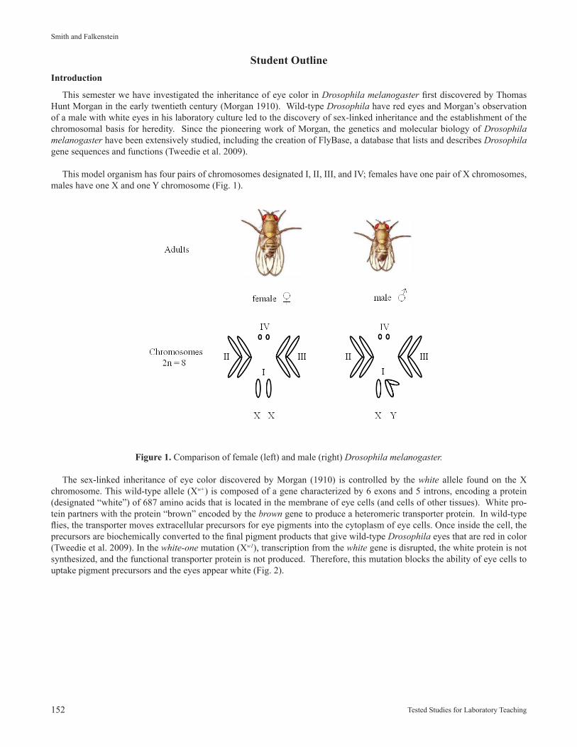
This model organism has four pairs of chromosomes designated I, II, III, and IV; females have one pair of X chromosomes, males have one X and one Y chromosome (Fig. 1).
Figure 1. Comparison of female (left) and male (right) Drosophila melanogaster.
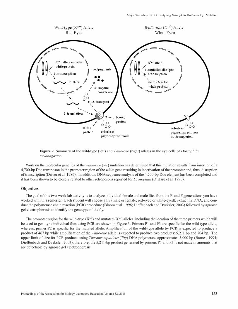
The sex-linked inheritance of eye color discovered by Morgan (1910) is controlled by the white allele found on the X chromosome. This wild-type allele (Xw+) is composed of a gene characterized by 6 exons and 5 introns, encoding a protein (designated “white”) of 687 amino acids that is located in the membrane of eye cells (and cells of other tissues). White pro-tein partners with the protein “brown” encoded by the brown gene to produce a heteromeric transporter protein. In wild-type flies, the transporter moves extracellular precursors for eye pigments into the cytoplasm of eye cells. Once inside the cell, the precursors are biochemically converted to the final pigment products that give wild-type Drosophila eyes that are red in color (Tweedie et al. 2009). In the white-one mutation (Xw1), transcription from the white gene is disrupted, the white protein is not synthesized, and the functional transporter protein is not produced. Therefore, this mutation blocks the ability of eye cells to uptake pigment precursors and the eyes appear white (Fig. 2).
Proceedings of the Association for Biology Laboratory Education, Volume 32, 2011 153
Major Workshop: PCR Genotyping Drosophila White-one Eye Mutation
Figure 2. Summary of the wild-type (left) and white-one (right) alleles in the eye cells of Drosophila melanogaster.
Work on the molecular genetics of the white-one (w1) mutation has determined that this mutation results from insertion of a 4,700-bp Doc retroposon in the promoter region of the white gene resulting in inactivation of the promoter and, thus, disruption of transcription (Driver et al. 1989). In addition, DNA sequence analysis of the 4,700-bp Doc element has been completed and it has been shown to be closely related to other retroposons reported for Drosophila (O’Hare et al. 1990).
Objectives
The goal of this two-week lab activity is to analyze individual female and male flies from the F1 and F2 generations you have worked with this semester. Each student will choose a fly (male or female; red-eyed or white-eyed), extract fly DNA, and con-duct the polymerase chain reaction (PCR) procedure (Bloom et al. 1996; Dieffenbach and Dveksler, 2003) followed by agarose gel electrophoresis to identify the genotype of the fly.
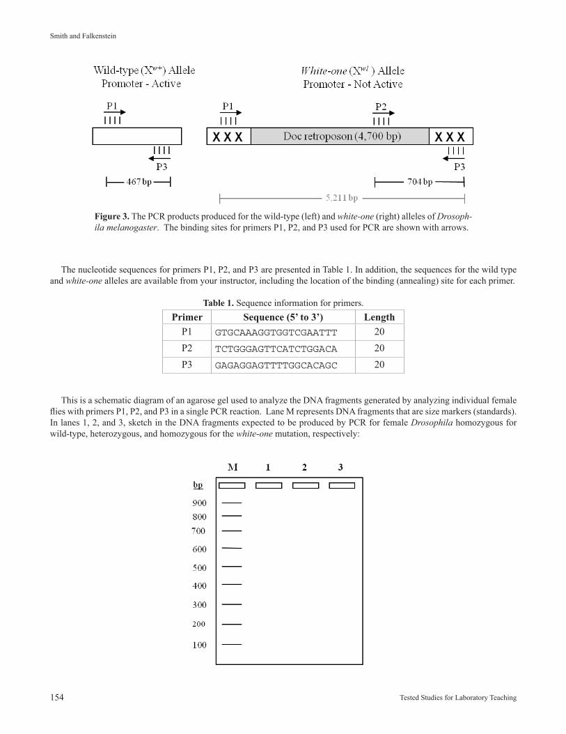
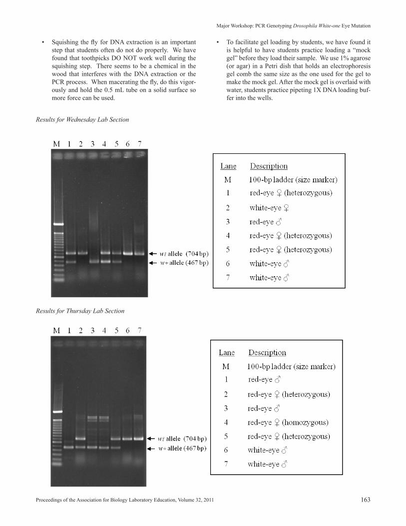
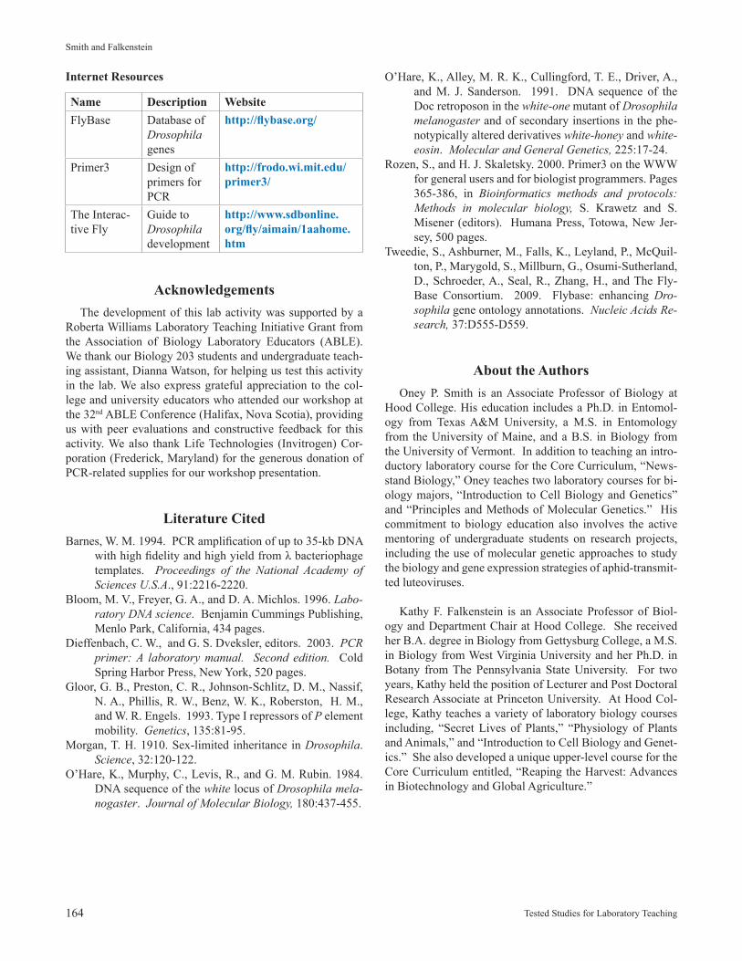
The promoter region for the wild-type (Xw+) and mutated (Xw1) alleles, including the location of the three primers which will be used to genotype individual flies using PCR are shown in Figure 3. Primers P1 and P3 are specific for the wild-type allele, whereas, primer P2 is specific for the mutated allele. Amplification of the wild-type allele by PCR is expected to produce a product of 467 bp while amplification of the white-one allele is expected to produce two products: 5,211 bp and 704 bp. The upper limit of size for PCR products using Thermus aquaticus (Taq) DNA polymerase approximates 5,000 bp (Barnes, 1994; Dieffenbach and Dveksler, 2003), therefore, the 5,211-bp product generated by primers P1 and P3 is not made in amounts that are detectable by agarose gel electrophoresis.
154 Tested Studies for Laboratory Teaching
Smith and Falkenstein
The nucleotide sequences for primers P1, P2, and P3 are presented in Table 1. In addition, the sequences for the wild type and white-one alleles are available from your instructor, including the location of the binding (annealing) site for each primer.
Table 1. Sequence information for primers.Primer Sequence (5’ to 3’) Length
P1 GTGCAAAGGTGGTCGAATTT 20P2 TCTGGGAGTTCATCTGGACA 20P3 GAGAGGAGTTTTGGCACAGC 20
This is a schematic diagram of an agarose gel used to analyze the DNA fragments generated by analyzing individual female flies with primers P1, P2, and P3 in a single PCR reaction. Lane M represents DNA fragments that are size markers (standards). In lanes 1, 2, and 3, sketch in the DNA fragments expected to be produced by PCR for female Drosophila homozygous for wild-type, heterozygous, and homozygous for the white-one mutation, respectively:
Figure 3. The PCR products produced for the wild-type (left) and white-one (right) alleles of Drosoph-ila melanogaster. The binding sites for primers P1, P2, and P3 used for PCR are shown with arrows.
Proceedings of the Association for Biology Laboratory Education, Volume 32, 2011 155
Major Workshop: PCR Genotyping Drosophila White-one Eye Mutation
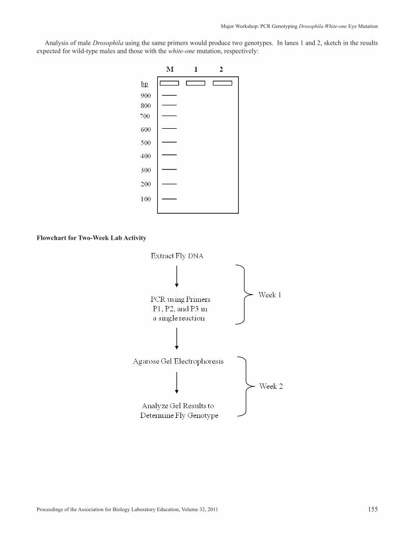
Analysis of male Drosophila using the same primers would produce two genotypes. In lanes 1 and 2, sketch in the results expected for wild-type males and those with the white-one mutation, respectively:
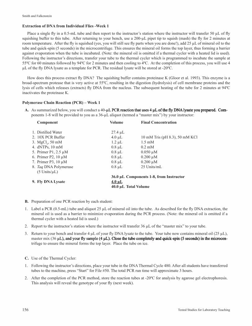
Flowchart for Two-Week Lab Activity
156 Tested Studies for Laboratory Teaching
Smith and Falkenstein
Extraction of DNA from Individual Flies -Week 1
Place a single fly in a 0.5-mL tube and then report to the instructor’s station where the instructor will transfer 50 µL of fly squishing buffer to this tube. After returning to your bench, use a 200-µL pipet tip to squish (mash) the fly for 2 minutes at room temperature. After the fly is squished (yes, you will still see fly parts when you are done!), add 25 µL of mineral oil to the tube and quick-spin (5 seconds) in the microcentrifuge. This ensures the mineral oil forms the top layer, thus forming a barrier against evaporation when the tube is incubated. (Note: the mineral oil is omitted if a thermal cycler with a heated lid is used). Following the instructor’s directions, transfer your tube to the thermal cycler which is programmed to incubate the sample at 55ºC for 60 minutes followed by 94ºC for 2 minutes and then cooling to 4ºC. At the completion of this process, you will use 4 µL of the fly DNA lysate as a template for PCR. The residual lysate will be stored at -20ºC.
How does this process extract fly DNA? The squishing buffer contains proteinase K (Gloor et al. 1993). This enzyme is a broad-spectrum protease that is very active at 55ºC, resulting in the digestion (hydrolysis) of cell membrane proteins and the lysis of cells which releases (extracts) fly DNA from the nucleus. The subsequent heating of the tube for 2 minutes at 94ºC inactivates the proteinase K.
Polymerase Chain Reaction (PCR) – Week 1
A. As summarized below, you will conduct a 40-µL PCR reaction that uses 4 µL of the fl y DNA lysate you prepared. Com-µL PCR reaction that uses 4 µL of the fl y DNA lysate you prepared. Com- PCR reaction that uses 4 µL of the fl y DNA lysate you prepared. Com-µL of the fl y DNA lysate you prepared. Com- of the fly DNA lysate you prepared. Com-ponents 1-8 will be provided to you as a 36-µL aliquot (termed a “master mix”) by your instructor:
Component Volume Final Concentration
1. Distilled Water 27.4 µL 2. 10X PCR Buffer 4.0 µL 10 mM Tris (pH 8.3), 50 mM KCl 3. MgCl2, 50 mM 1.2 µL 1.5 mM 4. dNTPs, 10 mM 0.8 µL 0.2 mM 5. Primer P1, 2.5 µM 0.8 µL 0.050 µM 6. Primer P2, 10 µM 0.8 µL 0.200 µM 7. Primer P3, 10 µM 0.8 µL 0.200 µM 8. Taq DNA Polymerase 0.8 µL 25 Units/mL (5 Units/µL) 36.0 µL Components 1-8, from Instructor 9. Fly DNA Lysate 4.0 µL 40.0 µL Total Volume
B. Preparation of one PCR reaction by each student:
1. Label a PCR (0.5-mL) tube and aliquot 25 µL of mineral oil into the tube. As described for the fly DNA extraction, the mineral oil is used as a barrier to minimize evaporation during the PCR process. (Note: the mineral oil is omitted if a thermal cycler with a heated lid is used.)
2. Report to the instructor’s station where the instructor will transfer 36 µL of the “master mix” to your tube.
3. Return to your bench and transfer 4 µL of your fly DNA lysate to the tube. Your tube now contains mineral oil (25 µL), master mix (36 µL), and your fl y sample (4 µL). Close the tube completely and quick-spin (5 seconds) in the microcen-µL), and your fl y sample (4 µL). Close the tube completely and quick-spin (5 seconds) in the microcen-), and your fly sample (4 µL). Close the tube completely and quick-spin (5 seconds) in the microcen-µL). Close the tube completely and quick-spin (5 seconds) in the microcen-). Close the tube completely and quick-spin (5 seconds) in the microcen-trifuge to ensure the mineral forms the top layer. Place the tube on ice.
C. Use of the Thermal Cycler:
1. Following the instructor’s directions, place your tube in the DNA Thermal Cycle 480. After all students have transferred tubes to the machine, press “Start” for File #50. The total PCR run time will approximate 3 hours.
2. After the completion of the PCR method, store the reaction tubes at -20ºC for analysis by agarose gel electrophoresis. This analysis will reveal the genotype of your fly (next week).
Proceedings of the Association for Biology Laboratory Education, Volume 32, 2011 157
Major Workshop: PCR Genotyping Drosophila White-one Eye Mutation
D. Description of PCR methood
File # Steps Description
50 1. 94ºC, 1 minute • Preamplification denaturation 2. 1 cycle of fly DNA 3. linked to File 49
49 1. 94ºC, 1 minute • Denaturation of DNA templates 2. 55ºC, 1 minute • Annealing (binding) of primers 3. 72ºC, 2 minute • Extension (synthesis) of new DNA 4. 35 cycles • Amplification (steps 1 to 3, repeated 35X) 5. linked to File 48
48 1. 72ºC, 5 minutes • Final extension (synthesis) of DNA 2. linked to File 47 to ensure products (amplicons) are completely synthesized 47 1. 4ºC, hold • Method completed
Gel Electrophoresis – Week 2
A. Sample Preparation
Label a 1.5-mL tube and transfer 8 µL of your PCR reaction to this tube followed by 2 µL of 5X DNA loading buffer (25% glycerol, 0.05% bromophenol blue) to produce a total volume of 10 µL. The addition of loading buffer adjusts the sample to 5% glycerol and 0.01% bromophenol blue. The glycerol increases the density of the sample for loading into the well of the agarose gel and the bromophenol blue provides a “dye front” to monitor the length of time for electrophoresis. (Bromophenol blue migrates at a rate equivalent to a 300-bp fragment of DNA.)
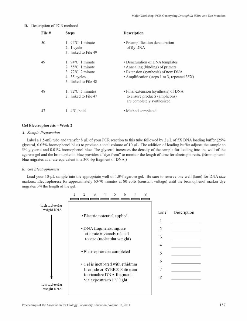
B. Gel Electrophoresis
Load your 10-µL sample into the appropriate well of 1.0% agarose gel. Be sure to reserve one well (lane) for DNA size markers. Electrophorese for approximately 60-70 minutes at 80 volts (constant voltage) until the bromophenol marker dye migrates 3/4 the length of the gel.
158 Tested Studies for Laboratory Teaching
Smith and Falkenstein
C. Gel Staining and Documentation
The lab instructor will demonstrate how to stain the gel with ethidium bromide or Sybr® Safe stain and to produce a gel photograph. The final photograph will be posted on Blackboard.
Questions
1. Based on the results of agarose gel electrophoresis, what is the genotype of the fly you processed by PCR. Explain your reasoning.
2. The DNA extraction protocol includes the heat inactivation of proteinase K. Why is this step necessary?
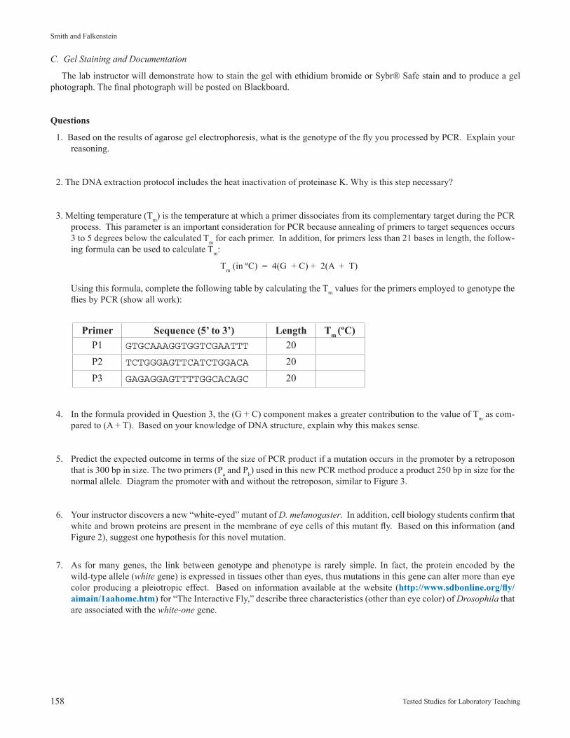
3. Melting temperature (Tm) is the temperature at which a primer dissociates from its complementary target during the PCR process. This parameter is an important consideration for PCR because annealing of primers to target sequences occurs 3 to 5 degrees below the calculated Tm for each primer. In addition, for primers less than 21 bases in length, the follow-ing formula can be used to calculate Tm:
Tm (in ºC) = 4(G + C) + 2(A + T)
Using this formula, complete the following table by calculating the Tm values for the primers employed to genotype the flies by PCR (show all work):
Primer Sequence (5’ to 3’) Length Tm (ºC)P1 GTGCAAAGGTGGTCGAATTT 20P2 TCTGGGAGTTCATCTGGACA 20P3 GAGAGGAGTTTTGGCACAGC 20
4. In the formula provided in Question 3, the (G + C) component makes a greater contribution to the value of Tm as com-pared to (A + T). Based on your knowledge of DNA structure, explain why this makes sense.
5. Predict the expected outcome in terms of the size of PCR product if a mutation occurs in the promoter by a retroposon that is 300 bp in size. The two primers (Pa and Pb) used in this new PCR method produce a product 250 bp in size for the normal allele. Diagram the promoter with and without the retroposon, similar to Figure 3.
6. Your instructor discovers a new “white-eyed” mutant of D. melanogaster. In addition, cell biology students confirm that white and brown proteins are present in the membrane of eye cells of this mutant fly. Based on this information (and Figure 2), suggest one hypothesis for this novel mutation.
7. As for many genes, the link between genotype and phenotype is rarely simple. In fact, the protein encoded by the wild-type allele (white gene) is expressed in tissues other than eyes, thus mutations in this gene can alter more than eye color producing a pleiotropic effect. Based on information available at the website (http://www.sdbonline.org/fly/aimain/1aahome.htm) for “The Interactive Fly,” describe three characteristics (other than eye color) of Drosophila that are associated with the white-one gene.
Proceedings of the Association for Biology Laboratory Education, Volume 32, 2011 159
Major Workshop: PCR Genotyping Drosophila White-one Eye Mutation
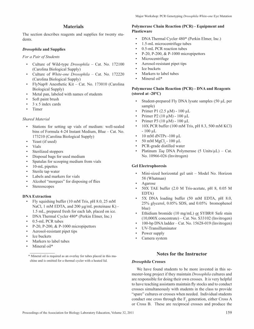
Polymerase Chain Reaction (PCR) - Equipment and Plasticware
• DNA Thermal Cycler 480* (Perkin Elmer, Inc.) • 1.5-mL microcentrifuge tubes• 0.5-mL PCR reaction tubes• P-20, P-200, & P-1000 micropipettors• Microcentrifuge• Aerosol-resistant pipet tips • Ice buckets• Markers to label tubes • Mineral oil*
Polymerase Chain Reaction (PCR) - DNA and Reagents (stored at -20ºC)
• Student-prepared Fly DNA lysate samples (50 µL per sample)
• Primer P1 (2.5 µM) - 100 µL• Primer P2 (10 µM) - 100 µL • Primer P3 (10 µM) - 100 µL• 10X PCR buffer (100 mM Tris, pH 8.3, 500 mM KCl)
- 100 µL• 10 mM dNTPs -100 µL• 50 mM MgCl2 - 100 µL• PCR-grade distilled water • Platinum Taq DNA Polymerase (5 Units/µL) – Cat.
No. 10966-026 (Invitrogen)
Gel Electrophoresis
• Mini-sized horizontal gel unit – Model No. Horizon 58 (Whatman)
• Agarose• 50X TAE buffer (2.0 M Tris-acetate, pH 8, 0.05 M
EDTA)• 5X DNA loading buffer (50 mM EDTA, pH 8.0,
25% glycerol, 0.05% SDS, and 0.05% bromophenol blue)
• Ethidium bromide (10 mg/mL) or SYBR® Safe stain (10,000X concentrate) – Cat. No. S33102 (Invitrogen)
• 100-bp DNA ladder – Cat. No. 15628-019 (Invitrogen)• UV-Transilluminator• Power supply• Camera system
Notes for the InstructorDrosophila Crosses
We have found students to be more invested in this se-mester-long project if they maintain Drosophila cultures and are responsible for doing their own crosses. It is very helpful to have teaching assistants maintain fly stocks and to conduct crosses simultaneously with students in the class to provide “spare” cultures or crosses when needed. Individual students conduct one cross through the F2 generation, either Cross A or Cross B. These are reciprocal crosses and produce the
MaterialsThe section describes reagents and supplies for twenty stu-dents.
Drosophila and Supplies
For a Pair of Students
• Culture of Wild-type Drosophila – Cat. No. 172100 (Carolina Biological Supply)
• Culture of White-one Drosophila – Cat. No. 172220 (Carolina Biological Supply)
• FlyNap® Anesthetic Kit – Cat. No. 173010 (Carolina Biological Supply)
• Metal pan, labeled with names of students• Soft paint brush• 3 x 5 index cards• Timer
Shared Material
• Stations for setting up vials of medium: well-sealed bins of Formula 4-24 Instant Medium, Blue – Cat. No. 173210 (Carolina Biological Supply)
• Yeast (if used)• Vials• Sterilized stoppers• Disposal bags for used medium• Spatulas for scooping medium from vials• 10-mL pipettes• Sterile tap water• Labels and markers for vials • Alcohol “morgues” for disposing of flies • Stereoscopes
DNA Extraction• Fly squishing buffer (10 mM Tris, pH 8.0, 25 mM
NaCl, 1 mM EDTA, and 200 µg/mL proteinase K) - 1.5 mL, prepared fresh for each lab, placed on ice.
• DNA Thermal Cycler 480* (Perkin Elmer, Inc.) • 0.5-mL PCR tubes • P-20, P-200, & P-1000 micropipettors• Aerosol-resistant pipet tips • Ice buckets• Markers to label tubes• Mineral oil* ____________* Mineral oil is required as an overlay for tubes placed in this ma-chine and is omitted for a thermal cycler with a heated lid.
160 Tested Studies for Laboratory Teaching
Smith and Falkenstein
Note: 10X TNE is 100 mM Tris, pH 8.0, 250 mM NaCl, 10 mM EDTA. Proteinase K (Cat. No. 25530-015, Invitro-gen) is from a stock solution (10 mg/mL) prepared in dis-tilled water and stored at -20ºC in 35 µL aliquots.
Polymerase Chain Reaction (PCR)
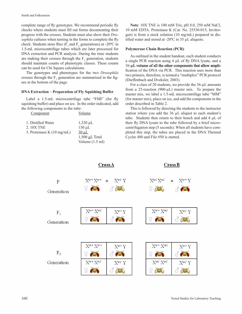
As outlined in the student handout, each student conducts a single PCR reaction using 4 µL of fly DNA lysate, and a 36-µL volume of all the other components that allow ampli-µL volume of all the other components that allow ampli- volume of all the other components that allow ampli-fication of the DNA via PCR. This reaction uses more than two primers, therefore, is termed a “multiplex” PCR protocol (Dieffenbach and Dveksler, 2003). For a class of 20 students, we provide the 36-µL amounts from a 25-reaction (900-µL) master mix. To prepare the master mix, we label a 1.5-mL microcentrifuge tube “MM” (for master mix), place on ice, and add the components in the order described in Table 2. This is followed by directing the students to the instructor station where you add the 36 µL aliquot to each student’s tube. Students then return to their bench and add 4 µL of their fly DNA lysate to the tube followed by a brief micro-centrifugation step (5 seconds). When all students have com-pleted this step, the tubes are placed in the DNA Thermal Cycler 480 and File #50 is started.
complete range of fly genotypes. We recommend periodic fly checks where students must fill out forms documenting their progress with the crosses. Students must also show their Dro-sophila cultures when turning in the forms to complete the fly check. Students store flies (F1 and F2 generations) at -20ºC in 1.5-mL microcentrifuge tubes which are later processed for DNA extraction and PCR analysis. During the time students are making their crosses through the F2 generation, students should maintain counts of phenotypic classes. These counts can be used for Chi Square calculations. The genotypes and phenotypes for the two Drosophila crosses through the F2 generation are summarized in the fig-ure at the bottom of the page.
DNA Extraction – Preparation of Fly Squishing Buffer
Label a 1.5-mL microcentrifuge tube “FSB” (for fly squishing buffer) and place on ice. In the order indicated, add the following components to the tube: Component Volume
1. Distilled Water 1,320 µL 2. 10X TNE 150 µL 3. Proteinase K (10 mg/mL) 30 µL 1,500 µL Total Volume (1.5 ml)
Proceedings of the Association for Biology Laboratory Education, Volume 32, 2011 161
Major Workshop: PCR Genotyping Drosophila White-one Eye Mutation
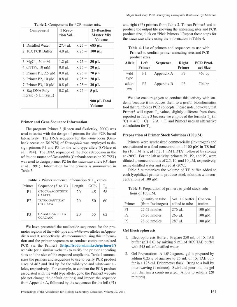
and right (P3) primers from Table 2. To run Primer3 and to produce the output file showing the annealing sites and PCR product size, click on “Pick Primers.” Repeat these steps for the white-one allele using the information in Table 4.
Table 4. List of primers and sequences to use with Primer3 to confirm primer annealing sites and PCR product sizes.
Allele Left Primer
Sequence Right Primer
PCR Prod-uct Size
wild type
P1 Appendix A P3 467 bp
white-one
P2 Appendix B P3 704 bp
We also encourage you to conduct this activity with stu-dents because it introduces them to a useful bioinformatics tool that reinforces PCR concepts. Please note, however, that Primer3 will report Tm values slightly different from those reported in Table 3 because we employed the formula Tm (in ºC) = 4(G + C) + 2(A + T) and Primer3 uses an alternative calculation for Tm.
Preparation of Primer Stock Solutions (100 µM)
Primers were synthesized commercially (Invitrogen) and reconstituted to a final concentration of 100 µM in TE buf-µM in TE buf-M in TE buf-fer (10 mM Tris, pH 7.2, 1 mM EDTA) followed by storage at -20ºC. For the lab activity, primers P1, P2, and P3, were diluted to concentrations of 2.5, 10, and 10 µM, respectively, using distilled water and stored at -20ºC. Table 5 summarizes the volume of TE buffer added to each lyophilized primer to produce stock solutions with con-centrations of 100 µM.
Table 5. Preparation of primers to yield stock solu-tions of 100 µM.
PrimerQuantity in tube (from Invitrogen)
Vol. TE buffer added to tube
Concen-tration
P1 27.62 nmoles 276 µL 100 µMP2 26.26 nmoles 263 µL 100 µMP3 28.66 nmoles 287 µL 100 µM
Gel Electrophoresis
1. Electrophoresis Buffer: Prepare 250 mL of 1X TAE buffer (pH 8.0) by mixing 5 mL of 50X TAE buffer with 245 mL of distilled water.
2. Gel Preparation: A 1.0% agarose gel is prepared by adding 0.25 g of agarose to 25 mL of 1X TAE buf-fer in a 125-mL Erlenmeyer flask. Bring to a boil by microwaving (1 minute). Swirl and pour into the gel unit that has a comb inserted. Allow to solidify (20 minutes).
Table 2. Components for PCR master mix.Component 1 Reac-
tion Vol.25-Reaction Master Mix
Volume1. Distilled Water 27.4 µL x 25 = 685 µL2. 10X PCR Buffer 4.0 µL x 25 = 100 µL
3. MgCl2, 50 mM 1.2 µL x 25 = 30 µL4. dNTPs, 10 mM 0.8 µL x 25 = 20 µL5. Primer P1, 2.5 µM 0.8 µL x 25 = 20 µL6. Primer P2, 10 µM 0.8 µL x 25 = 20 µL7. Primer P3, 10 µM 0.8 µL x 25 = 20 µL8. Taq DNA Poly-merase (5 Units/µL)
0.2 µL x 25 = 5 µL
900 µL Total Volume
Primer and Gene Sequence Information
The program Primer 3 (Rozen and Skaletsky, 2000) was used to assist with the design of primers for this PCR-based lab activity. The DNA sequence for the white locus (Gen-bank accession X02974) of Drosophila was employed to de-sign primers P1 and P3 for the wild-type allele (O’Hare et al., 1984). The DNA sequence of the Doc retroposon in the white-one mutant of Drosophila (Genbank accession X17551) was used to design primer P2 for the white-one allele (O’Hare et al., 1991). Information for the primers is summarized in Table 3.
Table 3. Primer sequence information & Tm values.Primer Sequence (5’ to 3’) Length GC% Tm
P1 GTGCAAAGGTGGTC GAATTT
20 45 58
P2 TCTGGGAGTTCAT CTGGACA
20 50 60
P3 GAGAGGAGTTTTGGCACAGC
20 55 62
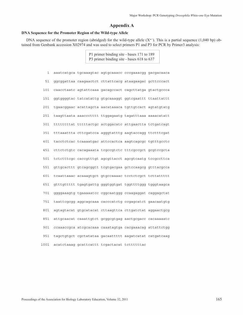
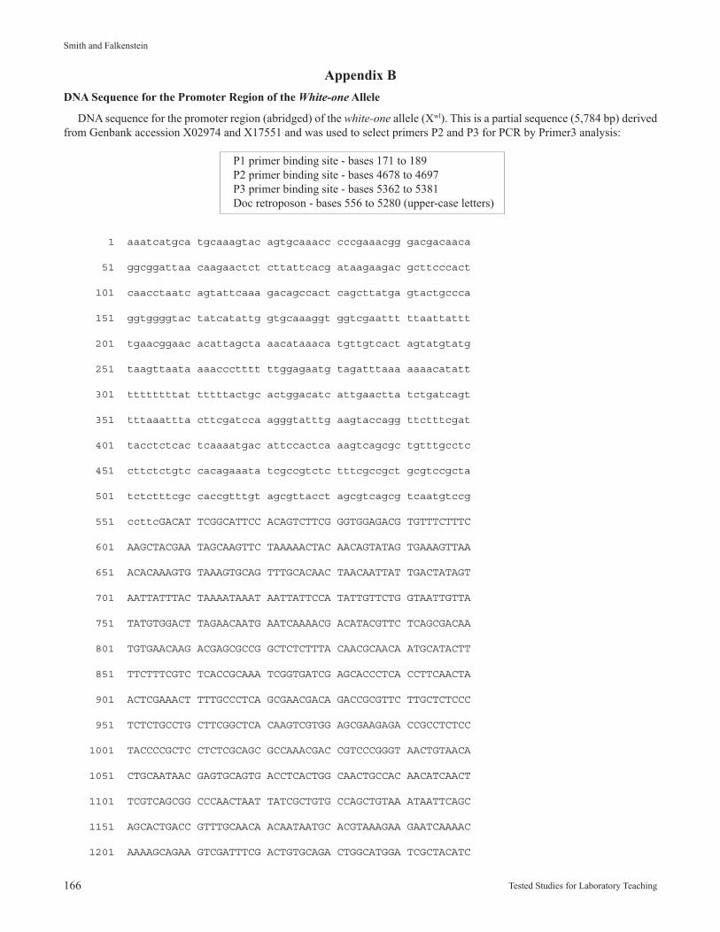
We have presented the nucleotide sequences for the pro-moter regions of the wild-type and white-one alleles in Appen-dix A and B, respectively. We recommend using this informa-tion and the primer sequences to conduct computer-assisted PCR via the Primer3 (http://frodo.wi.mit.edu/primer3/) website (or a similar website) to verify the primer annealing sites and the size of the expected amplicons. Table 4 summa-rizes the primers and sequences to use to verify PCR product sizes of 467 and 704 bp for the wild-type and white-one al-leles, respectively. For example, to confirm the PCR product associated with the wild type allele, go to the Primer3 website (do not change the default options) and import the sequence from Appendix A, followed by the sequences for the left (P1)
162 Tested Studies for Laboratory Teaching
Smith and Falkenstein
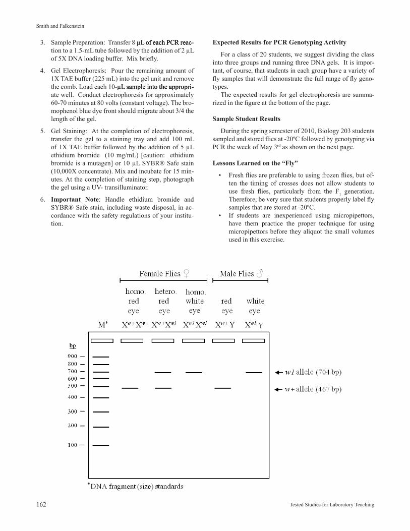
Expected Results for PCR Genotyping Activity
For a class of 20 students, we suggest dividing the class into three groups and running three DNA gels. It is impor-tant, of course, that students in each group have a variety of fly samples that will demonstrate the full range of fly geno-types. The expected results for gel electrophoresis are summa-rized in the figure at the bottom of the page.
Sample Student Results
During the spring semester of 2010, Biology 203 students sampled and stored flies at -20ºC followed by genotyping via PCR the week of May 3rd as shown on the next page.
Lessons Learned on the “Fly”
• Fresh flies are preferable to using frozen flies, but of-ten the timing of crosses does not allow students to use fresh flies, particularly from the F1 generation. Therefore, be very sure that students properly label fly samples that are stored at -20ºC.
• If students are inexperienced using micropipettors, have them practice the proper technique for using micropipettors before they aliquot the small volumes used in this exercise.
3. Sample Preparation: Transfer 8 µL of each PCR reac-µL of each PCR reac- of each PCR reac-tion to a 1.5-mL tube followed by the addition of 2 µL of 5X DNA loading buffer. Mix briefly.
4. Gel Electrophoresis: Pour the remaining amount of 1X TAE buffer (225 mL) into the gel unit and remove the comb. Load each 10-µL sample into the appropri-µL sample into the appropri- sample into the appropri-ate well. Conduct electrophoresis for approximately 60-70 minutes at 80 volts (constant voltage). The bro-mophenol blue dye front should migrate about 3/4 the length of the gel.
5. Gel Staining: At the completion of electrophoresis, transfer the gel to a staining tray and add 100 mL of 1X TAE buffer followed by the addition of 5 µL ethidium bromide (10 mg/mL) [caution: ethidium bromide is a mutagen] or 10 µL SYBR® Safe stain (10,000X concentrate). Mix and incubate for 15 min-utes. At the completion of staining step, photograph the gel using a UV- transilluminator.
6. Important Note: Handle ethidium bromide and SYBR® Safe stain, including waste disposal, in ac-cordance with the safety regulations of your institu-tion.
Proceedings of the Association for Biology Laboratory Education, Volume 32, 2011 163
Major Workshop: PCR Genotyping Drosophila White-one Eye Mutation
Results for Wednesday Lab Section
Results for Thursday Lab Section
• Squishing the fly for DNA extraction is an importantstep that students often do not do properly. We havefound that toothpicks DO NOT work well during thesquishing step. There seems to be a chemical in thewood that interferes with the DNA extraction or thePCR process. When macerating the fly, do this vigor-ously and hold the 0.5 mL tube on a solid surface somore force can be used.
• To facilitate gel loading by students, we have found itis helpful to have students practice loading a “mockgel” before they load their sample. We use 1% agarose(or agar) in a Petri dish that holds an electrophoresisgel comb the same size as the one used for the gel tomake the mock gel. After the mock gel is overlaid withwater, students practice pipeting 1X DNA loading buf-fer into the wells.
164 Tested Studies for Laboratory Teaching
Smith and Falkenstein
Internet Resources
Name Description WebsiteFlyBase Database of
Drosophila genes
http://flybase.org/
Primer3 Design of primers for PCR
http://frodo.wi.mit.edu/primer3/
The Interac-tive Fly
Guide to Drosophila development
http://www.sdbonline.org/fly/aimain/1aahome.htm
Acknowledgements The development of this lab activity was supported by a Roberta Williams Laboratory Teaching Initiative Grant from the Association of Biology Laboratory Educators (ABLE). We thank our Biology 203 students and undergraduate teach-ing assistant, Dianna Watson, for helping us test this activity in the lab. We also express grateful appreciation to the col-lege and university educators who attended our workshop at the 32nd ABLE Conference (Halifax, Nova Scotia), providing us with peer evaluations and constructive feedback for this activity. We also thank Life Technologies (Invitrogen) Cor-poration (Frederick, Maryland) for the generous donation of PCR-related supplies for our workshop presentation.
Literature CitedBarnes, W. M. 1994. PCR amplification of up to 35-kb DNA
with high fidelity and high yield from λ bacteriophage templates. Proceedings of the National Academy of Sciences U.S.A., 91:2216-2220.
Bloom, M. V., Freyer, G. A., and D. A. Michlos. 1996. Labo-ratory DNA science. Benjamin Cummings Publishing, Menlo Park, California, 434 pages.
Dieffenbach, C. W., and G. S. Dveksler, editors. 2003. PCR primer: A laboratory manual. Second edition. Cold Spring Harbor Press, New York, 520 pages.
Gloor, G. B., Preston, C. R., Johnson-Schlitz, D. M., Nassif, N. A., Phillis, R. W., Benz, W. K., Roberston, H. M., and W. R. Engels. 1993. Type I repressors of P element mobility. Genetics, 135:81-95.
Morgan, T. H. 1910. Sex-limited inheritance in Drosophila. Science, 32:120-122.
O’Hare, K., Murphy, C., Levis, R., and G. M. Rubin. 1984. DNA sequence of the white locus of Drosophila mela-nogaster. Journal of Molecular Biology, 180:437-455.
O’Hare, K., Alley, M. R. K., Cullingford, T. E., Driver, A., and M. J. Sanderson. 1991. DNA sequence of the Doc retroposon in the white-one mutant of Drosophila melanogaster and of secondary insertions in the phe-notypically altered derivatives white-honey and white-eosin. Molecular and General Genetics, 225:17-24.
Rozen, S., and H. J. Skaletsky. 2000. Primer3 on the WWW for general users and for biologist programmers. Pages 365-386, in Bioinformatics methods and protocols: Methods in molecular biology, S. Krawetz and S. Misener (editors). Humana Press, Totowa, New Jer-sey, 500 pages.
Tweedie, S., Ashburner, M., Falls, K., Leyland, P., McQuil-ton, P., Marygold, S., Millburn, G., Osumi-Sutherland, D., Schroeder, A., Seal, R., Zhang, H., and The Fly-Base Consortium. 2009. Flybase: enhancing Dro-sophila gene ontology annotations. Nucleic Acids Re-search, 37:D555-D559.
About the Authors Oney P. Smith is an Associate Professor of Biology at Hood College. His education includes a Ph.D. in Entomol-ogy from Texas A&M University, a M.S. in Entomology from the University of Maine, and a B.S. in Biology from the University of Vermont. In addition to teaching an intro-ductory laboratory course for the Core Curriculum, “News-stand Biology,” Oney teaches two laboratory courses for bi-ology majors, “Introduction to Cell Biology and Genetics” and “Principles and Methods of Molecular Genetics.” His commitment to biology education also involves the active mentoring of undergraduate students on research projects, including the use of molecular genetic approaches to study the biology and gene expression strategies of aphid-transmit-ted luteoviruses.
Kathy F. Falkenstein is an Associate Professor of Biol-ogy and Department Chair at Hood College. She received her B.A. degree in Biology from Gettysburg College, a M.S. in Biology from West Virginia University and her Ph.D. in Botany from The Pennsylvania State University. For two years, Kathy held the position of Lecturer and Post Doctoral Research Associate at Princeton University. At Hood Col-lege, Kathy teaches a variety of laboratory biology courses including, “Secret Lives of Plants,” “Physiology of Plants and Animals,” and “Introduction to Cell Biology and Genet-ics.” She also developed a unique upper-level course for the Core Curriculum entitled, “Reaping the Harvest: Advances in Biotechnology and Global Agriculture.”
Proceedings of the Association for Biology Laboratory Education, Volume 32, 2011 165
Major Workshop: PCR Genotyping Drosophila White-one Eye Mutation
Appendix ADNA Sequence for the Promoter Region of the Wild-type Allele
DNA sequence of the promoter region (abridged) for the wild-type allele (Xw+). This is a partial sequence (1,040 bp) ob-tained from Genbank accession X02974 and was used to select primers P1 and P3 for PCR by Primer3 analysis:
P1 primer binding site - bases 171 to 189P3 primer binding site - bases 618 to 637
1 aaatcatgca tgcaaagtac agtgcaaacc cccgaaacgg gacgacaaca
51 ggcggattaa caagaactct cttattcacg ataagaagac gcttcccact
101 caacctaatc agtattcaaa gacagccact cagcttatga gtactgccca
151 ggtggggtac tatcatattg gtgcaaaggt ggtcgaattt ttaattattt
201 tgaacggaac acattagcta aacataaaca tgttgtcact agtatgtatg
251 taagttaata aaaccctttt ttggagaatg tagatttaaa aaaacatatt
301 ttttttttat tttttactgc actggacatc attgaactta tctgatcagt
351 tttaaattta cttcgatcca agggtatttg aagtaccagg ttctttcgat
401 tacctctcac tcaaaatgac attccactca aagtcagcgc tgtttgcctc
451 cttctctgtc cacagaaata tcgccgtctc tttcgccgct gcgtccgcta
501 tctctttcgc caccgtttgt agcgttacct agcgtcaatg tccgccttca
551 gttgcacttt gtcagcggtt tcgtgacgaa gctccaagcg gtttacgcca
601 tcaattaaac acaaagtgct gtgccaaaac tcctctcgct tcttattttt
651 gtttgttttt tgagtgattg gggtggtgat tggttttggg tgggtaagca
701 ggggaaagtg tgaaaaatcc cggcaatggg ccaagaggat caggagctat
751 taattcgcgg aggcagcaaa cacccatctg ccgagcatct gaacaatgtg
801 agtagtacat gtgcatacat cttaagttca cttgatctat aggaactgcg
851 attgcaacat caaattgtct gcggcgtgag aactgcgacc cacaaaaatc
901 ccaaaccgca atcgcacaaa caaatagtga cacgaaacag attattctgg
951 tagctgtgct cgctatataa gacaattttt aagatcatat catgatcaag
1001 acatctaaag gcattcattt tcgactacat tcttttttac
166 Tested Studies for Laboratory Teaching
Smith and Falkenstein
Appendix BDNA Sequence for the Promoter Region of the White-one Allele
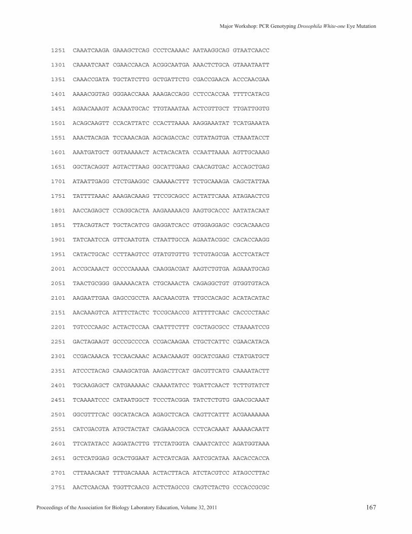
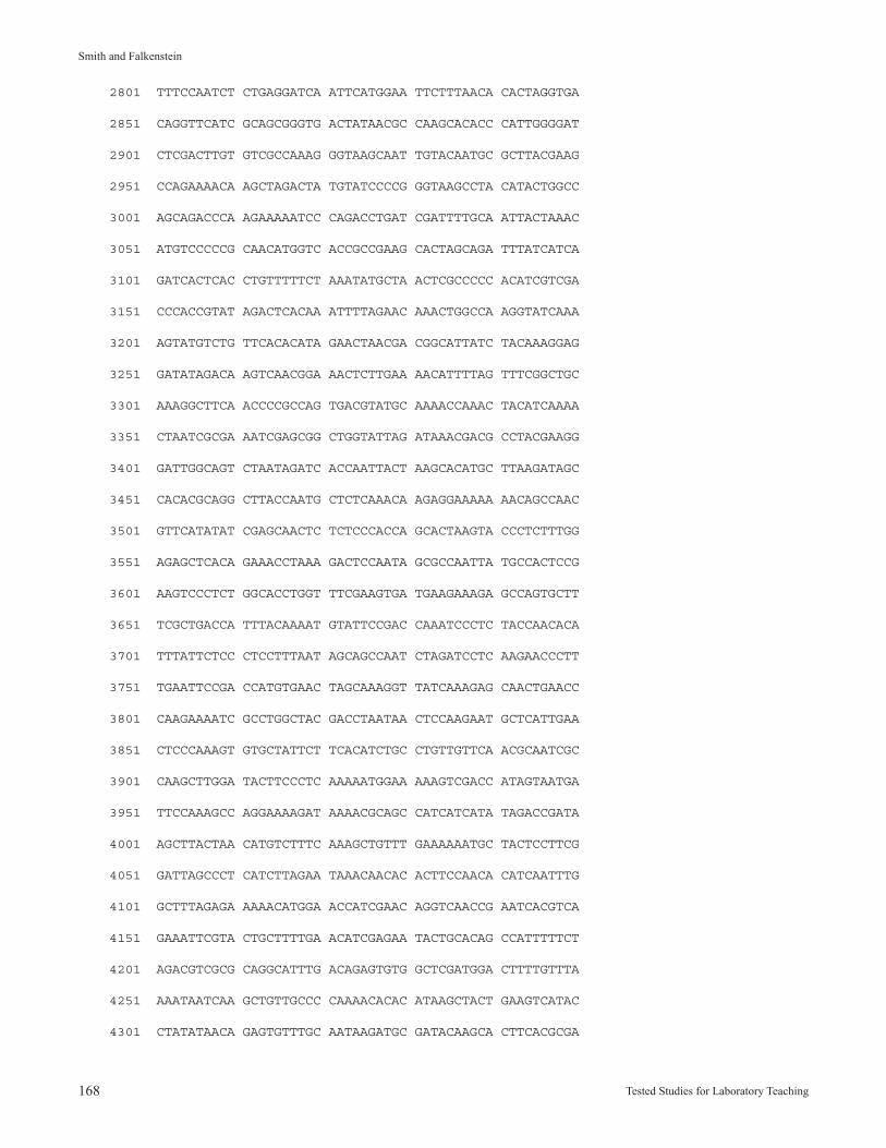
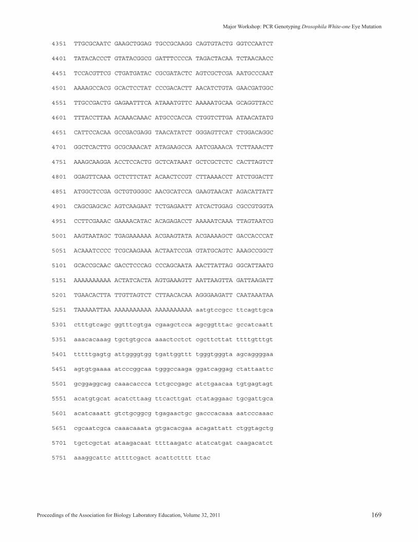
DNA sequence for the promoter region (abridged) of the white-one allele (Xwl). This is a partial sequence (5,784 bp) derived from Genbank accession X02974 and X17551 and was used to select primers P2 and P3 for PCR by Primer3 analysis:
P1 primer binding site - bases 171 to 189 P2 primer binding site - bases 4678 to 4697 P3 primer binding site - bases 5362 to 5381 Doc retroposon - bases 556 to 5280 (upper-case letters)
1 aaatcatgca tgcaaagtac agtgcaaacc cccgaaacgg gacgacaaca
51 ggcggattaa caagaactct cttattcacg ataagaagac gcttcccact
101 caacctaatc agtattcaaa gacagccact cagcttatga gtactgccca
151 ggtggggtac tatcatattg gtgcaaaggt ggtcgaattt ttaattattt
201 tgaacggaac acattagcta aacataaaca tgttgtcact agtatgtatg
251 taagttaata aaaccctttt ttggagaatg tagatttaaa aaaacatatt
301 ttttttttat tttttactgc actggacatc attgaactta tctgatcagt
351 tttaaattta cttcgatcca agggtatttg aagtaccagg ttctttcgat
401 tacctctcac tcaaaatgac attccactca aagtcagcgc tgtttgcctc
451 cttctctgtc cacagaaata tcgccgtctc tttcgccgct gcgtccgcta
501 tctctttcgc caccgtttgt agcgttacct agcgtcagcg tcaatgtccg
551 ccttcGACAT TCGGCATTCC ACAGTCTTCG GGTGGAGACG TGTTTCTTTC
601 AAGCTACGAA TAGCAAGTTC TAAAAACTAC AACAGTATAG TGAAAGTTAA
651 ACACAAAGTG TAAAGTGCAG TTTGCACAAC TAACAATTAT TGACTATAGT
701 AATTATTTAC TAAAATAAAT AATTATTCCA TATTGTTCTG GTAATTGTTA
751 TATGTGGACT TAGAACAATG AATCAAAACG ACATACGTTC TCAGCGACAA
801 TGTGAACAAG ACGAGCGCCG GCTCTCTTTA CAACGCAACA ATGCATACTT
851 TTCTTTCGTC TCACCGCAAA TCGGTGATCG AGCACCCTCA CCTTCAACTA
901 ACTCGAAACT TTTGCCCTCA GCGAACGACA GACCGCGTTC TTGCTCTCCC
951 TCTCTGCCTG CTTCGGCTCA CAAGTCGTGG AGCGAAGAGA CCGCCTCTCC
1001 TACCCCGCTC CTCTCGCAGC GCCAAACGAC CGTCCCGGGT AACTGTAACA
1051 CTGCAATAAC GAGTGCAGTG ACCTCACTGG CAACTGCCAC AACATCAACT
1101 TCGTCAGCGG CCCAACTAAT TATCGCTGTG CCAGCTGTAA ATAATTCAGC
1151 AGCACTGACC GTTTGCAACA ACAATAATGC ACGTAAAGAA GAATCAAAAC
1201 AAAAGCAGAA GTCGATTTCG ACTGTGCAGA CTGGCATGGA TCGCTACATC
Proceedings of the Association for Biology Laboratory Education, Volume 32, 2011 167
Major Workshop: PCR Genotyping Drosophila White-one Eye Mutation
1251 CAAATCAAGA GAAAGCTCAG CCCTCAAAAC AATAAGGCAG GTAATCAACC
1301 CAAAATCAAT CGAACCAACA ACGGCAATGA AAACTCTGCA GTAAATAATT
1351 CAAACCGATA TGCTATCTTG GCTGATTCTG CGACCGAACA ACCCAACGAA
1401 AAAACGGTAG GGGAACCAAA AAAGACCAGG CCTCCACCAA TTTTCATACG
1451 AGAACAAAGT ACAAATGCAC TTGTAAATAA ACTCGTTGCT TTGATTGGTG
1501 ACAGCAAGTT CCACATTATC CCACTTAAAA AAGGAAATAT TCATGAAATA
1551 AAACTACAGA TCCAAACAGA AGCAGACCAC CGTATAGTGA CTAAATACCT
1601 AAATGATGCT GGTAAAAACT ACTACACATA CCAATTAAAA AGTTGCAAAG
1651 GGCTACAGGT AGTACTTAAG GGCATTGAAG CAACAGTGAC ACCAGCTGAG
1701 ATAATTGAGG CTCTGAAGGC CAAAAACTTT TCTGCAAAGA CAGCTATTAA
1751 TATTTTAAAC AAAGACAAAG TTCCGCAGCC ACTATTCAAA ATAGAACTCG
1801 AACCAGAGCT CCAGGCACTA AAGAAAAACG AAGTGCACCC AATATACAAT
1851 TTACAGTACT TGCTACATCG GAGGATCACC GTGGAGGAGC CGCACAAACG
1901 TATCAATCCA GTTCAATGTA CTAATTGCCA AGAATACGGC CACACCAAGG
1951 CATACTGCAC CCTTAAGTCC GTATGTGTTG TCTGTAGCGA ACCTCATACT
2001 ACCGCAAACT GCCCCAAAAA CAAGGACGAT AAGTCTGTGA AGAAATGCAG
2051 TAACTGCGGG GAAAAACATA CTGCAAACTA CAGAGGCTGT GTGGTGTACA
2101 AAGAATTGAA GAGCCGCCTA AACAAACGTA TTGCCACAGC ACATACATAC
2151 AACAAAGTCA ATTTCTACTC TCCGCAACCG ATTTTTCAAC CACCCCTAAC
2201 TGTCCCAAGC ACTACTCCAA CAATTTCTTT CGCTAGCGCC CTAAAATCCG
2251 GACTAGAAGT GCCCGCCCCA CCGACAAGAA CTGCTCATTC CGAACATACA
2301 CCGACAAACA TCCAACAAAC ACAACAAAGT GGCATCGAAG CTATGATGCT
2351 ATCCCTACAG CAAAGCATGA AAGACTTCAT GACGTTCATG CAAAATACTT
2401 TGCAAGAGCT CATGAAAAAC CAAAATATCC TGATTCAACT TCTTGTATCT
2451 TCAAAATCCC CATAATGGCT TCCCTACGGA TATCTCTGTG GAACGCAAAT
2501 GGCGTTTCAC GGCATACACA AGAGCTCACA CAGTTCATTT ACGAAAAAAA
2551 CATCGACGTA ATGCTACTAT CAGAAACGCA CCTCACAAAT AAAAACAATT
2601 TTCATATACC AGGATACTTG TTCTATGGTA CAAATCATCC AGATGGTAAA
2651 GCTCATGGAG GCACTGGAAT ACTCATCAGA AATCGCATAA AACACCACCA
2701 CTTAAACAAT TTTGACAAAA ACTACTTACA ATCTACGTCC ATAGCCTTAC
2751 AACTCAACAA TGGTTCAACG ACTCTAGCCG CAGTCTACTG CCCACCGCGC
168 Tested Studies for Laboratory Teaching
Smith and Falkenstein
2801 TTTCCAATCT CTGAGGATCA ATTCATGGAA TTCTTTAACA CACTAGGTGA
2851 CAGGTTCATC GCAGCGGGTG ACTATAACGC CAAGCACACC CATTGGGGAT
2901 CTCGACTTGT GTCGCCAAAG GGTAAGCAAT TGTACAATGC GCTTACGAAG
2951 CCAGAAAACA AGCTAGACTA TGTATCCCCG GGTAAGCCTA CATACTGGCC
3001 AGCAGACCCA AGAAAAATCC CAGACCTGAT CGATTTTGCA ATTACTAAAC
3051 ATGTCCCCCG CAACATGGTC ACCGCCGAAG CACTAGCAGA TTTATCATCA
3101 GATCACTCAC CTGTTTTTCT AAATATGCTA ACTCGCCCCC ACATCGTCGA
3151 CCCACCGTAT AGACTCACAA ATTTTAGAAC AAACTGGCCA AGGTATCAAA
3201 AGTATGTCTG TTCACACATA GAACTAACGA CGGCATTATC TACAAAGGAG
3251 GATATAGACA AGTCAACGGA AACTCTTGAA AACATTTTAG TTTCGGCTGC
3301 AAAGGCTTCA ACCCCGCCAG TGACGTATGC AAAACCAAAC TACATCAAAA
3351 CTAATCGCGA AATCGAGCGG CTGGTATTAG ATAAACGACG CCTACGAAGG
3401 GATTGGCAGT CTAATAGATC ACCAATTACT AAGCACATGC TTAAGATAGC
3451 CACACGCAGG CTTACCAATG CTCTCAAACA AGAGGAAAAA AACAGCCAAC
3501 GTTCATATAT CGAGCAACTC TCTCCCACCA GCACTAAGTA CCCTCTTTGG
3551 AGAGCTCACA GAAACCTAAA GACTCCAATA GCGCCAATTA TGCCACTCCG
3601 AAGTCCCTCT GGCACCTGGT TTCGAAGTGA TGAAGAAAGA GCCAGTGCTT
3651 TCGCTGACCA TTTACAAAAT GTATTCCGAC CAAATCCCTC TACCAACACA
3701 TTTATTCTCC CTCCTTTAAT AGCAGCCAAT CTAGATCCTC AAGAACCCTT
3751 TGAATTCCGA CCATGTGAAC TAGCAAAGGT TATCAAAGAG CAACTGAACC
3801 CAAGAAAATC GCCTGGCTAC GACCTAATAA CTCCAAGAAT GCTCATTGAA
3851 CTCCCAAAGT GTGCTATTCT TCACATCTGC CTGTTGTTCA ACGCAATCGC
3901 CAAGCTTGGA TACTTCCCTC AAAAATGGAA AAAGTCGACC ATAGTAATGA
3951 TTCCAAAGCC AGGAAAAGAT AAAACGCAGC CATCATCATA TAGACCGATA
4001 AGCTTACTAA CATGTCTTTC AAAGCTGTTT GAAAAAATGC TACTCCTTCG
4051 GATTAGCCCT CATCTTAGAA TAAACAACAC ACTTCCAACA CATCAATTTG
4101 GCTTTAGAGA AAAACATGGA ACCATCGAAC AGGTCAACCG AATCACGTCA
4151 GAAATTCGTA CTGCTTTTGA ACATCGAGAA TACTGCACAG CCATTTTTCT
4201 AGACGTCGCG CAGGCATTTG ACAGAGTGTG GCTCGATGGA CTTTTGTTTA
4251 AAATAATCAA GCTGTTGCCC CAAAACACAC ATAAGCTACT GAAGTCATAC
4301 CTATATAACA GAGTGTTTGC AATAAGATGC GATACAAGCA CTTCACGCGA
Proceedings of the Association for Biology Laboratory Education, Volume 32, 2011 169
Major Workshop: PCR Genotyping Drosophila White-one Eye Mutation
4351 TTGCGCAATC GAAGCTGGAG TGCCGCAAGG CAGTGTACTG GGTCCAATCT
4401 TATACACCCT GTATACGGCG GATTTCCCCA TAGACTACAA TCTAACAACC
4451 TCCACGTTCG CTGATGATAC CGCGATACTC AGTCGCTCGA AATGCCCAAT
4501 AAAAGCCACG GCACTCCTAT CCCGACACTT AACATCTGTA GAACGATGGC
4551 TTGCCGACTG GAGAATTTCA ATAAATGTTC AAAAATGCAA GCAGGTTACC
4601 TTTACCTTAA ACAAACAAAC ATGCCCACCA CTGGTCTTGA ATAACATATG
4651 CATTCCACAA GCCGACGAGG TAACATATCT GGGAGTTCAT CTGGACAGGC
4701 GGCTCACTTG GCGCAAACAT ATAGAAGCCA AATCGAAACA TCTTAAACTT
4751 AAAGCAAGGA ACCTCCACTG GCTCATAAAT GCTCGCTCTC CACTTAGTCT
4801 GGAGTTCAAA GCTCTTCTAT ACAACTCCGT CTTAAAACCT ATCTGGACTT
4851 ATGGCTCCGA GCTGTGGGGC AACGCATCCA GAAGTAACAT AGACATTATT
4901 CAGCGAGCAC AGTCAAGAAT TCTGAGAATT ATCACTGGAG CGCCGTGGTA
4951 CCTTCGAAAC GAAAACATAC ACAGAGACCT AAAAATCAAA TTAGTAATCG
5001 AAGTAATAGC TGAGAAAAAA ACGAAGTATA ACGAAAAGCT GACCACCCAT
5051 ACAAATCCCC TCGCAAGAAA ACTAATCCGA GTATGCAGTC AAAGCCGGCT
5101 GCACCGCAAC GACCTCCCAG CCCAGCAATA AACTTATTAG GGCATTAATG
5151 AAAAAAAAAA ACTATCACTA AGTGAAAGTT AATTAAGTTA GATTAAGATT
5201 TGAACACTTA TTGTTAGTCT CTTAACACAA AGGGAAGATT CAATAAATAA
5251 TAAAAATTAA AAAAAAAAAA AAAAAAAAAA aatgtccgcc ttcagttgca
5301 ctttgtcagc ggtttcgtga cgaagctcca agcggtttac gccatcaatt
5351 aaacacaaag tgctgtgcca aaactcctct cgcttcttat ttttgtttgt
5401 tttttgagtg attggggtgg tgattggttt tgggtgggta agcaggggaa
5451 agtgtgaaaa atcccggcaa tgggccaaga ggatcaggag ctattaattc
5501 gcggaggcag caaacaccca tctgccgagc atctgaacaa tgtgagtagt
5551 acatgtgcat acatcttaag ttcacttgat ctataggaac tgcgattgca
5601 acatcaaatt gtctgcggcg tgagaactgc gacccacaaa aatcccaaac
5651 cgcaatcgca caaacaaata gtgacacgaa acagattatt ctggtagctg
5701 tgctcgctat ataagacaat ttttaagatc atatcatgat caagacatct
5751 aaaggcattc attttcgact acattctttt ttac
170 Tested Studies for Laboratory Teaching
Smith and Falkenstein
Appendix CSupplier Addresses
Carolina Biological Supply Co.2700 York RoadBurlington, North Carolina 27215 Phone: 800-334-5551Website: http://www.carolina.com
Invitrogen Corp.5791 Van Allen WayP.O. Box 6482Carlsbad, California 92008 Phone: 800-955-8288Website: http://www.invitrogen.com
Whatman, Inc.Building 1800 Centennial AvenuePiscataway, New Jersey 08854 Phone: 800-942-8626Website: http://www.whatman.com
Mission, Review Process & Disclaimer The Association for Biology Laboratory Education (ABLE) was founded in 1979 to promote information exchange among university and college educators actively concerned with teaching biology in a laboratory setting. The focus of ABLE is to improve the undergraduate biology laboratory experience by promoting the development and dissemination of interesting, in-novative, and reliable laboratory exercises. For more information about ABLE, please visit http://www.ableweb.org/ Papers published in Tested Studies for Laboratory Teaching: Proceedings of the Conference of the Association for Biology Laboratory Education are evaluated and selected by a committee prior to presentation at the conference, peer-reviewed by participants at the conference, and edited by members of the ABLE Editorial Board. Although the laboratory exercises in this proceedings volume have been tested and due consideration has been given to safety, individuals performing these exercises must assume all responsibilities for risk. ABLE disclaims any liability with re-gards to safety in connection with the use of the exercises in this volume.
Citing This Article Smith, O.P. and K.F. Falkenstein. 2011. Linking Genotype to Phenotype in Drosophila melanogaster: PCR Genotyping the White-one Eye Mutation. Pages 151-170, in Tested Studies for Laboratory Teaching, Volume 32 (K. McMahon, Editor). Proceedings of the 32nd Conference of the Association for Biology Laboratory Education (ABLE), 445 pages. http://www. ableweb.org/volumes/vol-32/?art=13
Compilation © 2011 by the Association for Biology Laboratory Education, ISBN 1-890444-14-6. All rights reserved. No part of this publication may be reproduced, stored in a retrieval system, or transmitted, in any form or by any means, electronic, mechanical, photocopying, recording, or otherwise, without the prior written permission of the copyright owner. Use solely at one’s own institution with no intent for profit is excluded from the preceding copyright restriction, unless otherwise noted on the copyright notice of the individual chapter in this volume. Proper credit to this publication must be included in your laboratory outline for each use; a sample citation is given above. Upon obtaining permission or with the “sole use at one’s own institution” exclusion, ABLE strongly encourages individuals to use the exercises in this proceedings volume in their teaching program.End Adams
Copyright © 2022 FDOKUMEN