Transcription-controlled gene therapy against tumor angiogenesis
Upload
independentCategory
view
3download
0
VIEWPOINTS IN DIGESTIVE DISEASES
Tissue Plasminogen Activator Is Required for the Growth,Invasion, and Angiogenesis of Pancreatic Tumor Cells
VICTOR M. DIAZ,*,‡ JESUS PLANAGUMA,* TIMOTHY M. THOMSON,*,‡ JAUME REVENTOS,*and ROSANNA PACIUCCI**Unitat de Recerca Biomedica, Hospital Materno-Infantil, Hospitals Vall d’Hebron, and ‡Instituto de Biologıa Molecular Consejo Superior deInvestigaciones Cientıficas, Barcelona, Spain
Background & Aims: Overexpression of tissue-type plas-minogen activator (t-PA) in exocrine pancreatic tumorsmight be a determinant of the aggressive biologicalbehavior of these tumors. Methods: Endogenous t-PAproduction was suppressed by antisense oligonucleo-tides or transcripts in CAPAN-1 and RWP-1 cell lines.Reciprocally, the t-PA non-expressing BxPC-3 andPANC-1 cells were stably transfected to overexpresst-PA. Recombinant t-PA and chemical inhibitors werealso used on these cells. Clones were assayed for inva-sion and growth in vitro and in vivo. Results: In vitro,specific inhibition of t-PA expression or activity signifi-cantly inhibited the proliferation of t-PA–producingRWP-1, CAPAN-1, and SK-PC-1 cells. Antisense con-structs were used to generate RWP-1 clones stably sup-pressed for t-PA expression (AS clones). These cloneshad a significantly reduced invasion and proliferation onplastic and in soft agar. The addition of recombinantt-PA rescued the growth of the AS clones to parentallevels and was mitogenic for other independent pan-creas cell lines. This effect did not require plasmin ac-tivity. In athymic mice, RWP-1 AS clones produced tu-mors fivefold smaller than control clones. AS tumorscontained a significantly reduced number of Ki67-posi-tive nuclei, fewer mitotic cells, and a remarkably re-duced angiogenic network. Finally, the generation oftetracycline-repressed t-PA transfectants in PANC-1 cellsconfirmed the activity of t-PA in invasion and prolifera-tion in vitro and in vivo. Conclusions: t-PA, in addition toits known role in invasion, plays other critical roles inpancreas tumor progression, stimulating cancer cell pro-liferation and tumor-associated angiogenesis.
Tissue-type plasminogen activator (t-PA) and uroki-nase (u-PA) are serine proteases that proteolytically
activate the zymogen plasminogen to plasmin in vivo.1
Plasmin plays a prominent role in cancer invasion, withinvolvement in the degradation of the extracellular ma-trix,2 the activation of several prometalloproteases,3,4 themetastatic process,5,6 and the activation of latent growth
factors such as hepatocyte growth factor and transform-ing growth factor b7–9 or the liberation of vascularendothelial growth factor, fibroblast growth factor, plate-let-derived growth factor, and insulin growth factor IIfrom the extracellular matrix.10–12 The role of u-PA andits specific membrane receptor, urokinase receptor, intumor growth and invasion is well established.6,13–15 Thepossible role of t-PA in cancer progression is less wellunderstood. t-PA expression is elevated in patients withfamilial adenomatous polyposis coli, but it decreases inadenocarcinomas of the colon.16 In breast carcinomas,patients with high u-PA and low t-PA levels in thetumors have significantly higher relapse rates than thosewith low u-PA and high t-PA levels.17 In contrast, inmelanoma, neuroblastoma, acute myeloblastic leukemia,and pancreatic cancer, higher levels of t-PA have beenassociated with invasive and/or metastatic behavior,18–22
suggesting that t-PA contributes to tumor progression.In pancreatic adenocarcinomas, overexpression of t-PA isspecifically observed in cancer cells but not in epithelialcells from the areas of pancreatitis or in primary culturesderived from normal exocrine pancreas.21 The observa-tion that t-PA expression is strictly associated with thetumor phenotype suggests that if most pancreas tumorcells activate de novo t-PA expression, this proteasemight be required for processes important in tumorprogression. To gain further insight into the role of t-PAin the growth characteristics of pancreas tumors, we havedeveloped in vitro models in which endogenous t-PA
Abbreviations used in this paper: DMEM, Dulbecco’s modified Eaglemedium; DOTAP, N-[1-(2,3-Dioleoyloxy)-propyl]-N,N,N-trimethylammo-nium methylsulfate; FBS, fetal bovine serum; MTT, 3-(4,5-dimethylthia-zol-2-yl)-2,5-diphenyltetrazolium bromide; rt-PA, recombinant tissue-type plasminogen activator; t-PA, tissue-type plasminogen activator;u-PA, urokinase.
© 2002 by the American Gastroenterological Association0016-5085/02/$35.00
doi:10.1053/gast.2002.31885
GASTROENTEROLOGY 2002;122:806–819
levels have been artificially altered. Using several inde-pendently derived pancreas tumor cell lines in whichexpression of t-PA was engineered in vitro, we confirmthe role of t-PA in in vitro invasion; most importantly,we report previously undescribed roles for this proteasein tumor progression, namely that endogenous t-PAstimulates cell proliferation and that it is required for thedevelopment of in vivo tumor-associated neoangiogen-esis.
Materials and MethodsCell Culture and Reagents
Cell lines obtained from the American Type CultureCollection (Rockville, MD) were maintained in Dulbecco’smodified Eagle medium (DMEM; GIBCO-BRL, Gaithesburg,NY) supplemented with 10% heat-inactivated fetal bovineserum (FBS; GIBCO-BRL) at 37°C in an atmosphere of 5%CO2. Pefabloc/t-PA (2,7-Bis-[4-amidinobenzylidene]-cyclo-heptanone-[1] dihydrochloride salt) was from Pentapharm(Basel, Switzerland). Matrigel was from Becton Dickinson(Bedford, MA). Recombinant t-PA (rt-PA; Actilyse) was fromBoehringer Mannheim (Barcelona, Spain). Agar Noble wasfrom Difco (Detroit, MI). Neutralizing goat antibodies recog-nizing t-PA or u-PA were from American Diagnostica (Green-wich, CT). Anti–plasminogen activator inhibitor 1 (ESP-1)was kindly provided by Dr. N. Booth (University of Aberdeen,United Kingdom). Mouse monoclonal anti-annexin II anti-body was from Transduction Laboratories (Lexington, KY).Rabbit anti-human Ki67 was from DAKO (Glostrup, Den-mark). Rat monoclonal antibody to PECAM-1/CD31 was fromPharmingen (San Diego, CA). Peroxidase-coupled anti-goatantibodies were from DAKO. Biotin-labeled mouse anti-goatimmunoglobulin was from DAKO, ABC staining kit was fromPierce (Rockford, IL), and peroxidase-coupled streptavidin wasfrom Zymed (San Francisco, CA).
Plasmid Constructions and Transfection
Antisense inhibition of t-PA expression. The com-plementary DNA (cDNA) encoding the t-PA gene23 was usedto amplify a 440–base pair DNA fragment that included thefirst codon (upper primer, 59-TATCTAGACCCACCCCCT-GCCTGGAAACTT-39; lower primer, 59-ATGGATCCGT-GCCCCCGTTGAAACACCTTG-39) and subcloned intopcDNA3 (Invitrogen, Carlsbad, CA) in antisense orientationunder the control of the cytomegalovirus promoter. PlasmidDNA was transfected into RWP-1, expressing approximately5.1 ng of t-PA z 24 hours z mg of total protein by means ofcationic liposomes (Lipofectamine; GIBCO-BRL) and stabletransfectants selected in medium with 10% FBS containingG418 (600 mg/mL) (GIBCO-BRL). Overall, 38 clones wereselected, slow-growing clones were discarded, and the remain-ing 33 clones were analyzed by enzyme-linked immunosorbentassay and Western blotting to determine the t-PA proteinlevels. Five clones showed a reduction of more than 90% of
secreted protein (,0.3 ng of t-PA z 24 h z mg of total protein),11 had a decrease in secreted t-PA that ranged between 70%and 90%, and 17 produced t-PA levels that were not signifi-cantly different from parental cells. Also, specific inhibition oft-PA expression was achieved by transfecting CAPAN-1 cellswith antisense oligonucleotides directed to the 59 region ofthe coding sequence of t-PA (t-PA-AS, 59-CCCTCTCTTCAT-TGCATCCAT-39). Oligonucleotides with phosphorothioatemodification at the 4 external bases (TIB MOLBIOMOL,Berlin, Germany) were transfected in combination withDOTAP (Boehringer-Mannheim, Mannheim, Germany) ac-cording to described protocols.24 The optimal ratio concentra-tion of the transfection mix was determined to be 2 mmol/L ofoligonucleotides and 8 mmol/L of DOTAP in DMEM mediumwith 1% FBS. Inhibition of t-PA expression was confirmed byWestern blotting of cells exposed to transfection mix for 72hours.
Overexpression of t-PA in nonexpressing celllines. The full-length t-PA cDNA23 inserted into pcDNA3in sense orientation was transfected into BxPC-3 cells aspreviously described and stable clones selected in G418-con-taining medium (400 mg/mL). Tetracycline-regulated overex-pression of t-PA was obtained in PANC-1 cells by first trans-fecting the pTet-Off plasmid (Clontech, Palo Alto, CA) andselecting transfectants in medium containing G418 (400 mg/mL). A second transfection with pTRE-tPA and pTK-hygro-mycin plasmids was then performed in a chosen Tet-Offtransfectant, and a second selection medium containing hy-gromycin (100 mg/mL) and G418 was applied for 3 weeks.Three of 45 clones were further selected for showing, byWestern blotting, high induction of t-PA expression in theabsence of tetracycline and complete shutdown of t-PA expres-sion in the presence of tetracycline. These clones were routinelymaintained in medium containing tetracycline (2 mg/mL;Sigma Chemical Co., St. Louis, MO), G418, and hygromycin.
RNA Analysis
Total RNA was obtained by the acid guanidiniumthiocyanate-phenol-chloroform extraction procedure.25 North-ern blot analysis of total RNA (15 mg) was performed accord-ing to standard procedures.20 RNA quality was evaluated byethidium bromide staining, and filters were normalized byhybridization with a probe for glyceraldehyde-3-phosphatedehydrogenase. Reverse transcriptions were performed with 2mg of total RNA using oligo(dT) and M-MLVRT (Promega,Madison, WI). Amplification reactions were performed withT7 and SP6 primers.
Western Blotting
Cells were lysed with Laemmli sample buffer (50mmol/L Tris-HCl, pH 6.8, 2% [wt/vol] sodium dodecyl sul-fate, 10% [vol/vol] glycerol) heated at 85°C and centrifuged at10,000g. Protein (40 mg) gel electrophoresis and transfer tonitrocellulose were performed as described.21 Filters wereblocked with 3% bovine serum albumin in phosphate-bufferedsaline/0.1% Tween 20 and incubated with primary antibody,
March 2002 TUMOR t-PA PROMOTES GROWTH AND ANGIOGENESIS 807
and antigen was detected by horseradish peroxidase–conju-gated secondary antibody. After washing, reactivity was devel-oped with a chemiluminescent substrate (ECL; AmershamPharmacia Biotech, Buckinghamshire, England). For sequen-tial Western blotting, membranes were stripped at 50°C ac-cording to the manufacturer’s instructions.
Cell Proliferation Assays
Cells (1.5 3 104/well) were plated in 24-well plates incomplete medium, and replica plates were switched to 1% FBSmedium after 24 hours. Medium was replaced every other day.A total of 0.5 mg/mL 3-(4,5-dimethylthiazol-2-yl)-2,5-diphe-nyltetrazolium bromide (MTT; Sigma Chemical Co.) wasadded for 3 hours at 37°C to determine the percentage ofviable cells. After solubilization of cells in acidic isopropanol(0.1N HCl), the absorbance at 549–630 nm was determined.Proliferation was also determined by total protein quantifica-tion using the method of Bradford (Bio-Rad, Richmond, VA)and by cell counting after trypsinization. The treatment ofcells with Pefabloc/t-PA (10 or 30 mmol/L) for the indicatedlengths of time was determined not to be toxic, as shown bythe lack of release of lactate dehydrogenase (not shown). Todetermine the rate of DNA synthesis, cells grown in 0.5% FBSfor 72 hours were exposed to factors for 22 hours in thepresence of 0.5 mCi [3H]thymidine/well (Amersham Pharma-cia Biotech). Labeled DNA was precipitated with 15% (wt/vol) trichloroacetic acid.26 The effect of the addition of mito-gen was expressed as a percentage of the incorporation of[3H]thymidine in cells grown at 0.5% FBS.
In Vitro Invasion Assay
The invasive potential of cultured tumor cells wastested using Matrigel-coated Transwell filters (Costar, Cam-bridge, MA). Quantitative determinations were obtained usingovernight [3H]thymidine-labeled cells (2 mCi z 105 cells z mL)according to the procedure previously described.21 Results areexpressed as the percentage of labeled cells on the bottomsurface of the filter with respect to the total represented by thecells present in the upper and lower chambers.
Soft-Agar Growth Assay
Anchorage-independent growth was evaluated as de-scribed.27 Cells were plated at different dilutions (1, 4, or 20 3103 cells/mL in complete DMEM containing 0.3% agar) in6-well plates. Medium was added (100 mL/well) twice a weekto maintain constant humidity. After 3 weeks, colonies $0.2mm in diameter containing approximately 30–50 cells werecounted by staining with MTT (0.5 mg/mL) for 3 hours at37°C.
Tumorigenic Assay in Nude Mice
RWP-1 clones were grown in medium without G418for 6 days, trypsinized when 70% confluence was reached,washed, and resuspended in sterile phosphate-buffered saline.These cells as well as SK-PC-1, CAPAN-1, BxPC-3, andPANC-1 cells (2 3 106) were inoculated in the subcutis of
5-week-old athymic female BALB/c mice (Criffa, Barcelona,Spain). Two injections per mouse were performed, one on eachthigh of the animal, and 3 mice per clone were used. Tumorvolume was measured externally with a caliper once a week andcalculated by the formula of an ovoid in which L equalsmidaxis length and W equals midaxis width: Tumor Vol-ume 5 4/3p 3 L/2 (W/2)2. After 15 weeks, mice were killedby cervical dislocation, and the site of injection as well as theliver, lungs, and lymph nodes were examined. Tissues weresnap-frozen in liquid nitrogen using OCT or formalin-fixed,embedded in paraffin, and stained with H&E.
To decrease the latency of tumor formation, PANC-1 Tet-Off clone 21 (4 3 106 cells) grown in the presence or absenceof tetracycline were inoculated into nude mice after resuspen-sion in 200 mL growth factor–depleted Matrigel (BectonDickinson). When indicated, mice received tetracycline in thedrinking water (2 mg/mL) with a change every other day.Tumor mass was monitored every week and tumor volumecalculated as previously described.
Immunohistochemical Methods
Ki67 antigen was detected by the indirect immuno-peroxidase assay in 5-mm paraffin-embedded sections treatedwith heat in citrate buffer for antigen retrieval. Endogenousperoxidase activity was quenched with 3% H2O2; sections wereincubated with primary antibody, washed, and thereafter in-cubated with peroxidase-conjugated goat anti-rabbit immuno-globulin (EnVision; DAKO). Subsequently, sections werewashed and reactions were developed with diaminobenzidine,followed by counterstaining with hematoxylin. Quantitativedeterminations for Ki67-positive nuclei were performed by 2independent investigators by counting the number of positivenuclei per number of total cells per high-power field (magni-fication 4003). A minimum of 700 total nuclei was countedfor each sample. PECAM-1/CD31 antigen was detected on10-mm cryostat sections, and stained cells and microvesselswere counted according to previously described methods.28
Areas of highest vascularization were chosen at low magnifi-cation (1003) and microvessel counting performed at 2003on 3 chosen fields. Three consecutive sections were used forquantitative determinations. The microvessel score was theaverage of the vessel counts obtained in the 3 sections. Mi-crovessels adjacent to normal tissue were excluded from theappraisal. Vessels with a clearly defined lumen or well-definedlinear vessel shape, but not single endothelial cells, were takeninto account for microvessel counting. We separately assessedthe endothelial cell score by counting single endothelial cellsin the same fields in which microvessel counting was per-formed. In all assays, matched isotype control antibodies wereused and found to be unreactive in all cases.
Statistics
Results are expressed as mean 6 SEM, and the Studentt test was used for statistical analysis. P , 0.05 was taken aslevel of significance.
808 DIAZ ET AL. GASTROENTEROLOGY Vol. 122, No. 3
ResultsEndogenously Produced t-PA Is Necessaryfor Proliferation of Pancreas Cancer Cellsin Low Serum Medium
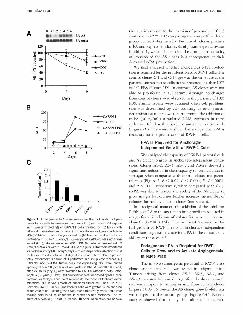
t-PA stimulates the proliferation of untrans-formed cells in vitro.29,30 To study the function of t-PAin pancreatic adenocarcinomas, we have inhibited itsexpression by transfecting CAPAN-1 and SK-PC-1 cells,which overexpress the gene,21 with oligonucleotidescomplementary to the 59 region of the t-PA messengerRNA (mRNA) that encompasses the translational startsite (t-PA-AS). As shown by Western blotting (Figure1A ), the oligonucleotide t-PA-AS at 2 mmol/L caused aninhibition of t-PA protein in CAPAN-1 cells of morethan 90%, as determined by densitometric analysis withnormalization to cells treated with oligonucleotide t-PA-sense. No decrease of t-PA expression was observed incells transfected with t-PA-sense or in sham-transfectedcells (Figure 1A ) or with oligonucleotides complemen-tary to several other regions of the gene (not shown).
Down-regulation of t-PA expression was stable for atleast 72 hours after transfection of t-PA-AS oligonucle-otides. To determine if the reduced t-PA production inCAPAN-1 cells affects their growth, cell proliferationwas monitored for 6 days by MTT. Because the presenceof serum may mask the activities of growth factors, cellswere grown in low (1%) serum. Control cells not trans-fected, sham transfected, or transfected with the t-PA-sense oligonucleotide grew at the same rate and reachedconfluency at day 6 (Figure 1A and data not shown). Incontrast, t-PA-AS–treated cells had a significant decreasein their proliferative capacity at day 6 of growth. Theaverage from 3 independent experiments showed a de-crease in proliferation of 53% compared with sham-transfected cells.
To determine if active t-PA is necessary for prolifera-tion, we treated cells with Pefabloc/t-PA, a syntheticderivative of benzamidine that, at the concentrationused, inhibits t-PA but not u-PA or plasmin activi-ties.21,31 CAPAN-1 and SK-PC-1, expressing high levelsof t-PA,21 and RWP-1, expressing moderately high lev-els of t-PA, were grown in 1% FBS without or with theaddition of Pefabloc/t-PA. All cell lines treated with theinhibitor showed a dose-dependent decrease of prolifer-ation as compared with untreated cells, with inhibitionof proliferation of more than 90% at 30 mmol/L (Figure1B and data not shown). The effect of Pefabloc/t-PA wasnot observed when cells were grown in the presence of10% FBS (not shown). Thus, proteolitically active t-PAis required for proliferation of pancreas cancer cells invitro. In addition to these experiments, the t-PA–ex-pressing cell lines SK-PC-1, CAPAN-1, and RWP-1
(which produce 20, 18, and 5 ng of t-PA z 24 h z mg totalprotein, respectively) as well as BxPC-3 and PANC-1cell lines, which do not express t-PA,21 were injected inthe subcutis of nude mice, and growth was monitoredevery week (Figure 1C ). At 6 weeks after injection, onlythe high t-PA expressor cells SK-PC-1 and CAPAN-1formed tumors. At 14 weeks, tumors were observed onlyin the t-PA–producing SK-PC-1, CAPAN-1, andRWP-1 cells. BxPC-3 and PANC-1 cells showed a la-tency of tumor formation longer than 4 months. Theseresults indicate that there is a correlation between thefaster in vivo growth rate of t-PA–producing cancer cellsand their levels of t-PA expression.
Stable Inhibition of t-PA Expression inRWP-1 Cells by Antisense TranscriptsDecreases Invasion and Cell ProliferationAbilities
To confirm the growth-promoting effect of t-PAon pancreatic adenocarcinomas, we have stably inhibitedits expression by transfecting RWP-1 pancreas tumorcells with a construct expressing t-PA antisense tran-scripts under the control of a constitutive promoter (ASclones). Northern and Western blotting of 4 transfec-tants showed a marked decrease in t-PA levels (AS-2,AS-3, AS-7, AS-29) compared with parental cells andcontrol clones transfected with empty vector (C-1, C-13)(Figure 2A and B).
The levels of t-PA transcripts in the AS clones werereduced by more than 90% with respect to controlclones, as indicated by densitometric analysis with nor-malization to glyceraldehyde-3-phosphate dehydroge-nase signal. The suppression of t-PA expression wasdependent on the expression of antisense transcripts, asdetermined by reverse-transcription polymerase chain re-action with primers specific for the exogenous constructand transcript (not shown). No major variations in thelevels of u-PA mRNA were observed in the differentclones analyzed. A parallel significant reduction of t-PAprotein levels was detected in all AS clones. No majorvariations in the levels of annexin II, the putative mem-brane receptor for t-PA in endothelial cells,32 and plas-minogen activator inhibitor 1, the physiologic inhibitorof t-PA, were detected in the AS clones with respect tocontrol clones.
We have previously shown that t-PA contributes sig-nificantly to the in vitro invasiveness of SK-PC-1 pan-creas cancer cells.21 To confirm this activity for t-PA inRWP-1 cells, 2 control and 3 AS clones were tested fortheir capacity to invade through a reconstituted basalmembrane (Matrigel) in vitro. After 72 hours of incuba-tion, the invasive abilities of clones AS-2, AS-3, andAS-29 were reduced by 51%, 62%, and 42%, respec-
March 2002 TUMOR t-PA PROMOTES GROWTH AND ANGIOGENESIS 809
tively, with respect to the invasion of parental and C-13control cells (P 5 0.02 comparing the group AS with thegroup control) (Figure 2C ). Because all clones produceu-PA and express similar levels of plasminogen activatorinhibitor 1, we concluded that the diminished capacityof invasion of the AS clones is a consequence of theirdecreased t-PA production.
We next analyzed whether endogenous t-PA produc-tion is required for the proliferation of RWP-1 cells. Thecontrol clones C-1 and C-13 grew at the same rate as theparental untransfected cells in the presence of either 10%or 1% FBS (Figure 2D). In contrast, AS clones were notable to proliferate in 1% serum, although no changesfrom control clones were observed in the presence of 10%FBS. Similar results were obtained when cell prolifera-tion was determined by cell counting or total proteindetermination (not shown). Furthermore, the addition ofrt-PA (50 ng/mL) stimulated DNA synthesis in thesecells 2–2.8-fold with respect to untreated control cells(Figure 2E ). These results show that endogenous t-PA isnecessary for the proliferation of RWP-1 cells.
t-PA Is Required for Anchorage-Independent Growth of RWP-1 Cells
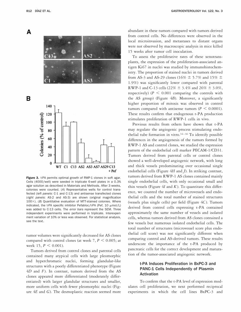
We analyzed the capacity of RWP-1 parental cellsand AS clones to grow in anchorage-independent condi-tions. Clones AS-2, AS-3, AS-7, and AS-29 showed asignificant reduction in their capacity to form colonies insoft agar when compared with control clones and paren-tal cells (Figure 3; P , 0.02, P , 0.004, P , 0.0001,and P , 0.01, respectively, when compared with C-1).rt-PA was able to restore the ability of the AS clones togrow in agar but did not further increase the number ofcolonies formed by control clones (not shown).
In a reciprocal manner, the addition of the inhibitorPefabloc/t-PA to the agar-containing medium resulted ina significant inhibition of colony formation in controlclone C-13 (P 5 0.024). Thus, active t-PA is required forfull growth of RWP-1 cells in anchorage-independentconditions, suggesting a role for t-PA in the tumorigenicability of these cells.33
Endogenous t-PA Is Required for RWP-1Cells to Grow and to Activate Angiogenesisin Nude Mice
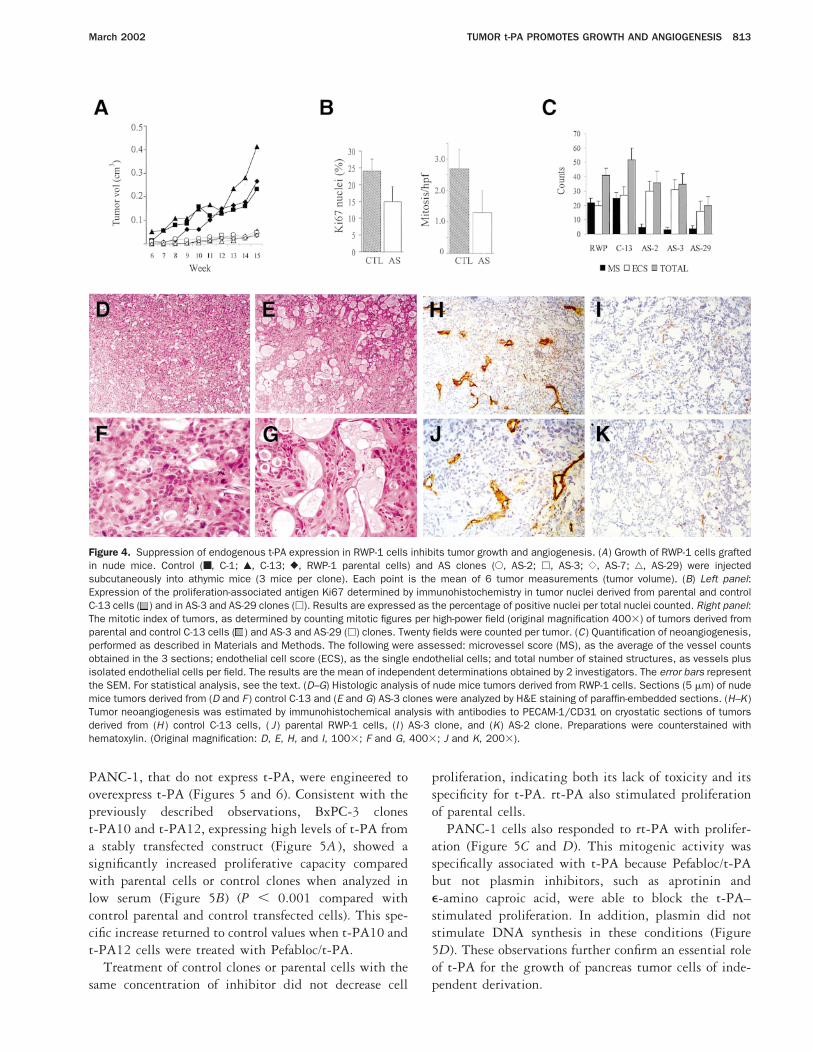
The in vivo tumorigenic potential of RWP-1 ASclones and control cells was tested in athymic mice.Tumors arising from clones AS-2, AS-3, AS-7, andAS-29 consistently showed a significantly slower growthrate with respect to tumors arising from control clones(Figure 4). At 15 weeks, the AS clones grew fivefold lesswith respect to the control group (Figure 4A ). Kineticanalyses showed that at any time after cell xenograft,
Figure 1. Endogenous t-PA is necessary for the proliferation of pan-creas tumor cells in low-serum medium. (A) Upper panel: t-PA expres-sion (Western blotting) of CAPAN-1 cells treated for 72 hours withdifferent concentrations (mmol/L) of the antisense oligonucleotide tot-PA (t-PA-AS) or control oligonucleotide (t-PA-sense) and a fixed con-centration of DOTAP (8 mmol/L). Lower panel: CAPAN-1 cells not trans-fected (CTL), sham-transfected (DOT, DOTAP only), or treated with 2mmol/L t-PA-AS or with 2 mmol/L t-PA-sense plus DOTAP were monitoredfor proliferation by MTT every 2 days with a change of transfection mix at72 hours. Results obtained at days 4 and 6 are shown. One represen-tative experiment is shown of 3 performed in quintuplicate replicas. (B)CAPAN-1 and SK-PC-1 tumor cells overexpressing t-PA were platedsparsely (1.5 3 104/well) in 24-well plates in DMEM plus 10% FBS and,after 24 hours (day 1), were switched to 1% FBS without or with Pefab-loc/t-PA (30 mmol/L, Pef). Cell proliferation was monitored by MTT incor-poration for 8 days. Each point represents the mean of triplicate deter-minations. (C) In vivo growth of pancreas tumor cell lines. SK-PC-1,CAPAN-1, RWP-1, BxPC-3, and PANC-1 cells were grafted in the subcutisof athymic mice. Tumor growth was monitored every week and tumorvolume calculated as described in Materials and Methods. The re-sults at 6 weeks (h) and 14 weeks (■) after inoculation are shown.
810 DIAZ ET AL. GASTROENTEROLOGY Vol. 122, No. 3
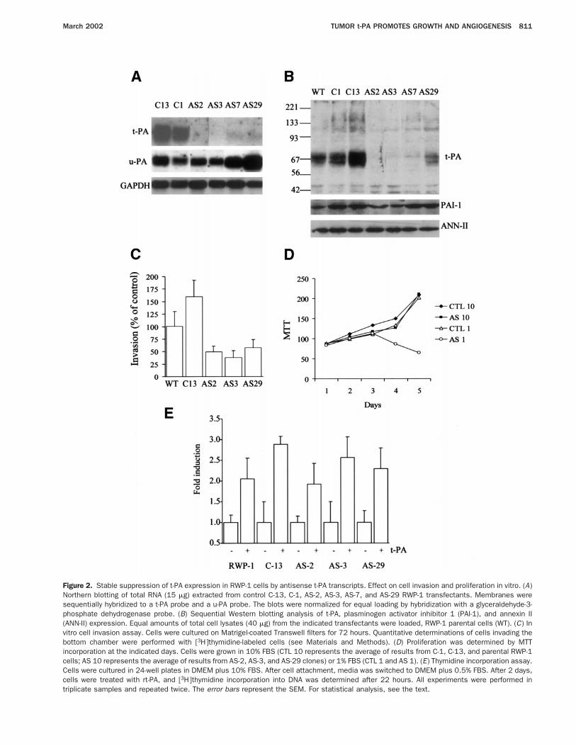
Figure 2. Stable suppression of t-PA expression in RWP-1 cells by antisense t-PA transcripts. Effect on cell invasion and proliferation in vitro. (A)Northern blotting of total RNA (15 mg) extracted from control C-13, C-1, AS-2, AS-3, AS-7, and AS-29 RWP-1 transfectants. Membranes weresequentially hybridized to a t-PA probe and a u-PA probe. The blots were normalized for equal loading by hybridization with a glyceraldehyde-3-phosphate dehydrogenase probe. (B) Sequential Western blotting analysis of t-PA, plasminogen activator inhibitor 1 (PAI-1), and annexin II(ANN-II) expression. Equal amounts of total cell lysates (40 mg) from the indicated transfectants were loaded, RWP-1 parental cells (WT). (C) Invitro cell invasion assay. Cells were cultured on Matrigel-coated Transwell filters for 72 hours. Quantitative determinations of cells invading thebottom chamber were performed with [3H]thymidine-labeled cells (see Materials and Methods). (D) Proliferation was determined by MTTincorporation at the indicated days. Cells were grown in 10% FBS (CTL 10 represents the average of results from C-1, C-13, and parental RWP-1cells; AS 10 represents the average of results from AS-2, AS-3, and AS-29 clones) or 1% FBS (CTL 1 and AS 1). (E) Thymidine incorporation assay.Cells were cultured in 24-well plates in DMEM plus 10% FBS. After cell attachment, media was switched to DMEM plus 0.5% FBS. After 2 days,cells were treated with rt-PA, and [3H]thymidine incorporation into DNA was determined after 22 hours. All experiments were performed intriplicate samples and repeated twice. The error bars represent the SEM. For statistical analysis, see the text.
March 2002 TUMOR t-PA PROMOTES GROWTH AND ANGIOGENESIS 811
tumor volumes were significantly decreased for AS clonescompared with control clones (at week 7, P , 0.005; atweek 15, P , 0.001).
Tumors derived from control clones and parental cellscontained many atypical cells with large pleomorphicand hyperchromatic nuclei, forming glandular-likestructures with a poorly differentiated phenotype (Figure4D and F ). In contrast, tumors derived from the ASclones appeared more differentiated (moderately differ-entiated) with larger glandular structures and smaller,more uniform cells with fewer pleomorphic nuclei (Fig-ure 4E and G). The desmoplastic reaction seemed more
abundant in these tumors compared with tumors derivedfrom control cells. No differences were observed in thelocal microinvasion, and metastases to distant organswere not observed by macroscopic analysis in mice killed15 weeks after tumor cell inoculation.
To assess the proliferative rates of these xenotrans-plants, the expression of the proliferation-associated an-tigen Ki67 in nuclei was studied by immunohistochem-istry. The proportion of stained nuclei in tumors derivedfrom AS-3 and AS-29 clones (16% 6 5.7% and 15% 61.9%) was significantly lower compared with parentalRWP-1 and C-13 cells (22% 6 3.4% and 26% 6 3.0%,respectively) (P , 0.001 comparing the controls withthe AS group) (Figure 4B). Moreover, a significantlyhigher proportion of mitosis was observed in controltumors compared with antisense tumors (P , 0.0001).These results confirm that endogenous t-PA productionstimulates proliferation of RWP-1 cells in vivo.
Previous results from others have shown that t-PAmay regulate the angiogenic process stimulating endo-thelial tube formation in vitro.34–36 To identify possibledifferences in the angiogenesis of the tumors formed byRWP-1 AS and control clones, we studied the expressionpattern of the endothelial cell marker PECAM-1/CD31.Tumors derived from parental cells or control clonesshowed a well-developed angiogenic network, with longand thick vessels predominating over occasional singleendothelial cells (Figure 4H and J). In striking contrast,tumors derived from RWP-1 AS clones contained mainlysingle endothelial cells, with only occasional small andthin vessels (Figure 4I and K ). To quantitate this differ-ence, we counted the number of microvessels and endo-thelial cells and the total number of stained structures(vessels plus single cells) per field (Figure 4C ). Tumorsderived from control cells expressing t-PA containedapproximately the same number of vessels and isolatedcells, whereas tumors derived from AS clones contained afew vessels but numerous isolated endothelial cells. Thetotal number of structures (microvessel score plus endo-thelial cell score) was not significantly different whencomparing control and AS-derived tumors. These resultsunderscore the importance of the t-PA produced bypancreatic cells for the correct development and matura-tion of the tumor-associated angiogenic network.
t-PA Induces Proliferation in BxPC-3 andPANC-1 Cells Independently of PlasminActivation
To confirm that the t-PA level of expression mod-ulates cell proliferation, we next performed reciprocalexperiments in which the cell lines BxPC-3 and
Figure 3. t-PA permits optimal growth of RWP-1 clones in soft agar.Cells (4000/well) were seeded in triplicate 6-well plates in a 0.3%agar solution as described in Materials and Methods. After 3 weeks,colonies were counted. (A) Representative wells for control trans-fected (left panels: C-1 and C-13) and antisense transfected clones(right panels: AS-2 and AS-3) are shown (original magnification1003). (B) Quantitative evaluation of MTT-stained colonies. Whereindicated, the t-PA specific inhibitor Pefabloc/t-PA (Pef, 10 mmol/L)was added to C-13 cells. The error bars represent the SEM. Threeindependent experiments were performed in triplicate. Interexperi-ment variation of 10% or less was observed. For statistical analysis,see the text.
812 DIAZ ET AL. GASTROENTEROLOGY Vol. 122, No. 3
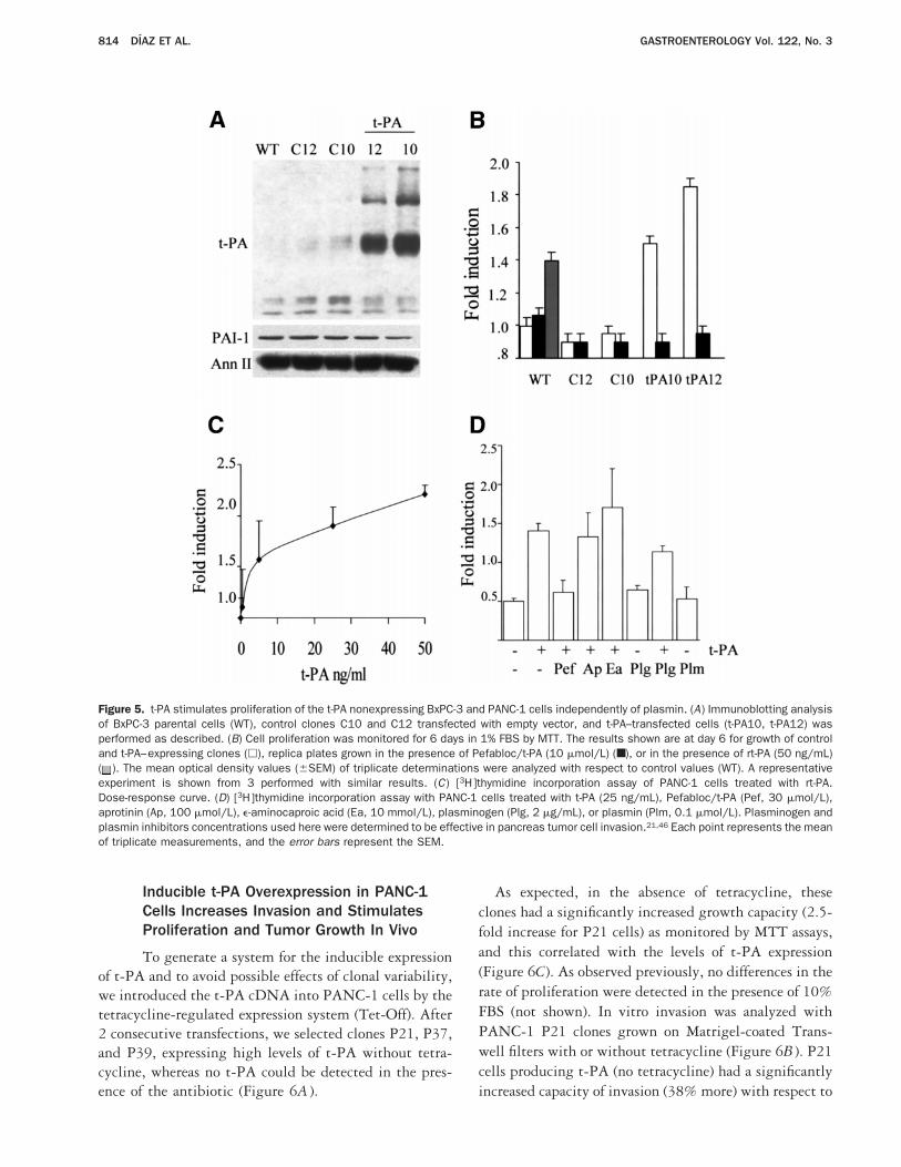
PANC-1, that do not express t-PA, were engineered tooverexpress t-PA (Figures 5 and 6). Consistent with thepreviously described observations, BxPC-3 clonest-PA10 and t-PA12, expressing high levels of t-PA froma stably transfected construct (Figure 5A ), showed asignificantly increased proliferative capacity comparedwith parental cells or control clones when analyzed inlow serum (Figure 5B) (P , 0.001 compared withcontrol parental and control transfected cells). This spe-cific increase returned to control values when t-PA10 andt-PA12 cells were treated with Pefabloc/t-PA.
Treatment of control clones or parental cells with thesame concentration of inhibitor did not decrease cell
proliferation, indicating both its lack of toxicity and itsspecificity for t-PA. rt-PA also stimulated proliferationof parental cells.
PANC-1 cells also responded to rt-PA with prolifer-ation (Figure 5C and D). This mitogenic activity wasspecifically associated with t-PA because Pefabloc/t-PAbut not plasmin inhibitors, such as aprotinin ande-amino caproic acid, were able to block the t-PA–stimulated proliferation. In addition, plasmin did notstimulate DNA synthesis in these conditions (Figure5D). These observations further confirm an essential roleof t-PA for the growth of pancreas tumor cells of inde-pendent derivation.
Figure 4. Suppression of endogenous t-PA expression in RWP-1 cells inhibits tumor growth and angiogenesis. (A) Growth of RWP-1 cells graftedin nude mice. Control (■, C-1; Œ, C-13; }, RWP-1 parental cells) and AS clones (E, AS-2; h, AS-3; {, AS-7; ‚, AS-29) were injectedsubcutaneously into athymic mice (3 mice per clone). Each point is the mean of 6 tumor measurements (tumor volume). (B) Left panel:Expression of the proliferation-associated antigen Ki67 determined by immunohistochemistry in tumor nuclei derived from parental and controlC-13 cells ( ) and in AS-3 and AS-29 clones (h). Results are expressed as the percentage of positive nuclei per total nuclei counted. Right panel:The mitotic index of tumors, as determined by counting mitotic figures per high-power field (original magnification 4003) of tumors derived fromparental and control C-13 cells ( ) and AS-3 and AS-29 (h) clones. Twenty fields were counted per tumor. (C) Quantification of neoangiogenesis,performed as described in Materials and Methods. The following were assessed: microvessel score (MS), as the average of the vessel countsobtained in the 3 sections; endothelial cell score (ECS), as the single endothelial cells; and total number of stained structures, as vessels plusisolated endothelial cells per field. The results are the mean of independent determinations obtained by 2 investigators. The error bars representthe SEM. For statistical analysis, see the text. (D–G) Histologic analysis of nude mice tumors derived from RWP-1 cells. Sections (5 mm) of nudemice tumors derived from (D and F ) control C-13 and (E and G) AS-3 clones were analyzed by H&E staining of paraffin-embedded sections. (H–K)Tumor neoangiogenesis was estimated by immunohistochemical analysis with antibodies to PECAM-1/CD31 on cryostatic sections of tumorsderived from (H ) control C-13 cells, ( J ) parental RWP-1 cells, (I ) AS-3 clone, and (K) AS-2 clone. Preparations were counterstained withhematoxylin. (Original magnification: D, E, H, and I, 1003; F and G, 4003; J and K, 2003).
March 2002 TUMOR t-PA PROMOTES GROWTH AND ANGIOGENESIS 813
Inducible t-PA Overexpression in PANC-1Cells Increases Invasion and StimulatesProliferation and Tumor Growth In Vivo
To generate a system for the inducible expressionof t-PA and to avoid possible effects of clonal variability,we introduced the t-PA cDNA into PANC-1 cells by thetetracycline-regulated expression system (Tet-Off). After2 consecutive transfections, we selected clones P21, P37,and P39, expressing high levels of t-PA without tetra-cycline, whereas no t-PA could be detected in the pres-ence of the antibiotic (Figure 6A ).
As expected, in the absence of tetracycline, theseclones had a significantly increased growth capacity (2.5-fold increase for P21 cells) as monitored by MTT assays,and this correlated with the levels of t-PA expression(Figure 6C). As observed previously, no differences in therate of proliferation were detected in the presence of 10%FBS (not shown). In vitro invasion was analyzed withPANC-1 P21 clones grown on Matrigel-coated Trans-well filters with or without tetracycline (Figure 6B ). P21cells producing t-PA (no tetracycline) had a significantlyincreased capacity of invasion (38% more) with respect to
Figure 5. t-PA stimulates proliferation of the t-PA nonexpressing BxPC-3 and PANC-1 cells independently of plasmin. (A) Immunoblotting analysisof BxPC-3 parental cells (WT), control clones C10 and C12 transfected with empty vector, and t-PA–transfected cells (t-PA10, t-PA12) wasperformed as described. (B) Cell proliferation was monitored for 6 days in 1% FBS by MTT. The results shown are at day 6 for growth of controland t-PA–expressing clones (h), replica plates grown in the presence of Pefabloc/t-PA (10 mmol/L) (■), or in the presence of rt-PA (50 ng/mL)( ). The mean optical density values (6SEM) of triplicate determinations were analyzed with respect to control values (WT). A representativeexperiment is shown from 3 performed with similar results. (C) [3H]thymidine incorporation assay of PANC-1 cells treated with rt-PA.Dose-response curve. (D) [3H]thymidine incorporation assay with PANC-1 cells treated with t-PA (25 ng/mL), Pefabloc/t-PA (Pef, 30 mmol/L),aprotinin (Ap, 100 mmol/L), e-aminocaproic acid (Ea, 10 mmol/L), plasminogen (Plg, 2 mg/mL), or plasmin (Plm, 0.1 mmol/L). Plasminogen andplasmin inhibitors concentrations used here were determined to be effective in pancreas tumor cell invasion.21,46 Each point represents the meanof triplicate measurements, and the error bars represent the SEM.
814 DIAZ ET AL. GASTROENTEROLOGY Vol. 122, No. 3
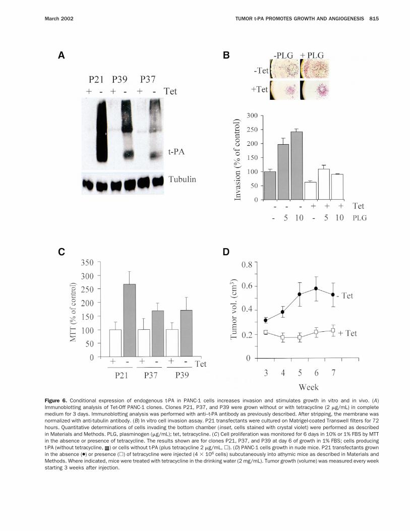
Figure 6. Conditional expression of endogenous t-PA in PANC-1 cells increases invasion and stimulates growth in vitro and in vivo. (A)Immunoblotting analysis of Tet-Off PANC-1 clones. Clones P21, P37, and P39 were grown without or with tetracycline (2 mg/mL) in completemedium for 3 days. Immunoblotting analysis was performed with anti–t-PA antibody as previously described. After stripping, the membrane wasnormalized with anti-tubulin antibody. (B) In vitro cell invasion assay. P21 transfectants were cultured on Matrigel-coated Transwell filters for 72hours. Quantitative determinations of cells invading the bottom chamber (inset, cells stained with crystal violet) were performed as describedin Materials and Methods. PLG, plasminogen (mg/mL); tet, tetracycline. (C) Cell proliferation was monitored for 6 days in 10% or 1% FBS by MTTin the absence or presence of tetracycline. The results shown are for clones P21, P37, and P39 at day 6 of growth in 1% FBS; cells producingt-PA (without tetracycline, ) or cells without t-PA (plus tetracycline 2 mg/mL, h). (D) PANC-1 cells growth in nude mice. P21 transfectants grownin the absence (●) or presence (h) of tetracycline were injected (4 3 106 cells) subcutaneously into athymic mice as described in Materials andMethods. Where indicated, mice were treated with tetracycline in the drinking water (2 mg/mL). Tumor growth (volume) was measured every weekstarting 3 weeks after injection.
March 2002 TUMOR t-PA PROMOTES GROWTH AND ANGIOGENESIS 815
the same cells with tetracycline. Addition of plasmino-gen greatly stimulated invasion of cells in the t-PA–producing state although only poorly stimulated inva-sion of cells with no t-PA, indicating that plasmin,generated via t-PA, is a major activator of invasion inthese cells.
Finally, to confirm the role of t-PA in the ability ofpancreas cells to form tumors in vivo, PANC-1 P21clones were grown in athymic mice and tumor growthwas monitored for 7 weeks. As expected, tumors express-ing t-PA in mice that received no tetracycline showed asignificantly faster growth with respect to tumors arisingin mice treated with the antibiotic (Figure 6D). At week7 after inoculation, P21 tumors without tetracycline hadgrown 2.3-fold more than control P21 tumors, confirm-ing our previous findings.
DiscussionThere is much evidence that the plasminogen
activator system plays a major role in tumor cell inva-sion. The results shown here indicate that t-PA, inaddition to being required for a full invasive phenotypeof cancer cells, is necessary for the growth of differentpancreas tumor cell lines in vitro and in vivo and isrequired for tumor-associated neoangiogenesis in vivo.
The mitogenic effect of t-PA on pancreas tumor cellsis shown here in vitro and in vivo by a number ofcomplementary experimental approaches performed oncells with different genetic backgrounds. In 1 set ofexperiments, the inhibition of expression of the endog-enous t-PA by antisense oligonucleotides in CAPAN-1cells or by an antisense expression vector stably inte-grated into RWP-1 cells resulted in a marked reductionof the proliferation capacity on plastic and in soft agar.These capacities were rescued to parental cell values inRWP-1 AS clones by adding exogenous t-PA back intothe culture medium, thus arguing that suppression ofendogenous t-PA expression is the only responsible factorfor the compromised growth of the AS clones. Consistentwith these observations, blocking t-PA proteolytic activ-ity with the specific inhibitor Pefabloc/t-PA, used atconcentrations that do not inactivate u-PA or plasmin,31
also inhibited proliferation of several pancreas tumor celllines expressing t-PA. In reciprocal experiments, we haveshown that the addition of rt-PA is mitogenic forRWP-1, BxPC-3, and PANC-1 cells and that the stableexpression of the full-length cDNA in BxPC-3 andPANC-1 cells caused an increase in the proliferativeabilities of these cells, which could be blocked by theinhibition of the t-PA proteolytic activity. These find-ings are strongly supported by the results in vivo;
RWP-1 AS clones form tumors significantly smaller thancontrols and, reciprocally, t-PA–expressing PANC-1 P21clones form faster-growing tumors than the same cellsnot expressing t-PA. The mitogenic activity of t-PA invitro was only detected in cells growing at low serumconcentration, indicating that serum is a source of activegrowth factors that may rescue low endogenous produc-tion of such factors.2,37 The impaired growth of antisenset-PA clones in the subcutis in vivo, an environment withlimited growth factor supply, further reinforces the no-tion that the endogenous production of t-PA is therelevant factor for the growth of RWP-1 cells.
t-PA itself, but not plasmin, the major product of thecatalytic activity of t-PA, seems to be responsible for thestimulation of growth in the condition tested, as consis-tently shown by the specific inhibition produced byPefabloc/t-PA but not by plasmin inhibitors. A numberof reports by other laboratories are in agreement with ourpresent observations.30,34,38,39 On the other hand, ourresults do not allow us to conclude whether this is adirect effect, or the result of processing or induction byt-PA of other factors that would be more directly in-volved in the observed mitogenic effects. t-PA has beeninvolved in the proteolytic activation of several growthfactors8,10,12; thus, it is conceivable that its growth-stimulating effect on the cell lines used in this study ismediated by 1 or more peptide growth factors. In addi-tion, in smooth muscle cells, platelet-derived growthfactor stimulates proliferation and activates t-PA expres-sion. In t-PA–depleted smooth muscle cells, platelet-derived growth factor is unable to stimulate cell prolif-eration, suggesting that t-PA can operate through otherless-known mechanisms.38
Taken together, our observations indicate a prominentrole for t-PA in the maintenance of pancreas tumor cellsin a state of proliferative competence. An increasing bodyof evidence indicates that proteases of the extracellularmatrix with well-established roles in tumor invasion,such as stromelysin 3, stromelysin 1, and matrilysin, playin addition a crucial role in early stages of tumorigenesis,promoting tumor formation and/or growth.40–42 Evi-dence for a role of t-PA or u-PA and/or plasmin in theprocess of tumor growth varies with particular experi-mental models and individual tumor types. Thus, abla-tion of u-PA but not of t-PA causes resistance to mela-noma induction,14 the development of mammary tumorsby the viral polyoma middle T antigen in plasminogen-deficient mice is not appreciably altered,43 endogenouslyproduced u-PA or t-PA stimulate melanoma and neuro-blastoma cell proliferation in vitro,12,44 and the com-bined loss of t-PA and u-PA activities results in a sig-
816 DIAZ ET AL. GASTROENTEROLOGY Vol. 122, No. 3
nificant reduction in proliferation rates of transformedendothelial cells in vitro and a reduced efficiency intumor formation.15 These observations lend support toour conclusion, based on the present direct evidence, thatendogenous production of t-PA is necessary for the op-timum growth of pancreas cancer cells in vitro and invivo.
The second relevant new finding reported here is theeffect of t-PA on the formation of the angiogenic net-work associated with tumor growth. Eventually, thegrowth of a tumor depends on the availability of nutri-ents in the local microenvironment, which in turn re-quires the development of an adequate vascular network.Formation of functional microvessels is a stepwise processin tumor progression that includes endothelial cell mi-gration and/or proliferation.45 In pancreatic tumors, pro-duction of angiogenic factors by neoplastic cells, includ-ing epidermal growth factor, fibroblast growth factor,platelet-derived growth factor, hepatocyte growth factor,and PAs,21,46,47 can stimulate endothelial cell prolifera-tion and migration. In this way, a paracrine loop ofreciprocal activation could be established between tumorand endothelial cells, which permits neovascularizationand, together with the perfusion of nutrients and oxygen,permits growth and progression of the tumor. Studies invitro by others have indicated that t-PA regulates angio-genesis by stimulating endothelial cell tube forma-tion.34–36 Our observations provide the first in vivoevidence showing that the tumor-produced t-PA is re-quired for the correct development of angiogenesis at thetumor site. Although the proportion of isolated endothe-lial cells in the tumors derived from RWP-1 control andAS clones is similar, the well-developed vascular networkof the control tumors stands in marked contrast with thescant vessels of the AS tumors. These results indicate thatthe tumor-produced t-PA is required for the phase ofendothelial tube formation and/or maturation but not forthe recruitment of endothelial cells. The recent observa-tion that t-PA might be the target protein inactivated bymaspin48,49 and by angiostatin,50 both effective inhibi-tors of angiogenesis, supports our findings. The mecha-nisms by which t-PA would facilitate or induce theformation of tumor-associated vessels are not clear atpresent. t-PA may facilitate the availability of growthfactors present in the extracellular matrix for endothelialcell usage,11,51 or it may control matrix degradation toallow the formation of capillary structures.37
Tumors derived from RWP-1 AS clones were mod-erately differentiated with respect to control tumors(poorly differentiated), suggesting that a change in theendogenous t-PA levels can affect the cell-differentiation
program. Tumor progression in carcinomas has beensuggested to imply a gradual transition from an epithe-lial- to a mesenchymal-like phenotype; several growthfactors, including hepatocyte growth factor, fibroblastgrowth factor 1, epidermal growth factor, and transform-ing growth factor a, which are also overexpressed inpancreatic adenocarcinomas,47 have been implicated inthis process.52 Thus, it is conceivable that t-PA avail-ability, allowing the activation of latent growth factors,could also affect the cell-differentiation program andtumor progression.
In conclusion, the findings reported in this studysupport an extension of the previously known role oft-PA in tumor invasion to an earlier phase of tumorigen-esis in the pancreas, the initial stages in the growth of theprimary tumor when cell proliferation and activation ofangiogenesis are required. At later stages in which inva-sion and metastasis occur, t-PA and other proteases, suchas u-PA, might be determinant. Future therapeutic strat-egies aimed at the inhibition of t-PA might be usefuladjuvants to use in addition to conventional therapies inthese tumors.
References1. Carmeliet P, Schoonjans L, Kieckens L, Ream B, Degen J, Bron-
son R, De Vos R, van den Oord JJ, Collen D, Mulligan RC.Physiological consequences of loss of plasminogen activatorgene function in mice. Nature 1994;36:419–424.
2. Liotta LA, Rao CN, Barsky SH. Tumor invasion and the extracel-lular matrix. Lab Invest 1983;49:636–649.
3. DeClerck YA, Imren S, Montgomery AM, Mueller BM, Reisfeld RA,Laug WE. Proteases and protease inhibitors in tumor progres-sion. Adv Exp Med Biol 1997;425:89–97.
4. Carmeliet P, Moons L, Lijnen R, Baes M, Lemaitre V, Tipping P,Drew A, Eeckhout Y, Shapiro S, Lupu F, Collen D. Urokinase-generated plasmin activates matrix metalloproteinases duringaneurysm formation. Nat Genet 1997;17:439–444.
5. Dano K, Andreasen PA, Grondahl-Hansen J, Kristensen P, NielsenLS, Skriver L. Plasminogen activators, tissue degradation, andcancer. Adv Cancer Res 1985;44:139–266.
6. Ossowski L. Plasminogen activator dependent pathways in thedissemination of human tumor cells in the chick embryo. Cell1988;52:321–328.
7. Naldini L, Tamagnone L, Vigna E, Sachs M, Hartmann G, Birch-meier W, Daikuhara Y, Tsubouchi H, Blasi F, Comoglio P. Extra-cellular proteolytic cleavage by urokinase is required for activa-tion of hepatocyte growth factor/scatter factor. EMBO J 1992;11:4825–4833.
8. Mars WM, Zarnegar R, Michalopoulos GK. Activation of hepato-cyte growth factor by the plasminogen activators uPA and tPA.Am J Pathol 1993;143:949–958.
9. Sato Y, Tsuboi R, Lyons R, Moses H, Rifkin DB. Characterizationof the activation of latent TGF-beta by co-cultures of endothelialcells and pericytes or smooth muscle cells: a self-regulatingsystem. J Cell Biol 1990;111:757–763.
10. Houck KA, Leung DW, Rowland AM, Winer J, Ferrara N. Dualregulation of vascular endothelial growth factor bioavailability bygenetic and proteolytic mechanisms. J Biol Chem 1992;267:26031–26037.
11. Flaumenhaft R, Moscatelli D, Saksela O, Rifkin DB. Role of
March 2002 TUMOR t-PA PROMOTES GROWTH AND ANGIOGENESIS 817
extracellular matrix in the action of basic fibroblast growth factor:matrix as a source of growth factor for long-term stimulation ofplasminogen activator production and DNA synthesis. J CellPhysiol 1989;140:75–81.
12. Menouny M, Binoux M, Babajko S. Role of insulin-like growthfactor binding protein-2 and its limited proteolysis in neuroblas-toma cell proliferation: modulation by transforming growth factor-beta and retinoic acid. Endocrinology 1997;138:683–690.
13. Andreasen PA, Kjoller L, Christensen L, Duffy MJ. The urokinase-type plasminogen activator system in cancer metastasis: a re-view. Int J Cancer 1997;72:1–22.
14. Shapiro RL, Duquette JG, Roses DF, Nunes I, Harris MN, KaminoH, Wilson EL, Rifkin DB. Induction of primary cutaneous melano-cytic neoplasms in urokinase-type plasminogen activator (uPA)-deficient and wild-type mice: cellular blue nevi invade but do notprogress to malignant melanoma in uPA-deficient animals. Can-cer Res 1996;56:3597–3604.
15. Sabapathy KT, Pepper MS, Kiefer F, Mohle-Steinlein U, Tacchini-Cottier F, Fetka I, Breier G, Risau W, Carmeliet P, Montesano R,Wagner EF. Polyoma middle T-induced vascular tumor formation:the role of the plasminogen activator/plasmin system. J Cell Biol1997;137:953–963.
16. Sier CF, Vloedgraven HJ, Griffioen G, Ganesh S, Nagengast FM,Lamers CB, Verspaget HW. Plasminogen activators and inhibitortype 1 in neoplastic colonic tissue from patients with familialadenomatous polyposis. Br J Cancer 1995;71:393–396.
17. Kim SJ, Shiba E, Kobayashi T, Yayoi E, Furukawa J, Takatsuka Y,Shin E, Koyama H, Inaji H, Takai S. Prognostic impact of uroki-nase-type plasminogen activator (PA), PA inhibitor type-1, andtissue-type PA antigen levels in node-negative breast cancer: aprospective study on multicenter basis. Clin Cancer Res 1998;4:177–182.
18. Wilson EL, Jacobs P, Dowdle EB. The secretion of plasminogenactivators by human myeloid leukemic cells in vitro. Blood 1983;61:568–574.
19. Sugiura Y, Ma L, Sun B, Shimada H, Laug WE, Seeger RC,DeClerck YA. The plasminogen-plasminogen activator (PA) sys-tem in neuroblastoma: role of PA inhibitor-1 in metastasis. Can-cer Res 1999;59:1327–1336.
20. Paciucci R, Berrozpe G, Tora M, Navarro E, Garcia de Herreros A,Real FX. Isolation of tissue-type plasminogen activator, cathepsinH, and non-specific cross-reacting antigen from SK-PC-1 pan-creas cancer cells using subtractive hybridization. FEBS Lett1996;385:72–76.
21. Paciucci R, Tora M, Diaz VM, Real FX. The plasminogen activatorsystem in pancreas cancer: role of t-PA in the invasive potentialin vitro. Oncogene 1998;16:625–633.
22. Tiberio A, Farina AR, Tacconelli A, Cappabianca L, Gulino A,Mackay AR. Retinoic acid-enhanced invasion through reconsti-tuted basement membrane by human SK-N-SH neuroblastomacells involves membrane-associated tissue-type plasminogen ac-tivator. Int J Cancer 1997;73:740–748.
23. Collen D, Lijnen HR, Bulens F, Vandamme AM, Tulinsky A, NellesL. Biochemical and functional characterization of human tissue-type plasminogen activator variants with mutagenized kringledomains. J Biol Chem 1990;265:12184–12191.
24. Rodrıguez M, Noe V, Alemany C, Miralles A, Bemi V, Caragol I,Ciudad C. Effects of antisense oligonucleotides directed towarddihydrofolate reductase RNA in mammalian cultured cells. Int JCancer 1999;81:785–792.
25. Chomczynski P, Sacchi N. Single-step method of RNA isolation byacid guanidinium thiocyanate-phenol-chloroform extraction. AnalBiochem 1987;162:156–159.
26. Hiraki Y, Rosen OM, Birnbaum MJ. Growth factors rapidly induceexpression of the glucose transporter gene. J Biol Chem 1988;263:13655–13662.
27. Rizzino A. Soft agar growth assays for transforming growth fac-
tors and mitogenic peptides. Methods Enzymol 1987;146:341–352.
28. Weidner N, Semple JP, Welch WR, Folkman J. Tumor angiogene-sis and metastasis–correlation in invasive breast carcinoma.N Engl J Med 1991;324:1–8.
29. Herbert JM, Lamarche I, Prabonnaud V, Dol F, Gauthier T. Tissue-type plasminogen activator is a potent mitogen for human aorticsmooth muscle cells. J Biol Chem 1994;269:3076–3080.
30. De Petro G, Copeta A, Barlati S. Urokinase-type and tissue-typeplasminogen activators as growth factors of human fibroblasts.Exp Cell Res 1994;213:286–294.
31. Sturzebecker J, Markwardt F. Inhibition of tissue-plasminogenactivator by benzamidine derivatives [abstr]. Fibrinolysis 1988;2:49.
32. Hajjar KA, Jacovina AT, Chacko J. An endothelial cell receptor forplasminogen/tissue plasminogen activator. I. Identity with an-nexin II. J Biol Chem 1994;269:21191–21197.
33. Cifone MA, Fidler IJ. Correlation of patterns of anchorage-inde-pendent growth with in vivo behavior of cells from a murinefibrosarcoma. Proc Natl Acad Sci U S A 1980;77:1039–1043.
34. Sato Y, Okamura K, Morimoto A, Hamanaka R, Hamaguchi K,Shimada T, Ono M, Kohno K, Sakata T. Indispensable role oftissue-type plasminogen activator in growth factor-dependenttube formation of human microvascular endothelial cells in vitro.Exp Cell Res 1993;204:223–229.
35. Ito K, Ryuto M, Ushiro S, Ono M, Sugenoya A, Kuraoka A, ShibataY, Kuwano M. Expression of tissue-type plasminogen activatorand its inhibitor couples with development of capillary network byhuman microvascular endothelial cells on Matrigel. J Cell Physiol1995;162:213–224.
36. Schnaper HW, Barnathan ES, Mazar A, Maheshwari S, Ellis S,Cortez SL, Baricos WH, Kleinman HK. Plasminogen activatorsaugment endothelial cell organization in vitro by two distinctpathways. J Cell Physiol 1995;165:107–118.
37. Mignatti P, Rifkin DB. Plasminogen activators and angiogenesis.Curr Top Microbiol Immunol 1996;213:33–50.
38. Herbert JM, Lamarche I, Carmeliet P. Urokinase and tissue-typeplasminogen activator are required for the mitogenic and chemo-tactic effects of bovine fibroblast growth factor and platelet-derived growth factor-BB for vascular smooth muscle cells. J BiolChem 1997;272:23585–23591.
39. Welling TH, Huber TS, Messina LM, Stanley JC. Tissue plasmin-ogen activator increases canine endothelial cell proliferation ratethrough a plasmin-independent, receptor-mediated mechanism.J Surg Res 1996;66:36–42.
40. Wilson CL, Heppner KJ, Labosky PA, Hogan BL, Matrisian LM.Intestinal tumorigenesis is suppressed in mice lacking the met-alloproteinase matrilysin. Proc Natl Acad Sci U S A 1997;94:1402–1407.
41. Masson R, Lefebvre O, Noel A, Fahime ME, Chenard MP, WendingC, Kebers F, LeMeur M, Dierich A, Foidart JM, Basset P, Rio MC.In vivo evidence that stromelysin-3 metalloproteinase contributein a paracrine manner to epithelial cell malignancy. J Cell Biol1998;140:1535–1541.
42. Sternlicht MD, Lochter A, Sympson CJ, Huey B, Rougier JP, GrayJW, Pinkel D, Bissell MJ, Werb Z. The stromal proteinase MMP3/stromelysin-1 promotes mammary carcinogenesis. Cell 1999;98:137–146.
43. Bugge TH, Lund LR, Kombrinck KK, Nielsen BS, Holmback K,Drew AF, Flick MJ, Witt DP, Dano K, Degen JL. Reduced metas-tasis of Polyoma virus middle T antigen-induced mammary cancerin plasminogen-deficient mice. Oncogene 1998;16:3097–3104.
44. Kirchheimer JC, Wojta J, Christ G, Binder BR. Functional inhibitionof endogenously produced urokinase decreases cell proliferationin a human melanoma cell line. Proc Natl Acad Sci U S A 1989;86:5424–5428.
818 DIAZ ET AL. GASTROENTEROLOGY Vol. 122, No. 3
45. Carmeliet P. Mechanisms of angiogenesis and arteriogenesis.Nat Med 2000;6:389–395.
46. Paciucci R, Vila MR, Adell T, Diaz VM, Tora M, Nakamura T, RealFX. Activation of the urokinase plasminogen activator/urokinaseplasminogen activator receptor system and redistribution of E-cadherin are associated with hepatocyte growth factor-inducedmotility of pancreas tumor cells overexpressing Met. Am J Pathol1998;153:201–212.
47. Frazier ML, Longnecker DS. Cell lines of the human and rodentexocrine pancreas. In: Go VLW, DiMagno EP, Gardner JD, Leb-enthal E, Reber HA, Scheele GA, eds. The pancreas. Biology,pathobiology, and disease. 2nd ed. New York: Raven, 1993:565–574.
48. Zhang M, Volpert O, Shi YH, Bouck N. Maspin is an angiogenesisinhibitor. Nat Med 2000;6:196–199.
49. Sheng S, Truong B, Fredrickson D, Wu R, Pardee AB, Sager R.Tissue-type plasminogen activator is a target of the tumor sup-pressor gene maspin. Proc Natl Acad Sci U S A 1998;95:499–504.
50. Stack MS, Gately S, Bafetti LM, Enghild JJ, Soff GA. Angiostatininhibits endothelial and melanoma cellular invasion by blockingmatrix-enhanced plasminogen activation. Biochem J 1999;340:77–84.
51. Rifkin DB, Gleizes PE, Harpel J, Nunes I, Munger J, Mazzieri R,Noguera I. Plasminogen/plasminogen activator and growth factoractivation. Ciba Found Symp 1997;212:105–118.
52. Thiery JP, Chopin D. Epithelial cell plasticity in development andtumor progression. Cancer Metastasis Rev 1999;18:31–42.
Received March 15, 2001. Accepted November 8, 2001.Address requests for reprints to: Rosanna Paciucci, Ph.D., Unitat de
Recerca Biomedica, Hospital Materno-Infantil Vall d’Hebron, Pg.Vall d’Hebron, 119-129, 08035 Barcelona, Spain. e-mail: [email protected]; fax: (34) 93-4894064.
Supported in part by grants from Comision Interministerial de Cien-cia y Tecnologıa (SAF97-0084 and SAF2000-0195), a grant from theFundacio Marato-TV3, and a grant from the Fondos de InvestigacionSanitaria (FIS 99/0897). Dr. Dıaz was a recipient of a Beca FundacionCientıfica AECC and of a Beca de Formacion del Personal Investigador(Ministerio de Educacion y Cultura).
The authors thank C. Lopez-Otın for critically reviewing the manu-script; L. Calderon, N. Toran, I. De Torres, and J. Vila for valuablecontributions; and the investigators mentioned in the text for providingreagents.
March 2002 TUMOR t-PA PROMOTES GROWTH AND ANGIOGENESIS 819
Copyright © 2022 FDOKUMEN