Vitamins C and E attenuate plasminogen activator inhibitor-1 (PAI-1) expression in a...
9
Cardiovascular Research 49 (2001) 484–492 www.elsevier.com / locate / cardiores www.elsevier.nl / locate / cardiores Vitamins C and E attenuate plasminogen activator inhibitor-1 (PAI-1) expression in a hypercholesterolemic porcine model of angioplasty a b c b b Josune Orbe , Jose A. Rodriguez , Alfonso Calvo , Andres Grau , Maria S. Belzunce , b a, * ´ Diego Martinez-Caro , Jose A. Paramo a Laboratory of Atherosclerosis Research, School of Medicine, University of Navarra, Pamplona, Spain b Department of Cardiology, School of Medicine, University of Navarra, Pamplona, Spain c Department of Histology, School of Medicine, University of Navarra, Pamplona, Spain Received 15 June 2000; accepted 18 October 2000 Abstract Background: The plasminogen activator inhibitor-1 (PAI-1), which modulates fibrinolysis and cell migration, may influence proteolysis and neointimal formation in the arterial wall contributing to restenosis after vascular injury. Antioxidants have been proposed as inhibiting multiple proatherogenic events.We explore the effect of vitamins C and E on PAI-1 expression in an experimental model of angioplasty in hypercholesterolemic pigs. Methods and results: A total of 44 Yucatan minipigs were divided into three diet groups: a normal-cholesterol (NC), a high-cholesterol (HC), and a high-cholesterol plus vitamins C1E (HCV) group. Balloon injury was induced in the right internal iliac artery 4 weeks after initiation of either dietary regimen, and plasma and tissue samples were taken at different time periods to measure PAI-1 activity and vascular inhibitor expression. The cholesterol-rich diet induced an increased in vascular PAI-1 expression in the intima, media and adventitia which was markedly reduced in the HCV group. After injury, severe structural changes were observed in NC and HC animals associated with increased systemic PAI-1 activity ( P,0.001) and local PAI-1 expression being more intense in HC group. Vitamins C and E significantly reduced plasma PAI-1 activity ( P50.018) and attenuated the inhibitor expression as compared with HC. Conclusions: This experimental study in a porcine model of hypercholesterolemia demonstrates that vitamins C and E reduce local and systemic PAI-1 induced after angioplasty as well as the hypercholesterolemia-induced vascular PAI-1. 2001 Elsevier Science B.V. All rights reserved. Keywords: Atherosclerosis; Cholesterol; Angioplasty; Hemostasis; Restenosis 1. Introduction An injury-related healing response mediated by smooth muscle cell (SMC) proliferation and neointima formation, Percutaneous angioplasty is an established treatment for leading to luminal narrowing, has been considered an atherothrombosis. Despite a primary success rate, angio- essential factor in the development of late restenosis. plasty induces early reocclusion in 3–13% and late re- Recent data also indicate that vascular remodeling is an stenosis in more than 30% of patients treated. The mecha- important pathophysiological mechanism for restenosis [4– nisms promoting these lesions are not completely under- 7]. stood and different pharmacological strategies have not The plasminogen activator / plasmin system has been proved effective in preventing these complications al- implicated in several cardiovascular disorders, including though the introduction of intracoronary stents has reduced wound healing after vascular reconstruction as suggested their incidence in recent years [1–3]. from data obtained in PAI-1 knockout mice [8,9]. Recent observations indicate that components of this system may influence the process of restenosis by either promoting or ´ *Corresponding author. Present address: Clınica Universitaria, Servicio inhibiting the development of the neointima [10–12]. PAI- ´ Hematologıa, C / Pio XII s / n, 31008-Pamplona, Spain. Tel.: 134-948- 296-397; fax: 134-948-296-500. ´ E-mail address: [email protected] (J.A. Paramo). Time for primary review 21 days. 0008-6363 / 01 / $ – see front matter 2001 Elsevier Science B.V. All rights reserved. PII: S0008-6363(00)00260-1 by guest on June 15, 2016 Downloaded from
-
Upload
independent -
Category
Documents
-
view
0 -
download
0
Transcript of Vitamins C and E attenuate plasminogen activator inhibitor-1 (PAI-1) expression in a...
Cardiovascular Research 49 (2001) 484–492www.elsevier.com/ locate /cardiores
www.elsevier.nl / locate /cardiores
Vitamins C and E attenuate plasminogen activator inhibitor-1 (PAI-1)expression in a hypercholesterolemic porcine model of angioplasty
a b c b bJosune Orbe , Jose A. Rodriguez , Alfonso Calvo , Andres Grau , Maria S. Belzunce ,b a ,*´Diego Martinez-Caro , Jose A. Paramo
aLaboratory of Atherosclerosis Research, School of Medicine, University of Navarra, Pamplona, SpainbDepartment of Cardiology, School of Medicine, University of Navarra, Pamplona, Spain
cDepartment of Histology, School of Medicine, University of Navarra, Pamplona, Spain
Received 15 June 2000; accepted 18 October 2000
Abstract
Background: The plasminogen activator inhibitor-1 (PAI-1), which modulates fibrinolysis and cell migration, may influenceproteolysis and neointimal formation in the arterial wall contributing to restenosis after vascular injury. Antioxidants have been proposedas inhibiting multiple proatherogenic events. We explore the effect of vitamins C and E on PAI-1 expression in an experimental model ofangioplasty in hypercholesterolemic pigs. Methods and results: A total of 44 Yucatan minipigs were divided into three diet groups: anormal-cholesterol (NC), a high-cholesterol (HC), and a high-cholesterol plus vitamins C1E (HCV) group. Balloon injury was inducedin the right internal iliac artery 4 weeks after initiation of either dietary regimen, and plasma and tissue samples were taken at differenttime periods to measure PAI-1 activity and vascular inhibitor expression. The cholesterol-rich diet induced an increased in vascular PAI-1expression in the intima, media and adventitia which was markedly reduced in the HCV group. After injury, severe structural changeswere observed in NC and HC animals associated with increased systemic PAI-1 activity (P,0.001) and local PAI-1 expression beingmore intense in HC group. Vitamins C and E significantly reduced plasma PAI-1 activity (P50.018) and attenuated the inhibitorexpression as compared with HC. Conclusions: This experimental study in a porcine model of hypercholesterolemia demonstrates thatvitamins C and E reduce local and systemic PAI-1 induced after angioplasty as well as the hypercholesterolemia-induced vascular PAI-1. 2001 Elsevier Science B.V. All rights reserved.
Keywords: Atherosclerosis; Cholesterol; Angioplasty; Hemostasis; Restenosis
1. Introduction An injury-related healing response mediated by smoothmuscle cell (SMC) proliferation and neointima formation,
Percutaneous angioplasty is an established treatment for leading to luminal narrowing, has been considered anatherothrombosis. Despite a primary success rate, angio- essential factor in the development of late restenosis.plasty induces early reocclusion in 3–13% and late re- Recent data also indicate that vascular remodeling is anstenosis in more than 30% of patients treated. The mecha- important pathophysiological mechanism for restenosis [4–nisms promoting these lesions are not completely under- 7].stood and different pharmacological strategies have not The plasminogen activator /plasmin system has beenproved effective in preventing these complications al- implicated in several cardiovascular disorders, includingthough the introduction of intracoronary stents has reduced wound healing after vascular reconstruction as suggestedtheir incidence in recent years [1–3]. from data obtained in PAI-1 knockout mice [8,9]. Recent
observations indicate that components of this system mayinfluence the process of restenosis by either promoting or
´*Corresponding author. Present address: Clınica Universitaria, Servicioinhibiting the development of the neointima [10–12]. PAI-´Hematologıa, C/Pio XII s /n, 31008-Pamplona, Spain. Tel.: 134-948-
296-397; fax: 134-948-296-500.´E-mail address: [email protected] (J.A. Paramo). Time for primary review 21 days.
0008-6363/01/$ – see front matter 2001 Elsevier Science B.V. All rights reserved.PI I : S0008-6363( 00 )00260-1
by guest on June 15, 2016D
ownloaded from
J. Orbe et al. / Cardiovascular Research 49 (2001) 484 –492 485
1, the primary physiological inhibitor of plasminogen leaving left internal iliac arteries as control. A total of fouractivators, is an important determinant of fibrinolysis at animals from NC and HC groups were sacrificed at days 3,sites of vascular injury and has been implicated in the 7, 14 and 28 after the first intervention with a lethal dosepathogenesis of thrombosis at the arterial site [13,14]. of 65 mg/kg pentobarbital (Sigma, USA) followed by aClinical studies have suggested a relationship between bolus of KCl. Experiments were not performed at day 3 inPAI-1 in plasma and cardiovascular risk [15–17], and it the HCV group (total number included512) based on thehas also been reported that the levels of PAI-1 mRNA are unlikely short-term effect of the treatment.significantly elevated in atherosclerotic vessels and corre-late with the severity of atherosclerotic lesions [14,18–20]. 2.3. Arterial injury
Antioxidant vitamins C and E have been evaluatedthrough clinical studies for their effect on the prevention Each animal was fasted overnight before the day of theand treatment of atherosclerotic disease, although there is study but allowed ad libitum access to tap water. Animalscontroversy surrounding the results [21]. At the ex- were sedated, intubated and ventilated through an endotra-perimental level, treatment with antioxidants has been cheal tube. Anesthetic induction was completed by i.v.shown to reduce intimal lesions after injury in hyper- injection of 2 mg/kg metomidate (Esteve Veterinaria,cholesterolemic animals [22,23] and may contribute to Spain); 0.2 mg/ l pancuronium bromide (Organon, Theplaque stabilization [24], but its possible effect on the Netherlands) and 0.05 mg fentanyl (Roche, Switzerland)regulation of fibrinolysis has not been properly assessed. were given to maintain a constant level of anesthesia. The
We therefore tried to analyze the local and systemic right internal jugular vein and carotid artery were exposedchanges of PAI-1 in an experimental model of angioplasty by cutdown and cannulated with a 7F venous sheath andinduced in hypercholesterolemic pigs, on the basis they 9F arterial sheath, respectively.would likely predispose to thrombosis and possibly re- After iliac angiography, endothelial injury was per-stenosis [25]. We tested further the hypothesis that vita- formed by three 30-s inflations of a balloon catheter at 8mins C and E would favorably alter vascular PAI-1 atm, separated by 30-s intervals, in the medial portion ofexpression induced by hypercholesterolemia and mechani- the internal iliac artery. The balloon diameter was chosencal injury in this experimental model. in order to achieve a balloon/artery diameter rate higher
than 1.2.Arterial blood samples were drawn before and after
2. Methods injury for analytical measurements and vascular tissueswere obtained for histological purposes.
2.1. Animals2.4. Tissue processing
A total of 44 Yucatan minipigs (4 months old, meanweight 33.2961.69 kg), procured from our breeding The iliac arterial tree was dissected, perfused with 4%center, were maintained in the animal facilities of CIFA paraformaldehyde at 48C for 10 min and left in 500 ml of(GLP accredited center at the University of Navarra, 4% paraformaldehyde at 48C for 4 h. Arteries were cut toSpain) in accordance with the Guide for the Care and Use 1-cm-long pieces and paraffin embedded (up to eightof Laboratory Animals published by US National Institutes pieces per block). Serial sections were cut (5 mm) for inof Health (NIH Publication No. 85-23, revised 1996). situ hybridization (ISH) and immunohistochemistry, and
mounted on microscope slides (ProbeOn Plus, Fisher-2.2. Hypercholesterolemic model Scientific, USA). Adjacent sections were stained with
hematoxylin-eosin or elastin (van Giesson) for morphomet-Animals were divided into three groups and fed on ric studies. Sections with maximal lesion were chosen for
different dietary formulations for 4 weeks: a normal- analysis.cholesterol (NC) control group (n516), fed standardporcine chow (Porcisanders, Sanders, Spain); a high- 2.5. Morphometric analysischolesterol (HC) group (n516) fed with a diet containing24.5% animal lard, 4% cholesterol (Roig Farma, Spain) Stained arterial sections were examined with a micro-and 1.5% bile extract (Roig Farma, Spain); and a high- scope (Nikon Optiphot-2, Japan). Images were acquiredcholesterol plus vitamins C and E (HCV) group (n512) with a video camera (Sony DX-151 AP, Japan) attached tofed in the same way as HC group but supplemented during a computer, and analyzed with Optimas 5.2 (Mediathe final week with 1 g vitamin C and 1000 IU vitamin E Cybernetics, USA) software. The following parameters(Roig Farma, Spain) per animal per day. At this time, were measured: lumen area circumscribed by the intima/animals were subjected to catheterization and allowed to neointima lumina border; intima/neointima area betweenrecover by staying on each diet until sacrifice. Vascular the lumen and the internal elastic lamina; media areainjury was induced in the right internal iliac arteries, between internal and external elastic lamina; cell density,
by guest on June 15, 2016D
ownloaded from
486 J. Orbe et al. / Cardiovascular Research 49 (2001) 484 –492
2number of cells in 0.01 mm of the media; percentage 2.7.1. Semi-quantitative assessmentsstenosis, defined as 1003(12lumen area / lumen area1 A visual grading scale for the assessment of the intensityneointima area). of ISH and immunohistochemistry staining was performed
in batches of 12 slides and compared with internal control2.6. In situ hybridization (ISH) sections (scored 0 and 3). Sections were characterized by
two observers independently, who were blinded to theA 434-bp fragment (nucleotides 217–651) derived from characteristics of the samples. The mean scoring system of
human PAI-1 cDNA [26] was amplified by PCR and PAI-1 staining in relation to the number of cells / layer wassubcloned in the PGEM 3Z vector (Promega, USA). Sense established as follows: 0, no staining; 1, positive stainingand antisense probes were prepared by linearizing the ,25%; 2, positive staining .25%,75%; 3, positiveconstructs with appropriate restriction enzymes and tran- staining .75%.scribed with either T7 and SP6 RNA polymerase (Roche,Switzerland) in the presence of DIG-11-UTP (Roche, 2.8. PAI-1 activity, cholesterol and vitamin ESwitzerland). ISH was performed according to Dijkman et measurementsal. [27], with some modifications. Arterial tissue sectionsfrom animals were deparaffinized in xylene, rehydrated in Blood samples were collected in 0.13 mol / l sodiumethanol, postfixated with paraformaldehyde for 10 min at citrate (9 /1, v /v) kept on ice for no longer than 2 h and the48C and treated with proteinase K 10 mg/ml (30 min at platelet-poor plasma, obtained by centrifugation at 20003g378C) (Roche, Switzerland). Samples were acetylated, for 20 min at 48C, was stored at 2708C until used.washed and air dried. The cRNA probes were prepared at PAI-1 activity was measured by an amidolytic assaythe concentration of 2.5 ng/ml (in 50% formamide, SSC [30], using a commercially available kit (Coatest-PAI,23 /dextran 10%, EDTA 5 mM, 10 mM DTT, 0.01% Chromogenix, Sweden). Assay specificity was assessed bySDS, 13 Denhardt’s solution and 250 mg/ml salmon using serial dilutions of human t-PA (0–100 IU/ml).sperm). Sections were covered with 20 ml of probe Serum cholesterol levels were determined by the stan-solution, sealed with coverslips and left hybridizing over- dard enzymatic assay of Siedel et al. [31], using a fullynight, at 438C in a humidified chamber. Samples were automated clinical analyzer (Hitachi-717, Roche, Switzer-washed with 50% formamide /23 SSC at 428C, 13 SSC at land).RT and 0.53 SSC before immunological detection with Vitamin E was measured in plasma using a reverse-anti-DIG antibody and X-Phosphate /NBT as substrate. phase high-performance liquid chromatography (HPLC)The reaction was stopped 2 h after the addition of substrate method [32].solution.
2.9. Statistical analysis2.7. Immunohistochemistry
All data were expressed as mean6S.E.M. ComparisonsSections were deparaffinized, rehydrated, and treated between different time periods within the same diet were
with 3% H O solution for 10 min to inhibit endogenous done by the Wilcoxon signed rank test. Differences2 2
peroxidase. Slides were covered with a solution containing between groups were calculated by analysis of variance0.1% w/v trypsin (T-4799, Sigma, USA), 0.1% w/v (ANOVA) followed by Tukey posthoc test or by Kruska–CaCl , 0.05 mol / l Tris–HCl, pH 7.6, for 10 min at 378C Wallis, for parametric and non-parametric conditions,2
[28]. Non-specific binding was blocked by incubation with respectively. Statistical significance was accepted at thenormal rabbit serum diluted 1:20 in TBS for 30 min and 95% confidence level (P,0.05). The correlation coeffi-sections were incubated overnight at 48C with 100 mg/ml cient between variables was calculated using Spearman’santi-PAI-1 ([3785, American Diagnostica, USA) which correlation test for independent data. Histomorphologicalrecognizes inhibitor both free and complexed to plas- differences in response to vascular injury among groups
2minogen activators. After washing with TBS, sections were assessed using x -test. Analysis was performed bywere incubated for 1 h in 1:200 biotinylated rabbit anti- using the computer program SPSS for Windows (SPSS,mouse Fc (Dako, Denmark). Slides were treated for 1 h USA).with the avidin–biotin–peroxidase complex (ABC, Dako,Denmark) diluted 1:100. Peroxidase activity was detectedwith 3-39-diaminobenzidine hydrochloride (Sigma, USA) 3. Resultsand H O , with nickel enhancement, the time of develop-2 2
ment being the same for all samples [29]. Slides were 3.1. Hypercholesterolemia-induced vascular changescounterstained with Harris’ hematoxylin, dehydrated andmounted in DPX (BDH, UK). Negative controls (without 3.1.1. Morphometric analysisprimary antibody) yielded no immunohistochemical re- Hypercholesterolemic diet substantially increased plas-action. ma cholesterol levels (1.560.1 in NC vs. 4.660.5 mM in
by guest on June 15, 2016D
ownloaded from
J. Orbe et al. / Cardiovascular Research 49 (2001) 484 –492 487
HC, P,0.01) after 4 weeks of diet. Hematoxylin-eosin vascular PAI-1 was higher in HC than in NC group (25 vs.staining of control arterial sections revealed intact endo- 6%).thelial structure in all groups. The main morphological The plasma PAI-1 activity, which was similar in bothchanges in the HC group as compared with NC group groups in baseline conditions, did not experience furtherconsisted of a significant increase in the percentage increase despite the cholesterol feeding (28.160.7 vs.stenosis (14.763.3 vs. 5.760.3%, P,0.05) and intimal 28.660.2 AU/ml). Cholesterol rich diet did not induce
2area (0.09 vs. 0.05 mm , P,0.01), as well as a significant significant changes in plasma vitamin E levels between HCreduction in media cell density (38.867.9 vs. 47.363.9 and NC groups (1.760.2 vs. 2.060.4 mM, respectively).
2 210 cells /mm , P,0.05), without differences betweengroups for other morphologic parameters analyzed. 3.2. Mechanical-induced vascular injury
3.2.1. Morphometric analysis3.1.2. PAI-1 expression Angioplasty induced severe structural changes in the
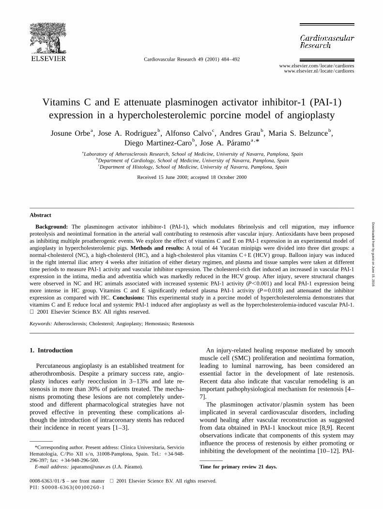
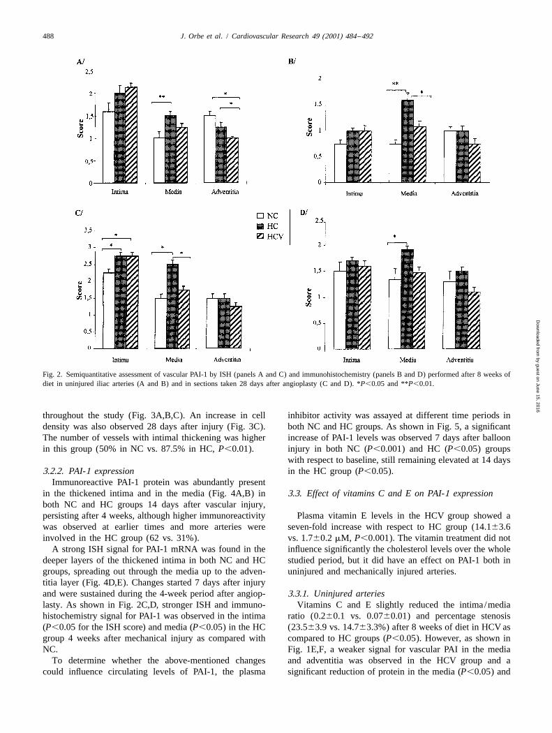
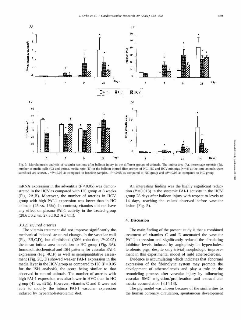
An intense and extensive vascular PAI-1 positive signal vascular wall of both groups. A significant increase in thein the media area was observed both by immunohisto- intimal area and percentage stenosis (P,0.05) was presentchemistry (Fig. 1A,B) and ISH (Fig. 1D,E) in the HC after vascular lesion as compared with non-injured arteriesgroup as compared with NC after 8 weeks of cholesterol in NC animals. Changes were evident at 7 days reachingrich diet. Results regarding semiquantitative assessment of significance (P,0.05) 28 days after injury (Fig. 3A,B), butimmunohistochemistry and ISH in both groups are shown occurred earlier in the HC than in the NC group. Ain Fig. 2A,B. Increased PAI-1 expression was observed in significant (P,0.05) time-dependent increase in intimalmedia layer of HC with respect to NC (P,0.01). Further- area, percentage stenosis and intima/media ratio wasmore, the number of arteries that exhibited increased already observed 7 days after angioplasty and persisted
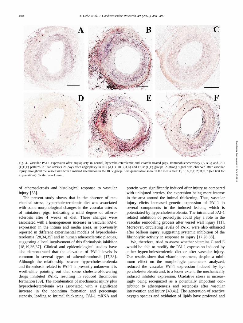
Fig. 1. Immunohistochemical and ISH patterns in vascular sections from hypercholesterolemic pigs. Panels A (NC), B (HC) and C (HCV) show theimmunohistochemical distribution of PAI-1 in uninjured iliac arteries after 8 weeks of diet. Panels D (NC), E (HC) and F (HCV) show the ISH patterncorresponding to the same period. Panel E (magnification 320) also shows an adventitia section with scattered positive cells and strong positive signal invasa-vasorum endothelial cells (arrowhead). Representative score in the media area: F, 0; C and D, 1; A and E, 2; B, 3 (see text for explanations). Scalebar51 mm.
by guest on June 15, 2016D
ownloaded from
488 J. Orbe et al. / Cardiovascular Research 49 (2001) 484 –492
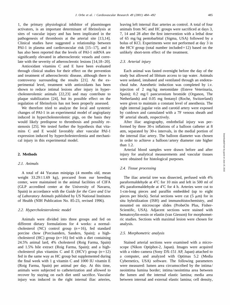
Fig. 2. Semiquantitative assessment of vascular PAI-1 by ISH (panels A and C) and immunohistochemistry (panels B and D) performed after 8 weeks ofdiet in uninjured iliac arteries (A and B) and in sections taken 28 days after angioplasty (C and D). *P,0.05 and **P,0.01.
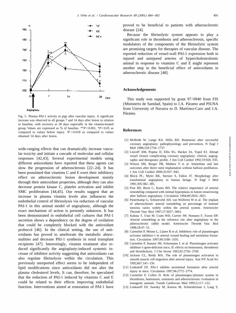
throughout the study (Fig. 3A,B,C). An increase in cell inhibitor activity was assayed at different time periods indensity was also observed 28 days after injury (Fig. 3C). both NC and HC groups. As shown in Fig. 5, a significantThe number of vessels with intimal thickening was higher increase of PAI-1 levels was observed 7 days after balloonin this group (50% in NC vs. 87.5% in HC, P,0.01). injury in both NC (P,0.001) and HC (P,0.05) groups
with respect to baseline, still remaining elevated at 14 days3.2.2. PAI-1 expression in the HC group (P,0.05).
Immunoreactive PAI-1 protein was abundantly presentin the thickened intima and in the media (Fig. 4A,B) in 3.3. Effect of vitamins C and E on PAI-1 expressionboth NC and HC groups 14 days after vascular injury,persisting after 4 weeks, although higher immunoreactivity Plasma vitamin E levels in the HCV group showed awas observed at earlier times and more arteries were seven-fold increase with respect to HC group (14.163.6involved in the HC group (62 vs. 31%). vs. 1.760.2 mM, P,0.001). The vitamin treatment did not
A strong ISH signal for PAI-1 mRNA was found in the influence significantly the cholesterol levels over the wholedeeper layers of the thickened intima in both NC and HC studied period, but it did have an effect on PAI-1 both ingroups, spreading out through the media up to the adven- uninjured and mechanically injured arteries.titia layer (Fig. 4D,E). Changes started 7 days after injuryand were sustained during the 4-week period after angiop- 3.3.1. Uninjured arterieslasty. As shown in Fig. 2C,D, stronger ISH and immuno- Vitamins C and E slightly reduced the intima/mediahistochemistry signal for PAI-1 was observed in the intima ratio (0.260.1 vs. 0.0760.01) and percentage stenosis(P,0.05 for the ISH score) and media (P,0.05) in the HC (23.563.9 vs. 14.763.3%) after 8 weeks of diet in HCV asgroup 4 weeks after mechanical injury as compared with compared to HC groups (P,0.05). However, as shown inNC. Fig. 1E,F, a weaker signal for vascular PAI in the media
To determine whether the above-mentioned changes and adventitia was observed in the HCV group and acould influence circulating levels of PAI-1, the plasma significant reduction of protein in the media (P,0.05) and
by guest on June 15, 2016D
ownloaded from
J. Orbe et al. / Cardiovascular Research 49 (2001) 484 –492 489
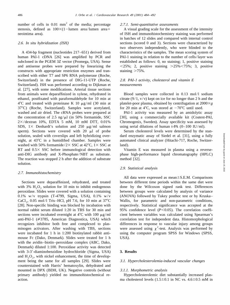
Fig. 3. Morphometric analysis of vascular sections after balloon injury in the different groups of animals. The intima area (A), percentage stenosis (B),number of media cells (C) and intima/media ratio (D) in the balloon injured iliac arteries of NC, HC and HCV minipigs (n54) at the time animals weresacrificed are shown. . *P,0.05 as compared to baseline samples, †P,0.05 as compared to NC group and ‡P,0.05 as compared to HC group.
mRNA expression in the adventitia (P,0.05) was demon- An interesting finding was the highly significant reduc-strated in the HCV as compared with HC group at 8 weeks tion (P50.018) in the systemic PAI-1 activity in the HCV(Fig. 2A,B). Moreover, the number of arteries in HCV group 28 days after balloon injury with respect to levels atgroup with high PAI-1 expression was lower than in HC 14 days, reaching the values observed before vascularanimals (25 vs. 16%). In contrast, vitamins did not have lesion (Fig. 5).any effect on plasma PAI-1 activity in the treated group(28.660.2 vs. 27.560.2 AU/ml).
4. Discussion3.3.2. Injured arteries
The vitamin treatment did not improve significantly the The main finding of the present study is that a combinedmechanical-induced structural changes in the vascular wall treatment of vitamins C and E attenuated the vascular(Fig. 3B,C,D), but diminished (30% reduction, P,0.05) PAI-1 expression and significantly reduced the circulatingthe mean intima area in relation to HC group (Fig. 3A). inhibitor levels induced by angioplasty in hypercholes-Immunohistochemical and ISH patterns for vascular PAI-1 terolemic pigs, despite only trivial morphologic improve-expression (Fig. 4C,F) as well as semiquantitative assess- ment in this experimental model of mild atherosclerosis.ment (Fig. 2C, D) showed weaker PAI-1 expression in the Evidence is accumulating which indicates that abnormalmedia layer in the HCV group as compared to HC (P,0.05 expression of the fibrinolytic system may promote thefor the ISH analysis), the score being similar to that development of atherosclerosis and play a role in theobserved in control animals. The number of arteries with remodeling process after vascular injury by influencinghigh PAI-1 expression was also lower in HVC than in HC vascular SMC migration /proliferation and extracellulargroup (41 vs. 62%). However, vitamins C and E were not matrix accumulation [8,14,18].able to modify the intima PAI-1 vascular expression The pig model was chosen because of the similarities toinduced by hypercholesterolemic diet. the human coronary circulation, spontaneous development
by guest on June 15, 2016D
ownloaded from
490 J. Orbe et al. / Cardiovascular Research 49 (2001) 484 –492
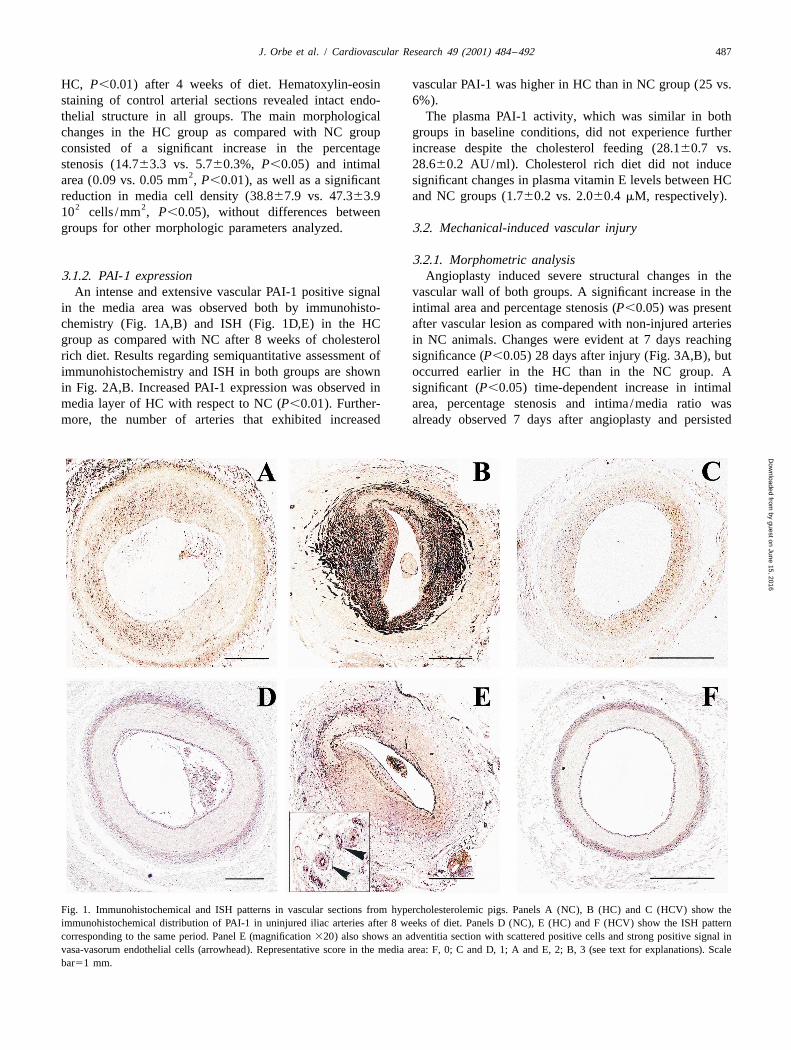
Fig. 4. Vascular PAI-1 expression after angioplasty in normal, hypercholesterolemic and vitamin-treated pigs. Immunohistochemistry (A,B,C) and ISH(D,E,F) patterns in iliac arteries 28 days after angioplasty in NC (A,D), HC (B,E) and HCV (C,F) groups. A strong signal was observed after vascularinjury throughout the vessel wall with a marked attenuation in the HCV group. Semiquantitative score in the media area: D, 1; A,C,F, 2; B,E, 3 (see text forexplanations). Scale bar51 mm.
of atherosclerosis and histological response to vascular protein were significantly induced after injury as comparedinjury [33]. with uninjured arteries, the expression being more intense
The present study shows that in the absence of me- in the area around the intimal thickening. Thus, vascularchanical stress, hypercholesterolemic diet was associated injury elicits increased genetic expression of PAI-1 inwith some morphological changes in the vascular arteries several components in the induced lesions, which isof miniature pigs, indicating a mild degree of athero- potentiated by hypercholesterolemia. The intramural PAI-1sclerosis after 4 weeks of diet. These changes were related inhibition of proteolysis could play a role in theassociated with a homogeneous increase in vascular PAI-1 vascular remodeling process after vessel wall injury [11].expression in the intima and media areas, as previously Moreover, circulating levels of PAI-1 were also enhancedreported in different experimental models of hypercholes- after balloon injury, suggesting systemic inhibition of theterolemia [28,34,35] and in human atherosclerotic plaques, fibrinolytic activity in response to injury [17,28,38].suggesting a local involvement of this fibrinolysis inhibitor We, therefore, tried to assess whether vitamins C and E[18,19,36,37]. Clinical and epidemiological studies have would be able to modify the PAI-1 expression induced byalso demonstrated that the elevation of PAI-1 levels is either hypercholesterolemic diet or after vascular injury.common in several types of atherothrombosis [17,38]. Our results show that vitamin treatment, despite a mini-Although the relationship between hypercholesterolemia mum effect on the morphologic parameters analyzed,and thrombosis related to PAI-1 is presently unknown it is reduced the vascular PAI-1 expression induced by hy-worthwhile pointing out that some cholesterol-lowering percholesterolemia and, to a lesser extent, the mechanicallydrugs inhibited PAI-1, resulting in reduced thrombosis induced inhibitor expression. Oxidative stress is increas-formation [39]. The combination of mechanical injury plus ingly being recognized as a potentially important con-hypercholesterolemia was associated with a significant tributor to atherogenesis and restenosis after vascularincrease in the neointima formation and percentage intervention and injury [40,41]. The generation of reactivestenosis, leading to intimal thickening. PAI-1 mRNA and oxygen species and oxidation of lipids have profound and
by guest on June 15, 2016D
ownloaded from
J. Orbe et al. / Cardiovascular Research 49 (2001) 484 –492 491
proved to be beneficial to patients with atheroscleroticdisease [24].
Because the fibrinolytic system appears to play asignificant role in thrombosis and atherosclerosis, specificmodulators of the components of the fibrinolytic systemare promising targets for therapies of vascular disease. Thereported reduction of vessel-wall PAI-1 expression both ininjured and uninjured arteries of hypercholestrolemicanimal in response to vitamins C and E might representanother step in the beneficial effect of antioxidants inatherosclerotic disease [48].
Acknowledgements
This study was supported by grant 97 /0940 from FIS´(Ministerio de Sanidad, Spain) to J.A. Paramo and PIUNA´from University of Navarra to D. Martınez-Caro and J.A.
´Paramo.Fig. 5. Plasma PAI-1 activity in pigs after vascular injury. A significantincrease was observed in all groups 7 and 14 days after lesion in relationto baseline, with recovery at 28 days especially in the vitamin-treatedgroup. Values are expressed as % of baseline. **P,0.001, *P,0.05 as Referencescompared to values before injury. †P50.018 as compared to valuesobtained 14 days after lesion.
[1] McBride W, Lange RA, Hillis RD. Restenosis after successfulcoronary angioplasty: pathophysiology and prevention. N Engl JMed 1988;318:1734–1737.wide-ranging effects that can dramatically increase vascu-
[2] Lincoff AM, Popma JJ, Ellis SG, Hacker JA, Topol EJ. Abruptlar toxicity and initiate a cascade of molecular and cellularvessel closure complicating coronary angioplasty: clinical, angiog-
responses [42,43]. Several experimental models using raphic and therapeutic profile. J Am Coll Cardiol 1992;19:926–935.different antioxidants have reported that these agents can [3] Wilson SM, Berger PB, Mathew V et al. Immediate and late
outcomes after direct stent implantation without balloon predilation.slow the progression of atherosclerosis [22–24]. It hasJ Am Coll Cardiol 2000;35:937–943.been postulated that vitamins C and E exert their inhibitory
[4] Block PC, Myler RK, Stertzer S, Fallon JT. Morphology aftereffect on atherosclerotic lesion development mainlytransluminal angioplasty in human beings. N Engl J Med
through their antioxidant properties, although they can also 1981;305:382–385.decrease protein kinase C, platelet activation and inhibit [5] Post MJ, Borst C, Kuntz RD. The relative importance of arterialSMC proliferation [44,45]. Our results suggest that an remodeling compared with intimal hyperplasia in lumen renarrowing
after balloon angioplasty. Circulation 1994;89:2816–2821.increase in plasma vitamin levels also influences the[6] Pasterkamp G, Schoneveld AH, van Wolferen W et al. The implantendothelial control of fibrinolysis via reduction of vascular
of atherosclerotic arterial remodeling on percentage of luminalPAI-1 in this animal model of angioplasty, although the stenosis varies widely within the arterial system. Arteriosclerexact mechanism of action is presently unknown. It has Thromb Vasc Biol 1997;17:3057–3063.been demonstrated in endothelial cell cultures that PAI-1 [7] Kakuta T, Usui M, Coats WD, Currier JW, Numano F, Faxon DP.
Arterial remodeling at the reference site after angioplasty in thesecretion shows a dependency on the degree of oxidationatherosclerotic rabbit model. Arterioscler Thromb Vasc Biolthat could be completely blocked with the antioxidant1998;18:47–51.
probucol [46]. In the clinical setting, the use of anti- [8] Carmeliet P, Moons L, Lijnen R et al. Inhibitory role of plasminogenoxidants has proved to ameliorate the metabolic abnor- activator inhibitor-1 in arterial wound healing and neointima forma-malities and decrease PAI-1 synthesis in renal transplant tion. Circulation 1997;96:3180–3191.
[9] Carmeliet P, Stassen JM, Schoonjans L et al. Plasminogen activatorrecipients [47]. Interestingly, vitamin treatment also re-inhibitor-1-gene-deficient mice, II: effects on hemostasis, thrombosisduced significantly the angioplasty-induced systemic in-and thrombolysis. J Clin Invest 1993;92:2756–2760.
crease of inhibitor activity suggesting that antioxidants can [10] Jackson CL, Reidy MA. The role of plasminogen activation inalso regulate fibrinolysis within the circulation. This smooth muscle cell migration after arterial injury. Ann NY Acad Scipreviously unreported effect seems to be independent of 1992;667:141–150.
[11] Loskutoff DJ. PAI-1 inhibits neointimal formation after arteriallipid modifications since antioxidants did not alter theinjury in mice. Circulation 1997;96:2772–2774.plasma cholesterol levels. It can, therefore, be speculated
[12] Carmeliet P, Collen D. Role of plasminogen/plasmin system inthat the reduction of PAI-1 induced by vitamins C and E thrombosis, hemostasis, restenosis and atherosclerosis: evaluation incould be related to their effects improving endothelial transgenic animals. Trends Cardiovasc Med 1995;5:117–122.function. Interventions aimed at restoration of PAI-1 have [13] Loskutoff DJ, Sawdey M, Keeton M, Schneiderman J, Lang Y,
by guest on June 15, 2016D
ownloaded from
492 J. Orbe et al. / Cardiovascular Research 49 (2001) 484 –492
¨Schleef R. Analysis of PAI-1 gene expression in vivo using in situ [31] Siedel J, Hagele EO, Ziegenhorn J, Wahlefeld AW. Reagent for thehybridization. Ann NY Acad Sci 1994;714:259–264. enzymatic determination of serum total cholesterol with improved
[14] Schneiderman J, Sawdey MS, Keeton MR et al. Increased type 1 lipolytic efficiency. Clin Chem 1983;29:1075–1080.plasminogen activator inhibitor gene expression in atherosclerotic [32] Palace VP, Brown SB. HPLC determination of tocopherol, retinol,human arteries. Proc Natl Acad Sci USA 1992;89:6998–7002. dehydroretinol and retinyl palmitate in tissues of lake char (Sal-
´[15] Paramo JA, Colucci M, Collen D, van de Werf F. Plasminogen velinus namaycush) exposed to coplanar 3,39,4,49,5-pentachloro-activator inhibitor in the blood of patients with coronary artery biphenyl. Environ Toxicol Chem 1994;13:473–476.disease. Br Med J 1985;291:573–574. [33] De Smet BJ, van der Zande J, van der Helm YJ, Kuntz RE, Borst C,
[16] Thompson SG, Van De Loo JCW. ECAT angina pectoris study: Post MJ. The atherosclerotic Yucatan animal model to study thebaseline associations of haemostatic factors with extent of coronary arterial response after balloon angioplasty: the natural history ofarteriosclerosis and other coronary risk factors in 3000 patients with remodeling. Cardiovasc Res 1998;39:224–232.angina pectoris undergoing coronary angiography. Eur Heart J [34] Farrehi PM, Ozaki K, Carmeliet P, Fay WP. Regulation of arterial1993;14:8–17. thrombolysis by plasminogen activator-1 in mice. Circulation
[17] Juhan-Vague I, Pyke SD, Alessi MC, Jespersen J, Haverkate F, 1998;97:1002–1008.Thompson SG. Fibrinolytic factors and the risk of myocardial [35] Sawa H, Lundgren C, Sobel BE, Fujii S. Increased intramuralinfarction or sudden death in patients with angina pectoris. ECAT expression of plasminogen activator inhibitor type 1 after balloonStudy Group. European Concerted Action on Thrombosis and injury: a potential progenitor of restenosis. J Am Coll CardiolDisabilities. Circulation 1996;94:2057–2063. 1994;24:1742–1748.
[18] Robbie LA, Booth NA, Brown PAJ, Bennet B. Inhibitors of [36] Schneider DJ, Ricci MA, Taatjes DJ et al. Changes in arterialfibrinolysis are elevated in atherosclerotic plaque. Arterioscler expression of fibrinolytic system proteins in atherogenesis. Arterios-Thromb Vasc Biol 1996;16:539–545. cler Thromb Vasc Biol 1997;17:3294–3301.
¨¨ ¨[19] Lupu F, Bergonzelli GE, Heim DA et al. Localization and pro- [37] Falkenberg M, Tjarnstrom J, Ortenwall P, Olausson M. Localizationduction of plasminogen activator inhibitor-1 in human healthy and of fibrinolytic activators and inhibitors in normal and atheroscleroticatherosclerotic arteries. Arterioscler Thromb 1993;13:1090–1100. vessels. Thromb Haemost 1996;75:933–938.
[20] Padro T, Steins M, Li CX et al. Comparative analysis of plas- [38] Salomaa V, Stinson V, Kark JD, Folsom AR, Davis CE, Wu KK.minogen activator inhibitor-1 expression in different types of Association of fibrinolytic parameters with early atherosclerosis.atherosclerotic lesions in coronary arteries from human heart The ARIC Study. Atherosclerosis Risk in Communities Study.explants. Cardiovasc Res 1997;36:28–36. Circulation 1995;91:284–290.
´[21] Dıaz MN, Frei B, Vita JA, Keaney Jr. JF. Antioxidants and [39] Dangas G, Smith DA, Unger AM et al. Pravastatin: an antithrom-atherosclerotic heart disease. N Engl J Med 1997;337:408–416. botic effect independent of the cholesterol-lowering effect. Thromb
[22] Nunes GL, Sgoutas DS, Redden RA et al. Combination of vitamin C Haemost 2000;83:688–692.and E alters the response to coronary balloon injury in the pig. [40] Holvoet P, Theilmeier G, Shivalkar B, Flameng W, Collen D.Arterioscler Thromb Vasc Biol 1995;15:156–165. LDL-hypercholesterolemia is associated with accumulation of oxi-
[23] Hennekens CH, Gaziano JM, Manson JE, Buring JE. Antioxidant dized LDL, atherosclerotic plaque growth, and compensatory vesselvitamin-cardiovascular disease hypothesis is still promising, but still enlargement in coronary arteries of miniature pigs. Arteriosclerunproven: the need for randomized trials. Am J Clin Nutr Thromb Vasc Biol 1998;18:415–422.1995;62:1377S–1380S. [41] Edelman ER. Vessel size, antioxidants and restenosis. Circulation
[24] Rabbani R, Topol EJ. Strategies to achieve coronary arterial plaque 1998;97:416–420.stabilization. Cardiovasc Res 1999;41:402–417. [42] Steinberg D. Oxidative modification of LDL and atherogenesis.
[25] Strauss BH, Lau HK, Bowman KA et al. Plasma urokinase antigen Circulation 1997;95:1062–1071.and plasminogen activator inhibitor-1 antigen levels predict angiog- [43] Nunes GL, Robinson K, Kalynych A, King III SB, Sgoutas DS,raphic coronary restenosis. Circulation 1999;100:1616–1622. Berk BC. Vitamins C and E inhibit O production in the pig2
[26] Bosmat PJ, van den Berg EA, Kooistra T, Siemieniak DR, Slightom coronary artery. Circulation 1997;96:3593–3601.JL. Human plasminogen activator inhibitor-1 gene. J Biol Chem [44] Freedman JE, Farhat JH, Loscalzo J, Keaney JR. a-Tocopherol1988;263:9129–9141. inhibits agregation of human platelets by a protein kinase C-
[27] Dijkman HBPM, Mentzel S, de Jong AS, Assmann KJM. RNA in dependent mechanism. Circulation 1996;94:2434–2440.situ hybridization using digoxigenin-labeled cRNA probes. Bio- [45] Boskoboinik D, Szewezyk A, Azzi A. Alpha-tocopherol (vitamin E)chemica 1995;2:21–25. regulates vascular smooth muscle cell proliferation and protein
[28] Sawa H, Sobel BE, Fujii S. Potentiation by hypercholesterolemia of kinase C activity. Arch Biochem Biophys 1991;286:264–269.the induction of aortic intramural synthesis of plasminogen activator [46] Grafe M, Auch Schwelk W, Hertel H et al. Human cardiacinhibitor type 1 by endothelial injury. Circ Res 1993;73:671–680. microvascular and macrovascular endothelial cells respond different-
[29] Shu SY, Ju G, Fan LZ. The glucose-oxidase DAB nickel method in ly to oxidatively modified LDL. Atherosclerosis 1998;137:87–95.peroxidase histochemistry of the nervous system. Neurosci Lett [47] Dimeny E, Fellstrom B. Metabolic abnormalities in renal transplant1988;85:169–171. recipients. Risk factors and predictors of chronic graft dysfunction?
[30] Chmielewska J, Ranby M, Wiman B. Evidence for a rapid inhibitor Nephrol Dial Transplant 1997;12:21–24.to tissue plasminogen activator in plasma. Thromb Res [48] Sobel BE. Increased plasminogen activator inhibitor-1 and vas-1983;31:417–436. culopathy. A reconcilable paradox. Circulation 1999;99:2496–2498.
by guest on June 15, 2016D
ownloaded from